OUTLINE FIBROADENOMA FIBROADENOMA. FIBROEPITHELIAL LESIONS OF THE BREAST UCSF Current Issues in Anatomic Pathology 2015 FIBROADENOMA PHYLLODES TUMOR
|
|
|
- Darlene Hall
- 6 years ago
- Views:
Transcription


 Predisposing factors No known inherited genetic")
 Carney complex (myxoid fibroadenomas) Solitary, mobile, rubbery and")

1 OUTLINE FIBROADENOMA FIBROEPITHELIAL LESIONS OF THE BREAST UCSF Current Issues in Anatomic Pathology 2015 Gregor Krings, MD PhD Assistant Professor PHYLLODES TUMOR DIFFERENTIAL DIAGNOSIS CELLULAR FIBROEPITHELIAL LESIONS MALIGNANT PHYLLODES TUMORS EXCISION VERSUS CORE NEEDLE BIOPSY IMMUNOHISTOCHEMISTRY FIBROADENOMA FIBROADENOMA Very common Most common fibroepithelial lesions Most common benign tumors of the breast Broad age group Incidence highest in women <30 years old Can occur at any age (18.5% of women >40 years old in Breast Cancer Surveillance Consortium) Predisposing factors No known inherited genetic alterations but risk in some families Hormonal influence Rare in men but associated with gynecomastia, exogenous hormones, drugs Cyclosporin A (organ transplant) Carney complex (myxoid fibroadenomas) Solitary, mobile, rubbery and painless palpable mass Non-palpable, mammographically detected Calcifications (hyalinized fibroadenomas) Rarely pain and/or bloody nipple discharge Infarction Pregnancy, prior aspiration procedure, spontaneous Often <3 cm but larger tumors not uncommon Giant fibroadenomas up to 20 cm Larger tumors in adolescents (juvenile fibroadenoma) 1
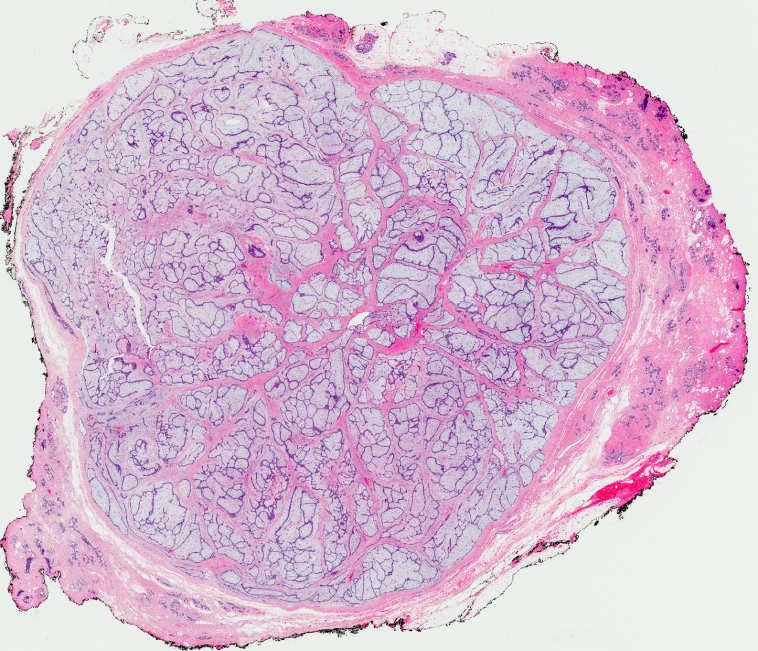
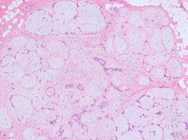
2 Usual-type Hyalinized Intracanalicular Pericanalicular Mixed Myxoid Mixed 2


3 Myxoid FA Mucinous carcinoma Myxoid FA Mucinous carcinoma Myxoid fibroadenoma may mimic invasive mucinous carcinoma Misdiagnosis on imaging - 16/17 myxoid fibroadenomas with rapid growth or size >3 cm misdiagnosed as mucinous carcinoma on ultrasound Yamaguchi Human Pathology 2011;42: Misdiagnosis on FNA and core biopsy Simsir 2001 Diagn Cytopathol. 2001;25: COMPLEX FIBROADENOMA Sclerosing adenosis, papillary apocrine metaplasia, cysts >3mm or epithelial calcifications COMPLEX FIBROADENOMA Managed like typical FA in absence of atypia or rad-path discordance We do not use this term in diagnosis Sklair-Levy M et al. AJR 2008;190(1):


 Overlapping")


4 CELLULAR FIBROADENOMA Focal or diffuse mildly increased stromal cellularity without stromal atypia No threshold criteria for defining hypercellularity Stromal atypia is subjective Stromal mitotic figures may be present (up to 2 MF/10 HPF typically acceptable) Overlapping features with benign phyllodes tumors Uniform cellularity and epithelial:stromal distribution JUVENILE FIBROADENOMA More common in adolescents and women <20 years old Usual-type fibroadenoma most common in all age groups May mimic phyllodes tumor Rapid growth, large size, histologic features Cellular stroma with pericanalicular growth Stromal mitotic activity may be present No stromal cytologic atypia Uniform cellularity and epithelial:stromal distribution Gynecomastoid usual ductal hyperplasia Excision with preservation of adjacent breast 4


5 ALH E-cadherin Atypia or carcinoma may involve fibroadenomas primarily or secondarily - ALH/LCIS most common - ADH/DCIS - Invasive carcinoma ALH ADH E-cadherin Tangential PHYLLODES TUMORS Rare <1% primary breast tumors <2.5% fibroepithelial lesions in tertiary centers Age years (but wide range, adolescence to 90) years older than FA, on average Tumors in adolescents often benign More common in Asian and Latina women May present at younger age in this group Li-Fraumeni Syndrome (p53 mutations) predisposed 5


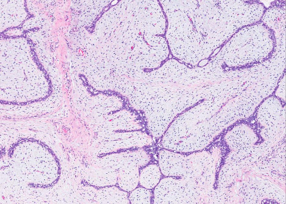
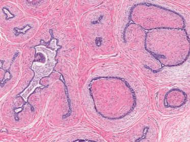
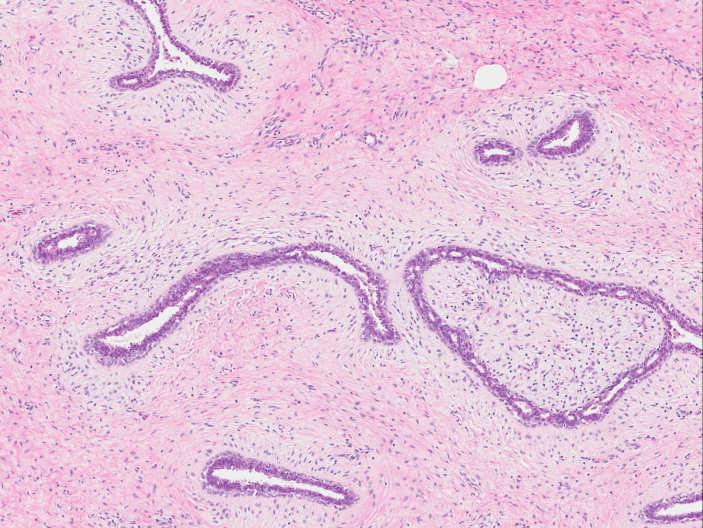
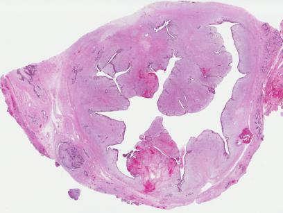
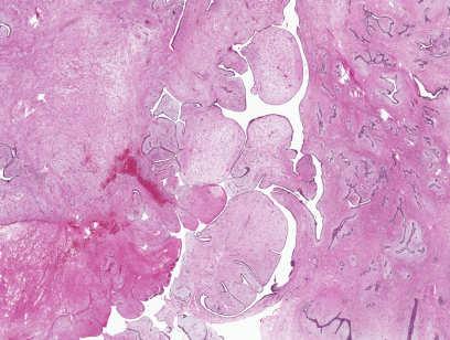
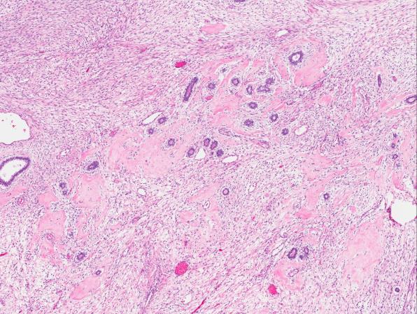
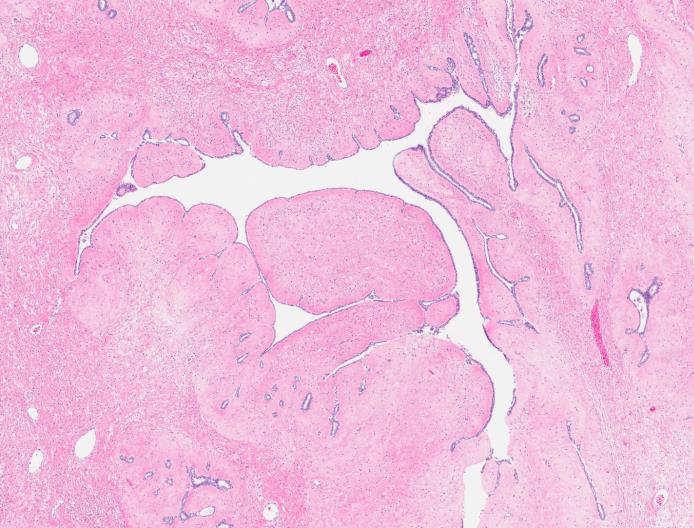
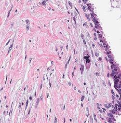
6 PHYLLODES TUMORS Present as mass lesion Rapidly growing or accelerated growth of previously stable lesion 4-5 cm in size, but wide range (<3-20+ cm) Smaller lesions increasingly detected by screening Not reliably distinguished from fibroadenoma by imaging PHYLLODES TUMORS fibroepithelial neoplasms, histologically resembling intracanalicular fibroadenomas, characterized by a doublelayered epithelial component arranged in clefts surrounded by a hypercellular stromal/mesenchymal component which in combination elaborate leaf-like structures PHYLLODES TUMOR DIAGNOSIS BASED ON A CONSTELLATION OF FEATURES Increased stromal cellularity* Leaf-like growth ± periductal stromal condensation Stromal heterogeneity +/- mitotic activity* +/- infiltrative border* +/- stromal overgrowth* +/- stromal cytologic atypia* +/- malignant heterologous stroma* * Used to establish grade LEAF-LIKE GROWTH 6

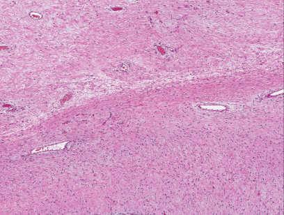
7 LEAF-LIKE GROWTH LEAF-LIKE GROWTH SUBEPITHELIAL STROMAL CONDENSATION SUBEPITHELIAL STROMAL CONDENSATION 7

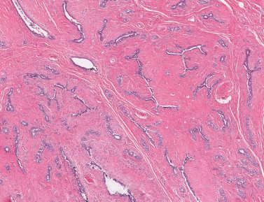

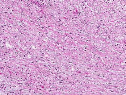
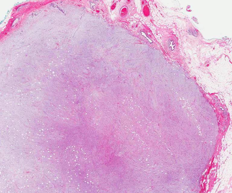
8 INTRALOBULAR STROMAL COMPRESSION OF FIBROADENOMA STROMAL HETEROGENEITY GRADING PHYLLODES TUMORS BENIGN PHYLLODES TUMOR Adapted from WHO Classification of Tumours of the Breast, 4 th ed


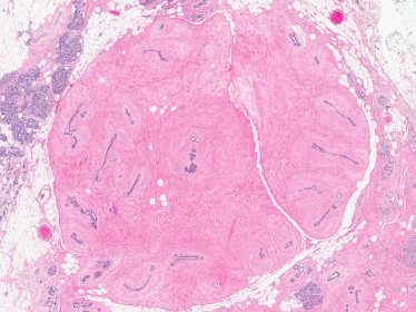
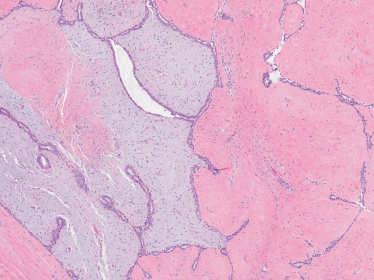
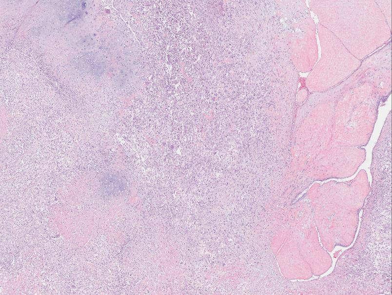
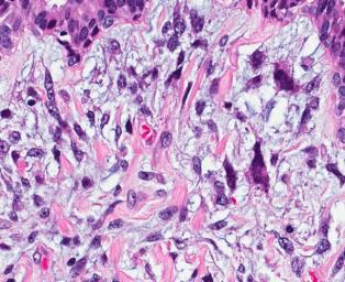
9 MALIGNANT PHYLLODES TUMOR INFILTRATIVE BORDERS Stromal overgrowth (4x low power field) often diffuse 9


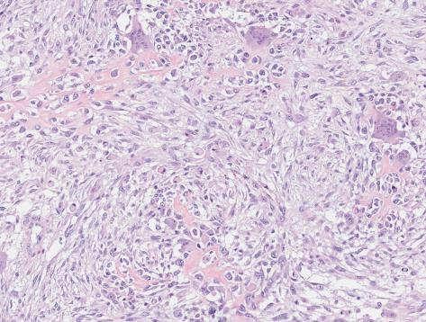
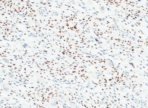
10 5/23/2015 MALIGNANT HETEROLOGOUS STROMA Most commonly liposarcomatous SATB2 is a useful marker of osseous differentiationsatb2 BORDERLINE PHYLLODES TUMOR SATB2 10
11 Phyllodes tumor histologic grade predicts local recurrence FEATURES PREDICTIVE OF PHYLLODES TUMOR RECURRENCE phyllodes tumors (diagnosed over 18 years, ) patients with clinical follow-up /24.6 months mean/median time to recurrence A.M.O.S. criteria Tan PH et al J Clin Pathol 2012;65:69-76 NOMOGRAM FOR PREDICTING PHYLLODES TUMOR RECURRENCE FREE SURVIVAL NOMOGRAM FOR PREDICTING PHYLLODES TUMOR RECURRENCE FREE SURVIVAL A. M. O. S. * Positive margin status best predictor of recurrence* A. M. O. S. REQUIRES ADDITIONAL VALIDATION IN OTHER POPULATIONS Tan PH et al J Clin Pathol 2012;65:69-76 Tan PH et al J Clin Pathol 2012;65:
12 PHYLLODES TUMOR: HISTOLOGIC GRADE AND PROGNOSIS Benign Borderline Malignant Local recurrence* 4-17% 14-25% 23-30% * Margin status remains the best predictor of local recurrence 4/48 (8.3%) benign tumors recurred as malignant Tan PH et al J Clin Pathol 2012;65: /48 (43.8%) initially benign tumors recurred as higher grade 2/16 (12.8%) initially borderline tumors recurred as malignant Hart WR et al. Am J Clin Pathol 1978;70(2):211-6 Moffat CJ et al. Histopathology 1995;27(3): De Roos WK et al. British J Surg 1999;86(3):396-9 Tan PH et al. J Clin Pathol 2012;65:69-76 Barth RJ Jr. Breast Cancer Res Treat 1999;57(3):291-5 Kim S et al. Breast Cancer Res Treat ; WHO 2012 Other studies with similar results % benign tumors reported to recur as malignant Highlights importance of preventing local tumor recurrence DISTANT PHYLLODES TUMOR METASTASIS PHYLLODES TUMOR: HISTOLOGIC GRADE AND PROGNOSIS Benign Borderline Malignant % of phyllodes 65-70% 15-20% 10-20% Metastasis** (<10% overall) 0% 0-4% 13-29% Stromal overgrowth and malignant heterologous stromal elements are best predictors of distant spread Metastasis essentially always stromal component only ** Essentially only malignant tumors metastasize Tan PH et al. J Clin Pathol (1):69-76 Kim S et al. Breast Cancer Res Treat ; WHO 2012 Lung/pleura (>75%) and skeletal system most common sites 12
13 Lung Vulva PHYLLODES TUMOR TREATMENT Excision with negative margins to minimize recurrence risk 1 cm normal rim preferable (but no data to support this arbitrary margin width) Rationale Margin status primary predictor of recurrence Recurrences may be of higher grade Metastatic tumors may be preceded by local recurrences No routine role for radiation or chemotherapy DIFFERENTIAL DIAGNOSIS OF FIBROEPITHELIAL LESIONS benign borderline malignant Benign phyllodes Fibroadenoma Mean age ~45-50 (but any age) ~30 y (but any age) Size Few cm up to 20 cm <3 cm; rarely up to 20 cm Growth May be rapid; rapid growth of previously stable mass OVERLAP OVERLAP NOT RELIABLE Stable Clinical and radiologic features do not reliably distinguish between phyllodes tumor and fibroadenoma MAY BE PROBLEMATIC IN EXCISIONS AND CORE BIOPSIES Jacobs et al Am J Clin Pathol Sep;124(3): WHO
14 Leaf-like architecture BENIGN PHYLLODES FIBROADENOMA Present, well-developed ± periductal condensation Usually absent, may be focal CELLULAR FIBROADENOMA BENIGN PHYLLODES TUMOR Stromal heterogeneity May be present Absent Distribution of epithelium and stroma Often non-uniform Uniform Stromal cellularity Stromal mitoses Mild Few (0-4/10 HPF) Hypocellular or Mild (*cellular and juvenile fibroadenoma) Rare-up to 2/10 HPF allowed (*cellular and juvenile fibroadenoma) Cellular atypia Mild Absent (*stromal giant cells) Squamous metaplasia Rarely present Virtually absent Leaf-like architecture BENIGN PHYLLODES FIBROADENOMA Present, well-developed ± periductal condensation Usually absent, may be focal Stromal heterogeneity May be present Absent Distribution of epithelium and stroma Often non-uniform Uniform Stromal cellularity Stromal mitoses Mild Few (0-4/10 HPF) Hypocellular or Mild (*cellular and juvenile fibroadenoma) Rare-up to 2/10 HPF allowed (*cellular and juvenile fibroadenoma) Cellular atypia Mild Absent (*stromal giant cells) Squamous metaplasia Rarely present Virtually absent 14

15 PITFALLS 1. Fibroadenomas may have focal leaf-like growth. 2. Phyllodes tumors may lack leaf-like growth. 3. Phyllodes tumors may be less cellular or mimic fibroadenomas in some areas due to heterogeneity. 4. Benign multinucleated stromal giant cells in fibroadenomas should not be mistaken for atypia. 5. Mitotic activity does not equate with phyllodes tumor. PITFALLS 1. Fibroadenomas may have focal leaf-like growth. 2. Phyllodes tumors may lack leaf-like growth. 3. Phyllodes tumors may be less cellular or mimic fibroadenomas in some areas due to heterogeneity. 4. Benign multinucleated stromal giant cells in fibroadenomas should not be mistaken for atypia. 5. Mitotic activity does not equate with phyllodes tumor. FIBROADENOMA WITH FOCAL LEAF-LIKE GROWTH FIBROADENOMA WITH FOCAL LEAF-LIKE GROWTH 15


16 Juvenile fibroadenoma with focal leaf-like growth PITFALLS 1. Fibroadenomas may have focal leaf-like growth. 2. Phyllodes tumors may lack leaf-like growth. 3. Phyllodes tumors may be less cellular or mimic fibroadenomas in some areas due to heterogeneity. 4. Benign multinucleated stromal giant cells in fibroadenomas should not be mistaken for atypia. 5. Mitotic activity does not equate with phyllodes tumor. PHYLLODES TUMOR WITHOUT LEAF-LIKE GROWTH PHYLLODES TUMOR WITHOUT LEAF-LIKE GROWTH 16


17 PITFALLS 1. Fibroadenomas may have focal leaf-like growth. 2. Phyllodes tumors may lack leaf-like growth. 3. Phyllodes tumors may be less cellular or mimic fibroadenomas in some areas due to heterogeneity. 4. Benign multinucleated stromal giant cells in fibroadenomas should not be mistaken for atypia. 5. Mitotic activity does not equate with phyllodes tumor. PHYLLODES TUMOR HETEROGENEITY PHYLLODES TUMOR HETEROGENEITY PHYLLODES TUMOR HETEROGENEITY CNB DIAGNOSIS: FIBROADENOMATOUS CHANGE * Adequate sampling required 17


18 PHYLLODES TUMOR HETEROGENEITY PITFALLS 1. Fibroadenomas may have focal leaf-like growth. 2. Phyllodes tumors may lack leaf-like growth. 3. Phyllodes tumors may be less cellular or mimic fibroadenomas in some areas due to heterogeneity. 4. Benign multinucleated stromal giant cells in fibroadenomas should not be mistaken for atypia. 5. Mitotic activity does not equate with phyllodes tumor. Benign multinucleated stromal giant cells PITFALLS 1. Fibroadenomas may have focal leaf-like growth. 2. Phyllodes tumors may lack leaf-like growth. CD34 Atypical stromal cells of phyllodes tumor 3. Phyllodes tumors may be less cellular or mimic fibroadenomas in some areas due to heterogeneity. 4. Benign multinucleated stromal giant cells in fibroadenomas should not be mistaken for atypia. 5. Mitotic activity does not equate with phyllodes tumor. - Overlap with cellular/juvenile fibroadenomas 18

: 1 fibroadenoma, 1 phyllodes 4 (19%) cases equally split between cellular fibroadenoma and phyllodes")


19 SOME FIBROEPITHELIAL LESIONS CANNOT BE EASILY CLASSIFIED AS FIBROADENOMA OR PHYLLODES TUMOR 21 pre-selected challenging cellular fibroepithelial lesions separately reviewed by 10 breast pathologists (1-2 slides per case) Uniform agreement in only 2 cases (9.5%): 1 fibroadenoma, 1 phyllodes 4 (19%) cases equally split between cellular fibroadenoma and phyllodes 43% of cases, diagnoses ranged from fibroadenoma to borderline phyllodes DIAGNOSIS OF FIBROADENOMA IS PREFERABLE WHEN THERE IS HISTOLOGICAL AMBIGUITY TO AVOID OVERTREATMENT. WHO 2012 OUR APPROACH: CELLULAR FIBROEPITHELIAL LESION; SEE COMMENT. - Describe features and diagnostic difficulty/ambiguity - Relate low recurrence potential, especially if margins frankly positive CORE BIOPSY OF CELLULAR FIBROEPITHELIAL LESIONS Cellular FA Benign PT benign borderline malignant Cellular FA Benign PT 19

 Ill-defined borders Stromal cell atypia Ki-67 index 5% Ki-67 6% (range 10-18%) in PT versus 1.")
 in FA Jacobs TW et al Am J Clin Pathol 2005;124:342-354 Lee AHS et al Histopathology 2007; 51; 336-244 Jara-Lazaro et al Histopathology 2010; 57:220-232 Tsang AK et al Histopathology.")
 Increased mitotic activity (>2 MF/10 HPF) Stromal heterogeneity Tissue 2011;59(4):600-8 Yasir S et al Am J Clin Pathol 2014;142:362-369 (favor")
20 Features of cellular fibroepithelial lesions on core biopsy that may predict phyllodes tumor??? PHYLLODES TUMOR Excision with rim of normal tissue Recurrence potential FIBROADENOMA Enucleation No (?) recurrence potential Optimize cosmesis and avoid additional surgery Increased stromal cellularity (marked>moderate) Increased mitotic activity (>2 MF/10 HPF) Stromal heterogeneity Tissue fragmentation Adipose tissue within lesional stroma Stromal expansion (no epithelium in 100x field) Ill-defined borders Stromal cell atypia Ki-67 index 5% Ki-67 6% (range 10-18%) in PT versus 1.6% (range 0-4.4%) in FA Jacobs TW et al Am J Clin Pathol 2005;124: Lee AHS et al Histopathology 2007; 51; Jara-Lazaro et al Histopathology 2010; 57: Tsang AK et al Histopathology. 2011;59(4):600-8 Yasir S et al Am J Clin Pathol 2014;142: Features of cellular fibroepithelial lesions on core biopsy that may predict phyllodes tumor??? Increased stromal cellularity (marked>moderate) Increased mitotic activity (>2 MF/10 HPF) Stromal heterogeneity Tissue fragmentation Adipose tissue within lesional stroma Stromal expansion (no epithelium in 100x field) Ill-defined borders Stromal cell atypia Ki-67 index 5% Ki-67 6% (range 10-18%) in PT versus 1.6% (range 0-4.4%) in FA Jacobs TW et al Am J Clin Pathol 2005;124: Lee AHS et al Histopathology 2007; 51; Jara-Lazaro et al Histopathology 2010; 57: Tsang AK et al Histopathology. 2011;59(4):600-8 Yasir S et al Am J Clin Pathol 2014;142: (favor phyllodes tumor) 20
 Increased mitotic activity (>2 MF/10 HPF)")
 in PT versus 1.6% (range 0-4.")


21 Features of cellular fibroepithelial lesions on core biopsy that may predict phyllodes tumor??? Stromal heterogeneity of cellular fibroepithelial lesions on CNB Increased stromal cellularity (marked>moderate) Increased mitotic activity (>2 MF/10 HPF) Stromal heterogeneity Tissue fragmentation Adipose tissue within lesional stroma Stromal expansion (no epithelium in 100x field) Ill-defined borders Stromal cell atypia Ki-67 index 5% Ki-67 6% (range 10-18%) in PT versus 1.6% (range 0-4.4%) in FA Jacobs TW et al Am J Clin Pathol 2005;124: Lee AHS et al Histopathology 2007; 51; Jara-Lazaro et al Histopathology 2010; 57: Tsang AK et al Histopathology. 2011;59(4):600-8 Yasir S et al Am J Clin Pathol 2014;142: CNB Excision No histologic features can reliably predict phyllodes tumor over cellular fibroadenoma on CNB - Cellularity and mitotic activity most useful - Stromal heterogeneity DIFFERENTIAL DIAGNOSIS OF FIBROEPITHELIAL LESIONS Constellation of features may favor phyllodes tumor in some cases benign borderline malignant DESCRIPTIVE DIAGNOSIS: FIBROEPITHELIAL LESION WITH CELLULAR STROMA Recommend excision for final classification 41% probability of PT on excision Jacobs TW et al Am J Clin Pathol 2005;124: MAY BE PROBLEMATIC IN EXCISIONS AND CORE BIOPSIES 21


22 AMPLE TUMOR SAMPLING TO IDENTIFY EPITHELIAL COMPONENT 22

 CK and p63 staining in all cases Auger M et al.")
:474-81 Barbareschi M et al. Am J Surg Path.")
:1506-12 Leibl S et al. Am J Surg Path 2005;29(3):347-53 Chia Y et al.")
23 DIFFERENTIAL DIAGNOSIS Metaplastic carcinoma Phyllodes tumor Sarcoma Potential neoadjuvant chemotherapy Sentinel lymph node Surgical management No sentinel lymph node DIFFERENTIAL DIAGNOSIS Metaplastic carcinoma Phyllodes tumor Sarcoma KERATIN AND p63 EXPRESSION IN PHYLLODES TUMORS Cytokeratin Focal (1-5% of stromal cells) CK and p63 staining in all cases Auger M et al. Arch Pathol Lab Med 1989 Nov;113(11): Aranda FI et al. Path Res Pract 1994;190(5): Barbareschi M et al. Am J Surg Path. 2001;25(8): Dunne B et al. Human Pathology 2003;34(10): Koker MM et al. Am J Surg Path 2004;28(11): Leibl S et al. Am J Surg Path 2005;29(3): Chia Y et al. J Clin Pathol 2012;65(4): Cimino-Mathews A et al. Am J Surg Path 2014;38(12):
 malignant PT were")

 cytokeratin MALIGNANT")
 Cytokeratin or p63")

24 Sarcomatoid carcinoma Malignant PT Borderline PT Benign PT Fibroadenoma MALIGNANT PHYLLODES TUMOR 8/14 (57%) malignant PT were p63 positive - Most (63%) focal (1-5%) - None with >30% p63+ stromal cells - p40 more specific but less sensitive for sarcomatoid carcinoma 3/14 (21%) malignant PT were CK positive - Cytokeratins AE1/3, 34βE12 and CK8/18 - All focal (1-5%) cytokeratin MALIGNANT PHYLLODES TUMOR Phyllodes tumors may express cytokeratins and/or p63 - Expression is typically focal or patchy (<5%) Cytokeratin or p63 expression, especially in core biopsies, cannot be used to exclude phyllodes tumor Strong, diffuse CK staining may favor metaplastic carcinoma p63 cytokeratin 24


:84-91 Moore T and Lee AH.")
:152-8 Chia Y et al.")

:1689-96 Lee AHS.")
25 5/23/2015 CD34 in Fibroepithelial Lesions Strong diffuse CD34 expression essentially excludes metaplastic carcinoma * alternate grading scheme ** patchy staining in all PT; median 40% cells staining in malignant PT vs 80% cells staining in benign/borderline PT *** TMA Focal staining only (<5% cells) Spindle cell carcinoma are essentially always CD34 negative Silverman JS et al. Histopathology. 1996;29(5):411-9 Chen et al. J Surg Res. 2000;94(2):84-91 Moore T and Lee AH. Histopathology. 2001;38(1):62-7 Noronha Y et al. Int J Surg Path. 2011;19(2):152-8 Chia Y et al. J Clin Pathol. 2012;65(4): Ho SK et al. Histopathology. 2013;63(3): Cimino-Mathews A et al. Am J Surg Path. 2014;38(12): Lee AHS. Histopathology 2008;52:45-57 CD34 EXCISION MALIGNANT PT MALIGNANT PHYLLODES TUMOR CD34 pankeratin p63 25

 Aberrant nuclear β-catenin can be seen in both phyllodes tumor and")

 Sawyer EJ et al.")
:1438-48 Abraham SW et al.")
26 5/23/2015 CD34 positivity essentially excludes metaplastic carcinoma Malignant phyllodes tumors are often CD34 negative CD34 expression is negatively correlated with phyllodes tumor grade If positive, staining in malignant tumors is often focal or patchy (<5%) Aberrant nuclear β-catenin can be seen in both phyllodes tumor and metaplastic carcinoma % of mammary fibromatosis 23% metaplastic carcinomas 72-83% of phyllodes tumors More common in benign than malignant PT 94% benign vs 57% malignant PT (Lacroix-Triki et al 2010) 12.5% malignant PT (Sawyer et al 2002) Sawyer EJ et al. J Pathol 2002;196(4): Lacroix-Triki M et al. Modern Pathology;2010;23(11): Abraham SW et al. Hum Pathol; 2002:33:39-46 Excision Malignant phyllodes tumor CD34 β-catenin Multiple keratins (AE1/3, Cam 5.2, MNF116, CK5/6) negative 26


27 Fibroadenoma and variants SUMMARY Phyllodes tumor Diagnosis requires constellation of features Pitfalls in diagnosis Diagnosis and categorization has clinical significance Some lesions may defy accurate categorization: err on conservative side Core biopsy of cellular fibroepithelial lesions requires excision for definitive classification Malignant phyllodes tumor may mimic metaplastic carcinoma Additional sampling and/or immunohistochemistry may be useful Core biopsy diagnosis of spindle cell neoplasm Questions? 27
A Practical Approach to the Evaluation of Fibroepithelial Lesions. Edi Brogi MD PhD Attending Pathologist Director of Breast Pathology
 A Practical Approach to the Evaluation of Fibroepithelial Lesions Edi Brogi MD PhD Attending Pathologist Director of Breast Pathology Overview Fibroadenomas (FAs) Phyllodes Tumors (PTs) Morphology and
57th Annual HSCP Spring Symposium 4/16/2016
 An Unusual Malignant Spindle Cell Lesion to Involve the Breast Erinn Downs-Kelly, D.O. Associate Professor of Pathology University of Utah & ARUP Laboratories No disclosures Case 39 y/o female with no
Treatment options for the precancerous Atypical Breast lesions. Prof. YOUNG-JIN SUH The Catholic University of Korea
 Treatment options for the precancerous Atypical Breast lesions Prof. YOUNG-JIN SUH The Catholic University of Korea Not so benign lesions? Imaging abnormalities(10% recall) lead to diagnostic evaluation,
Breast pathology. 2nd Department of Pathology Semmelweis University
 Breast pathology 2nd Department of Pathology Semmelweis University Breast pathology - Summary - Benign lesions - Acute mastitis - Plasma cell mastitis / duct ectasia - Fat necrosis - Fibrocystic change/
FIBROEPITHELIAL LESIONS
 DEFINITIONS FIBROEPITHELIAL LESIONS Suzanne Moore FIBROADENOMA- A discrete benign tumour showing evidence of connective tissue and epithelial proliferation- WHO Fibrous stromal element of these tumours
Breast Pathology. Breast Development
 Breast Pathology Lecturer: Hanina Hibshoosh, M.D. Reading: Kumar, Cotran, Robbins, Basic Pathology, 6th Edition, pages 623-635 Breast Development 5th week - thickening of the epidermis - milk line 5th
Controversies and Problematic Issues in Core Needle Biopsies (To excise or not to excise)
") Controversies and Problematic Issues in Core Needle Biopsies (To excise or not to excise) Laura C. Collins, M.D. Beth Israel Deaconess Medical Center and Harvard Medical School Boston, MA Schematic Representation
Controversies and Problematic Issues in Core Needle Biopsies (To excise or not to excise) Laura C. Collins, M.D. Beth Israel Deaconess Medical Center and Harvard Medical School Boston, MA Schematic Representation
3/27/2017. Disclosure of Relevant Financial Relationships. Papilloma???
 Management of Papillary Lesions Diagnosed at Rad Path Concordant Core Biopsy (CNB) Disclosure of Relevant Financial Relationships USCAP requires that all planners (Education Committee) in a position to
Division of Pathology
 Case 38 Adult woman with a 35mm right breast lump at the 10 o clock position. Excision performed. (Case contributed by Dr Mihir Gudi, KKH) Division of Pathology Merlion, One Fullerton Singapore Diagnosis
Diagnosis of Fibroepithelial and Mesenchymal Lesions on Core Needle Biopsy
 Diagnosis of Fibroepithelial and Mesenchymal Lesions on Core Needle Biopsy Emmanuel Agosto-Arroyo, MD Assistant Member Department of Anatomic Pathology 3/3/2018 Disclosure There are no conflicts of interest.
04/10/2018. Intraductal Papillary Neoplasms Of Breast INTRADUCTAL PAPILLOMA
 Intraductal Papillary Neoplasms Of Breast Savitri Krishnamurthy MD Professor of Pathology Deputy Division Head The University of Texas MD Anderson Cancer Center 25 th Annual Seminar in Pathology Pittsburgh,
Papillary Lesions of the Breast A Practical Approach to Diagnosis. (Arch Pathol Lab Med. 2016;140: ; doi: /arpa.
 Papillary Lesions of the Breast A Practical Approach to Diagnosis (Arch Pathol Lab Med. 2016;140:1052 1059; doi: 10.5858/arpa.2016-0219-RA) Papillary lesions of the breast Span the spectrum of benign,
Benign Mimics of Malignancy in Breast Pathology
 Arthur Purdy Stout Society of Surgical Pathologists Companion Meeting Benign Mimics of Malignancy in Breast Pathology Stuart J. Schnitt, M.D. Beth Israel Deaconess Medical Center and Harvard Medical School,
MALIGNANT PHYLLODES TUMOUR OF BREAST WITH LIPOSARCOMATOUS DIFFERENTIATION PRESENTING CLINICALLY AS FIBROADENOMA REPORT OF A RARE CASE
 IJCRR Vol 06 issue 10 Section: Healthcare Category: Case Report Received on: 13/04/14 Revised on: 28/04/14 Accepted on: 13/05/14 MALIGNANT PHYLLODES TUMOUR OF BREAST WITH LIPOSARCOMATOUS DIFFERENTIATION
Low-Grade Periductal Stromal of Breast: a case report
 Low-Grade Periductal Stromal of Breast: a case report Rosanna Nenna 1 Cosimo Damiano Inchingolo 1 Domenico Palmieri 2 Annalisa De Lucia 1 Giusy Elicio 1 Pina Miscioscia 1 ( 1 ) U.O.C. di Anatomia Patologica,
Mousa. Israa Ayed. Abdullah AlZibdeh. 0 P a g e
 1 Mousa Israa Ayed Abdullah AlZibdeh 0 P a g e Breast pathology The basic histological units of the breast are called lobules, which are composed of glandular epithelial cells (luminal cells) resting on
Diseases of the breast (1 of 2)
") Diseases of the breast (1 of 2) Introduction A histology introduction Normal ducts and lobules of the breast are lined by two layers of cells a layer of luminal cells overlying a second layer of myoepithelial
Diseases of the breast (1 of 2) Introduction A histology introduction Normal ducts and lobules of the breast are lined by two layers of cells a layer of luminal cells overlying a second layer of myoepithelial
Malignant Phyllodes tumor with necrosis a rare case report
 Quest Journals Journal of Medical and Dental Science Research Volume 1 ~ Issue 1 (2014) pp: 01-06 ISSN(Online) : 2394-076X ISSN (Print):2394-0751 www.questjournals.org Research Paper Malignant Phyllodes
Proliferative Breast Disease: implications of core biopsy diagnosis. Proliferative Breast Disease
 Proliferative Breast Disease: implications of core biopsy diagnosis Jean F. Simpson, M.D. Breast Pathology Consultants, Inc. Nashville, TN Proliferative Breast Disease Must be interpreted in clinical and
Interpretation of Breast Pathology in the Era of Minimally Invasive Procedures
 Shahla Masood, M.D. Professor and Chair Department of Pathology and Laboratory Medicine University of Florida College of Medicine Jacksonville Medical Director, UF Health Breast Center Chief of Pathology
LYMPHATIC DRAINAGE AXILLARY (MOSTLY) INTERNAL MAMMARY SUPRACLAVICULAR
 INTERNAL MAMMARY SUPRACLAVICULAR") BREAST LYMPHATIC DRAINAGE AXILLARY (MOSTLY) INTERNAL MAMMARY SUPRACLAVICULAR HISTOLOGY LOBE: (10 in whole breast) LOBULE: (many per lobe) ACINUS/I, aka ALVEOLUS/I: (many per lobule) DUCT(S): INTRA- or
BREAST LYMPHATIC DRAINAGE AXILLARY (MOSTLY) INTERNAL MAMMARY SUPRACLAVICULAR HISTOLOGY LOBE: (10 in whole breast) LOBULE: (many per lobe) ACINUS/I, aka ALVEOLUS/I: (many per lobule) DUCT(S): INTRA- or
BREAST PATHOLOGY. Fibrocystic Changes
 BREAST PATHOLOGY Lesions of the breast are very common, and they present as palpable, sometimes painful, nodules or masses. Most of these lesions are benign. Breast cancer is the 2 nd most common cause
Papillary Lesions of the breast
 Papillary Lesions of the breast Emad Rakha Professor of Breast Pathology The University of Nottingham Papillary lesions of the breast are a heterogeneous group of disease, which are characterised by neoplastic
Spindle Cell Lesions Of The Breast. Emad Rakha Professor of Breast Pathology and Consultant Pathologist
 Spindle Cell Lesions Of The Breast Emad Rakha Professor of Breast Pathology and Consultant Pathologist * SCLs comprise a wide spectrum of diseases, ranging from reactive processes to aggressive malignant
Overview of Pathology Evaluation of Breast Lesions and Quality Assurance
 Overview of Pathology Evaluation of Breast Lesions and Quality Assurance 2 Michael O. Idowu, Jaime A. Singh, and Margaret M. Grimes Masses/Densities/Distortions: General Considerations Radiologic evaluation
Non-mass Enhancement on Breast MRI. Aditi A. Desai, MD Margaret Ann Mays, MD
 Non-mass Enhancement on Breast MRI Aditi A. Desai, MD Margaret Ann Mays, MD Breast MRI Important screening and diagnostic tool, given its high sensitivity for breast cancer detection Breast MRI - Indications
Lesion Imaging Characteristics Mass, Favoring Benign Circumscribed Margins Intramammary Lymph Node
 Lesion Imaging Characteristics Mass, Favoring Benign Circumscribed Margins Intramammary Lymph Node Oil Cyst Mass, Intermediate Concern Microlobulated Margins Obscured Margins Mass, Favoring Malignant Indistinct
Abid Irshad, MD Director Breast Imaging. Medical University of South Carolina Charleston
 Abid Irshad, MD Director Breast Imaging Medical University of South Carolina Charleston Cases Financial disclosure: I or my family have no financial interest related to the material discussed in this presentation
Enterprise Interest None
 Enterprise Interest None B3 lesions of the breast What are they at surgery? Case 4 Edi Brogi MD PhD Attending Pathologist - Director of Breast Pathology Memorial Sloan Kettering Cancer Center New York
Papillary Lesions of the Breast
 Papillary Lesions of the Breast Laura C. Collins, M.D. Associate Professor of Pathology Associate Director, Division of Anatomic Pathology Beth Israel Deaconess Medical Center and Harvard Medical School
Disclosures. Parathyroid Pathology. Objectives. The normal parathyroid 11/10/2012
 Disclosures Parathyroid Pathology I have nothing to disclose Annemieke van Zante MD/PhD Assistant Professor of Clinical Pathology Associate Chief of Cytopathology Objectives 1. Review the pathologic features
Ductal Carcinoma in Situ. Laura C. Collins, M.D. Department of Pathology Beth Israel Deaconess Medical Center and Harvard Medical School Boston, MA
 Ductal Carcinoma in Situ Laura C. Collins, M.D. Department of Pathology Beth Israel Deaconess Medical Center and Harvard Medical School Boston, MA Definition of DCIS WHO 2012 A neoplastic proliferation
The role of the cytologist in breast cancer screening
 The role of the cytologist in breast cancer screening I.Seili-Bekafigo, MD, PhD Clinical cytologist KBC Rijeka Croatian Society for Clinical Cytology Fine needle aspiration (FNA, FNAB, FNAC) Fine needle
6/3/2010. Outline of Talk. Lobular Breast Cancer: Definition of lobular differentiation. Common Problems in Diagnosing LCIS in Core Biopsies
 Outline of Talk Lobular Breast Cancer: Common Problems in Diagnosing LCIS in Core Biopsies Definition of lobular differentiation Variants of LCIS that: carry risk for unsampled invasive cancer mimic DCIS
EQA circulation 35 educational cases. Dr. A Graham Aberdeen Royal Infirmary
 EQA circulation 35 educational cases Dr. A Graham Aberdeen Royal Infirmary Case E1 Female 52 Polypoid mass right side of cervix, adjacent to os 70 Biphasic lesion 4 No answer 3 Prolapsed tube 2 Endometriosis
A rare presentation of malignant phyllodes tumor with bloody nipple discharge report of a case
 Case Report A rare presentation of malignant phyllodes tumor with bloody nipple discharge report of a case Wei-Hsin Chen Division of General Surgery, Department of Surgery, Chung Shan Medical University
Basement membrane in lobule.
 Bahram Memar, MD Basement membrane in lobule. Normal lobule-luteal phase Normal lobule-follicular phase Lactating breast Greater than 95% are adenocarcinomas in situ carcinomas and invasive carcinomas.
Benign Breast Disease. David Anderson, MD Assistant Professor of Clinical Surgery
 Benign Breast Disease David Anderson, MD Assistant Professor of Clinical Surgery Overview Nipple Discharge Breast infection Breast Pain Gynecomastia Fibroepithelial lesions High Risk Lesions-Papilloma,
04/10/2018 HIGH RISK BREAST LESIONS. Pathology Perspectives of High Risk Breast Lesions ELEVATED RISK OF BREAST CANCER HISTORICAL PERSPECTIVES
 Pathology Perspectives of High Risk Breast Lesions Savitri Krishnamurthy MD Professor of Pathology Deputy Division Head Director of Clinical Trials, Research and Development The University of Texas MD
Case year old female presented with asymmetric enlargement of the left lobe of the thyroid
 Case 4 22 year old female presented with asymmetric enlargement of the left lobe of the thyroid gland. No information available relative to a prior fine needle aspiration biopsy. A left lobectomy was performed.
Case 4 Diagnosis 2/21/2011 TGB
 Case 4 22 year old female presented with asymmetric enlargement of the left lobe of the thyroid gland. No information available relative to a prior fine needle aspiration biopsy. A left lobectomy was performed.
PAAF vs Core Biopsy en Lesiones Mamarias Case #1
 5/19/2014 PAAF vs Core Biopsy en Lesiones Mamarias Case #1 Fine Needle Aspiration Cytology of Breast: Correlation with Needle Core Biopsy 64-year-old woman Mass in breast Syed Hoda, MD CD31 Post-Radiation
CPC 4 Breast Cancer. Rochelle Harwood, a 35 year old sales assistant, presents to her GP because she has noticed a painless lump in her left breast.
 CPC 4 Breast Cancer Rochelle Harwood, a 35 year old sales assistant, presents to her GP because she has noticed a painless lump in her left breast. 1. What are the most likely diagnoses of this lump? Fibroadenoma
A712(19)- Test slide, Breast cancer tissues with corresponding normal tissues
- Test slide, Breast cancer tissues with corresponding normal tissues") A712(19)- Test slide, Breast cancer tissues with corresponding normal tissues (formalin fixed) For research use only Specifications: No. of cases: 12 Tissue type: Breast cancer tissues with corresponding
A712(19)- Test slide, Breast cancer tissues with corresponding normal tissues (formalin fixed) For research use only Specifications: No. of cases: 12 Tissue type: Breast cancer tissues with corresponding
Benign Breast Disease and Breast Cancer Risk
 Benign Breast Disease and Breast Cancer Risk Jean F. Simpson, M.D. Vanderbilt University Nashville, Tennessee December 1, 2011 Nashville Nashville Lebanon 1 Cedars of Lebanon State Park The American University
Papillary Lesions of the Breast: WHO Update
 Papillary Lesions of the Breast: WHO Update Stuart J. Schnitt, M.D. Department of Pathology Beth Israel Deaconess Medical Center and Harvard Medical School Boston, MA, USA Papillary Lesions of the Breast
Pleomorphic adenoma of breast - a case report and distinction with metaplastic carcinoma D Gupta, S Agrawal, N Trivedi, A Tewari
 of breast - a case report and distinction with metaplastic carcinoma D Gupta, S Agrawal, N Trivedi, A Tewari Introduction, also known as mixed tumour, is a benign tumour which typically presents as a painless,
Invasive Cribriform Carcinoma Arising in Malignant Phyllodes Tumor of Breast: A Case Report
 The Korean Journal of Pathology 2012; 46: 205-209 CASE REPORT Invasive Cribriform Carcinoma Arising in Malignant Phyllodes Tumor of Breast: A Case Report Yoomi Choi Kyoung Yul Lee Min Hye Jang Hyesil Seol
HISTOMORPHOLOGICAL SPECTRUM OF BREAST LESIONS
 HISTOMORPHOLOGICAL SPECTRUM OF BREAST LESIONS Kiran H. S, Jayaprakash Shetty, Chandrika Rao Assistant Professor, Department of Pathology, Yenepoya Medical College, Mangalore. Professor, Department of Pathology,
COMMON BENIGN DISORDERS AND DISEASES OF THE BREAST
 COMMON BENIGN DISORDERS AND DISEASES OF THE BREAST Aberrations of Normal Development and Involution (ANDI). The basic principles underlying the aberrations of normal development and involution (ANDI) classification
Cytyc Corporation - Case Presentation Archive - March 2002
 FirstCyte Ductal Lavage History: 68 Year Old Female Gail Index: Unknown Clinical History: Negative Mammogram in 1995 6 yrs. later presents with bloody nipple discharge Subsequent suspicious mammogram Suspicious
1 NORMAL HISTOLOGY AND METAPLASIAS
 1 NORMAL HISTOLOGY AND METAPLASIAS, MD Anatomy and Histology 1 Metaplasias 2 ANATOMY AND HISTOLOGY The female breast is composed of a branching duct system, which begins at the nipple with the major lactiferous
This case presentation reviews a challenging case of. Metaplastic Carcinomas of the Breast: Diagnostic Challenges and New Translational Insights
 Metaplastic Carcinomas of the Breast: Diagnostic Challenges and New Translational Insights Comprising less than 1% of invasive carcinomas of the breast, metaplastic carcinomas are a heterogeneous group
Disclosures 5/27/2012. Outline of Talk. Outline of Talk. When Is LCIS Clinically Significant? Classic LCIS. Classic LCIS
 When Is LCIS Clinically Significant? Disclosures I have nothing to disclose Yunn-Yi Chen, MD, PhD Professor Outline of Talk Outline of Talk Classic LCIS Classic LCIS Definition of lobular differentiation
Phyllodes Tumor. Setting: King Abdul Aziz University hospital.
 Bahrain Medical Bulletin, Vol. 26,. 3, September 2004 Phyllodes Tumor Fadwa Jameel Altaf, FRCPC / FIAC* ura Daffa, MBChB** Introduction: Phyllodes tumor (PT) is a rare tumor of. It has many terminologies
Phyllodes Tumour; A Rare Finding
 Phyllodes Tumour; A Rare Finding 1 Dr. K. Padmavathi, 2 Dr. D. Asha latha 1 Assistant professor, Gynaecology and Obsterics; 2 Head of the Deparment, Anatomy, Andhra Medical College, Andhra Pradesh, India
Salivary Glands 3/7/2017
 Salivary Glands 3/7/2017 Goals and objectives Focus on the entities unique to H&N Common board type facts Information for your future practice Salivary Glands Salivary Glands Major gland. Paratid. Submandibular.
RSNA, /radiol Appendix E1. Methods
 RSNA, 2016 10.1148/radiol.2016151097 Appendix E1 Methods US and Near-infrared Data Acquisition Four optical wavelengths (740 nm, 780 nm, 808 nm, and 830 nm) were used to sequentially deliver the light
Triple Negative Breast Cancer
 Triple Negative Breast Cancer Prof. Dr. Pornchai O-charoenrat Division of Head-Neck & Breast Surgery Department of Surgery Faculty of Medicine Siriraj Hospital Breast Cancer Classification Traditional
Papillary Lesions of the Breast
 Papillary Lesions of the Breast Texas Society of Pathologists 2013 Laura C. Collins, M.D. Associate Professor of Pathology Associate Director, Division of Anatomic Pathology Beth Israel Deaconess Medical
Ultrasound of the Breast BASICS FOR THE ORDERING CLINICIAN
 Ultrasound of the Breast BASICS FOR THE ORDERING CLINICIAN Breast Ultrasound Anatomy Skin Breast Parenchyma Pectoralis Fascia Pectoralis Breast Ultrasound Anatomy Indications for Breast Ultrasound Palpable
Mody. AIS vs. Invasive Adenocarcinoma of the Cervix
 Common Problems in Gynecologic Pathology Michael T. Deavers, M.D. Houston Methodist Hospital, Houston, Texas Common Problems in Gynecologic Pathology Adenocarcinoma in-situ (AIS) of the Cervix vs. Invasive
University Journal of Pre and Para Clinical Sciences
 ISSN 2455 2879 Volume 2 Issue 1 2016 Metaplastic carcinoma breast a rare case report Abstract : Metaplastic carcinoma of the breast is a rare malignancy with two distinct cell lines described as a breast
Benign, Reactive and Inflammatory Lesions of the Breast
 Benign, Reactive and Inflammatory Lesions of the Breast Marilin Rosa, MD Associate Member Section Head of Breast Pathology Department of Anatomic Pathology Program Director, Breast Pathology Fellowship
INDEX. in this web service Cambridge University Press
 abscess. See also subareolar abscess acute mastitis, 44 lactational/puerperal mastitis, 55 mammary tuberculosis, 42 tuberculous, 43 adeno gastric, 198, 200 invasive, 157 lung, 197, 200 prostatic, 199 200
Pathology of Lobular & Ductal Preneoplasia. Syed A Hoda, MD Weill-Cornell, New York, NY
 Pathology of Lobular & Ductal Preneoplasia Syed A Hoda, MD Weill-Cornell, New York, NY Proliferative Epithelial Changes in Breast A wide range of proliferative epithelial changes occur in the breast There
Pitfalls in the diagnosis of well-differentiated hepatocellular lesions
 2013 Colorado Society of Pathology Pitfalls in the diagnosis of well-differentiated hepatocellular lesions Sanjay Kakar, MD University of California, San Francisco Outline Hepatocellular adenoma: new WHO
BREAST PATHOLOGY MCQS
 BREAST PATHOLOGY MCQS 1) :The most important factor in breast enlargement during pregnancy is A. stromal edema B. secretion of chorionic gonadotropin C. glandular hyperplasia D. proliferation of stroma
PSA. HMCK, p63, Racemase. HMCK, p63, Racemase
 Case 1 67 year old male presented with gross hematuria H/o acute prostatitis & BPH Urethroscopy: small, polypoid growth with a broad base emanating from the left side of the verumontanum Serum PSA :7 ng/ml
Columnar Cell Lesions
 Columnar Cell Lesions Laura C. Collins, M.D. Department of Pathology Beth Israel Deaconess Medical Center and Harvard Medical School Boston, MA Question? Columnar cell lesions are: a) Annoying lesions
Management of recurrent phyllodes with full thickness chest wall resection
 ORIGINAL ARTICLES Management of recurrent phyllodes with full thickness chest wall resection R Awwal a, SA Shashi b, MS Khondokar c, SH Khundkar d Abstract: Phyllodes tumours are biphasic fibroepithelial
Disclosure. Relevant Financial Relationship(s) None. Off Label Usage None MFMER slide-1
 None. Off Label Usage None MFMER slide-1") Disclosure Relevant Financial Relationship(s) None Off Label Usage None 2013 MFMER slide-1 Case Presentation A 43 year old male, with partial nephrectomy for a right kidney mass 2013 MFMER slide-2 2013
Disclosure Relevant Financial Relationship(s) None Off Label Usage None 2013 MFMER slide-1 Case Presentation A 43 year old male, with partial nephrectomy for a right kidney mass 2013 MFMER slide-2 2013
Pathology of the Thyroid
 Pathology of the Thyroid Thyroid Carcinoma Arising from Follicular Cells 2015-01-19 Prof. Dr. med. Katharina Glatz Pathologie Carcinomas Arising from Follicular Cells Differentiated Carcinoma Papillary
Immunohistochemical Phenotypes of Phyllodes Tumor of the Breast
 The Korean Journal of Pathology 2008; 42: 151-6 Immunohistochemical Phenotypes of Phyllodes Tumor of the Breast Joo Yeon Song Hye-Kyoung Yoon Department of Pathology, Pusan Paik Hospital, College of Medicine,
Criteria of Malignancy. Evaluation Score
 30 5 Diagnostic Criteria Criteria of Malignancy Table 5.2 lists criteria in contrast-enhancing MR mammography that strongly indicate the presence of malignancy or are unspecific. Unifactorial evaluation
Objectives. Atypical Glandular Cells. Atypical Endocervical Cells. Reactive Endocervical Cells
 2013 California Society of Pathologists 66 th Annual Meeting San Francisco, CA Atypical Glandular Cells to Early Invasive Adenocarcinoma: Cervical Cytology and Histology Christina S. Kong, MD Associate
3/24/2017. Disclosure of Relevant Financial Relationships. Mixed Epithelial Endometrial Carcinoma. ISGyP Endometrial Cancer Project
 Disclosure of Relevant Financial Relationships USCAP requires that all planners (Education Committee) in a position to influence or control the content of CME disclose any relevant financial relationship
A rare case of a large breast phyllodes tumor
 CASE REPORT Ferreira et al. 1 PEER REVIEWED OPEN ACCESS A rare case of a large breast phyllodes tumor Manuel Alexandre Viana Ferreira, Mariana Leite, Sandra Ferreira, Alberto Midões, Ana Gonçalves ABSTRACT
International Society of Gynecological Pathologists Symposium 2007
 International Society of Gynecological Pathologists Symposium 2007 Anais Malpica, M.D. Department of Pathology The University of Texas M.D. Anderson Cancer Center Grading of Ovarian Cancer Histologic grade
64 YO lady THBSO for prolapse At gross : A 3 cm endometrial polyp in the fundus
 Case 6 64 YO lady THBSO for prolapse At gross : A 3 cm endometrial polyp in the fundus Numerous irregular, large glands with leaf-like pattern Large glands with broad-based papillary infolding into the
Case #1: 75 y/o Male (treated and followed by prostate cancer oncology specialist ).
.") SOLID TUMORS WORKSHOP Cases for review Prostate Cancer Case #1: 75 y/o Male (treated and followed by prostate cancer oncology specialist ). January 2009 PSA 4.4, 20% free; August 2009 PSA 5.2; Sept 2009
SOLID TUMORS WORKSHOP Cases for review Prostate Cancer Case #1: 75 y/o Male (treated and followed by prostate cancer oncology specialist ). January 2009 PSA 4.4, 20% free; August 2009 PSA 5.2; Sept 2009
PATHOLOGY OF THE BREAST
 UROGENITAL SYSTEM MBBS 2 nd Yr. Lecture Dr. U.S. Khoo October 3, 2002-9:45 AM Underground Lecture Theatre 1 New Clinical Building, QMH PATHOLOGY OF THE BREAST Learning Objectives 1. To understand the normal
Gross appearance of nodular hyperplasia in material obtained from suprapubic prostatectomy. Note the multinodular appearance and the admixture of
 Tiền liệt tuyến Tiền liệt tuyến Gross appearance of nodular hyperplasia in material obtained from suprapubic prostatectomy. Note the multinodular appearance and the admixture of solid and microcystic areas.
Surgical Pathology Issues of Practical Importance
 Surgical Pathology Issues of Practical Importance Anne Moore, MD Medical Oncology Syed Hoda, MD Surgical Pathology The pathologist is central to the team approach needed to manage the patient with breast
Disclosures. Outline. What IS tumor budding?? Tumor Budding in Colorectal Carcinoma: What, Why, and How. I have nothing to disclose
 Tumor Budding in Colorectal Carcinoma: What, Why, and How Disclosures I have nothing to disclose Soo-Jin Cho, MD, PhD Assistant Professor UCSF Dept of Pathology Current Issues in Anatomic Pathology 2017
Invasive Papillary Breast Carcinoma
 410 This is an Open Access article licensed under the terms of the Creative Commons Attribution- NonCommercial-NoDerivs 3.0 License (www.karger.com/oa-license), applicable to the online version of the
CLINICAL SIGNIFICANCE OF BENIGN EPITHELIAL CHANGES
 Papillomas. Papillomas are composed of multiple branching fibrovascular cores, each having a connective tissue axis lined by luminal and myoepithelial cells ( Fig. 23-11 ). Growth occurs within a dilated
POORLY DIFFERENTIATED, HIGH GRADE AND ANAPLASTIC CARCINOMAS: WHAT IS EVERYONE TALKING ABOUT?
 POORLY DIFFERENTIATED, HIGH GRADE AND ANAPLASTIC CARCINOMAS: WHAT IS EVERYONE TALKING ABOUT? AGGRESSIVE THYROID CANCERS PAPILLARY CARCINOMA CERTAIN SUBTYPES POORLY DIFFERENTIATED CARCINOMA HIGH GRADE DIFFERENTIATED
Diplomate of the American Board of Pathology in Anatomic and Clinical Pathology
 A 33-year-old male with a left lower leg mass. Contributed by Shaoxiong Chen, MD, PhD Assistant Professor Indiana University School of Medicine/ IU Health Partners Department of Pathology and Laboratory
Maram Abdaljaleel, MD Dermatopathologist and Neuropathologist University of Jordan, School of Medicine
 Maram Abdaljaleel, MD Dermatopathologist and Neuropathologist University of Jordan, School of Medicine The most common non-skin malignancy of women 2 nd most common cause of cancer deaths in women, following
JMSCR Vol 05 Issue 07 Page July 2017
 www.jmscr.igmpublication.org Impact Factor 5.84 Index Copernicus Value: 83.27 ISSN (e)-2347-176x ISSN (p) 2455-0450 DOI: https://dx.doi.org/10.18535/jmscr/v5i7.88 Phyllodes Tumor of Breast: A Case Series
5/21/2018. Difficulty in Underdiagnosing Prostate Cancer. Diagnosis of Prostate Cancer. Evaluation of Prostate Cancer and Atypical on Needle Biopsy
 Evaluation of Prostate Cancer and Atypical on Needle Biopsy Jonathan I. Epstein Difficulty in Underdiagnosing Prostate Cancer Limited tissue on needle biopsy (1 cm. x
Update on Thyroid FNA The Bethesda System. Shikha Bose M.D. Associate Professor Cedars Sinai Medical Center
 Update on Thyroid FNA The Bethesda System Shikha Bose M.D. Associate Professor Cedars Sinai Medical Center Thyroid Nodules Frequent occurrence Palpable: 4-7% of adults Ultrasound: 10-31% Majority benign
Minimizing Errors in Diagnostic Pathology
 Shahla Masood, M.D. Professor and Chair Department of Pathology and Laboratory Medicine University of Florida College of Medicine-Jacksonville Medical Director, Shands Jacksonville Breast Health Center
INTRADUCTAL LESIONS OF THE PROSTATE. Jonathan I. Epstein
 INTRADUCTAL LESIONS OF THE PROSTATE Jonathan I. Epstein Topics Prostatic intraepithelial neoplasia (PIN) Intraductal adenocarcinoma (IDC-P) Intraductal urothelial carcinoma Ductal adenocarcinoma High Prostatic
Differential Diagnosis of Oral Masses. Palatal Lesions
 Differential Diagnosis of Oral Masses Palatal Lesions Palatal Masses Periapical Abscess Torus Palatinus Mucocele Lymphoid Hyperplasia Adenomatous Hyperplasia Benign Salivary Neoplasms Malignant Salivary
Recent advances in breast cancers
 Recent advances in breast cancers Breast cancer is a hetrogenous disease due to distinct genetic alterations. Similar morphological subtypes show variation in clinical behaviour especially in response
Columnar Cell Lesions. Columnar Cell Lesions and Flat Epithelial Atypia
 Columnar Cell Lesions and Stuart J. Schnitt, M.D. Beth Israel Deaconess Medical Center and Harvard Medical School Boston, MA, USA Columnar Cell Lesions Lesions characterized by columnar epithelial cells
Breast Evaluation & Management Guidelines
 Breast Evaluation & Management Guidelines Pamela L. Kurtzhals, M.D. F.A.C.S. Head, Dept. of General Surgery Scripps Clinic, La Jolla Objective Review screening & diagnostic guidelines Focused patient complaints
Case study 1. Rie Horii, M.D., Ph.D. Division of Pathology Cancer Institute Hospital, Japanese Foundation for Cancer Research
 NCCN/JCCNB Seminar in Japan April 15, 2012 Case study 1 Rie Horii, M.D., Ph.D. Division of Pathology Cancer Institute Hospital, Japanese Foundation for Cancer Research Present illness: A 50y.o.premenopausal