Basics of MRI Part I
|
|
|
- Owen Moore
- 6 years ago
- Views:
Transcription
1 Basics of MRI Part I Mathew J. Dixon, D.O. Chairman Department of Radiology Memorial Health University Medical Center Savannah, GA
2 Objectives Brief History Concept of MRI Creation of a Magnetic Field Concepts of T1 and T2 Contrast Clinically Useful Information Image identification Safety Common Artifacts
3 History First scan performed July 3, 1977 on first scanner that was named Indomitable There were appx.. 20,000 clinical MR units in the U.S. in 2000
4 How does MRI work? About 2/3 of the body is composed of water There are differences in the composition of water in various tissues and organs Pathological processes frequently have a water composition that is different from normal tissues
5 How does MRI work? Water is composed of hydrogen and oxygen Hydrogen nuclei are capable of acting like a microscopic compass When placed in a magnetic field, the hydrogen nuclei will align with the field like a compass When submitted to radiowaves,, the nuclei will change their alignment/energy state This allows differences in tissues to be measured since the nuclei will re-align at different rates when in different tissues

6 HUH!
7 Let s s Review

8 Place water containing tissues in scanner images/mri-scanner.jpg
9 Figure 1. Electrons flowing along a wire Pooley, R. A. Radiographics 2005;25: Copyright Radiological Society of North America, 2005
10 Figure 3. Main magnetic field Pooley, R. A. Radiographics 2005;25: Copyright Radiological Society of North America, 2005

11 Figure 2. Hydrogen proton Pooley, R. A. Radiographics 2005;25: Copyright Radiological Society of North America, 2005

12 Figure 4. Alignment of protons with the B0 field Pooley, R. A. Radiographics 2005;25: Copyright Radiological Society of North America, 2005

13 Figure 6. Precession Pooley, R. A. Radiographics 2005;25: Copyright Radiological Society of North America, 2005
14 Figure 7. Larmor equation Pooley, R. A. Radiographics 2005;25: Copyright Radiological Society of North America, 2005
15 Figure 8. Absorption of RF energy Pooley, R. A. Radiographics 2005;25: Copyright Radiological Society of North America, 2005
16 Figure 9. Longitudinal (T1) relaxation Pooley, R. A. Radiographics 2005;25: Copyright Radiological Society of North America, 2005
17 Figure 10. Definition of T1 Pooley, R. A. Radiographics 2005;25: Copyright Radiological Society of North America, 2005
18 Figure 11. T1-weighted contrast Pooley, R. A. Radiographics 2005;25: Copyright Radiological Society of North America, 2005
19 Figure 12. Transverse (T2*) relaxation Pooley, R. A. Radiographics 2005;25: Copyright Radiological Society of North America, 2005

20 T2* and T2 Dephasing
21 Figure 14. Definition of T2 Pooley, R. A. Radiographics 2005;25: Copyright Radiological Society of North America, 2005
22 Figure 15. T2-weighted contrast Pooley, R. A. Radiographics 2005;25: Copyright Radiological Society of North America, 2005
23 T1 and T2 Relaxation Processes Occur Simultaneously
24 Figure 13. Measurement of the MR signal Pooley, R. A. Radiographics 2005;25: Copyright Radiological Society of North America, 2005

25 Still Confused?
26 Good News!
27 You only need to understand about 10% of everything we just covered to interpret MRI

28 Hey, this MRI stuff is just like making decisions at The White House!
29 Image Identification

30 Figure 11. T1-weighted contrast Pooley, R. A. Radiographics 2005;25: Copyright Radiological Society of North America, 2005

31 Figure 15. T2-weighted contrast Pooley, R. A. Radiographics 2005;25: Copyright Radiological Society of North America, 2005
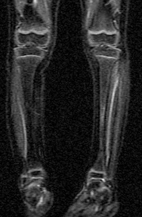
32 Image Identification STIR
33 Image Identification FLAIR
34 Image Identification
35 Image Identification

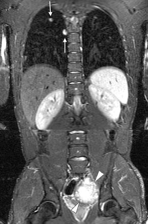
36 Image Identification Qayyum, A. et al. Radiology 2005;237: Copyright Radiological Society of North America, 2005

37 Image Identification Qayyum, A. et al. Radiology 2005;237: Copyright Radiological Society of North America, 2005
38 Common Artifacts Motion-Respiration, Flow, Cardiac, Patient Susceptibility Chemical Shift Aliasing (Wraparound)

39 Figure 7a. Motion-related artifacts (Respiratory) Zhuo, J. et al. Radiographics 2006;26: Copyright Radiological Society of North America, 2006

40 Figure 7b. Motion-related artifacts (Pulsation) Zhuo, J. et al. Radiographics 2006;26: Copyright Radiological Society of North America, 2006

41 Figure 14. Sagittal MR image shows a magnetic susceptibility artifact that resulted from the presence of metallic dental fillings Copyright Radiological Society of North America, 2006 Zhuo, J. et al. Radiographics 2006;26:
42 Safety General Safety During Pregnancy Orbital Foreign Bodies Foreign Bodies Elsewhere Aneurysm Clips Orthopedic Implants Pacers Stents Heart Valves Spine Stimulators
43
44 Question #1 Electric current flowing through a wire will produce: A. Hydrogen protons B. The gyromagnetic ratio C. A magnetic field D. T1 contrast
45 Question #1 Electric current flowing through a wire will produce: A. Hydrogen protons B. The Exist in the body with or without current The gyromagnetic ratio Is unaltered by current It is fixed for different nuclei 42.6 MHz/T for hydrogen protons C. A magnetic field D. T1 contrast Correct Answer Further proton manipulation will be necessary to create T1 contrast
46 Explanation #1 Electrons flowing in a wire will create a magnetic field perpendicular to the current. Pooley, R. A. Radiographics 2005;25:
47 Question #2 Under normal circumstances and with respect to their magnetization, how are protons oriented in the body? A. Randomly B. Along the Z-axis Z (longitudinal) C. Along the X-axis X (transverse) D. Along the Y-axis Y (transverse)
48 Question #2 Under normal circumstances and with respect to their magnetization, how are protons oriented in the body? A. Randomly Correct answer B. Along the Z-axis Z (longitudinal) Protons may align along this axis, but this will not be universal and should be temporary until an external magnetic field is applied C. Along the X-axis X (transverse) Protons may align along this axis, but this typically occurs after an external magnetic field and a 90 degree RF pulse are applied D. Along the Y-axis Y (transverse) Protons may align along this axis, but this typically occurs after an external magnetic field and a 90 degree RF pulse are applied
49 Explanation #2 Protons are distributed randomly in the body until their nuclei are manipulated with external forces. Pooley, R. A. Radiographics 2005;25:
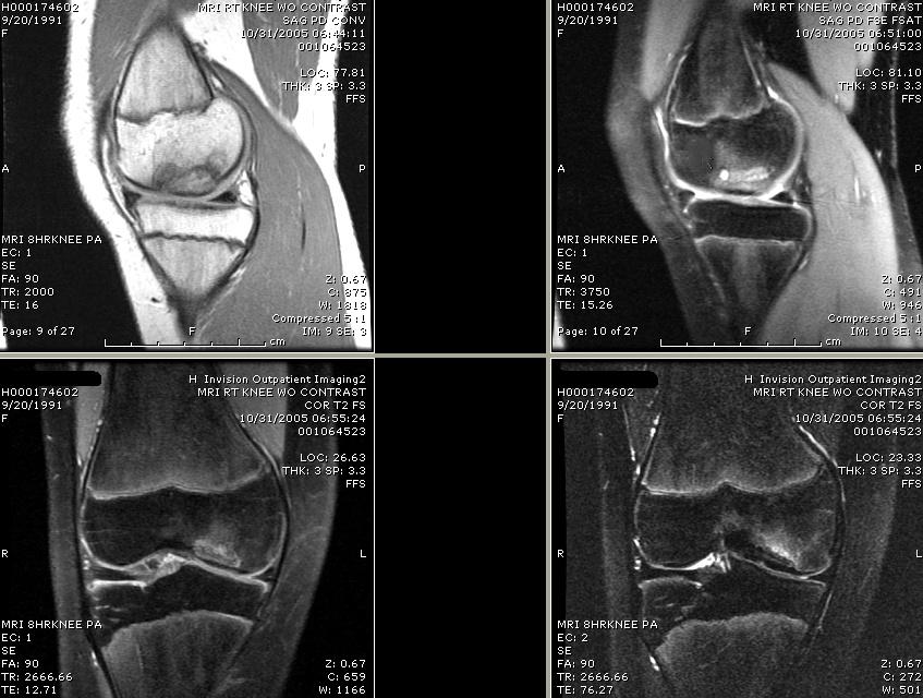
50 Question #3 Which of the following image(s) is/are T1-weighted? A. B. C. D.
51 Question #3 Which of the following image(s) is/are T1-weighted? A. B. C. D.
52 Explanation #3 Which of the following image(s) is/are T1-weighted? A. B. C. D. On T1 white matter and fat are bright and CSF and fluid are much darker, which is seen on image A. and D. The CSF in B. is dark because this is a FLAIR sequence, which is heavily T2 weighted B. and C. are incorrect because the white matter is very dark on both Kaplan et al. Musculoskeletal MRI 2001; 1:4.
53 Question #4 Which imaging artifact is frequently utilized in adrenal imaging? A. Aliasing B. Pulsation C. Gibbs D. Chemical Shift
54 Question #4 Which imaging artifact is frequently utilized in adrenal imaging? A. Aliasing is incorrect because it refers to imaging wrap and has no place in adrenal imaging B. Pulsation is incorrect since this is an artifact created by repetitive movement such as arterial flow and is also not useful in adrenal imaging unless attempting to detect vascularity within a lesion C. Gibbs is incorrect since Gibbs artifact is a group of bright or dark lines seen adjacent and parallel to regions of abrupt signal change D. Chemical Shift is correct
55 Explanation #4 D. Chemical shift is correct because it is caused by the difference of chemical shift between fat and water This artifact manifests itself as a misregistration between fat and water pixels in an image This artifact allows identification of microscopic fat in lesions, which is very useful in identifying lipid rich adrenal adenomas
 In-phase MR image acquired with an")

56 Figure 18a. (a) In-phase MR image acquired with an echo time of 2.2 msec Zhuo, J. et al. Radiographics 2006;26: Copyright Radiological Society of North America, 2006
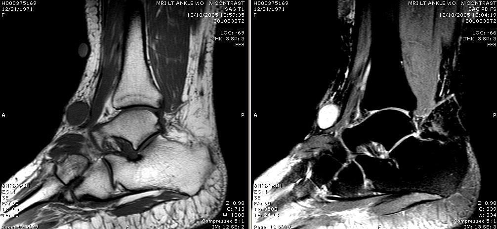
57 Question #5 Which of the following image(s) is/are T2-weighted? A. B. C. D.
58 Question #5 Which of the following image(s) is/are T2-weighted? A. B. C. D.
59 Explanation #5 Which of the following image(s) is/are T2-weighted? A. B. C. D. On T1 white matter and fat are bright and CSF and fluid are much darker, which fits images A. and D. The white matter in B. and C. is very dark, indicating that they are T2 weighted. CSF in B is dark because this is a FLAIR sequence, which suppresses CSF signal, but this is still T2 weighted. Kaplan et al. Musculoskeletal MRI 2001; 1:4.
Syllabus References. Resources. Video: MRI Introduction
 MRI Lesson Outline Syllabus References 9.6.4.2.5 Define precessing and relate the frequency of the precessing to the composition of the nuclei and the strength of the applied external magnetic field 9.6.4.2.6
Magnetic Resonance Imaging. Basics of MRI in practice. Generation of MR signal. Generation of MR signal. Spin echo imaging. Generation of MR signal
 Magnetic Resonance Imaging Protons aligned with B0 magnetic filed Longitudinal magnetization - T1 relaxation Transverse magnetization - T2 relaxation Signal measured in the transverse plane Basics of MRI
FOR CMS (MEDICARE) MEMBERS ONLY NATIONAL COVERAGE DETERMINATION (NCD) FOR MAGNETIC RESONANCE IMAGING:
 MEMBERS ONLY NATIONAL COVERAGE DETERMINATION (NCD) FOR MAGNETIC RESONANCE IMAGING:") National Imaging Associates, Inc. Clinical guidelines BONE MARROW MRI Original Date: July 2008 Page 1 of 5 CPT Codes: 77084 Last Review Date: September 2014 NCD 220.2 MRI Last Effective Date: July 2011
National Imaging Associates, Inc. Clinical guidelines BONE MARROW MRI Original Date: July 2008 Page 1 of 5 CPT Codes: 77084 Last Review Date: September 2014 NCD 220.2 MRI Last Effective Date: July 2011
Basics of MR Imaging. Dynamic MRI. MRI Closed. The bed rotates from Upright to Recumbent, stopping at any angle in between.
 Basics of MR Imaging Dynamic MRI MRI Closed The bed rotates from Upright to Recumbent, stopping at any angle in between MRI Open Patient with Low Back Pain After Surgery Extremity MRI Sagittal T2 WI of
Clinical Applications
 C H A P T E R 16 Clinical Applications In selecting pulse sequences and measurement parameters for a specific application, MRI allows the user tremendous flexibility to produce variations in contrast between
FOR CMS (MEDICARE) MEMBERS ONLY NATIONAL COVERAGE DETERMINATION (NCD) FOR MAGNETIC RESONANCE IMAGING:
 MEMBERS ONLY NATIONAL COVERAGE DETERMINATION (NCD) FOR MAGNETIC RESONANCE IMAGING:") National Imaging Associates, Inc. Clinical guidelines SINUS MRI Original Date: November 2007 Page 1 of 5 CPT Codes: 70540, 70542, 70543 Last Review Date: July 2014 NCD 220.2 MRI Last Effective Date: July
National Imaging Associates, Inc. Clinical guidelines SINUS MRI Original Date: November 2007 Page 1 of 5 CPT Codes: 70540, 70542, 70543 Last Review Date: July 2014 NCD 220.2 MRI Last Effective Date: July
MR Advance Techniques. Vascular Imaging. Class II
 MR Advance Techniques Vascular Imaging Class II 1 Vascular Imaging There are several methods that can be used to evaluate the cardiovascular systems with the use of MRI. MRI will aloud to evaluate morphology
MR Advance Techniques. Cardiac Imaging. Class IV
 MR Advance Techniques Cardiac Imaging Class IV Heart The heart is a muscular organ responsible for pumping blood through the blood vessels by repeated, rhythmic contractions. Layers of the heart Endocardium
Orthopedic Hardware Imaging Part II: MRI v. Metal
 Orthopedic Hardware Imaging Trent Roth, MD And Lauren Ladd, MD Indiana University School of Medicine IU Health Physicians-Radiology Recap: Imaging Techniques Radiography Standard for initial and surveillance
FOR CMS (MEDICARE) MEMBERS ONLY NATIONAL COVERAGE DETERMINATION (NCD) FOR MAGNETIC RESONANCE IMAGING:
 MEMBERS ONLY NATIONAL COVERAGE DETERMINATION (NCD) FOR MAGNETIC RESONANCE IMAGING:") National Imaging Associates, Inc. Clinical guidelines TEMPOROMANDIBULAR JOINT (TMJ) MRI Original Date: May 23, 2003 Page 1 of 5 CPT Code: 70336 Last Review Date: May 2016 NCD 220.2 MRI Last Effective Date:
National Imaging Associates, Inc. Clinical guidelines TEMPOROMANDIBULAR JOINT (TMJ) MRI Original Date: May 23, 2003 Page 1 of 5 CPT Code: 70336 Last Review Date: May 2016 NCD 220.2 MRI Last Effective Date:
Table 1. Summary of PET and fmri Methods. What is imaged PET fmri BOLD (T2*) Regional brain activation. Blood flow ( 15 O) Arterial spin tagging (AST)
 Regional brain activation. Blood flow ( 15 O) Arterial spin tagging (AST)") Table 1 Summary of PET and fmri Methods What is imaged PET fmri Brain structure Regional brain activation Anatomical connectivity Receptor binding and regional chemical distribution Blood flow ( 15 O)
Table 1 Summary of PET and fmri Methods What is imaged PET fmri Brain structure Regional brain activation Anatomical connectivity Receptor binding and regional chemical distribution Blood flow ( 15 O)
RADIOLOGY TEACHING CONFERENCE
 RADIOLOGY TEACHING CONFERENCE John Athas, MD Monica Tadros, MD Columbia University, College of Physicians & Surgeons Department of Otolaryngology- Head & Neck Surgery September 27, 2007 CT SCAN IMAGING
*smith&nephew. MRI Safety Information & Parameters for Smith & Nephew Orthopaedics AG. Knee Implants
 Knee Implants MRI Safety Information & Parameters for Smith & Nephew Orthopaedics AG Knee Implants *smith&nephew Supporting healthcare professionals for over 150 years Summary All knee implants of Smith
MSRS 6473 Vascular Noninvasive Imaging Procedures
 MSRS 6473 Vascular Noninvasive Imaging Procedures Rex T. Christensen MHA RT (R) (MR) (CT) (ARRT) CIIP Basic Physics Equipment Cardiac Positioning Perfusion Pathology MRI 1 Animal Magnetism MRI Basic Physics
RECENT ADVANCES IN CLINICAL MR OF ARTICULAR CARTILAGE
 In Practice RECENT ADVANCES IN CLINICAL MR OF ARTICULAR CARTILAGE By Atsuya Watanabe, MD, PhD, Director, Advanced Diagnostic Imaging Center and Associate Professor, Department of Orthopedic Surgery, Teikyo
Magnetic Resonance Imaging (MRI) Dynamic Pelvic Floor
 Dynamic Pelvic Floor") Scan for mobile link. Magnetic Resonance Imaging (MRI) Dynamic Pelvic Floor Dynamic pelvic floor magnetic resonance imaging (MRI) is a noninvasive test that uses a powerful magnetic field, radio waves
Scan for mobile link. Magnetic Resonance Imaging (MRI) Dynamic Pelvic Floor Dynamic pelvic floor magnetic resonance imaging (MRI) is a noninvasive test that uses a powerful magnetic field, radio waves
CARDIAC MRI. Cardiovascular Disease. Cardiovascular Disease. Cardiovascular Disease. Overview
 CARDIAC MRI Dr Yang Faridah A. Aziz Department of Biomedical Imaging University of Malaya Medical Centre Cardiovascular Disease Diseases of the circulatory system, also called cardiovascular disease (CVD),
*smith&nephew. MRI Safety Information & Parameters for Smith & Nephew Orthopaedics AG. Shoulder Implants
 Shoulder Implants MRI Safety Information & Parameters for Smith & Nephew Orthopaedics AG Shoulder Implants *smith&nephew Supporting healthcare professionals for over 150 years Summary All shoulder implants
8/4/2016. MRI for Radiotherapy: MRI Basics. Nuclear Magnetic Resonance. Nuclear Magnetic Resonance. Wilson Miller
 MRI for Radiotherap: MRI asics Wilson Miller Universit of Virginia Department of Radiolog & Medical Imaging AAPM 2016 August 4, 2016 Nuclear Magnetic Resonance Magnetic resonance images are created using
Magnetic Resonance Angiography
 Magnetic Resonance Angiography 1 Magnetic Resonance Angiography exploits flow enhancement of GR sequences saturation of venous flow allows arterial visualization saturation of arterial flow allows venous
Assessment of Adipose Tissue from Whole Body 3T MRI Scans
 Assessment of Adipose Tissue from Whole Body 3T MRI Scans Ting Song 1, Jing An 2, Qun Chen 2, Vivian Lee 2, Andrew Laine 1 1 Department of Biomedical Engineering, Columbia University, New York, NY, USA
High Field MR of the Spine
 Department of Radiology University of California San Diego 3T for MR Applications Advantages High Field MR of the Spine Increased signal-to-noise Better fat suppression Increased enhancement with gadolinium
Abdominal MRI Techniques in Pediatric Oncology
 Abdominal MRI Techniques in Pediatric Oncology Jonathan R. Dillman, M.D. Assistant Professor Departments of Radiology & Urology Section of Pediatric Radiology C.S. Mott Children s Hospital Disclosures
Fat Suppression in the Abdomen
 Clinical How I do it? Fat Suppression in the Abdomen Wilhelm Horger Siemens Medical Solutions, Erlangen, Germany Introduction Due to the different chemical environment, hydrogen nuclei in - and in -tissue
1Pulse sequences for non CE MRA
 MRI: Principles and Applications, Friday, 8.30 9.20 am Pulse sequences for non CE MRA S. I. Gonçalves, PhD Radiology Department University Hospital Coimbra Autumn Semester, 2011 1 Magnetic resonance angiography
ACR MRI Accreditation: Medical Physicist Role in the Application Process
 ACR MRI Accreditation: Medical Physicist Role in the Application Process Donna M. Reeve, MS, DABR, DABMP Department of Imaging Physics University of Texas M.D. Anderson Cancer Center Educational Objectives
Introduction. Cardiac Imaging Modalities MRI. Overview. MRI (Continued) MRI (Continued) Arnaud Bistoquet 12/19/03
 MRI (Continued) Arnaud Bistoquet 12/19/03") Introduction Cardiac Imaging Modalities Arnaud Bistoquet 12/19/03 Coronary heart disease: the vessels that supply oxygen-carrying blood to the heart, become narrowed and unable to carry a normal amount
Introduction Cardiac Imaging Modalities Arnaud Bistoquet 12/19/03 Coronary heart disease: the vessels that supply oxygen-carrying blood to the heart, become narrowed and unable to carry a normal amount
Functional Chest MRI in Children Hyun Woo Goo
 Functional Chest MRI in Children Hyun Woo Goo Department of Radiology and Research Institute of Radiology Asan Medical Center, University of Ulsan College of Medicine, Seoul, Korea No ionizing radiation
Non Contrast MRA. Mayil Krishnam. Director, Cardiovascular and Thoracic Imaging University of California, Irvine
 Non Contrast MRA Mayil Krishnam Director, Cardiovascular and Thoracic Imaging University of California, Irvine No disclosures Non contrast MRA-Why? Limitations of CTA Radiation exposure Iodinated contrast
Personal use only. MRI Metal Artifact Reduction: Shoulder Implants and Arthroplasty. Reto Sutter, MD
 MRI Metal Artifact Reduction: Shoulder Implants and Arthroplasty Reto Sutter, MD University Hospital Balgrist Zurich University of Zurich Cor PD fat sat 56-year old male patient with positive lift-off
*smith&nephew. MRI Safety Information & Parameters for Smith & Nephew Orthopaedics AG. Hip Implants
 Hip Implants MRI Safety Information & Parameters for Smith & Nephew Orthopaedics AG Hip Implants *smith&nephew Supporting healthcare professionals for over 150 years Summary All hip implants of Smith &
The Low Sensitivity of Fluid-Attenuated Inversion-Recovery MR in the Detection of Multiple Sclerosis of the Spinal Cord
 The Low Sensitivity of Fluid-Attenuated Inversion-Recovery MR in the Detection of Multiple Sclerosis of the Spinal Cord Mark D. Keiper, Robert I. Grossman, John C. Brunson, and Mitchell D. Schnall PURPOSE:
Terminology Tissue Appearance
 By Marc Nielsen, MD Advantages/Disadvantages Generation of Image Ultrasound Machine/Transducer selection Modes of Ultrasound Terminology Tissue Appearance Scanning Technique Real-time Portable No ionizing
Keep Imaging Simple: An Introduction To Neuroimaging
 Keep Imaging Simple: An Introduction To Neuroimaging Meghan Elkins, OD, FAAO Please silence all mobile devices and remove items from chairs so others can sit. Unauthorized recording of this session is
Magnetic Resonance Imaging (MRI) - Body
 - Body") Scan for mobile link. Magnetic Resonance Imaging (MRI) - Body What is MRI of the Body? Magnetic resonance imaging (MRI) is a noninvasive medical test that physicians use to diagnose and treat medical conditions.
Scan for mobile link. Magnetic Resonance Imaging (MRI) - Body What is MRI of the Body? Magnetic resonance imaging (MRI) is a noninvasive medical test that physicians use to diagnose and treat medical conditions.
This presentation is the intellectual property of the author. Contact them for permission to reprint and/or distribute.
 MRI of the Knee Jennifer Swart, M.D. Musculoskeletal Radiology South Texas Radiology Group Outline Coils, Patient Positioning Acquisition Parameters, Planes and Pulse Sequences Knee Arthrography Normal
PATIENT CARE AND MRI SAFETY Module 7
 PATIENT CARE AND MRI SAFETY Module 7 1 Biological Considerations There are no reported adverse biological effects of extended exposure to MRI. However, several inconsequential and reversible effects of
High Signal Intensity of the Infundibular Stalk on Fluid-Attenuated Inversion Recovery MR
 High Signal Intensity of the Infundibular Stalk on Fluid-Attenuated Inversion Recovery MR Yutaka Araki, Ryuichirou Ashikaga, Satoru Takahashi, Jun Ueda, and Osamu Ishida PURPOSE: To determine the MR imaging
Managing Metallic Artifacts in MRI
 Managing Metallic Artifacts in MRI William Faulkner, BS, RT (R) (MR) (CT), FSMRT Background Magnetism is a fundamental property of matter. As such, all substances have some type of magnetic property. Magnetic
This presentation is the intellectual property of the author. Contact them at for permission to reprint and/or distribute.
 MRI of the Knee Jennifer Swart, M.D. Musculoskeletal Radiology South Texas Radiology Group Financial Disclosure Dr. Jennifer Swart has no relevant financial relationships with commercial interests to disclose.
MR imaging the post operative spine - What to expect!
 MR imaging the post operative spine - What to expect! Poster No.: C-2334 Congress: ECR 2012 Type: Educational Exhibit Authors: A. Jain, M. Paravasthu, M. Bhojak, K. Das ; Warrington/UK, 1 1 1 2 1 2 Liverpool/UK
MRI for Patients with MRI-Conditional Pacing System: Radiographers Role. Lawrance Yip DM, DR, QMH
 MRI for Patients with MRI-Conditional Pacing System: Radiographers Role Lawrance Yip DM, DR, QMH MRI & Pacemaker MRI is a crucial and growing imaging modality Plays an important role in clinical management
The physiology of the BOLD signal What do we measure with fmri?
 The physiology of the BOLD signal What do we measure with fmri? Methods and Models in fmri, 10.11.2012 Jakob Heinzle Translational Neuromodeling Unit (TNU) Institute for Biomedical Engineering (IBT) University
Principles of Ultrasound. Cara C. Prideaux, M.D. University of Utah PM&R Sports Medicine Fellow March 14, 2012
 Principles of Ultrasound Cara C. Prideaux, M.D. University of Utah PM&R Sports Medicine Fellow March 14, 2012 None Disclosures Outline Introduction Benefits and Limitations of US Ultrasound (US) Physics
3/22/2017. MR Protocol Review Clinical Opportunities for Physicists. AAPM Newsletter March/April Technical to Clinical Transition - How?
 AAPM Newsletter March/April 2017 Clinical Opportunities for Physicists Anshuman Panda, Ph.D. AAPM Spring Clinical Meeting New Orleans, LA March 2017 No conflict of interest to declare Physics Summit on
MRI Physics: Basic to Advanced
 Annual Meeting of the American Society of Neuroimaging MRI Physics: Basic to Advanced Mike Moseley, PhD Department of Radiology Stanford University CA 94305-5488 USA http://www-radiology.stanford.edu moseley@stanford.edu
Tools for cardiovascular magnetic resonance imaging
 Review Article Tools for cardiovascular magnetic resonance imaging Ramkumar Krishnamurthy, Benjamin Cheong, Raja Muthupillai Department of Diagnostic and Interventional Radiology, CHI St. Luke s Health,
MRI Sequences: What to use for what
 MRI Sequences: What to use for what MRI basics T 1 and T 2 relaxation Common Imaging Protocols Mechanical function (cine) Tissue characterization LGE Edema imaging (T 2 weighted) T1 Special protocols MRA
Correction of CSF Motion Artifact on MR Images of the Brain and Spine by Pulse Sequence Modification: Clinical Evaluation
 1069 Correction of CSF Motion Artifact on MR Images of the Brain and Spine by Pulse Sequence Modification: Clinical Evaluation Nikolaus M. Szeverenyi 1 Stephen A. Kieffer Edwin D. Cacayorin A modification
Daniel Bulte. Centre for Functional Magnetic Resonance Imaging of the Brain. University of Oxford
 Daniel Bulte Centre for Functional Magnetic Resonance Imaging of the Brain University of Oxford Overview Signal Sources BOLD Contrast Mechanism of MR signal change FMRI Modelling Scan design details Factors
Magnetic Resonance Imaging (MRI) - Head
 - Head") Scan for mobile link. Magnetic Resonance Imaging (MRI) - Head Magnetic resonance imaging (MRI) of the head uses a powerful magnetic field, radio waves and a computer to produce detailed pictures of the
Scan for mobile link. Magnetic Resonance Imaging (MRI) - Head Magnetic resonance imaging (MRI) of the head uses a powerful magnetic field, radio waves and a computer to produce detailed pictures of the
HSC Physics. Module 9.6. Medical Physics
 HSC Physics Module 9.6 Medical Physics Contextual Outline 9.6 Medical Physics (28 indicative hours) The use of other advances in technology, developed from our understanding of the electromagnetic spectrum,
Magnetic Resonance Imaging (MRI) - Breast
 - Breast") Scan for mobile link. Magnetic Resonance Imaging (MRI) - Breast Magnetic resonance imaging (MRI) of the breast uses a powerful magnetic field, radio waves and a computer to produce detailed pictures of
Scan for mobile link. Magnetic Resonance Imaging (MRI) - Breast Magnetic resonance imaging (MRI) of the breast uses a powerful magnetic field, radio waves and a computer to produce detailed pictures of
醫用磁振學 MRM 肌肉骨骼磁振造影簡介 肌肉骨骼磁振造影. 本週課程內容 General Technical Considerations 肌肉骨骼磁振造影簡介 盧家鋒助理教授國立陽明大學生物醫學影像暨放射科學系
 本週課程內容 http://www.ym.edu.tw/~cflu 肌肉骨骼磁振造影簡介 醫用磁振學 MRM 肌肉骨骼磁振造影 盧家鋒助理教授國立陽明大學生物醫學影像暨放射科學系 alvin4016@ym.edu.tw MRI of the musculoskeletal system (5th/6th edition) Editor: Thomas H. Berquist MD 2 General
Magnetic Resonance Imaging (MRI) - Body
 - Body") Scan for mobile link. Magnetic Resonance Imaging (MRI) - Body Magnetic resonance imaging (MRI) of the body uses a powerful magnetic field, radio waves and a computer to produce detailed pictures of the
Scan for mobile link. Magnetic Resonance Imaging (MRI) - Body Magnetic resonance imaging (MRI) of the body uses a powerful magnetic field, radio waves and a computer to produce detailed pictures of the
Magnetic Resonance Imaging
 Magnetic Resonance Imaging The purpose of structured education is to provide the opportunity for individuals to develop mastery of discipline-specific knowledge that, when coupled with selected clinical
Why Cardiac MRI? Presented by:
 Why Cardiac MRI? Presented by: Lisa G. Carkner, MD, FACC 1 Disclosures I have no financial disclosures Objectives Review basic principles of Cardiac MRI. What patient characteristics do I need to consider
Spatially Varying Saturation Pulse (SVSP) for Fat Suppression in Breast MRI
 for Fat Suppression in Breast MRI") Spatially Varying Saturation Pulse (SVSP) for Fat Suppression in Breast MRI by Tse Chiang Chen A thesis submitted in conformity with the requirements for the degree of Master of Science Department of Medical
Spatially Varying Saturation Pulse (SVSP) for Fat Suppression in Breast MRI by Tse Chiang Chen A thesis submitted in conformity with the requirements for the degree of Master of Science Department of Medical
Magnetic Resonance, Functional (fmri) - Brain
 - Brain") Scan for mobile link. Magnetic Resonance, Functional (fmri) - Brain Functional magnetic resonance imaging (fmri) measures the metabolic changes that occur within the brain. It may be used to examine the
Scan for mobile link. Magnetic Resonance, Functional (fmri) - Brain Functional magnetic resonance imaging (fmri) measures the metabolic changes that occur within the brain. It may be used to examine the
GUIDELINE 2-G-004 (formerly APPENDIX 3) HSREB (March 2005) Page 1
 HSREB (March 2005) Page 1") HSREB (March 2005) Page 1 Guidelines for Ethics Approval of Research Protocols Involving Human Exposure to Magnetic Resonance Imaging Required Wording in Informed Consent Documentation; The Food & Drug
HSREB (March 2005) Page 1 Guidelines for Ethics Approval of Research Protocols Involving Human Exposure to Magnetic Resonance Imaging Required Wording in Informed Consent Documentation; The Food & Drug
MR Angiography in the evaluation of Lower Extremity Arterial Disease
 March 2001 MR Angiography in the evaluation of Lower Extremity Arterial Disease Ted Mau, Harvard Medical School Year III Objectives We will cover: Indications for Magnetic Resonance Angiography (MRA) Basic
Study of the CNS. Bent O. Kjos' Richard L. Ehman Michael Brant-Zawadzki William M. Kelly David Norman Thomas H. Newton
 271 Reproducibility of Relaxation Times and Spin Density Calculated from Routine MR Imaging Sequences: Clinical Study of the CNS Bent O. Kjos' Richard L. Ehman Michael Brant-Zawadzki William M. Kelly David
Introduction & Physics of ED Ultrasound. Objectives. What? - Limited Studies. Who? - ED Docs
 Introduction & Physics of ED Ultrasound Martine Sargent, MD Ultrasound Director, Assistant Professor UCSF Department of Emergency Medicine San Francisco General Hospital & Trauma Center Objectives Who?
MR Advance Techniques. Cardiac Imaging. Class III
 MR Advance Techniques Cardiac Imaging Class III Black Blood Imaging & IR Blue= O2 poor blood Red=O2 rich blood Inversion pulses can produce black blood imaging in GRE pulse sequences. Specially on the
Magnetic Resonance Imaging (MRI) - Breast
 - Breast") Scan for mobile link. Magnetic Resonance Imaging (MRI) - Breast What is MRI of the Breast? Magnetic resonance imaging (MRI) is a noninvasive medical test that physicians use to diagnose and treat medical
Scan for mobile link. Magnetic Resonance Imaging (MRI) - Breast What is MRI of the Breast? Magnetic resonance imaging (MRI) is a noninvasive medical test that physicians use to diagnose and treat medical
1 Normal Anatomy and Variants
 1 Normal Anatomy and Variants 1.1 Normal Anatomy MR Technique. e standard MR protocol for a routine evaluation of the spine always comprises imaging in sagittal and axial planes, while coronal images are
Objectives 8/17/2011. Challenges in Cardiac Imaging. Challenges in Cardiac Imaging. Basic Cardiac MRI Sequences
 8/17/2011 Traditional Protocol Model for Tomographic Imaging Cardiac MRI Sequences and Protocols Frandics Chan, M.D., Ph.D. Stanford University Medical Center Interpretation Lucile Packard Children s Hospital
MRI Scan. Patient Information. MRI Department Cobalt Imaging Centre. Registered Charity No:
 MRI Scan Patient Information MRI Department Cobalt Imaging Centre www.cobalthealth.co.uk Registered Charity No: 1090790 This leaflet aims to answer questions about having an MRI scan What is an MRI scan?
MR Angiography 1. What is MR Angiography? What are some common uses of the procedure? August 17, 2007
 http://www.radiologyinfo.org MR Angiography (MRA) This procedure is reviewed by a physician with expertise in the area presented and is further reviewed by committees from the American College of Radiology
DETECTION AND CHARACTERIZATION OF RETINAL DISRUPTION IN MICE USING HIGH ANGULAR RESOLUTION DIFFUSION MICROSCOPY (HARDM)
") DETECTION AND CHARACTERIZATION OF RETINAL DISRUPTION IN MICE USING HIGH ANGULAR RESOLUTION DIFFUSION MICROSCOPY (HARDM) By SAURAV BIMALKUMAR CHANDRA A DISSERTATION PRESENTED TO THE GRADUATE SCHOOL OF THE
DETECTION AND CHARACTERIZATION OF RETINAL DISRUPTION IN MICE USING HIGH ANGULAR RESOLUTION DIFFUSION MICROSCOPY (HARDM) By SAURAV BIMALKUMAR CHANDRA A DISSERTATION PRESENTED TO THE GRADUATE SCHOOL OF THE
Diagnostic approach to heart disease
 Diagnostic approach to heart disease Initial work up History Physical exam Chest radiographs ECG Special studies Echocardiography Cardiac catheterization Echocardiography principles Technique of producing
Physics COPYRIGHTED MATERIAL. Comparative Imaging. Patrick R. Gavin. Diagnostic Radiology. Nuclear Medicine or Gamma Scintigraphy CHAPTER ONE
 CHPTER ONE Physics Diagnostic imaging has always been a mainstay of the armamentarium for the veterinarian. Veterinarians have limited resources available as regards history and routine screening procedures.
P2 Visual - Perception
 P2 Visual - Perception 2014 SOSE Neuroimaging of high-level visual functions gyula.kovacs@uni-jena.de 11/09/06 Functional magnetic resonance imaging (fmri) The very basics What is fmri? What is MRI? The
Diffusion-weighted MR Imaging Offers No Advantage over Routine Noncontrast MR Imaging in the Detection of Vertebral Metastases
 AJNR Am J Neuroradiol 1:948 953, May Diffusion-weighted MR Imaging Offers No Advantage over Routine Noncontrast MR Imaging in the Detection of Vertebral Metastases Mauricio Castillo, Andres Arbelaez, J.
Cochlear Implants. Medical Procedures. for MED EL Implant Systems. AW33290_2.0 (English US)
") Cochlear Implants Medical Procedures for MED EL Implant Systems AW33290_2.0 (English US) This manual provides important instructions and safety information for MED EL Implant System users who have to undergo
Cochlear Implants Medical Procedures for MED EL Implant Systems AW33290_2.0 (English US) This manual provides important instructions and safety information for MED EL Implant System users who have to undergo
Medical imaging X-ray, CT, MRI, scintigraphy, SPECT, PET Györgyi Műzes
 Medical imaging X-ray, CT, MRI, scintigraphy, SPECT, PET Györgyi Műzes Semmelweis University, 2nd Dept. of Medicine Medical imaging: definition technical process of creating visual representations about
MR Angiography (MRA)
") MR Angiography (MRA) What is MR Angiography? What are some common uses of the procedure? How should I prepare? What does the equipment look like? How does the procedure work? How is the procedure performed?
MR Angiography (MRA) What is MR Angiography? What are some common uses of the procedure? How should I prepare? What does the equipment look like? How does the procedure work? How is the procedure performed?
ACR MRI Accreditation Program. ACR MRI Accreditation Program Update. Educational Objectives. ACR accreditation. History. New Modular Program
 ACR MRI Accreditation Program Update Donna M. Reeve, MS, DABR, DABMP Department of Imaging Physics University of Texas M.D. Anderson Cancer Center Educational Objectives Present requirements of the new
Initial Clinical Experience of TOSHIBA 3T MRI
 The 21st Conference of the Japanese Society of Cardiovascular Imaging & Dynamics Sponsored Seminar The Leading Edge of CT/MRI Diagnosis for the Cardiovascular System Initial Clinical Experience of TOSHIBA
Magnetic Resonance Imaging
 STRUCTURED SELF ASSESSMENT CONTENT SPECIFICATIONS ARRT BOARD APPROVED: PENDING IMPLEMENTATION DATE: JANUARY 1, 2018 Magnetic Resonance Imaging The purpose of continuing qualifications requirements (CQR)
Magnetization Preparation Sequences
 Magnetization Preparation Sequences Acquisition method may not give desired contrast Prep block adds contrast (and/or encoding) MP-RAGE = Magnetization prepared rapid acquisition with gradient echo (Mugler,
Stroke Imaging Basics. Jeremy Hopkin M.D.
 Stroke Imaging Basics Jeremy Hopkin M.D. Goals Introduce the basic physical properties of imaging used in stroke. Understand why each modality is used in the setting of stroke. Understand some strengths
Magnetic Resonance Imaging (MRI) - Cardiac (Heart)
 - Cardiac (Heart)") Scan for mobile link. Magnetic Resonance Imaging (MRI) - Cardiac (Heart) Cardiac magnetic resonance imaging (MRI) uses a powerful magnetic field, radio waves and a computer to produce detailed pictures
Scan for mobile link. Magnetic Resonance Imaging (MRI) - Cardiac (Heart) Cardiac magnetic resonance imaging (MRI) uses a powerful magnetic field, radio waves and a computer to produce detailed pictures
The Role of IDEAL and DTI in Peripheral Nerve MR Imaging
 In Practice The Role of IDEAL and DTI in Peripheral Nerve MR Imaging y Darryl. Sneag, MD, Assistant Attending Radiologist, and Hollis G. Potter, MD, Chairman and The Coleman Chair, MRI Research, Department
MR imaging techniques
 Eur Radiol (2002) 12:2866 2882 DOI 10.1007/s00330-002-1428-9 MAGNETIC RESONANCE W. R. Nitz Fast and ultrafast non-echo-planar MR imaging techniques Received: 21 August 2001 Revised: 21 February 2002 Accepted:
FieldStrength. Achieva 3.0T enables cutting-edge applications, best-in-class MSK images
 FieldStrength Publication for the Philips MRI Community Issue 33 December 2007 Achieva 3.0T enables cutting-edge applications, best-in-class MSK images Palo Alto Medical Clinic Sports Medicine Center employs
The safe and accurate measurement of liver iron concentration. Information for Patients
 The safe and accurate measurement of liver iron concentration Information for Patients What you need to know What is iron overload? Iron overload is the accumulation of too much iron in the body. People
Cervical Spondylosis: Three-dimensional Gradient-Echo MR with Magnetization Transfer
 Cervical Spondylosis: Three-dimensional Gradient-Echo MR with Magnetization Transfer Elias R. Melhem, Mark L. Benson, Norman J. Beauchamp, and Roland R. Lee PURPOSE: To compare a three-dimensional Fourier
By Mohammed D. Alenezy
 Modeling Left Ventricle Wall Motion Using Tagged Magnetic Resonance Imaging By Mohammed D. Alenezy Submitted to the Department of Physics and Astronomy and the Faculty of the Graduate School of the University
Problem Set #8 Rad 226
 Problem Set #8 1. J-editing. In class, we discussed J-editing for the doublet resonance of lactate. Other in vivo peaks (e.g. GABA) are more complicated (triplets, quartets, etc.). In this problem, we
T2, T2*, ute. Yeo Ju Kim. Radiology, Inha University Hospital, Incheon, Korea
 SY28-1 T2, T2*, ute Yeo Ju Kim Radiology, Inha University Hospital, Incheon, Korea T2 relaxation times relate to the rate of transverse magnetization decay, caused by the loss of phase coherence induced
Introduction to the Course and the Techniques. Jeffry R. Alger, PhD Ahmanson-Lovelace Brain Mapping Center Department of Neurology
 Introduction to the Course and the Techniques Jeffry R. Alger, PhD Ahmanson-Lovelace Brain Mapping Center Department of Neurology (jralger@ucla.edu) CTSI Neuroimaging April 2014 Rationale for the Course
Attenuation value in HU From -500 To HU From -10 To HU From 60 To 90 HU. From 200 HU and above
 Brain Imaging Common CT attenuation values Structure Air Fat Water Brain tissue Recent hematoma Calcifications Bone Brain edema and infarction Normal liver parenchyma Attenuation value in HU From -500
The Essentials Tissue Characterization and Knobology
 The Essentials Tissue Characterization and Knobology Randy E. Moore, DC, RDMS RMSK No relevant financial relationships Ultrasound The New Standard of Care Musculoskeletal sonography has become the standard
10/3/2016. T1 Anatomical structures are clearly identified, white matter (which has a high fat content) appears bright.
 appears bright.") H2O -2 atoms of Hydrogen, 1 of Oxygen Hydrogen just has one single proton and orbited by one single electron Proton has a magnetic moment similar to the earths magnetic pole Also similar to earth in that
H2O -2 atoms of Hydrogen, 1 of Oxygen Hydrogen just has one single proton and orbited by one single electron Proton has a magnetic moment similar to the earths magnetic pole Also similar to earth in that
Essentials of Clinical MR, 2 nd edition. 99. MRA Principles and Carotid MRA
 99. MRA Principles and Carotid MRA As described in Chapter 12, time of flight (TOF) magnetic resonance angiography (MRA) is commonly utilized in the evaluation of the circle of Willis. TOF MRA allows depiction
Echelon Oval provides a robust suite of leading musculoskeletal imaging capabilities for detailed assessment of all anatomy for your most challenging
 Echelon Oval provides a robust suite of leading musculoskeletal imaging capabilities for detailed assessment of all anatomy for your most challenging cases. Hitachi Medical Systems America, Inc. 1959 Summit
BREAST MRI PROTOCOLS
 BREAST MRI PROTOCOLS Updated 03/14/2018 Routine Bilateral w/wo Silicone Breast Implant Post Biopsy Additional View Right Unilateral w/wo Left Unilateral w/wo Saline Breast Implant Biopsy Breast (Bilateral
MATHEMATICAL MODEL AND VALIDATION OF MICROVASCULAR FUNCTION IN LOW PERFUSION TISSUE
 MATHEMATICAL MODEL AND VALIDATION OF MICROVASCULAR FUNCTION IN LOW PERFUSION TISSUE by Eric Zakher A thesis submitted in conformity with the requirements for the degree of Master of Applied Science The
Quantification of liver steatosis in MRI: available techniques and use of transverse magnetization decay curve in patients with iron overload
 Quantification of liver steatosis in MRI: available techniques and use of transverse magnetization decay curve in patients with iron overload Poster No.: C-1302 Congress: ECR 2013 Type: Educational Exhibit