Evidence based imaging of the pancreas
|
|
|
- Mervin Horton
- 6 years ago
- Views:
Transcription

1 Evidence based imaging of the pancreas D.Vanbeckevoort, D.Bielen, K.Op de beeck, R.Vanslembrouck Department of Radiology Chairman Prof. Dr. R.Oyen
2 Non-invasive imaging tests available for the diagnosis of pancreatic disease 1. Ultrasound Transabdominal Color Doppler US, Power Doppler US, CE-US Endoscopic ultrasound Conventional EUS, Color Doppler EUS, CE-EUS, Intraductal US, EUS-guided fine needle aspiration (FNAC) 2. Computed tomography MSCT PET-CT 3. Magnetic Resonance Imaging Ultrafast breath-hold dynamic contrast-enhanced imaging High field strength magnet systems (3T)
3 Learning objectives 1. Current status of basic imaging techniques Application Value 2. How to determine a logical approach Imaging strategies pancreatic inflammatory disease pancreatic malignancy
4 Acute pancreatitis
5 Question 1 Is US a useful diagnostic imaging technique to confirm the clinical suspicion of acute pancreatitis? ECR Vienna, 2006


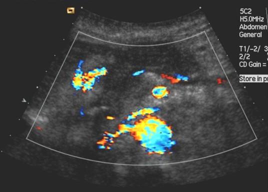
6 Transabdominal ultrasound (US) Can identify: Pancreatic enlargement Reduction in parenchymal reflectivity Fluid accumulation ECR Vienna, 2006
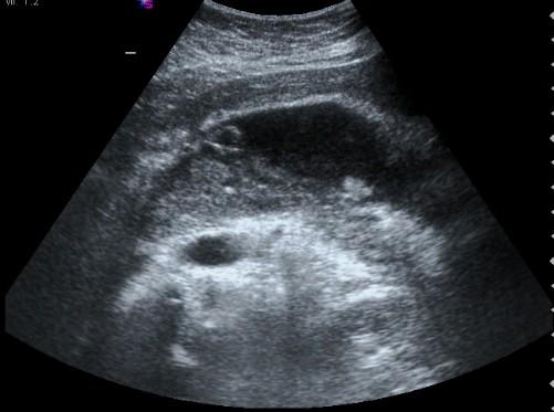
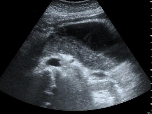
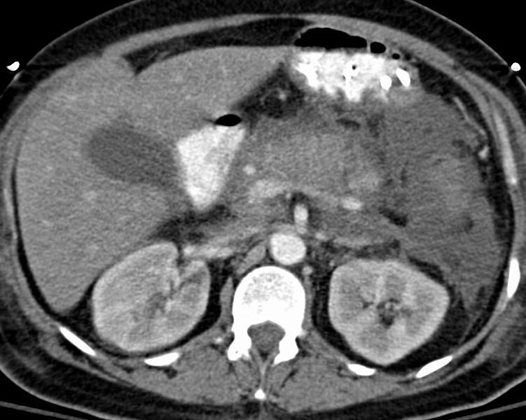
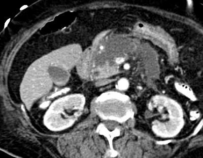
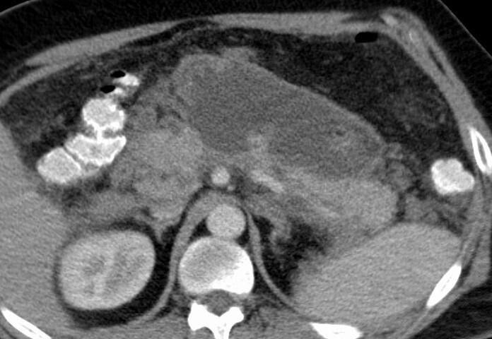
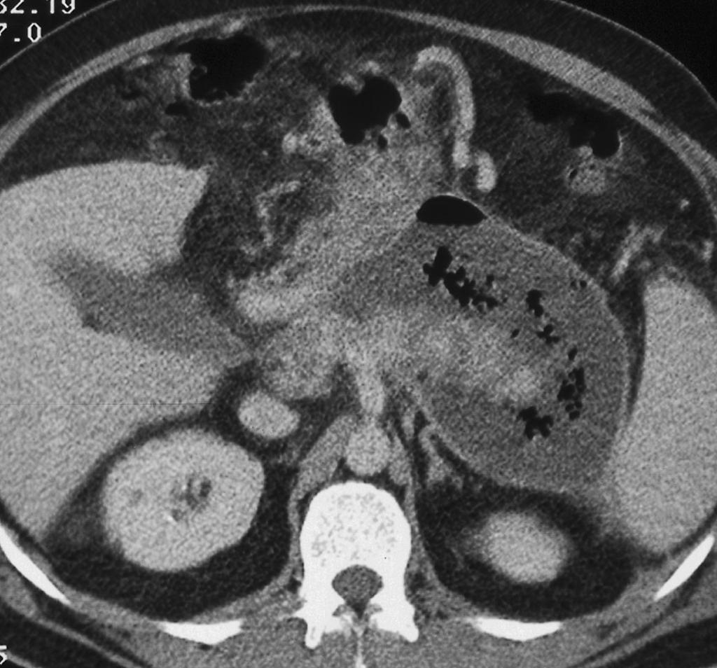
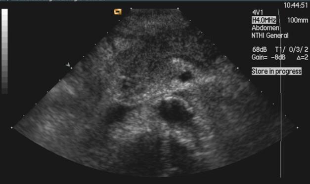
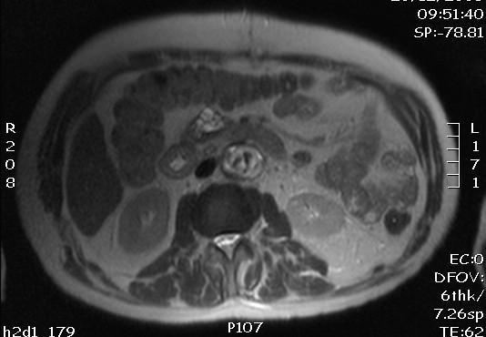
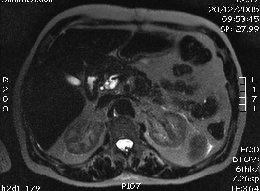
7 Severe pancreatitis ECR Vienna, 2006
8 Transabdominal ultrasound (US) A. Limited in the initial phase less successful in early diagnosis/staging effective in diagnosing gallbladder stones and biliary obstruction ERCP in extremely ill patients too unstable to undergo CT B. Valuable in the follow-up pseudocysts vascular complications Maher M. et al. Cardiovasc. Intervent. Radiol. (2004) 27:
9 Endoscopic ultrasound (EUS) Pro: More sensitive than transabdominal US in the identification of cholelithiasis Can distinguish mild from severe pancreatitis Contra: Little information regarding the viability of pancreatic tissue Bhutani et al. Gastro-enterol Clin North Am (1999) 28:747-77O Hayakawa et al. JOP (2000) 1: 46-48
10 Question 2: What is the imaging technique of choice for the diagnosis and staging of acute pancreatitis? ECR Vienna, 2006
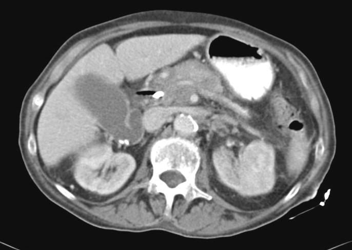
11 Computer tomography (CT) Modality of choice accuracy, reproducibility, availability Imaging features: pancreatic swelling, stranding acute fluid collections pancreatic necrosis Piironen A et al. Eur Radiol 2001; 26:
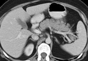
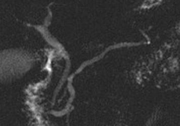
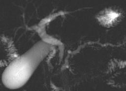
12 A B C D
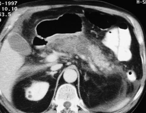
13 Severe pancreatitis E E
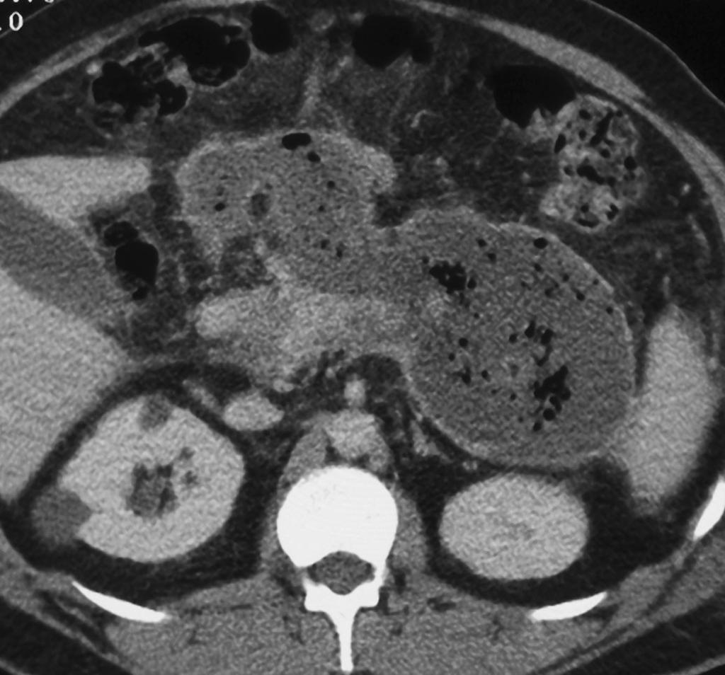
14 The Balthazar classification and CT severity index Prognostic indicators Characteristics P pancreatic inflammation normal pancreas 0/A focal or diffuse enlargement of the pancreas 1/B intrinsic pancreatic abnormalities with 2/C inflammatory changes in peripancreatic fat single, ill-defined fluid collection or phlegmon 3/D two or more poorly defined collections or 4/E presence of gas in or adjacent to the pancreas pancreatic necrosis no necrosis 0 30 % or less 2 30 %- 50 % 4 greater than 50 % 6

15 < 30 % < 50 % > 50 %
16 The Balthazar classification and CT severity index mortality morbidity points 4-6 points 7-10 points Balthazar EJ, et al. Imaging and intervention in acute pancreatitis. Radiology (1994) 193:
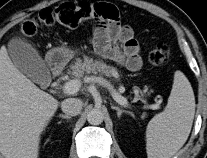
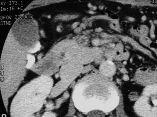
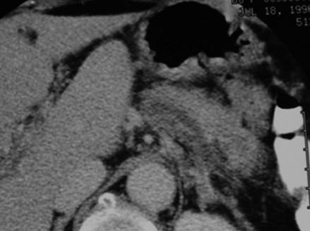
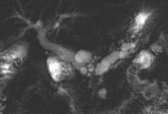
17 CT: complications Pseudocyst Collection of pancreatic juice enclosed by a wall of granulation tissue Infected necrosis Infection of a focal or diffuse area of nonviable pancreatic parenchyma Pancreatic abscess Circumscribed collection of pus containing little or no necrosis vansonnenberg et al. Radiology (1989) 170:
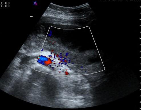
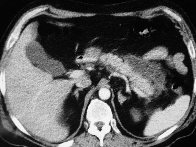
18 Pancreatic abscess


19 Role of interventional radiology in the management of acute pancreatitis ECR Vienna, 2006
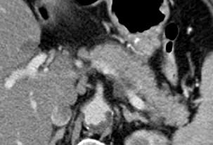
20 CT: complications Vascular complications
21 Question 3: At what timepoint is CT best performed? ECR Vienna, 2006
22 Recommendations for the use of CT CT should be guided by the natural history of the disease Should be timed to yield maximum diagnostic information, which will alter clininal management Maher M. et al. Cardiovasc. Intervent. Radiol. (2004) 27: ECR Vienna, 2006
23 Recommendations for the use of CT Balthazar et al. Radiology (1994);193: ECR Vienna, 2006
24 Question 4: Is MRI MRCP useful in patients with acute pancreatitis? ECR Vienna, 2006
 hemorrhage (T1 high) pancreatic necrosis ECR Vienna,")
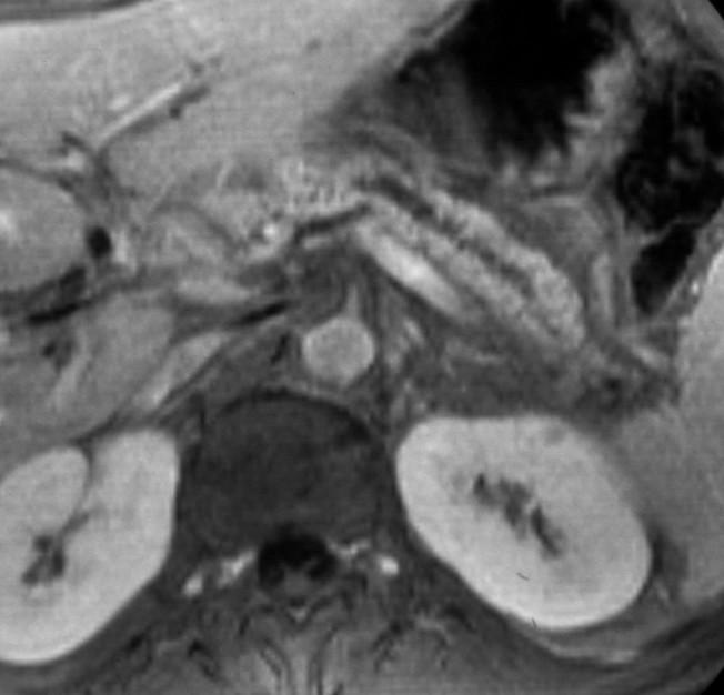
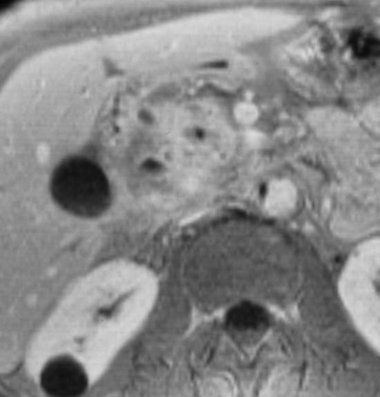
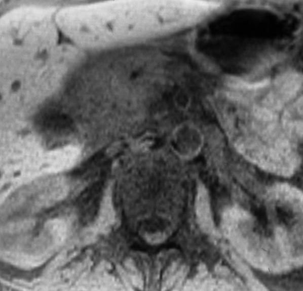
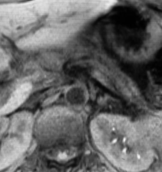
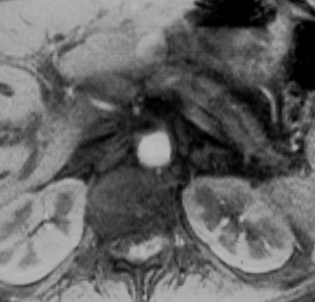
25 Magnetic resonance imaging (MRI) Imaging features pancreatic edema (low T1 / high T2) acute fluid collections (T2 WI) hemorrhage (T1 high) pancreatic necrosis ECR Vienna, 2006

26 T2 T2 FS-T1 Gad-T1
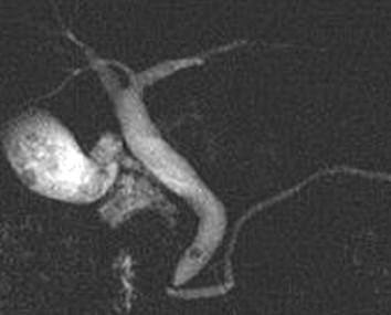
27
28 Role of MRI Limited BUT superior to CT for detection of mild acute pancreatitis for staging in the depiction of necrosis drainability of the collections for choledocholithiasis MRCP Replace CT in patients with renal failure Amano et al. Abdom Imaging (2001) 26:59-63 Kalra et al. JCAT (2002) 26:
? patients with severe pancreatitis are generally too ill to cooperate for an MRI examination")
29 Acute pancreatitis: imaging strategies (E)US in the early management to select those who would benefit from endoscopic stone extraction and drainage CT in severe pancreatitis to detect complications (MRI)? patients with severe pancreatitis are generally too ill to cooperate for an MRI examination
30 Chronic pancreatitis
31 Question 1: Is US a useful diagnostic imaging technique to confirm the clinical suspicion of chronic pancreatitis? ECR Vienna, 2006

32 Transabdominal US Features Calcifications Parenchymal atrophy Pancreatic duct dilatation ECR Vienna, 2006
33 Transabdominal US Accurate for the diagnosis of: advanced/complicated pancreatitis Rather limited: For the diagnosis of early chronic pancreatitis For differentiating adenocarc >< chron pancreatitis Frulloni et al. Digestive and Liver Disease 42 (2010) S381-S406

34 Endoscopic US High sensitivity in diagnosing CP ALSO of early chronic pancreatitis * Problem solving For differentiating adenocarc >< chron pancreatitis EUS + FNAC * Raimondo et al. JOP (2004) 5:1-7
35 Question 2: What is the most appropriate imaging technique for the identification of the site and the topography of pancreatic stones? ECR Vienna, 2006


36 Computed Tomography (CT) Superior for detecting calcifications ONLY in advanced stages Insensitive in the diagnosis of early CP Glasbrenner et al. Eur J gastroenterol Hepatol (2002) 14:

37 CT features Features calcifications duct dilation parenchymal atrophy intra- and peripancreatic pseudocysts tapering of common bile duct enhancement (heterogeneous)


38 CT: complicated chronic pancreatitis inflammatory masses, pseudocysts, calculi in the pancreatic duct or pseudo-aneurysms
39 Endoscopic US EUS is comparable to CT in depicting site and topography of pancreatic stones also very small stones (< 3mm) Frulloni et al. Digestive and Liver Disease 42 (2010) S381-S406
40 Question 3: What is the imaging technique of choice to diagnose early chronic pancreatitis? ECR Vienna, 2006

 214: 849-55 Cappeliez et al.")
41 MRI after secretin stimulation enhance the diagnosis of early chronic pancreatitis in the absence of marked ductal alteration Manfredi et al. Radiology (2000) 214: Cappeliez et al. Radiology (2000) 215:
42 MRI Imaging features T1: decreased signal intensity T2: variable (may be normal) enhancement (less and delayed) MRCP: extent of morphologic ductal changes classifies severity of disease << calcifications ECR Vienna, 2006
43
44
45 MRI MRI is less sensitive than CT for detection of calcifications * MRI is more sensitive than CT for parenchymal changes ** abnormal low signal intensity on fat-suppressed T1-weighted sequence for glandular enhancement ** decreased enhancement on the immediate and delayed postgadolinium images * Remer et al. Radiol Clin N Am (2002) ** Ly et al. Radiol Clin N Am (2002)
46 Endoscopic US Has recently shown its ability to diagnose early chronic pancreatitis by assessing morphological and structural changes of the pancreatic parenchyma Risk of overdiagnosis? Frulloni et al. Digestive and Liver Disease 42 (2010) S381-S406
47 Question 4: What is the imaging technique of choice to identify pancreatic malignancy in patients with chronic pancreatitis? ECR Vienna, 2006


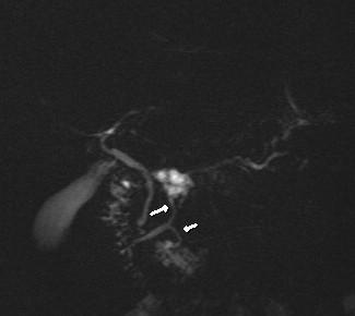
48 Mass lesion in chronic pancreatic: benign or malignant? Duct penetrating sign


49 Mass lesion in chronic pancreatic: benign or malignant? Negative duct-penetrating sign corona sign
50 Mass lesion in chronic pancreatic: MRI & MRCP The duct penetrating sign on MRCP images was more helpful to distinguish inflammatory mass from pancreatic adenocarcinoma than were the enhancement patterns on CT or MR images. * Ichikawa et al. Radiology (2001) 221:
51 Endoscopic US Used as problem solving Indeterminate mass on CT and MRI EUS-guided FNAB can increase the diagnostic accuracy. Frulloni et al. Digestive and Liver Disease 42 (2010) S381-S406
52 Chronic pancreatitis: imaging strategies US as initial step for screening CT/EUS more accurate for the detection of calcifications in the diagnosis of complications in the differential diagnosis of focal pancreatitis and pancreatic cancer
53 Chronic pancreatitis: imaging strategies MRI + MRCP: best modality EARLY detection of chronic pancreatitis prior to the development of calcifications PROBLEM SOLVING atypical presentation of CP differentiation CP from neoplasms FOLLOW-UP of parenchymal and duct abnormalities associated with CP
54 Pancreatic tumors
55 Key issues Early diagnosis of cancer Differentiation between cancer and inflammation Correct staging appropriate management options Identification of patients who would profit from radical surgery and of those who would not ultimate prognosis of the disease
56 Question 1: Is US a useful diagnostic imaging technique to confirm the clinical suspicion of a pancreatic tumor? ECR Vienna, 2006

57 Transabdominal US Features Echo-poor/heterogeneous mass Duct dilatation Atrophy Abnormal contour ECR Vienna, 2006
58 Transabdominal US Primary screening method Detection of pancreatic carcinoma Sensitivity : 44-95% 80-95% (head) < 50 % for lesions < 1 cm Lynch H.T. et al. Journal of Hepatobiliary Pancreatic Surgery 9 (2002), 12-31
59 BUT, sensitivity of US depends on : Size and location of the tumor small tumors tumors in the body and tail Patient-dependent factors Overlying bowel gas Obesity Operator s degree of training Technical quality of US equipment
60 Transabdominal US Less frequently employed for staging US is inferior to CT staging

61 Endoscopic US: complementary to CT and MRI Important imaging modality in assessing small pancreatic tumors (< 2 cm), undetectable by CT or MRI in the management of cystic lesions Insulinoma Hunt G. et al.gastrointestine Endoscopy 55 (2002),
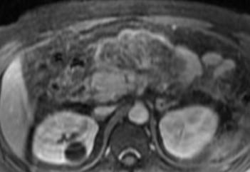
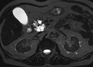
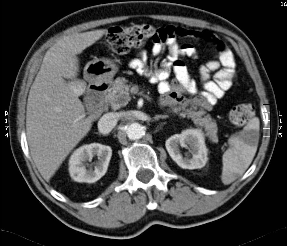
62 Limitations of EUS Need for patient sedation Operator dependency Inability to examine the entire liver To detect peritoneal metastases
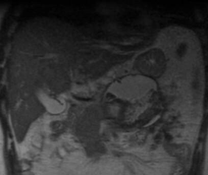
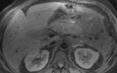
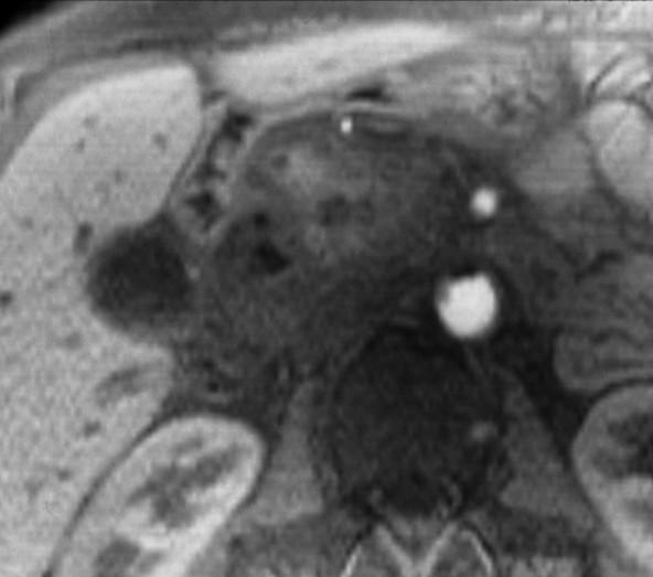
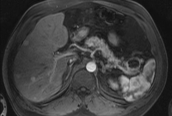
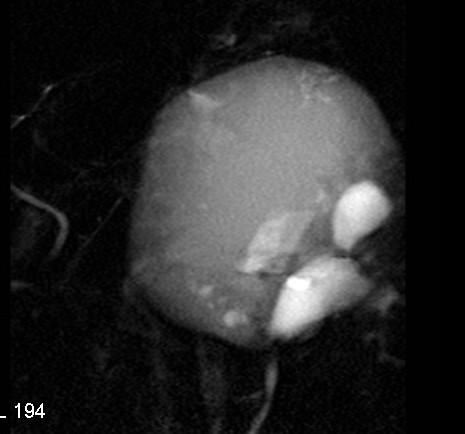
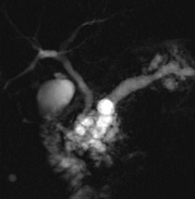
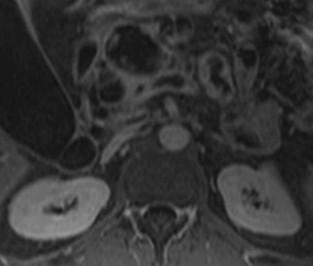
63 Question 2: What is the imaging technique of choice for the diagnosis and staging of suspected pancreatic cancer? ECR Vienna, 2006
64 Helical CT: primary imaging modality Detection Overall accuracy of CT: 80% to 97% Sensitivity lower for small tumors Staging Conv Helical PPV unresectability % % PPV resectability 45-72% 76-90% Legmann et al. AJR (1998) 170: Kalra et al. JCAT (2002) 26: Freeny et al. Pancreatology (2001) 1:
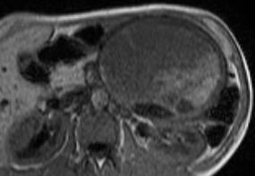
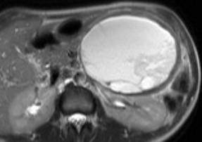
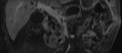
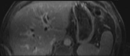
65 Ductal adenocarcinoma: CT findings Primary tumor focal mass (95%) low density area (75%) ill-defined borders Secondary findings duct dilation pancreatic duct (50%) CBD (40%) atrophy of the tail (20%) dilated collateral veins (12%) duodenal invasion

66 Staging: tumor is resectable or not? Need to access Vascular involvement Tumor contact > ½ circumference of vessel Dilated peripancreatic veins Focal decrease in vessel calibre Local invasion into retroperitoneal structures Metastases to liver, peritoneum, lymph nodes


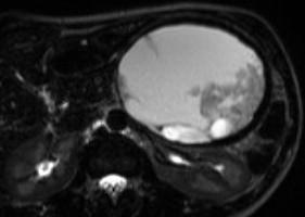
67 Staging: tumor is resectable or not? Need to access Vascular involvement Tumor contact > ½ circumference of vessel Dilated peripancreatic veins Focal decrease in vessel calibre Local invasion into retroperitoneal structures Metastases to liver, peritoneum, lymph nodes
68 CT-limitations in the staging of pancreatric carcinoma Poor sensitivity for detecting small metastatic peritoneal implants Multi-slice CT earlier detection of enlarged lymph nodes BUT does not allow differentiation between benign and malignant causes
69 PET- CT: add-on procedure? Additional staging information detection of unsuspected metastases Helps in the evaluation of masses with equivocal CT and MRI diagnosis locoregional recurrence Documentation of tumor cell vitality susceptibility to adjuvant treatment
70 Question 3: Is MRI with MRCP useful in patients with suspected pancreatic cancer? ECR Vienna, 2006
 Cancer")

71 MRI Diagnosis of tumor Sensitivity: 95% (42/44) Cancer non-resectability PPV: 90% NPV: 83% * Hänninen et al. Radiology (2002) 224: 34-41


72 MRI superior to CT For detection of small pancreatic lesions small liver metastasis

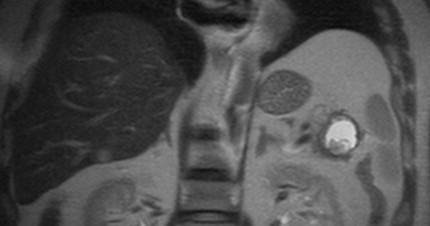
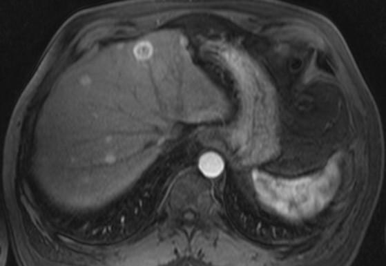
73 Malignant cystic neuroendocrine tumor with small liver metastasis ECR Vienna, 2006


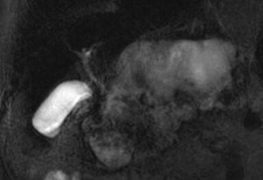
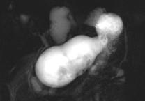
74 MRI is superior to CT: in the differential diagnosis of cystic neoplasms ECR Vienna, 2006

75 IPMN ECR Vienna, 2006

76 IPMN ECR Vienna, 2006


77 Malignant IPMN ECR Vienna, 2006

78 Limitations of MRI Availability Detection of small peripancreatic lymph nodes peritoneal implants
 ECR Vienna,")
79 Pancreatic tumors: imaging strategies Detection US (large/head) CT EUS, MRI: smaller/cystic pancreatic tumors Staging CT and MRI (EUS) ECR Vienna, 2006
80 CONCLUSION Single correct approach does not exist Imaging tests have a much higher PPV than NPV All of the available diagnostic methods have their strenghts and weakness Ultimate choosen strategy depends on local expertise and availibility of equipment ECR Vienna, 2006
81 Acknowlegments O.Van Rillaer F.Claus D.Bielen A.L.Baert S.Dymarkowski K.Op de beeck M.Thijs E. Nijs R. Van Slembrouck L.Van Hoe G.Marchal ECR Vienna, 2006
Anatomical and Functional MRI of the Pancreas
 Anatomical and Functional MRI of the Pancreas MA Bali, MD, T Metens, PhD Erasme Hospital Free University of Brussels Belgium mbali@ulb.ac.be Introduction The use of MRI to investigate the pancreas has
Role of Imaging Methods in Diagnosis of Acute Pancreatitis. Válek V. Radiologická klinika, FN Brno a LF MU v Brně
 Role of Imaging Methods in Diagnosis of Acute Pancreatitis Válek V. Radiologická klinika, FN Brno a LF MU v Brně New Classification: Acute Pancreatitis 2007 revision of Atlanta classification and definitions
Dr Claire Smith, Consultant Radiologist St James University Hospital Leeds
 Dr Claire Smith, Consultant Radiologist St James University Hospital Leeds Imaging in jaundice and 2ww pathway Image protocol Staging Limitations Pancreatic cancer 1.2.4 Refer people using a suspected
Common and unusual CT and MRI manifestations of pancreatic adenocarcinoma: a pictorial review
 Review Article Common and unusual CT and MRI manifestations of pancreatic adenocarcinoma: a pictorial review Min-Jie Yang, Su Li, Yong-Guang Liu, Na Jiao, Jing-Shan Gong Department of Radiology, Shenzhen
Biliary tree dilation - and now what?
 Biliary tree dilation - and now what? Poster No.: C-1767 Congress: ECR 2012 Type: Educational Exhibit Authors: I. Ferreira, A. B. Ramos, S. Magalhães, M. Certo; Porto/PT Keywords: Pathology, Diagnostic
Imaging in gastric cancer
 Imaging in gastric cancer Gastric cancer remains a deadly disease because of late diagnosis. Adenocarcinoma represents 90% of malignant tumors. Diagnosis is based on endoscopic examination with biopsies.
Pancreatic Adenocarcinoma: Everything You Need to Know From Cross-Sectional Imaging to Treatment
 Pancreatic Adenocarcinoma: Everything You Need to Know From Cross-Sectional Imaging to Treatment Andrew W. Bowman, MD PhD Assistant Professor of Radiology Mayo Clinic Florida SCBT-MR Annual Meeting Nashville,
Cystic Pancreatic Lesions: Approach to Diagnosis
 Cystic Pancreatic Lesions: Approach to Diagnosis Poster No.: R-0130 Congress: RANZCR-AOCR 2012 Type: Educational Exhibit Authors: A. AGARWAL, R. M. Mendelson; Perth/AU Keywords: Cysts, Biopsy, Endoscopy,
ENDOSCOPY IN COMPETITION DIAGNOSTICS. Dr. med. Dirk Hartmann Klinikum Ludwigshafen
 Falk Symposium 166 GI Endoscopy Standards and Innovations Mainz, 18. 19. September 2008 ENDOSCOPY IN COMPETITION DIAGNOSTICS Dr. med. Dirk Hartmann Klinikum Ludwigshafen ENDOSCOPY IN COMPETITION Competing
Autoimmune Pancreatitis: A Great Imitator
 Massachusetts General Hospital Harvard Medical School Autoimmune Pancreatitis: A Great Imitator Dushyant V Sahani MD dsahani@partners.org Autoimmune Pancreatitis: Learning Objectives Clinical manifestations
Evaluation of Suspected Pancreatic Cancer
 Evaluation of Suspected Pancreatic Cancer October 15, 2015 If you experience technical difficulty during the presentation: Contact WebEx Technical Support directly at: US Toll Free: 1-866-779-3239 Toll
40th European Congress of Cytology Liverpool, UK, 2-5 th October 2016
 40th European Congress of Cytology Liverpool, UK, 2-5 th October 2016 EUS FNA of abdominal organs: An approach to reporting and triage for ancillary testing Date and time: Sunday 2 nd October 2016 15.00-16.30
CT 101 :Pancreas and Spleen
 CT 101 :Pancreas and Spleen Shikha Khullar,, MD, MPH Division of Radiology University of South Alabama The Pancreas Normal Pancreas 3 Phase Pancreatic CT Non contrast Arterial phase : 30-35 35 second
X-ray Corner. Imaging of The Pancreas. Pantongrag-Brown L
 X-ray Corner 125 Imaging of The Pancreas Modern imaging modalities commonly used in pancreas include ultrasound (US), CT, and MRI. Pancreas is a retroperitoneal organ which makes it difficult to visualize
Intraoperative staging of GIT cancer using Intraoperative Ultrasound
 Intraoperative staging of GIT cancer using Intraoperative Ultrasound Thesis For Fulfillment of MSc Degree In Surgical Oncology By Abdelhalim Salah Abdelhalim Moursi M.B.B.Ch (Cairo University ) Supervisors
Diagnosis of chronic Pancreatitis. Christoph Beglinger, University Hospital Basel, Switzerland
 Diagnosis of chronic Pancreatitis Christoph Beglinger, University Hospital Basel, Switzerland Pancreatitis Pancreas Pancreas - an organ that makes bicarbonate to neutralize gastric acid, enzymes to digest
IMAGING OF ACUTE AND CHRONIC PANCREATITIS, INCLUDING EXOCRINE FUNCTION
 IMAGING OF ACUTE AND CHRONIC PANCREATITIS, INCLUDING EXOCRINE FUNCTION Andrew T. Trout, MD @AndrewTroutMD Disclosures Grant support National Pancreas Foundation In-kind support - ChiRhoClin modified from:
Appendix 9: Endoscopic Ultrasound in Gastroenterology
 Appendix 9: Endoscopic Ultrasound in Gastroenterology This curriculum is intended for clinicians who perform endoscopic ultrasonography (EUS) in gastroenterology. It includes standards for theoretical
Multidetector CT evaluation of acute pancreatitis and its complications and its correlation with clinical outcome
 INTERNATIONAL JOURNAL OF CURRENT RESEARCH IN BIOLOGY AND MEDICINE ISSN: 2455-944X www.darshanpublishers.com DOI:10.22192/ijcrbm Volume 3, Issue 1-2018 Original Research Article Multidetector CT evaluation
Endoscopic Ultrasonography Clinical Impact. Giancarlo Caletti. Gastroenterologia Università di Bologna. Caletti
 Clinical Impact Giancarlo Gastroenterologia Università di Bologna AUSL di Imola,, Castel S. Pietro Terme (BO) 1982 Indications Diagnosis of Submucosal Tumors (SMT) Staging of Neoplasms Evaluation of Pancreato-Biliary
The Pancreas. Basic Anatomy. Endocrine pancreas. Exocrine pancreas. Pancreas vasculature. Islets of Langerhans. Acinar cells Ductal System
 SGNA: Back to Basics Rogelio G. Silva, MD Assistant Clinical Professor of Medicine University of Illinois at Chicago Department of Medicine Division of Gastroenterology Advocate Christ Medical Center GI
Magnetic resonance cholangiopancreatography (MRCP) is an imaging. technique that is able to non-invasively assess bile and pancreatic ducts,
 is an imaging. technique that is able to non-invasively assess bile and pancreatic ducts,") SECRETIN AUGMENTED MRCP Riccardo MANFREDI, MD, MBA, FESGAR Magnetic resonance cholangiopancreatography (MRCP) is an imaging technique that is able to non-invasively assess bile and pancreatic ducts, in
SECRETIN AUGMENTED MRCP Riccardo MANFREDI, MD, MBA, FESGAR Magnetic resonance cholangiopancreatography (MRCP) is an imaging technique that is able to non-invasively assess bile and pancreatic ducts, in
Imaging of Neuroendocrine Metastases
 Imaging of Neuroendocrine Metastases Aoife Kilcoyne, Shaunagh McDermott, Colin McCarthy,Manuel Patino, Dushyant Sahani, Michael Blake Abdominal Imaging Division Massachusetts General Hospital Disclosure
ACG Clinical Guideline: Diagnosis and Management of Pancreatic Cysts
 ACG Clinical Guideline: Diagnosis and Management of Pancreatic Cysts Grace H. Elta, MD, FACG 1, Brintha K. Enestvedt, MD, MBA 2, Bryan G. Sauer, MD, MSc, FACG (GRADE Methodologist) 3 and Anne Marie Lennon,
U Nordic Forum - Trauma & Emergency Radiology. Lecture Objectives. MDCT in Acute Pancreatitis. Acute Pancreatitis: Etiologies
 Nordic Forum - Trauma & Emergency Radiology Lecture Objectives MDCT in Acute Pancreatitis Borut Marincek Institute of Diagnostic Radiology niversity Hospital Zurich, Switzerland To describe the role of
Newcastle HPB MDM updated radiology imaging protocol recommendations. Author Dr John Scott. Consultant Radiologist Freeman Hospital
 Newcastle HPB MDM updated radiology imaging protocol recommendations Author Dr John Scott. Consultant Radiologist Freeman Hospital This document is intended as a guide to aid radiologists and clinicians
The role of endoscopy in the diagnosis and treatment of cystic pancreatic neoplasms
 The role of endoscopy in the diagnosis and treatment of cystic pancreatic neoplasms CYSTIC LESIONS AND FLUID COLLECTIONS OF THE PANCREAS Their pathology ranges from pseudocysts and pancreatic necrosis
Chronic pancreatitis mimicking intraductal papillary mucinous neoplasm of the pancreas; Report of tow cases
 Jichi Medical University Journal Chronic pancreatitis mimicking intraductal papillary mucinous neoplasm of the pancreas; Report of tow cases Noritoshi Mizuta, Hiroshi Noda, Nao Kakizawa, Nobuyuki Toyama,
Case Scenario 1. Discharge Summary
 Case Scenario 1 Discharge Summary A 69-year-old woman was on vacation and noted that she was becoming jaundiced. Two months prior to leaving on that trip, she had had a workup that included an abdominal
Case Study: #3: Gallbladder Carcinoma?
 Case Study: #3: Gallbladder Carcinoma? By: Megan Wyatt K. SON Wyatt 225 2B1 RDMS, RVT Patient: Male 85 YOA Caucasian Indication: Elevated Alkaline Phosphatase History Annual physical showed elevated alkaline
Imaging of liver and pancreas
 Imaging of liver and pancreas.. Disease of the liver Focal liver disease Diffusion liver disease Focal liver disease Benign Cyst Abscess Hemangioma FNH Hepatic adenoma HCC Malignant Fibrolamellar carcinoma
Imaging Pancreatic Neuroendocrine Tumors (PNETs): CT, MRI, EUS, Nuclear
: CT, MRI, EUS, Nuclear") Imaging Pancreatic Neuroendocrine Tumors (PNETs): CT, MRI, EUS, Nuclear Eric Tamm, M.D. Department of Diagnostic Radiology Division of Diagnostic Imaging MD Anderson Cancer Center Houston, TX Disclosure
Imaging Pancreatic Neuroendocrine Tumors (PNETs): CT, MRI, EUS, Nuclear Eric Tamm, M.D. Department of Diagnostic Radiology Division of Diagnostic Imaging MD Anderson Cancer Center Houston, TX Disclosure
EFSUMB EUROPEAN FEDERATION OF SOCIETIES FOR ULTRASOUND IN MEDICINE AND BIOLOGY Building a European Ultrasound Community
 MINIMUM TRAINING REQUIREMENTS FOR THE PRACTICE OF MEDICAL ULTRASOUND IN EUROPE Appendix 9: Endoscopic Ultrasound in Gastroenterology This curriculum is intended for clinicians who perform endoscopic ultrasonography
Appendix 5. EFSUMB Newsletter. Gastroenterological Ultrasound
 EFSUMB Newsletter 87 Examinations should encompass the full range of pathological conditions listed below A log book listing the types of examinations undertaken should be kept Training should usually
Chronic Pancreatitis: When to Scope? Gregory A. Cote, MD, MS Assistant Professor of Medicine Indiana University School of Medicine
 Chronic Pancreatitis: When to Scope? Gregory A. Cote, MD, MS Assistant Professor of Medicine Indiana University School of Medicine Endoscopy & Chronic Pancreatitis Diagnosis EUS ERCP Exocrine Function
Pancreas Case Scenario #1
 Pancreas Case Scenario #1 An 85 year old white female presented to her primary care physician with increasing abdominal pain. On 8/19 she had a CT scan of the abdomen and pelvis. This showed a 4.6 cm mass
An Approach to Pancreatic Cysts. Introduction
 An Approach to Pancreatic Cysts Nalini M. Guda, MD Aurora St. Luke s Medical Center, Milwaukee Clinical Adjunct Professor of Medicine, University of Wisconsin School of Medicine and Public Health Introduction
Navigating the Biliary Tract with CT & MR: An Imaging Approach to Bile Duct Obstruction
 Navigating the Biliary Tract with CT & MR: An Imaging Approach to Bile Duct Obstruction Ann S. Fulcher, MD Medical College of Virginia Virginia Commonwealth University Richmond, Virginia Objectives To
CASE 1 11/1/2016 HEPATOBILIARY IMAGING CASE PRESENTATIONS DECLARATION. Dr. Chirag Patel ORGAN IMAGING yr old lady
 HEPATOBILIARY IMAGING CASE PRESENTATIONS DECLARATION No financial disclosures or affiliations with commercial organisations No discussion of investigational or off-label use of medical devices, products
Diagnostics of Pancreatic Carcinoma
 Diagnostics of Pancreatic Carcinoma Jens Ricke Charité - Universitätsmedizin Berlin Klinik für Strahlenheilkunde (Director: Prof. Dr. Dr. Roland Felix) Imaging in Pancreatic Carcinoma CT MRT ERCP? FDG-PET
Contrast Enhanced Ultrasound of Parenchymal Masses in Children
 Contrast Enhanced Ultrasound of Parenchymal Masses in Children Sue C Kaste, DO On behalf of Beth McCarville, MD St. Jude Children s Research Hospital Memphis, TN Overview Share St. Jude experience with
Anatomy of the biliary tract
 Harvard-MIT Division of Health Sciences and Technology HST.121: Gastroenterology, Fall 2005 Instructors: Dr. Jonathan Glickman Anatomy of the biliary tract Figure removed due to copyright reasons. Biliary
Case 1. Intro to Gallbladder & Pancreas Pathology. Case 1 DIAGNOSIS??? Acute Cholecystitis. Acute Cholecystitis. Helen Remotti M.D.
 Cholecystitis acute chronic Gallbladder tumors Adenomyoma (benign) Adenocarcinoma Pancreatitis acute chronic Pancreatic tumors Intro to Gallbladder & Pancreas Pathology Helen Remotti M.D. Case 1 70 year
EUS FNA NEUROENDOCRINE TUMORS. A. Ginès Endocopy Unit Hospital Cínic. Barcelona (Spain)
") EUS FNA NEUROENDOCRINE TUMORS A. Ginès Endocopy Unit Hospital Cínic. Barcelona (Spain) GI NEUROENDOCRINE TUMORS GENERAL CONCEPTS Rare neoplasms arising from the neuroendocrine cells of the GI tract Include:
EUS FNA NEUROENDOCRINE TUMORS A. Ginès Endocopy Unit Hospital Cínic. Barcelona (Spain) GI NEUROENDOCRINE TUMORS GENERAL CONCEPTS Rare neoplasms arising from the neuroendocrine cells of the GI tract Include:
Lesions of the pancreaticoduodenal groove, a pictorial review
 Lesions of the pancreaticoduodenal groove, a pictorial review Poster No.: C-2131 Congress: ECR 2013 Type: Educational Exhibit Authors: E. Ni Mhurchu, L. Lavelle, I. Murphy, S. Skehan ; IE, Dublin/ IE Keywords:
Evaluation of AGA and Fukuoka Guidelines for EUS and surgical resection of incidental pancreatic cysts
 Evaluation of AGA and Fukuoka Guidelines for EUS and surgical resection of incidental pancreatic cysts Authors Alexander Lee 1, Vivek Kadiyala 2,LindaS.Lee 3 Institutions 1 Texas Digestive Disease Consultants,
The role of ERCP in chronic pancreatitis
 The role of ERCP in chronic pancreatitis Marianna Arvanitakis Erasme University Hospital, ULB, Brussels, Belgium 10 th Nottingham Endoscopy Masterclass SPEAKER DECLARATIONS This presenter has the following
Frank Burton Memorial Update on Pancreato-biliary Cancers
 Frank Burton Memorial Update on Pancreato-biliary Cancers Diagnosis and management of pancreatic cancer: common dilemmas Moderators: Banke Agarwal, MD Paul Buse, MD Evaluation of patients with obstructive
Upper GI Malignancies Imaging Guidelines for the Management of Gastric, Oesophageal & Pancreatic Cancers 2012
 Upper GI Malignancies Imaging Guidelines for the Management of Gastric, Oesophageal & Pancreatic Cancers 2012 Version Control This is a controlled document please destroy all previous versions on receipt
Chronic Pancreatitis
 Gastro Foundation Fellows Weekend 2017 Chronic Pancreatitis Jose Ramos University of the Witwatersrand Donald Gordon Medical Centre Aetiology in SA Alcohol (up to 80%) Idiopathic Tropical Obstruction Autoimmune
performed to help sway the clinician in what the appropriate diagnosis is, which can substantially alter the treatment of management.
 Hello, I am Maura Polansky at the University of Texas MD Anderson Cancer Center. I am a Physician Assistant in the Department of Gastrointestinal Medical Oncology and the Program Director for Physician
ROLE OF RADIOLOGY IN INVESTIGATION OF JAUNDICE
 ROLE OF RADIOLOGY IN INVESTIGATION OF JAUNDICE Dr. Sohan kumar sah *, Dr. Liu Sibin, Dr. sumendra raj pandey, Dr. Prakashmaan shah, Dr. Gaurishankar pandit, Dr. Suraj kurmi and Dr. Sanjay kumar jaiswal
Hepatobiliary and Pancreatic Malignancies
 Hepatobiliary and Pancreatic Malignancies Gareth Eeson MD MSc FRCSC Surgical Oncologist and General Surgeon Kelowna General Hospital Interior Health Consultant, Surgical Oncology BC Cancer Agency Centre
Imaging Guided Biopsy. Edited & Presented by ; Hussien A.B ALI DINAR. Msc Lecturer,Reporting Sonographer
 Imaging Guided Biopsy Edited & Presented by ; Hussien A.B ALI DINAR. Msc Lecturer,Reporting Sonographer Objective By the End of this lessons you should : Define what biopsy Justify Aim to perform biopsy
Radiology Pathology Conference
 Radiology Pathology Conference Nadia F. Yusaf, M.D. PGY-3 1/29/2010 Presentation material is for education purposes only. All rights reserved. 2010 URMC Radiology Page 1 of 90 Case 1 60 year- old man presents
State of the Art Imaging for Hepatic Malignancy: My Assignment
 State of the Art Imaging for Hepatic Malignancy: My Assignment CT vs MR vs MRCP Which one to choose for HCC vs Cholangiocarcinoma What special protocols to use for liver tumors Role of PET and Duplex US
Imaging of common diseases of hepatobiliary and GI system
 Imaging of common diseases of hepatobiliary and GI system Natthaporn Tanpowpong, M.D. Diagnostic radiology Faculty of Medicine, Chulalongkorn University Normal plain radiograph A = Common bile duct
4/9/2018 OBJECTIVES PANCREAOTO BILIARY ULTRASOUND: BEYOND CHOLECYSTITIS
 PANCREAOTO BILIARY ULTRASOUND: BEYOND CHOLECYSTITIS Jean Yves Sewah Kaiser Permanente West Los Angeles 1 OBJECTIVES Discuss the role of ultrasound in the evaluation of the gallbladder, biliary tree and
Intraductal papillary neoplasms in the bile ducts
 Intraductal papillary neoplasms in the bile ducts Seok Hwa Youn Myunghee Yoon Dong Hoon Shin Kosin University Gospel Hospital Department of general surgery Hepato-biliary-pancreatic division Introduction
Pancreatic Cancer. What is pancreatic cancer?
 Scan for mobile link. Pancreatic Cancer Pancreatic cancer is a tumor of the pancreas, an organ that is located behind the stomach in the abdomen. Pancreatic cancer does not always cause symptoms until
The Use of Pancreatoscopy in the Diagnosis of Intraductal Papillary Mucinous Tumor Lesions of the Pancreas
 CLINICAL GASTROENTEROLOGY AND HEPATOLOGY 2005;3:S53 S57 The Use of Pancreatoscopy in the Diagnosis of Intraductal Papillary Mucinous Tumor Lesions of the Pancreas KENJIRO YASUDA, MUNEHIRO SAKATA, MOOSE
What Is Pancreatitis?
 What Is Pancreatitis? Pancreatitis is inflammation (swelling) of the pancreas that is most often caused by gallstones or alcohol abuse. There are other causes that your gastroenterologist will look for,
Disclosures. Extra-hepatic Biliary Disease and the Pancreas. Objectives. Pancreatitis 10/3/2018. No relevant financial disclosures to report
 Extra-hepatic Biliary Disease and the Pancreas Disclosures No relevant financial disclosures to report Jeffrey Coughenour MD FACS Clinical Associate Professor of Surgery and Emergency Medicine Division
6 th August 2018 Day 1 - Gallbladder & Bile duct Topic
 Venue: Sterling Hospital Auditorium, Sterling Hospitals, Gurukul Road Ahmedabad, Gujarat 6 th August 2018 Day 1 - Gallbladder & Bile duct Registration(8:00am-8:15am) Inauguration(8:15am-8:30am) Welcome
Role of imaging in RCC. Ultrasonography. Solid lesion. Cystic RCC. Solid RCC 31/08/60. From Diagnosis to Treatment: the Radiologist Perspective
 Role of imaging in RCC From Diagnosis to Treatment: the Radiologist Perspective Diagnosis Staging Follow up Imaging modalities Limitations and pitfalls Duangkamon Prapruttam, MD Department of Therapeutic
Virtual MR Pancreatoscopy in the Evaluation of the Pancreatic Duct in Chronic Pancreatitis
 MULTIMEDIA ARTICLE - Videoclips Virtual MR Pancreatoscopy in the Evaluation of the Pancreatic Duct in Chronic Pancreatitis Rakesh Kalapala 1, Lingareddy Sunitha 2, Reddy D Nageshwar 1, Guduru V Rao 1,
A patient with an unusual congenital anomaly of the pancreaticobiliary tree
 A patient with an unusual congenital anomaly of the pancreaticobiliary tree Thomas Hocker, HMS IV BIDMC Core Radiology Case Presentation September 17, 2007 Review of Normal Pancreaticobiliary Tract Anatomy
Pancreatic Cysts. Darius C. Desai, MD FACS St. Luke s University Health Network
 Pancreatic Cysts Darius C. Desai, MD FACS St. Luke s University Health Network None Disclosures Incidence Widespread use of cross sectional imaging Seen in over 2% of patients having abdominal imaging
Original Policy Date 12:2013
 MP 6.01.30 Magnetic Resonance Cholangiopancreatography Medical Policy Section Radiology Is12:2013sue 3:2005 Original Policy Date 12:2013 Last Review Status/Date 12:2013 Return to Medical Policy Index Disclaimer
Endoscopic Ultrasonography Assessment for Ampullary and Bile Duct Malignancy
 Diagnostic and Therapeutic Endoscopy, Vol. 3, pp. 35-40 Reprints available directly from the publisher Photocopying permitted by license only (C) 1996 OPA (Overseas Publishers Association) Amsterdam B.V.
The Bile Duct (and Pancreas) and the Physician
 and the Physician") The Bile Duct (and Pancreas) and the Physician Javaid Iqbal Consultant in Gastroenterology and Pancreato-biliary Medicine University Hospital South Manchester Not so common?! Two weeks 38 ERCP s 20 15
The Bile Duct (and Pancreas) and the Physician Javaid Iqbal Consultant in Gastroenterology and Pancreato-biliary Medicine University Hospital South Manchester Not so common?! Two weeks 38 ERCP s 20 15
IMAGING OF LIVER, BILIARY TREE, PANCREAS
 IMAGING OF LIVER, BILIARY TREE, PANCREAS Department of Radiology West China Hospital, Sichuan University Yao Jin Learning Points The methodology for imaging the LBP (liver, biliary tree, and pancreas )
Pictorial review of Benign Biliary tract abnormality on MRCP/MRI Liver with Endoscopic (including splyglass) and Endoscopic Ultrasound correlation
 and Endoscopic Ultrasound correlation") Pictorial review of Benign Biliary tract abnormality on MRCP/MRI Liver with Endoscopic (including splyglass) and Endoscopic Ultrasound correlation Poster No.: C-2617 Congress: ECR 2015 Type: Educational
Pictorial review of Benign Biliary tract abnormality on MRCP/MRI Liver with Endoscopic (including splyglass) and Endoscopic Ultrasound correlation Poster No.: C-2617 Congress: ECR 2015 Type: Educational
Management A Guideline Based Approach to the Incidental Pancreatic Cysts. Common Cystic Pancreatic Neoplasms.
 Management 2016 A Guideline Based Approach to the Incidental Pancreatic Cysts ISMRM 2016 Masoom Haider, MD, FRCP(C) Professor of Radiology, University of Toronto Clinician Scientist, Ontario Institute
Endoscopic ultrasonography in suspected pancreatic malignancy and indecisive CT
 O r i g i n a l a r t i c l e Endoscopic ultrasonography in suspected pancreatic malignancy and indecisive CT O.L.M. Meijer 1, R.K. Weersma 1, E.J. van der Jagt 2, H.M. van Dullemen 1 * Departments of
Biliary cancers: imaging diagnosis. Study of 30 cases
 Biliary cancers: imaging diagnosis. Study of 30 cases N Hammoune, S Semlali, M Eddarai, T. Amil, M Zentar, S. El Kandri,, M Benameur,, S Chaouir. Radiology Department. Mohamed V Military Hospital. Rabat-
CASE 01 LA Path Slide Seminar 13 March, 08. Deepti Dhall, MD Department of Pathology and Laboratory Medicine Cedars-Sinai Medical Center
 CASE 01 LA Path Slide Seminar 13 March, 08 Deepti Dhall, MD Department of Pathology and Laboratory Medicine Cedars-Sinai Medical Center Clinical History 60 year old male presented with obstructive jaundice
Acute Pancreatitis: Role of Imaging Modalities
 International Journal of Interdisciplinary and Multidisciplinary Studies (IJIMS), 2015, Vol 2, No.9,109-114. 109 Available online at http://www.ijims.com ISSN: 2348 0343 Abstract Acute Pancreatitis: Role
Biliary Tree Ultrasound - In a nutshell. Pamela Parker Lead Sonographer
 Biliary Tree Ultrasound - In a nutshell Pamela Parker Lead Sonographer Aims Review what we know about the biliary system Common pathologies Pitfalls Reporting tips The Nutshell Background Biliary examinations
A LEADER IN ADVANCED ENDOSCOPY AND HEPATOBILIARY SURGERY
 A LEADER IN ADVANCED ENDOSCOPY AND HEPATOBILIARY SURGERY St. Peter s Hospital Advanced Endoscopy & Hepatobiliary Center Welcome The St. Peter s Hospital Advanced Endoscopy & Hepatobiliary Center is a leader
8/3/2016. Consultant for / research support from: Astellas Bayer Bracco GE Healthcare Guerbet Medrad Siemens Healthcare. Single Energy.
 U. Joseph Schoepf, MD Prof. (h.c.), FAHA, FSCBT-MR, FNASCI, FSCCT Professor of Radiology, Medicine, and Pediatrics Director, Division of Cardiovascular Imaging Consultant for / research support from: Astellas
Diagnostic performance of endoscopic ultrasound-guided fine-needle aspiration in pancreatic lesions
 European Review for Medical and Pharmacological Sciences 2018; 22: 1397-1401 Diagnostic performance of endoscopic in pancreatic lesions Q.-M. WU 1, Y.-N. GUO 1, Y.-Q. XU 1 Digestive Department of Beijing
MRI Abdomen Protocol Pancreas/MRCP with Contrast
 MRI Abdomen Protocol Pancreas/MRCP with Contrast Reviewed By: Brett Mollard, MD; Anna Ellermeier, MD Last Reviewed: July 2018 Contact: (866) 761-4200 Standard uses: 1. Characterization of cystic and solid
Intraductal papillary mucinous neoplasm (IPMN) is a distinct
 is a distinct") CLINICAL GASTROENTEROLOGY AND HEPATOLOGY 2008;6:815 819 Evaluation of the Guidelines for Management of Pancreatic Branch-Duct Intraductal Papillary Mucinous Neoplasm RAYMOND S. TANG,* BENJAMIN WEINBERG,
CLINICAL GASTROENTEROLOGY AND HEPATOLOGY 2008;6:815 819 Evaluation of the Guidelines for Management of Pancreatic Branch-Duct Intraductal Papillary Mucinous Neoplasm RAYMOND S. TANG,* BENJAMIN WEINBERG,
PANCREATIC CANCER GUIDELINES
 PANCREATIC CANCER GUIDELINES North-East London Cancer Network & Barts and the London HPB Centre PROTOCOL FOR MANAGEMENT OF PANCREATIC CANCER (SEPTEMBER 2010) I. PRE-REFERRAL GUIDELINES Screening 1. Offer
Multi modality Imaging in Acute Pancreatitis. Marsha Lynch, HMS III Gillian Lieberman, MD BIDMC Core Clerkship in Radiology March 2009
 Multi modality Imaging in Acute Pancreatitis Marsha Lynch, HMS III Gillian Lieberman, MD BIDMC Core Clerkship in Radiology March 2009 Our Patient R: Introduction 52M with 10d history of nausea, vomiting
Abdomen Sonography Examination Content Outline
 Abdomen Sonography Examination Content Outline (Outline Summary) # Domain Subdomain Percentage 1 2 3 Anatomy, Perfusion, and Function Pathology, Vascular Abnormalities, Trauma, and Postoperative Anatomy
CASE REPORT. Abstract. Introduction. Case Report
 CASE REPORT Branch Duct Intraductal Papillary Mucinous Neoplasms of the Pancreas Involving Type 1 Localized Autoimmune Pancreatitis with Normal Serum IgG4 Levels Successfully Diagnosed by Endoscopic Ultrasound-guided
General Surgery PURPLE SERVICE MUHC-RVH Site
 Preamble HPB is a clinical teaching unit with several different vocations: It regroups all solid organ Transplantation as well as most advanced Hepatobiliary and Pancreatic clinical activities performed
Radiological staging of lung cancer. Shukri Loutfi,MD,FRCR Consultant Thoracic Radiologist KAMC-Riyadh
 Radiological staging of lung cancer Shukri Loutfi,MD,FRCR Consultant Thoracic Radiologist KAMC-Riyadh Bronchogenic Carcinoma Accounts for 14% of new cancer diagnoses in 2012. Estimated to kill ~150,000
Imaging of Biliary Tract Emergencies in Jorge A. Soto, MD Professor of Radiology Boston University Medical Center.
 Imaging of Biliary Tract Emergencies in 2011 Jorge A. Soto, MD Professor of Radiology Boston University Medical Center Introduction Biliary emergencies are: Common Come in many flavors Deceiving: frequent
IT 의료융합 1 차임상세미나 복부질환초음파 이재영
 IT 의료융합 1 차임상세미나 2013-4-3 복부질환초음파 이재영 나는오늘누구를위하여 종을울리나? 전통적의료 의사 공학설계자 의사 최첨단진단장비들 USG, CT, MRI 환자 환자 현대의료 사용자중심의사고 US in the Abdomen Detection DDx Look Behavior Response by external stimuli Guiding Tool
EUS in the Management of
 EUS in the Management of Pancreaticobiliary Cancers Frank Gress, MD Professor of Medicine and Chief Division of Gastroenterology and Hepatology State University of New York Downstate Medical Center Brooklyn,
D DAVID PUBLISHING. Groove Pancreatitis: A Case Report. 1. Introduction. 2. Case Report
 Journal of Pharmacy and Pharmacology 6 (2018) 415-419 doi: 10.17265/2328-2150/2018.04.013 D DAVID PUBLISHING Luciana Leony Valente, Mariama Alves Dantas Fagundes, Camila Medrado Pereira Barbosa, Hélio
COMPUTED TOMOGRAPHY FINDINGS IN ACUTE PANCREATITIS
 ORIGINAL ARTICLE COMPUTED TOMOGRAPHY FINDINGS IN ACUTE PANCREATITIS Noorul Hadi, Kalsoom Nawab, Ayesha Amin Department Of Radiology, Post Graduate Medical Institute, Hayatabad Medical Complex, Peshawar
PET IMAGING (POSITRON EMISSION TOMOGRAPY) FACT SHEET
 FACT SHEET") Positron Emission Tomography (PET) When calling Anthem (1-800-533-1120) or using the Point of Care authorization system for a Health Service Review, the following clinical information may be needed to
Positron Emission Tomography (PET) When calling Anthem (1-800-533-1120) or using the Point of Care authorization system for a Health Service Review, the following clinical information may be needed to
Hepatobiliary Ultrasound Rimon Bengiamin, MD, RDMS Assistant Clinical Professor Director of Emergency Ultrasound UCSF Fresno. Objectives. Why?
 Hepatobiliary Ultrasound Rimon Bengiamin, MD, RDMS Assistant Clinical Professor Director of Emergency Ultrasound UCSF Fresno Objectives Discuss the goals of point-of-care biliary ultrasound Review the
CHOLANGIOCARCINOMA (CCA)
") CHOLANGIOCARCINOMA (CCA) Deepak Hariharan MD (Research), FRCS, Locum Consultant HPB Surgeon AIM Outline essential facts & principles Present 4 cases Discuss Challenges /Controversies INTRODUCTION Most
CHOLANGIOCARCINOMA (CCA) Deepak Hariharan MD (Research), FRCS, Locum Consultant HPB Surgeon AIM Outline essential facts & principles Present 4 cases Discuss Challenges /Controversies INTRODUCTION Most
Personal Profile. Name: 劉 XX Gender: Female Age: 53-y/o Past history. Hepatitis B carrier
 Personal Profile Name: 劉 XX Gender: Female Age: 53-y/o Past history Hepatitis B carrier Chief complaint Fever on and off for 2 days Present illness 94.10.14 Sudden onset of epigastric pain 94.10.15 Fever
Color Codes Pathology and Genetics Medicine and Clinical Pathology Surgery Imaging
 Saturday, November 5, 2005 8:30-10:30 a. m. Poorly Differentiated Endocrine Carcinomas Chairman: E. Van Cutsem, Leuven, Belgium 9:00-9:30 a. m. Working Group Sessions Pathology and Genetics Group leaders: