Università degli Studi di Napoli Federico II. Dipartimento di Fisica "Ettore Pancini"
|
|
|
- Diane Baker
- 6 years ago
- Views:
Transcription

1 Università degli Studi di Napoli Federico II Scuola Politecnica e delle Scienze di Base Collegio di Scienze Dipartimento di Fisica "Ettore Pancini" Corso di Laurea Magistrale in Fisica TESI DI LAUREA SPERIMENTALE IN FISICA MEDICA Dose intercomparison at hadrontherapy centres Relatore Prof. Paolo Russo Candidato Dott. Arcangelo Barbato Matr. N94/334 Anno Accademico 2016/2017
2 All ombra oscillante sul muro che mi ha spinto fuori dalla caverna.
3 List of contents Introduction... 1 Chapter 1 Dosimetry for particle beams in hadrontherapy IAEA TRS-398 Code of Practice for Dosimetry to Water Design of an intercomparison study for proton and Carbon ion beams Chapter 2 Intercomparison study on reference dosimetry in water Measurements at Centro Nazionale di Adroterapia Oncologica Measurements at Fondazione Maugeri Measurements at Centro di Protonterapia di Trento Intercomparison between CNAO, FM, and CPT Plan for measurements at Centro di Adroterapia ed Applicazioni Nucleari Avanzate di Catania (CATANA) and Heidelberg Ion Therapy Centre (HIT) Conclusions Appendix References Acknowledgements... 61
4 Introduction In the last few years there has been a growing interest in oncological hadrontherapy. The reason lies in the encouraging physical properties of particle beams, namely the sharp lateral penumbra and the large specific energy deposition that occurs mostly at the end of the particles range in matter, allowing a lower surface dose if compared to conventional radiotherapy with photon beams. Hadrontherapy was born in 1938, when neutron beams were used in cancer therapy, but only recently it has become an accepted therapeutical modality. Charged hadron beams have been observed to be more favorable then neutron beams, since the dose gradient near the Bragg peak is high, with a rapid falloff in dose beyond. The most used hadrons are at present protons and Carbon ions, which permit a dose deposition conforming to the tumour target. Nowadays, in the 56 centres currently dedicated to delivering therapeutic beams worldwide, more than patients have been treated with protons and with Carbon ions [9]. Absorbed dose to water relates closely to the biological effects of radiations, and is therefore the quantity of main interest in hadrontherapy. The absolute determination of absorbed dose to water is currently made by calorimetry, chemical dosimetry or ionization dosimetry. In particular, the ionization chamber standard consists of a graphite cavity chamber with known active volume, designed to fulfill as far as possible the requirements of a Bragg-Gray detector, placed in a water phantom at the reference point. Since the conversion from free-air air kerma to the in-phantom absorbed dose to water 1
5 involves several coefficients and perturbation factors, it is preferred [3] to calibrate the ionization chambers in terms of absorbed dose to water and then to apply correction factors for all influence quantities, thus offering a simple formalism and reduced uncertainty, compared to the air kerma calibration. Moreover, in quality assurance and beam verification procedures, radiochromic films are routinely adopted by most treatment facilities. The aim of this thesis is to plan and partly carry out the first intercomparison study on nominal doses delivered by the Treatment Planning Systems ( ) in various European Hadrontherapy Centres (Centro Nazionale di Adroterapia Oncologica- in Pavia, Centro di Protonterapia di Trento-, Heidelberg Ion Therapy Centre-, Centro di Adroterapia ed Applicazioni Nucleari Avanzate- in Catania), including Fondazione Maugeri- in Pavia for photon radiotherapy, using ionization chamber reference dosimetry in water and film dosimetry with - radiochromic films. A series of measurements was made in each facility, using a Farmer chamber and some - pieces, adopting the same protocol, in order to compare the results in terms of absorbed dose to water. The first chapter of this work illustrates the physics and the measurement protocols in dosimetry for particle beams in hadrontherapy, in reference to the - Code of Practice here applied, and describes the equipment and the design for the intercomparison study. The second chapter contains a description of the therapy facilities involved in the intercomparison, the details of the measurements and data elaboration methods, the intercomparison results, and a planning for further measurements. 2
6 Chapter 1 Dosimetry for particle beams in hadrontherapy In cancer therapy, it is indispensable to achieve a precise and accurate dose determination and delivery, in order to locally control the tumour and avoid postirradiation complications to normal tissue. Due to the dependence of the response of films and solid-state detectors, dosimetric measurements are mostly based on ionization chambers. Alongside the widespread adoption of intensity-modulated radiation therapy (IMRT), ion radiotherapy (hadrontherapy) is rapidly developing due to the favourable physical properties of high energy particles such as protons and Carbon ions; unlike photons, they show in fact an increasing energy deposition with penetration distance leading to the Bragg peak, near the end of the beam range. The surface dose is therefore much lower than with photons, and the dose distal to the peak is essentially zero. The Bragg peak depth is an increasing function of the initial energy of the particle, but the peak is generally too narrow to treat clinically relevant volumes. Hence, a Spread-out Bragg peak ( ), a uniform dose region of well-designed width, formed by modulating the range with superimposition of several peaks of successively lower energies, is used. At present, there are two main clinical applications for proton beams [1]: the treatment of ocular tumours using low energies (less than ) at high dose rates, with field sizes smaller than, and the treatment of large and deepseated tumours using energies between and, corresponding to a range 3
7 in water up to (in mass length units, product of the actual thickness in and the material density in ). Figure 1.1 Physical and biological dose distributions of therapeutic Carbon beams of energy. SOBPs of to width are designed to yeld uniform biological effect in the peaks, according to the tumour dimensions [3]. In the case of high energy protons, incident particles interact with target nuclei and produce low-energy protons or heavy ions. Carbon ions are heavier than protons and are known to exhibit a significantly increased biological effectiveness in the Bragg peak; but, on the other hand, passing through human tissues or beam modulating devices, they produce fragmented nuclei that reach deeper regions than Carbon ions themselves, resulting in a tail, in the depth-dose distribution, at the distal end of the typical Bragg peak. In order to quantify the biological effects of radiations, it is common in hadrontherapy to use a Biological effective dose (DBE), defined as: 4
8 where is the Relative Biological Effectiveness (the ratio of biological effectiveness of one type of ionizing radiation relative to a reference one, typically photons) and is the (physical) absorbed dose (Fig. 1.1), defined as the mean energy imparted to matter per unit mass by ionizing radiation and measured by the unit Gray. By now, primary standards of absorbed dose to water for proton and Carbon beams still remain to be established. Nevertheless, the - Code of Practice [3] provides recommendations for reference dosimetry in particle beams, with similar protocols as for photons and electrons, using the same formalism as specifically introduced by Hartmann et al. [2] in 1999 for scanned particle beams. It is based on ion chambers calibrated in terms of absorbed dose to water in (reference beam quality ). Radiochromic films (e. g. - ) are widely used to measure 2-D dose distributions with high spatial resolution, and for pre-treatment radiotherapy verification and quality assurance. To measure dose, the increase in film s optical density produced by the radiation is converted into absorbed dose using a calibration curve, mostly nonlinear. The film response strongly depends on beam quality. 1.1 IAEA TRS-398 Code of Practice for Dosimetry to Water The - Code of Practice [3] was published in 1996 by the International Atomic Energy Agency, in response to the need for a unified approach to the calibration and the use of ionization chambers in determining the absorbed dose to water for external beam radiotherapy and hadrontherapy. Cylindrical ionization chambers are 5
9 recommended for reference dosimetry, provided that the measure is limited to the entrance plateau region of a sufficiently high-energy particle beam, or the SOBP width is in water. If not, chambers with better spatial resolution, like planeparallel chambers, must be used. In a clinical treatment for tumour eradication, it is generally required an accuracy of at the level in the delivery of an absorbed dose to a target volume, as concluded by the International Commission on Radiation Units and Measurement ( ) in its in Report 24 [4]. Many factors are involved in the dosimetric chain that starts with a calibration factor in terms of absorbed dose to water ( ), measured in air using a beam and ends with the absorbed dose to water ( ), measured in water in clinical beams, according to the simple formalism: where is a factor that corrects for the effects of the differences between the reference beam quality and the actual used quality and is the reading of the dosimeter, corrected for all the influence quantities, using the respective correction factors. The subscript will be often omitted if referred to. As the chamber recommended in this document are open to the ambient air, the factor: is used for conversion of the cavity air mass at temperature and pressure to the reference conditions and. The factor accounts for the effect, on a chamber reading, of using opposite polarity potentials. When a chamber is used in a beam that produces a measurable 6
10 polarity effect, the true reading is taken to be the mean of the absolute values of readings and taken at both polarities: is the electrometer reading obtained with the polarity used routinely, positive or negative. The last the factor corrects for the incomplete collection of charge in the cavity due to the recombination of ions. In pulsed-scanned beam the dose rate during a pulse is relatively high and recombination is often significant, and it is recommended that the correction factor is derived using the two-voltage method. This method assumes a linear dependence of on and uses the measured values, under the same irradiation conditions, of the collected charges and at the polarizing voltages and respectively. is the normal operating voltage and a lower voltage, so that is equal to or larger than. The factor is obtained from: The constants depend on the pulsed or pulsed-scanned technique and on the voltages applied, according to tabulated values. The most important factor is, defined as the ratio, at the qualities and, of the calibration factors in terms of adsorbed dose to water of the ionization chamber: In many cases, when no direct calibration at the user quality and no experimental data are available, and where Bragg-Gray theory can be applied, the correction 7
11 factors can be calculated theoretically as the ratio, for the qualities and, of the products of Spencer-Attix water/air stopping power ratio, of the main energy expended in air per ion pair formed, and the perturbation factor at the different beam qualities: In this Code of Practice, tabulated values for are given for the most common used ionization chambers, depending on the beam quality index, called residual range, defined as: where is the depth of measurement and is the practical range (both expressed in ), which is defined as the depth at which the absorbed dose beyond the Bragg peak or falls to of its maximum value. The overall perturbation factor includes all departures from the ideal Bragg-Gray detector conditions: In particular, the perturbation of the electron fluence entering the cavity is accounted by the factor ; the shift of the effective point of measurement,, due to the presence of the central electrode, is accounted by the displacing factor or by shifting the reference point of the chamber (the center of the cavity volume of the chamber on the chamber axis) by a proper compensating amount, recommended as times the internal radius of the cylindrical cavity; the water-equivalent 8
12 thickness of the phantom window or the waterproofing sleeves must also be taken into account in or in shift. The factor corrects the response for the effect of the central electrode, an energy-dependent variation of the chamber response related to the size and composition of the central electrode [5]: the value is used in the Code of Practice along with the stated uncertainty of which is adopted for all cylindrical ionization chambers; lastly, the factor accounts for the non medium-equivalence, that is the different radiation response of the chamber wall material from that of the phantom material. Experimental information on perturbation factors in proton beams is currently only available for a limited number of ionization chambers at a specific proton energy. When no data are available, all components are taken to be unity, with uncertainties: for, for, for, and negligible uncertainty for. For Carbon beams, because of the unavailability of experimental information, is always taken to be unity, with an overall uncertainty of. The determination of all correction and perturbation factors for this thesis work will be discussed in the second chapter, together with the description of the measurements at the different medical facilities. Uncertainties of measurements are expressed as standard or relative standard uncertainties (corresponding to one standard deviation). Estimates of the uncertainty in dose determination for the different radiation types are given in the appropriate sections of the - Code of Practice. The uncertainties associated with the physical quantities and procedures involved in the determination of the absorbed dose to water in the user proton beam can be divided into two steps. Step 9
13 considers uncertainties up to the calibration of the user chamber in terms of at a standards laboratory. Step deals with the subsequent calibration of the user proton beam using this chamber and includes the uncertainty of as well as that associated with measurements at the reference depth in a water phantom. Estimates of the uncertainties in these two steps are given in Table 1.1, yielding a combined standard uncertainty of for the determination of the absorbed dose to water in a clinical proton beam with a cylindrical ionization chamber. For Carbon ions beams, uncertainties are larger compared with the dosimetry of proton beams, because of the larger contribution from uncertainty ( rather than ), leading to a global standard uncertainty of. For photon beams, uncertainty is, and the global standard uncertainty is. Table 1.1 Estimated relative standard uncertainty of at the reference depth in water for a clinical proton beam, based on a chamber calibration in Gamma radiation [3]. 10
14 1.2 Design of an intercomparison study for proton and Carbon ion beams The intercomparison study designed in this work aims to compare for the first time nominal doses delivered by the Treatment Planning Systems ( ) in various European therapy centres ( and in Pavia, in Trento, in Heidelberg and in Catania), using the same experimental setup and choosing the same reference conditions for dosimetry in water. Reference conditions are described by a set of values of influence, for example, the geometrical arrangement (distance and depth of the reference point), the field size, the material and dimensions of the irradiated phantom, and the ambient temperature, pressure and relative humidity. Figure 1.2 Experimental setup for reference dosimetry in water. The reference measuring plan has then been designed as follows: a water phantom is placed in a proton or Carbon ions beam, so that the beam s central axis crosses the center of the entrance window. A ionization chamber (or a - film piece) is placed in the phantom at a reference depth of in water, and then irradiated. For 11
, a fully guarded (i.e. electric current leaking through the HV insulator is intercepted by a grounded guard electrode) cylindrical")
15 the camber in particular, as earlier discussed, the effective point of measurement must be displaced by the amount (Fig. 1.2) in order to account the correction factor. The ionization chamber is a PTW Farmer Type (Fig. 1.3), a fully guarded (i.e. electric current leaking through the HV insulator is intercepted by a grounded guard electrode) cylindrical chamber used for absolute dosimetry in radiotherapy beams, with a sensitive volume, vented to air, not waterproof. The acrylic wall ensures the ruggedness of the chamber, protecting it from mechanical damage and dirt; it shields the active volume from charged particles that originate outside the wall, and is a source of secondary charged particles that contribute to the dose in, providing charged-particle equilibrium ( ), which is essential for kerma-dose equivalence. Figure 1.3 Farmer Type [6]. 12
 is made of ( ) and graphite ( ) (Fig. 1.4).")
16 The reference point is on chamber axis, from chamber tip; the nominal response is, and the maximum chamber voltage is. The wall of the sensitive volume (radius and length ) is made of ( ) and graphite ( ) (Fig. 1.4). The central electrode is in (diameter ) and the build up cap (which has been replaced by the waterproof sleeve of the water phantom) is made of PMMA ( thick). Figure 1.4 Scheme of a cylindrical thimble cavity ionization chamber, fully guarded radiography (, ) of the Farmer Type. [7] and The electrometer used for the intercomparison is the by PTW-Freiburg (Fig. 1.5), a high quality dosemeter for universal use in radiation therapy and diagnostic radiology. It complies with the International Electrotechnical Commission 13
![( ) Standard [8] and can be remotely controlled with the PTW DosiCom software via Serial Cable (RS-232). The measuring range is and the resolution is.](/docs-images/73/68640657/images/17-0.jpg "The electrometer and the ionization chamber have been both calibrated measuring absorbed dose to water ( ) in a a beam at temperature (cf. Appendix) is and pressure.")
 and the dose rate. Figure 1.")
17 ( ) Standard [8] and can be remotely controlled with the PTW DosiCom software via Serial Cable (RS-232). The measuring range is and the resolution is. The electrometer and the ionization chamber have been both calibrated measuring absorbed dose to water ( ) in a a beam at temperature (cf. Appendix) is and pressure. The factory-provided calibration factor. In electrical mode, the UNIDOS E measures charge and current; in radiological mode, it gives the dose as the product (corresponding to uncorrected) and the dose rate. Figure 1.5 Universal Dosemeter by [6] and PTW DosiCom software for serial control. The dose determination must always be referred to the absorbed dose to water at the reference depth in a homogeneous water phantom. The phantom should extend to at least beyond all four sides of the largest field size employed at the depth of measurement. In - Code of Pratice, the use of plastic phantoms is strongly discouraged for reference measurements, as in general they are responsible for the largest discrepancies in the determination of absorbed dose for most beam types. This is mainly due to density variations between different batches and to the 14
 is a water phantom specifically designed for calibration measurements in radiation therapy using a horizontal beam.")
18 approximate nature of the procedures for scaling depths and absorbed dose (or fluence) from plastic to water. All the stationary water phantoms used for this work have the same specifications. For example, the (Fig. 1.6) is a water phantom specifically designed for calibration measurements in radiation therapy using a horizontal beam. The ionization chamber can be placed at different water depths by using waterproof acrylic adapter and a calliper on the phantom top, from less than up to. The sleeve has a sufficiently thin wall to allow the chamber to achieve thermal equilibrium with the water in less than 10 min, and is designed so as to allow the air pressure in the chamber to reach ambient air pressure quickly. The external phantom dimensions are approximately entrance window in one of the walls has the thickness of, and the and the size of. Figure 1.6 Water Phantom for horizontal beams by [6]. 15
19 The latest - model of (Ashland Inc. [10]) film was introduced in [11]. They are near tissue-equivalent and can map absorbed dose (dynamic dose range: to ) by a self-developing radiation-induced color change, determined by the presence of organic di-acetylene monomers (which polymerize under irradiation) thus determining an increased optical absorption with increased absorbed dose. The active layer of the film consists of a matrix throughout which monomer (and dye) particles are uniformly distributed, and is coated by a symmetrical external layer of polyester (Fig. 1.7). - films can measure absolute dose as long as there is an established conversion (a calibration curve) of the film response to dose deposited within reference medium that caused measured change in transmittance. Figure 1.7 The composition of the latest -3model of [11]. The response of the films is -dependent. In particular, in case of hadron clinical beams, irradiations lead to a strongly inhomogeneous dose deposition and a quenching effect affects the film response at the Bragg peak region. 16
![Nevertheless, in the plateau region of the depth-dose curve for monoenergetic proton beams, the response has been observed to coincide with the curve for beam [12].](/docs-images/73/68640657/images/20-0.jpg "Instead, in the plateau region of monoenergetic Carbon beams, the relative efficiency, defined as the ratio of doses of photons to that of ions needed to produce the same film darkening, ranges from")
20 Nevertheless, in the plateau region of the depth-dose curve for monoenergetic proton beams, the response has been observed to coincide with the curve for beam [12]. Instead, in the plateau region of monoenergetic Carbon beams, the relative efficiency, defined as the ratio of doses of photons to that of ions needed to produce the same film darkening, ranges from about to about (energies ), corresponding to an energy-dependent under-response from to [13]. No dependence on the dose and thus on the fluence has been observed. Figure 1.8 Sampling raw pixel data from scanned film images over multiple s. Anyway, even if a calibration curve is not available, provided that the film batch and the beam quality are the same, - films can be used for relative dosimetric intercomparison. Films from were cut into pieces and, to measure change in their response, the same protocol as in Devic et al. (here reported from Devic [11]) was adopted. After irradiation, films were reproducibly placed at the 17
 scans were obtained both pre- and post irradiations, in order to get a single average image with reduced noise.")
21 center of the flatbed scanner Perfection Pro, using a paper guide. Three different -bit RGB ( dpi) scans were obtained both pre- and post irradiations, in order to get a single average image with reduced noise. Since the polymer created after irradiation has the highest absorption in the red part of the optical spectrum, centered at the wavelength of analyzed in the red channel ( -bit), sampling raw data from, the images were different regions of interest (, Fig. 1.8) as the average pixel value ( ) and its corresponding standard deviation ( ). Figure 1.9 EPSON Perfection V850 Pro flatbed scanner [21]. For the -bit depth images, the minimum value would correspond to and the maximum would be. While the maximum value is commonly observed within scanned images without any absorber, the minimum of is never observed. Even in the case of absolute zero transmission, the detector will possess a dark signal governed by the thermal noise of detector array. This so called dark signal can be measured by placing an absolutely opaque sheet between the light 18
22 source and the detector. As mentioned earlier, the response of radiochromic films to radiation is described by transmittance (the transmitted light intensity relative to initial intensity): Since the quantity of interest in film dosimetry is the actual change of it is preferable to use the change in optical density after irradiation, for every single : If one considers the impact of the dark (or background) signal and the influence of environmental factors (using a control film piece not irradiated), one has: and the standard deviation: 19
23 The average optical density change can be calculated using a weighted mean of the sampled s: with the corresponding normalized weights calculated as: and the corresponding standard deviation on : Perfection Pro is a flatbed scanner with scanning resolution of (Horizontal x Vertical), with a Matrix CCD with Micro Lens and High Pass Optics as optical sensor, and White LED, IR LED with ReadyScan LED Technology as light source (Fig. 1.9). 20

24 Chapter 2 Intercomparison study on reference dosimetry in water In this chapter, the therapy facilities involved in the intercomparison will be described, together with the specific reference conditions of every single session of measurements and the corresponding results, according to the protocols previously exposed; then, the outcome of the intercomparison will be discussed. In all centres, we asked to deliver a beam of protons and/or Carbon ions, at available energies as similar as possible between the different facilities, both as single monoenergetic layer (to be measured at plateau region) and, in order to obtain a uniform field with a reference dose ( ) at the reference depth in water ( ). In addition, at Fondazione Maugeri ( ), a photon beam was delivered for the intercomparison. Figure 2.1 Coronal view of the spot beam scanning, from a Schwarz) software [20] (courtesy of Dr. Marco 21

![simulated dose map in water [20].](/docs-images/73/68640657/images/25-1.jpg "3 Axial, Sagittal and Coronal dose map;")
25 Figure 2.2 Actively beam spot delivery in water with simulated dose map in water [20]. Figure 2.3 Axial, Sagittal and Coronal dose map; Longitudinal and Transverse depth-dose distribution[20]. 22
26 Particle beams in those hadrontherapy centres are spot scanned. In the actively beam spot scanning, the tumour is ideally divided into monoenergetic layers painted with the pencil beam, spot by spot (Figs., ). At each facility, the software can also provide dose distributions, mostly based on Monte Carlo simulations. In Figure a dose map for a single layer proton beam is shown in color scale, highlighting uniform dose regions. Longitudinal dose distribution shows the usual Bragg peak, while the transverse profile shows a very uniform nominal central dose profile ( ) along the layer s side. The measurements were made using PTW Farmer chamber Type , with PTW UNIDOS E dosemeter, and - films. 2.1 Measurements at Centro Nazionale di Adroterapia Oncologica ( ) Measurements at Centro Nazionale di Adroterapia Oncologica ( ), in Pavia, Italy, were taken on May,, according to the worksheet in Table. The facility [14], completed at the end of, is equipped with a synchrotron and beam monitor ( ) systems, providing actively scanned proton ( ) and Carbon ions ( ) pencil beams. The energy is varied by the synchrotron, and within the slice the beam moves horizontally and vertically thanks to the finely laminated scanning magnets, painting the slice with a pencil beam. Scanned beams can be classified as quasiparallel, because the scanning magnets are positioned approximately upstream from the isocenter, so that the beam 23

27 divergence is small. Inside the synchrotron ring, the sources, the injection lines and the linear accelerator are housed. Figure 2.4 Layout of the high technology of [14]. Table 2.1 main medical parameters [14]. 24
28 Outside the ring, four extraction lines, each about beam into three treatment rooms (Fig. 2.4). The Table long, lead the extracted summarizes the main clinical parameters of the hadron beams used at. Table 2.2 Measuring plan worksheet for absorbed dose to water determination for protons and Carbon ion beam at. The plan of measurements consists in determination of the absorbed dose to water: using the - - Farmer chamber (Serial number- ), the electrometer ( ) and the water phantom. Eq. contains the corrected reading in charge and can be rewritten, expliciting the correction factors, as: 25
29 is the result of the chamber reading given in electrical units (charge). The nominal dose delivered by the refers to the reference dosimetry of the center [15] made using, among others, the Farmer chamber (with the same specifications of chamber type used for this work) and the PTW UNIDOS Webline electrometer. The shift of the reference point, due to the, is, so that the effective measuring depth is. As reported in Section, the quality factor can be calculated as a function of the beam quality index and it varies by approximately. Tabulated values for protons are reported in -. For Carbon ions, due to lack in experimental data, is assumed to be equal to and independent of the beam quality. The determination of and factors, for recombination and polarity correction, was based on a published work by Mirandola et al. [15] in which a Farmer chamber was used at for beam commissioning [15]. Following the - Code of Practice, the approximation to a continuous scanned beam can be applied, as the spill duration is, much longer than the chamber collection time; in that work, the ion recombination is evaluated using the two-voltage method, resulting in values as in Table 2.4. The polarity effect is negligible, both for protons and ions, and the factors are near unity. Due to the different values for reported in literature [17], and to the underestimation of, when using ion chamber and Carbon beams, found by independent studies [18] [19], an additional comprehensive multiplying term is recommended to be applied for Carbon ions. 26
30 factor was calculated as in Eq. measuring the pressure inside the treatment room and the phantom water temperature, with which the cavity air of the ionization chamber reaches thermal equilibrium. Each measurement was repeated three times (with the exception of film irradiations), and the result is their average value, with predicted overall relative standard uncertainty of for protons and for Carbon ions, as estimated in - Code of Practice, which includes, among the appropriate chambers for the standard measuring protocol, the ion chamber used here. But actually, the dosemeter response and the beam delivery system has proven to be much stable, and the standard deviation of the three values measures mostly produces a relative uncertainty on mean value less than predicted in the Code of Practice (cf. Table 1.1). As result, combining the other estimated uncertainty contributions as reported in the Code of practice, the overall uncertainty is slightly lower for protons (about instead of ), since the greatest contribution comes from beam quality correction factor. According to the measurements plan (Tab. 2.2), the beams have been delivered in order to obtain a nominal dose of (standard uncertainty of for protons, for Carbon ions) at the reference depth in the water phantom (for single layer beams). To take into account the effective point of measurement of the cylindrical Farmer chamber, the measurement position was displaced by an additional depth of. For the single layers of proton beam (monoenergetic scanned pencil beam), selected 27


31 energies were and, corresponding to a Bragg peak depth in water of about and respectively, so that the measurements were made in plateau region. Figure 2.5 Water phantom positioning in front of the beam window. 28
32 Square uniform dose fields of were created using spots large, with a fluence of and for the low and the high energy respectively. For the single layers of Carbon ions beam, selected energies were and (Bragg peak depth of and in water respectively), and square uniform dose fields of were created using spots (spot size ), with a fluence of and for the low and the high energy beams, respectively. The modulated scanning proton beam (energies from to ) was delivered to create a cubic uniform dose field of ( width ), using isoenergetic layers, spaced. The reference depth is at the center of the,. In order to take in account the biological effectiveness of modulated beams, and to obtain a biological dose of at, a physical dose of multiplication factor of was delivered. For the dose intercomparison, a will be used, considered as representative. For the Carbon ions, a cubic uniform dose field of was created, and a physical dose of was delivered ( ). The water phantom was placed in front of the beam exit window (Fig. ), and the center of the phantom was aligned at the isocenter using the room lasers (Fig. ). The needs a warm-up period of 5 minutes, after which the offset current is sufficiently stable. 29


33 Figure 2.6 Water phantom alignment at the isocenter using the room lasers. 30
, and an automatic zeroing was performed by pressing the key. The measurements results show high reproducibility, especially for SOBP beams, as can be seen from Table. Figure 2.")
34 After selecting the chamber voltage and the measurement range, the ion chamber was connected and placed inside the chamber sleeve (Fig. 2.7), and an automatic zeroing was performed by pressing the key. The measurements results show high reproducibility, especially for SOBP beams, as can be seen from Table. Figure 2.7 Chamber positioning at the isocenter for taking measurements in water phantom. 31
35 Table 2.3 Ion chamber measurements results at. (protons) (Carbon ions) Table 2.4 Results for measurements, relative standard deviation and correction factors to convert mean charge to absorbed dose to water. 32


36 Figure 2.8 Film positioning at the isocenter ( ) for taking measurements in water phantom. 33

. Carbon ions beam) Table 2.")
37 Afterwards, the chamber and the sleeve was removed, and - films were placed inside the water phantom and aligned at the isocenter, using a PMMA support with adjustable extensions; films were then irradiated under single layer beams, at the reference depth, without additional depth displacement (Fig. ). The darkened region on the films shows the lateral dose profile (Fig. ). Figure 2.9 Post-irradiation film darkening (Label A1, Single layer and gray value profiles (vertical and horizontal). Carbon ions beam) Table 2.5 Films scanning results at. 34
38 The results of films scanning using Perfection Pro, according to the protocol described in the first chapter, are listed in Table. 2.2 Measurements at Fondazione Maugeri Measurements at Fondazione Maugeri ( ), in Pavia, Italy, were taken on May The clinical institute is equipped with a linear accelerator ( ) for and photons, and electrons for Intraoperative Radiation Therapy ( ), Three-Dimensional Conformal Radiation Therapy ( ) and Intensity Modulated Radiation Therapy ( ). The plan of measurements, according to the photon beam clinical parameters listed in Table, consists in determination of the absorbed dose to water, as was done at center: using the - - Farmer chamber (Serial number- ), the electrometer ( ) and the water phantom. The factor was calculated as previously, measuring the pressure inside the treatment room and the phantom water temperature, with which the cavity air of the ionization chamber reaches thermal equilibrium. Other correction factors were taken to be equal to the ones used at for reference dosimetry, made using the Farmer chamber (with the same specifications of chamber) and the Webline electrometer. A uniform square field ( ) of photons was delivered in order to obtain a nominal dose of (standard 35

39 uncertainty of ) at the reference depth in the water phantom. To take into account the effective point of measurement of the cylindrical Farmer chamber, the measurement position was displaced by an additional depth of. Table 2.6 Measuring plan worksheet for absorbed dose to water determination photons at. Figure 2.10 Water phantom horizontal positioning in front of the rotating gantry. 36

, and the center of the phantom was aligned at the isocenter using the room lasers (Fig. ).")
40 Figure 2.11 Water phantom and chamber alignment at the isocenter using the room lasers. Once again, the water phantom was placed in front of the exit window of the Gantry in horizontal position (Fig. ), and the center of the phantom was aligned at the isocenter using the room lasers (Fig. ). The measurements results are shown in Table. Then, an - film piece was irradiated at the reference depth 37
 for measurements in water phantom. Table 2.")
41 , without additional depth displacement (Fig. ). The scanning results using Perfection Pro are included in Table. Figure 2.12 Film positioning at the isocenter ( ) for measurements in water phantom. Table 2.7 Reference dosimetry measurements and our measurements results, with relative standard deviation and correction factors to convert mean charge to absorbed dose to water. 38
42 . Table 2.8 Film scanning results at. 2.3 Measurements at Centro di Protonterapia di Trento Measurements at Centro di Protonterapia di Trento ( ), in Trento, Italy, were taken on June The Trento Proton Therapy facility is the first proton therapy center with isocentric gantries in Italy, provided with two Gantry Treatment rooms. Proton therapy treatments started on October,. The site is equipped with Pencil Beam Scanning ( ) technology ( spot sizes available), based on cyclotron delivery system by [16], a leading provider of proton therapy solutions for the treatment of cancer. Along the transfer line, a degrader provides the selection of the protons in the range. The facility boasts advanced features such as multiple spot and high precision positioning. Table 2.9 Measuring plan worksheet for absorbed dose to water determination photons at. 39
43 As before, the plan of measurements consists in determination of the absorbed dose to water: using the - - Farmer chamber (Serial number- ), the electrometer ( ) and the water phantom, having the same specifications (i.e. dimensions and wall materials) as. Since the beam qualities for proton beams are the same as for the measurements (cf. Section ), the same calibration factors will be applied, except for the factor, calculated by measuring the pressure inside the treatment room and the phantom water temperature. According to the measurements plan (Tab. 2.9), proton beams were delivered in order to obtain a nominal biological dose of (standard uncertainty of ) at the reference depth in the water phantom. To take into account the effective point of measurement of the cylindrical Farmer chamber, the effective measuring depth was displaced by an additional amount of. Figure the shows Axial, Sagittal and Coronal simulated dose map distributions, for single-layer proton beam, taken from Treatment Planning System software used at (Raystation by Raysearch Laboratories [20]) in color scale, highlighting uniform dose region. Longitudinal and transverse depth-dose distributions are shown in Figure : the first shows the usual Bragg peak, while the transverse profile shows a very uniform nominal central dose profile ( ) along the layer s side. As usual, the water phantom was placed in front of the beam exit 40

44 window (Fig. ), and the center of the phantom was aligned at the isocenter using the room lasers (Fig. ). For the single-layer proton beams, square uniform dose fields of were created using spots wide, and measurement were made, followed by the SOBP measurement. Then, - film pieces were irradiated for single-layer proton beams. In Tables, and the measurements and scanning results using Perfection Pro are listed. Figure 2.12 Axial, Sagittal and Coronal simulated dose map distributions, for the single-layer proton beam, taken software used at (Raystation by Raysearch Laboratories [20]). 41

45 Figure 2.13 longitudinal and transverse depth-dose distributions for the protons [20]. 42


46 Figure 2.14 Water phantom horizontal positioning in front of the beam exit window. 43

47 Figure 2.15 Water phantom and chamber alignment at the isocenter using the room lasers. Table 2.10 Ion chamber measurements results at. 44
48 Table 2.11 Results at, relative standard deviation and correction factors to convert mean charge to absorbed dose to water. Table 2.12 Films scanning results at. 2.4 Intercomparison between, and In all facilities involved in the intercomparison, beams have been delivered to obtain a nominal physical or biological dose of at the reference depth in the water phantom. The weighted mean of nominal doses at the different facilities is. Table includes data for the corrected doses calculated for the ionization chamber measurements. Data show consistency within the experimental uncertainties (Fig. ). The data weighted mean is and the standard deviation is (standard error = ), consistent with the mean nominal dose. The largest data deviation relative to the mean nominal dose is:. 45
49 Dose to water, D W,Q (Gy) Table 2.13 Corrected dose and dose standard uncertainty calculation for the different facilities, for the photon beam, the proton beams (p) and the Carbon ion beams (C) Mean Std. dev. = } p 118 MeV CNAO FM CPT p 173 MeV p SOBP C 209 MeV/u Mean Std. err. = C 332 MeV/u C SOBP photons p 118 MeV p 173 MeV p SOBP Figure 2.16 Dose intercomparison results. The mean nominal dose is 46
50 netod Table 2.14 Films response for the different facilities CNAO FM CPT EBT-3 Lot# A3 A4 A1 A2 A5 C7 C8 Film label Figure 2.17 Radiochromic films response for the different facilities and beams. 47
51 net OD net OD 1 EBT3 lot # Epson V750 Protons vs. 6 MV photons MV - HSR MeV - CNAO 90.0 MeV - CPT Dose to water (Gy) EBT3 lot # Epson V750 This work - protons MeV - CNAO MV - HSR MeV - CNAO 90.0 MeV - CPT Dose to water (Gy) Figure 2.18 Comparison between experimental data ( protons- ) and calibration curves (protons at and photons at S. Raffaele Hospital, Milan) from Castriconi et al. [13] 48
52 net OD net OD 1 EBT3 lot # Epson V750 6 MV vs. 12 C ions MV - HSR MeV/u - CNAO Dose to water (Gy) EBT3 lot # Epson V This work - Carbon ions MeV/u - CNAO 6 MV - HSR MeV/u - CNAO Dose to water (Gy) Figure 2.19 Comparison between experimental data ( Carbon ions- ) and calibration curves ( Carbon ions- ) from Castriconi et al. [13] 49
53 A dose-response calibration curve is not available for - films, so they cannot be used for absolute dosimetry but just for relative response intercomparison. Since the film batch is the same, only discrepancies due to quality-related effects are expected to emerge (Tab. 2.14, Fig. ). And actually, if experimental points from this work are plotted on calibration curves from a previous one (Castriconi et al. [13]), for a different film lot and very similar beam qualities, at the same facilities, a slight underresponse, clearly due to the difference in the film batch, can be observed (Figs. and ). For protons, the relative deviation between this work ( - ) and Castriconi s work ( - ) is. For Carbon ions, the relative deviation between this work ( - ) and Castriconi s ( - ) is. The differences among film responses (Fig. ) will be discussed. For proton irradiations at correspond to, doses were expressed as biological doses, so that of physical dose. If one assumes that, for a very short range of dose, film response variation as a function of dose is linear, films response can be multiplied by the factor. For Carbon irradiations at, the results confirm the expected energy-dependent under-response from to [13] in the energy range (cf. Section ). A correction factor of was applied to account a global under-response of. As expected [12], for proton beams at, the film response has been observed to coincide, within uncertainties, with the curve for beam at. 50
54 netod Corrected film response data, shown in Table and Figure, are consistent within uncertainties. The data weighted mean is and the standard deviation is. The data deviation relative to the mean is at most: 0.169/0.169=6.5% Table 2.16 Corrected films response for the different facilities Mean Std. dev. = CNAO FM CPT EBT-3 Lot# A3 A4 A1 A2 A5 C7 C8 Film label Figure 2.20 Corrected films response for the different facilities. 51
55 2.5 Plan for measurements at Centro di Adroterapia ed Applicazioni Nucleari Avanzate di Catania ( ) and Heidelberg Ion Therapy Centre (HIT) The Centro di Adroterapia e Applicazioni Nucleari Avanzate ( ) was built in Catania, at the Istituto Nazionale di Fisica Nucleare, Laboratori Nazionali del Sud ( ). Here a - proton beam, produced by a Superconducting Cyclotron ( ), is used for the treatment of shallow tumors like those of the ocular region [22]. Because of the low energy of the proton beams, it is impossible to make a measurement with a Farmer ionization chamber at the entrance plateau region of the monoenergetic beam s Bragg curve in water. So, a proton SOBP beam must be employed, whose width should be in water as required by - Code of Practice [3], in order to obtain a nominal biological dose of at the reference depth in the water phantom. The plan of measurements must be the same, consisting in the determination of the absorbed dose to water: using the - - Farmer chamber, the electrometer and a water phantom having the same specifications (i.e. dimensions and wall materials) as. Since the beam qualities for proton beams are note the same as and measurements, all the correction factors must be calculated following the - prescriptions. 52

56 The facility was built on over of land directly adjacent and connected to the neighboring buildings of all clinical departments of the University Hospital on the Campus of Heidelberg. A library of pencil beams variable in energy, focus and intensity, ion species ranging from protons to Oxygen are provided by a linacsynchrotron combination with a diameter of [23]. Within the center, radiation therapy is offered in three treatment rooms, two equipped with a horizontal beamline, and one with the world s first carbon ion gantry. Figure provides a summary of beam characteristics. Figure 2.21 facility characteristics for active beam delivery [23]. The experimental plan is similar as previous CNAO measurements, since the same beam qualities are available (Fig. ). 53
57 Conclusions This thesis aimed to intercompare nominal absorbed dose to water delivered in various European hadrontherapy and radiotherapy centres using ionization chamber reference dosimetry in water, according to the - Code of Practice, and film dosimetry with - radiochromic films. In all centres involved in the study, we asked to deliver a beam, at available energies as similar as possible between the different facilities, both as single monoenergetic layer (to be measured at plateau region) and Spread Out Bragg Peak ( ), in order to obtain a uniform field with a reference dose ( ) at the reference depth in water ( for single layers). The measurements were made using the same experimental setup, according to the same dosimetric protocol and formalism. Ion chamber measured values, corrected for influence quantities (temperature, pressure and humidity, beam quality, ion recombination and polarity effects, cavity effects), show consistency within the experimental uncertainties, with a maximum relative deviation of The data weighted mean is, consistent with the weighted mean nominal dose of. Films response data, corrected for energy-dependent effects and -related effects, are consistent within uncertainties. The data deviation relative to the mean is. If compared to a previous work on - film dosimetry (Castriconi et al. [13]) 54
58 at the same facilities, a slight under-response, due to the difference in the film batch, can be observed (the relative deviation between values ranges from to ). This comparison program verified a good agreement, between the different facilities involved, of the ionization chamber and film measurements for determination of absorbed dose to water. Further results, that could confirm the accord with other European facilities, like (Catania, Italy) and (Heidelberg, Germany), are foreseen, since the consistency of nominal doses is essential for the comparison of biological effects and clinical results. 55
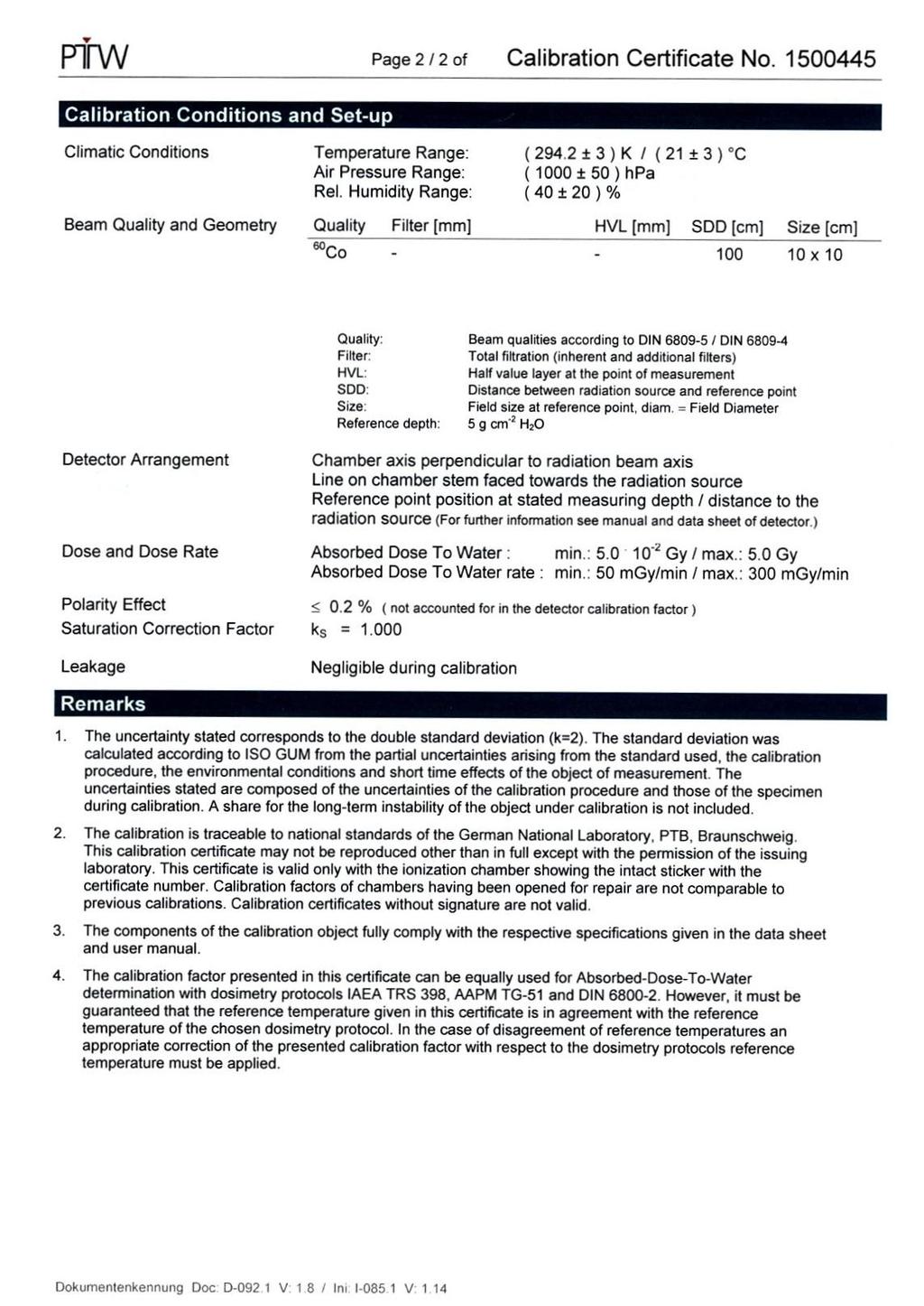
59 Appendix This appendix contains the calibration certificate, for the Ionization Chamber used during this thesis work, by the chamber producer itself ( -Freiburg). 56
60 57
61 References [1] E. Pedroni et al., The 200 MeV proton therapy project at the Paul Scherrer Institute: Conceptual design and practical realization, Med Phys 22 (1995) [2] G. H. Hartmann et al., Determination of water absorbed dose in a Carbon ion beam using thimble ionization chambers, Phys. Med. Biol. 44(5), (1999). [3] International Atomic Energy Agency (IAEA), TRS-398: Absorbed Dose Determination in External Beam Radiotherapy: An International Code of Practice for Dosimetry based on Standards of Absorbed Dose to Water, V. 12, June [4] International Commission on Radiation Units and Measurements (ICRU), Report 24: Determination of absorbed dose in a patient irradiated by beams of X or gamma rays in radiotherapy procedures, Bethesda (1976). [5] C. Ma, A. E. Nahum, Effect of size and composition of the central electrode on the response of cylindrical ionization chambers in high-energy photon and electron beams, Physics in Medicine and Biology, Vol. 38, Num. 2 (1993). [6] PTW Freiburg Germany. [7] F. H. Attix, Introduction to radiological physics and radiation dosimetry, Wiley- VCH, [8] International Electrotechnical Commission (IEC), Medical electrical equipment - Dosimeters with ionization chambers as used in radiotherapy, [9] A. Degiovanni, A. Ugo, History of hadron therapy accelerators, Physica Medica 31 (2015)
62 [10] and [11] S. Devic et al., Reference radiochromic film dosimetry: Review of technical aspects, Physica Medica 32 (2016) [12] M. Martišíková and O. Jäkel, Dosimetric properties of Gafchromic EBT films in monoenergetic medical ion beams, Phys. Med. Biol. 55(13) (2010). [13] R. Castriconi et al., Dose response of EBT3 radiochromic films to proton and Carbon ion clinical beams, Physics in Medicine & Biology, Vol. 62, Num. 2 (2016). [14] S. Rossi, The National Centre for Oncological Hadrontherapy (CNAO): Status and perspectives, Physica Medica 31 (2015) [15] A. Mirandola et al., Dosimetric commisioning and quality assurance of scanned ion beams at the Italian National Center for Oncological Hadrontherapy, Med. Phys. 42 (9) (2015). [16] -International. [17] C. P. Karger et al., Dosimetry for ion beam radiotherapy, Phys. Med. Biol. 55(21), R (2010). [18] O. Geithner et al., Calculation of stopping power ratios for carbon ion dosimetry, Phys. Med. Biol. 51(9), (2006). [19] D. Sanchez-Parcerisa et al., Influence of the delta ray production threshold on water-to-air stopping power ratio calculations for carbon ion beam radiotherapy, Phys. Med. Biol. 58(1), (2013). 59
Topics covered 7/21/2014. Radiation Dosimetry for Proton Therapy
 Radiation Dosimetry for Proton Therapy Narayan Sahoo Department of Radiation Physics University of Texas MD Anderson Cancer Center Proton Therapy Center Houston, USA Topics covered Detectors used for to
Dosimetric characterization with 62 MeV protons of a silicon segmented detector for 2D dose verifications in radiotherapy
 Dosimetric characterization with 62 MeV protons of a silicon segmented detector for 2D dose verifications in radiotherapy C. Talamonti a*, M. Bucciolini a, L. Marrazzo a, D. Menichelli a. a) Department
Standard calibration of ionization chambers used in radiation therapy dosimetry and evaluation of uncertainties
 Standard calibration of ionization chambers used in radiation therapy dosimetry and evaluation of uncertainties A. Solimanian and M. Ghafoori * Iran. J. Radiat. Res., 2010; 8 (3): 195-199 Radiation Dosimetry
Recent advances in dosimetry in reference conditions for proton and light-ion beams
 Recent advances in dosimetry in reference conditions for proton and light-ion beams S. Vatnitskiy a), P. Andreo b) and D.T.L. Jones c) a) MedAustron, Wiener Neustadt, Austria b) Medical Radiation Physics,
COMPARISON OF RADIOBIOLOGICAL EFFECTS OF CARBON IONS TO PROTONS ON A RESISTANT HUMAN MELANOMA CELL LINE
 COMPARISON OF RADIOBIOLOGICAL EFFECTS OF CARBON IONS TO PROTONS ON A RESISTANT HUMAN MELANOMA CELL LINE I. Petrovi a, A. Risti -Fira a, L. Kori anac a, J. Požega a, F. Di Rosa b, P. Cirrone b and G. Cuttone
Practical Reference Dosimetry Course April 2015 PRDC Program, at a glance. Version 1.0. Day 1 Day 2 Day 3 Day 4
 Practical Reference Dosimetry Course 21-24 April 2015 PRDC 2015 Program, at a glance Version 1.0 Day 1 Day 2 Day 3 Day 4 Quantities and Units Free air chambers Uncertainties Brachytherapy traceability
Semiflex 3D. Always perfectly oriented. 3D Thimble Ionization Chamber for Relative and Absolute Dosimetry
 DETECTORS Always perfectly oriented. Semiflex 3D 3D Thimble Ionization Chamber for Relative and Absolute Dosimetry The New Reference Class Full 3D Geometry For FFF and FF Beams Semiflex 3D 3D Detector
Active Scanning Beam 3 Checking Delivery/Dosimetry
 Active Scanning Beam 3 Checking Delivery/Dosimetry C Algranati, J Salk, A Coray, A Lomax, E Pedroni, T Boeringer, S Lin, E Hug Center for Proton Radiation Therapy Checking Delivery/Dosimetry Contents Absolute
Semiflex 3D. Always perfectly oriented. 3D Thimble Ionization Chamber for Relative and Absolute Dosimetry
 DETECTORS Always perfectly oriented. Semiflex 3D 3D Thimble Ionization Chamber for Relative and Absolute Dosimetry The New Reference Class Full 3D Geometry For FFF and FF Beams Semiflex 3D 3D Detector
Dosimetry and QA of proton and heavier ion beams
 Dosimetry and QA of proton and heavier ion beams Stanislav Vatnitskiy EBG MedAustron GmbH Wiener Neustadt, Austria Content Introduction Reference dosimetry Methods, detectors, protocols Dosimetry in non-reference
Y FILMS DOSIMETR Nederland België / Belgique
 DOSIMETRY FILMS GAFCHROMIC Dosimetry Films The Self-developing Dosimetry Films that Allow You to Go Filmless Do away with the cost and headache of calibrating and maintaining a processor Do away with hazardous
Characterization and implementation of Pencil Beam Scanning proton therapy techniques: from spot scanning to continuous scanning
 Characterization and implementation of Pencil Beam Scanning proton therapy techniques: from spot scanning to continuous scanning Supervisors Prof. V. Patera PhD R. Van Roermund Candidate Annalisa Patriarca
ABSTRACTS FOR RADIOTHERAPY STANDARDS USERS MEETING. 5 th June 2007
 ABSTRACTS FOR RADIOTHERAPY STANDARDS USERS MEETING 5 th June 2007 An Overview of Radiotherapy Dosimetry at the NPL Hugo Palmans In relation to radiotherapy applications, The National Physical Laboratory
III. Proton-therapytherapy. Rome SB - 5/5 1
 Outline Introduction: an historical review I Applications in medical diagnostics Particle accelerators for medicine Applications in conventional radiation therapy II III IV Hadrontherapy, the frontier
Protons Monte Carlo water-equivalence study of two PRESAGE formulations for proton beam dosimetry J. Phys.: Conf. Ser.
 Protons Monte Carlo water-equivalence study of two PRESAGE formulations for proton beam dosimetry T Gorjiara, Z Kuncic, J Adamovics and C Baldock 2013 J. Phys.: Conf. Ser. 444 012090 PRESAGE is a radiochromic
GAFCHROMIC DOSIMETRY MEDIA TYPE MD-V3
 GAFCHROMIC DOSIMETRY MEDIA TYPE MD-V3 WARNING: Store below 25ºC Store away from radiation sources Avoid exposure of film to sunlight Handle film carefully, creasing may cause damage Do not expose to temperatures
Calibration of dosimeters for small mega voltage photon fields at ARPANSA
 Calibration of dosimeters for small mega voltage photon fields at ARPANSA G Ramanathan 1, C.Oliver 1, D J Butler 1, P D Harty 1, Viliami Takau 1 Tracy Wright 1 and Tom Kupfer 2 1 Australian Radiation Protection
GAFCHROMIC MD-55 RADIOCHROMIC DOSIMETRY FILM FOR HIGH-ENERGY PHOTONS CONFIGURATION, SPECIFICATIONS AND PERFORMANCE DATA
 GAFCHROMIC MD-55 RADIOCHROMIC DOSIMETRY FILM FOR HIGH-ENERGY PHOTONS CONFIGURATION, SPECIFICATIONS AND PERFORMANCE DATA DESCRIPTION GAFCHROMIC MD-55 radiochromic dosimetry film is designed for the measurement
PROGRESS IN HADRONTHERAPY
 PROGRESS IN HADRONTHERAPY Saverio Braccini TERA Foundation for Oncological Hadrontherapy IPRD06 - Siena - 01.10.06 - SB 1 Outline Introduction Radiation therapy with X rays and hadrontherapy Hadrontherapy
Dosimetric characterization with 62 MeV protons of a silicon segmented detector for 2D dose verifications in radiotherapy
 Dosimetric characterization with 62 MeV protons of a silicon segmented detector for 2D dose verifications in radiotherapy C. Talamonti M. Bruzzi,M. Bucciolini, L. Marrazzo, D. Menichelli University of
Proton and helium beams: the present and the future of light ion beam therapy
 Proton and helium beams: the present and the future of light ion beam therapy Dr. Andrea Mairani Group Leader Biophysics in Particle Therapy Heidelberg Ion Beam Therapy Center HIT Department of Radiation
Monte Carlo water-equivalence study of two PRESAGE formulations for proton beam dosimetry
 Monte Carlo water-equivalence study of two PRESAGE formulations for proton beam dosimetry T Gorjiara 1, Z Kuncic 1, J Adamovics 2 and C Baldock 1,3 1 Institute of Medical Physics, School of Physics, University
Two-Dimensional Thermoluminescence Dosimetry System for Proton Beam Quality Assurance
 Two-Dimensional Thermoluminescence Dosimetry System for Proton Beam Quality Assurance Jan Gajewski Institute of Nuclear Physics, Kraków, Poland German Cancer Research Center, Heidelberg, Germany Existing
Radiotherapy. Marta Anguiano Millán. Departamento de Física Atómica, Molecular y Nuclear Facultad de Ciencias. Universidad de Granada
 Departamento de Física Atómica, Molecular y Nuclear Facultad de Ciencias. Universidad de Granada Overview Introduction Overview Introduction Brachytherapy Radioisotopes in contact with the tumor Overview
M. J. Maryanski, Three Dimensional BANG Polymer Gel Dosimeters AAPM'99, CE Course
 Three Dimensional BANG Polymer Gel Dosimeters Marek J. Maryanski MGS Research, Inc. Guilford, CT Educational objectives: Describe the need for high-resolution 3D dosimetry in 3D CRT. Explain the physics
A comparison of dose distributions measured with two types of radiochromic film dosimeter MD55 and EBT for proton beam of energy 175 MeV
 A comparison of dose distributions measured with two types of radiochromic film dosimeter MD55 and EBT for proton beam of energy 175 MeV M. Mumot, G. V. Mytsin, Y. I. Luchin and A. G. Molokanov Medico-Technical
Intensity Modulated Radiation Therapy: Dosimetric Aspects & Commissioning Strategies
 Intensity Modulated Radiation Therapy: Dosimetric Aspects & Commissioning Strategies ICPT School on Medical Physics for Radiation Therapy Justus Adamson PhD Assistant Professor Department of Radiation
Abstract: A National Project for the in-vivo dosimetry in radiotherapy: first results
 Abstract: A National Project for the in-vivo dosimetry in radiotherapy: first results A.Piermattei 1, L. Azario 1, S. Cilla 2, A.Fidanzio 1, F.Greco 1, M.T.Russo 3, S. Zucca 4 1 Istituto di Fisica e Unità
Verification of treatment planning system parameters in tomotherapy using EBT Radiochromic Film
 Verification of treatment planning system parameters in tomotherapy using EBT Radiochromic Film E.B.Rajmohan¹, Pratik Kumar¹, Bhudatt Paliwal,² David Westerly², N.Gopishankar³, R.K.Bisht³, D.Tewatia²,
JOURNAL OF APPLIED CLINICAL MEDICAL PHYSICS, VOLUME 4, NUMBER 4, FALL 2003
 JOURNAL OF APPLIED CLINICAL MEDICAL PHYSICS, VOLUME 4, NUMBER 4, FALL 2003 Reference photon dosimetry data: A preliminary study of in-air off-axis factor, percentage depth dose, and output factor of the
Venue: IEEE NSS/MIC/RTSD Conference, Seoul, South Korea, 27 th October 2013 Workshop: NWK3/RD1 Radiation Protection and Dosimetry
 Venue: IEEE NSS/MIC/RTSD Conference, Seoul, South Korea, 27 th October 2013 Workshop: NWK3/RD1 Radiation Protection and Dosimetry M. Caresana a, A. Sashala Naik a,c, S. Rollet b, M. Ferrarini a,d a Polytechnic
OPERATION AND PATIENT TREATMENTS AT CNAO FACILITY
 OPERATION AND PATIENT TREATMENTS AT CNAO FACILITY Abstract The CNAO (National Centre for Oncological Hadrontherapy) has been realized in Pavia. It is a clinical facility created and financed by the Italian
PREDICTION OF ABSORBED DOSE DISTRIBUTIONS AND NEUTRON DOSE EQUIVALENT VALUES IN PROTON BEAM RADIATION THERAPY
 PREDICTION OF ABSORBED DOSE DISTRIBUTIONS AND NEUTRON DOSE EQUIVALENT VALUES IN PROTON BEAM RADIATION THERAPY IDENTIFICATION NUMBER: ANS-RT-PROTON-01 BENCHMARK CLASSIFICATION: Radiation Therapy BENCHMARK
Calibration of Radiation Instruments Used in Radiation Protection and Radiotherapy in Malaysia
 Abstract Calibration of Radiation Instruments Used in Radiation Protection and Radiotherapy in Malaysia Taiman Bin Kadni (taiman@mint.gov.my) Secondary Standard Dosimetry Laboratory (SSDL) Malaysian Institute
EXPERIMENTAL RESULTS IN PERCENTAGE DEPTH DOSE (PDD) DETERMINATION AT THE EXTENDED DISTANCES *
 DETERMINATION AT THE EXTENDED DISTANCES *") Romanian Reports in Physics, Vol. 66, No. 1, P. 157 165, 2014 EXPERIMENTAL RESULTS IN PERCENTAGE DEPTH DOSE (PDD) DETERMINATION AT THE EXTENDED DISTANCES * M. SPUNEI 1,2, M. MIHAI 3, I. MĂLĂESCU 1 1 West
Romanian Reports in Physics, Vol. 66, No. 1, P. 157 165, 2014 EXPERIMENTAL RESULTS IN PERCENTAGE DEPTH DOSE (PDD) DETERMINATION AT THE EXTENDED DISTANCES * M. SPUNEI 1,2, M. MIHAI 3, I. MĂLĂESCU 1 1 West
A preliminary clinic dosimetry study for synchrotron radiation therapy at SSRF
 Nuclear Science and Techniques 24 (2013) 060102 A preliminary clinic dosimetry study for synchrotron radiation therapy at SSRF LI Zhaobin SHI Zeliang ZHANG Qing WANG Yong FU Shen * The 6 th People s Hospital
Energy response of EBT3 radiochromic films: implications for dosimetry in kilovoltage range
 JOURNAL OF APPLIED CLINICAL MEDICAL PHYSICS, VOLUME 15, NUMBER 1, 2014 Energy response of EBT3 radiochromic films: implications for dosimetry in kilovoltage range J. Eduardo Villarreal-Barajas, a and Rao
DOSIMETRIC COMPARISION FOR RADIATION QUALITY IN HIGH ENERGY PHOTON BEAMS
 DOSIMETRIC COMPARISION FOR RADIATION QUALITY IN HIGH ENERGY PHOTON BEAMS EUGENIA BADITA 1, CATALIN VANCEA 1,3, ION CALINA 1,3, DANIELA STROE 2, MIHAELA DUMITRACHE 2,3, MIRABELA DUMITRACHE 1,3 1 National
Measurements of Air Kerma Index in Computed Tomography: A comparison among methodologies
 Measurements of Air Kerma Index in Computed Tomography: A comparison among methodologies Thêssa C. Alonso 1, 2, Arnaldo P. Mourão 1, 3, Teógenes A. Da Silva 1, 2 1 Program of Nuclear Science and Techniques
D DAVID PUBLISHING. Uncertainties of in vivo Dosimetry Using Semiconductors. I. Introduction. 2. Methodology
 Journal of Life Sciences 9 (2015) 120-126 doi: 10.17265/1934-7391/2015.03.005 D DAVID PUBLISHING Uncertainties of in vivo Dosimetry Using Semiconductors Zeina Al Kattar, Hanna El Balaa and Saeed Zahran
Assessment of Dosimetric Functions of An Equinox 100 Telecobalt Machine
 2017 IJSRST Volume 3 Issue 3 Print ISSN: 2395-6011 Online ISSN: 2395-602X Themed Section: Science and Technology Assessment of Dosimetric Functions of An Equinox 100 Telecobalt Machine Samuel Nii Adu Tagoe
PROCEDURE FOR ABSORBED DOSE TO WATER DETERMINATION IN HIGH ENERGY PHOTON AND ELECTRON BEAMS BY FERROUS SULPHATE DOSIMETER AT INMRI-ENEA
 PROCEDURE FOR ABSORBED DOSE TO WATER DETERMINATION IN HIGH ENERGY PHOTON AND ELECTRON BEAMS BY FERROUS SULPHATE DOSIMETER AT INMRI-ENEA M. Pimpinella, A. S. Guerra, S. La Civita and R. F. Laitano Istituto
Volumetric Modulated Arc Therapy - Patient Specific QA
 Volumetric Modulated Arc Therapy - Patient Specific QA Daliang Cao, PhD, DABR Swedish Cancer Institute, Seattle, WA VMAT plan QA methods Composite dose measurement Film & ion chamber diode array Mapcheck
Proton Treatment. Keith Brown, Ph.D., CHP. Associate Director, Radiation Safety University of Pennsylvania
 Proton Treatment Keith Brown, Ph.D., CHP Associate Director, Radiation Safety University of Pennsylvania Proton Dose vs. Depth Wilson,. R.R. Radiological use of fast protons. Radiology 47:487-491, 1946.
Application(s) of Alanine
 of Alanine") Application(s) of Alanine Simon Duane Radiotherapy Standards User Group, 5 June 2007 Outline Alanine/EPR dosimetry characteristics, usage (dis)advantages for reference dosimetry Traceable dosimetry for
Application(s) of Alanine Simon Duane Radiotherapy Standards User Group, 5 June 2007 Outline Alanine/EPR dosimetry characteristics, usage (dis)advantages for reference dosimetry Traceable dosimetry for
Learning Objectives. Clinically operating proton therapy facilities. Overview of Quality Assurance in Proton Therapy. Omar Zeidan
 Overview of Quality Assurance in Proton Therapy Omar Zeidan AAPM 2012 Charlotte, NC July 30 st, 2012 Learning Objectives Understand proton beam dosimetry characteristics and compare them to photon beams
RADIATION PROTECTION IN DIAGNOSTIC AND INTERVENTIONAL RADIOLOGY. L19: Optimization of Protection in Mammography
 IAEA Training Material on Radiation Protection in Diagnostic and Interventional Radiology RADIATION PROTECTION IN DIAGNOSTIC AND INTERVENTIONAL RADIOLOGY L19: Optimization of Protection in Mammography
Diode calibration for dose determination in total body irradiation
 Iran. J. Radiat. Res., 2008; 6 (1): 43-50 Diode calibration for dose determination in total body irradiation M.Allahverdi 1*, Gh. Geraily 1, M. Esfehani 3,A. Sharafi 2,A. Shirazi 1 1 Department of Medical
Large-area Two-Dimensional Thermoluminescence Dosimetry System in Ion Beam Quality Assurance
 Large-area Two-Dimensional Thermoluminescence Dosimetry System in Ion Beam Quality Assurance J. Gajewski1, M. Kłosowski1, S. Greilich2, P. Olko1 Institute of Nuclear Physics, Kraków, Poland German Cancer
Radiochromic film dosimetry in water phantoms
 INSTITUTE OF PHYSICS PUBLISHING PHYSICS IN MEDICINE AND BIOLOGY Phys. Med. Biol. 46 (2001) N27 N31 www.iop.org/journals/pb PII: S0031-9155(01)16858-2 NOTE Radiochromic film dosimetry in water phantoms
 This article was published in an Elsevier journal. The attached copy is furnished to the author for non-commercial research and education use, including for instruction at the author s institution, sharing
Peak temperature ratio of TLD glow curves to investigate the spatial variation of LET in a clinical proton beam
 Peak temperature ratio of TLD glow curves to investigate the spatial variation of LET in a clinical proton beam University of Chicago CDH Proton Center LET study C. Reft 1, H. Ramirez 2 and M. Pankuch
Specific Aspects of Radiochromic Film Dosimetry AAPM Task Group 235 An Update to Task Group 55 (1998)
") Azam Niroomand-Rad: General Aspects of RCF (TG-235), AAPM July 2015 1 Specific Aspects of Radiochromic Film Dosimetry AAPM Task Group 235 An Update to Task Group 55 (1998) Azam Niroomand-Rad, PhD, FAAPM,
Azam Niroomand-Rad: General Aspects of RCF (TG-235), AAPM July 2015 1 Specific Aspects of Radiochromic Film Dosimetry AAPM Task Group 235 An Update to Task Group 55 (1998) Azam Niroomand-Rad, PhD, FAAPM,
ROPES eye plaque dosimetry: commissioning and verification of an ophthalmic brachytherapy treatment planning system
 University of Wollongong Research Online Faculty of Engineering and Information Sciences - Papers: Part A Faculty of Engineering and Information Sciences 2013 ROPES eye plaque dosimetry: commissioning
5th ADAMAS Workshop at GSI December 15-16, 2016, Darmstadt, Germany
 Evaluation of 3D diamond detectors for application in medical radiation dosimetry K. Kanxheri (1,2), L. Servoli (2), C. Zucchetti (5), A. C. Dipilato (5), M. Iacco (5), S. Lagomarsino (3,4), A. Morozzi
Nuclear energy, nuclear technologies, and radiological protection Vocabulary. Part 4: Dosimetry for radiation processing
 Provläsningsexemplar / Preview INTERNATIONAL STANDARD ISO 12749-4 First edition 2015-08-15 Nuclear energy, nuclear technologies, and radiological protection Vocabulary Part 4: Dosimetry for radiation processing
Report to the 18 th Meeting of the CCRI(I), May 2007 Recent Activities in Measurement Standards and Dosimetry at METAS,
, May 2007 Recent Activities in Measurement Standards and Dosimetry at METAS,") Report to the 18 th Meeting of the CCRI(I), May 2007 Recent Activities in Measurement Standards and Dosimetry at, 2005-2007 1. Introduction Gerhard Stucki, Solange Gagnebin, Damian Twerenbold, Sándor Vörös
Report to the 18 th Meeting of the CCRI(I), May 2007 Recent Activities in Measurement Standards and Dosimetry at, 2005-2007 1. Introduction Gerhard Stucki, Solange Gagnebin, Damian Twerenbold, Sándor Vörös
Heavy Ion Tumor Therapy
 Heavy Ion Tumor Therapy Applications Bence Mitlasoczki 25.06.2018 Heidelberg 1. Source (H 2 /CO 2 ) 2. Linac 3. Synchrotron 4. Guide 5. Treatment rooms 6. X-ray system 7. Gantry 8. Treatment room with
CHARACTERIZATION OF THE ELECTRON BEAM RADIATION FIELD BY CHEMICAL DOSIMETRY
 CHARACTERIZATION OF THE ELECTRON BEAM RADIATION FIELD BY CHEMICAL DOSIMETRY M. R. NEMTANU, C. OPROIU, M. BRASOVEANU, M. OANE National Institute for Laser, Plasma and Radiation Physics, Electron Accelerator
Traceability and absorbed dose standards for small fields, IMRT and helical tomotherapy
 Traceability and absorbed dose standards for small fields, IMRT and helical tomotherapy Simon Duane, Hugo Palmans, Peter Sharpe NPL, UK Stefaan Vynckier UCL, Brussels, Belgium LNE-LNHB / BIPM workshop,
Skyscan 1076 in vivo scanning: X-ray dosimetry
 Skyscan 1076 in vivo scanning: X-ray dosimetry DOSIMETRY OF HIGH RESOLUTION IN VIVO RODENT MICRO-CT IMAGING WITH THE SKYSCAN 1076 An important distinction is drawn between local tissue absorbed dose in
Neutron dose evaluation in radiotherapy
 Neutron dose evaluation in radiotherapy Francesco d Errico University of Pisa, Italy Yale University, USA Radiation therapy with a linear accelerator (LINAC) Photoneutron production in accelerator head
An anthropomorphic head phantom with a BANG polymer gel insert for dosimetric evaluation of IMRT treatment delivery
 An anthropomorphic head phantom with a BANG polymer gel insert for dosimetric evaluation of IMRT treatment delivery G. Ibbott a, M. Beach a, M. Maryanski b a M.D. Anderson Cancer Center, Houston, Texas,
PMP. Use of Cylindrical Chambers as Substitutes for Parallel- Plate Chambers in Low-Energy Electron Dosimetry. Original Article.
 Original Article PMP Progress in Medical Physics 29(1), March 218 https://doi.org/1.14316/pmp.218.29.1.16 pissn 258-4445, eissn 258-4453 Use of Cylindrical Chambers as Substitutes for Parallel- Plate Chambers
Small Field Dosimetry & Radiochromic film
 Small Field Dosimetry & Radiochromic film Michael Kissick, Ph.D., Assistant Professor, Medical Physics & Human Oncology & Affiliated with the Morgridge Institute for Research North Central AAPM Chapter
MEASUREMENT OF THE EQUIVALENT INDIVIDUAL DOSES FOR PATIENTS IN ANGIOGRAPHY PROCEDURE AND INTERVENTIONAL RADIOLOGY WITH THERMOLUMINESCENT SYSTEMS
 RADIOPROTECTION AND DOSIMETRY MEASUREMENT OF THE EQUIVALENT INDIVIDUAL DOSES FOR PATIENTS IN ANGIOGRAPHY PROCEDURE AND INTERVENTIONAL RADIOLOGY WITH THERMOLUMINESCENT SYSTEMS DANIELA ADAM 1, ANA STOCHIOIU
Monte Carlo simulation of 192 Ir radioactive source in a phantom designed for brachytherapy dosimetry and source position evaluation.
 Monte Carlo simulation of 192 Ir radioactive source in a phantom designed for brachytherapy dosimetry and source position evaluation Samuel Chiquita 1 1 University of Porto, Porto, Portugal Abstract In
Limits of Precision and Accuracy of Radiation Delivery Systems
 Limits of Precision and Accuracy of Radiation Delivery Systems Jean M. Moran, Ph.D. 1 and Timothy Ritter, Ph.D. 2 1 University of Michigan, Ann Arbor, Michigan 2 Ann Arbor Veterans Affairs Hospital, Ann
Dosimetric study of 2D ion chamber array matrix for the modern radiotherapy treatment verification
 JOURNAL OF APPLIED CLINICAL MEDICAL PHYSICS, VOLUME 11, NUMBER 2, SPRING 2010 Dosimetric study of 2D ion chamber array matrix for the modern radiotherapy treatment verification Sathiyan Saminathan, a Ravikumar
ADVANCES IN RADIATION TECHNOLOGIES IN THE TREATMENT OF CANCER
 ADVANCES IN RADIATION TECHNOLOGIES IN THE TREATMENT OF CANCER Bro. Dr. Collie Miller IARC/WHO Based on trends in the incidence of cancer, the International Agency for Research on Cancer (IARC) and WHO
Use of Bubble Detectors to Characterize Neutron Dose Distribution in a Radiotherapy Treatment Room used for IMRT treatments
 Use of Bubble Detectors to Characterize Neutron Dose Distribution in a Radiotherapy Treatment Room used for IMRT treatments Alana Hudson *1 1 Tom Baker Cancer Centre, Department of Medical Physics, 1331
Multi-Channel Film Dosimetry and Gamma Map Verification
 Multi-Channel Film Dosimetry and Gamma Map Verification Micke A International Specialty Products Ashland proprietary technology, patents pending Single Channel Film Dosimetry Calibration Curve X=R R ave
Quality assurance of volumetric modulated arc therapy using Elekta Synergy
 Acta Oncologica, 2009; 48: 11931197 ORIGINAL ARTICLE Quality assurance of volumetric modulated arc therapy using Elekta Synergy AKIHIRO HAGA 1, KEIICHI NAKAGAWA 1, KENSHIRO SHIRAISHI 1, SAORI ITOH 1, ATSURO
A brief presentation of The TERA Foundation
 A brief presentation of The TERA Foundation David Watts on behalf of Prof. Ugo Amaldi and all my colleagues at TERA TERA Overview Direction: Prof. Ugo Amaldi AQUA (Advanced QUAlity Assurance) Cyclinac
Unrivaled, End-to-End
 PHANTOMS Unrivaled, End-to-End Stereotactic QA Industry-leading 0.1mm accuracy minimizes errors at each link in the stereotactic quality assurance chain. Stereotactic radiosurgery (SRS) is governed by
Small field diode dosimetry
 Small field diode dosimetry Parham Alaei, Ph.D. Department of Radiation Oncology University of Minnesota NCCAAPM Symposium-October 10, 2013 1 Diodes as beam data collection detectors Diodes as in vivo
Amendment No. 2. Item No. 2 (Rfx/ Event number )
") Amendment 2 Sub: Amendment to the Document 06.12.2018 Ref.: Notice Inviting Bid ref. HITES/PCD/NCI-AIIMS/36/18-19 dated 26.09.2018 read with its Amendment no. 1 dated 19.11.18 The following changes have
Amendment 2 Sub: Amendment to the Document 06.12.2018 Ref.: Notice Inviting Bid ref. HITES/PCD/NCI-AIIMS/36/18-19 dated 26.09.2018 read with its Amendment no. 1 dated 19.11.18 The following changes have
Hampton University Proton Therapy Institute
 Hampton University Proton Therapy Institute Brief introduction to proton therapy technology, its advances and Hampton University Proton Therapy Institute Vahagn Nazaryan, Ph.D. Executive Director, HUPTI
SCIENTIFIC AND TECHNOLOGICAL DEVELOPMENT OF HADRONTHERAPY
 SCIENTIFIC AND TECHNOLOGICAL DEVELOPMENT OF HADRONTHERAPY SAVERIO BRACCINI * Albert Einstein Centre for Fundamental Physics, Laboratory for High Energy Physics (LHEP), University of Bern, Sidlerstrasse
 This article was published in an Elsevier journal. The attached copy is furnished to the author for non-commercial research and education use, including for instruction at the author s institution, sharing
A VMAT PLANNING SOLUTION FOR NECK CANCER PATIENTS USING THE PINNACLE 3 PLANNING SYSTEM *
 Romanian Reports in Physics, Vol. 66, No. 2, P. 401 410, 2014 A VMAT PLANNING SOLUTION FOR NECK CANCER PATIENTS USING THE PINNACLE 3 PLANNING SYSTEM * M. D. SUDITU 1,2, D. ADAM 1,2, R. POPA 1,2, V. CIOCALTEI
Verification of micro-beam irradiation
 Journal of Physics: Conference Series OPEN ACCESS Verification of micro-beam irradiation To cite this article: Qiongge Li et al 2015 J. Phys.: Conf. Ser. 573 012047 View the article online for updates
Protection of the contralateral breast during radiation therapy for breast cancer
 Protection of the contralateral breast during radiation therapy for breast cancer Edgardo Garrigó a*, Alejandro Germanier b, Silvia Zunino a a Instituto Privado de Radioterapia, Ob Oro 423 (5000) Córdoba,
Dependence of the thermoluminescent high-temperature ratio (HTR) of LiF:Mg,Ti detectors on proton energy and dose
 of LiF:Mg,Ti detectors on proton energy and dose") Submitted to Radiation Measurements Dependence of the thermoluminescent high-temperature ratio (HTR) of LiF:Mg,Ti detectors on proton energy and dose P. Bilski 1, M. Sadel 1, J. Swakon 1, A. Weber 2 1
Submitted to Radiation Measurements Dependence of the thermoluminescent high-temperature ratio (HTR) of LiF:Mg,Ti detectors on proton energy and dose P. Bilski 1, M. Sadel 1, J. Swakon 1, A. Weber 2 1
Absolute Dosimetry. Versatile solid and water phantoms. Introduction Introduction Introduction Introtro Intro Intro
 In Phantoms Vivo treatment for Absolute Dosimetry verification Versatile solid and water phantoms Introduction Introduction Introduction Introtro Intro Intro The rapid development of advanced treatment
LONG TERM STABILITY OF THE PERFORMANCE OF A CLINICAL LINEAR ACCELERATOR AND Z-SCORE ASSESSEMENT FOR ABSORBED DOSE TO WATER QUANTITY
 LONG TERM STABILITY OF THE PERFORMANCE OF A CLINICAL LINEAR ACCELERATOR AND Z-SCORE ASSESSEMENT FOR ABSORBED DOSE TO WATER QUANTITY E. BADITA 1, C.VANCEA 1,3, I. CALINA 1,3, D. STROE 2, M. DUMITRACHE 2,3,
MODELLING A GAMMA IRRADIATION PROCESS USING THE MONTE CARLO METHOD
 2011 International Nuclear Atlantic Conference - INAC 2011 Belo Horizonte, MG, Brazil, October 24-28, 2011 ASSOCIAÇÃO BRASILEIRA DE ENERGIA NUCLEAR - ABEN ISBN: 978-85-99141-04-5 MODELLING A GAMMA IRRADIATION
Disclosure. Outline. Machine Overview. I have received honoraria from Accuray in the past. I have had travel expenses paid by Accuray in the past.
 Clinical Implementation of the CyberKnife Disclosure I have received honoraria from Accuray in the past. I have had travel expenses paid by Accuray in the past. Mary Ellen Masterson-McGary McGary CyberKnife
In vivo dosimetry and shielding disk alignment verification by EBT3 GAFCHROMIC film in breast IOERT treatment
 JOURNAL OF APPLIED CLINICAL MEDICAL PHYSICS, VOLUME 16, NUMBER 1, 2015 In vivo dosimetry and shielding disk alignment verification by EBT3 GAFCHROMIC film in breast IOERT treatment Mara Severgnini, 1a
Verification of the PAGAT polymer gel dosimeter by photon beams using magnetic resonance imaging
 Iran. J. Radiat. Res., 2008; 6 (2): 83-87 Verification of the PAGAT polymer gel dosimeter by photon beams using magnetic resonance imaging B. Azadbakht 1, M.H. Zahmatkesh 2 *, k. Hadad 1, S. Bagheri 2
Radiation qualities in carbon-ion radiotherapy at NIRS/HIMAC
 Radiation qualities in carbon-ion radiotherapy at NIRS/ Shunsuke YONAI Radiological Protection Section Research Center for Charged Particle Therapy National Institute of Radiological Sciences (NIRS) E-mail:
Monte Carlo Modelling: a reliable and efficient tool in radiation dosimetry
 Monte Carlo Modelling: a reliable and efficient tool in radiation dosimetry G. Gualdrini, P. Ferrari ENEA Radiation Protection Institute, Bologna (Italy) Contribution to the Italy in Japan 2011 initiative
SOMATOM Drive System Owner Manual Dosimetry and imaging performance report
 www.siemens.com/healthcare SOMATOM Drive System Owner Manual Dosimetry and imaging performance report Table of contents 1 Dosimetry and imaging performance report 5 1.1 Dose information 5 1.1.1 General
Assessment of a three-dimensional (3D) water scanning system for beam commissioning and measurements on a helical tomotherapy unit
 water scanning system for beam commissioning and measurements on a helical tomotherapy unit") JOURNAL OF APPLIED CLINICAL MEDICAL PHYSICS, VOLUME 16, NUMBER 1, 2015 Assessment of a three-dimensional (3D) water scanning system for beam commissioning and measurements on a helical tomotherapy unit
JOURNAL OF APPLIED CLINICAL MEDICAL PHYSICS, VOLUME 16, NUMBER 1, 2015 Assessment of a three-dimensional (3D) water scanning system for beam commissioning and measurements on a helical tomotherapy unit
A new geometric and mechanical verification device for medical LINACs
 JOURNAL OF APPLIED CLINICAL MEDICAL PHYSICS, VOLUME 3, NUMBER 2, SPRING 2002 A new geometric and mechanical verification device for medical LINACs Keith T. Welsh,* Robert A. Wlodarczyk, and L. E. Reinstein
Radiation Monitoring Instruments
 Radiation Monitoring Instruments This set of slides is based on Chapter 4 authored byg. Rajan, J. Izewska of the IAEA publication (ISBN 92-0-107304-6): Radiation Oncology Physics: A Handbook for Teachers
Introduction. Measurement of Secondary Radiation for Electron and Proton Accelerators. Introduction - Photons. Introduction - Neutrons.
 Measurement of Secondary Radiation for Electron and Proton Accelerators D. Followill, Ph.D. Radiological Physics Center U. T. M. D. Anderson Cancer Center Introduction Patients undergoing radiation therapy
Investigation of the clinical performance of a novel solid-state diagnostic dosimeter
 JOURNAL OF APPLIED CLINICAL MEDICAL PHYSICS, VOLUME 16, NUMBER 4, 2015 Investigation of the clinical performance of a novel solid-state diagnostic dosimeter Jason Tse, a Donald McLean Medical Physics and
Evaluation of Dosimetry Check software for IMRT patient-specific quality assurance
 JOURNAL OF APPLIED CLINICAL MEDICAL PHYSICS, VOLUME 16, NUMBER 3, 2015 Evaluation of Dosimetry Check software for IMRT patient-specific quality assurance Ganesh Narayanasamy, Travis Zalman, Chul S. Ha,
IMRT point dose measurements with a diamond detector
 IMRT point dose measurements with a diamond detector Erin Barnett, Marc MacKenzie, B. Gino Fallone Department of Physics, University of Alberta, and Department of Medical Physics, Cross Cancer Institute,