Traps in the Immunohistochemical Work-up of Spindle Cell Tumours
|
|
|
- Geraldine Gibson
- 6 years ago
- Views:
Transcription

1 BRENDAN C. DICKSON BA, BSc, MD, MSc, FCAP, FRCPC Staff Pathologist Head, Section of Immunohistochemistry Pathology and Laboratory Medicine Mount Sinai Hospital Assistant Professor Laboratory Medicine and Pathobiology University of Toronto Traps in the Immunohistochemical Work-up of Spindle Cell Tumours
2 DISCLOSURE I have no actual, or poten al, conflicts of interest in rela on to this presenta on to disclose
3 OUTLINE Goal Approach to avoid common immunohistochemistryrelated pi alls in the evalua on of spindle cell neoplasms Introduc on Three cases Case presenta on Differen al diagnosis audience par cipa on Discussion of pi alls and pearls Summary
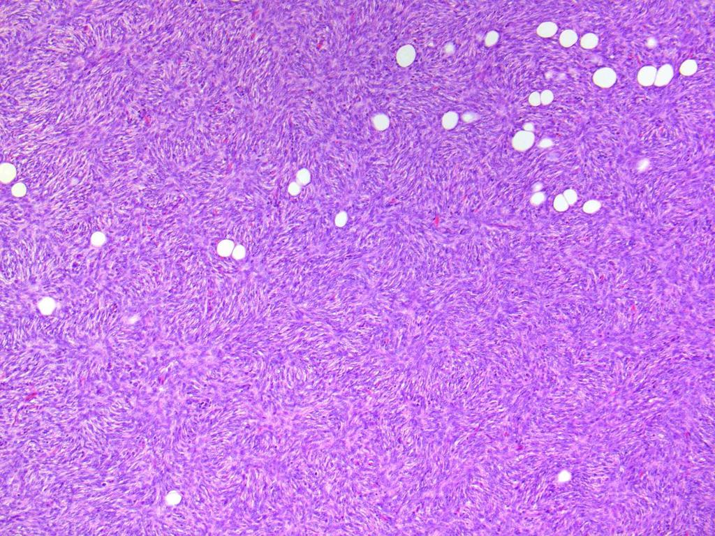
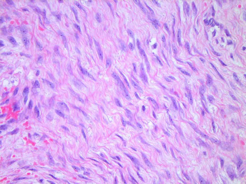
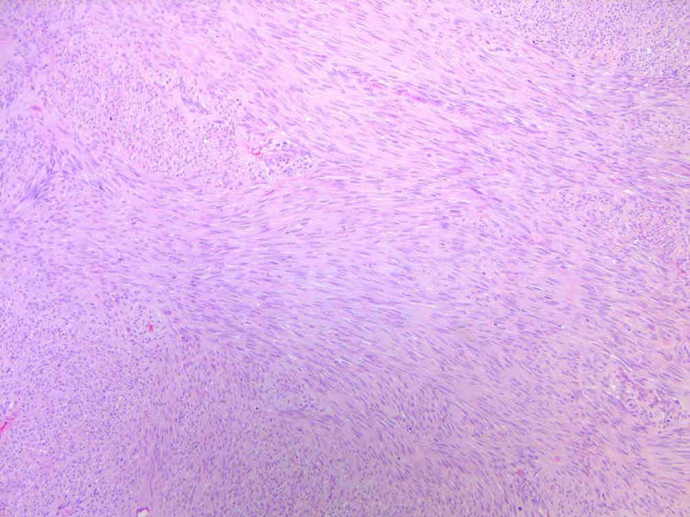
4 INTRODUCTION Spindle cell Descrip ve term deno ng a bipolar cell with tapered ends
5 INTRODUCTION Architecture Cells Nuclei Fascicular Plump Round-ovoid-elongated Storiform Elongated Monomorphic-pleomorphic Herringbone Colour of cytoplasm Wavy Pa ernless Inclusions Ends: blunt, tapered, asymmetric Plexiform Nucleoli Mito c ac vity (incl. atypical figures) Ancillary findings Margins: circumscribed-infiltra ve Necrosis Vascular pa ern Inflamma on Extracellular matrix / stroma
6 INTRODUCTION Clinical history Diagnos c imaging Pathology Morphology Does it look like sarcoma, carcinoma, melanoma? Immunohistochemistry Addi onal ancillary studies Electron microscopy Molecular and cytogene cs
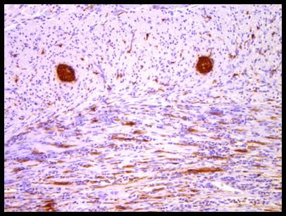
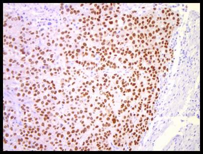
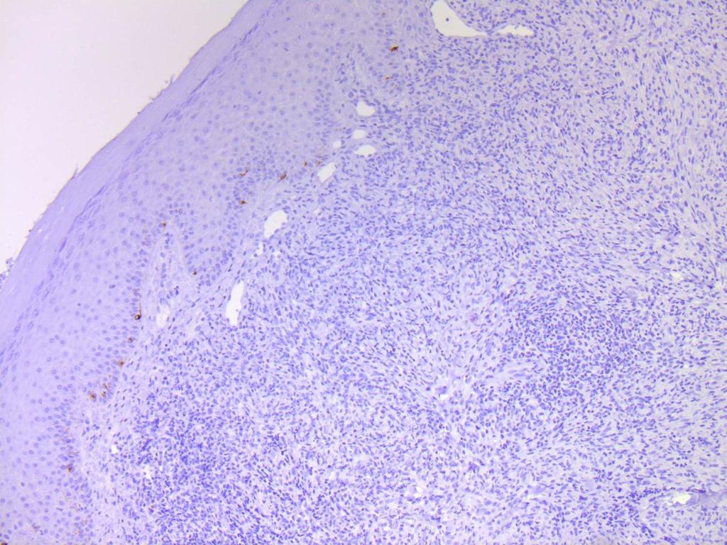
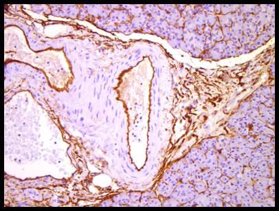
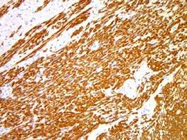
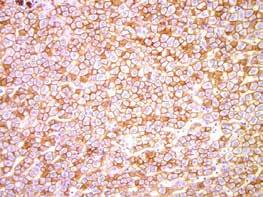
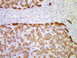
7 INTRODUCTION Basic spindle cell immunohistochemistry panel: Desmin Smooth muscle ac n CD34 S100 Kera n (AE1/AE3) Supplement & swap based on clinical context, morphologic impression, differen al diagnosis and/or need for redundancy
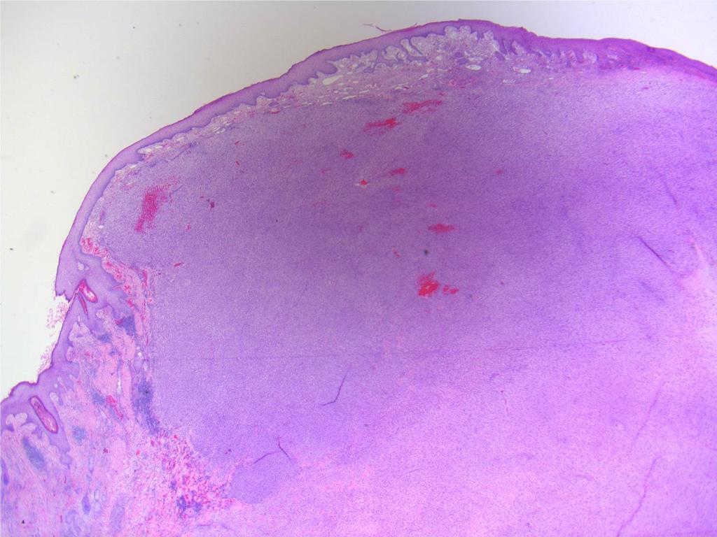
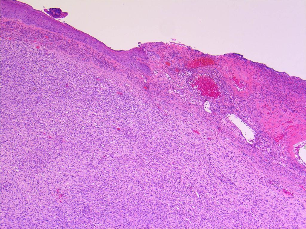
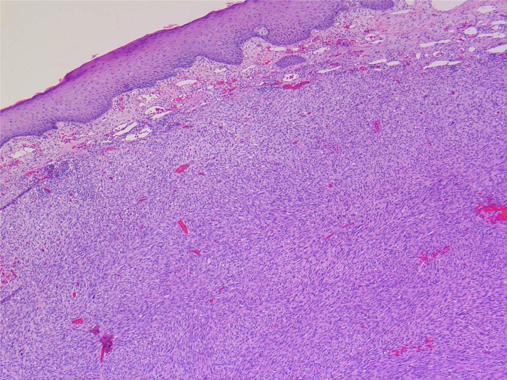
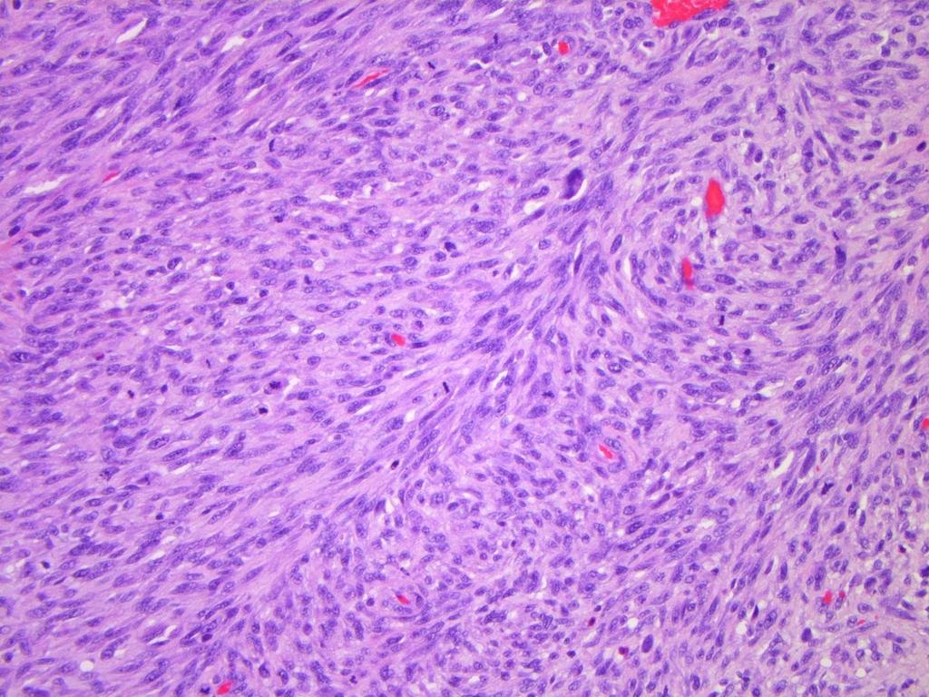
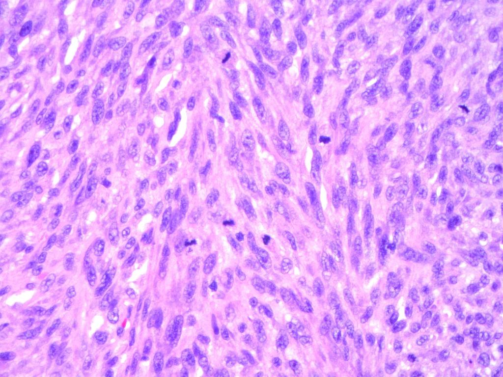
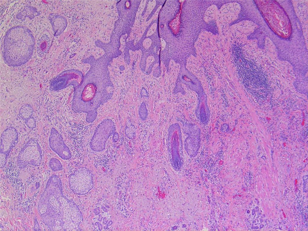
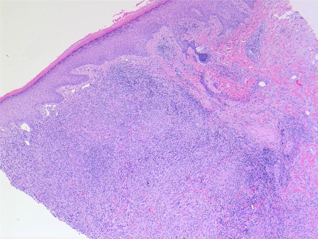
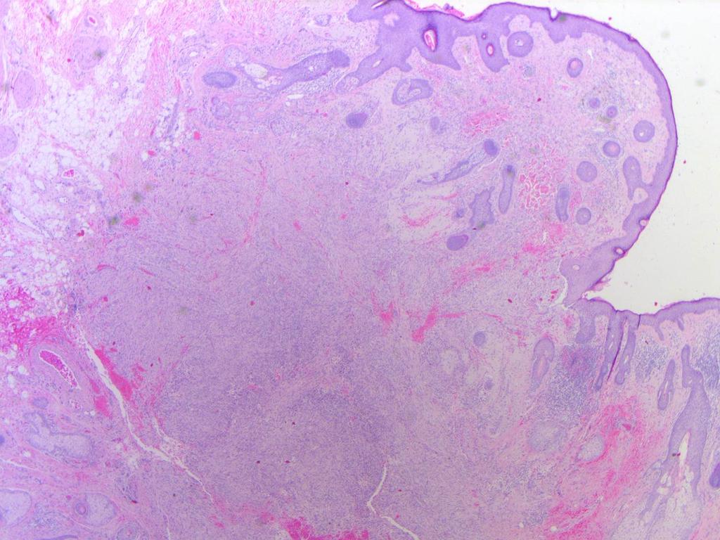
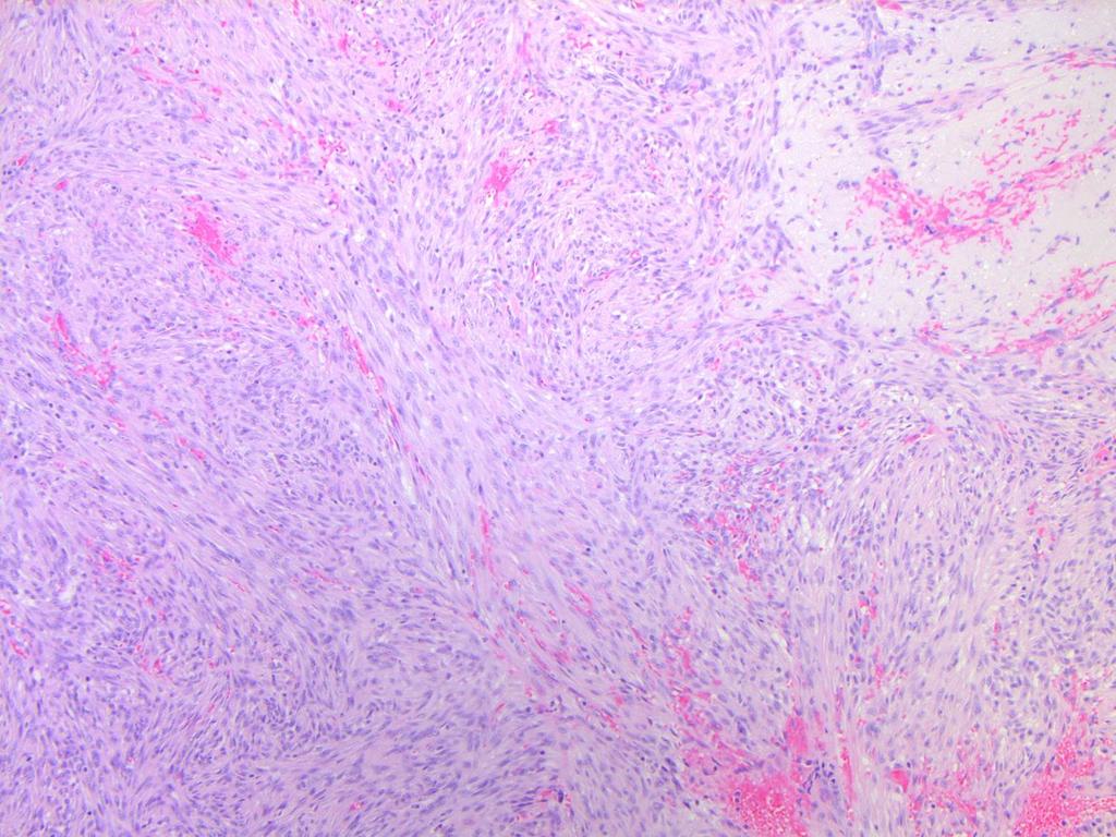
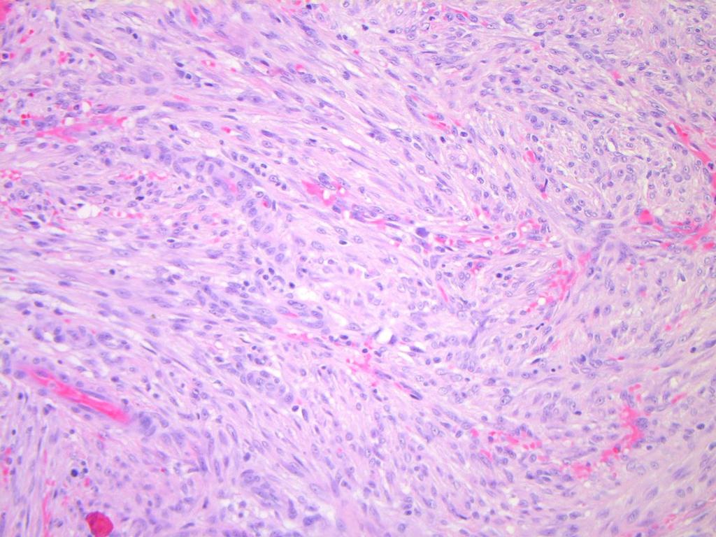
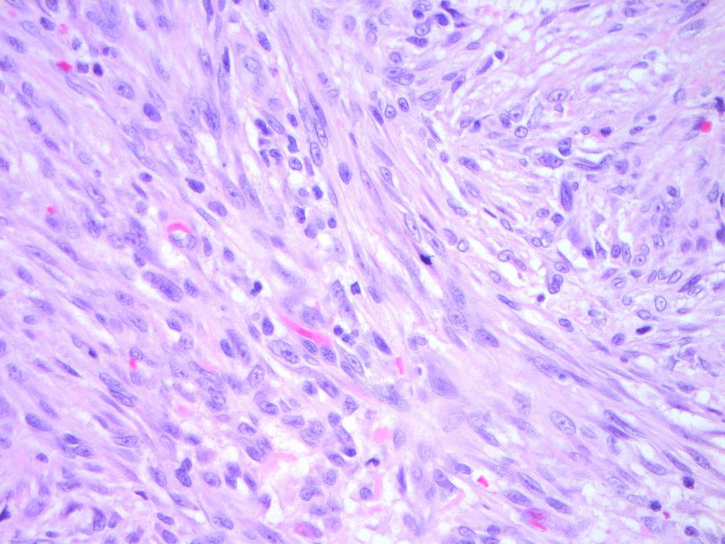
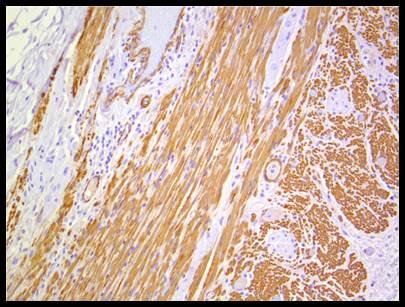
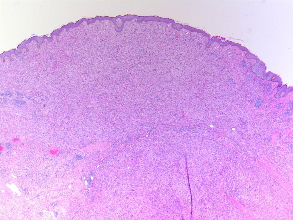
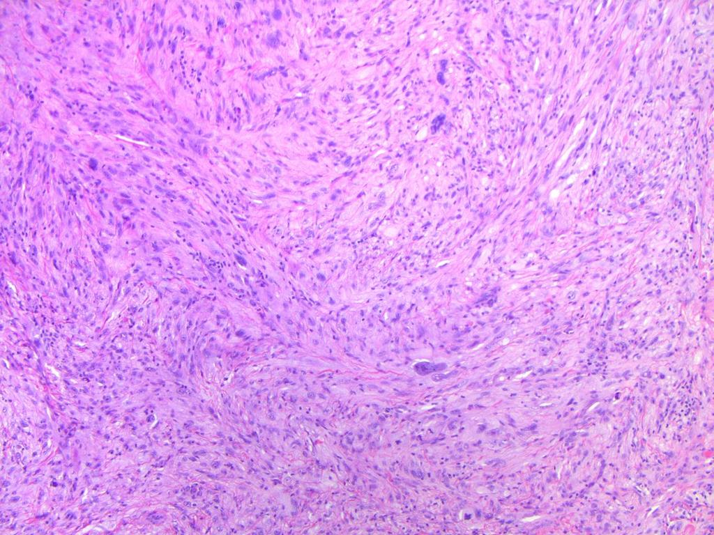
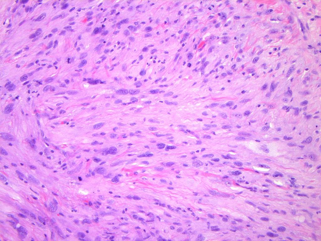
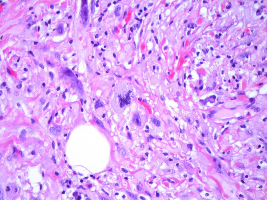
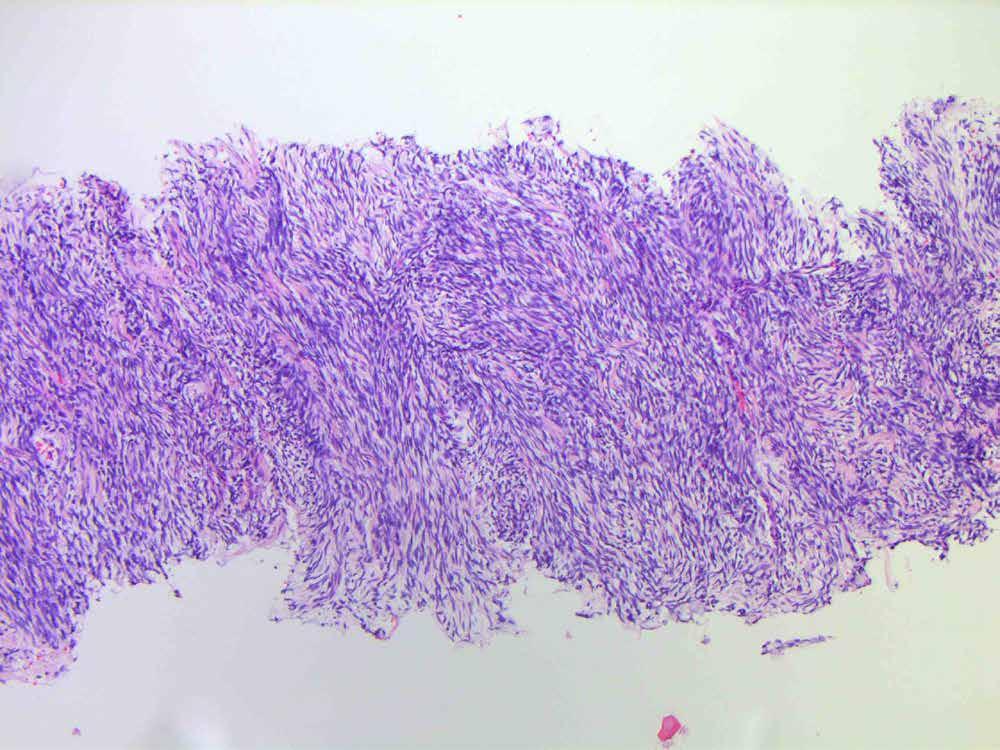
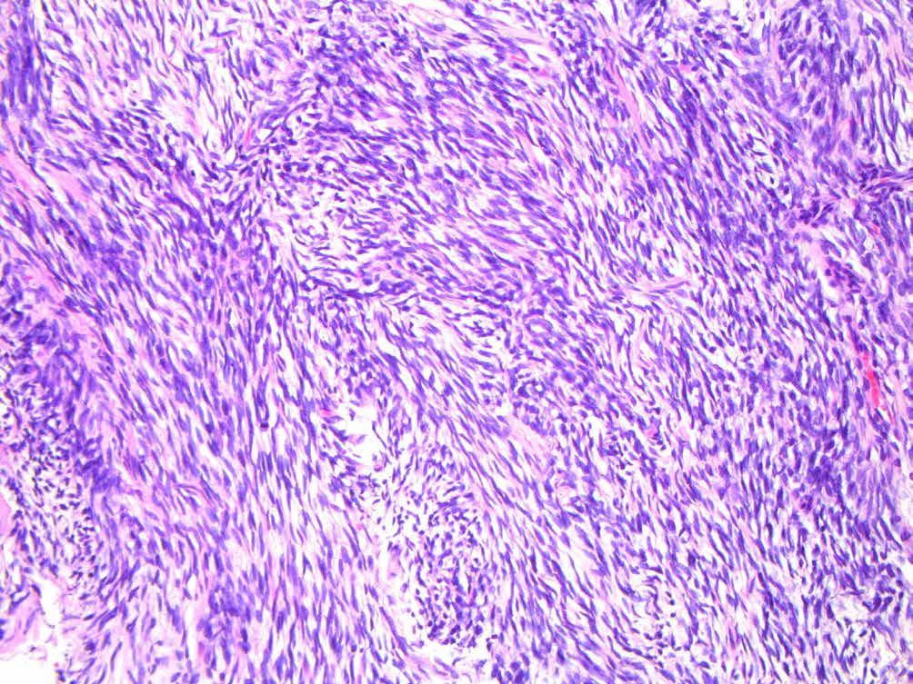
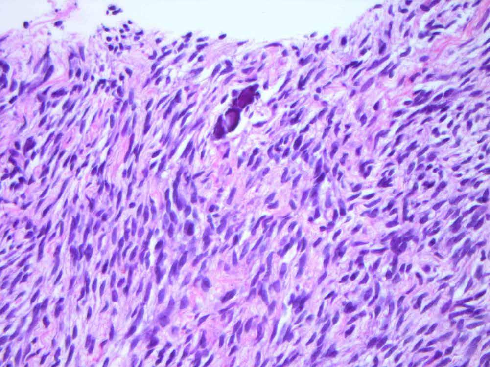
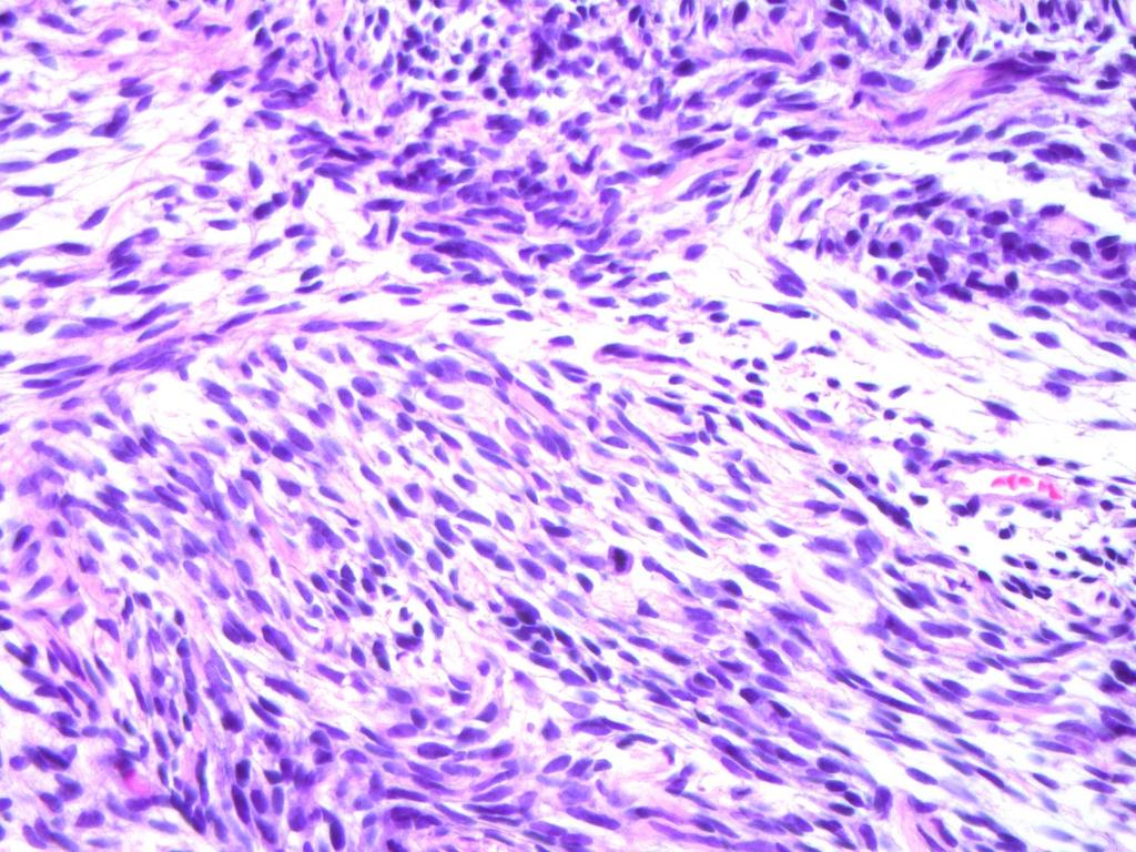
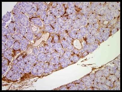
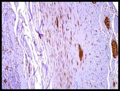
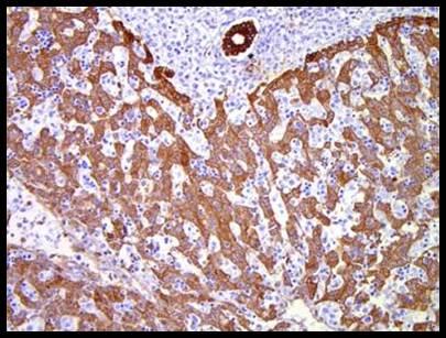
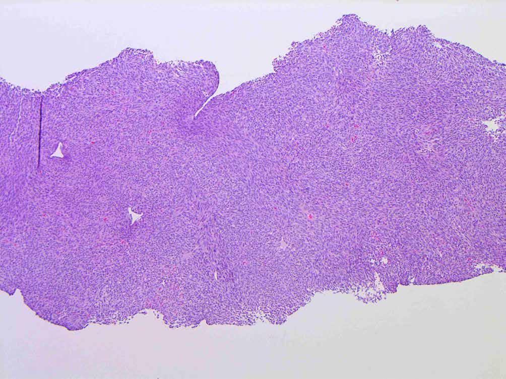
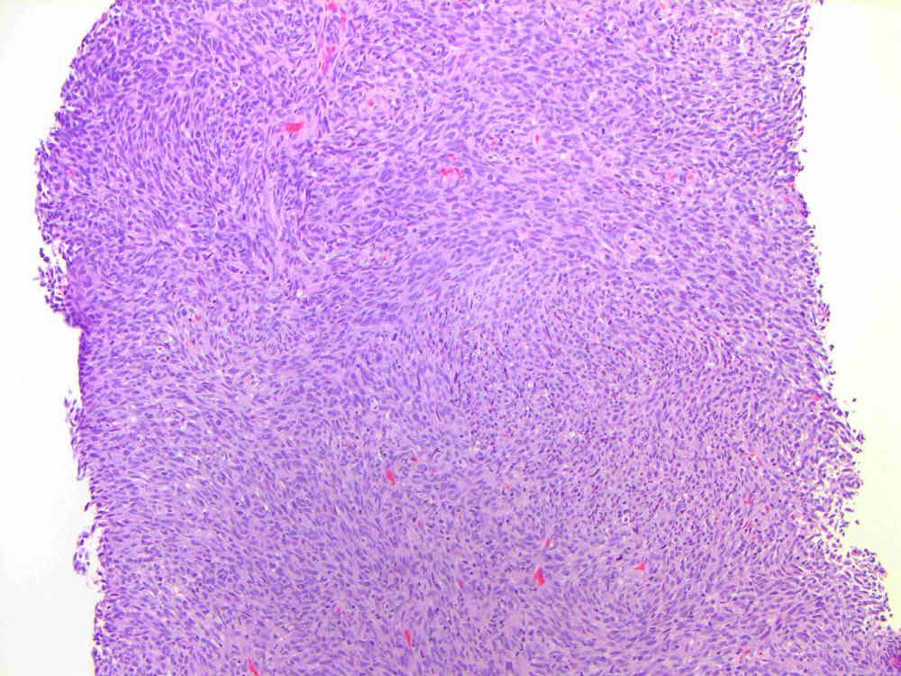
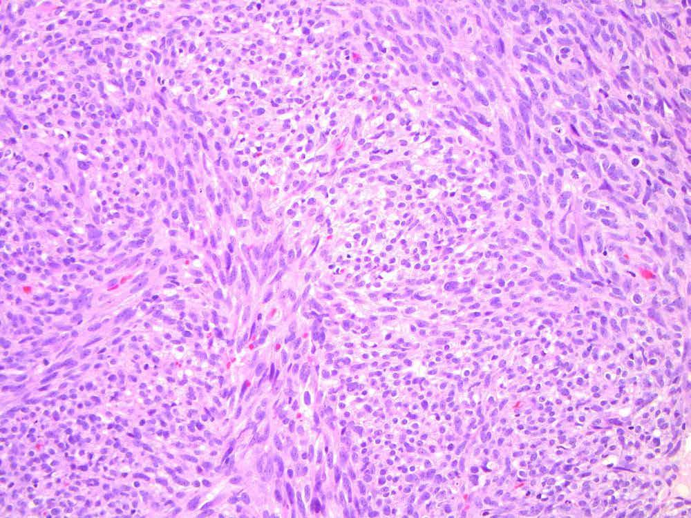
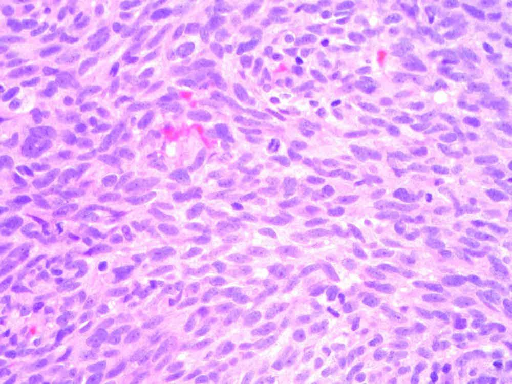
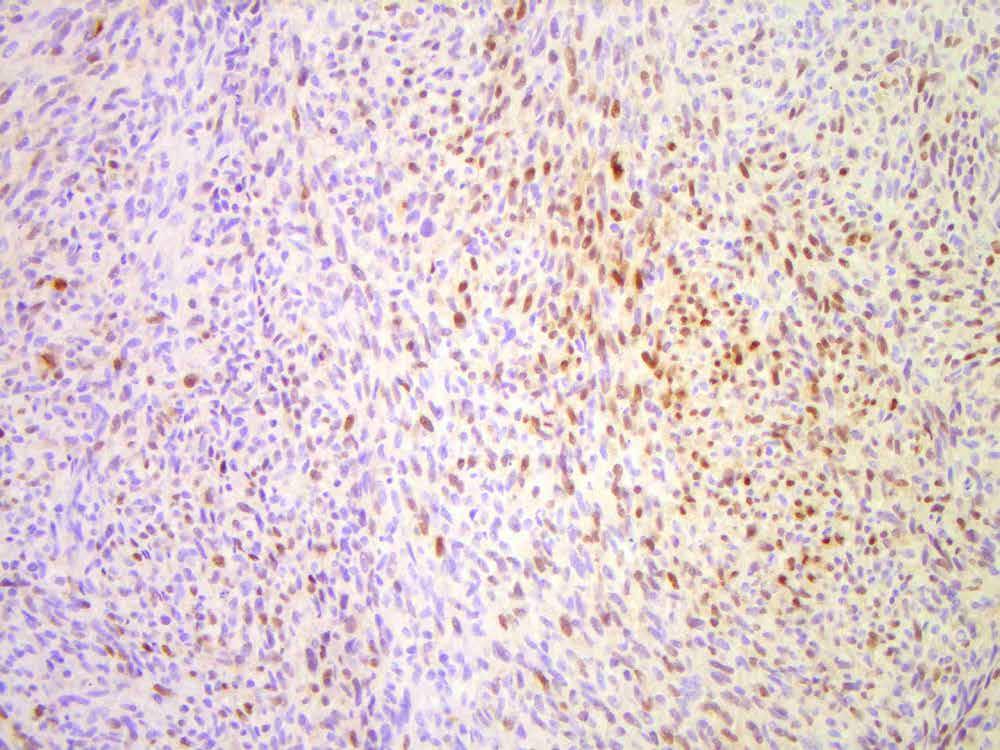
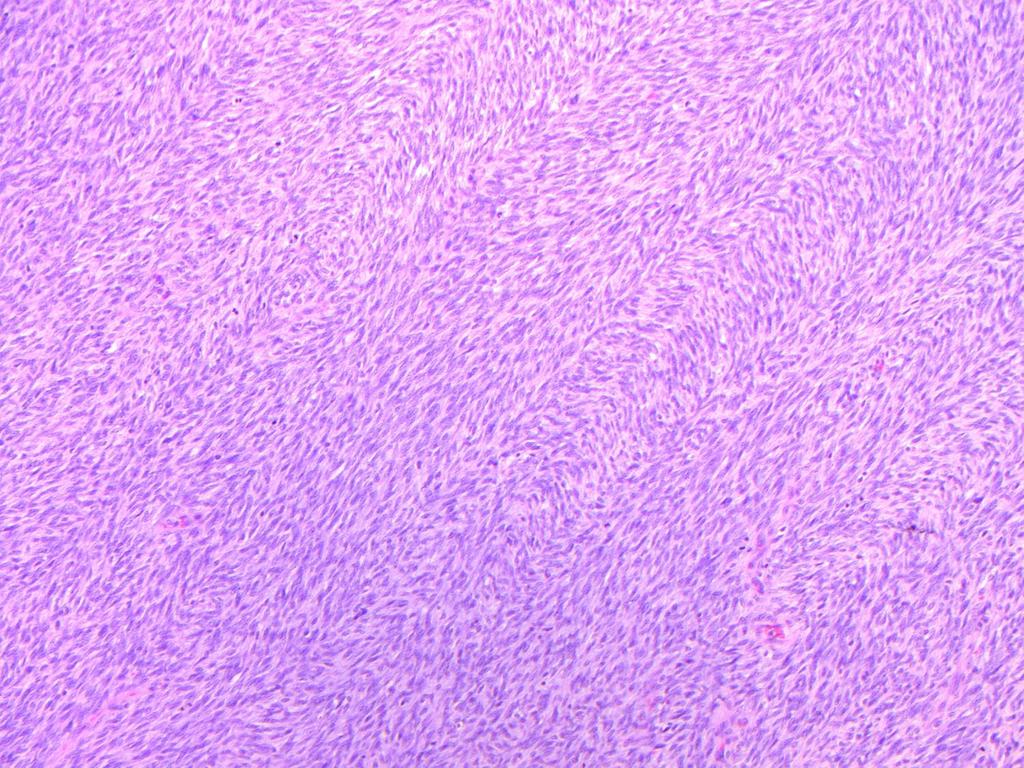
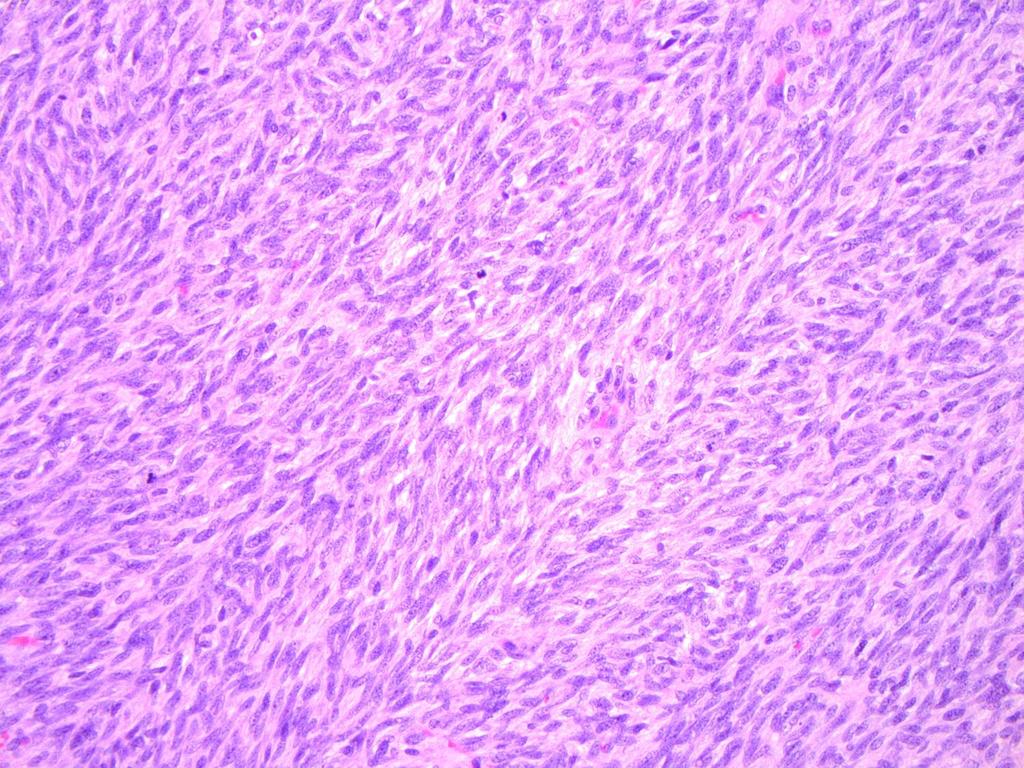
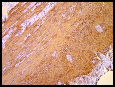
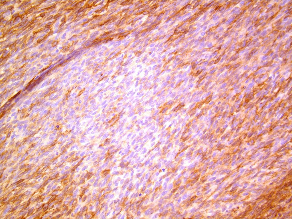
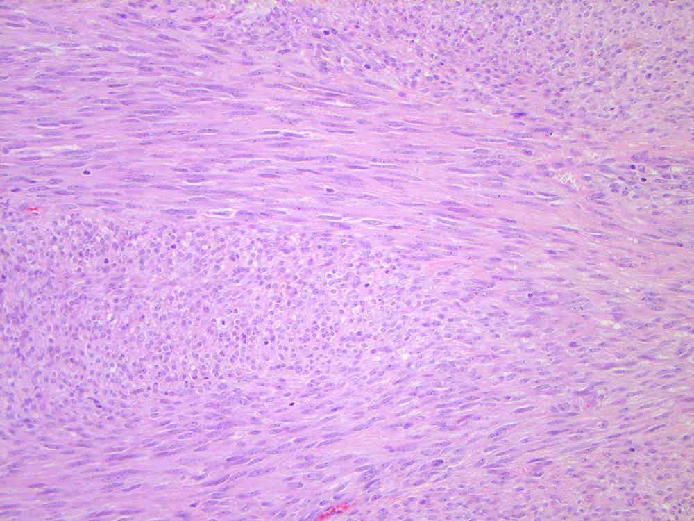
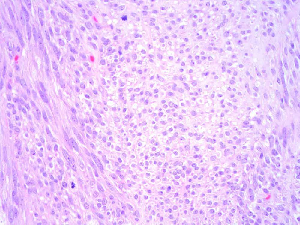
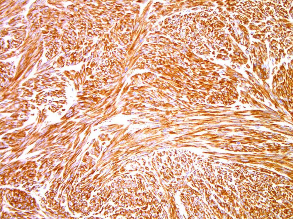
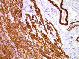
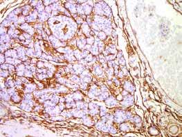
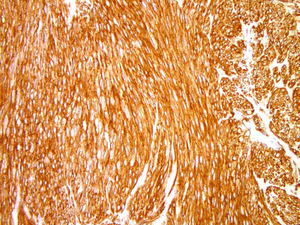
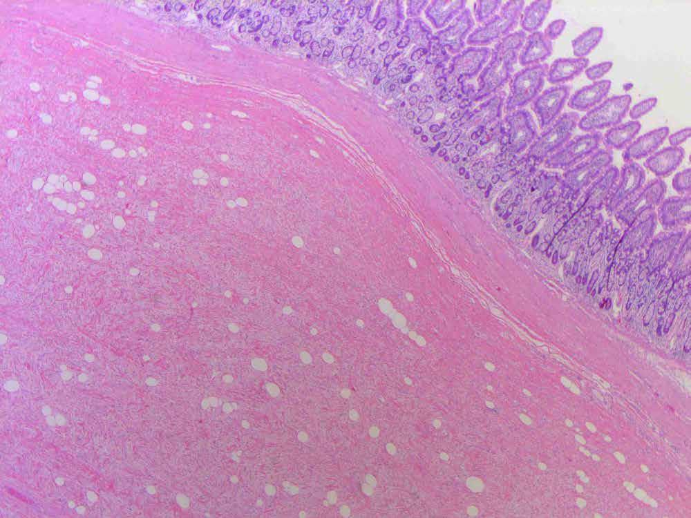
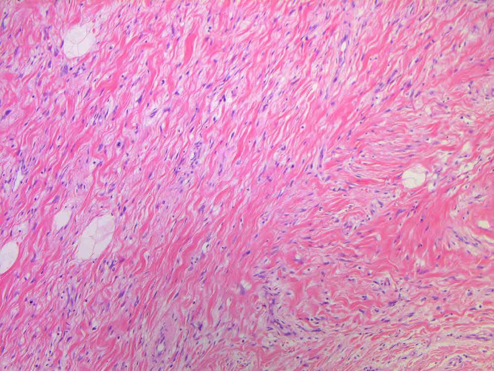
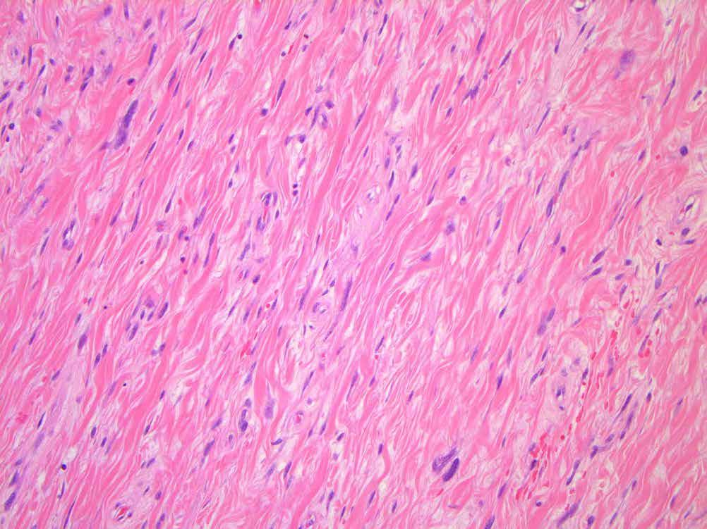
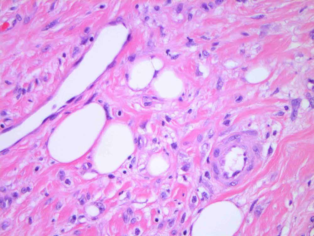
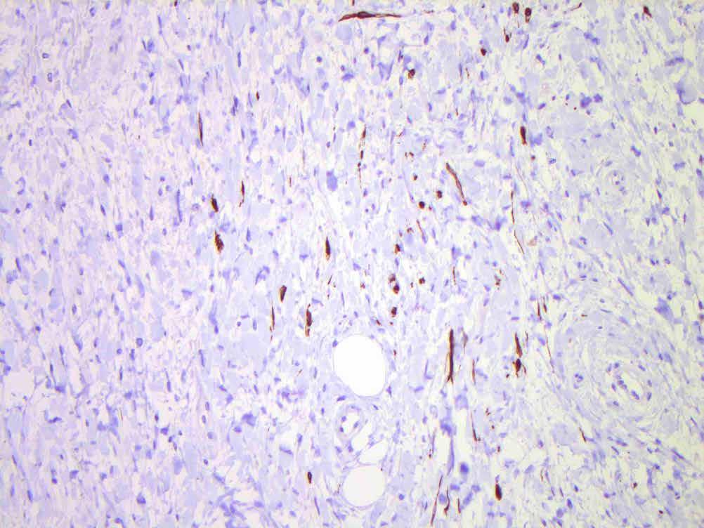
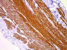
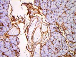
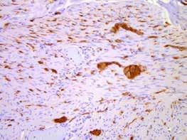
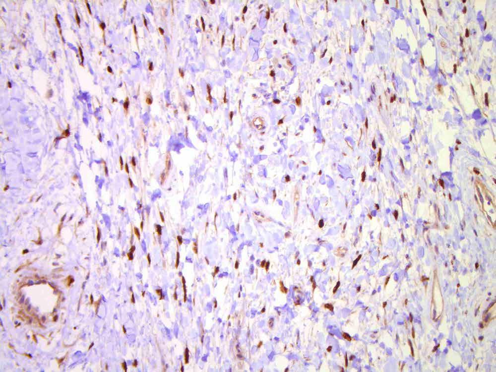
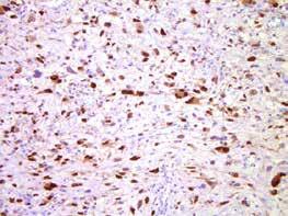
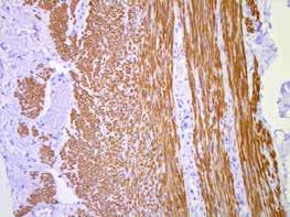
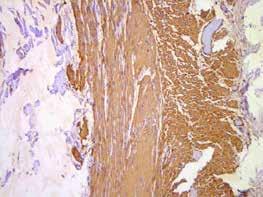
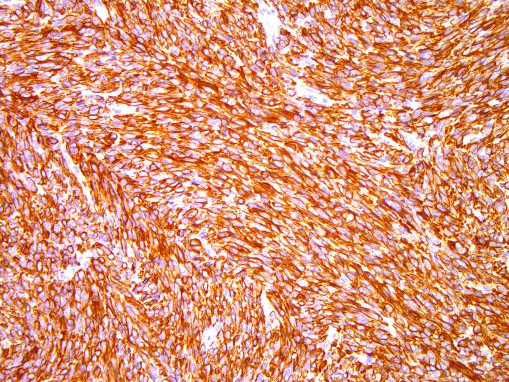
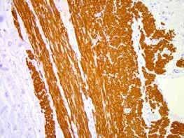
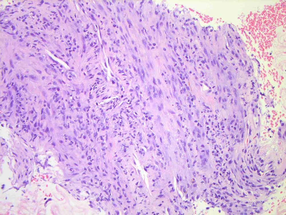
8 CASE 1 96 M. Excision right facial tumor. Spindle cell tumour, probable recurrent melanoma.
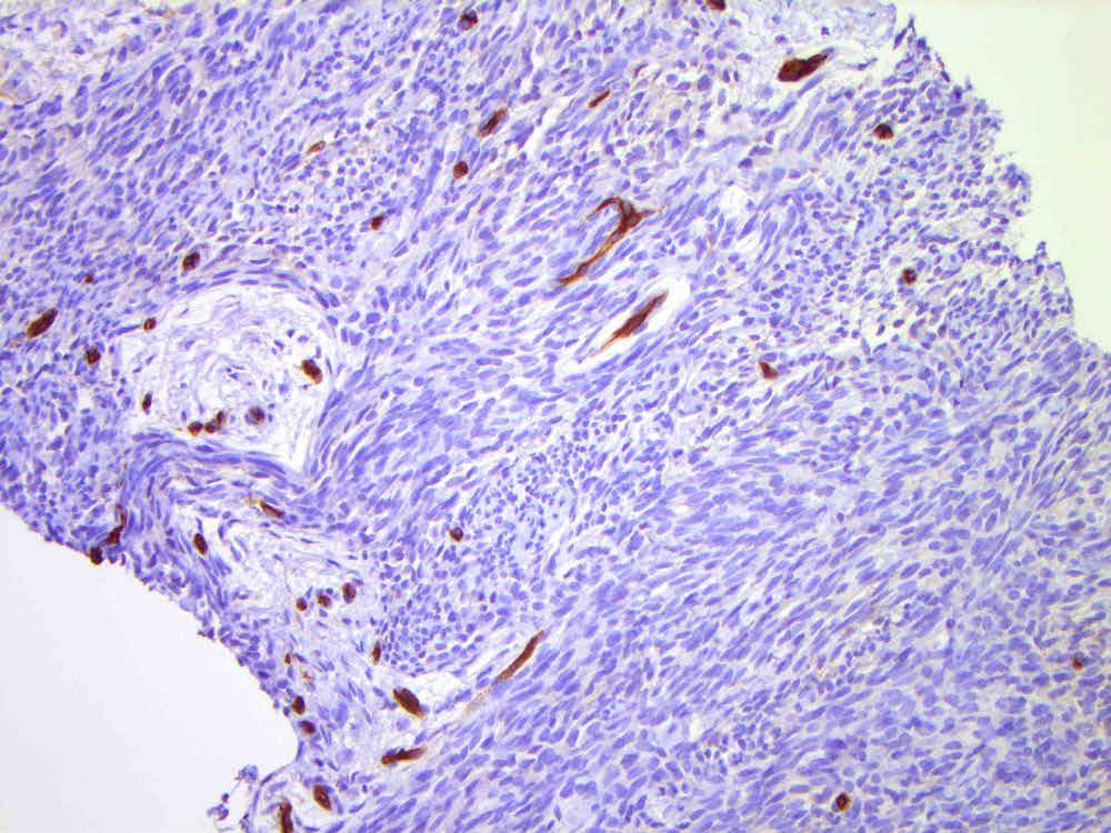
9
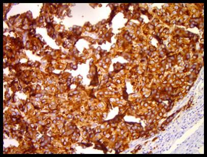
10
11
12
13
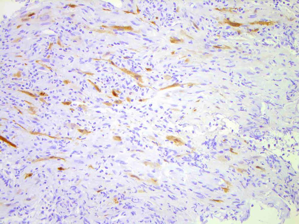
14
15 CASE 1 Differen al diagnosis a) Desmoplas c / spindle cell melanoma b) Squamous cell carcinoma, spindle cell variant c) Atypical fibroxanthoma, spindle cell variant d) Pleomorphic dermal sarcoma e) I do not know, and require a panel of immunohistochemistry


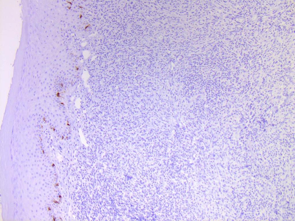
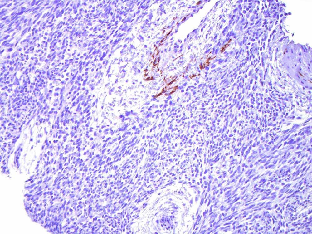
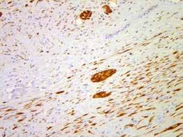
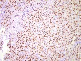
16 S100 (original lab)

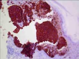
17 panmel (original lab)
")
")
18 Keratin (AE1/AE3) (original lab)


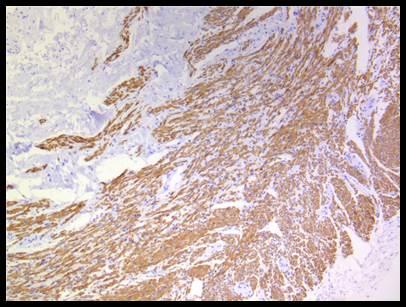
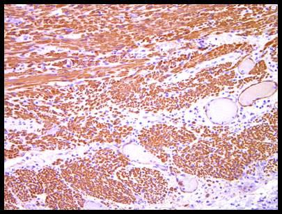
19 Smooth muscle actin (original lab)

20 Desmin (original lab)

21 CD34 (original lab)


22 Vimentin (original lab)
23 CASE 1 Differen al diagnosis a) Desmoplas c / spindle cell melanoma b) Squamous cell carcinoma, spindle cell variant c) Atypical fibroxanthoma, spindle cell variant d) Pleomorphic dermal sarcoma e) I STILL do not know, and require ANOTHER panel of immunohistochemistry

24 SOX10
")
25 Keratin (AE1/AE3)

26 p63
27 CASE 1 Differen al diagnosis a) Desmoplas c / spindle cell melanoma b) Squamous cell carcinoma, spindle cell variant c) Atypical fibroxanthoma, spindle cell variant d) Pleomorphic dermal sarcoma
28 CASE 1 Squamous cell carcinoma, spindle cell variant Pi alls: Loss of kera n(> 30%); NB: controls O en posi ve for vimen n, smooth muscle ac n Infiltra ng S100 posi ve dendri c cells Pearls: CK5/6 more sensi ve that AE1/AE3 p63 posi ve in majority (83-100%) p40 sensi vity, may be more specific Gleason BC, Calder KB, Cibull TL et al. J Cutan Pathol. 2009;36(5): Henderson SA, Torres-Cabala CA, Curry JL et al. Am J Surg Pathol. 2014;38(8): Ha Lan TT, Chen SJ, Arps DP et al. J Cutan Pathol. 2014;41(11):831-8.
29 CASE 1 Differen al diagnosis a) Desmoplas c / spindle cell melanoma b) Squamous cell carcinoma, spindle cell variant c) Atypical fibroxanthoma, spindle cell variant d) Pleomorphic dermal sarcoma
30

31

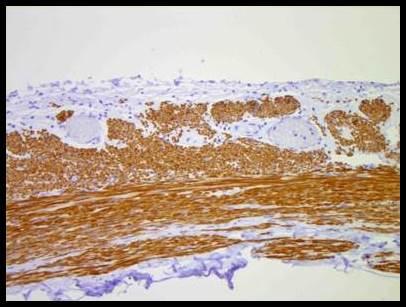
32 S100

33 SOX10
34 HMB45

35 MART-1
36 CASE 1 Desmoplas c / spindle cell melanoma Pi alls: Loss of HMB45, MART-1 MiTF not sensi ve / specific Gain of smooth muscle ac n (30%), CD56 (50%) Vimen n posi ve Pearls: O en accompanied by lymphoplasmacy c inflamma on S100 and SOX10 typically robust Presently limited role for molecular Shain AH, Garrido M, Bo on T et al. Nat Genet. 2015;47(10): Hu enbach Y, Prieto VG, Reed JA. J Cutan Pathol. 2002;29(9): Longacre TA, Egbert BM, Rouse RV. Am J Surg Pathol. 1996;20(12):
37 CASE 1 Differen al diagnosis a) Desmoplas c / spindle cell melanoma b) Squamous cell carcinoma, spindle cell variant c) Atypical fibroxanthoma, spindle cell variant d) Pleomorphic dermal sarcoma
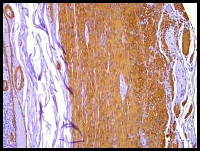
38
39
40
41

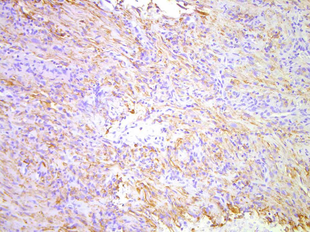
42 Smooth muscle actin
43 CASE 1 Atypical fibroxanthoma, spindle cell variant Pi alls: Diagnosis of exclusion Pearls: Be skep cal outside prototypic clinical context Should not see kera n Moot point? Controversy as to whether represents dedifferen ated squamous cell carcinoma Calonje E, Wadden C, Wilson-Jones E, Fletcher CD. Histopathology. 1993;22(3): Gru AA, Santa Cruz DJ. Semin Diagn Pathol. 2013;30(1):4-12.
44 CASE 1 Differen al diagnosis a) Desmoplas c / spindle cell melanoma b) Squamous cell carcinoma, spindle cell variant c) Atypical fibroxanthoma, spindle cell variant d) Pleomorphic dermal sarcoma
45
46
47
48

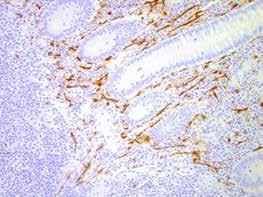
49 Smooth muscle actin
50 CASE 1 Pleomorphic dermal sarcoma Pi alls: Smooth muscle ac n o en posi ve (70%) CD31 o en posi ve (48%) Pearls: Deep, necrosis, LVI, perineural invasion may be present Miller K, Goodlad JR, Brenn T. Am J Surg Pathol. 2012;36(9):
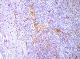
51 CASE 2 32 M. Right arm mass involving brachial plexus
52
53
54
55
56 CASE 2 Differen al diagnosis a) Malignant peripheral nerve sheath tumour b) Dermatofibrosarcoma, fibrosarcomatous variant c) Synovial sarcoma, monophasic d) Desmoid-type fibromatosis e) I do not know, and require a panel of immunohistochemistry
57 Desmin

58 Smooth muscle actin
59 CD34

60 S100
")
61 Keratin (AE1/AE3)
62 CASE 2 Differen al diagnosis a) Malignant peripheral nerve sheath tumour b) Dermatofibrosarcoma, fibrosarcomatous variant c) Synovial sarcoma, monophasic d) Desmoid-type fibromatosis e) I STILL do not know, and require ANOTHER panel of immunohistochemistry AND/OR molecular
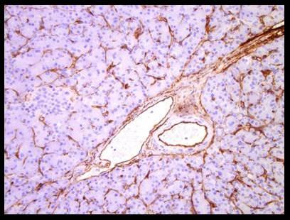
63 CASE 2 Differen al diagnosis a) Malignant peripheral nerve sheath tumour b) Dermatofibrosarcoma, fibrosarcomatous variant c) Synovial sarcoma, monophasic d) Desmoid-type fibromatosis
64 CASE 2 Synovial sarcoma, monophasic variant Pi alls: Kera n (AE1/AE3) may be lost (30%) or focal S100 (38%), smooth muscle ac n (21%), CD117 (11%) Pearls: Dystrophic calcifica on; wiry collagen Plump overlapping nuclei EMA more sensi ve than panck (near 100%) TLE1 is a robust biomarker May show decreased INI-1 Pelmus M, Guillou L, Hostein I et al. Am J Surg Pathol. 2002;26(11): Terry J, Saito T, Subramanian S et al. Am J Surg Pathol. 2007;31(2):240-6.
65 CASE 2 Differen al diagnosis a) Malignant peripheral nerve sheath tumour b) Dermatofibrosarcoma, fibrosarcomatous variant c) Synovial sarcoma, monophasic d) Desmoid-type fibromatosis
66
67
68
69

70 S100
71 SOX10
72 CASE 2 Malignant peripheral nerve sheath tumour Pi alls: S100 (30-67% of tumours), may be focal SOX10 (40-67% of tumours), may be weak/focal Heterologous smooth muscle, rhabdomyoblas c and epithelial differen a on Pearls: Clinical history occasionally points to NF1 Radiology / OR report may point to nerve origin Kang Y, Pekmezci M, Folpe AL et al. Mod Pathol. 2014;27(1): Nonaka D, Chiriboga L, Rubin BP. Am J Surg Pathol Sep;32(9): Karamchandani JR, Nielsen TO, van de Rijn M, West RB. Appl Immunohistochem Mol Morphol. 2012;20(5):
73 CASE 2 Differen al diagnosis a) Malignant peripheral nerve sheath tumour b) Dermatofibrosarcoma, fibrosarcomatous variant c) Synovial sarcoma, monophasic d) Desmoid-type fibromatosis
74

75 CD34
76
77

78 Smooth muscle actin
79 CD34
80 CASE 2 Dermatofibrosarcoma protuberans, fibrosarcomatous variant Pi alls: Loss of storiform pa ern Loss of CD34 (19%) Myoid / myofibroblas c differen a on Pearls: Transloca on immutable Abbo JJ, Oliveira AM, Nascimento AG. Am J Surg Pathol. 2006;30(4): O'Connell JX, Tro er MJ. Mod Pathol. 1996;9(3): Calonje E, Fletcher CD. J Cutan Pathol. 1996;23(1):30-6.
81 CASE 2 Differen al diagnosis a) Malignant peripheral nerve sheath tumour b) Dermatofibrosarcoma, fibrosarcomatous variant c) Synovial sarcoma, monophasic d) Desmoid-type fibromatosis
82
83
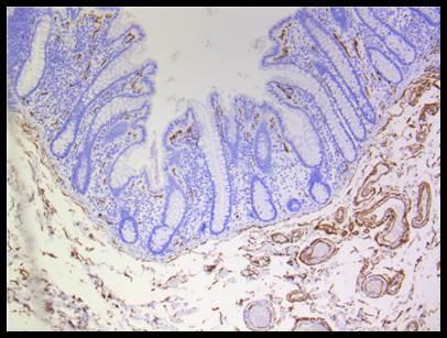
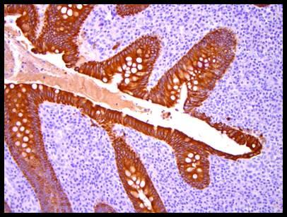
84
85

86 Desmin


87 Smooth muscle actin

88 Beta-catenin
89 CASE 2 Desmoid-type fibromatosis Pi alls: Entrapped skeletal muscle (myogenin and desmin may be misinterpreted as RMS or LGmyofibrosarcoma) Desmin may be focally posi ve Focal smooth muscle ac n O en nega ve for beta-catenin (~70% nuclear; lower in sporadic) Pearls: Early lesions may be myxoid +/- keloid-like collagen Late lesions have dense sclero c collagen Carlson JW, Fletcher CD. Histopathology. 2007;51(4): Ng TL, Gown AM, Barry T et al. Mod Pathol. 2005;18(1):68-74.
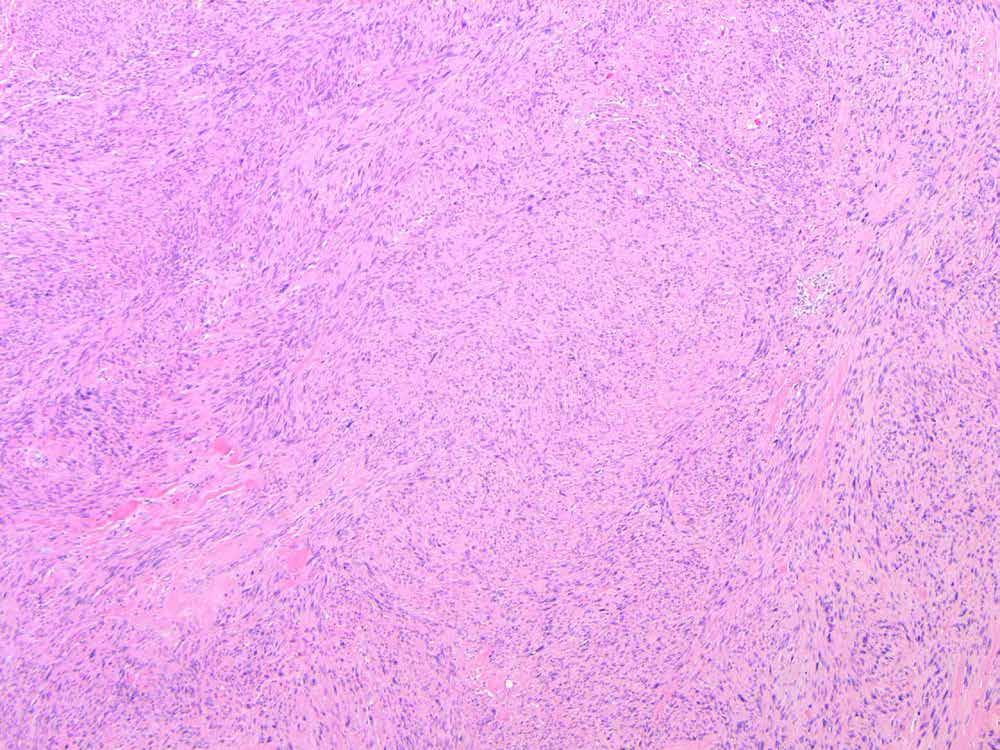
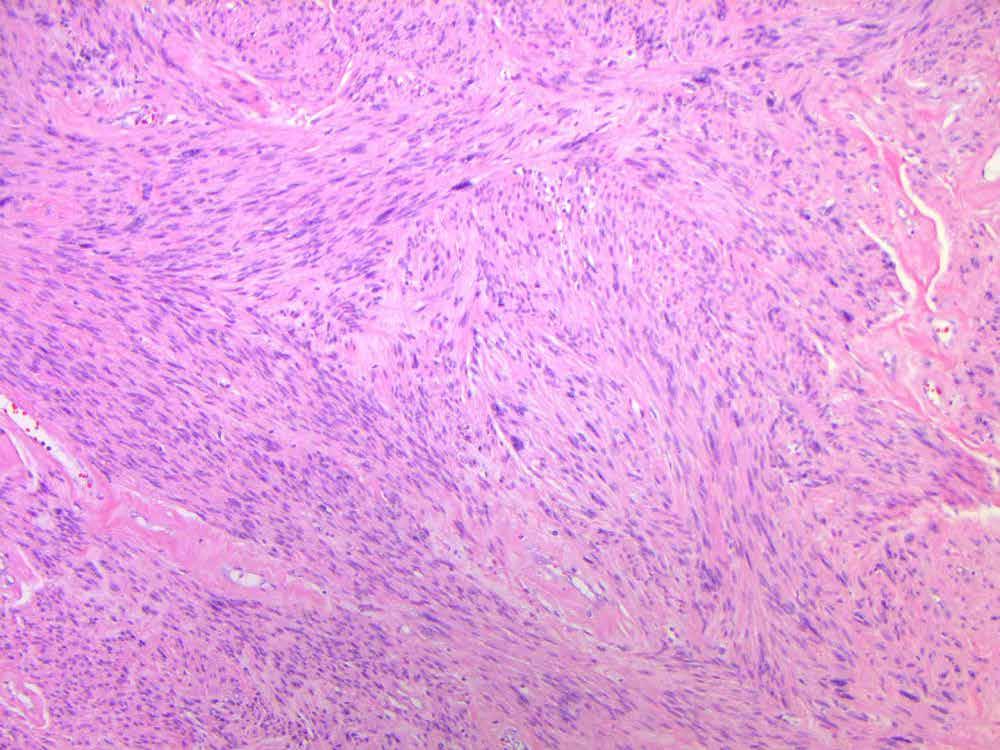
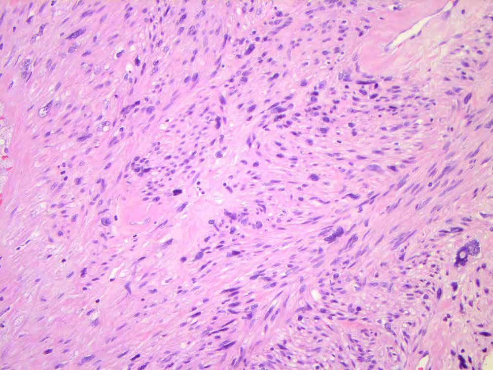
90 CASE 3 63 F. Two mesenteric masses
91
92
93
94 CASE 3 Differen al diagnosis a) Dedifferen ated liposarcoma b) Leiomyosarcoma c) Mesothelioma, sarcomatoid variant d) Gastrointes nal stromal cell tumour, spindle cell variant e) I do not know, and require a panel of immunohistochemistry
95 H-caldesmon

96 CD34
97 CD117

98 DOG1
99 CASE 3 Differen al diagnosis a) Dedifferen ated liposarcoma b) Leiomyosarcoma c) Mesothelioma, sarcomatoid variant d) Gastrointes nal stromal cell tumour, spindle cell variant e) I STILL do not know, and require ANOTHER panel of immunohistochemistry AND/OR molecular
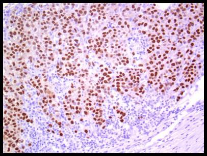
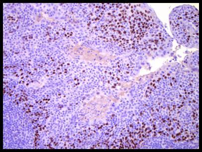
100 CASE 3 Differen al diagnosis a) Dedifferen ated liposarcoma b) Leiomyosarcoma c) Mesothelioma, sarcomatoid variant d) Gastrointes nal stromal cell tumour, spindle cell variant
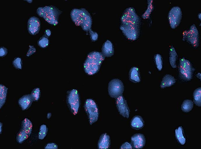
101 CASE 3 Gastrointes nal stromal tumour, spindle cell variant Pi alls: 5% may be nega ve for CD117 O en posi ve for smooth muscle markers, including H- caldesmon (80%) Pearls: Pleomorphism is rare CD34 frequently posi ve (par cularly in spindle cell variant) In contrast to smooth muscle tumours, smooth muscle ac n and desmin are rarely diffuse Molecular o en reassuring Mie nen M, Lasota J. Arch Pathol Lab Med. 2006;130(10):
102 CASE 3 Differen al diagnosis a) Dedifferen ated liposarcoma b) Leiomyosarcoma c) Mesothelioma, sarcomatoid variant d) Gastrointes nal stromal cell tumour, spindle cell variant
103
104
105
106
107 Desmin


108 Smooth muscle actin

109 CD34

110 S100
111 MDM2
112 in situ hybridization
113 CASE 3 Dedifferen ated liposarcoma Pi alls: Nonsensical immunoprofile (may express desmin, smooth muscle ac n, CD34, S100, others) Heterologous differen a on (may show: smooth muscle, rhabdomyoblas c, osteosarcomatous, others) Pearls: Search surrounding fat for atypia Recogni on of enlarged polygonal cells with dense eosinophilic cytoplasm MDM2 (IHC / FISH)
114 CASE 3 Differen al diagnosis a) Dedifferen ated liposarcoma b) Leiomyosarcoma c) Mesothelioma, sarcomatoid variant d) Gastrointes nal stromal cell tumour, spindle cell variant
115
116
117

118 Desmin

119 Smooth muscle actin
120 H-caldesmon
121 DOG1
122 CASE 3 Leiomyosarcoma Pi alls: Differing criteria (gynecologic vs so ssue [M vs F in retroperitoneum]) Dedifferen ated leiomyosarcoma may show loss of typical immunophenotype May express CD117 (11.5% uterine LMS), and DOG1 (27% uterine LMS) Pearls: Perinuclear cytoplasmic vacuoles o en present H-caldesmon generally helpful Hormone receptors occasionally relevant Chen E, O'Connell F, Fletcher CD. Histopathology. 2011;59(6): Sah SP, McCluggage WG. J Clin Pathol. 2013;66(1):40-3
123 CASE 3 Differen al diagnosis a) Dedifferen ated liposarcoma b) Leiomyosarcoma c) Mesothelioma, sarcomatoid variant d) Gastrointes nal stromal cell tumour, spindle cell variant
124
125
126

127
")
128 Keratin (AE1/AE3)
129 Calretinin
130 D2-40
131 CASE 3 Mesothelioma, sarcomatoid variant Pi alls: May have decreased expression of tradi onal markers: kera n (93%), D240 (79%), WT-1 (46%), calre nin (31%), Vimen n (91%) and SMA (60%) o en posi ve Pearls: Clinical History/ Radiology o en essen al to correct diagnosis Search for epithelioid cells / biphasic component Klebe S, Brownlee NA, Mahar A et al. Mod Pathol. 2010;23(3): Padge DM, Cathro HP, Wick MR, Mills SE. Am J Surg Pathol. 2008;32(1):123-7.
 Synthesize clinical informa on, diagnos c imaging and pathology findings; flesh-out immunohistochemical incongrui es Beware of morphologic mimics and employ redundancy in")
132 SUMMARY Technique Rigorous immunohistochemistry standards, including on-slide posi ve controls impera ve Interpreta on Tumours may aberrantly over / under express markers Review controls (including endogenous) Synthesize clinical informa on, diagnos c imaging and pathology findings; flesh-out immunohistochemical incongrui es Beware of morphologic mimics and employ redundancy in immunohistochemical panels Draw on literature for unexpected findings


133 Carlson JW, Fletcher CD. Histopathology. 2007;51(4): Ng TL, Gown AM, Barry T et al. Mod Pathol. 2005;18(1):68-74.
Diplomate of the American Board of Pathology in Anatomic and Clinical Pathology
 A 33-year-old male with a left lower leg mass. Contributed by Shaoxiong Chen, MD, PhD Assistant Professor Indiana University School of Medicine/ IU Health Partners Department of Pathology and Laboratory
Update on Cutaneous Mesenchymal Tumors. Thomas Brenn
 Update on Cutaneous Mesenchymal Tumors Thomas Brenn Cutaneous Mesenchymal Tumours Wide morphological and biological spectrum Myofibroblastic, smooth muscle, neural, vascular, apidocytic, undifferentiated;
Spindle Cell Lesions Of The Breast. Emad Rakha Professor of Breast Pathology and Consultant Pathologist
 Spindle Cell Lesions Of The Breast Emad Rakha Professor of Breast Pathology and Consultant Pathologist * SCLs comprise a wide spectrum of diseases, ranging from reactive processes to aggressive malignant
57th Annual HSCP Spring Symposium 4/16/2016
 An Unusual Malignant Spindle Cell Lesion to Involve the Breast Erinn Downs-Kelly, D.O. Associate Professor of Pathology University of Utah & ARUP Laboratories No disclosures Case 39 y/o female with no
GUT-C 11/30/2017. Debasmita Das, M.D. PGY-1 Danbury Hospital
 GUT-C 11/30/2017 Debasmita Das, M.D. PGY-1 Danbury Hospital CLINICAL SUMMARY 8/2017 59 year old female Presented to the ED with 1 month history of general malaise, fever and weight loss PMH: Significant
A 25 year old female with a palpable mass in the right lower quadrant of her abdomen
 May 2016 A 25 year old female with a palpable mass in the right lower quadrant of her abdomen Contributed by: Paul Ndekwe, MD, Resident Physician, Indiana University School of Department of Pathology and
3/27/2017. Disclosure of Relevant Financial Relationships
 Ophthalmic Pathology Evening Specialty Conference USCAP 2017 5 th March, 2017 Mukul K. Divatia, MD Assistant Professor Department of Pathology & Genomic Medicine Weill Cornell Medical College Houston Methodist
Desmoplastic Melanoma R/O BCC. Clinical Information. 74 y.o. man with lesion on left side of neck r/o BCC
 R/O BCC Sabine Kohler, M.D. Professor of Pathology and Dermatology Dermatopathology Service Stanford University School of Medicine Clinical Information 74 y.o. man with lesion on left side of neck r/o
Enterprise Interest Nothing to declare
 Enterprise Interest Nothing to declare Diagnoses one would not like to miss in soft tissue pathology early in your career Marta Sbaraglia, MD Department of Pathology Hospital of Treviso University of Padua
أملس عضلي غرن = Leiomyosarcoma. Leiomyosarcoma 1 / 5
 Leiomyosarcoma 1 / 5 EPIDEMIOLOGY Exact incidence is unknown, but older studies suggest that leiomyosarcomas comprise approximately 3 percent of soft-tissue sarcomas. Superficial leiomyosarcoma occurs
1/10/2018. Soft Tissue Tumors Showing Melanocytic Differentiation. Overview. Desmoplastic/ Spindle Cell Melanoma
 2016 MFMER slide-1 2016 MFMER slide-2 2016 MFMER slide-3 Soft Tissue Tumors Showing Melanocytic Differentiation Andrew L. Folpe, M.D. Professor of Laboratory Medicine and Pathology Mayo Clinic, Rochester,
Evening Specialty Conference Bone and Soft Tissue Pathology. Diagnostic pitfalls in bone and soft tissue pathology
 Evening Specialty Conference Bone and Soft Tissue Pathology. Case 1 Elizabeth G Demicco, MD, PhD Mount Sinai Hospital, New York Disclosure of Relevant Financial Relationships USCAP requires that all planners
Financial disclosures
 Mesenchymal Neoplasms with Melanocytic Differentiation By Konstantinos Linos MD, FCAP, FASDP Bone, Soft Tissue and Dermatopathology Assistant Professor of Pathology Dartmouth-Hitchcock Medical Center Geisel
I have nothing to disclose
 A 47 year old female with multiple lung nodules Disclosure of Relevant Financial Relationships Tamar Giorgadze, MD, PhD Professor of Pathology Medical College of Wisconsin Milwaukee, Wisconsin USCAP requires
Mesothelioma: diagnostic challenges from a pathological perspective. Naseema Vorajee August 2016
 Mesothelioma: diagnostic challenges from a pathological perspective Naseema Vorajee August 2016 Naseema.vorajee@nhls.ac.za Pleural diseases (whether neoplastic, reactive or infective) may have similar
Case of Pleomorphic Dermal Sarcoma of the Eyelid Treated with Micrographic Surgery and Secondary Intention Healing
 JI Kim, et al pissn 1013-9087ㆍeISSN 2005-3894 Ann Dermatol Vol. 28, No. 5, 2016 http://dx.doi.org/10.5021/ad.2016.28.5.632 CASE REPORT Case of Pleomorphic Dermal Sarcoma of the Eyelid Treated with Micrographic
Diagnostic Value of Immunohistochemistry in Soft Tissue Tumors
 Original Article DOI: 10.21276/APALM.1637 Diagnostic Value of Immunohistochemistry in Soft Tissue Tumors Sridevi. V*., Susruthan Muralitharan., and Thanka. J Dept of Pathology, SriMuthukumaran Medical
Disclosures. An update on ancillary techniques in the diagnosis of soft tissue tumors. Ancillary techniques. Introduction
 Disclosures An update on ancillary techniques in the diagnosis of soft tissue tumors. I have nothing to disclose. Andrew Horvai, MD, PhD Clinical Professor, Pathology Introduction Ancillary techniques
Immunohistochemistry and Bladder Tumours
 Immunohistochemistry and Bladder Tumours Dr. Andrew J. Evans MD PhD FRCPC Consultant in Genitourinary Pathology University Health Network Toronto, ON Objec ves Review markers of urothelial differen a on
The Relevance of Cytologic Atypia in Cutaneous Neural Tumors
 The Relevance of Cytologic Atypia in Cutaneous Neural Tumors Recent Findings - New Developments New Problems Zsolt B. Argenyi, M.D. Professor of Pathology & Dermatology Director of Dermatopathology Department
SOFT TISSUE TUMOR PATHOLOGY: AN UPDATE
 SOFT TISSUE TUMOR PATHOLOGY: AN UPDATE Jason L. Hornick, MD, PhD July 18, 2013 Department of Pathology Brigham and Women s Hospital Harvard Medical School Boston, MA, USA I have no disclosures. New Soft
Disclosure. Relevant Financial Relationship(s) None. Off Label Usage None MFMER slide-1
 None. Off Label Usage None MFMER slide-1") Disclosure Relevant Financial Relationship(s) None Off Label Usage None 2013 MFMER slide-1 Case Presentation A 43 year old male, with partial nephrectomy for a right kidney mass 2013 MFMER slide-2 2013
Disclosure Relevant Financial Relationship(s) None Off Label Usage None 2013 MFMER slide-1 Case Presentation A 43 year old male, with partial nephrectomy for a right kidney mass 2013 MFMER slide-2 2013
Keywords solitary fibrous tumor, dedifferentiation, dedifferentiated solitary fibrous tumor, STAT6, GRIA2, cytokeratin, rhabdomyosarcomatous
 758452IJSXXX10.1177/1066896918758452International Journal of Surgical PathologyCreytens et al research-article2018 Pitfalls in Pathology Multifocal Cytokeratin Expression in a Dedifferentiated Solitary
Biopsy Interpretation of Spindle cell proliferations of the Serosa
 Biopsy Interpretation of Spindle cell proliferations of the Serosa Richard Attanoos, Cardiff. U.K. Disclosure of Relevant Financial Relationships USCAP requires that all planners (Education Committee)
Soft Tissue Perineurioma
 The Korean Journal of Pathology 2009; 43: 266-70 DOI: 10.4132/KoreanJPathol.2009.43.3.266 Soft Tissue Perineurioma - A Case Report - Jun Mo Kim Joon Hyuk Choi Department of Pathology, Yeungnam University
Selected Pseudomalignant Soft Tissue Tumors of the Skin and Subcutis
 Selected Pseudomalignant Soft Tissue Tumors of the Skin and Subcutis Andrew L. Folpe, M.D. Professor of Laboratory Medicine and Pathology Mayo Clinic, Rochester, MN folpe.andrew@mayo.edu 2016 MFMER slide-1
Classification (1) Classification (3) Classification (2) Spindle cell lesions. Spindle cell lesions of bladder (Mills et al.
 Classification (3) Classification (2) Spindle cell lesions. Spindle cell lesions of bladder (Mills et al.") Non-epithelial tumours and nonepithelial tumour-like lesions of the bladder Dr Jonathan H Shanks The Christie NHS Foundation Trust, Manchester, UK Classification (1) Myofibroblastic proliferations and
Non-epithelial tumours and nonepithelial tumour-like lesions of the bladder Dr Jonathan H Shanks The Christie NHS Foundation Trust, Manchester, UK Classification (1) Myofibroblastic proliferations and
Cellular Neurothekeoma
 Cellular Neurothekeoma Scott W Binder, MD Pritzker Professor of Pathology & Dermatology Sr. Vice Chair Director, Pathology Clinical Services Chief, Dermatopathology Geffen/UCLA School of Medicine Clinical
From Morphology to Molecular Pathology: A Practical Approach for Cytopathologists Part 1-Cytomorphology. Songlin Zhang, MD, PhD LSUHSC-Shreveport
 From Morphology to Molecular Pathology: A Practical Approach for Cytopathologists Part 1-Cytomorphology Songlin Zhang, MD, PhD LSUHSC-Shreveport I have no Conflict of Interest. FNA on Lymphoproliferative
History A 89 year old gentleman presenting with a scalp/forehead nodule. Patient had squamous cell carcinoma 18 m at same site, excised. Outside diagn
 Case III History A 89 year old gentleman presenting with a scalp/forehead nodule. Patient had squamous cell carcinoma 18 m at same site, excised. Outside diagnoses: Squamous cell carcinoma. R/O: SCC, Melanoma,
59 yo male with past medical history of prostate carcinoma, presented with upper abdominal pain
 December 2016 59 yo male with past medical history of prostate carcinoma, presented with upper abdominal pain Contributed by: Divya Sharma, MD. Fellow, Gastrointestinal Pathology, Department of Pathology
ESS: Pathologic Insights
 GEIS XVI INTERNATIONAL SYMPOSIUM Seville 4th October 2018 ESS: Pathologic Insights Sílvia Bagué The Royal Marsden Hospital London (United Kingdom) I have no conflicts of interest Endometrial stromal sarcoma
Enterprise Interest No disclosures.
 Enterprise Interest No disclosures. Secondary Tumours in Uropathology Case 2 PRESENTED AT: EUROPEAN CONGRESS OF PATHOLOGY 18 #ECP2018 Slides are the property of the author. Permission required for reuse.
ACCME/Disclosures. Diagnosing Mesothelioma in Limited Tissue Samples. Papanicolaou Society of Cytopathology Companion Meeting March 12 th, 2016
 Diagnosing Mesothelioma in Limited Tissue Samples Papanicolaou Society of Cytopathology Companion Meeting March 12 th, 2016 Sanja Dacic, MD, PhD University of Pittsburgh ACCME/Disclosures GENERAL RULES
3/24/2017 DENDRITIC CELL NEOPLASMS: HISTOLOGY, IMMUNOHISTOCHEMISTRY, AND MOLECULAR GENETICS. Disclosure of Relevant Financial Relationships
 DENDRITIC CELL NEOPLASMS: HISTOLOGY, IMMUNOHISTOCHEMISTRY, AND MOLECULAR GENETICS Jason L. Hornick, M.D., Ph.D. Director of Surgical Pathology and Immunohistochemistry Brigham and Women s Hospital Professor
No financial or other disclosures
 Case 2014-5 Esther N. Bit-Ivan, DO Northwestern University Jason Wang, MD Jason Park, MD Korgun Koral, MD Children s Medical Center Charles Timmons, MD Veena Rajaram, MD No financial or other disclosures
Disclosures. An update on ancillary techniques in the diagnosis of soft tissue tumors. Ancillary techniques. Introduction
 Disclosures An update on ancillary techniques in the diagnosis of soft tissue tumors. I have nothing to disclose. Andrew Horvai, MD, PhD Clinical Professor, Pathology Introduction Ancillary techniques
IMMUNOHISTOCHEMISTRY IN THE DIAGNOSIS OF SOFT TISSUE TUMORS
 IMMUNOHISTOCHEMISTRY IN THE DIAGNOSIS OF SOFT TISSUE TUMORS Nicolas de Saint Aubain Somerhausen Institut Jules Bordet / Hôpital Erasme nicolas.desaintaubain@synet.be ForPath 2005 1 I. Ancillary techniques
Pathology of Sarcoma ELEANOR CHEN, MD, PHD, ASSISTANT PROFESSOR DEPARTMENT OF PATHOLOGY UNIVERSITY OF WASHINGTON
 Pathology of Sarcoma ELEANOR CHEN, MD, PHD, ASSISTANT PROFESSOR DEPARTMENT OF PATHOLOGY UNIVERSITY OF WASHINGTON Presentation outline Background and epidemiology of sarcomas Sarcoma classification Sarcoma
Special slide seminar
 Special slide seminar Tomáš Rozkoš The Fingerland Department of Pathology Charles University Medical Faculty and Faculty Hospital in Hradec Králové Czech Republic Case history, 33 years old resistance
Lung Tumor Cases: Common Problems and Helpful Hints
 Lung Tumor Cases: Common Problems and Helpful Hints Brandon T. Larsen, MD, PhD Senior Associate Consultant Department of Laboratory Medicine and Pathology Mayo Clinic Arizona Arizona Society of Pathologists
Klinisch belang van chromosomale translocatie detectie in sarcomen
 Translocations in sarcomas Klinisch belang van chromosomale translocatie detectie in sarcomen Judith V.M.G. Bovée, M.D., Ph.D. Department of Pathology Leiden University Medical Center RNA binding DNA binding
Financial disclosures
 Cutaneous Mesenchymal Neoplasms with EWSR1 Rearrangement By Konstantinos Linos MD, FCAP, FASDP Bone, Soft Tissue and Dermatopathology Assistant Professor of Pathology Dartmouth-Hitchc Geisel School of
Pathology Mystery and Surprise
 Pathology Mystery and Surprise Tim Smith, MD Director Anatomic Pathology Medical University of South Carolina Disclosures No conflicts to declare Some problem cases Kidney tumor Scalp tumor Bladder tumor
4/12/2018. MUSC Pathology Symposium Kiawah Island April 18, Jesse K. McKenney, MD
 MUSC Pathology Symposium Kiawah Island April 18, 2018 Jesse K. McKenney, MD 1 Urothelial Carcinoma with Alternative Differentiation 2 Urothelial Carcinoma with Alternative Differentiation Recognition as
Part 1. Slides 1-38, Rita Alaggio Soft tissue tumors Trondheim 14. mars 2013
 Part 1 Slides 1-38, Rita Alaggio Soft tissue tumors Trondheim 14. mars 2013 Pediatric Pathology Soft Tissue Tumors AN UPDATE Rita Alaggio Azienda Ospedaliera Università di Padova Soft Tissue Tumors More
IMMUNOPROFILES OF THE MAJOR RENAL NEOPLASMS (%staining)
") Stain Clear Cell Papillary IMMUNOPROFILES OF THE MAJOR RENAL NEOPLASMS (%staining) Chromophobe Collecting Duct Carcinom a Sarcomatoid Xp11 Translocat ion Dr Jon Oxley See also www.jonoxley.com Page 1 MTSCC
Stain Clear Cell Papillary IMMUNOPROFILES OF THE MAJOR RENAL NEOPLASMS (%staining) Chromophobe Collecting Duct Carcinom a Sarcomatoid Xp11 Translocat ion Dr Jon Oxley See also www.jonoxley.com Page 1 MTSCC
The role of immunohistochemistry in surgical pathology of the uterine corpus and cervix
 The role of immunohistochemistry in surgical pathology of the uterine corpus and cervix Prof. Ben Davidson, MD PhD Department of Pathology, Norwegian Radium Hospital, Oslo University Hospital, Oslo, Norway
Case Presentation. Maha Akkawi, MD, Fatima Obeidat, MD, Tariq Aladily, MD. Department of Pathology Jordan University Hospital Amman, Jordan
 Case Presentation Maha Akkawi, MD, Fatima Obeidat, MD, Tariq Aladily, MD Department of Pathology Jordan University Hospital Amman, Jordan The 25th Annual Congress of the ADIAP The 8/11/2013 1 5th International
LOOK-ALIKES IN SPINDLE AND EPITHELIOID TUMORS: Immunohistochemistry. Cytogenetics Flow cytometry Molecular diagnostics
 LOOK-ALIKES IN SPINDLE AND EPITHELIOID TUMORS: Ultrastructural value and pitfalls in diagnosis Guillermo A Herrera Department of Pathology and Translational Pathobiology Louisiana State University Health
CASE REPORT Benign epithelioid peripheral nerve sheath tumour resembling schwannoma
 Malaysian J Pathol 2014; 36(3) : 217 221 CASE REPORT Benign epithelioid peripheral nerve sheath tumour resembling schwannoma Thejasvi KRISHNAMURTHY MD and SR NIVEDITHA MD, DNB Department of Pathology,
3/24/2017. Disclosure of Relevant Financial Relationships. Mixed Epithelial Endometrial Carcinoma. ISGyP Endometrial Cancer Project
 Disclosure of Relevant Financial Relationships USCAP requires that all planners (Education Committee) in a position to influence or control the content of CME disclose any relevant financial relationship
Cutaneous Mesenchymal Neoplasms with EWSR1 Rearrangement
 Cutaneous Mesenchymal Neoplasms with EWSR1 Rearrangement By Konstantinos Linos MD, FCAP, FASDP Bone, Soft Tissue and Dermatopathology Assistant Professor of Pathology Dartmouth-Hitchcock Medical Center
Shintaro Sugita *, Hiroko Asanuma and Tadashi Hasegawa
 Sugita et al. Diagnostic Pathology (2016) 11:37 DOI 10.1186/s13000-016-0486-2 RESEARCH Open Access Diagnostic use of fluorescence in situ hybridization in expert review in a phase 2 study of trabectedin
Notice of Faculty Disclosure
 Mesenchymal Tumors of the Vulva: Old, New, Something(s) Different Napa Valley Conference Pathology Education Partners Inc May 15, 2018 Teri A. Longacre, M.D. longacre@stanford.edu Stanford University,
Diagnostic problems in uterine smooth muscle tumors
 Diagnostic problems in uterine smooth muscle tumors Marina Kos Ljudevit Jurak Clinical Department of Pathology, Clinical Hospital Center Sestre milosrdnice, Zagreb Institute of Pathology, University of
5/10. Pathology Soft tissue tumors. Farah Bhani. Mohammed Alorjani
 5/10 Pathology Soft tissue tumors Mohammed Alorjani Farah Bhani Slides are included in this sheet. Objectives: Soft tissue tumors 1. Describe soft tissue tumors. 2. Understand the classification of soft
Newer soft tissue entities
 Newer soft tissue entities Examples among fibroblastic tumors Turku, May 6, 2010 Markku Miettinen, M.D. AFIP, Washington, DC Fibroblastic neoplasms Solitary fibrous tumor /Hemangiopericytoma Low-grade
Tumores de células pequeñas, redondas y azules: diagnóstico diferencial cuando el tiempo apremia
 Tumores de células pequeñas, redondas y azules: diagnóstico diferencial cuando el tiempo apremia Sílvia Bagué Servei de Patologia Hospital de Sant Pau Barcelona Soft tissue sarcomas Heterogeneous group
Mesothelioma Pathobasic. Lukas Bubendorf Pathology
 Mesothelioma Pathobasic Lukas Bubendorf Pathology Mechanisms of Asbestos Carcinogenesis in Mesothelioma Asprin High-mobility group protein B1 master switch HMGB1 Initiation/ perpetuation of inflamm. response
Breast - ductal carcinoma CK7 ER PR GATA3 Mammaglobin (50-70%) GCDFP-15 (50-70%) E-cadherin HMWCK CK20 PAX2 ER/PR/HER2 on all newly diagnosed cases
 GCDFP-15 (50-70%) E-cadherin HMWCK CK20 PAX2 ER/PR/HER2 on all newly diagnosed cases") Adrenal cortical carcinoma Inhibin Synap Melan-A Calretinin Vimentin Chromogr CK7 CK20 Breast - ductal carcinoma CK7 ER PR GATA3 Mammaglobin (50-70%) GCDFP-15 (50-70%) E-cadherin HMWCK CK20 PAX2 ER/PR/HER2
Adrenal cortical carcinoma Inhibin Synap Melan-A Calretinin Vimentin Chromogr CK7 CK20 Breast - ductal carcinoma CK7 ER PR GATA3 Mammaglobin (50-70%) GCDFP-15 (50-70%) E-cadherin HMWCK CK20 PAX2 ER/PR/HER2
05/07/2018. Types of challenges. Challenging cases in uterine pathology. Case 1 ` 65 year old female Post menopausal bleeding Uterine Polyp
 Types of challenges Challenging cases in uterine pathology Nafisa Wilkinson Gynaecological Pathologist UCLH London Lack of complete history often, NO clinical history at all! Cases from other centres often
Case 27 Male 42. Painless, static, well-circumscribed, subcutaneous nodule right lower leg,?lipoma. The best diagnosis is:
 Case 27 Male 42. Painless, static, well-circumscribed, subcutaneous nodule right lower leg,?lipoma. The best diagnosis is: A. Angiosarcoma B. Haemangiopericytoma C.Myopericytoma D.Myofibroma E. Angioleiomyoma
Article begins on next page
 Leiomyoma of the Vulva Rutgers University has made this article freely available. Please share how this access benefits you. Your story matters. [https://rucore.libraries.rutgers.edu/rutgers-lib/50624/story/]
An Overview of Genital Stromal Tumors
 An Overview of Genital Stromal Tumors By Konstantinos Linos MD, FCAP, FASDP Bone, Soft Tissue and Dermatopathology Assistant Professor of Pathology Dartmouth-Hitchcock Medical Center Geisel School of Medicine
Differential diagnosis of HCC
 Hepatocellular Carcinoma Quest for an Ideal Immunohistochemical Panel Sanjay Kakar, MD UCSF Differential diagnosis of HCC Hepatocellular lesions Adenoma, FNH, HG dysplasia Adenocarcinoma CholangioCA, metastasis
Slide Seminar Spanish Society of Pathology
 Slide Seminar Spanish Society of Pathology John R. Goldblum, M.D. Chairman, Department of Anatomic Pathology Cleveland Clinic Professor of Pathology Cleveland Clinic Lerner College of Medicine 1921 Original
Inflammatory Myofibroblastic Tumor of the Bladder
 Inflammatory Myofibroblastic Tumor of the Bladder We illustrate a case of an inflammatory myofibroblastic tumor (IMT) involving the bladder in a woman with dysuria and review the literature and differential
Mayo Medical Laboratories
 Mayo Medical Laboratories Virtual Lectures 2014 MFMER 2016 MFMER slide-1 Virtual Lectures Planning Committee Disclosure Summary As a provider accredited by ACCME, College of Medicine, Mayo Clinic (Mayo
Affiliazione autori0. Riccardo Ricci Journal Club GIPAD, settore GIST Anatomia Patologica, Università Cattolica, Roma
 GIST Manifesting as a Retroperitoneal Tumor: Clinicopathologic Immunohistochemical, and Molecular Genetic Study of 112 Cases American Journal of Surgical Pathology, 2017, 41:577-585 Miettinen M*; Felisiak-Golabek
Pancreas. Atrophy, acinar cell. Pathogenesis: Diagnostic key features:
 Pancreas Atrophy, acinar cell Pathogenesis: Decrease in number and/or size of acinar cells may be due to spontaneous or experimentally induced degenerative changes, apoptosis, or a sequel of chronic inflammation.
Melanocytic Lesions: Use of Immunohistochemistry and Special Studies Napa Valley 2018
 Melanocytic Lesions: Use of Immunohistochemistry and Special Studies Napa Valley 2018 Victor G. Prieto, MD, PhD Professor Depts. of Pathology and Dermatology University of Texas - MD Anderson Cancer Center
Dermatopathology. Dr. Rafael Botella Estrada. Hospital La Fe de Valencia
 Dermatopathology Dr. Rafael Botella Estrada. Hospital La Fe de Valencia DERMATOPATHOLOGY CASE CHALLENGE: RECOGNIZING MIMIS AND MASQUERADERS Rosalie Elenitsas. University of Pennsylvania Spectrum Lupus
ACCME/Disclosures ALK FUSION-POSITIVE MESENCHYMAL TUMORS. Tumor types with ALK rearrangements. Anaplastic Lymphoma Kinase. Jason L.
 Companion Meeting of the International Society of Bone and Soft Tissue Pathology The Evolving Concept of Mesenchymal Tumors ALK FUSION-POSITIVE MESENCHYMAL TUMORS Jason L. Hornick, MD, PhD March 13, 2016
When Immunostains Can Get You in Trouble: Gynecologic Pathology p16: Panacea or Pandora s Box?
 When Immunostains Can Get You in Trouble: Gynecologic Pathology p16: Panacea or Pandora s Box? Teri A. Longacre, MD Stanford Medicine Stanford California pi6 in Gynecologic Pathology: Panacea or Pandora
USCAP 2011: ASDP companion meeting. Steven D. Billings 1
 USCAP 2011: ASDP companion meeting. Steven D. Billings (billins@ccf.org) 1 Spindle cell tumors that make you say, Oh $*&%! This lecture will focus on examples of cutaneous tumors that present particular
Enterprise Interest None
 Enterprise Interest None What are triple negative breast cancers? A synopsis of their histological patterns Ian Ellis Molecular Medical Sciences, University of Nottingham Department of Histopathology,
ACCME/Disclosures. Case 4 USCAP Pulmonary Panel Case 4 History
 Case 4 USCAP Pulmonary Panel 2016 Andrew Churg, MD Department of Pathology Vancouver General Hospital & University of British Columbia Vancouver, BC achurg@mail.ubc.ca. ACCME/Disclosures The USCAP requires
6/3/2010. Outline of Talk. Lobular Breast Cancer: Definition of lobular differentiation. Common Problems in Diagnosing LCIS in Core Biopsies
 Outline of Talk Lobular Breast Cancer: Common Problems in Diagnosing LCIS in Core Biopsies Definition of lobular differentiation Variants of LCIS that: carry risk for unsampled invasive cancer mimic DCIS
21/07/2017. Hobnail endothelial cells are not the same as epithelioid endothelial cells
 UPDATE IN CUTANEOUS VASCULAR S DERMATOPATHOLOGY SESSION BELFAST PATHOLOGY JUNE 21/2017 Dr E Calonje St John s Institute of Dermatology, London, United Kingdom THE FAMILY OF VASCULAR S WITH EPITHELIOID
Financial disclosures
 An update on immunohistochemical markers in mesenchymal neoplasms By Konstantinos Linos MD, FCAP, FASDP Assistant Professor of Pathology Geisel School of Medicine at Dartmouth Dartmouth-Hitchcock Medical
Accepted Manuscript. Low grade spindle cell sarcoma of the true vocal folds. Samuel R. Barber, Pavel Kopach, Elizabeth M. Genega, Thomas L.
 Accepted Manuscript Low grade spindle cell sarcoma of the true vocal folds Samuel R. Barber, Pavel Kopach, Elizabeth M. Genega, Thomas L. Carroll PII: S2468-5488(17)30121-2 DOI: 10.1016/j.xocr.2017.11.006
Rhabdomyomas and Rhabdomyosarcomas (RMS) David M. Parham, MD Chief of Anatomic Pathology
 David M. Parham, MD Chief of Anatomic Pathology") Rhabdomyomas and Rhabdomyosarcomas (RMS) David M. Parham, MD Chief of Anatomic Pathology Tumors of skeletal muscle: Rhabdomyomas and rhabdomyosarcomas Embryonal muscle 2 3 4 5 6 7 8 Rhabdomyoma Benign
Rhabdomyomas and Rhabdomyosarcomas (RMS) David M. Parham, MD Chief of Anatomic Pathology Tumors of skeletal muscle: Rhabdomyomas and rhabdomyosarcomas Embryonal muscle 2 3 4 5 6 7 8 Rhabdomyoma Benign
Carcinoma of Unknown Primary (CUP)
") Metasta c Carcinoma of Unknown Primary: Diagnos c Approach Using Immunohistochemistry James R. Conner, MD, PhD Mount Sinai Hospital Toronto, ON Carcinoma of Unknown Primary (CUP) 3-5% of all new malignant
Metasta c Carcinoma of Unknown Primary: Diagnos c Approach Using Immunohistochemistry James R. Conner, MD, PhD Mount Sinai Hospital Toronto, ON Carcinoma of Unknown Primary (CUP) 3-5% of all new malignant
DIAGNOSTIC SLIDE SEMINAR: PART 1 RENAL TUMOUR BIOPSY CASES
 DIAGNOSTIC SLIDE SEMINAR: PART 1 RENAL TUMOUR BIOPSY CASES Dr. Andrew J. Evans MD, PhD, FACP, FRCPC Consultant in Genitourinary Pathology University Health Network, Toronto, ON Case 1 43 year-old female,
Myxo-inflammatory Fibroblastic sarcoma
 AKA Myxo-inflammatory Fibroblastic sarcoma Acral Myxoinflammatory fibroblastic sarcomaam.j.surg.path1998; 22; 911-924 Inflammatory myxoid tumour of soft parts with bizarre giant cells [Pathol.Res.Pract.
Disclosure of Relevant Financial Relationships
 Neuropathology Evening Specialty Conference Disclosure of Relevant Financial Relationships The USCAP requires that anyone in a position to influence or control the content of all CME activities disclose
Case 1. Disclosure. Imaging. Clinical history 5/10/2016. USCAP 2016 Annual Meeting Evening Specialty Conference Bone and Soft tissue Pathology
 Disclosure Dr. Agaram has nothing to disclose Case 1 Narsi Agaram, MBBS USCAP 2016 Annual Meeting Evening Specialty Conference Bone and Soft tissue Pathology Clinical history Imaging 1998 A three month
Maligna Melanoma and Atypical Fibroxanthoma: An Unusual Collision Tumour G Türkcü 1, A Keleş 1, U Alabalık 1, D Uçmak 2, H Büyükbayram 1 ABSTRACT
 Maligna Melanoma and Atypical Fibroxanthoma: An Unusual Collision Tumour G Türkcü 1, A Keleş 1, U Alabalık 1, D Uçmak 2, H Büyükbayram 1 ABSTRACT Two different neoplasia in the same biopsy material called
Sarcomatoid (spindle cell) carcinoma of the cricopharynx presenting as dysphagia
 carcinoma of the cricopharynx presenting as dysphagia") Case Report Sarcomatoid (spindle cell) carcinoma of the cricopharynx presenting as dysphagia Jagtap Sunil V. 1, Shukla Dhirajkumar B. 2, Jagtap Swati S. 3, Havle Abhay D. 4 1 Associate Professor, Department
Case Report Sarcomatoid (spindle cell) carcinoma of the cricopharynx presenting as dysphagia Jagtap Sunil V. 1, Shukla Dhirajkumar B. 2, Jagtap Swati S. 3, Havle Abhay D. 4 1 Associate Professor, Department
Epithelial Columnar Breast Lesions: Histopathology and Molecular Markers
 29th Annual International Conference Advances in the Application of Monoclonal Antibodies in Clinical Oncology and Symposium on Cancer Stem Cells 25 th -27t h June, 2012, Mykonos, Greece Epithelial Columnar
Low-Grade Periductal Stromal of Breast: a case report
 Low-Grade Periductal Stromal of Breast: a case report Rosanna Nenna 1 Cosimo Damiano Inchingolo 1 Domenico Palmieri 2 Annalisa De Lucia 1 Giusy Elicio 1 Pina Miscioscia 1 ( 1 ) U.O.C. di Anatomia Patologica,
Hemangioendothelioma with a Prominent Lymphoid Infiltrate Mimicking Follicular Dendritic Cell Tumor: Report of a Case
 Journal of Cancer Research Updates, 2013, 2, 135-139 135 Hemangioendothelioma with a Prominent Lymphoid Infiltrate Mimicking Follicular Dendritic Cell Tumor: Report of a Case Justin Kerstetter 1, Mia Perez
Endometrial Stromal Tumors
 Endometrial Stromal Tumors WHO Categories: Endometrial Stromal Nodule (ESN) Endometrial Stromal Sarcoma, low grade (LGESS) Endometrial Stromal Sarcoma, high grade (HGESS) Undifferentiated Uterine Sarcoma
Mody. AIS vs. Invasive Adenocarcinoma of the Cervix
 Common Problems in Gynecologic Pathology Michael T. Deavers, M.D. Houston Methodist Hospital, Houston, Texas Common Problems in Gynecologic Pathology Adenocarcinoma in-situ (AIS) of the Cervix vs. Invasive
Division of Pathology
 Case 38 Adult woman with a 35mm right breast lump at the 10 o clock position. Excision performed. (Case contributed by Dr Mihir Gudi, KKH) Division of Pathology Merlion, One Fullerton Singapore Diagnosis
1 NORMAL HISTOLOGY AND METAPLASIAS
 1 NORMAL HISTOLOGY AND METAPLASIAS, MD Anatomy and Histology 1 Metaplasias 2 ANATOMY AND HISTOLOGY The female breast is composed of a branching duct system, which begins at the nipple with the major lactiferous
Disclosures. Giant Cell Rich Tumors of Bone. Outline. The osteoclast. Giant cell rich tumors 5/21/11
 Disclosures Giant Cell Rich Tumors of Bone Andrew Horvai, MD, PhD Associate Clinical Professor, Pathology This lecture discusses "off label" uses of a number of pharmaceutical agents. The speaker is describing
The Genetics of Myoepithelial Tumors: salivary glands, soft tissue and bone
 The Genetics of Myoepithelial Tumors: salivary glands, soft tissue and bone Cristina Antonescu, MD Memorial Sloan-Kettering Cancer Center, New York Nothing to declare Disclosure Spectrum of Myoepithelial
Loss of H3K27 tri-methylation is a diagnostic marker for malignant peripheral nerve sheath tumors and an indicator for an inferior survival
 Modern Pathology (2016), 1 9 2016 USCAP, Inc All rights reserved 0893-3952/16 $32.00 1 Loss of H3K27 tri-methylation is a diagnostic marker for malignant peripheral nerve sheath tumors and an indicator