FDOPA, C11Choline, C11 Methionine. Dr K.G.Kallur
|
|
|
- Julius Bates
- 6 years ago
- Views:
Transcription
1 FDOPA, C11Choline, C11 Methionine Dr K.G.Kallur


2 Why? 11C Methionine scan Had undergone resection Earlier. Post op recurrent hypercalcemia

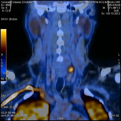
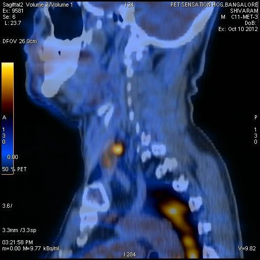
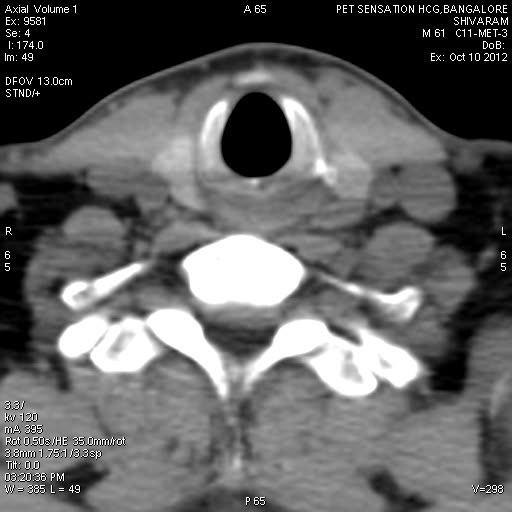
3 C11 Methionine Unable to see in Sestamibi scan
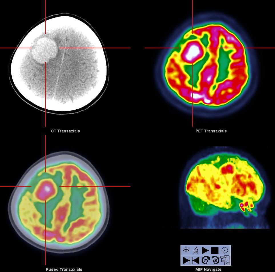
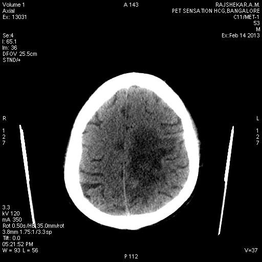
4 Brain Tumor After treatment questions to be answered Necrosis? Viable tumor? Recurrence? Biopsy site Prognostication
5 Lymphoma
6 Limitations of FDG PET Imaging of brain tumors with 18F-FDG was the first oncologic application of PET Diagnostic limitations Relatively lower sensitivity of lesion detection Variability in 18F-FDG uptake Increase in inflammatory lesions


7 Radiation necrosis - usually seen between 2 to 32 months after radiation therapy MRI: ring enhancing region in the left parietal-temporal area on post-gadolinium enhanced images FDG PET exam- no tracer uptake consistent with postsurgical and post radiation change. MR spectroscopy can also be performed to evaluate for radiation necrosis versus tumor recurrence. The cholinecreatinine ratio and choline-nacetyl aspartate ratio are significantly higher in recurrent tumor and in radiation necrosis. A combined diagnostic threshold of a choline-creatine ratio greater than 1.11 and a choline-n-acetyl aspartate ratio greater than 1.17 has a sensitivity of 89% and a specificity of 83% for the identification of tumor. An elevated lipid-lactate peak and a generalized decrease in other metabolite levels suggests radiation necrosis
8 Biologic process and tracers Energy metabolism [18F]-2-fluoro-2-deoxyglucose (FDG) 1-[11C]glucose [15O]O2, [15O]H2O, [15O]CO Amino acid transport and incorporation L-[methyl-11C]methionine (MET) L-[11C]tyrosine L-[18F]fluorotyrosine L-[11C]leucine Neuroreceptor DNA synthesis F Dopa 2-[11C]thymidine [18F]3Vdeoxy-3V-fluorothymidine (FLT) [18F]- or [11C]-2V-fluoro-5-methyl-1-b-D-arabinofuranosyluracil Membrane/lipid biosynthesis Hypoxia Tumor Neovascularity 1-[1-11C]acetate [11C]choline [18F]fluorocholine [18F]fluoromisonidazole RGD Peptides
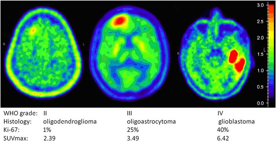
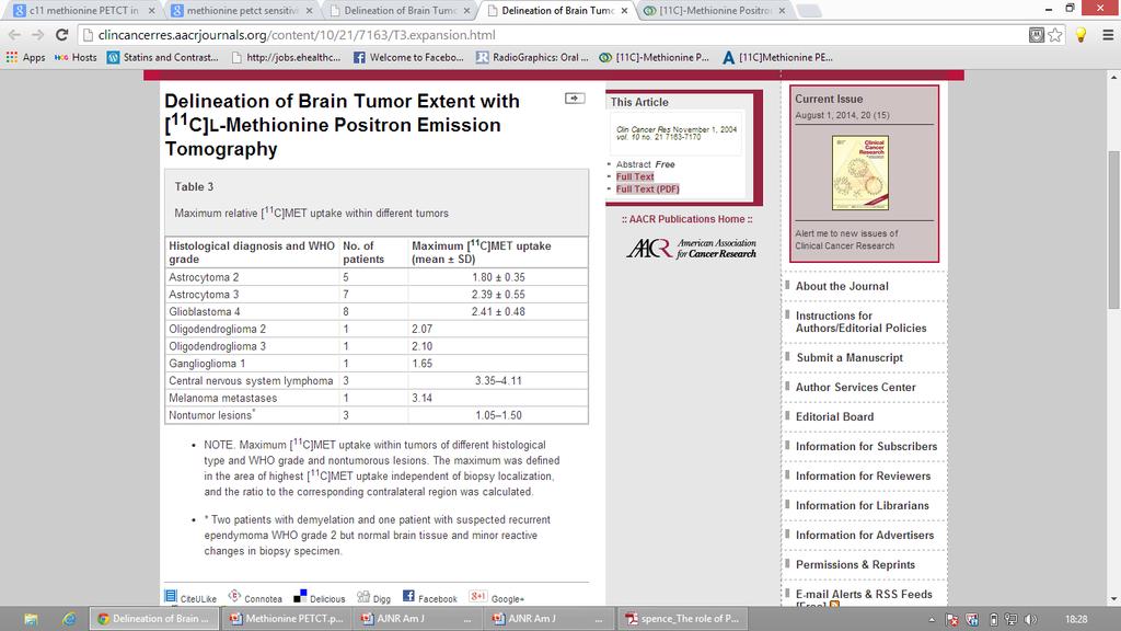
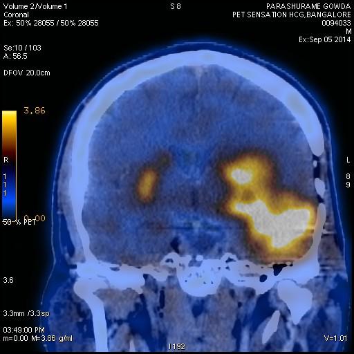
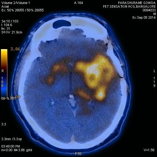
9 C11 Methionine PET 11CMET is considered to accumulate preferentially in tumor tissue, with a low level of accumulation in normal brain tissue, providing good contrast to highlight tumor uptake. The margins of tumors, as assessed by positron emission tomography (PET) with amino acid tracers like methyl-[ 11C]Lmethionine ([11C]MET), are frequently wider than those assessed by MRI or CT. This phenomenon is even more pronounced in low-grade tumors and in diffuse gliomatosis because of their frequent lack of contrast enhancement in MRI.
10 C]MET is a sensitive tracer in tumor detection, and it differentiates benign from malignant lesions with high sensitivity and specificity with relatively low background activity in normal brain tissue. 11 [11C]MET uptake correlates to cell proliferation in cell culture, Ki-67 expression, proliferating cell nuclear antigen expression and microvessel density, indicating its role as a marker for active tumor proliferation and angiogenesis. Patients with tumors that showed stable of reduced MET uptake after radiotherapy lived longer MET PET is sensitive in detecting changes in tumor volume over time in low grade gliomas.
11
12

13 11C-MET PET can differentiate tumor recurrence from radiation necrosis. The L/N mean of 11CMET PET may be the most valuable index for this differential diagnosis for both metastatic brain tumors and gliomas.
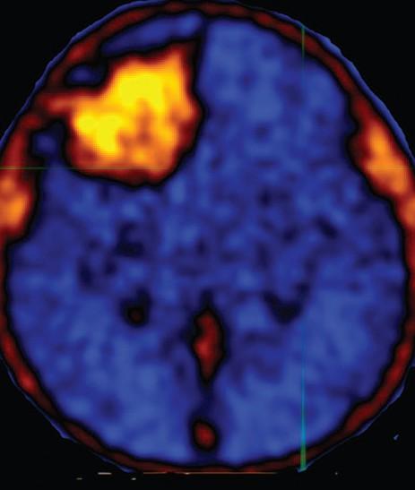
14 Low grade oligodendroglioma FDG PET

15 Right frontal high grade glioma

16 High grade glioma FDG PET

17 FDG & C11 Met Glioma


18 High grade tumor MET & FDG PET


19 FDG & C11 Met PET in low grade Glioma FDG C11 MET


20 FDG & C11 Met PET in low grade Glioma


21 PETMRI Met PET in Insular Glioma MRI C11 Met PETMRI

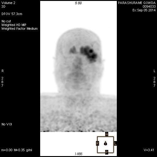
22 Recurrent right frontal glioma FDG PET, C11 MET PET

23 Bicentric gliosarcoma MET PET
24 FDG & C11 MET glioma You can plan biopsy site Based on metabolic info
25 F18 DOPA Uptake reflects the activity of the decarboxylating enzyme and the storage capacity of dopamine. 18F-DOPA is an analog of L-DOPA, this positron emitting compound is clinically used to trace the dopaminergic pathway and to evaluate striatal dopaminergic presynaptic function Useful for monitoring disease progression
26 F FOPA PET SCAN The large neutral amino acid transport system is highly expressed not only in the nigrostriatal region as a physiologic feature of normal brain but also in brain tumors as a pathologic feature, causing an increased uptake of amino acids, compared with that in normal brain. Longer half-life compared to C11 methionine.
.")
27 FDOPA PET FDOPA PET helps in differentiating recurrent or progressive brain metastases from late or delayed radiation injury. The mean time to progression was 4.6 times longer for lesions with negative 18F-FDOPA PET results than for lesions with positive 18FFDOPA PET results (76.5 vs mo; P, 0.001).18F-FDOPA PET findings tended to predict overall survival. Among the 83 lesions included in the study, 32 (39%) were classified as recurrences and 51 (61%) were determined to be radiation injury. FDOPAPET was able to differentiate between recurrence and radiation injury with a sensitivity of 81%, specificity of 84%, and accuracy of 83%. The results of the study "demonstrated that F-18 FDOPA PET could distinguish between Recurrent or Progressive BM and Late or Delayed Radiation Injury with a high diagnostic accuracy in a population of patients in whom RPBM was suggested by MR imaging," the authors concluded.

28 Results: Both high-grade and low-grade tumors were well visualized with 18F-FDOPA. The sensitivity for identifying tumors was substantially higher with 18F-FDOPA PET than with 18F-FDG PET at comparable specificities, as determined by simple visual inspection, especially for the assessment of low-grade tumors The high diagnostic accuracy of 18F-FDOPA PET at these thresholds was confirmed with the additional 51 patients (a total of 81 patients: sensitivity,98%; specificity, 86%; positive predictive value, 95%;negative predictive value, 95%). No significant difference in tumor uptake on 18F-FDOPA scans was seen between low-grade and high-grade tumors (P ) or between contrast-enhancing and non-enhancing tumors (P50.97). Radiation necrosis was generally distinguishable from tumors on 18F-FDOPA scans (P, ). Conclusion: 18F-FDOPA PET was more accurate than18f-fdg PET for imaging of lowgrade tumors and evaluating recurrent tumors. 18F-FDOPA PET may prove especially useful for imaging of recurrent low-grade tumors and for distinguishing tumor recurrence from radiation necrosis.

29 Conclusion: 18F-FDOPA uptake is significantly higher in high-grade than in low-grade tumors in newly diagnosed but not recurrent tumors that had been treated previously. A significant correlation between 18F-FDOPA uptake and tumor proliferation in newly diagnosed tumors was observed, whereas this correlation was not identified for recurrent tumors. Thus, 18F-FDOPA PET might serve as a noninvasive marker of tumor grading and might provide a useful surrogate of tumor proliferative activity in newly diagnosed gliomas. J Nucl Med 2010; 51:

30 Conclusion: 18F-DOPA PET/CT changed the intended management of 41% of patients with brain tumors, and intended management changes were implemented in 75% of these. These changes suggest a potentially important clinical role of imaging amino acid transport in the management of brain tumor patients. J Nucl Med 2012; 53:


31 If only this image is considered for radiotherapy planning then one will treat this region GEOGRAPHICAL MISS CAN BE AVOIDED For want of C11 MET One will miss this


32 FDOPA PET SCAN
33 Demonstration of tumor neovascularity


34 DOPA PSMA MRI FINDINGS- elsewhere Porencephalic cavity in the right frontal lobe involving the cortex and subcortical white matter with significant adjoining gliosis in the deep white matter of right frontal lobe. Altered signal intensity in the periventricular region of frontal horn of left lateral ventricle with enhancement. Focal nodular hyperintense lesion in the subcortical white matter of left frontal lobe with post contrast enhancement. Other MRI findings as described above. Findings are likely secondary to post therapy changes, suggested close follow up.


35 LOW GRADE GLIOMA BRAIN STEM CT MRI PSMAPET FDOPA PET Comments: Findings are consistent with brain stem glioma. F-DOPA uptake in the periphery of the cystic lesion in the brain stem suggestive of residual disease. PSMA uptake described above in the periphery of this lesion is due to PSMA expression in the vasculature surrounding the tumor.

36 Glioblastoma multiforme C11 Choline PET
37 C11 Choline PET Membrane synthesis. Why we stopped it? HCG

38 Intense accumulation in Necrosis.


39 (Dotanoc) Post therapy Response Meningioma Treated by Lu177 Dotatate (PET CT)
40 PETMRI

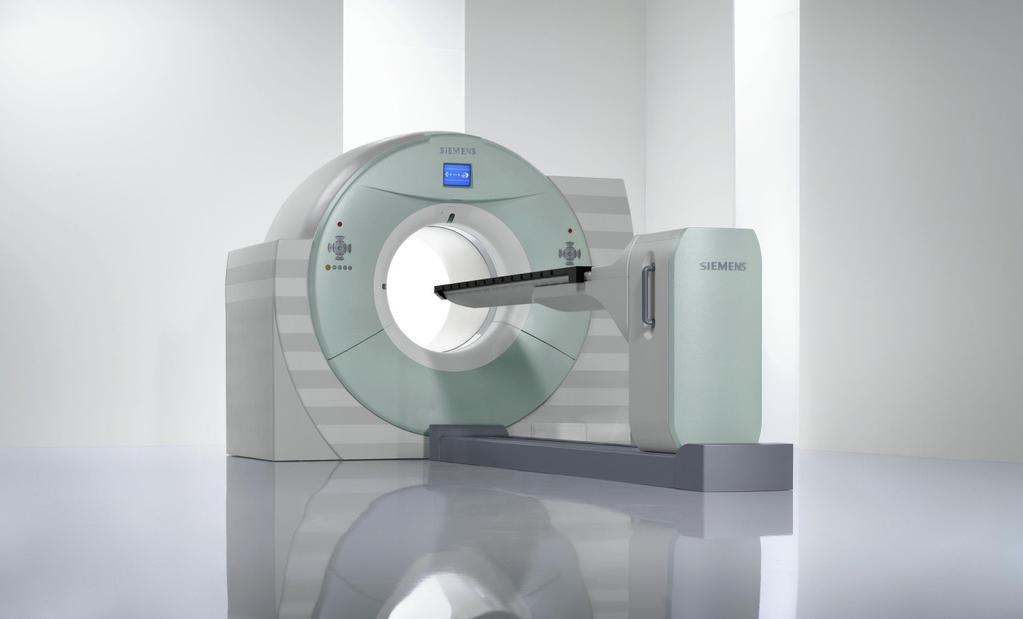
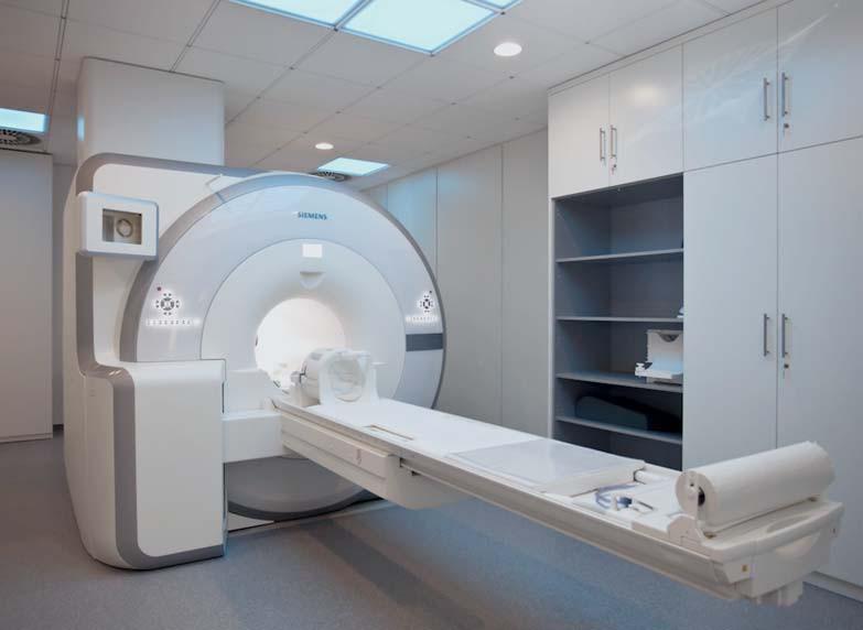
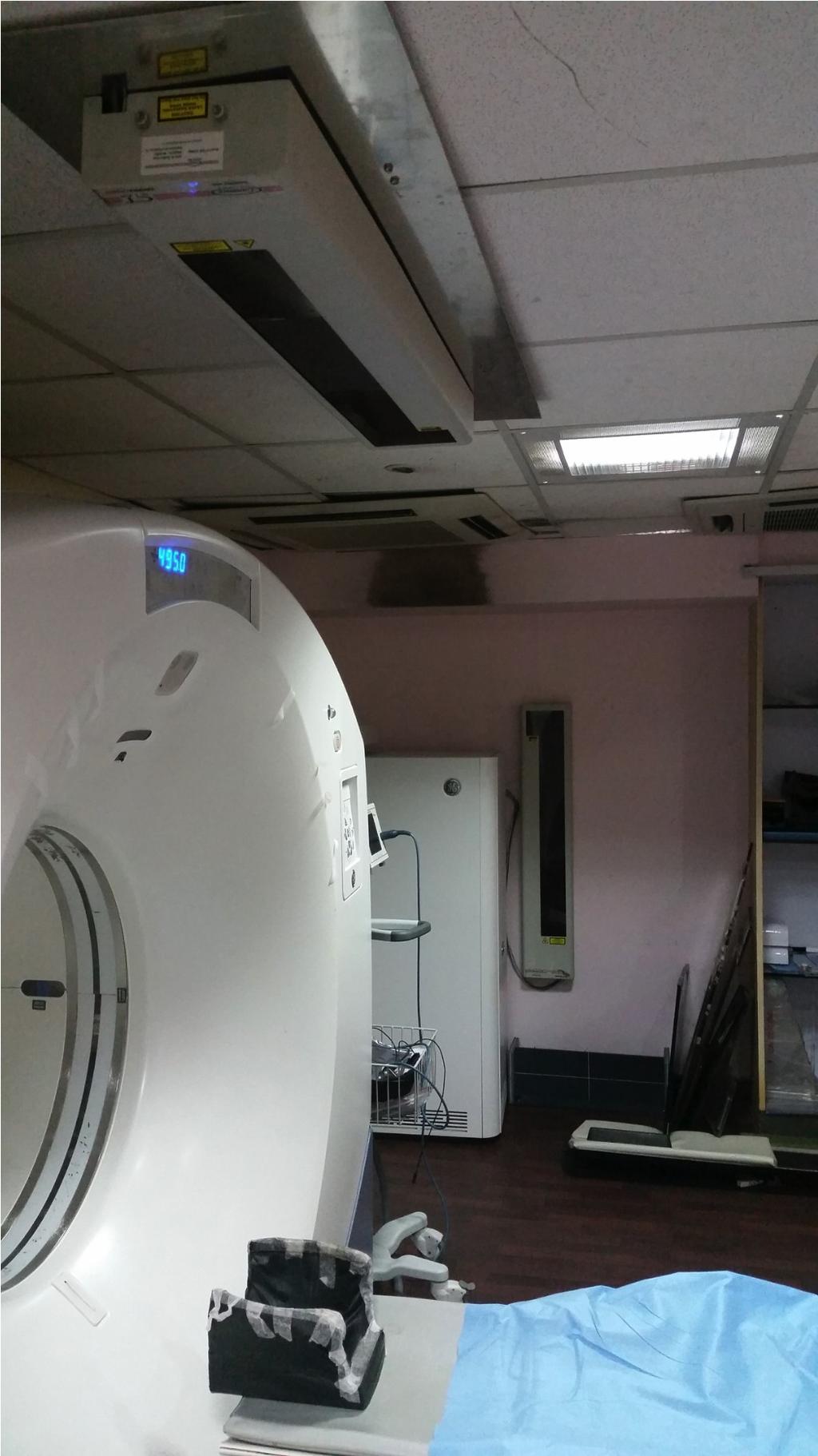
41 3 T Skyra 128 mct mmr
42 DAT -123I-FP-CIT,TRODAT Uptake reflects the activity of the transmembrane dopamine transporter. Dopamine transporter is downregulated in early disease, making 123I-FP-CIT SPECT more sensitive for the early detection of Parkinson s disease. For monitoring disease progression, the 2 compounds are theoretically equivalent.


43 FDOPA SCAN IN EVALUATION FOR PARKINSONISM Normal Control image FDOPA IMAGES OF PATIENT FDOPA uptake in brain showing uniform symmetrical uptake in the Caudate nucleus and Putamen. Reduced tracer uptake seen in the bilateral Putamen-left more reduced than right


44 Normal Control image FDOPA uptake in brain showing uniform symmetrical uptake in the Caudate nucleus and Putamen. MRI Images


45 Normal Control image FDOPA IMAGES FDOPA uptake in brain showing uniform symmetrical uptake in the Caudate nucleus and Putamen. Reduced tracer uptake seen in the bilateral Putamen. Right more reduced than left
46 Consider F-DOPA scan
 previously unresected tumors, and 11/11 (100%) of recurrent tumors(the sensitivity of 18F-FDOPA was 95.2% and that of MRI was 90.5%).")
47 Results: Fusion technology facilitated precise anatomical localization of 18FFDOPA activity. In group I, all 21 cases showed pathology-confirmed tumor. Of these, 18FFDOPA scans were positive in 9/10 (90%) previously unresected tumors, and 11/11 (100%) of recurrent tumors(the sensitivity of 18F-FDOPA was 95.2% and that of MRI was 90.5%). Of the 70 patients in group II, concordance between MRI and 18F-FDOPA was found in 49/54 (90.1%) of patients with sufficient follow-up; in the remaining 16 patients concordance could not be determined due to lack of follow-up. 18FFDOPA labeling was comparable in both high- and low-grade gliomas and identified both enhancing and non-enhancing tumor equally well. In some cases, 18F-FDOPA activity preceded tumor detection on MRI.
 show high 18F-FDOPA activity on the fused images (B, arrow) posterior to the resection cavity, without")
, but only the region of T2W signal posterior to the")
48 FDOPA PET may have utility in distinguishing non-enhancing tumor from other causes of T2W signal change such as gliosis, edema, etc Non-enhancing tumor regions on contrast-enhanced T1W MRI (A) show high 18F-FDOPA activity on the fused images (B, arrow) posterior to the resection cavity, without associated contrast enhancement. There is T2W signal abnormality anterior and posterior to the resection cavity (C, arrowheads), but only the region of T2W signal posterior to the resection cavity demonstrates abnormal 18F-FDOPA activity (D, arrow). This region was resected and confirmed to be recurrent tumor.
 also exhibits increased 18F-FDOPA activity (B, arrow).")
49 In this patient, elevated 18FFDOPA activity is seen at the margins of the resection cavity corresponding to a region of abnormal contrast enhancement (not shown). More superiorly, a non-enhancing area subtly expands the cortex in the parasagittal frontal lobe as shown on axial T2W MRI (A, arrow) also exhibits increased 18F-FDOPA activity (B, arrow). There was significant interval tumor growth into this previously PETdelineated tumor region on 7month followup MRI scan (T2W MRI (C, arrow) and contrasted-enhanced T1W MRI (D, FDOPA uptake delineated a region of tumor that was inconspicuous on MRI and which preceded abnormal contrast-enhancement on MRI
 that might represent either postsurgical change or recurrent tumor; however 18F-FDOPA")
50 18F-FDOPA labeling differentiates post-surgical changes from recurrent tumor. Seven month follow-up MRI for a resected grade III oligodendroglioma exhibits contrast-enhanced areas (A, arrow) that might represent either postsurgical change or recurrent tumor; however 18F-FDOPA activity on PET-MRI fusion (B, arrow) suggests the possibility of tumor recurrence. A subsequent follow-up MRI 2 months (C, arrow) and 3 months (D, arrow) later shows increased enhancement into the area previously delineated by 18FFDOPAactivity the metabolic abnormality on 18FFDOPA PET preceded the local tumor recurrence on MRI, suggesting that 18F-FDOPA may detect residual tumor not clearly defined by MRI alone.
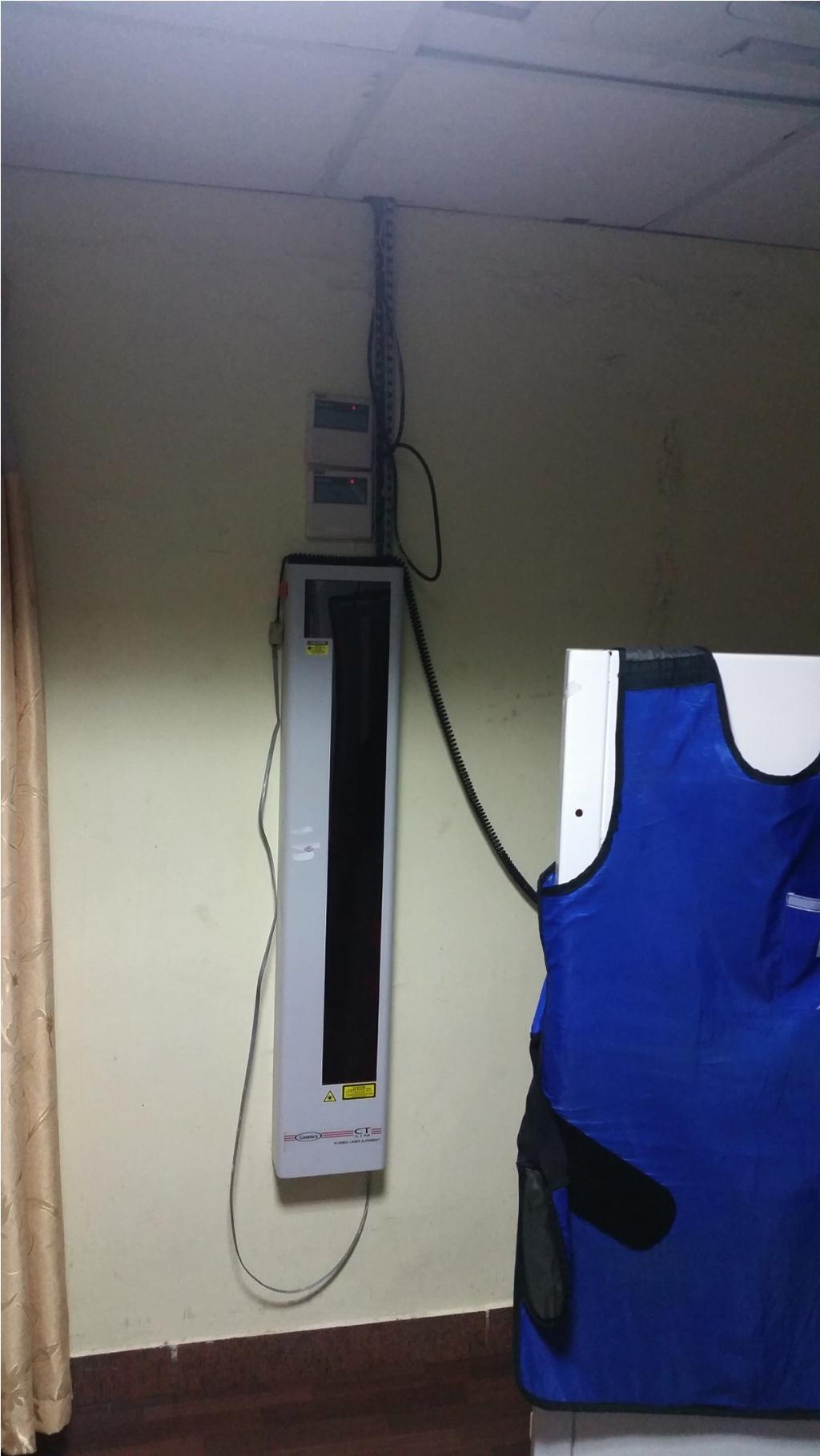
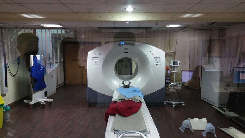
51 PETCT Radiotherapy plan How we do it FDOPA injected 15 min - One image acquired Finish diagnostic scan & call for RT - plan after 2 hours. If already done earlier then inject again on the day of the plan Casts, Radiotherapist,Physicist or Technician usually accompanies Flat couch like Radiotherapy couch is used. Moving lasers are used to mark fiducials on the cast used for immobilization
52 Steps in RT Planning Immobilization CT Simulation [PET CT] Image acquisition [DICOM] Image fusion [CT/MRI/PETCT] Anatomical+Functional Volume delineation [GTV,CTV,PTV,OAR] Planning [Optimal plan selection] Data transfer [Lantis] Set up and verification [EPID] Treatment delivery Monitoring Follow up
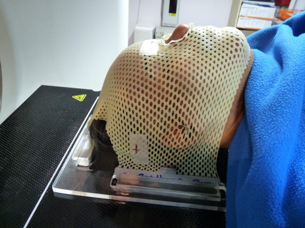
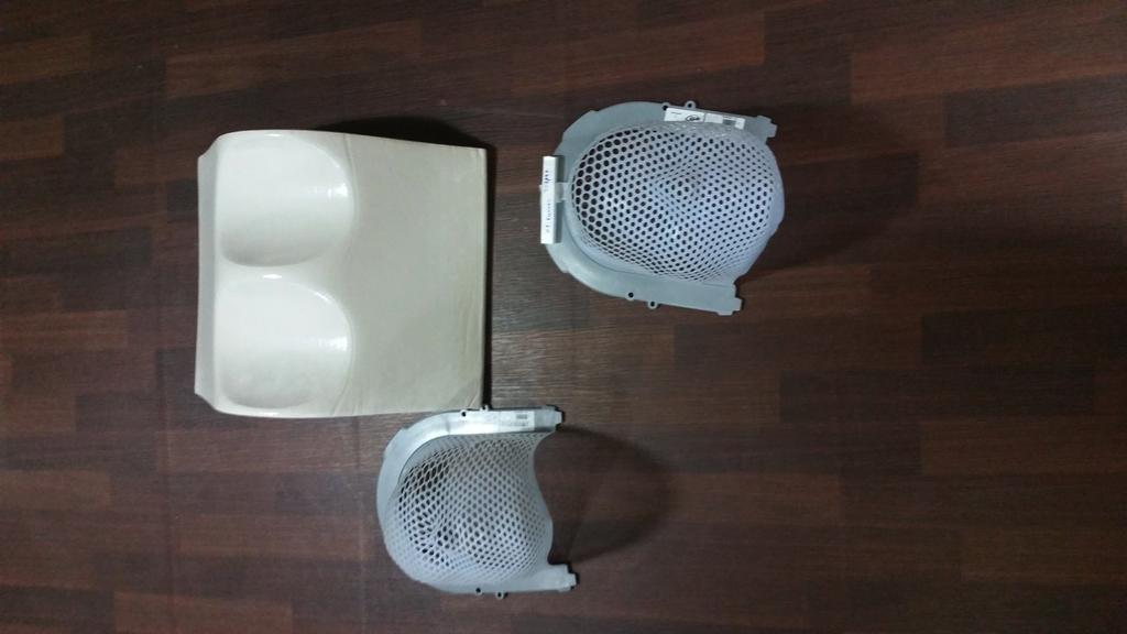
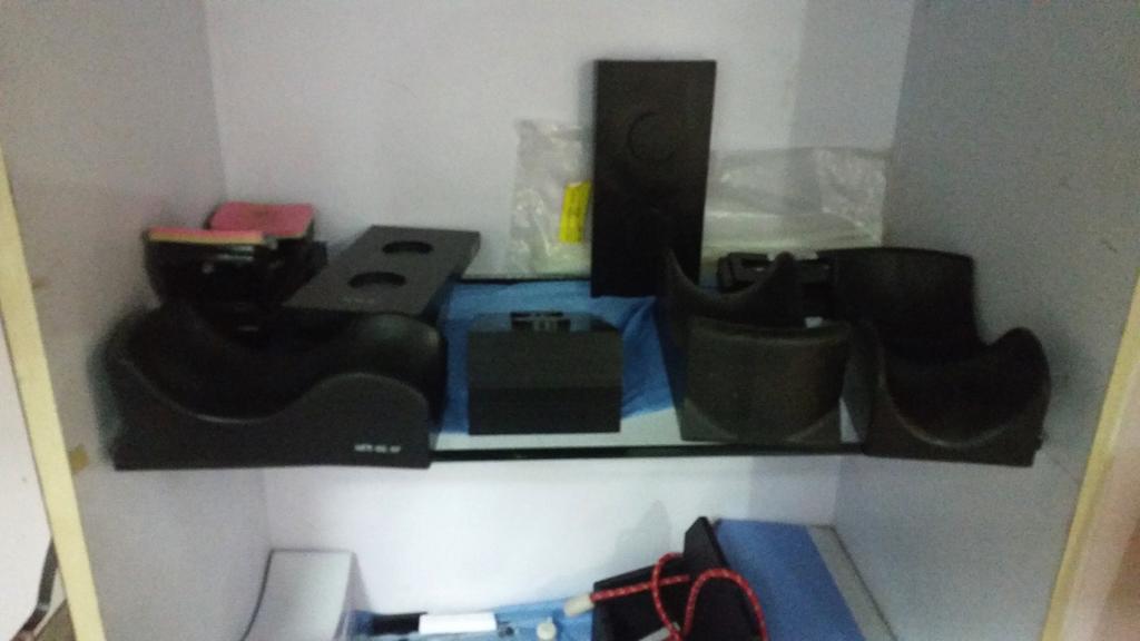
53 Immobilization
![Simulation [CT,PET CT] Image acquisition C11 MET : 15 min Plan and therapy set up finished](/docs-images/73/69096212/images/54-0.jpg "same time Extremely demanding. FDOPA : Image at 15 min and then RT plan at 1-2 hours later.")
54 Simulation [CT,PET CT] Image acquisition C11 MET : 15 min Plan and therapy set up finished same time Extremely demanding. FDOPA : Image at 15 min and then RT plan at 1-2 hours later.
55
56
57 Response evaluation for Chemoradiation HCG


58


59 C11 Met PET Post Rx
60 Thank You HCG
8/10/2016. PET/CT for Tumor Response. Staging and restaging Early treatment response evaluation Guiding biopsy
 PET/CT for Tumor Response Evaluation August 4, 2016 Wei Lu, PhD Department of Medical Physics www.mskcc.org Department of Radiation Oncology www.umaryland.edu FDG PET/CT for Cancer Imaging Staging and
Laura Tormoehlen, M.D. Neurology and EM-Toxicology Indiana University
 Laura Tormoehlen, M.D. Neurology and EM-Toxicology Indiana University Disclosures! No conflicts of interest to disclose Neuroimaging 101! Plain films! Computed tomography " Angiography " Perfusion! Magnetic
Assessment of renal cell carcinoma by two PET tracer : dual-time-point C-11 methionine and F-18 fluorodeoxyglucose
 Assessment of renal cell carcinoma by two PET tracer : dual-time-point C-11 methionine and F-18 fluorodeoxyglucose Poster No.: C-0805 Congress: ECR 2015 Type: Scientific Exhibit Authors: S. Ito, K. Kato,
Dr Sneha Shah Tata Memorial Hospital, Mumbai.
 Dr Sneha Shah Tata Memorial Hospital, Mumbai. Topics covered Lymphomas including Burkitts Pediatric solid tumors (non CNS) Musculoskeletal Ewings & osteosarcoma. Neuroblastomas Nasopharyngeal carcinomas
PSMA PET SCANNING AND THERANOSTICS IN PROSTATE CANCER KEVIN TRACEY, MD, FRCPC PRECISION DIAGNSOTIC IMAGING REGIONAL PET/CT CENTRE
 PSMA PET SCANNING AND THERANOSTICS IN PROSTATE CANCER KEVIN TRACEY, MD, FRCPC PRECISION DIAGNSOTIC IMAGING REGIONAL PET/CT CENTRE DISCLOSURES/CONFLICTS NONE OBJECTIVES Understand current diagnostic role
Dosimetry, see MAGIC; Polymer gel dosimetry. Fiducial tracking, see CyberKnife radiosurgery
 Subject Index Acoustic neuroma, neurofibromatosis type 2 complications 103, 105 hearing outcomes 103, 105 outcome measures 101 patient selection 105 study design 101 tumor control 101 105 treatment options
Molecular Imaging and Cancer
 Molecular Imaging and Cancer Cancer causes one in every four deaths in the United States, second only to heart disease. According to the U.S. Department of Health and Human Services, more than 512,000
Chapter 10. Summary, conclusions and future perspectives
 Chapter 10 Summary, conclusions and future perspectives 10.1 SUMMARY In this thesis, a new tumor imaging tracer in nuclear medicine is studied. This 123 tracer, L-3-[ I]Iodo-alpha-methyl-tyrosine (IMT),
Principles of nuclear metabolic imaging. Prof. Dr. Alex Maes AZ Groeninge Kortrijk and KULeuven Belgium
 Principles of nuclear metabolic imaging Prof. Dr. Alex Maes AZ Groeninge Kortrijk and KULeuven Belgium I. Molecular imaging probes A. Introduction - Chemical disturbances will precede anatomical abnormalities
PET-CT for radiotherapy planning in lung cancer: current recommendations and future directions
 PET-CT for radiotherapy planning in lung cancer: current recommendations and future directions Gerry Hanna Centre for Cancer Research and Cell Biology Queen s University of Belfast @gerryhanna Talk Outline
PET-MRI in malignant bone tumours. Lars Stegger Department of Nuclear Medicine University Hospital Münster, Germany
 PET-MRI in malignant bone tumours Lars Stegger Department of Nuclear Medicine University Hospital Münster, Germany Content From PET to PET/MRI General considerations Bone metastases Primary bone tumours
Nuclear neurology. Zámbó Katalin Department of Nuclear Medicine
 Nuclear neurology Zámbó Katalin Department of Nuclear Medicine To refresh your memory Brain has a high rate of oxidative metabolism. It has no reserves either of oxygen or of glucose and has a very limited
F18 FET PET/CT in Brain Tumors
 Editorial F18 FET PET/CT in Brain Tumors Abdelwahab, M.A and Omar, W. NCI, Cairo University. Egypt. 18F-fluoro-ethyl-tyrosine (18F-FET) was developed in the late 1990s first synthesized by Wester and colleagues
MRI Applications in Radiation Oncology:
 MRI Applications in Radiation Oncology: Physician s Perspective Jeff Olsen, MD Department of Radiation Oncology Washington University, St. Louis, MO Disclosures Washington University has research and service
KEY WORDS gamma knife surgery metastatic brain tumor radiation injury tumor recurrence thallium-201 single-photon emission computerized tomography
 J Neurosurg (Suppl) 102:266 271, 2005 Diagnostic value of thallium-201 chloride single-photon emission computerized tomography in differentiating tumor recurrence from radiation injury after gamma knife
PET in Prostate Cancer
 PET in Prostate Cancer Tom R. Miller, M.D., Ph.D. Mallinckrodt Institute of Radiology Washington University School of Medicine St. Louis, Missouri, USA Prostate Imaging Bone Scintigraphy primarily for
SUPPLEMENTARY INFORMATION
 VOLUME: 1 ARTICLE NUMBER: 0027 In the format provided by the authors and unedited. Rapid intraoperative histology of unprocessed surgical specimens via fibre-laser-based stimulated Raman scattering microscopy
PET/CT in oncology. Positron emission tomography
 Clinical Medicine 2012, Vol 12, No 4: 368 72 PET/CT in oncology Fahim-Ul-Hassan, SpR Nuclear Medicine, Guy s Hospital, London; Gary J Cook, professor of Clinical PET, KCL Division of Imaging Sciences &
Functional aspects of anatomical imaging techniques
 Functional aspects of anatomical imaging techniques Nilendu Purandare Associate Professor & Consultant Radiologist Tata Memorial Centre Functional/metabolic/molecular imaging (radioisotope scanning) PET
NEUROIMAGING IN PANS/PANDAS
 NEUROIMAGING IN PANS/PANDAS Harry T. Chugani, M.D. Chief, Pediatric Neurology Nemours A.I. dupont Hospital for Children Wilmington, Delaware, USA Professor of Pediatrics and Neurology Thomas Jefferson
Ruolo dell imaging nella pianificazione del trattamento
 Simposio AIRO-SIRM: Diagnostica per immagini morfologica e funzionale nella stadiazione, terapia e follow-up dei sarcomi delle parti molli Ruolo dell imaging nella pianificazione del trattamento Marco
Indications of PET/CT in oncology
 Monday, August 27, 2012 Session 1, 10:00-10:40 Indications of PET/CT in oncology Helle Westergren Hendel MD, PhD, assistant professor Bacelor in Leadership & Health Ecomomics Head of Clinical PET, Herlev
New imaging techniques: let there be light. Felix M. Mottaghy Department of Nuclear Medicine University Hospital KU Leuven
 New imaging techniques: let there be light Felix M. Mottaghy Department of Nuclear Medicine University Hospital KU Leuven Medical imaging and the pathology cascade Molecular/Cellular disturbance Alterations
Structural and functional imaging for the characterization of CNS lymphomas
 Structural and functional imaging for the characterization of CNS lymphomas Cristina Besada Introduction A few decades ago, Primary Central Nervous System Lymphoma (PCNSL) was considered as an extremely
Bone PET/MRI : Diagnostic yield in bone metastases and malignant primitive bone tumors
 Bone PET/MRI : Diagnostic yield in bone metastases and malignant primitive bone tumors Lars Stegger, Benjamin Noto Department of Nuclear Medicine University Hospital Münster, Germany Content From PET to
PET imaging of cancer metabolism is commonly performed with F18
 PCRI Insights, August 2012, Vol. 15: No. 3 Carbon-11-Acetate PET/CT Imaging in Prostate Cancer Fabio Almeida, M.D. Medical Director, Arizona Molecular Imaging Center - Phoenix PET imaging of cancer metabolism
Molecular Imaging and Breast Cancer
 Molecular Imaging and Breast Cancer Breast cancer forms in tissues of the breast usually in the ducts, tubes that carry milk to the nipple, and lobules, the glands that make milk. It occurs in both men
Ga68 Imaging. Roland HUSTINX Division of Nuclear Medicine and Oncologic Imaging Centre Hospitalier Universitaire de Liège Belgium
 Ga68 Imaging Roland HUSTINX Division of Nuclear Medicine and Oncologic Imaging Centre Hospitalier Universitaire de Liège Belgium 68 Ga Produced by a 68 Ge/ 68 Ga generator Decays by positron emission
Nuclear medicine in oncology. 1. Diagnosis 2. Therapy
 Nuclear medicine in oncology 1. Diagnosis 2. Therapy Diagnosis - Conventional methods - Nonspecific radiopharmaceuticals cumulating in tumours - Specific radiopharmaceuticals (receptor- and immunoscintigraphy)
General Identification. Name: 江 X X Age: 29 y/o Gender: Male Height:172cm, Weight: 65kg Date of admission:95/09/27
 General Identification Name: 江 X X Age: 29 y/o Gender: Male Height:172cm, Weight: 65kg Date of admission:95/09/27 Chief Complaint Sudden onset of seizure for several minutes Present illness This 29-year
PET Guidance of Therapy for BNCT and in vivo B-10 imaging
 INFN LNL Legnaro 17-19 Novembre 2009 Principles of Positron Emission Tomography and Radiopharmaceuticals PET Guidance of Therapy for BNCT and in vivo B-10 imaging Luca Menichetti, Ph.D C.N.R. Institute
PET/MR:Techniques, Indications and Applications
 PET/MR:Techniques, Indications and Applications Franz Wolfgang Hirsch Professor and Head of the Department of Pediatric Radiology University Hospital Leipzig / Germany Children s Hospital University Leipzig
Optimized. clinical pathway. propels high utilization of PET/MR at Pitié-Salpêtrière Hospital
 Optimized propels high utilization of PET/MR at Pitié-Salpêtrière Hospital clinical pathway As one of Europe s largest teaching hospitals, Pitié-Salpêtrière Hospital is renowned for its innovative research
Esophageal Cancer Initially Thought to be Accompanied by a Solitary Metastasis to an Intrathoracic Paraaortic Lymph Node
 2012 66 5 417 421 Esophageal Cancer Initially Thought to be Accompanied by a Solitary Metastasis to an Intrathoracic Paraaortic Lymph Node a b* a a a a a a a b ʼ 418 Horio et al. Acta Med. Okayama Vol.
CNS SESSION 3/8/ th Multidisciplinary Management of Cancers: A Case based Approach
 CNS SESSION Chair: Ruben Fragoso, MD/PhD UC Davis Fellow: Michael Cardenas, MD UC Davis Panel: Gordon Li, MD Stanford Seema Nagpal, MD Stanford Jennie Taylor, MD UCSF HPI: 46 yo right handed woman who
New Visions in PET: Surgical Decision Making and PET/CT
 New Visions in PET: Surgical Decision Making and PET/CT Stanley J. Goldsmith, MD Director, Nuclear Medicine Professor, Radiology & Medicine New York Presbyterian Hospital- Weill Cornell Medical Center
Understanding L-dopa L and Metabolism in the Human Brain
 Understanding L-dopa L Transport and Metabolism in the Human Brain Final Presentation for REU program August 3, 2006 Megan Mary Mekarski Advisor: Professor Linninger Dr. Libin Zhang Laboratory for Product
POSITRON EMISSION TOMOGRAPHY (PET)
") Status Active Medical and Behavioral Health Policy Section: Radiology Policy Number: V-27 Effective Date: 08/27/2014 Blue Cross and Blue Shield of Minnesota medical policies do not imply that members should
Status Active Medical and Behavioral Health Policy Section: Radiology Policy Number: V-27 Effective Date: 08/27/2014 Blue Cross and Blue Shield of Minnesota medical policies do not imply that members should
CT & MRI Evaluation of Brain Tumour & Tumour like Conditions
 CT & MRI Evaluation of Brain Tumour & Tumour like Conditions Dr. Anjana Trivedi 1, Dr. Jay Thakkar 2, Dr. Maulik Jethva 3, Dr. Ishita Virda 4 1 M.D. Radiology, Professor and Head, P.D.U. Medical College
MRI to fit your planning. Philips Panorama HFO Oncology Configuration
 MRI to fit your planning Philips Panorama HFO Oncology Configuration MR Imaging that fits Philips Panorama HFO Oncology Configuration allows radiation oncologists to take full advantage of MRI s excellent
Ronald C. Walker, MD, Prof of Radiology Vanderbilt University Medical Center Nashville, TN. Ga-DOTATATE PET/CT imaging Initial Vanderbilt experience
 Ronald C. Walker, MD, Prof of Radiology Vanderbilt University Medical Center Nashville, TN 68 Ga-DOTATATE PET/CT imaging Initial Vanderbilt experience Disclosures: No financial disclosures or conflicts
PET/CT in Breast Cancer
 PET/CT in Breast Cancer Rodolfo Núñez Miller, M.D. Nuclear Medicine and Diagnostic Imaging Section Division of Human Health International Atomic Energy Agency Vienna, Austria Overview Introduction Locorregional
Applications of PET in medicine and biomedical studies
 Lecture 21 Applications of PET in medicine and biomedical studies PET imaging is a technique for observing biological processes. Because disease is a biological process, molecular imaging is a sensitive
Defining Target Volumes and Organs at Risk: a common language
 Defining Target Volumes and Organs at Risk: a common language Eduardo Rosenblatt Section Head Applied Radiation Biology and Radiotherapy (ARBR) Section Division of Human Health IAEA Objective: To introduce
MRI and CT of the CNS
 MRI and CT of the CNS Dr.Maha ELBeltagy Assistant Professor of Anatomy Faculty of Medicine The University of Jordan 2018 Computed Tomography CT is used for the detection of intracranial lesions. CT relies
World Molecular Imaging Society (WMIS) Comment on Proposed Decision Memorandum for Positron Emission Tomography (CAG-00065R2)
 Comment on Proposed Decision Memorandum for Positron Emission Tomography (CAG-00065R2)") January 11, 2013 Director, Coverage and Analysis Group Centers for Medicare & Medicaid Services 7500 Security Blvd Baltimore, MD 21244 By Online Submission RE: World Molecular Imaging Society (WMIS) Comment
January 11, 2013 Director, Coverage and Analysis Group Centers for Medicare & Medicaid Services 7500 Security Blvd Baltimore, MD 21244 By Online Submission RE: World Molecular Imaging Society (WMIS) Comment
The Radiologic Evaluation of Glioblastoma (GBM) and Differentiation from Pseudoprogression
 and Differentiation from Pseudoprogression") The Radiologic Evaluation of Glioblastoma (GBM) and Differentiation from Pseudoprogression Alexis Roy, Harvard Medical School, Year III Our Patient AB: Clinical Presentation 53 year old female with a past
The Radiologic Evaluation of Glioblastoma (GBM) and Differentiation from Pseudoprogression Alexis Roy, Harvard Medical School, Year III Our Patient AB: Clinical Presentation 53 year old female with a past
PRINCESS MARGARET CANCER CENTRE CLINICAL PRACTICE GUIDELINES
 PRINCESS MARGARET CANCER CENTRE CLINICAL PRACTICE GUIDELINES CENTRAL NERVOUS SYSTEM LOW GRADE GLIOMAS CNS Site Group Low Grade Gliomas Author: Dr. Norm Laperriere 1. INTRODUCTION 3 2. PREVENTION 3 3. SCREENING
Evaluation of Lung Cancer Response: Current Practice and Advances
 Evaluation of Lung Cancer Response: Current Practice and Advances Jeremy J. Erasmus I have no financial relationships, arrangements or affiliations and this presentation will not include discussion of
Molecular Imaging as a Cancer Biomarker:
 Molecular Imaging as a Cancer Biomarker: Imaging to Guide Targeted Cancer Therapy 51 st Annual AAPM Meeting, Anaheim CA David A. Mankoff, MD, PhD Seattle Cancer Care Alliance University of Washington Seattle,
Using PET/CT in Prostate Cancer
 Using PET/CT in Prostate Cancer Legal Disclaimer These materials were prepared in good faith by MITA as a service to the profession and are believed to be reliable based on current scientific literature.
Testicular relapse of non-hodgkin Lymphoma noted on FDG-PET
 Testicular relapse of non-hodgkin Lymphoma noted on FDG-PET Stephen D. Scotti 1*, Jennifer Laudadio 2 1. Department of Radiology, North Carolina Baptist Hospital, Winston-Salem, NC, USA 2. Department of
Facility IBA 18/9 Cyclotron Accelerates p and d. Facility GMP Grade Clean Room Automated Dispenser Synthera Rig FDG 07/11/2014
 Dr Chris Marshall Director Introduction to PET Positron Emission Tomography Positron is anti matter equivalent of the electron Isotopes that are proton rich can decay by emission of a positron Positron
στη σταδιοποίηση του καρκίνου του προστάτη Γ. Αρσος, Γ Εργ. Πυρηνικής Ιατρικής ΑΠΘ, ΓΝΘ Παπαγεωργίου
 Η θέση του PET/CT στη σταδιοποίηση του καρκίνου του προστάτη Γ. Αρσος, Γ Εργ. Πυρηνικής Ιατρικής ΑΠΘ, ΓΝΘ Παπαγεωργίου 2014 : the Guidelines year. PRINCIPLES OF IMAGING Imaging is performed for the detection
Evaluation of Three-dimensional Conformal Radiotherapy and Intensity Modulated Radiotherapy Techniques in High-Grade Gliomas
 1 Carol Boyd Comprehensive Case Study July 11, 2013 Evaluation of Three-dimensional Conformal Radiotherapy and Intensity Modulated Radiotherapy Techniques in High-Grade Gliomas Abstract: Introduction:
Update on functional brain imaging in Movement Disorders
 Update on functional brain imaging in Movement Disorders Mario Masellis, MSc, MD, FRCPC, PhD Assistant Professor & Clinician-Scientist Sunnybrook Health Sciences Centre University of Toronto 53 rd CNSF
Index. Surg Oncol Clin N Am 16 (2007) Note: Page numbers of article titles are in boldface type.
 Note: Page numbers of article titles are in boldface type.") Surg Oncol Clin N Am 16 (2007) 465 469 Index Note: Page numbers of article titles are in boldface type. A Adjuvant therapy, preoperative for gastric cancer, staging and, 339 B Breast cancer, metabolic
Surg Oncol Clin N Am 16 (2007) 465 469 Index Note: Page numbers of article titles are in boldface type. A Adjuvant therapy, preoperative for gastric cancer, staging and, 339 B Breast cancer, metabolic
The Use of PET Scanning in Urologic Oncology
 The Use of PET Scanning in Urologic Oncology Dr Nicholas C. Buchan Uro-oncology Fellow 1 2 Aims To understand the basic concepts underlying PET scanning. Understand the emerging role of PET Scanning for
IAEA RTC. PET/CT and Planning of Radiation Therapy 20/08/2014. Sarajevo (Bosnia & Hercegovina) Tuesday, June :40-12:20 a.
 Tuesday, June :40-12:20 a.") IAEA RTC PET/CT and Planning of Radiation Therapy Sarajevo (Bosnia & Hercegovina) Tuesday, June 17 2014 11:40-12:20 a.m María José García Velloso Servicio de Medicina Nuclear Clínica Universidad de Navarra
IAEA RTC PET/CT and Planning of Radiation Therapy Sarajevo (Bosnia & Hercegovina) Tuesday, June 17 2014 11:40-12:20 a.m María José García Velloso Servicio de Medicina Nuclear Clínica Universidad de Navarra
What Radiologists do?
 Multimodality Imaging in Oncology 2018 March 5 th 9th Diagnostic Imaging in Oncology What Radiologists do? Chikako Suzuki, MD, PhD Department of Diagnostic Radiology, KS Solna Department of Molecular Medicine
Case Scenario 1 Worksheet. Primary Site C44.4 Morphology 8743/3 Laterality 0 Stage/ Prognostic Factors
 CASE SCENARIO 1 9/10/13 HISTORY: Patient is a 67-year-old white male and presents with lesion located 4-5cm above his right ear. The lesion has been present for years. No lymphadenopathy. 9/10/13 anterior
Brain Tumor Treatment
 Scan for mobile link. Brain Tumor Treatment Brain Tumors Overview A brain tumor is a group of abnormal cells that grows in or around the brain. Tumors can directly destroy healthy brain cells. They can
PET/CT Value: Rocky Mountain Cancer Centers
 PET/CT Value: Rocky Mountain Cancer Centers Glenn Balasky Executive Director Rocky Mountain Cancer Centers glenn.balasky@usoncology.com CANM/CAMRT Joint Conference March 22, 2018 Vancouver, British Columbia
Molecular Imaging and the Brain
 Molecular imaging technologies are playing an important role in neuroimaging, a branch of medical imaging, by providing a window into the living brain. Where CT and conventional MR imaging provide important
Positron emission tomography Medicare Services Advisory Committee
 Positron emission tomography Medicare Services Advisory Committee Authors' objectives To assess the effectiveness of positron emission tomography (PET), the report addressed the following (truncated) six
Imaging in gastric cancer
 Imaging in gastric cancer Gastric cancer remains a deadly disease because of late diagnosis. Adenocarcinoma represents 90% of malignant tumors. Diagnosis is based on endoscopic examination with biopsies.
It s s Always Something!
 It s s Always Something! New Approaches in Brain Tumor Treatment Virginia Stark-Vance, M.D. When Something Is a Brain Tumor Brain tumors aren t rare: there are over 100,000/yr Most originate as other cancers
Case Report PET/CT Imaging in Oncology: Exceptions That Prove the Rule
 Case Reports in Oncological Medicine Volume 2013, Article ID 865032, 4 pages http://dx.doi.org/10.1155/2013/865032 Case Report PET/CT Imaging in Oncology: Exceptions That Prove the Rule M. Casali, 1 A.
Oligodendroglioma: imaging findings, radio-pathological correlation and evolution
 Oligodendroglioma: imaging findings, radio-pathological correlation and evolution Poster No.: C-2104 Congress: ECR 2013 Type: Authors: Keywords: DOI: Scientific Exhibit A. Hernandez Castro, M. D. Monedero
Hybrid Imaging SPECT/CT PET/CT PET/MRI. SNMMI Southwest Chapter Aaron C. Jessop, MD
 Hybrid Imaging SPECT/CT PET/CT PET/MRI SNMMI Southwest Chapter 2014 Aaron C. Jessop, MD Assistant Professor, Department of Nuclear Medicine UT MD Anderson Cancer Center, Houston, Texas Complimentary role
Emerging contrasts at ultrahigh fields" A. Dean Sherry
 Emerging contrasts at ultrahigh fields" A. Dean Sherry Advanced Imaging Research Center Department of Radiology UT Southwestern Medical Center Department of Chemistry & Biochemistry, UT Dallas ADVANCED
Goals for this Lecture. Case 1. Key Points MRI TECHNIQUES FOR DIFFERENTIAL DIAGNOSIS OF RECURRENT BRAIN LESIONS
 MRI TECHNIQUES FOR DIFFERENTIAL DIAGNOSIS OF RECURRENT BRAIN LESIONS Goals for this Lecture 1. Review common appearances for recurrent tumor and treatment effects on conventional MRI 2. Discuss current
Clinical indications for positron emission tomography
 Clinical indications for positron emission tomography Oncology applications Brain and spinal cord Parotid Suspected tumour recurrence when anatomical imaging is difficult or equivocal and management will
Pitfalls in Oncologic Diagnosis with PET CT. Nononcologic Hypermetabolic Findings
 Pitfalls in Oncologic Diagnosis with PET CT. Nononcologic Hypermetabolic Findings Poster No.: C-1359 Congress: ECR 2012 Type: Educational Exhibit Authors: J. Rossi 1, C. A. Mariluis 1, E. Delgado 1, R.
LYMPHATIC DRAINAGE IN THE HEAD & NECK
 LYMPHATIC DRAINAGE IN THE HEAD & NECK Like other parts of the body, the head and neck contains lymph nodes (commonly called glands). Which form part of the overall Lymphatic Drainage system of the body.
C-methionine PET/CT for tumors of the skull base: Comparison with F-FDG PET/CT
 11 C-methionine PET/CT for tumors of the skull base: Comparison with 18 F-FDG PET/CT Award: Certificate of Merit Poster No.: C-0287 Congress: ECR 2013 Type: Scientific Exhibit Authors: N. Tomura, T. Saginoya,
magnetic resonance (MR) imaging, since both J. L. Kline, R. B. Noto, and M. Glantz
 imaging, since both J. L. Kline, R. B. Noto, and M. Glantz") Single-Photon Emission CT in the Evaluation of Recurrent Brain Tumor in Patients Treated with Gamma Knife Radiosurgery or Conventional Radiation Therapy J. L. Kline, R. B. Noto, and M. Glantz PURPOSE:
Single-Photon Emission CT in the Evaluation of Recurrent Brain Tumor in Patients Treated with Gamma Knife Radiosurgery or Conventional Radiation Therapy J. L. Kline, R. B. Noto, and M. Glantz PURPOSE:
FDG PET/CT STAGING OF LUNG CANCER. Dr Shakher Ramdave
 FDG PET/CT STAGING OF LUNG CANCER Dr Shakher Ramdave FDG PET/CT STAGING OF LUNG CANCER FDG PET/CT is used in all patients with lung cancer who are considered for curative treatment to exclude occult disease.
HSV1716 Dose levels and Cohort size Dose level No of Patients HSV1716 Dosage 1* 3 to 6 1 ml of 1 x 10 5 infectious units HSV1716 per ml 2
 Abstract and Schema: Description and Rationale: Pediatric high grade gliomas have a progressive initial course and high risk of relapse/ progression; making the 5-year overall survival rate 15-35% with
FDG-PET/CT in Gynaecologic Cancers
 Friday, August 31, 2012 Session 6, 9:00-9:30 FDG-PET/CT in Gynaecologic Cancers (Uterine) cervical cancer Endometrial cancer & Uterine sarcomas Ovarian cancer Little mermaid (Edvard Eriksen 1913) honoring
Molecular Imaging in the Development of Cancer Therapeutics. Johannes Czernin
 Molecular Imaging in the Development of Cancer Therapeutics Johannes Czernin Ahmanson Biological Imaging Division University of California Los Angeles Cancer Statistics Cancer Type 5-year Survival Rate
REVIEW. Distinguishing benign from malignant adrenal masses
 Cancer Imaging (2003) 3, 102 110 DOI: 10.1102/1470-7330.2003.0006 CI REVIEW Distinguishing benign from malignant adrenal masses Isaac R Francis Professor of Radiology, Department of Radiology, University
Case Report PET/MRI for Preoperative Planning in Patients with Soft Tissue Sarcoma: A Technical Report of Two Patients
 Volume 2013, Article ID 791078, 4 pages http://dx.doi.org/10.1155/2013/791078 Case Report PET/MRI for Preoperative Planning in Patients with Soft Tissue Sarcoma: A Technical Report of Two Patients Annika
Multiparametric imaging in oncology
 Multiparametric imaging in oncology p1 T p2 p2 T T p3 p1 p3 T Marco Ravanelli Roberto Maroldi The goal of traditional imaging is high spatial and contrast resolution diagnosis, tumor extent treatment planning,
Nuclear Sciences and Medicine
 Nuclear Sciences and Medicine Rethy Chhem, MD, PhD (Edu), PhD (His), FRCPC Division of Human Health Guest Professor, Medical University of Vienna International Atomic Energy Agency Medical Imaging X-rays
Nuclear Medicine: Basics to therapy
 Nuclear Medicine: Basics to therapy RCP Medical careers day Dr Sabina Dizdarevic MD MSc PhD FRCP Dr Deena Neriman MBBS FRCR Ms Charlotte Weston CEO BNMS On behalf of the British Nuclear Medicine Society
CT PET SCANNING for GIT Malignancies A clinician s perspective
 CT PET SCANNING for GIT Malignancies A clinician s perspective Damon Bizos Head, Surgical Gastroenterology Charlotte Maxeke Johannesburg Academic Hospital Case presentation 54 year old with recent onset
Novel Imaging in Advanced Prostate Cancer
 Novel Imaging in Advanced Prostate Cancer Robert J. Hamilton, MD MPH FRCSC Princess Margaret Cancer Centre ICUC Saturday January 21, 2017 Company/Organizati Details Faculty/Presenter on Disclosures I am
IEHP UM Subcommittee Approved Authorization Guidelines Magnetic Resonance Spectroscopy
 Policy: Based on the information reviewed, IEHP s UM Subcommittee consider Magnetic Resonance Spectroscopy (MRS) to be investigational and not medically necessary. Although MRS can accurately delineate
Short summary of published results of PET with fluoromethylcholine (18F) in prostate cancer
 in prostate cancer") Short summary of published results of PET with fluoromethylcholine (18F) in prostate cancer JN TALBOT and all the team of Service de Médecine Nucléaire Hôpital Tenon et Université Pierre et Marie Curie,
Short summary of published results of PET with fluoromethylcholine (18F) in prostate cancer JN TALBOT and all the team of Service de Médecine Nucléaire Hôpital Tenon et Université Pierre et Marie Curie,
Radionuclide detection of sentinel lymph node
 Radionuclide detection of sentinel lymph node Sophia I. Koukouraki Assoc. Professor Department of Nuclear Medicine Medicine School, University of Crete 1 BACKGROUND The prognosis of malignant disease is
Theranostics in Nuclear Medicine
 Theranostics in Nuclear Medicine Patrick FLAMEN, MD, PhD Head Nuclear Medicine Institut Jules Bordet Université Libre de Bruxelles (U.L.B.) n Theranostics in Nuclear Medicine n A form of (nuclear) diagnostic
Evaluation of Whole-Field and Split-Field Intensity Modulation Radiation Therapy (IMRT) Techniques in Head and Neck Cancer
 Techniques in Head and Neck Cancer") 1 Charles Poole April Case Study April 30, 2012 Evaluation of Whole-Field and Split-Field Intensity Modulation Radiation Therapy (IMRT) Techniques in Head and Neck Cancer Abstract: Introduction: This study
1 Charles Poole April Case Study April 30, 2012 Evaluation of Whole-Field and Split-Field Intensity Modulation Radiation Therapy (IMRT) Techniques in Head and Neck Cancer Abstract: Introduction: This study
ORIGINAL ARTICLE. Key words: malignant glioma, recurrence, PET, methionine, stereotactic radiosurgery, radiation necrosis
 ORIGINAL ARTICLE Annals of Nuclear Medicine Vol. 18, No. 4, 291 296, 2004 Methionine positron emission tomography for differentiation of recurrent brain tumor and radiation necrosis after stereotactic
Image Fusion, Contouring, and Margins in SRS
 Image Fusion, Contouring, and Margins in SRS Sarah Geneser, Ph.D. Department of Radiation Oncology University of California, San Francisco Overview Review SRS uncertainties due to: image registration contouring
Update on RECIST and Staging of Common Pediatric Tumors Ethan A. Smith, MD
 Update on RECIST and Staging of Common Pediatric Tumors Ethan A. Smith, MD Section of Pediatric Radiology C.S. Mott Children s Hospital University of Michigan ethans@med.umich.edu Disclosures No relevant
FUNCTIONAL MAGNETIC RESONANCE IMAGING IN FOLLOW-UP OF CEREBRAL GLIAL TUMORS
 Anvita Bieza FUNCTIONAL MAGNETIC RESONANCE IMAGING IN FOLLOW-UP OF CEREBRAL GLIAL TUMORS Summary of Doctoral Thesis to obtain PhD degree in medicine Specialty Diagnostic Radiology Riga, 2013 Doctoral thesis
Fusion Ultrasound: Characterization of Abdominal Masses with MR, CT, PET, and Contrast Ultrasound
 Fusion Ultrasound: Characterization of Abdominal Masses with MR, CT, PET, and Contrast Ultrasound Mollie Rashid, MD Corinne Deurdulian, MD Hisham Tchelepi, MD Keck School of Medicine, University of Southern