UW Radiology Review Course Breast Calcifications. BI-RADS 5 th Edition
|
|
|
- Isabella McKenzie
- 6 years ago
- Views:
Transcription
1 UW Radiology Review Course Breast Calcifications Grace Kalish, MD Vantage Radiology BI-RADS 5 th Edition Benign Skin Vascular Large rod like Coarse popcorn Suspicious Amorphous Coarse heterogenous Fine pleomorphic Fine linear or fine linear branching Round and punctate Rim Milk of calcium Dystrophic 1
2 Changes Rim (previously rim, eggshell and lucent centered ) Round and punctate clarified: Round >0.5mm but <1mm Punctate <0.5mm Amorphous (no indistinct) Elimination of the Indeterminate category Distribution Diffuse Regional Clustered (formerly Grouped ) Linear Segmental 2

3 Case 1 Question: What is the best BI-RADS description of these calcifications? 1. Coarse, diffuse 2. Round, diffuse 3. Dystrophic, diffuse 4. Coarse heterogenous, diffuse 3
 Round usually between 0.")

4 Discussion: Coarse Coarse-- classically large (>2-3mm) Round usually between 0.5-1mm Dystrophic irregular in shape Coarse heterogenous between 0.5-1mm Case 2. Diagnostic Mag CC Mag ML 4


5 Question: What is the best BI-RADS description of these calcifications? 1. Fine linear, clustered 2. Milk of Calcium, clustered 3. Coarse heterogenous, clustered 4. Amorphous, clustered Discussion: Milk of Calcium Milk of Calcium, clustered Apparent change is the most important feature CC CC ML ML 5


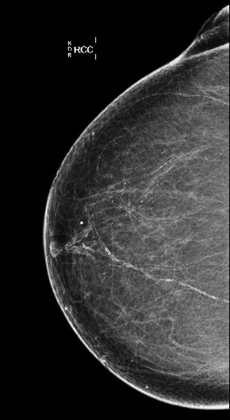
6 Case 3. Baseline Screen Question: What is the best BI-RADS assessment category? 1. BI-RADS 2 2. BI-RADS 3 3. BI-RADS 0 4. BI-RADS 1 6
7 Question: What is the best BI-RADS assessment category? 1. BI-RADS 2 2. BI-RADS 3 3. BI-RADS 0 4. BI-RADS 1 Discussion: Screening Mammogram Recommendations Options are BI-RADS 0, BI-RADS 2, or BI-RADS 1 These calcifications are indeterminate without diagnostic evaluation No need to describe morphology on screen Sufficient to state that there are calcifications in the right breast at 2 to 3 o clock 7

8 Case 3. Diagnostic Question: What is the best BI-RADS assessment category? 1. Benign BI-RADS 2 2. Short term follow up BI-RADS 3 3. Biopsy BI-RADS 4 4. Biopsy BI-RADS 5 8
9 Question: What is the best BI-RADS assessment category? 1. Benign BI-RADS 2 2. Short term follow up BI-RADS 3 3. Biopsy BI-RADS 4 4. Biopsy BI-RADS 5 Discussion: Coarse Heterogenous Biopsy. BI-RADS 4 Coarse heterogenous, clustered. 13% likelihood of malignancy upon biopsy BI-RADS 5 reserved for fine pleomorphic/linear 70% likelihood of malignancy upon biopsy Source: D Orsi CJ, Sickles EA, Mendelson EB, Morris EA. ACR BI-RADS Atlas. Breast Imaging Reporting System. 5 th Edition pg.61 9

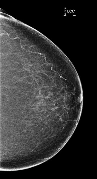
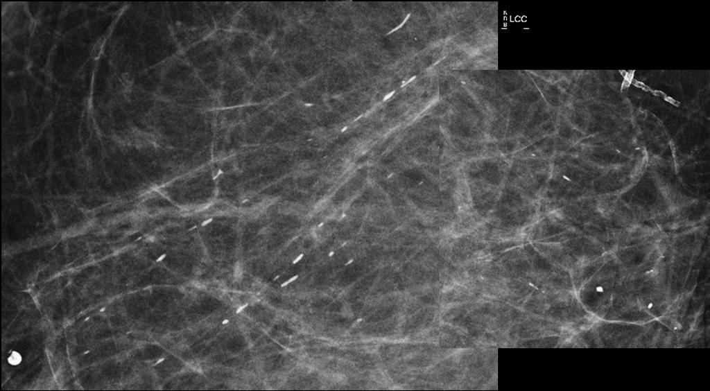
10 Case 4. Screening. Question: What is the best BI-RADS assessment category? 1. BI-RADS 2 2. BI-RADS 1 3. BI-RADS 0 4. BI-RADS 3 10
, parasternal, axilla, and")

11 Discussion BI-RADS 2 or B-RADS 1 Skin, diffuse Inframammary fold (IMF), parasternal, axilla, and around the areola Individual groups <5mm If you mention them, BI-RADS 2 Skin Calcs - Atypical CC ML 11


12 Tangential view shows the calcifications to be in the skin Case 5. Diagnostic. 55 year old with new calcifications CC Mag ML Mag Each calcification is <0.5mm 12
13 Question: What is the best BI-RADS description of these calcifications? 1. Round, clustered 2. Punctate, clustered 3. Amorphous, clustered 4. Milk of calcium, clustered Discussion: Punctate Round are >0.5mm but <1mm Margins are visible Do not layer 13
14 Question: What is the best BI-RADS assessment category? 1. BI-RADS 0 2. BI-RADS 2 3. BI-RADS 3 4. BI-RADS 4 Discussion If new clustered, linear, segmental, or adjacent to known cancer = must biopsy These were DCIS upon biopsy 14

15 Case 6. Diagnostic ML Mag Each calcification is <0.5mm Question: What is the best BI-RADS description of these calcifications? 1. Coarse heterogenous, clustered 2. Fine pleomorphic, clustered 3. Round, clustered 4. Fine linear/branching, clustered 15

16 Discussion: Fine pleomorphic Fine pleomorphic, clustered Each calcification usually less than 0.5mm 29% of all biopsied positive Coarse heterogenous Usually >0.5mm but <1mm Fine linear/branching Contains a branch point Source: D Orsi CJ, Sickles EA, Mendelson EB, Morris EA. ACR BI-RADS Atlas. Breast Imaging Reporting System. 5 th Edition pg.61 Case 7. Screening. 16

17 Case 7. Screening. Case 7 17
18 Question: What is the best BI-RADS description of these calcifications? 1. Fine linear/branching, segmental 2. Vascular, segmental 3. Large, rod-like, segmental 4. Coarse heterogenous, segmental Discussion: Large rod-like Large, rod-like calcifications Associated with duct ectasia Most are >0.5mm Ductal distribution, radiating Occasionally branching Usually seen in women who are >60 BI-RADS 2 or BI-RADS 1 18


19 Case 8. Diagnostic. New Calcifications. Question: What is the best BI-RADS description of these calcifications? 1. Fine pleomorphic, regional 2. Fine linear/branching, regional 3. Milk of calcium, regional 4. Large, rod-like, regional 19


20 Discussion: Fine linear/branching <0.5mm versus large/rod-like >0.5mm Branching PPV of 70% Invasive carcinoma upon biopsy Source: D Orsi CJ, Sickles EA, Mendelson EB, Morris EA. ACR BI-RADS Atlas. Breast Imaging Reporting System. 5 th Edition pg.61 Case 9. Screening
21 Question: What is the best BI-RADS description of these calcifications? 1. Coarse heterogenous 2. Dystrophic 3. Coarse, popcorn 4. Rim Discussion: Coarse popcorn Increasing but conforming to expected pattern Large >2-3 mm Associated with benign-appearing stable mass BI-RADS 2 21


22 Case 10. Screening. 73 year old with history of prior breast cancer, status post lumpectomy 10 years ago MLO CC Question: What is the best BI-RADS description of these calcifications? 1. Rim 2. Fine pleomorphic 3. Amorphous 4. Coarse heterogenous 22

23 Discussion: Rim Formerly eggshell, and lucent centered Calcium <1mm when viewed on edge Fat necrosis Calcifications in walls of cysts Case 12. Diagnostic. New Calcifications. ML Mag CC Mag 23
24 Question: What is the best BI-RADS description of these calcifications? 1. Fine pleomorphic, clustered 2. Round, clustered 3. Milk of calcium, clustered 4. Amorphous, clustered Discussion: Amorphous Formerly indistinct Hazy Bilateral diffuse can be benign otherwise biopsy PPV is 20% These were benign 24

25 Case 12. Diagnostic after Baseline Mag ML Mag CC Question: What is the best BI-RADS description of these calcifications? 1. Punctate, diffuse 2. Punctate, regional 3. Amorphous, diffuse 4. Round, regional 25
26 Discussion: Punctate, Regional Regional usually >2cm Punctate <0.5mm Regional PPV 26% OK to BI-RADS 3 since baseline. If not, biopsy. These were biopsied and were benign. 26
Leonard M. Glassman MD Analysis of Breast Calcifications
 Importance of Calcification Leonard M. Glassman MD FACR American Institute for Radiologic Pathology Washington Radiology Associates, PC Washington DC 45% of all breast cancers present as calcification
BI-RADS Update. Martha B. Mainiero, MD, FACR, FSBI Brown University Rhode Island Hospital
 BI-RADS Update Martha B. Mainiero, MD, FACR, FSBI Brown University Rhode Island Hospital No Disclosures BI-RADS History 1980s Quality Issues ACR Accreditation BI-RADS 1994 2003 4 th Edition MRI, US January
Amammography report is a key component of the breast
 Review Article Writing a Mammography Report Amammography report is a key component of the breast cancer diagnostic process. Although mammographic findings were not clearly differentiated between benign
Imaging in breast cancer. Mammography and Ultrasound Donya Farrokh.MD Radiologist Mashhad University of Medical Since
 Imaging in breast cancer Mammography and Ultrasound Donya Farrokh.MD Radiologist Mashhad University of Medical Since A mammogram report is a key component of the breast cancer diagnostic process. A mammogram
Leonard M. Glassman MD
 BI-RADS The New BI-RADS Leonard M. Glassman MD FACR Former Chief of Breast Imaging American Institute for Radiologic Pathology Washington Radiology Associates, PC Breast Imaging Reporting and Data System
Armed Forces Institute of Pathology.
 Armed Forces Institute of Pathology www.radpath.com Armed Forces Institute of Pathology Breast Disease www.radpath.org Armed Forces Institute of Pathology Evaluation of Breast Calcifications Leonard M.
Ana Sofia Preto 19/06/2013
 Ana Sofia Preto 19/06/2013 Understanding the underlying pathophysiologic processes leading to the various types of calcifications Description and illustration of the several types of calcifications, according
Pictorial Essay Singapore Med J 2009; 50(9) :
 :") 907 Pictorial Essay CME Article Breast calcifications: which are malignant? Muttarak M, Kongmebhol P, Sukhamwang N ABSTRACT Most calcifications depicted on mammograms are benign. However, calcifications
907 Pictorial Essay CME Article Breast calcifications: which are malignant? Muttarak M, Kongmebhol P, Sukhamwang N ABSTRACT Most calcifications depicted on mammograms are benign. However, calcifications
S. Murgo, MD. Chr St-Joseph, Mons Erasme Hospital, Brussels
 S. Murgo, MD Chr St-Joseph, Mons Erasme Hospital, Brussels? Introduction Mammography reports are sometimes ambiguous and indecisive. ACR has developped the BIRADS. BIRADS consists of a lexicon in order
Benign, Reactive and Inflammatory Lesions of the Breast
 Benign, Reactive and Inflammatory Lesions of the Breast Marilin Rosa, MD Associate Member Section Head of Breast Pathology Department of Anatomic Pathology Program Director, Breast Pathology Fellowship
ORIGINAL ARTICLE EVALUATION OF BREAST LESIONS USING X-RAY MAMMOGRAM WITH HISTOPATHOLOGICAL CORRELATION
 Available online at www.journalijmrr.com INTERNATIONAL JOURNAL OF MODERN RESEARCH AND REVIEWS IJMRR ISSN: 2347-8314 Int. J. Modn. Res. Revs. Volume 3, Issue 10, pp 807-814, October, 2015 ORIGINAL ARTICLE
Breast Imaging Lexicon
 9//201 200 BI RADS th Edition 201 BI RADS th Edition Breast Imaging Lexicon Mammographic Pathology and Assessment Categories Deborah Thames, R.T.(R)(M)(QM) The Advanced Health Education Center Nonmember:
AMSER Case of the Month: November 2018
 AMSER Case of the Month: November 2018 52 year old female with an abnormal screening mammogram Areeg Rehman, MS 4 Nova Southeastern University Rebecca T. Sivarajah, MD Penn State University College of
Segmental Breast Calcifications
 Residents Section Pattern of the Month Chen et al. Segmental reast Calcifications Residents Section Pattern of the Month Residents inradiology Po-Hao Chen 1 Erica T. Ghosh 1,2 Priscilla J. Slanetz 1,2
Lesion Imaging Characteristics Mass, Favoring Benign Circumscribed Margins Intramammary Lymph Node
 Lesion Imaging Characteristics Mass, Favoring Benign Circumscribed Margins Intramammary Lymph Node Oil Cyst Mass, Intermediate Concern Microlobulated Margins Obscured Margins Mass, Favoring Malignant Indistinct
Mammographic imaging of nonpalpable breast lesions. Malai Muttarak, MD Department of Radiology Chiang Mai University Chiang Mai, Thailand
 Mammographic imaging of nonpalpable breast lesions Malai Muttarak, MD Department of Radiology Chiang Mai University Chiang Mai, Thailand Introduction Contents Mammographic signs of nonpalpable breast cancer
Breast calcification: Management and Pictorial Review
 Breast calcification: Management and Pictorial Review Poster No.: C-0692 Congress: ECR 2014 Type: Educational Exhibit Authors: V. de Lara Bendahan, M. F. Ramos Solis, A. Amador Gil, C. 1 2 3 2 4 4 Gómez
ACRIN 6666 IM Additional Evaluation: Additional Views/Targeted US
 Additional Evaluation: Additional Views/Targeted US For revised or corrected form check box and fax to 215-717-0936. Instructions: The form is completed based on recommendations (from ID form) for additional
BI-RADS Categorization As a Predictor of Malignancy 1
 Susan G. Orel, MD Nicole Kay, BA Carol Reynolds, MD Daniel C. Sullivan, MD BI-RADS Categorization As a Predictor of Malignancy 1 Index terms: Breast, biopsy, 00.1261 Breast neoplasms, localization, 00.125,
Non-mass Enhancement on Breast MRI. Aditi A. Desai, MD Margaret Ann Mays, MD
 Non-mass Enhancement on Breast MRI Aditi A. Desai, MD Margaret Ann Mays, MD Breast MRI Important screening and diagnostic tool, given its high sensitivity for breast cancer detection Breast MRI - Indications
Microcalcifications detected on mammography classified as BIRADS 4 and 5 and their correlations with histopatologic findigns
 Microcalcifications detected on mammography classified as BIRADS 4 and 5 and their correlations with histopatologic findigns Poster No.: C-0401 Congress: ECR 2010 Type: Educational Exhibit Topic: Breast
Radiologic and pathologic correlation of non-mass like breast lesions on US and MRI: Benign, high risk, versus malignant
 Radiologic and pathologic correlation of non-mass like breast lesions on US and MRI: Benign, high risk, versus malignant Poster No.: C-1161 Congress: ECR 2013 Type: Educational Exhibit Authors: J. Kwak,
CURRENTLY IN THE United States, 431,284
 Increased Breast Calcifications in Women With ESRD on Dialysis: Implications for Breast Cancer Screening Mario Castellanos, MD, Seema Varma, MD, Kathleen Ahern, PhD, RN, Sue-Jane Grosso, MD, Shalom Buchbinder,
Case study 1. Rie Horii, M.D., Ph.D. Division of Pathology Cancer Institute Hospital, Japanese Foundation for Cancer Research
 NCCN/JCCNB Seminar in Japan April 15, 2012 Case study 1 Rie Horii, M.D., Ph.D. Division of Pathology Cancer Institute Hospital, Japanese Foundation for Cancer Research Present illness: A 50y.o.premenopausal
Imaging the Symptomatic Patient. Avice M.O Connell MD,FACR,FSBI Professor of Imaging Sciences Director, Women s Imaging University of Rochester
 Imaging the Symptomatic Patient Avice M.O Connell MD,FACR,FSBI Professor of Imaging Sciences Director, Women s Imaging University of Rochester The four most common symptoms Mass Pain Discharge Infection
Armed Forces Institute of Pathology.
 Armed Forces Institute of Pathology www.radpath.com Armed Forces Institute of Pathology Breast Disease www.radpath.org Armed Forces Institute of Pathology Interpretation of Breast MRI Leonard M. Glassman
Pitfalls and Limitations of Breast MRI. Susan Orel Roth, MD Professor of Radiology University of Pennsylvania
 Pitfalls and Limitations of Breast MRI Susan Orel Roth, MD Professor of Radiology University of Pennsylvania Objectives Review the etiologies of false negative breast MRI examinations Discuss the limitations
Linear Breast Calcifications
 Residents Section Pattern of the Month Lai et al. Linear reast alcifications Residents Section Pattern of the Month Residents inradiology Kenny. Lai 1 Priscilla J. Slanetz Ronald L. Eisenberg Lai K, Slanetz
AB MR Interpretation Overview
 AB MR Interpretation Overview Goal of AB MR interpretation is to maintain high sensitivity and specificity In order to minimize false positives and short term follow ups, it is fundamental to focus only
Radiology Review Course Hotel del Coronado Coronado, California
 37 th Annual Radiology Review Course Hotel del Coronado Coronado, California Friday, April 21, 2017 - PM TABLE OF CONTENTS Friday, April 21, 2017 - PM SAM Session - Breast Imaging Update 12:45 PM 1:30
Is Probably Benign Really Just Benign? Peter R Eby, MD, FSBI Virginia Mason Medical Center Seattle, WA
 Is Probably Benign Really Just Benign? Peter R Eby, MD, FSBI Virginia Mason Medical Center Seattle, WA Disclosures: CONSULTANT FOR DEVICOR MEDICAL ARS Question 1 Is probably benign really just benign?
OF HEMATOMA INTRACRANIAL HEMORRHAGE
 SCOPE TUTORIAL RADIOLOGY CT BRIAN BREASTS US MRCP Natrada Rawdhetubhai, M.D. Radiology Department, Lerdsin General Hospital CT OF HEMATOMA INTRACRANIAL HEMORRHAGE Intra-axial Extra-axial Intraventricular
Evaluation of Breast Specimens Removed by Needle Localization Technique
 Evaluation of Breast Specimens Removed by Needle Localization Technique Specimen Handling: The breast specimen when received should be measured and grossly inspected for any orientation designated by the
CDIS: what's beyond microcalcifications? - Pictorial essay
 CDIS: what's beyond microcalcifications? - Pictorial essay Poster No.: C-1096 Congress: ECR 2014 Type: Educational Exhibit Authors: R. N. Lucas, C. A. S. Ruano, I. Oliveira, J. M. G. Lourenco, Z. 1 1 1
The Breast Imaging Reporting and Data System (BI-RADS) has standardized the description and management of findings identified on mammograms, thereby f
 has standardized the description and management of findings identified on mammograms, thereby f") ORIGINAL RESEARCH BREAST IMAGING Elizabeth S. Burnside, MD, MPH, MS Jennifer E. Ochsner, MD Kathryn J. Fowler, MD Jason P. Fine, PhD Lonie R. Salkowski, MD Daniel L. Rubin, MD, MS Gale A. Sisney, MD Use
ORIGINAL RESEARCH BREAST IMAGING Elizabeth S. Burnside, MD, MPH, MS Jennifer E. Ochsner, MD Kathryn J. Fowler, MD Jason P. Fine, PhD Lonie R. Salkowski, MD Daniel L. Rubin, MD, MS Gale A. Sisney, MD Use
Hiding in Plain Sight / Site: Archictectural Distortions and Breast Asymmetries
 Hiding in Plain Sight / Site: Archictectural Distortions and Breast Asymmetries Dianne Georgian-Smith MD Associate Professor Harvard Med School Brigham and Women s Hospital Financial Disclosures Book Publication
Screen Detected Breast Carcinoma, with Coarse Calcification
 Screen Detected Breast Carcinoma, with Coarse Calcification Poster No.: R-0168 Congress: RANZCR ASM 2013 Type: Educational Exhibit Authors: L. Ebrahim, D. Abeywardhana, D. Dissanayake, C. Metcalf, E. Wylie;
Retrospective Analysis on Malignant Calcification Previously Misdiagnosed as Benign on Screening Mammography 스크리닝유방촬영술에서양성으로진단되었던악성석회화에대한후향적분석
 Original Article pissn 1738-2637 / eissn 2288-2928 https://doi.org/10.3348/jksr.2017.76.4.251 Retrospective Analysis on Malignant Calcification Previously Misdiagnosed as Benign on Screening 스크리닝유방촬영술에서양성으로진단되었던악성석회화에대한후향적분석
AMSER Case of the Month: September 2018
 AMSER Case of the Month: September 2018 60-year-old woman with a left breast mass noted on screening mammography. Catherine McNulty, MS4 Tulane University School of Medicine Dr. Robin Sobolewski Breast
Pathologic outcomes of coarse heterogeneous calcifications detected on mammography
 Pathologic outcomes of coarse heterogeneous calcifications detected on mammography Poster No.: C-1957 Congress: ECR 2011 Type: Scientific Paper Authors: H. J. Lim, K. R. Cho, K. W. Hwang, B. K. Seo, O.
BI-RADS and Breast MRI. Kathy Borovicka, M.D. Thursday February 15, 2018
 BI-RADS and Breast MRI Kathy Borovicka, M.D. Thursday February 15, 2018 Learning Objectives Be familiar with the Breast Imaging Reporting and Data System (BI-RADS) Understand the components of a breast
PLACE LABEL HERE. ACRIN 6657 MRI Form: Pre-Treatment (MRI-1)
") M3 ACRIN 6657 MRI Form: Pre-Treatment (MRI-1) If this is a revised or corrected form,indicate by checking box. ACRIN Study 6657 Case # Instructions: In accordance with the protocol, four MRI exams are
M3 ACRIN 6657 MRI Form: Pre-Treatment (MRI-1) If this is a revised or corrected form,indicate by checking box. ACRIN Study 6657 Case # Instructions: In accordance with the protocol, four MRI exams are
MEDICAL IMAGING AND BREAST DISEASE HOW CAN WE HELP YOU?
 MEDICAL IMAGING AND BREAST DISEASE HOW CAN WE HELP YOU? Barbara M. Preston, M.D. SCREENING MAMMOGRAPHY AVERAGE RISK PATIENTS KAISER RECOMMENDATION: ALL WOMEN (INCLUDING TRANSGENDER FEMALES) Every 1-21
Diagnostic benefits of ultrasound-guided. CNB) versus mammograph-guided biopsy for suspicious microcalcifications. without definite breast mass
 versus mammograph-guided biopsy for suspicious microcalcifications. without definite breast mass") Volume 118 No. 19 2018, 531-543 ISSN: 1311-8080 (printed version); ISSN: 1314-3395 (on-line version) url: http://www.ijpam.eu ijpam.eu Diagnostic benefits of ultrasound-guided biopsy versus mammography-guided
Volume 118 No. 19 2018, 531-543 ISSN: 1311-8080 (printed version); ISSN: 1314-3395 (on-line version) url: http://www.ijpam.eu ijpam.eu Diagnostic benefits of ultrasound-guided biopsy versus mammography-guided
Breast Imaging Essentials
 Breast Imaging Essentials Module 5 Transcript 2016 ASRT. All rights reserved. Breast Imaging Essentials Module 5 Pathology 1. ASRT Animation 2. Welcome Welcome to Module 5 of Breast Imaging Essentials
CLINICAL SIGNIFICANCE OF BENIGN EPITHELIAL CHANGES
 Papillomas. Papillomas are composed of multiple branching fibrovascular cores, each having a connective tissue axis lined by luminal and myoepithelial cells ( Fig. 23-11 ). Growth occurs within a dilated
Index. C Calcifications fat necrosis 1, 61 fat necrosis 4, 69 nipple/peri-areolar involvement 1, 165
 A ADH. See Atypical ductal hyperplasia (ADH) American College of Radiology (ACR), BI-RADS background parenchymal enhancement, 8, 9, 81, 82 fibroglandular tissue guidelines, 6 American Joint Committee on
National Diagnostic Imaging Symposium 2013 SAM - Breast MRI 1
 National Diagnostic Imaging Symposium 2013 December 8-12, 2013 Disney s Yacht Club Resort Lake Buena Vista, Florida Self Assessment Module Questions, Answers and References Day SAM Title - Each SAM title
Ductal carcinoma in situ: ultrasound, mammography and MRI features with pathologic correlation
 Ductal carcinoma in situ: ultrasound, mammography and MRI features with pathologic correlation Poster No.: C-2252 Congress: ECR 2013 Type: Educational Exhibit Authors: L. Fernandes, H. A. M. R. Tinto,
Breast Cancer Screening with Mammography
 Progress in Public Health Breast Cancer Screening with Mammography JMAJ 44(7): 318 324, 2001 Tokiko ENDO Director, Department of Radiology, National Nagoya Hospital Abstract: Breast cancer has been increasing
Ultrasonography. Methods. Brief Description. Indications. Device-related Prerequisites. Technical Requirements. Evaluation Criteria
 1 Ultrasonography Brief Description Imaging modality using sound waves Tissue-specific wave reflection. Indications Evaluation of palpable breast nodules Evaluation of clinically occult mammographic findings
Breast MRI Update. Jeffrey C. Weinreb, MD, FACR Yale University School of Medicine
 Breast MRI Update Jeffrey C. Weinreb, MD, FACR jeffrey.weinreb@yale.edu Yale University School of Medicine I disclose the following financial relationships with relevant commercial interests: Bracco Bayer
Diagnostic Dilemmas of Breast Imaging
 Diagnostic Dilemmas of Breast Imaging Common Causes of Error in Breast Cancer Detection By: Jason Cord, M.D. Mammography: Initial Imaging The standard for detection of breast cancer Screening mammography
Cytyc Corporation - Case Presentation Archive - March 2002
 FirstCyte Ductal Lavage History: 68 Year Old Female Gail Index: Unknown Clinical History: Negative Mammogram in 1995 6 yrs. later presents with bloody nipple discharge Subsequent suspicious mammogram Suspicious
Malignant transformation of fibroadenomas
 Malignant transformation of fibroadenomas Poster No.: C-2503 Congress: ECR 2013 Type: Educational Exhibit Authors: L. N. Elias, M. A. Rudner, L. M. Yano, P. C. Moraes, Y. 1 1 1 1 1 1 2 1 2 Chang, M. B.
Atypical Ductal Hyperplasia of the Breast:
 Atypical Ductal Hyperplasia of the Breast: Radiologic and Histopathologic Correlation 1 Ji Young Lee, M.D., Bo Kyoung Seo, M.D. 2, Jung Hyck Kim, M.D., Yu Whan Oh, M.D., Kyu Ran Cho, M.D., Eun Jeong Choi,
8/31/2016 HIDING IN PLAIN SITE, ARCHITECTURAL DISTORTIONS AND BREAST ASYMMETRIES ARCHITECTURAL DISTORTIONS ARCHITECTURAL DISTORTIONS
 HIDING IN PLAIN SITE, ARCHITECTURAL DISTORTIONS AND BREAST ASYMMETRIES DEBORAH THAMES R.T. (R)(M)(QM) ARCHITECTURAL DISTORTIONS Definition is disruption of the natural flow of breast pattern towards the
COPE Library Sample
 Breast Anatomy LOBULE LOBE ACINI (MILK PRODUCING UNITS) NIPPLE AREOLA COMPLEX ENLARGEMENT OF DUCT AND LOBE LOBULE SUPRACLAVICULAR NODES INFRACLAVICULAR NODES DUCT DUCT ACINI (MILK PRODUCING UNITS) 8420
Breast pathology. 2nd Department of Pathology Semmelweis University
 Breast pathology 2nd Department of Pathology Semmelweis University Breast pathology - Summary - Benign lesions - Acute mastitis - Plasma cell mastitis / duct ectasia - Fat necrosis - Fibrocystic change/
of Thyroid Lesions Comet Tail Crystals
 2 Ultrasound Features of Thyroid Lesions There are many different features indicating a certain benign or malignant tumor type, but many of these are overlapping signs. Combining several features is considered
Breast Calcifications: The Focal Group
 Residents Section Pattern of the Month reast alcifications Residents Section Pattern of the Month Downloaded from www.ajronline.org by 37.44.196.164 on 01/08/18 from IP address 37.44.196.164. opyright
Accreditation Case Review: Mammography and Stereotactic Biopsy
 Accreditation Case Review: Mammography and Stereotactic Biopsy Brett T. Parkinson MD Breast Imaging Director Breast Care Services Intermountain Healthcare Chair, ACR Committee on Mammography Accreditation
Breast Health and Imaging Glossary
 Contact: Lorna Vaughan HerSpace Breast Imaging & Biopsy Associates 300 State Route 35 South W. Long Branch, NJ 07764 732-571-9100, ext. 104 lorna@breast-imaging.com Breast Health and Imaging Glossary Women
PLACE LABEL HERE BASELINE / PRE-TREATMENT. ACRIN 6657 Extension MRI Form: Baseline / Pre-Treatment MRI 1. o Unknown
 T1 ACRIN 6657 Extension MRI Form: Baseline / Pre-Treatment MRI 1 If this is a revised or corrected form, please box. ACRIN Study 6657 No. Instructions: In accordance with the protocol, four MRI exams are
Learning Objectives. 1. Identify which patients meet criteria for annual lung cancer screening
 Disclosure I, Taylor Rowlett, DO NOT have a financial interest /arrangement or affiliation with one or more organizations that could be perceived as a real or apparent conflict of interest in the context
DCIS of the Breast--MRI findings with mammographic correlation.
 DCIS of the Breast--MRI findings with mammographic correlation. Poster No.: C-1560 Congress: ECR 2013 Type: Educational Exhibit Authors: N. B. Ibrahim, P. Morris, S. ANANDAN; Burlington, MA/US Keywords:
Criteria of Malignancy. Evaluation Score
 30 5 Diagnostic Criteria Criteria of Malignancy Table 5.2 lists criteria in contrast-enhancing MR mammography that strongly indicate the presence of malignancy or are unspecific. Unifactorial evaluation
Radiologic Findings of Mucocele-like Tumors of the breast: Can we differentiate pure benign from associated with high risk lesions?
 Radiologic Findings of Mucocele-like Tumors of the breast: Can we differentiate pure benign from associated with high risk lesions? Poster No.: C-0332 Congress: ECR 2014 Type: Educational Exhibit Authors:
MRI in breast cancer: diagnosis and intervention. Dr Sue Barter Addenbrookes Hospital, Cambridge UK
 MRI in breast cancer: diagnosis and intervention Dr Sue Barter Addenbrookes Hospital, Cambridge UK Intervention will be discussed in High Risk Screening! Indications UK and Europe: Breast MRI is well established
Image guided core biopsies:
 Recommendations on the Surgical, Radiologic and Pathologic Approaches to Breast Disease: Using best practices based on multidisciplinary methodologies developed through the Allina Breast Committee. Image
Alena Levit MD Avice O Connell MD University of Rochester, Rochester, NY
 Alena Levit MD Avice O Connell MD University of Rochester, Rochester, NY Purpose Review imaging spectrum of both common benign and malignant breast lesions Describe and demonstrate CT features with mammogram,
Medical Education. CME Article Clinics in diagnostic imaging (125) Padungchaichote W, Kongmebhol P, Muttarak M
 Padungchaichote W, Kongmebhol P, Muttarak M") 1062 Medical Education CME Article Clinics in diagnostic imaging (125) Padungchaichote W, Kongmebhol P, Muttarak M la Ib Ic Fig. I (a) Bilateral mediolateral oblique mammograms; (b) spot right craniocaudal
1062 Medical Education CME Article Clinics in diagnostic imaging (125) Padungchaichote W, Kongmebhol P, Muttarak M la Ib Ic Fig. I (a) Bilateral mediolateral oblique mammograms; (b) spot right craniocaudal
OPTO-ACOUSTIC BREAST IMAGING
 OPTO-ACOUSTIC BREAST IMAGING A Novel Fusion of Functional and Morphologic Imaging Reni S. Butler, MD A. Thomas Stavros, MD F. Lee Tucker, MD Michael J. Ulissey, MD PURPOSE 1. Explain opto-acoustic (OA)
Diseases of the breast (1 of 2)
") Diseases of the breast (1 of 2) Introduction A histology introduction Normal ducts and lobules of the breast are lined by two layers of cells a layer of luminal cells overlying a second layer of myoepithelial
Diseases of the breast (1 of 2) Introduction A histology introduction Normal ducts and lobules of the breast are lined by two layers of cells a layer of luminal cells overlying a second layer of myoepithelial
BREAST MRI. VASILIKI FILIPPI RADIOLOGIST CT MRI & PET/CT Departments Hygeia Hospital, Athens, Greece
 BREAST MRI VASILIKI FILIPPI RADIOLOGIST CT MRI & PET/CT Departments Hygeia Hospital, Athens, Greece Breast ΜR Imaging (MRM) Breast MR imaging is an extremely powerful diagnostic tool, that when used in
RSNA, /radiol Appendix E1. Methods
 RSNA, 2016 10.1148/radiol.2016151097 Appendix E1 Methods US and Near-infrared Data Acquisition Four optical wavelengths (740 nm, 780 nm, 808 nm, and 830 nm) were used to sequentially deliver the light
Case Scenario 1 History and Physical 3/15/13 Imaging Pathology
 Case Scenario 1 History and Physical 3/15/13 The patient is an 84 year old white female who presented with an abnormal mammogram. The patient has a five year history of refractory anemia with ringed sideroblasts
Standard Breast Imaging Modalities. Lilian Wang, M.D. Breast Imaging Section Department of Radiology Northwestern Medicine
 Standard Breast Imaging Modalities Lilian Wang, M.D. Breast Imaging Section Department of Radiology Northwestern Medicine Overview Standard breast imaging modalities Mammography Ultrasound MRI Imaging
CHAPTER 2 MAMMOGRAMS AND COMPUTER AIDED DETECTION
 9 CHAPTER 2 MAMMOGRAMS AND COMPUTER AIDED DETECTION 2.1 INTRODUCTION This chapter provides an introduction to mammogram and a description of the computer aided detection methods of mammography. This discussion
GTC 2018, San Jose, USA
 Automated Segmentation of Suspicious Breast Masses from Ultrasound Images Viksit Kumar, Jeremy Webb, Adriana Gregory, Mostafa Fatemi, Azra Alizad GTC 2018, San Jose, USA SIGNIFICANCE Breast cancer is most
Development of a Bayesian Network for Diagnosis of Breast Cancer
 Development of a Bayesian Network for Diagnosis of Breast Cancer Linda M. Roberts, Charles E. Kahn, Jr., Peter Haddawy {roberts,kahn,haddawy}@cs.uwm.edu Department of Electrical Engineering and Computer
Can magnetic resonance imaging obviate the need for biopsy for microcalcifications?
 Original Article Can magnetic resonance imaging obviate the need for biopsy for microcalcifications? Shinya Yamamoto, Takashi Chishima Department of Breast Surgery, Yokohama Rosai Hospital, Yokohama 222-0036,
EARLY DETECTION: MAMMOGRAPHY AND SONOGRAPHY
 EARLY DETECTION: MAMMOGRAPHY AND SONOGRAPHY Elizabeth A. Rafferty, M.D. Avon Comprehensive Breast Center Massachusetts General Hospital Harvard Medical School Breast Cancer Screening Early detection of
Tips and Tricks to performing Magnetic Resonance Imaging Guided Breast Interventional Procedures Habib Rahbar, MD, FSBI October 23, 2018, 7:00pm ET
 Tips and Tricks to performing Magnetic Resonance Imaging Guided Breast Interventional Procedures Habib Rahbar, MD, FSBI October 23, 2018, 7:00pm ET SAM Questions/Answers/Rationales/References 1. Below
IBCM 2, April 2009, Sarajevo, Bosnia and Herzegovina
 Preoperative diagnosis and treatment planning in breast cancer The pathologist s perspective L. Mazzucchelli Istituto Cantonale di Patologia Locarno, Switzerland IBCM 2, 23-25 April 2009, Sarajevo, Bosnia
High Detection Rate of Breast Ductal Carcinoma In Situ Calcifications on Mammographically Directed High-Resolution Sonography
 Article High Detection Rate of Breast Ductal Carcinoma In Situ Calcifications on Mammographically Directed High-Resolution Sonography Beverly E. Hashimoto, MD, Dawna J. Kramer, MD, Vincent J. Picozzi,
Usefulness of MRI of Microcalcification Lesions to Determine the Indication for Stereotactic Mammotome Biopsy
 Usefulness of MRI of Microcalcification Lesions to Determine the Indication for Stereotactic Mammotome Biopsy MARIKO KIKUCHI 1, HIROKAZU TANINO 1, YOSHIMASA KOSAKA 1, NORIHIKO SENGOKU 1, KEISHI YAMASHITA
PAAF vs Core Biopsy en Lesiones Mamarias Case #1
 5/19/2014 PAAF vs Core Biopsy en Lesiones Mamarias Case #1 Fine Needle Aspiration Cytology of Breast: Correlation with Needle Core Biopsy 64-year-old woman Mass in breast Syed Hoda, MD CD31 Post-Radiation
MRI BI-RADS: How to make it out?
 MRI BI-RADS: How to make it out? Poster No.: C-1850 Congress: ECR 2016 Type: Educational Exhibit Authors: M. Ben Ammar, A. Ben Miled, O. Ghdes, S. Harguem, A. Gaja, N. Mnif; Tunis/TN Keywords: Breast,
Min Jung Kim Department of Medicine The Graduate School, Yonsei University
 Zoomed image of contact mammography versus magnification mammography in the diagnosis of microcalcifications with soft-copy full field digital mammography Min Jung Kim Department of Medicine The Graduate
Breast Density. Update 2018: Implications for Clinical Practice
 Breast Density Update 2018: Implications for Clinical Practice Matthew A. Stein, MD Assistant professor Breast Imaging Department of Radiology and Imaging Sciences University of Utah Health Disclosures
AMSER Case of the Month: November 2018
 AMSER Case of the Month: November 2018 42 year old with right breast mass Rina Kiyota Petek Lake Erie College of Osteopathic Medicine, OMS-III Kossivi Dantey, MD Bibianna Klepchick, MD Matthew Hartman,
Atypical ductal hyperplasia diagnosed at ultrasound guided biopsy of breast mass
 Atypical ductal hyperplasia diagnosed at ultrasound guided biopsy of breast mass Poster No.: C-1483 Congress: ECR 2014 Type: Authors: Keywords: DOI: Scientific Exhibit J. Cho, J. Chung, E. S. Cha, J. E.
RADIOLOGIC EVALUATION OF BREAST CANCER
 RADIOLOGIC EVALUATION OF BREAST CANCER Orsolya Farkas, Gabriella Bodrogi and Gábor Szalai Department of Radiology, Pécs University Orsifarkas@yahoo.com Complex evaluation of the breast Patient history
Breast Imaging! Ravi Adhikary, MD!
 Breast Imaging! Ravi Adhikary, MD! ACS Estimated Cancers Statistics 2014! Breast! New Cases in Women! 232,670 (+67,570 in situ)! Deaths in Women! 40,000! Colon! 48,380! 24,040! Cervical! 12,360! 4,020!
Atypical Ductal Hyperplasia and Papillomas: A Comparison of Ultrasound Guided Breast Biopsy and Stereotactic Guided Breast Biopsy
 Atypical Ductal Hyperplasia and Papillomas: A Comparison of Ultrasound Guided Breast Biopsy and Stereotactic Guided Breast Biopsy Breast Cancer is the most common cancer diagnosed in women in the United
SCBT-MR 2015 LungRADS : Basics
 SCBT-MR 2015 LungRADS : Basics Reginald F. Munden MD, DMD, MBA I have no conflicts of interest to report National Lung Cancer Screening Trial 20% lung cancer mortality reduction 6.9% all cause mortality
CPC 4 Breast Cancer. Rochelle Harwood, a 35 year old sales assistant, presents to her GP because she has noticed a painless lump in her left breast.
 CPC 4 Breast Cancer Rochelle Harwood, a 35 year old sales assistant, presents to her GP because she has noticed a painless lump in her left breast. 1. What are the most likely diagnoses of this lump? Fibroadenoma