Brain Pain Infections of the CNS
|
|
|
- Hillary Edwards
- 6 years ago
- Views:
Transcription


1 FRIDAY, OCTOBER 28, 2016 Brain Pain Infections of the CNS Suyash Mohan MD, PDCC Assistant Professor of Radiology & Neurosurgery Division of Neuroradiology, Department of Radiology Perelman School of Medicine at University of Pennsylvania
2 Why Important? Life-threatening problems with high associated mortality ξ morbidity Increasing incidence Primarily attributed to AIDS ξ widespread use of immunosuppressants Prognosis depends on rapid identification
3 What s Important? Meningitis Ventriculitis Abscess Empyema Encephalitis Toxoplasmosis Tuberculosis Fungal Cysticercosis Neonatal infections
4 BACTERIAL INFECTIONS Meningitis: Most common CNS infection S/S include: Headache Fever Neck stiffness Photophobia Vomiting Altered consciousness Diagnosis: Clinical ξ CSF findings Role of neuroimaging: To confirm suspected meningitis To rule out meningitis mimics To evaluate for complications To rule out increased ICP before LP


5 Case: 45 yr male presented with seizures ξ fever Tubercular meningitis


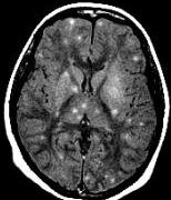
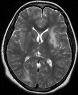
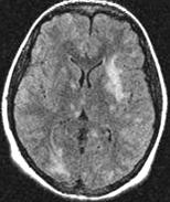
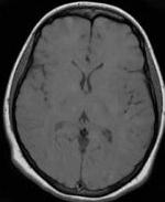
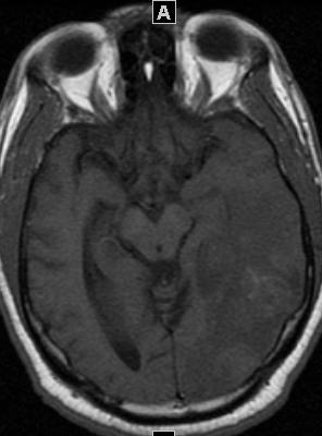
6 Case: 18 yr male presented with headaches ξ fever


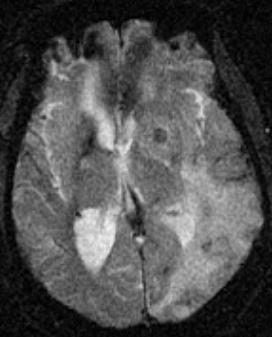
7 Case: 28 year-old male with mental status changes


8 Pre Post Pre Post Viral Meningitis: Symptoms improved with acyclovir
9 D/Dx of meningeal enhancement Meningeal enhancement is nonspecific, may be caused by the following 5 different etiologic subgroups: 1. Infectious 2. Carcinomatous 3. Reactive: surgery, shunt, trauma 4. Chemical: ruptured dermoid, cysticercus, intrathecal chemotherapy 5. Inflammatory: sarcoidosis, collagen vascular diseases, etc


10 Meningitis Nonspecific diffuse meningeal enhancement, FLAIR hyperintensity Cryptococcus Coccidiomycosis Tuberculosis Toxoplasmosis Listeria
11 How to differentiate between normal meningeal vessels/ cortical veins from abnormal leptomeningeal enhancement? Abnormal meningeal enhancement on MRI is: Thick Nodular/ Irregular Longer & continuous Asymmetrical Extends deep into the base of the sulci Long-segment (> 3 cm) or diffuse convexity meningeal enhancement If meningeal enhancement was present on more than three contiguous 1.5 T SE MR images Quint et al. Academic Radiology 1996: (3); Kamra et al. The British Journal of Radiology, 77 (2004),
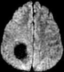
12 How to differentiate b/w acute vs chronic meningitis? Acute: meningeal enhancement is located over the cerebral convexity Chronic: meningeal enhancement is most prominent in the basal cisterns How to differentiate b/w infective vs carcinomatous meningitis? Carcinomatous meningitis typically presents with dural enhancement


13 34 Y/M with dizziness & loss of balance. Also history of ulcerative colitis & colon cancer
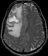
14 Imaging in Meningitis Most common: normal CSF FLAIR hyperintensity Meningeal enhancement Complications: Hydrocephalus, ventriculitis Effusion, empyema, cerebritis, abscess Cerebral edema, herniation Venous thrombosis, vasospasm, infarct


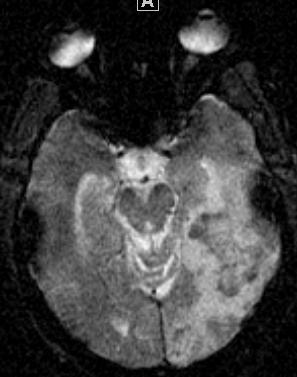
15 Case: year-old male with mental status changes. PMH - Sinusitis
16 Complications Effusion vs. Empyema Cerebritis
17 Complications Hydrocephalus Ventriculitis

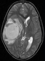
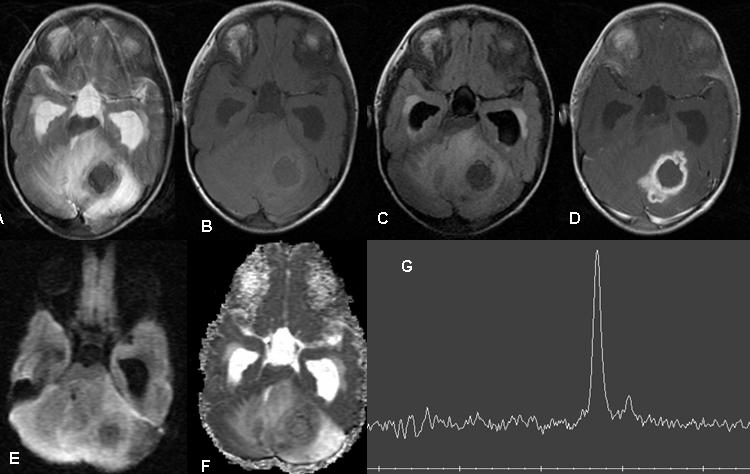
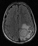
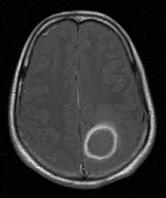
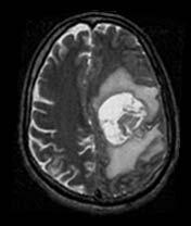
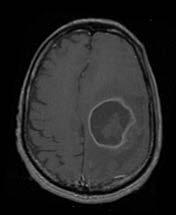
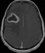
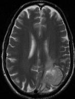
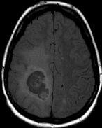
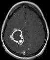
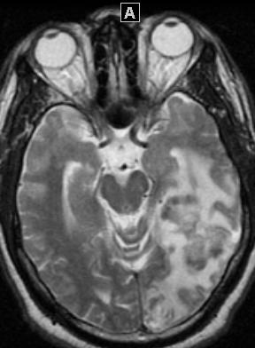
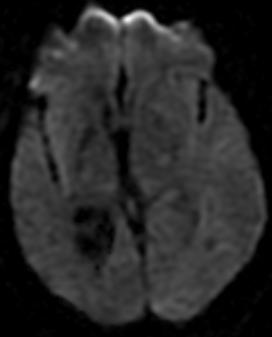
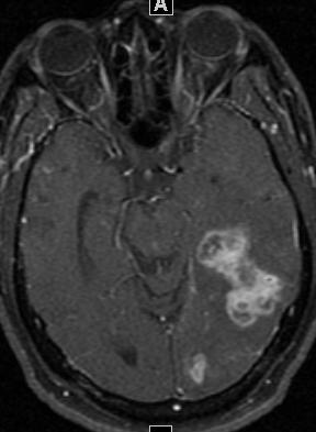
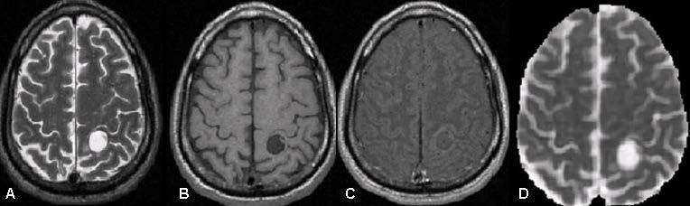
18 Case: 31 year-old woman with confusion and right sided weakness
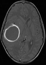

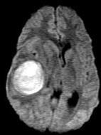
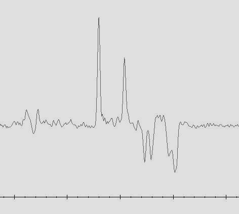
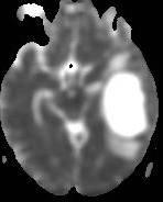
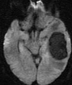
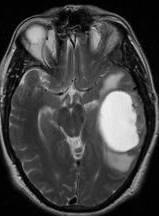
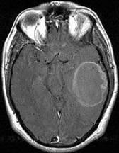

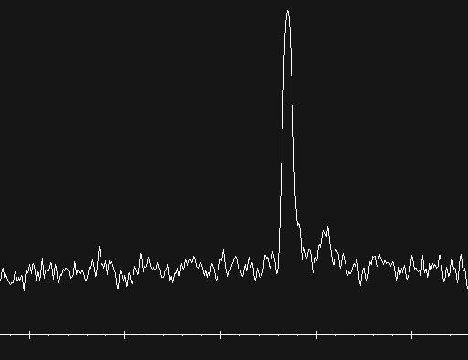
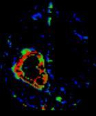
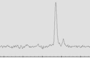
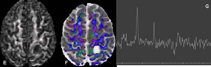
19 How to differentiate between cystic tumors and abscesses? MR Spetroscopy in combination of DWI Can differentiate cystic tumors from brain abscess Suc Ac Lac AA T2 PC T1 DWI ADC PMRS
20 Brain Abscesses ξ MRS Spectra from brain abscesses reveal Acetate (1.92 ppm) Succinate (2.4 ppm) Lactate (1.3 ppm) Amino acids (0.9 ppm) Main brain metabolites like NAA, Cr ξ Cho Usually not detectable Presence of acetate ξ succinate suggests anaerobic bacterial infection Garg M et al. Radiology 2004; 230:

21 CNS TUBERCULOSIS Incidence of TB has been increasing d/t increase in AIDS ξ other immunodeficiency states CNS involvement occurs in 2 5% of all TB patients ξ in 10-15% of AIDS patients Diffuse form: Leptomeningitis Localized parenchymal forms: Tuberculoma Miliary tuberculosis Abscess
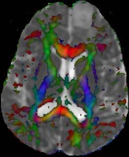
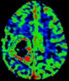
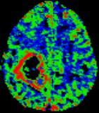
22 Tuberculoma Lip


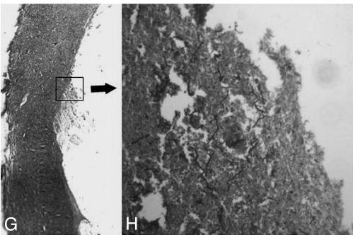
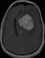
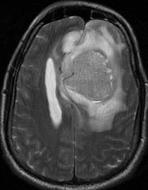
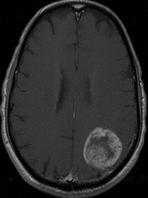
23 Tuberculoma HPE: Central caseous necrosis, surrounded by reactive epithelial cells, Langhans giant cells, PMN cells, plasma cells ξ lymphocytes

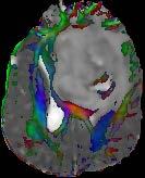
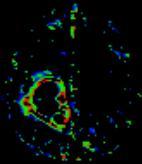
24 MTT1 Miliary Tuberculosis
25 Tubercular abscess T2 T1 MTT1 Flair PC-MTT1 Lip DWI ADC PMRS

26 Case: 39 year-old man with confusion. PMH of NHL on treatment

27 Case: 46 year-old immunocompetent man with headache and weakness T2 PCT1 DWI ADC PMRS HPE


28 How to diagnose a fungal abscess? Intracavitary projections: Characteristic Arise from the wall Restricted diffusion in projections ξ wall No restriction rest of the abscess core Contrast enhancement only in the wall These projections are a distinguishing feature of a fungal cause on conventional MRI
29 Role of MR Perfusion Abscesses show relative decreased CBV from neoplasms, which demonstrate significantly elevated CBV Infective lesions have lower values of rcbv as compared to high grade glioma Infective lesions have higher rcbv as compared to LGG As a result of differential expression of VEGF

30 Abscess Grade II Astrocytoma Grade III Astrocytoma Glioblastoma T2 PCT1 Color map of FA CBV
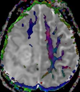
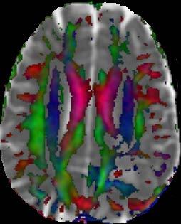
31 Permeability in Infections Diffusion Perfusion CBV uncorrected CBV corrected CBF K trans v e Angiogenesis in a Tuberculoma


32 Permeability in Infections Cerebral aspergillosis: patterns variable & depend upon immunological condition 1. Edematous lesions, 2. Hemorrhagic lesions, 3. Solid enhancing lesions aspergilloma Cho T2 T1 PCT1 PMRS CBV CBF K trans v e VEGF staining in the cytoplasm of giant cells
33 PARASITIC LESIONS Toxoplasmosis Neurocysticercosis Cerebral malaria Echinococcal Schistosomiasis


34 34 year old woman post renal transplant
 An enhancing nodule within and adjacent to the enhancing")

35 Toxoplasmosis MC opportunistic infection in HIV Frequently multifocal, predilection for BG, may hemorrhage Target sign: (30%) An enhancing nodule within and adjacent to the enhancing rim, represents an infolding of the cyst wall DWI Dark to isointense core, reflecting low viscosity ξ absence of purulent fluid, likley due to impaired immune response in immunocompromised patients


36 Cysticercosis MC and most widely disseminated parasitic infection in the world Humans become the definitive host when poorly cooked pork infected with cysticercosis is ingested Stages: 1. Vesicular stage 2. Colloidal-vesicular stage 3. Granular nodular stage 4. Nodular calcified stage
37 Cho Suc


38 Case: 48 year-old man with fever and rash
39 Herpes Simplex Encephalitis (HSE) Hemorrhage ± Contrast enhancement ± DWI is more sensitive for early detection Usually asymmetric Lesions involving bilateral anterior ξ medial temporal lobes, should be considered HSE until proven otherwise D/Dx of B/L temporal lobe lesions: 1. Herpes Encephalitis 2. Flavivirus Encephalitis 3. Limbic Encephalitis 4. Status epilepticus 5. Hypoxic ischemic injury 6. Hypoglycemia 7. Gliomatosis 8. Transient global amnesia 9. Mesial Temporal Sclerosis 10.Radiation Change
40 Flavivirus encephalitis Case fatality rate 10% to 60% Mostly disease of children Characteristic imaging findings: B/L thalamic ξ substantia nigra lesions B/L thalamic hemorrhages are considered highly specific for JE HSE vs JE: Unlike HSE, the anterior temporal lobe is usually spared ξ insular involvement is rare WD vs JE: Postero-medial thalamus is characteristically involved in JE ξ spared in WD, which involves anterolateral thalamus
41


42 2-day-old, 3 week premature, male with seizures, microcephaly & metabolic acidosis.
43 D/ Dx of subependymal & periventricular calcifications Toxoplasmosis: Basal ganglia & cortex are more commonly involved Rubella: Calcifications are evenly distributed in the necrotic brain Tuberous sclerosis: Mimics CMV Subependymal enhancing nodules in the region of the foramen of Monro & presence of hypodense cortical tubers help in differentiating the two conditions
44 What we have covered? Meningitis Ventriculitis Abscess Empyema Encephalitis Toxoplasmosis Tuberculosis Fungal Cysticercosis Neonatal infections
RINGS N THINGS: Imaging Patterns in Differential Diagnosis. Anne G. Osborn, M.D.
 RINGS N THINGS: Imaging Patterns in Differential Diagnosis Anne G. Osborn, M.D. ExpDDxs: Intra-axial (Parenchymal) Lesions Ring-enhancing lesions, solitary 1 Ring-enhancing lesion crossing corpus callosum
IMAGING OF INTRACRANIAL INFECTIONS
 IMAGING OF INTRACRANIAL INFECTIONS Dr Carolina Kachramanoglou LYSHOLM DEPARTMENT OF NEURORADIOLOGY NATIONAL HOSPITAL FOR NEUROLOGY AND NEUROSURGERY Plan Introduce MR sequences that are useful in the diagnosis
Imaging in a confused patient: Infections and Inflammation
 American Society of Neuroimaging Imaging in a confused patient: Infections and Inflammation January 21, 2017 Los Angeles, California Joshua P. Klein, MD, PhD, FANA, FAAN, FASN Chief, Division of Hospital
Disclosure. + Outline. Case-based approach to neurological emergencies that might present to the ED
 Kathleen R. Fink, MD University of Washington 5 th Nordic Emergency Radiology Course May 21, 2015 Disclosure My spouse receives research salary support from: Bracco BayerHealthcare Guerbet Outline Case-based
An Approach. to Brain. Infection. 37F found down. Disclosures. Approach to CNS Infection. Objectives. Parenchymal. None.
 An Approach Disclosures to Brain None. Infection Jason Shewchuk, MD Clinical Associate Professor Head of Neuroradiology UBC European Course in Neuroradiology 2018 Objectives Following this session the
Kathleen R. Fink, MD Virginia Mason Medical Center. 6 th Nordic Emergency Radiology Course 2017
 Kathleen R. Fink, MD Virginia Mason Medical Center 6 th Nordic Emergency Radiology Course 2017 Disclosure My spouse receives research salary support from: Guerbet Outline Indications for imaging CNS infections
Non-Traumatic Neuro Emergencies
 Department of Radiology University of California San Diego Non-Traumatic Neuro Emergencies John R. Hesselink, M.D. Nontraumatic Neuroemergencies 1. Acute focal neurological deficit 2. Worst headache of
Encephalitis. HSV Encephalitis. Encephalitis. Viral CNS Infection. WNV Encephalitis GRAY MATTER. Zoran Rumboldt
 Encephalitis Viral CNS Infection Hematogenous dissemination ( along peripheral nerves ) Zoran Rumboldt University of Rijeka Medical University of South Carolina Telemedicine Clinic MarinMed Clinic Many
Role of imaging (images) in my practice. Dr P Senthur Nambi Consultant Infectious Diseases
 in my practice. Dr P Senthur Nambi Consultant Infectious Diseases") Role of imaging (images) in my practice Dr P Senthur Nambi Consultant Infectious Diseases Medical images: My thoughts Images are just images Subject to the intellect of the interpreter View it in conjuction
Role of imaging (images) in my practice Dr P Senthur Nambi Consultant Infectious Diseases Medical images: My thoughts Images are just images Subject to the intellect of the interpreter View it in conjuction
Neuroradiology of AIDS
 Neuroradiology of AIDS Frank Minja,, HMS IV Gillian Lieberman MD September 2002 AIDS 90% of HIV patients have CNS involvement 1 10% of AIDS patients present first with neurological symptoms 2 73-80% of
CT and MR findings of systemic lupus erythematosus involving the brain: Differential diagnosis based on lesion distribution
 CT and MR findings of systemic lupus erythematosus involving the brain: Differential diagnosis based on lesion distribution Poster No.: C-2723 Congress: ECR 2010 Type: Educational Exhibit Topic: Neuro
Bacterial, viral, protoozal and fungal infections of the CNS
 Bacterial, viral, protoozal and fungal infections of the CNS Prof. Isidro Ferrer, Institut Neuropatologia, Servei Anatomia Patològica, IDIBELL-Hospital Universitari de Bellvitge, Universitat de Barcelona,
Disclosure. Learner Objectives. Congenital Infections. Question. Main Categories 4/26/2016
 Communicating Communicability: Imaging of CNS Infections Aaron P. Kamer, MD Assistant Professor of Clinical Radiology Neuroradiology Section April 26, 2016 Disclosure Within the past 12 months: I have
DES 9 janvier P. David. Clinic of Neuroradiology Erasme Hospital Université Libre de Bruxelles Belgium
 DES 9 janvier 2015 P. David Clinic of Neuroradiology Erasme Hospital Université Libre de Bruxelles Belgium CNS Infections Early recognition in children, infants Longterm effects on the brain :devastating
RING ENCHANCING LESION BY M.S. HEMHNATH
 RING ENCHANCING LESION BY M.S. HEMHNATH A 21 YRS FEMALE CAME WITH H/O HEADACHE AND SEIZURE FOR THE PAST ONE MONTH. NO OTHER FOCAL NEUROLOGICAL DEFICIT. DIFFERENTIAL DIAGNOSIS For this case are Neurocysticerosis
Central Nervous System Infection
 Central Nervous System Infection Ashley H. Aiken KEYWORDS CNS infections Meningitis Abscess Encephalitis Subdural empyema Infections of the brain and its linings pose a growing, worldwide health problem.
MRS and Perfusion of Brain Tumors
 Department of Radiology University of California San Diego MRS and Perfusion of Brain Tumors John R. Hesselink, M.D. MRS & Perfusion of Brain Tumors Tumor histology Degree of malignancy Delineate tumor
MR neuroimaging of HIV infected patients : A pictorial review
 MR neuroimaging of HIV infected patients : A pictorial review Poster No.: R-0198 Congress: 2014 CSM Type: Scientific Exhibit Authors: P. F. Kwan, R. Thomas, A. Dixon; SOUTH YARRA/AU Keywords: Neuroradiology
Pathologic Analysis of CNS Surgical Specimens
 2015 Kenneth M. Earle Memorial Neuropathology Review Pathologic Analysis of CNS Surgical Specimens Peter C. Burger, MD Interdisciplinary Quality Control Familiarity with entities Use of diagnostic algorithm
Imaging findings in CNS infections and differential diagnosis. M. Lequin
 Imaging findings in CNS infections and differential diagnosis M. Lequin OUTLINE Introduction and terminology Diagnosis & Differential diagnosis Pediatric brain infections viral infections Meningitis Encephalitis
Case 9 10/29/2018. CJD (Creutzfeldt -Jakob Disease) CJD (Creutzfeldt -Jakob Disease) CJD (Creutzfeldt -Jakob Disease)
 CJD (Creutzfeldt -Jakob Disease) CJD (Creutzfeldt -Jakob Disease)") CJD (Creutzfeldt -Jakob Disease) Rare fatal neurodegen dz caused by infectious protein Prion (lacks nucleic acid)- causes spongiform changes of the brain and neuronal death. 4 types: scjd- 85% of cases
CJD (Creutzfeldt -Jakob Disease) Rare fatal neurodegen dz caused by infectious protein Prion (lacks nucleic acid)- causes spongiform changes of the brain and neuronal death. 4 types: scjd- 85% of cases
CNS infections (1 of 2)
") CNS infections (1 of 2) How can microbes enter the nervous system? Hematogenous the most common mostly arterial can be from facial veins (through anastomoses with venous sinuses of the skull) Direct implantation
CNS infections (1 of 2) How can microbes enter the nervous system? Hematogenous the most common mostly arterial can be from facial veins (through anastomoses with venous sinuses of the skull) Direct implantation
General Identification. Name: 江 X X Age: 29 y/o Gender: Male Height:172cm, Weight: 65kg Date of admission:95/09/27
 General Identification Name: 江 X X Age: 29 y/o Gender: Male Height:172cm, Weight: 65kg Date of admission:95/09/27 Chief Complaint Sudden onset of seizure for several minutes Present illness This 29-year
Unit VIII Problem 6 Pathology: Meningitis
 Unit VIII Problem 6 Pathology: Meningitis - Important terms: Meningitis: it is inflammation of meninges (coverings of the central nervous system) caused by infection. They are classified to: Pachymeningitis:
Brain Imaging. IC calcifications. Mamdouh mahfouz MD
 Brain Imaging IC calcifications www.ssregypt.com Mamdouh mahfouz MD mamdouh.m5@gmail.com CT Hyper dense [ more than100 HU ] MRI Low signal in T1 and T2 WIs [non mobile protons] Exceptions Minute calcifications
Discovering the hippocampus with cranial-ct.
 Discovering the hippocampus with cranial-ct. Poster No.: C-0378 Congress: ECR 2018 Type: Educational Exhibit Authors: F. Pozo Piñon, A. B. Barba Arce, E. herrera romero, V. 1 2 3 1 3 3 Fernández Lobo,
Case 7391 Intraventricular Lesion
 Case 7391 Intraventricular Lesion Bastos Lima P1, Marques C1, Cabrita F2, Barbosa M2, Rebelo O3, Rio F1. 1Neuroradiology, 2Neurosurgery, 3Neuropathology, Coimbra University Hospitals, Portugal. University
Cerebral malaria: MR imaging spectrum
 Cerebral malaria: MR imaging spectrum Poster No.: C-2705 Congress: ECR 2010 Type: Educational Exhibit Topic: Neuro Authors: P. S. Naphade, M. D. Agrawal, S. S. Sankhe, K. M. Siva, B. K. Jain; Mumbai/IN
AMSER Case of the Month July 2018 Complicated Headache with Fever
 AMSER Case of the Month July 2018 Complicated Headache with Fever Benjamin Park, MS IV Dr. Karen Xie Department of Radiology University of Illinois College of Medicine at Chicago Patient Presentation CC:
TB Intensive Houston, Texas
 TB Intensive Houston, Texas October 15-17, 17 2013 Diagnosis of TB: Radiology Rosa M Estrada-Y-Martin, MD MSc FCCP October 16, 2013 Rosa M Estrada-Y-Martin, MD MSc FCCP, has the following disclosures to
Benign brain lesions
 Benign brain lesions Diagnostic and Interventional Radiology Hung-Wen Kao Department of Radiology, Tri-Service General Hospital, National Defense Medical Center Computed tomography Hounsfield unit (HU)
SWI including phase and magnitude images
 On-line Table: MRI imaging recommendation and summary of key features Sequence Pathologies Visible Key Features T1 volumetric high-resolution whole-brain reformatted in axial, coronal, and sagittal planes
Structural and functional imaging for the characterization of CNS lymphomas
 Structural and functional imaging for the characterization of CNS lymphomas Cristina Besada Introduction A few decades ago, Primary Central Nervous System Lymphoma (PCNSL) was considered as an extremely
Pediatric TB Intensive Houston, Texas
 Pediatric TB Intensive Houston, Texas November 13, 2009 Radiographic Manifestations of Pediatric TB Susan D. John, MD, FACR November 13, 2009 Radiologic Presentation of Childhood TB Susan D. John, MD,
Menigitidis. Dr Rodney Itaki Lecturer Anatomical Pathology Discipline
 Menigitidis Dr Rodney Itaki Lecturer Anatomical Pathology Discipline University of Papua New Guinea Division of Pathology School of Medicine & Health Sciences Review Normal Microanatomy Image Ref: www.histology-world.com
MRI OF THE THALAMUS. Mohammed J. Zafar, MD, FAAN Kalamazoo, MI
 1 MRI OF THE THALAMUS Mohammed J. Zafar, MD, FAAN Kalamazoo, MI Objectives: The thalamic nuclei can be involved in a wide variety of conditions. A systematic imaging approach would be useful for narrowing
Moath Darweesh. Zaid Emad. Anas Abu -Humaidan
 3 Moath Darweesh Zaid Emad Anas Abu -Humaidan Introduction: First two lectures we talked about acute and chronic meningitis, which is considered an emergency situation. If you remember, CSF examination
Imaging the Spinal Cord & Intradural Disease
 Department of Radiology University of California San Diego Imaging the Spinal Cord & Intradural Disease John R. Hesselink, M.D. Spinal Cord Diseases Tumors Syringohydromyelia Trauma Ischemia / Infarction
Themes Non-Traumatic Intracranial Emergencies
 Themes Non-Traumatic Intracranial Emergencies Diffuse Lesion: Infection vs Infarction Focal Lesion: Infection vs Tumor Kevin Abrams, M.D. Chief of Radiology Medical Director of Neuroradiology & MRI Baptist
Interactive Cases: Demyelinating Diseases and Mimics. Disclosures. Case 1 25 yo F with nystagmus; look for tumor 4/14/2017
 Interactive Cases: Demyelinating Diseases and Mimics Disclosures None Brad Wright, MD 27 March 2017 Case 1 25 yo F with nystagmus; look for tumor What do you suspect? A. Demyelinating disease B. Malignancy
EEG IN FOCAL ENCEPHALOPATHIES: CEREBROVASCULAR DISEASE, NEOPLASMS, AND INFECTIONS
 246 Figure 8.7: FIRDA. The patient has a history of nonspecific cognitive decline and multiple small WM changes on imaging. oligodendrocytic tumors of the cerebral hemispheres (11,12). Electroencephalogram
Outline. Neuroradiology. Diffusion Imaging in. Clinical Applications of. Basics of Diffusion Imaging. Basics of Diffusion Imaging
 Clinical Applications of Diffusion Imaging in Neuroradiology No disclosures Stephen F. Kralik Assistant Professor of Radiology Indiana University School of Medicine Department of Radiology and Imaging
Cerebral Toxoplasmosis in HIV-Infected Patients. Ahmed Saad,MD,FACP
 Cerebral Toxoplasmosis in HIV-Infected Patients Ahmed Saad,MD,FACP Introduction Toxoplasmosis: Caused by the intracellular protozoan, Toxoplasma gondii. Immunocompetent persons with primary infection
Vasculitides in Surgical Neuropathology Practice
 Vasculitides in Surgical Neuropathology Practice USCAP requires that all faculty in a position to influence or control the content of CME disclose any relevant financial relationship WITH COMMERCIAL INTERESTS
Role of MRI in acute disseminated encephalomyelitis
 Original Research Article Role of MRI in acute disseminated encephalomyelitis Shashvat Modiya 1*, Jayesh Shah 2, C. Raychaudhuri 3 1 1 st year resident, 2 Associate Professor, 3 HOD and Professor Department
Dilemmas in the Management of Meningitis & Encephalitis HEADACHE AND FEVER. What is the best initial approach for fever, headache, meningisums?
 Dilemmas in the Management of Meningitis & Encephalitis Paul D. Holtom, MD Professor of Medicine and Orthopaedics USC Keck School of Medicine HEADACHE AND FEVER What is the best initial approach for fever,
FOCAL NEUROLOGICAL DEFICIT in HIV PATIENTS -a case based approach. Dr Jency Maria Koshy, CMC, Ludhiana
 FOCAL NEUROLOGICAL DEFICIT in HIV PATIENTS -a case based approach Dr Jency Maria Koshy, CMC, Ludhiana Case 1 Middle aged gentleman Diagnosed to have HIV 5 months prior to admission CD4 at the time of detection-132
A Retrospective Study of Magnetic Resonance Imaging Findings in Acute Encephalitis Syndrome.
 Original article 95 A Retrospective Study of Magnetic Resonance Imaging Findings in Acute Encephalitis Syndrome. Songmen S, Panta OB, Maharjan S, Paudel S, Ansari MA, Ghimire RK. Department of Radiology
The Neurology of HIV Infection. Carolyn Barley Britton, MD, MS Associate Professor of Clinical Neurology Columbia University
 The Neurology of HIV Infection Carolyn Barley Britton, MD, MS Associate Professor of Clinical Neurology Columbia University HIV/AIDS Epidemiology World-wide pandemic, 40 million affected U.S.- Disproportionate
MRI and differential diagnosis in patients suspected of having MS
 Andrea Falini Italy MRI and differential diagnosis in patients suspected of having MS IMPROVING THE PATIENT S LIFE THROUGH MEDICAL EDUCATION www.excemed.org Outline of presentation - Diagnostic criteria
Head CT Scan Interpretation: A Five-Step Approach to Seeing Inside the Head Lawrence B. Stack, MD
 Head CT Scan Interpretation: A Five-Step Approach to Seeing Inside the Head Lawrence B. Stack, MD Five Step Approach 1. Adequate study 2. Bone windows 3. Ventricles 4. Quadrigeminal cistern 5. Parenchyma
The central nervous system
 Sectc.qxd 29/06/99 09:42 Page 81 Section C The central nervous system CNS haemorrhage Subarachnoid haemorrhage Cerebral infarction Brain atrophy Ring enhancing lesions MRI of the pituitary Multiple sclerosis
Pediatric TB Intensive Houston, Texas October 14, 2013
 Pediatric TB Intensive Houston, Texas October 14, 2013 Radiologic Presentation of Childhood TB Susan D. John, MD, FACR October 14, 2013 Disclosures I have no disclosures or conflicts of interest to report
CNS Infections in the Pediatric Age Group
 CNS Infections in the Pediatric Age Group Introduction CNS infections are frequently life-threatening In the Philippines, bacterial meningitis is one of the top leading causes of mortality in children
An Introduction to Radiology for TB Nurses
 An Introduction to Radiology for TB Nurses Garold O. Minns, MD September 14, 2017 TB Nurse Case Management September 12 14, 2017 EXCELLENCE EXPERTISE INNOVATION Garold O. Minns, MD has the following disclosures
MR Spectroscopy in Brain Infections
 MR Spectroscopy in Brain Infections Rakesh K. Gupta, MD*, Kamlesh J. Jobanputra, MD, Abhishek Yadav, PhD KEYWORDS Brain MR spectroscopy Infections MR imaging Neuro-infections KEY POINTS Magnetic resonance
Demyelinating Diseases of the Brain
 Department of Radiology University of California San Diego Demyelinating Diseases of the Brain John R. Hesselink, M.D. T1-Weighted Images Normal White Matter Contents Axons with envelope of myelin Neuroglia
CNS Infections. Philip Gothard Consultant in Infectious Diseases Hospital for Tropical Diseases, London. Hammersmith Acute Medicine 2011
 CNS Infections Philip Gothard Consultant in Infectious Diseases Hospital for Tropical Diseases, London Hammersmith Acute Medicine 2011 Case 1 HISTORY 27y man Unwell 3 days Fever Headache Photophobia Previously
brain MRI for neuropsychiatrists: what do you need to know
 brain MRI for neuropsychiatrists: what do you need to know Christoforos Stoupis, MD, PhD Department of Radiology, Spital Maennedorf, Zurich & Inselspital, University of Bern, Switzerland c.stoupis@spitalmaennedorf.ch
NEURO IMAGING 2. Dr. Said Huwaijah Chairman of radiology Dep, Damascus Univercity
 NEURO IMAGING 2 Dr. Said Huwaijah Chairman of radiology Dep, Damascus Univercity I. EPIDURAL HEMATOMA (EDH) LOCATION Seventy to seventy-five percent occur in temporoparietal region. CAUSE Most likely caused
Supplementary Appendix
 Supplementary Appendix This appendix has been provided by the authors to give readers additional information about their work. Supplement to: Carrera J-P, Forrester N, Wang E, et al. Eastern equine encephalitis
Laura Tormoehlen, M.D. Neurology and EM-Toxicology Indiana University
 Laura Tormoehlen, M.D. Neurology and EM-Toxicology Indiana University Disclosures! No conflicts of interest to disclose Neuroimaging 101! Plain films! Computed tomography " Angiography " Perfusion! Magnetic
The Many Faces of Central Nervous System Tuberculosis
 The Many Faces of Central Nervous System Tuberculosis Poster No.: C-2347 Congress: ECR 2013 Type: Educational Exhibit Authors: B. Alami, F. Belhoussine, O. Addou, M. Y. Alaoui Lamrani, M. 1 1 1 1 1 1 2
Fungal Meningitis. Stefan Zimmerli Institute for infectious diseases University of Bern Friedbühlstrasse Bern
 Fungal Meningitis Stefan Zimmerli Institute for infectious diseases University of Bern Friedbühlstrasse 51 3010 Bern Death due to infectious diseases in sub-saharan Africa Park BJ. Et al AIDS 2009;23:525
Cerebro-vascular stroke
 Cerebro-vascular stroke CT Terminology Hypodense lesion = lesion of lower density than the normal brain tissue Hyperdense lesion = lesion of higher density than normal brain tissue Isodense lesion = lesion
How to Analyse Difficult Chest CT
 How to Analyse Difficult Chest CT Complex diseases are:- - Large lesion - Unusual or atypical pattern - Multiple discordant findings Diffuse diseases are:- - Numerous findings in both sides 3 basic steps
Masses of the Corpus Callosum
 Masses of the Corpus Callosum Kesav Raghavan, HMS Year III Dr. Agenda Corpus Callosum Development and Anatomy Our Patient: Clinical Presentation Differential Diagnosis of Masses in the Corpus Callosum
TB Radiology for Nurses Garold O. Minns, MD
 TB Nurse Case Management Salina, Kansas March 31-April 1, 2010 TB Radiology for Nurses Garold O. Minns, MD April 1, 2010 TB Radiology for Nurses Highway Patrol Training Center Salina, KS April 1, 2010
Brain toxoplasmosis: typical and atypical imaging features.
 Brain toxoplasmosis: typical and atypical imaging features. Poster No.: C-1661 Congress: ECR 2011 Type: Educational Exhibit Authors: N. G. Macías, A. D. Sotomayor, J. berenguer, M. T. PUJOL 1 2 3 4 1 1
A challenging neurological complication in a young HIV-infected woman
 A challenging neurological complication in a young HIV-infected woman Ianache Irina-Cristiana Vi tor Ba es Clini al Hospital for Infectious and Tropical Diseases Bucharest - HIV/AIDS department Assessment
ISCHEMIC STROKE IMAGING
 ISCHEMIC STROKE IMAGING ผศ.พญ พญ.จ ร ร ตน ธรรมโรจน ภาคว ชาร งส ว ทยา คณะแพทยศาสตร มหาว ทยาล ยขอนแก น A case of acute hemiplegia Which side is the abnormality, right or left? Early Right MCA infarction
Human Herpes Virus-6 Limbic Encephalitis
 Case Studies [1] March 19, 2013 Case history: A 32-year-old Caucasian female with newly diagnosed acute myeloid leukemia (AML) was treated with induction chemotherapy and attained complete remission. She
Imaging in Epilepsy. Nucharin Supakul, MD Ramathibodi Hospital, Mahidol University August 22, 2015
 Imaging in Epilepsy Nucharin Supakul, MD Ramathibodi Hospital, Mahidol University August 22, 2015 Nothing to disclose Outline Role of Imaging and pitfalls Imaging protocol Case scenarios Clinical & Electrophysiologic
MR imaging of neonatal brain infections: Germ and specific signs
 MR imaging of neonatal brain infections: Germ and specific signs Poster No.: C-1646 Congress: ECR 2016 Type: Educational Exhibit Authors: N. Mama 1, A. Cherif 1, Y. BEN CHEIKH 2, A. Berrich 1, M. Gaha
NEURORADIOLOGY Part I
 NEURORADIOLOGY Part I Vörös Erika University of Szeged Department of Radiology SZEGED DISEASES OF CNS BRAIN Developmental anomalies Cerebrovascular disorders Tumours Inflammatory diseases Trauma DISEASES
Understanding general brain tumor pathology, Part I: The basics. Craig Horbinski, M.D., Ph.D. Department of Pathology University of Kentucky
 Understanding general brain tumor pathology, Part I: The basics Craig Horbinski, M.D., Ph.D. Department of Pathology University of Kentucky plan of attack what IS a pathologist, anyway? what s so special
Tumor-like Presentation of Tubercular Brain Abscess: Case Report
 pissn 2384-1095 eissn 2384-1109 imri 2015;19:231-236 http://dx.doi.org/10.13104/imri.2015.19.4.231 Tumor-like Presentation of Tubercular Brain Abscess: Case Report Dan B. Karki 1, Ghanashyam Gurung 2,
Vascular Disorders. Nervous System Disorders (Part B-1) Module 8 -Chapter 14. Cerebrovascular disease S/S 1/9/2013
 Module 8 -Chapter 14. Cerebrovascular disease S/S 1/9/2013") Nervous System Disorders (Part B-1) Module 8 -Chapter 14 Overview ACUTE NEUROLOGIC DISORDERS Vascular Disorders Infections/Inflammation/Toxins Metabolic, Endocrinologic, Nutritional, Toxic Neoplastic Traumatic
Nervous System Disorders (Part B-1) Module 8 -Chapter 14 Overview ACUTE NEUROLOGIC DISORDERS Vascular Disorders Infections/Inflammation/Toxins Metabolic, Endocrinologic, Nutritional, Toxic Neoplastic Traumatic
Stroke mimics. Case 1. Acute cases. History. 43 year old healthy male Shortly after awakening developed:
 Stroke mimics Acute cases Gothenburg 21. may 2007 History Case 1 43 year old healthy male Shortly after awakening developed: Left-sided lower facial weakness Left-sided arm paralysis and weakness in leg
Diagnosis of TB: Radiology David Finlay, MD
 TB Intensive Tyler, Texas June 2-4, 2010 Diagnosis of TB: Radiology David Finlay, MD June 3, 2010 2stages stages- Tuberculosis 1. primary infection 2. reactivation, or post primary disease 2 1 Primary
Automated Identification of Neoplasia in Diagnostic Imaging text reports
 Automated Identification of Neoplasia in Diagnostic Imaging text reports "This work has been funded in whole or in part with Federal funds from the National Cancer Institute, National Institutes of Health,
Role of imaging in RCC. Ultrasonography. Solid lesion. Cystic RCC. Solid RCC 31/08/60. From Diagnosis to Treatment: the Radiologist Perspective
 Role of imaging in RCC From Diagnosis to Treatment: the Radiologist Perspective Diagnosis Staging Follow up Imaging modalities Limitations and pitfalls Duangkamon Prapruttam, MD Department of Therapeutic
An Introduction to Imaging the Brain. Dr Amy Davis
 An Introduction to Imaging the Brain Dr Amy Davis Common reasons for imaging: Clinical scenarios: - Trauma (NICE guidelines) - Stroke - Tumours - Seizure - Neurological degeneration memory, motor dysfunction,
CNS TUMORS. D r. Ali Eltayb ( U. of Omdurman. I ). M. Path (U. of Alexandria)
. M. Path (U. of Alexandria)") CNS TUMORS D r. Ali Eltayb ( U. of Omdurman. I ). M. Path (U. of Alexandria) CNS TUMORS The annual incidence of intracranial tumors of the CNS ISmore than intraspinal tumors May be Primary or Secondary
CNS TUMORS D r. Ali Eltayb ( U. of Omdurman. I ). M. Path (U. of Alexandria) CNS TUMORS The annual incidence of intracranial tumors of the CNS ISmore than intraspinal tumors May be Primary or Secondary
Index. aneurysm, 92 carotid occlusion, 94 ICA stenosis, 95 intracranial, 92 MCA, 94
 A ADC. See Apparent diffusion coefficient (ADC) Aneurysm cerebral artery aneurysm, 93 CT scan, 93 gadolinium, 93 Angiography, 13 Anoxic brain injury, 25 Apparent diffusion coefficient (ADC), 7 Arachnoid
Imaging Acute Stroke and Cerebral Ischemia
 Department of Radiology University of California San Diego Imaging Acute Stroke and Cerebral Ischemia John R. Hesselink, M.D. Causes of Stroke Arterial stenosis Thrombosis Embolism Dissection Hypotension
ID Emergencies. BGSMC Internal Medicine Edwin Yu
 ID Emergencies BGSMC Internal Medicine Edwin Yu Learning Objectives Bacterial meningitis IDSA guidelines: Clin Infect Dis 2004; 39:1267-84 HSV encephalitis IDSA guidelines: Clin Infect Dis 2008; 47:303-27
Essentials of Clinical MR, 2 nd edition. 14. Ischemia and Infarction II
 14. Ischemia and Infarction II Lacunar infarcts are small deep parenchymal lesions involving the basal ganglia, internal capsule, thalamus, and brainstem. The vascular supply of these areas includes the
C. Douglas Phillips, MD FACR Director of Head and Neck Imaging Weill Cornell Medical College NewYork-Presbyterian Hospital
 C. Douglas Phillips, MD FACR Director of Head and Neck Imaging Weill Cornell Medical College NewYork-Presbyterian Hospital I have no financial disclosures Understand range of pathology that may present
Functional aspects of anatomical imaging techniques
 Functional aspects of anatomical imaging techniques Nilendu Purandare Associate Professor & Consultant Radiologist Tata Memorial Centre Functional/metabolic/molecular imaging (radioisotope scanning) PET
Role of Diffusion weighted Imaging in the Evaluation of Intracranial Tumors
 IOSR Journal of Dental and Medical Sciences (IOSR-JDMS) e-issn: 2279-0853, p-issn: 2279-0861.Volume 15, Issue 12 Ver. IX (December. 2016), PP 99-104 www.iosrjournals.org Role of Diffusion weighted Imaging
Paraneoplastic limbic encephalitis in Hodgkin s Lymphoma. Marc Wein, HMS III Dr. Gillian Lieberman, MD September 17, 2007
 Paraneoplastic limbic encephalitis in Hodgkin s Lymphoma Marc Wein, HMS III Dr. Gillian Lieberman, MD September 17, 2007 Our patient s CC: Disorientation and insomnia HPI: 27 year old man AW with no PMH
Characteristic features of CNS pathology. By: Shifaa AlQa qa
 Characteristic features of CNS pathology By: Shifaa AlQa qa Normal brain: - The neocortex (gray matter): six layers: outer plexiform, outer granular, outer pyramidal, inner granular, inner pyramidal, polymorphous
First clinical attack of inflammatory or demyelinating disease in the CNS. Alteration in consciousness ranging from somnolence or coma
 ADEM Clinical features First clinical attack of inflammatory or demyelinating disease in the CNS Acute or subacute onset Affects multifocal areas of the CNS Polysymptomatic presentation Must include encephalopathy:
May He Rest in Peace
 May He Rest in Peace Neurologic Complications of AIDS Medical Knowledge Fiesta 2012 Paul K. King MD pkingmd@yahoo.com Objectives definition of HIV/AIDS what are the neurologic complications of AIDS how
Intracranial spontaneous hemorrhage mechanisms, imaging and management
 Intracranial spontaneous hemorrhage mechanisms, imaging and management Dora Zlatareva Department of Diagnostic Imaging Medical University, Sofia, Bulgaria Intracranial hemorrhage (ICH) ICH 15% of strokes
MRI Findings Of An Atypical Cystic Meningioma A Rare Case
 ISPUB.COM The Internet Journal of Radiology Volume 14 Number 1 MRI Findings Of An Atypical Cystic Meningioma A Rare Case D Saxena, P Rout, K Pavan, B Philip Citation D Saxena, P Rout, K Pavan, B Philip.
COPYRIGHT 2012 THE TRANSVERSE MYELITIS ASSOCIATION. ALL RIGHTS RESERVED
 The Transverse Myelitis Association...advocating for those with acute disseminated encephalomyelitis, neuromyelitis optica, optic neuritis and transverse myelitis ACUTE DISSEMINATED ENCEPHALOMYELITIS (ADEM)
Case Report. Herpes simplex virus encephalitis presenting as frontal lobe hemorrhage
 1 Case Report Herpes simplex virus encephalitis presenting as frontal lobe hemorrhage Authors: Shila, MD, *Jessica Erfan, MPAS, PA-C, Ray Bogitch, MD, Jefferson T. Miley, MD Department of Neurology, Dell