Retroperitoneal Teratoma Heather Borders, MD
|
|
|
- Isaac Rodgers
- 6 years ago
- Views:
Transcription
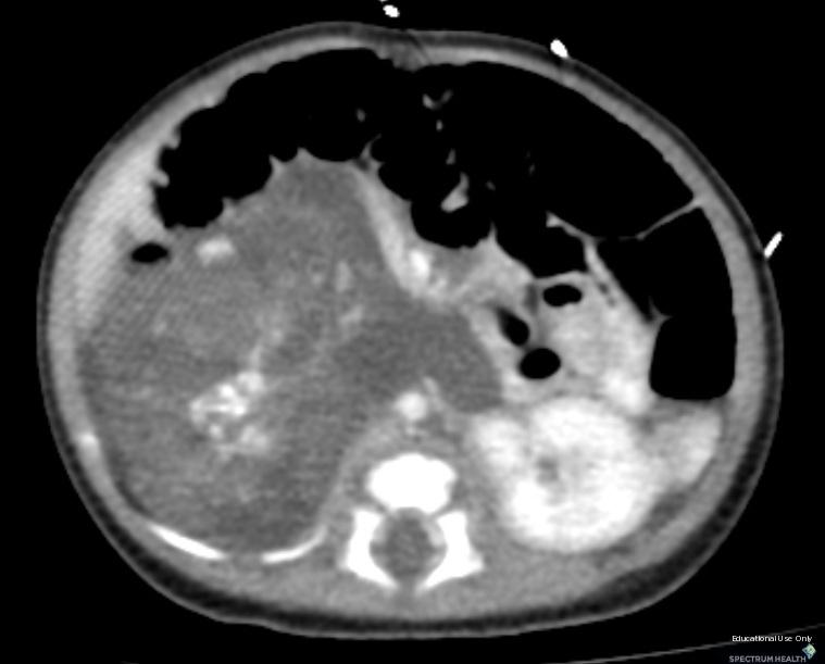
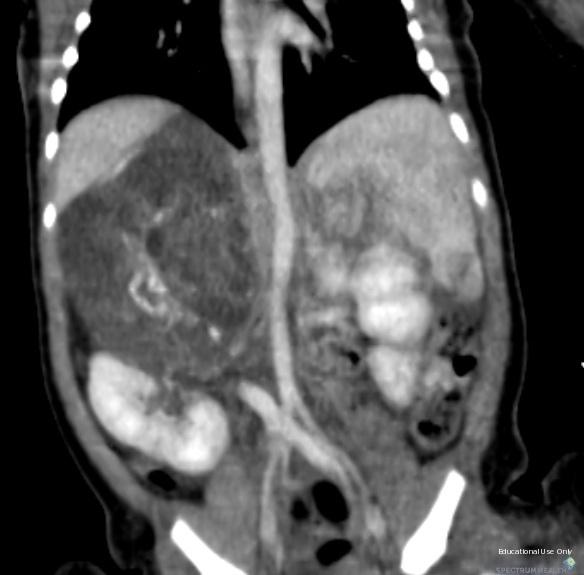
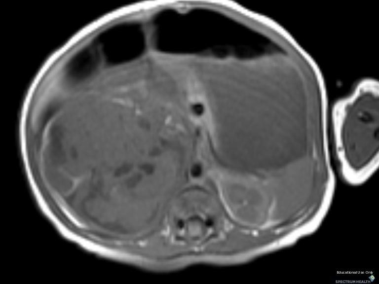
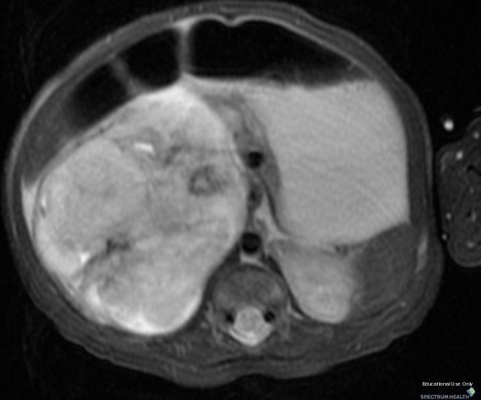
1 Retroperitoneal Teratoma Heather Borders, MD 03/04/2012 History Newborn with congenitally diagnosed mass. No other clinical symptoms. Diagnosis Retroperitoneal Teratoma; Immature teratoma, grade 1, with microscopic yolk sac tumor. Later resection demonstrating focus of secondary neuroglial malignancy. Additional Clinical Mass continued to grow and was resected. Discussion Teratoma is a germ cell tumor that originates from pluripotent germ cells. Less than 10% of teratomas are found in the retroperitoneum. Teratoma is the third most common tumor in the retroperitoneum in children, after neuroblastoma and Wilms tumor. Teratoma is more common in females, with a bimodal age distribution (<6 months and early adulthood). Teratoma can be benign or malignant, and benign teratoma can be either mature or immature. Mature teratoma (dermoid cyst) contains well-differentiated tissues from at least two germ cell layers. Ectodermal layers are seen in all, mesodermal layers in 90% of lesions, and endodermal layers in the majority of lesions. Mature teratomas are predominantly cystic. Calcification (toothlike or well defined) and fat can be seen in 56% and 93% of cases, respectively. A fat-fluid (sebum) level and chemical shift between fat and fluid are pathognomonic. Malignancy has been reported in 2% 3% of mature teratomas, more commonly in children (26%) than adults (10%), and is associated with wall thickening, irregular margins, and infiltration of adjacent organs. Compared with mature teratoma, immature teratoma is less common (<1%), contains more than 10% undifferentiated tissue, and is seen in a younger age group (<20 years). Immature teratoma is predominantly solid, with scattered areas of fat and calcification (coarse and ill defined), but cystic components are found occasionally. Malignant teratoma can have germ cell or non germ cell malignant tissue. Malignant transformation is less common in the retroperitoneum. Malignant tumors are irregular, with invasion of adjacent structures and vascular invasion. Surgical resection is required for definitive diagnosis and treatment. Findings CT: Hypodense heterogeneous mass with scattered calcifications located in the retroperitoneum with displacement of the kidney inferiorly and encasement of multiple vessels in a similar pattern to neuroblastoma. Multiple loculations are less dense than soft tissue but not as low in density as fat. MRI done to evaluate for fat demonstrates heterogeneous signal of fluid, soft tissue and calcium but no definitive fat that suppressed on T2FS. On histology, the mass contained brown fat (which likely explains the density/signal on initial studies representing brown fat, liquid fat) and this later transformed into mature fat on the CT. Reference
2 Imaging of Uncommon Retroperitoneal Masses Prabhakar Rajiah, MBBS, MD, FRCR, et al. July 2011 RadioGraphics, 31,
3
4
5
6
7 Sponsored By Disclaimer This teaching site is partially funded by an educational grant from GE Healthcare and Advanced Radiology Services, PC. The material on this site is independently controlled by Advanced Radiology Services, PC, and GE Healthcare and Spectrum Health have no influence over the content of this site Content Download Agreement The cases and images on this website are owned by Spectrum Health. Permission is granted (for nonprofit educational purposes) to download and print materials to distribute for the purpose of facilitating the education of health professionals. The authors retain all rights to the material and users are requested to acknowledge the source of the material. Site Disclaimer This site is developed to reach healthcare professionals and medical students. Nothing this site should be considered medical advice. Only your own doctor can help you make decisions about your medical care. If you have a specific medical question or are seeking medical care, please contact your physician. The information in this website is provided for general medical education purposes only and is not meant to substitute for the independent medical judgment of a physician relative to diagnostic and treatment options of a specific medical condition. The viewpoints expressed in these cases are those of the authors. They do not represent an endorsement. In no event will Advanced Radiology Associates, PC, Spectrum Health Hospitals (Helen Devos Children's Hospital) or GE Healthcare be liable for any decision made or action taken in reliance upon the information provided through this website.
Presacral Neuroblastoma Joseph Junewick, MD FACR
 Presacral Neuroblastoma Joseph Junewick, MD FACR 01/12/2010 History 16 month old male with irritability. Diagnosis Presacral Neuroblastoma Additional Clinical Initial US to evaluate for intussusception
Pleural Empyema Joseph Junewick, MD FACR
 Pleural Empyema Joseph Junewick, MD FACR 03/19/2010 History Teenager with persistent fever and cough. Pneumonia diagnosed 1 week ago. Diagnosis Pleural Empyema Additional Clinical Surgery-Clear fluid with
Paraspinal Venous Malformation Joseph Junewick, MD FACR
 Paraspinal Venous Malformation Joseph Junewick, MD FACR 06/04/2010 History 2 year old with history of fall. Rule out spinal injury. Diagnosis Paraspinal Venous Malformation Additional Clinical CT of the
Neuroblastoma Joseph Junewick, MD FACR
 Neuroblastoma Joseph Junewick, MD FACR 03/18/2011 History 15 month old with anemia. Diagnosis Neuroblastoma Discussion Neuroblastic tumors derive from primordial neural crest cells destined for sympathetic
Fallopian tube torsion and paratubal cyst Heather Borders, MD
 Fallopian tube torsion and paratubal cyst Heather Borders, MD 01/24/2012 History 13 year old female with one week of pelvic pain Diagnosis Fallopian tube torsion with paratubal cyst Additional Clinical
Spinal LCH Joseph Junewick, MD FACR
 Spinal LCH Joseph Junewick, MD FACR 05/16/2009 History 16 year old female with multiply recurrent Langerhans Cell Histiocytosis now with severe left sided neck pain. Diagnosis Langerhans Cell Histiocytosis
Diskitis Joseph Junewick, MD FACR
 Diskitis Joseph Junewick, MD FACR 09/20/2010 History 2 year old with fever, back pain and elevated sedimentation rate. Diagnosis Diskitis Discussion Diskitis is an inflammatory process of the intervertebral
Vein of Galen Malformation Joseph Junewick, MD FACR
 Vein of Galen Malformation Joseph Junewick, MD FACR 04/14/2018 History Midline cystic intracranial mass on prenatal ultrasound. Diagnosis Vein of Galen Malformation Discussion In normal fetal development,
Scrofula Joseph Junewick, MD FACR
 Scrofula Joseph Junewick, MD FACR 06/20/2012 History 4 year old male with refractory cervical adenopathy Diagnosis Scrofula Additional Clinical Positive PPD skin test. Discussion Scrofula refers to tuberculous
Chiari III Joseph Junewick, MD FACR
 Chiari III Joseph Junewick, MD FACR 07/02/2010 History Newborn with suboccipital mass. Diagnosis Chiari III Additional Clinical Surgery-Skin covered suboccipital cystic mass confined by the dura. Pathology-Leptomeningeal
Term Hypoxic Ischemic Injury Joseph Junewick, MD FACR
 Term Hypoxic Ischemic Injury Joseph Junewick, MD FACR 08/11/2010 History Term infant with perinatal distress and attempted forceps delivery. Diagnosis Term Hypoxic Ischemic Injury Discussion Encephalopathy
Transverse Dural Sinus Thrombosis Joseph Junewick, MD FACR
 Transverse Dural Sinus Thrombosis Joseph Junewick, MD FACR 03/19/2010 History Child with headache and otomastoiditis. Diagnosis Dural venous thrombosis secondary to mastoiditis Discussion The cerebral
Bilateral Retinoblastoma Joseph Junewick, MD FACR
 Bilateral Retinoblastoma Joseph Junewick, MD FACR 06/11/2010 History 17 month old adopted female with proptosis. Diagnosis Bilateral Retinoblastoma Discussion Retinoblastoma is the most common pediatric
Ulcerative Colitis Joseph Junewick, MD FACR
 Ulcerative Colitis Joseph Junewick, MD FACR 06/04/2010 History 16 year old male with hematochezia and anemia. Diagnosis Ulcerative Colitis Additional Clinical History of ulcerative colitis. Discussion
Pituitary Macroadenoma Joseph Junewick, MD FACR
 Pituitary Macroadenoma Joseph Junewick, MD FACR 08/13/2010 History 12 year old female with headache and visual disturbance. Diagnosis Pituitary Macroadenoma Additional Clinical Markedly elevated growth
Atlanto-occipital Dislocation Joseph Junewick, MD FACR
 Atlanto-occipital Dislocation Joseph Junewick, MD FACR 09/23/2009 History 12 year old male restrained back seat passenger in a car hit by a snowplow. Diagnosis Atlanto-occipital Dislocation Discussion
Chance Fracture Joseph Junewick, MD FACR
 Chance Fracture Joseph Junewick, MD FACR 08/02/2010 History Restrained teenager involved in motor vehicle accident. Diagnosis Chance Fracture (Hyperflexion-Distraction Injury) Discussion Chance-type spinal
Gastrointestinal Hemangiomatosis Joseph Junewick, MD FACR
 Gastrointestinal Hemangiomatosis Joseph Junewick, MD FACR 03/06/2010 History 3 month old with protuberant abdomen and anemia. Diagnosis Gastrointestinal Hemangiomatosis Discussion Gastrointestinal hemangiomatosis
Radiation Pneumonitis Joseph Junewick, MD FACR
 Radiation Pneumonitis Joseph Junewick, MD FACR 03/19/2010 History 16 year old with history of relapsed stage IV-A Hodgkin disease. Prior pulmonary involvement was irradiated. Diagnosis Radiation Pneumonitis
Tuberculous Meningitis Joseph Junewick, MD FACR
 Tuberculous Meningitis Joseph Junewick, MD FACR 08/11/2010 History 14 month old with fever and increasing lethargy. Diagnosis Tuberculous Meningitis Additional Clinical Grandmother with active tuberculosis.
Thymic Involvement in Chronic Granulomatous Disease of Childhood
 Thymic Involvement in Chronic Granulomatous Disease of Childhood Joseph Junewick, MD FACR 07/16/2010 History 3 year old male with multifocal osteomyelitis. Diagnosis Thymic Involvement in Chronic Granulomatous
Posterior Slipped Capital Femoral Epiphysis Joseph Junewick, MD FACR
 Posterior Slipped Capital Femoral Epiphysis Joseph Junewick, MD FACR 08/11/2010 History 6 year old male with intermittent hip pain for several months, acutely worsened after climbing the sand dunes. Diagnosis
Testicular Microlithiasis related to McCune-Albright Syndrome Joseph Junewick, MD FACR
 Testicular Microlithiasis related to McCune-Albright Syndrome Joseph Junewick, MD FACR 04/25/2010 History 12 year old with McCune-Albright syndrome. Diagnosis Testicular Microlithiasis related to Mcune-Albright
Pelvic tumor in childhood Classification, imaging approach and radiological findings
 Pelvic tumor in childhood Classification, imaging approach and radiological findings M. Mearadji International Foundation for Pediatric Imaging Aid Rotterdam, The Netherlands Solid pelvic masses in childhood
Patient Information. Age: 8 y/o Sex: Female. Date of Admission: Date of Discharge:
 Patient Information Age: 8 y/o Sex: Female Date of Admission: 92-10-08 Date of Discharge: 92-10-18 Chief Complaint Severe admominal pain and vomiting with dysuria since last afternoon Present Illness Lower
Pediatric Retroperitoneal Masses Radiologic-Pathologic Correlation
 Acta Radiológica Portuguesa, Vol.XVIII, nº 70, pág. 61-70, Abr.-Jun., 2006 Pediatric Retroperitoneal Masses Radiologic-Pathologic Correlation Marilyn J. Siegel Mallinckrodt Institute of Radiology, Washington
Scholars Journal of Medical Case Reports
 DOI: 10.21276/sjmcr.2016.4.12.15 Scholars Journal of Medical Case Reports Sch J Med Case Rep 2016; 4(12):926-930 Scholars Academic and Scientific Publishers (SAS Publishers) (An International Publisher
Mature Cystic Teratomas and the most common complications
 Mature Cystic Teratomas and the most common complications Poster No.: C-2230 Congress: ECR 2015 Type: Authors: Keywords: DOI: Educational Exhibit S. C. S. Silva 1, D. N. Silva 1, D. Garrido 1, I. C. S.
Update on RECIST and Staging of Common Pediatric Tumors Ethan A. Smith, MD
 Update on RECIST and Staging of Common Pediatric Tumors Ethan A. Smith, MD Section of Pediatric Radiology C.S. Mott Children s Hospital University of Michigan ethans@med.umich.edu Disclosures No relevant
Retroperitoneal Sarcomas - A pictorial review
 Retroperitoneal Sarcomas - A pictorial review Poster No.: C-1409 Congress: ECR 2013 Type: Educational Exhibit Authors: D. Douraghi-Zadeh, K. L. Shahabuddin, R. H. Thomas, E. Moskovic; London/UK Keywords:
Study of primary retroperitoneal masses. What I need to know?
 Study of primary retroperitoneal masses. What I need to know? Poster No.: C-1316 Congress: ECR 2013 Type: Educational Exhibit Authors: N. ROMERO FLOREZ, J. Soriano, L. Valls Masot, A. Maroto, M. Osorio,
Clinical summary. Male 30 year-old with past history of non-seminomous germ cell tumour. Presents with retroperitoneal lymphadenopathy on CT.
 Clinical summary Male 30 year-old with past history of non-seminomous germ cell tumour. Presents with retroperitoneal lymphadenopathy on CT. For restaging PET/CT. PET/CT findings No significant FDG uptake
A Practical Approach to Adnexal Masses
 A Practical Approach to Adnexal Masses Darcy J. Wolfman, MD Section Chief of Genitourinary Imaging American Institute for Radiologic Pathology Clinical Associate Johns Hopkins Community Radiology Division
J of Evolution of Med and Dent Sci/ eissn , pissn / Vol. 3/ Issue 46/Sep 22, 2014 Page 11296
 CT SPECTRUM OF GIANT RETROPERITONEAL LIPOSARCOMAS WITH HISTOPATHOLOGICAL CORRELATION Shashikumar M. R 1, Rajendra Kumar N. L 2, C. P. Nanjaraj 3, Nishanth R. K 4, Vishwanath Joshi 5 HOW TO CITE THIS ARTICLE:
3 cell types in the normal ovary
 Ovarian tumors 3 cell types in the normal ovary Surface (coelomic epithelium) the origin of the great majority of ovarian tumors 90% of malignant ovarian tumors Totipotent germ cells Sex cord-stromal cells
Prenatal and Neonatal MRI of Sacrococcygeal Teratoma With Surgical Correlation
 Prenatal and Neonatal MRI of Sacrococcygeal Teratoma With Surgical Correlation Ali Mahmood, M.D., and Nadia F. Mahmood, M.D. Citation: Mahmood A, Mahmood NF. Prenatal and Neonatal MRI of Sacrococcygeal
Imaging of Gastrointestinal Stromal Tumors (GIST) Amir Reza Radmard, MD Assistant Professor Shariati hospital Tehran University of Medical Sciences
 Amir Reza Radmard, MD Assistant Professor Shariati hospital Tehran University of Medical Sciences") Imaging of Gastrointestinal Stromal Tumors (GIST) Amir Reza Radmard, MD Assistant Professor Shariati hospital Tehran University of Medical Sciences Describe the typical imaging findings of GIST at initial
Imaging of Gastrointestinal Stromal Tumors (GIST) Amir Reza Radmard, MD Assistant Professor Shariati hospital Tehran University of Medical Sciences Describe the typical imaging findings of GIST at initial
OVARIES. MLS Basic histological diagnosis MLS HIST 422 Semester 8- batch 7 L13 Dr: Ali Eltayb.
 OVARIES MLS Basic histological diagnosis MLS HIST 422 Semester 8- batch 7 L13 Dr: Ali Eltayb. OBJECTIVES Recognize different disease of ovaries Classify ovarian cyst Describe the pathogenesis, morphology
Neckmasses in infancy and childhood: Clinical and radiological classification and imaging approaches M. Mearadji
 Neckmasses in infancy and childhood: Clinical and radiological classification and imaging approaches M. Mearadji International Foundation for Pediatric Imaging Aid Introduction Neck masses are a frequent
MRI IN THE CHARACTERIZATION OF SEMINOMATOUS AND NONSEMINOMATOUS GERM CELL TUMORS OF THE TESTIS
 MRI IN THE CHARACTERIZATION OF SEMINOMATOUS AND NONSEMINOMATOUS GERM CELL TUMORS OF THE TESTIS Ambesh Deshar *, Gyanendra KC and Zhang Lopsang *Department of Medical Imaging and Nuclear Medicine, First
Leonard M. Glassman MD
 BI-RADS The New BI-RADS Leonard M. Glassman MD FACR Former Chief of Breast Imaging American Institute for Radiologic Pathology Washington Radiology Associates, PC Breast Imaging Reporting and Data System
Germ cell tumours UK SH. Ivo Leuschner. Kiel Pediatric Tumor Registry, Institute of Pathology University Hospital of Schleswig-Holstein Campus Kiel
 Germ cell tumours Ivo Leuschner Kiel Pediatric Tumor Registry, Institute of Pathology University Hospital of Schleswig-Holstein Campus Kiel UK SH Old histogenetic Concept of Germ cell tumours Pluripotent
Personal data. Age : 63 Gender : male
 Personal data Age : 63 Gender : male Chief complain No specific symptom or discomfort A hepatic mass, found by abdominal sonography of routine health exam on 88-12-08 Past history 1984-3-3 Old CVA with
Imaging characterization of renal clear cell carcinoma
 Imaging characterization of renal clear cell carcinoma Poster No.: C-0327 Congress: ECR 2011 Type: Educational Exhibit Authors: S. Ballester 1, A. Gaser 2, M. Dotta 1, M. F. CAPPA 1, F. Hammar 1 ; 1 2
Mediastinal Tumors: Imaging
 Mediastinal Tumors: Imaging References Imaging in Oncology, Husband and Reznek Computed Tomography and Magnetic Resonance of the thorax, Naidich, Zerhouni, Siegelman, Mediastinal compartments Anterior:
STAGING AND FOLLOW-UP STRATEGIES
 ATHENS 4-6 October 2018 European Society of Urogenital Radiology STAGING AND FOLLOW-UP STRATEGIES Ahmet Tuncay Turgut, MD Professor of Radiology Hacettepe University, Faculty of Medicine Ankara 2nd ESUR
UW Radiology Review Course Breast Calcifications. BI-RADS 5 th Edition
 UW Radiology Review Course Breast Calcifications Grace Kalish, MD Vantage Radiology BI-RADS 5 th Edition Benign Skin Vascular Large rod like Coarse popcorn Suspicious Amorphous Coarse heterogenous Fine
A CASE OF A Huge Submandibular Pleomorphic Adenoma
 ISPUB.COM The Internet Journal of Head and Neck Surgery Volume 4 Number 2 S VERMA Citation S VERMA.. The Internet Journal of Head and Neck Surgery. 2009 Volume 4 Number 2. Abstract Pleomorphic adenoma
S. Murgo, MD. Chr St-Joseph, Mons Erasme Hospital, Brussels
 S. Murgo, MD Chr St-Joseph, Mons Erasme Hospital, Brussels? Introduction Mammography reports are sometimes ambiguous and indecisive. ACR has developped the BIRADS. BIRADS consists of a lexicon in order
CELL AND TISSUE INJURY COURSE-II PATHOLOGY LABORATORY. PATHOLOGY of MASS LESIONS and TISSUE DEFECTS -MACROSCOPY Assoc. Professor Rengin Ahıskalı
 CELL AND TISSUE INJURY COURSE-II PATHOLOGY LABORATORY PATHOLOGY of MASS LESIONS and TISSUE DEFECTS -MACROSCOPY Assoc. Professor Rengin Ahıskalı M1 - RENAL TUBERCULOSIS cavitary areas caseous necrosis fibrous
Testicular Germ Cell Tumors; A Simplistic Approach
 Testicular Germ Cell Tumors; A Simplistic Approach Merce Jorda, MD, PhD, MBA Professor and Vice Chair, Director of Anatomic Pathology Director of Genitourinary Pathology Service Interim Director of Cytopathology
Imaging in breast cancer. Mammography and Ultrasound Donya Farrokh.MD Radiologist Mashhad University of Medical Since
 Imaging in breast cancer Mammography and Ultrasound Donya Farrokh.MD Radiologist Mashhad University of Medical Since A mammogram report is a key component of the breast cancer diagnostic process. A mammogram
Malignant Focal Liver Lesions
 Malignant Focal Liver Lesions Other Than HCC Pablo R. Ros, MD, MPH, PhD Departments of Radiology and Pathology University Hospitals Cleveland Medical Center Case Western Reserve University Pablo.Ros@UHhospitals.org
 INTRAUTERINE DEVICE = IUD INTRAUTERINE DEVICE = IUD CONGENITAL DISORDERS Pyometra = pyometrea is a uterine infection, it is accumulation of purulent material in the uterine cavity. Ultrasound is usually
Imaging evaluation of ovarian masses.
 Imaging evaluation of ovarian masses. Poster No.: C-0988 Congress: ECR 2012 Type: Educational Exhibit Authors: M. Forment Navarro, C. La Parra Casado, A. Vera, C. Martínez 1 2 2 2 2 2 1 Rubio, M. Mazón
3 cell types in the normal ovary
 Ovarian tumors 3 cell types in the normal ovary Surface (coelomic epithelium) the origin of the great majority of ovarian tumors (neoplasms) 90% of malignant ovarian tumors Totipotent germ cells Sex cord-stromal
Case Report A Rare Adrenal Mass in a 3-Month-Old: A Case Report and Literature Review
 Hindawi Case Reports in Pediatrics Volume 2017, Article ID 4542321, 5 pages https://doi.org/10.1155/2017/4542321 Case Report A Rare Adrenal Mass in a 3-Month-Old: A Case Report and Literature Review Ashish
Retroperitoneal Ganglioneuroma Encasing the Celiac and Superior Mesenteric Arteries
 Case Study TheScientificWorldJOURNAL (2004) 4, 974 977 ISSN 1537-744X; DOI 10.1100/tsw.2004.198 Retroperitoneal Ganglioneuroma Encasing the Celiac and Superior Mesenteric Arteries Justin K. Nelms, Eric
Retroperitoneal masses
 Retroperitoneal masses Poster No.: C-0698 Congress: ECR 2015 Type: Educational Exhibit Authors: S. Dutra, R. Amaral, D. N. Silva, D. Garrido, S. Serpa, I. C. S. 1 1 1 1 2 1 1 1 2 P. Basto, P. Cordeiro
The diagnostic criteria of multilocular renal cysts
 Case Report 772 Multilocular Renal Cysts with Renal Cell Carcinoma: Report of Four Cases Chia-Hsi Chen, MD; Cheng-Keng Chuang, MD, PhD; Chun-Te Wu, MD; Kwai-Fong Ng 1, MD; Shuen-Kuei Liao 2, PhD According
The Incidental Renal lesion
 The Incidental Renal lesion BACKGROUND Increase in abdominal CT/US in last 15 years Resulted in detection of many (small) renal lesions 50% > 50yrs has at least 1 lesion majority simple cysts Renal lesions
Case Report Mature Cystic Teratoma in Douglas Pouch
 Case Reports in Pathology Volume 2015, Article ID 202853, 4 pages http://dx.doi.org/10.1155/2015/202853 Case Report Mature Cystic Teratoma in Douglas Pouch Kenji Ohshima, 1 Anna Umeda, 2 Ayako Hosoi, 2
Teratoma Formation Pluripotency Analysis Report
 Teratoma Formation Pluripotency Analysis Report Project and Customer Information Service Title: Teratoma formation pluripotency analysis Customer: PepGel LLC PI/Contact: Susan, Email: sun.pepgel@gmail.com
Adrenal masses in infancy and childhood: A clinical and radiological overview M. Mearadji
 Adrenal masses in infancy and childhood: A clinical and radiological overview M. Mearadji International Foundation for Pediatric Imaging Aid Introduction Neoplastic adrenal masses usually originate from
Incidental Esophageal Findings on Chest CT. Amira Hussien, MD, Elliot Fishman, MD, Bouchra Younes, MD, Ahmed Hatw. Johns Hopkins Medical Institution
 Incidental Esophageal Findings on Chest CT Amira Hussien, MD, Elliot Fishman, MD, ouchra Younes, MD, Ahmed Hatw. Johns Hopkins Medical Institution I have nothing to disclose. DISCLOSURE INTRODUCTION Although
How To Approach Renal Masses? - Differential Diagnosis On Image
 How To Approach Renal Masses? - Differential Diagnosis On Image Poster No.: C-1646 Congress: ECR 2015 Type: Educational Exhibit Authors: A. E. A. G. Costa, A. Gomes, A. Duarte, I. Távora; Lisbon/PT Keywords:
DUSTURBANCES OF GROWTH. MLS Basic histological diagnosis MLS HIST 422 Semester 8- batch 7 L8 Uz: Musa
 DUSTURBANCES OF GROWTH MLS Basic histological diagnosis MLS HIST 422 Semester 8- batch 7 L8 Uz: Musa Agnesia: means complete absence of an organ (Kidney). Aplasia: s defined in general as "defective development
Note: The cause of testicular neoplasms remains unknown
 - In the 15- to 34-year-old age group, they are the most common tumors of men. - Tumors of the testis are a heterogeneous group of neoplasms that include: I. Germ cell tumors : 95%; all are malignant.
CODING TUMOUR MORPHOLOGY. Otto Visser
 CODING TUMOUR MORPHOLOGY Otto Visser INTRODUCTION The morphology describes the tissue of the tumour closest to normal tissue Well differentiated tumours are closest to normal Undifferentiated tumours show
Medical Science, Volume 13, Number 53, October 29, Adult presentation of retroperitoneal cystic teratoma: a case report
 CASE REPORT PATHOLOGY Medical Science, Volume 13, Number 53, October 29, 2014 ISSN 2321 7359 EISSN 2321 7367 Medical Science The International W Adult presentation of retroperitoneal cystic teratoma: a
Incidental Discovery of Testicular Microlithiasis: What Is the Importance of Ultrasound Surveillance? Two Case Reports
 Published online: October 24, 2013 1662 6575/13/0063 0520$38.00/0 This is an Open Access article licensed under the terms of the Creative Commons Attribution-NonCommercial 3.0 Unported license (CC BY-NC)
Dr Claire Smith, Consultant Radiologist St James University Hospital Leeds
 Dr Claire Smith, Consultant Radiologist St James University Hospital Leeds Imaging in jaundice and 2ww pathway Image protocol Staging Limitations Pancreatic cancer 1.2.4 Refer people using a suspected
Amammography report is a key component of the breast
 Review Article Writing a Mammography Report Amammography report is a key component of the breast cancer diagnostic process. Although mammographic findings were not clearly differentiated between benign
Scrotum-like protrusion of lipoma arising from the proximal thigh
 Upsala J Med sci 109: 261 265, 2004 Scrotum-like protrusion of lipoma arising from the proximal thigh Report of two cases Koshi Hattori, 1 Masahito Hatori, 1 Mika Watanabe, 2 Toshihisa Osanai, 3 Shoichi
SUPPLEMENTARY FIG. S2. Teratoma. Portion of a teratoma composed of neural tissue. The large cells in the central part correspond to ganglion cells.
 Supplementary Data SUPPLEMENTARY FIG. S1. Teratoma. The tumor is composed predominantly of keratinizing squamous epithelium (Sq), which forms cysts filled with keratin (arrows). The tumor also contains
Leukaemia 35% Lymphoma 14%
 Distribution ib ti of Cancers in Children under 15 years Leukaemia 35% Lymphoma 14% Neuroblastoma 9% Other 5% Liver 1% Retinoblastoma 3% Bone and STS 15% CNS 20% Wilms' 8% 30-40% Mortality Germ Cell Tumours
Pediatric Abdominal Masses. Andrew Phelps MD Assistant Professor of Pediatric Radiology UCSF Benioff Children's Hospital
 Pediatric Abdominal Masses Andrew Phelps MD Assistant Professor of Pediatric Radiology UCSF Benioff Children's Hospital No Disclosures Take Home Message All you need to remember are the 5 common masses
IN THE NAME OF GOD POV: CYSTIC OVARIAN LESION
 IN THE NAME OF GOD POV: CYSTIC OVARIAN LESION CASE 1 20 years old girl with AUB and pelvic pain from 2 weeks ago Impression :Simple unilocular 6 cm ovarian cyst Next step? Almost certainly benign so FU
ADRENAL MEDULLARY DISORDERS: PHAEOCHROMOCYTOMAS AND MORE
 ADRENAL MEDULLARY DISORDERS: PHAEOCHROMOCYTOMAS AND MORE DR ANJU SAHDEV READER AND CONSULTANT RADIOLOGIST QUEEN MARY UNIVERSITY AND ST BARTHOLOMEW S HOSPITAL BARTS HEALTH, LONDON, UK DISCLOSURE OF CONFLICT
ADRENAL LESIONS 10/09/2012. Adrenal + lesion. Introduction. Common causes. Anatomy. Financial disclosure. Dr. Boraiah Sreeharsha. Nothing to declare
 ADRENAL LESIONS Financial disclosure Nothing to declare Dr. Boraiah Sreeharsha MBBS;FRCR;FRCPSC Introduction Adrenal + lesion Adrenal lesions are common 9% of the population Increase in the detection rate
See the latest estimates for new cases of ovarian cancer and deaths in the US and what research is currently being done.
 About Ovarian Cancer Overview and Types If you have been diagnosed with ovarian cancer or are worried about it, you likely have a lot of questions. Learning some basics is a good place to start. What Is
Case Report Delivery Induced Intraperitoneal Rupture of a Cystic Ovarian Teratoma and Associated Chronic Chemical Peritonitis
 Case Reports in Radiology, Article ID 189409, 4 pages http://dx.doi.org/10.1155/2014/189409 Case Report Delivery Induced Intraperitoneal Rupture of a Cystic Ovarian Teratoma and Associated Chronic Chemical
Matsunaga, Naofumi; Saito, Yutaka. Citation Acta medica Nagasakiensia. 1991, 36
 NAOSITE: Nagasaki University's Ac Title Author(s) A Large Periureteral Lipoma Associa Hydronephrosis. Hayashi, Tomayoshi; Imamura, Atushi Matsunaga, Naofumi; Saito, Yutaka Citation Acta medica Nagasakiensia.
Malignant transformation in benign cystic teratomas, dermoids of the ovary
 European JournalofObstetrics& Gynecology andreproductivebiology, 29 (1988) 197-206 197 Elsevier EJO 00716 Malignant transformation in benign cystic teratomas, dermoids of the ovary S. Chadha 1 and A. Schaberg
Clinicopathological and Histological Features of Ovarian Tumour- A Study
 IOSR Journal of Dental and Medical Sciences (IOSR-JDMS) e-issn: 2279-0853, p-issn: 2279-0861.Volume 16, Issue 9 Ver. IX (September. 2017), PP 56-60 www.iosrjournals.org Clinicopathological and Histological
Common and unusual CT and MRI manifestations of pancreatic adenocarcinoma: a pictorial review
 Review Article Common and unusual CT and MRI manifestations of pancreatic adenocarcinoma: a pictorial review Min-Jie Yang, Su Li, Yong-Guang Liu, Na Jiao, Jing-Shan Gong Department of Radiology, Shenzhen
7 Mousa. Obada Zalat. Mohammad Badi
 7 Mousa Obada Zalat Mohammad Badi Tumors of the ovaries Last lecture we talked about surface epithelial tumors of the ovaries (the most common type). But there are many other types of tumors of germ cell
CTA/MRA of Pediatric Hepatic Masses Radiology-Pathology Correlation
 Acta Radiológica Portuguesa, Vol.XVIII, nº70, pág. 41-50, Abr.-Jun., 2006 CTA/MRA of Pediatric Hepatic Masses Radiology-Pathology Correlation Marilyn J. Siegel Mallinckrodt Institute of Radiology, Washington
Essentials of Clinical MR, 2 nd edition. 51. Primary Neoplasms
 51. Primary Neoplasms As with spinal central canal neoplasms in other regions, those of the lumbar spine may be classified as extradural, intradural extramedullary, and medullary. If an extradural lesion
(2/3 PRCC!) (2/3 PRCC!)
 (2/3 PRCC!)") Approach to the Incidental Solid Renal Mass Stuart G. Silverman, MD, FACR Professor of Radiology Harvard ard Medical School Director, Abdominal Imaging and Intervention Brigham and Women s Hospital Boston,
Approach to the Incidental Solid Renal Mass Stuart G. Silverman, MD, FACR Professor of Radiology Harvard ard Medical School Director, Abdominal Imaging and Intervention Brigham and Women s Hospital Boston,
Pathology Slides. [Pathology]
![Pathology Slides. [Pathology]](/thumbs/94/120604575.jpg "Pathology Slides. [Pathology]") Pathology Slides MedicoNotes provides real laboratory pathological slides to aid you to differentiate between different pathological structures under microscope. www.mediconotes.com Histology slides example
Pathology Slides MedicoNotes provides real laboratory pathological slides to aid you to differentiate between different pathological structures under microscope. www.mediconotes.com Histology slides example
* I have no disclosures or any
 Howard Rosenthal, M.D. Associate Professor of Orthopedic Surgery University of Kansas Sarcoma Center I have no disclosures or any conflicts related to the content of this presentation. Objectives 1. Describe
Leonard M. Glassman MD Analysis of Breast Calcifications
 Importance of Calcification Leonard M. Glassman MD FACR American Institute for Radiologic Pathology Washington Radiology Associates, PC Washington DC 45% of all breast cancers present as calcification
Retroperitoneal tumors: Computed Tomography (CT) and Magnetic Resonance (MR) patterns
 and Magnetic Resonance (MR) patterns") Retroperitoneal tumors: Computed Tomography (CT) and Magnetic Resonance (MR) patterns Poster No.: C-1369 Congress: ECR 2011 Type: Educational Exhibit Authors: G. Cardone, A. Messina, D. Vergnaghi, P. Mangili,
Retroperitoneal tumors: Computed Tomography (CT) and Magnetic Resonance (MR) patterns Poster No.: C-1369 Congress: ECR 2011 Type: Educational Exhibit Authors: G. Cardone, A. Messina, D. Vergnaghi, P. Mangili,
Retroperitoneal tumors: Computed Tomography (CT) and Magnetic Resonance (MR) patterns
 and Magnetic Resonance (MR) patterns") Retroperitoneal tumors: Computed Tomography (CT) and Magnetic Resonance (MR) patterns Poster No.: C-1369 Congress: ECR 2011 Type: Educational Exhibit Authors: G. Cardone, A. Messina, D. VERGNAGHI, P. Mangili,
Retroperitoneal tumors: Computed Tomography (CT) and Magnetic Resonance (MR) patterns Poster No.: C-1369 Congress: ECR 2011 Type: Educational Exhibit Authors: G. Cardone, A. Messina, D. VERGNAGHI, P. Mangili,
Ovarian Malignant Germ Cell Tumors: Cellular Classification and Clinical and Imaging Features 1
 Note: This copy is for your personal non-commercial use only. To order presentation-ready copies for distribution to your colleagues or clients, contact us at www.rsna.org/rsnarights. Ovarian Malignant
BI-RADS Update. Martha B. Mainiero, MD, FACR, FSBI Brown University Rhode Island Hospital
 BI-RADS Update Martha B. Mainiero, MD, FACR, FSBI Brown University Rhode Island Hospital No Disclosures BI-RADS History 1980s Quality Issues ACR Accreditation BI-RADS 1994 2003 4 th Edition MRI, US January
MR Tumor Staging for Treatment Decision in Case of Wilms Tumor
 MR Tumor Staging for Treatment Decision in Case of Wilms Tumor G. Schneider, M.D., Ph.D.; P. Fries, M.D. Dept. of Diagnostic and Interventional Radiology, Saarland University Hospital, Homburg/Saar, Germany
Ó Journal of Krishna Institute of Medical Sciences University 112
 ISSN 2231-4261 CASE REPORT Large Pseudocyst of the Adrenal Gland: A Radiological Pathological Correlation 1* 1 2 1 1 Abhishek Dwivedi, Satveer Choudhary, Vikram Trehan, Narender Yadav, K. Leela Kanth 1
Adnexal Masses and Problem Solving Pelvic MRI
 28th Congress of the Hungarian Society of Radiologists RCR Session Budapest June 2016 Adnexal Masses and Problem Solving Pelvic MRI DrSarah Swift St James s University Hospital Leeds, UK Objectives Characterisation