INTRODUCTION BRAIN TUMOR INTRA KRANIAL, MED SPINALIS AND MENINGES TWO TYPE PRIMER AND SECUNDER PRIMER TUMOR ASTROCYTE, OLIGODENDROCYTE, EPENDEMOCYTE,
|
|
|
- Frank Jenkins
- 6 years ago
- Views:
Transcription
1 BRAIN TUMOR YUNELDI ANWAR DEPARTMENT NEUROLOGY MEDICAL FACULTY UNIVERSITY OF NORTH SUMATERA
2 INTRODUCTION BRAIN TUMOR INTRA KRANIAL, MED SPINALIS AND MENINGES TWO TYPE PRIMER AND SECUNDER PRIMER TUMOR ASTROCYTE, OLIGODENDROCYTE, EPENDEMOCYTE, ARACHNOID, NEUROBLAST AND MEDULLUBLAST SEKUNDER TUMOR METASTATIK FROM LUNG, BREAST, COLON AND SKIN
3 ETIOLOGY GENETIC GENE DEVIATION CONGENGITAL TERATOMA, CHORDOMA AGE PINEALOMA, MEDULLOBLASTOMA (< 20 yr). MENINGIOMA (> 50yr) CARSINOGEN HYDROCARBON, NITROSAMIN HEAD INYURY, INFECTION, TOKSIN, RADIATION, VIRUS
4 Risk factors related to CNS tumor Definitive ionizing radiation immune supression (HIV infection) Possible Electromagnetic field (high tension wires, cellular telepon) Diets (N-nitroso compounds, Aspartame) Occupation (petroleum industry, agricultural pesticides) Household chemicals (hairdyes and sprays, household pesticides) Head injury Medication (vitamin) Infections (Cysticercosis, varicella zoster, SV 40
5 CONCEPT OF PATOGENESIS PRIMER TUMOR HISTOGENIC THEORY (Bayley &Cushing) EMBRIOLOGICAL OF NERVE CELL AND GLIA RECENT THEORY TUMOR ARISES FROM TRANSFORMATION OF ASTROCYTE, OLIGODENDROCYTE, MICROGLIOCYTE, EPENDYMOCYTE NEOPLASM CELL MULTIPLICATION AND DIFFRENTIATION
6 PATHOFISIOLOGY NORMAL CAVUM CRANII CONSIST OF Brain tissue ( 1400 gr ) Cerebro spinal fluid ( 75 cc ) Blood ( 75 cc ) these component any time stabil Brain tumor increase intracranial pressure Brain tumor cerebral edema cerebral herniation


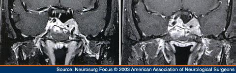
7 NORMAL BRAIN


8 Cerebral herniation A. Subfacial/cingulate hern B. Uncal herniation C. Transtentorial/central hern D. External herniation E. Tonsillar herniation
9 CLASSIFICATION HISTOPATOLOGICAL PATTERN Primary brain tumor histological benign or malformative meningioma pituitary adenoma, acustic neuroma, craniopharyngioma, pilocytic astrocytoma hemangioblastoma histological malignant glioma (anaplas tic astrocytoma, glioblastoma multiform), ependymoma, oligodendroglioma, medulloblasto ma, pineal cell tumor, chroid plexus carcinoma
10 MENINGIOMA
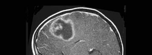
11 GLIOBLASTOMA
12 CLASSIFICATION CONT METASTATIC BRAIN TUMORS BREAST LUNG GI TRACT PROSTAT SKIN
13 INTRA CRANIAL METASTASIS Brain parenchyma breast, lung, melanoma Pituitary gland breast, melanoma, germ cell Dural based lung, prostat, breast Leptomeningeal disease breast, lung
14 SKULL METASTASIS Skull base breast, prostat, osteosarcoma of skull, head and neck cancer Calvarium breast, prostat
15 SPINE METASTASIS Epidural lung, breast, prostat, neuroblastoma, lymphoma. Leukemia Leptomeningeal breast, lung Intramedullary breast, colon, lung, prostat Brachial plexus lung, breast, lymphoma Lumbosacral plexus pelvic tumor
16 CLASSIFICATION BASED ON TOPOGRAPHY (LOCATION) SUPRA TENTORIAL TUMORS Cerebral lobe and deep hemispheric glioma, meningioma, metastatic tumor Sella turcica tumor pituitary tumor, craniopharyngiomas INFRATENTORIAL TUMOR MEDULLASPINALIS TUMOR
17 INFRATENTORIAL TUMORS ADULTS Cerebellopontine angel acoustic schwannoma Others sites brainstem gliomas, metastase, hemangioblastoma, ependymoma CHILDRENS Midline tumors medulloblastomas Cerebellar lobes astrocytomas
18 MEDULLA SPINALIS TUMORS EKSTRA DURAL Metastatic tumor INTRA DURAL Ekstra medulla meningioma Intra medulla neurofibromas ependymoma astrositoma
19 SYMPTOMS AND SIGNS Generalized symptoms caused by raised intracranial pressure Focal symptoms caused by invasion, ischemia and compression False localizing symptoms caused by shifts of cerebral structures
20 GENERALIZED SYMPTOMS AND SIGNS HEADACHE VOMITING DROWSINESS ( VERTIGO AND DIZZINESS) VISUAL OBSCURATION PERSONALITY CHANGE CONFUSION PAPILEDEMA APATHY
21 MECHANISM HEADACHE ASSOCIATED WITH BRAIN TUMOR 1.Traction on venous sinuses 2.Traction on meningeal arteries 3.Traction on large arteries at base of the brain 4.Pressure on cranial and cervical pain sensi tive structure 5.Dilatation of intracranial arteries 6.Inflamation of pain-sensitive structures
22 FOCAL SYMPTOMS AND SIGNS FRONTAL LOBE Generalized seizure Focal motor seizure (contra lateral) Expressive aphasia (dominant size) Behavioral changes Dementia Gait disorders, incotinence Hemiparese

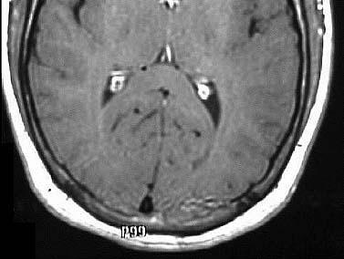
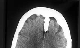
23 Frontal lobe tumor
24 FOCAL SYMPTOMS & SIGNS BASAL GANGLIA Hemiparesis (contralateral) Movement disorders (rare) PARIETAL LOBE Receptive aphasia (dominant size) Spatial disorientation (non dominant) Cortical sensory dysfungtion (contralat) Agnosias


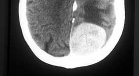
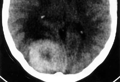
25 Metastase parietal lobe tumor
26 FOCAL SYMPTOMS & SIGNS OCCIPITAL LOBE Hemiparesis (contralateral) Visual disturbance TEMPORAL LOBE Complex partial (psychomotor) seizures Generalized seizures Behavioral changes Olfactory and complex seizures Visual auras Visual field defect

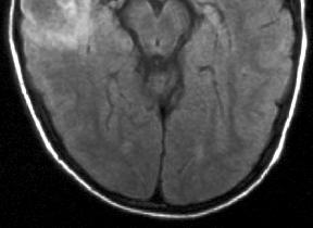
27 Occipital lobe tumor

28 Temporal lobe tumor
29 FOCAL SYMPTOMS & SIGNS CORPUS COLLOSUM Dementia (anterior) Behavioral changes Memory loss (posterior) Asymptomatic (mid) THALAMUS Sensory loss (contralateral) Behavioral changes (posterior) Languange disorders (dominant size)
30 FOCAL SYMPTOMS & SIGNS MIDBRAINS/PINEAL Paresis of vertical eyemovement Pupillary abnormalities Precocious puberity (boys) SELLA/OPTIC NERVE/PITUITARY Endocrinopathy Bitemporal hemianopia Monocular visual defect Opthalmoplegia (cavernous sinus)

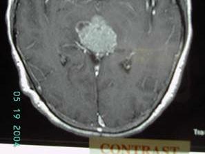
31 Pineal tumor

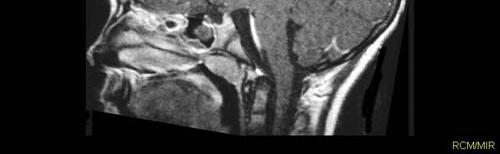
32 Pituitary tumor
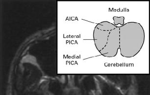
33 FOCAL SYMPYOMS & SIGNS PONS/MEDULLA Cranial nerve dysfungtion Ataxia, nystagmus, spasticity Weakness, sensory loss CERBELLO PONTINE ANGLE Deafness (ipsilateral) Loss of facial sensation (ipsilateral) Facial weakness (ipsilateral), ataxia CEREBELLUM Ataxia (ipsilateral) Nystagmus

34 Cerebellum tumor
35 FALSE LOCALIZING SIGNS CRANIAL NERVE Anosmia Diplopia, ptosis, anisocoria Face pain, numbness, and weakness Tnnitus, hearing loss
36 FALSE LOCALIZING SIGNS PARENCHYMAL SIGNS Ipsilateral hemiparesis Ipsilateral gaze palsy Visual field defect, cortical blindness Ataxia OTHER SIGNS Nuchal rigidity
37 SYMPTOM AND SIGN SPINAL INTRAMEDULLARY Sensory loss CORD TUMOR Early spinchter dysfungtion Pain + Decreased rectal tone spasticity upper motor neuron signs Common tumor -> Ependymoma, astrositoma
38 Symptom and signs spinal cord tumor Intradural and extramedullary Pain, metastase +++ Pain, primary tumor + Radicular or local sensory loss Radicular or segmental weakness and sensory loss Upper motor neuron signs Common tumors Meningioma
39 Symptom and sign spinal cord EPIDURAL Pain +++ tumor Radicular or local pain Common tumors Metastasis
40 DIAGNOSIS BRAIN TUMOR HISTORY GENERAL PHYSICAL EXAMINATION NEUROLOGIC EXAMINATION LABORATORY FINDING, ESPECIALLY IMAGING
41 Laboratory findings IMAGING CT Scan MRI Magnetic resonance spectroscopy (MRS) Fungtional MRI BIOPSY Defenitive diagnosis
42 DIFFRENTIAL DIAGNOSIS OF HEMATOMA ABCESS GRANULOMA BRAIN TUMORS PARASITIC INFECTION CYSTYCIRCOSIS VASCULAR MALFORMATION MULTIPLE SCLEROSIS CEREBRAL INFARCS
43 Therapy of Intra Cranial Tumors Definitive therapy Surgery Biopsi, resection Radiation External beam Streotactic radiosurgery Heavy particles Brachytherapy Chemotherapy Parentral, lokal Experimental modalities Supportive therapy
44 Experimental modalities Angiogenesis inhibitor Growth factor inhibitor Diffrentiating agents Immunotherapy Gene therapy Antisense oligonucleotide
45 Supportive Therapy Anticonvulsant Corticosteroids Anti-thrombosis thrombosis agent Psychotropic agents Physical therapy
46 Goals of Surgery Establish the diagnosis Cure the patient Decrease tumor burden Relieve symptoms Improve neurologic fungtion Extend duration and quality of live
47 Basic concepts in cancer chemotherapy Chemotherapeutic agent kill only a percentage of cancer cell Some drugs kill only dividing cells Multiple drugs with differing site of action are better than a single drugs High dosee are better than low doses
48 Brain tumors anticonvulsant Prophylaxis -- does not prevent first seizure -- probably useful in perioperative period Treatment -- efficacy unclear -- side effects more commons -- hard to control levels -- best drug unknown
49 Advantages and Disadvantages of corticosteroid Advantages -- control neurologic symptom by reduce edema -- decrease acute RT toxicity -- relieve emesis from chemotherapy -- oncolytic (lymphoma) Disadvantages -- side effects common -- decrease chemotherapy entry
Neuro-oncology Update Andrew Kokkino, MD Medical Director, The Neurosciences Institute at Sacred Heart at Riverbend May 20, 2013
 Neuro-oncology Update 2013 Andrew Kokkino, MD Medical Director, The Neurosciences Institute at Sacred Heart at Riverbend May 20, 2013 Case 1 58 year old man with recent facial droop and HA s Thin, cachectic
Tumors of the Nervous System
 Tumors of the Nervous System Peter Canoll MD. PhD. What I want to cover What are the most common types of brain tumors? Who gets them? How do they present? What do they look like? How do they behave? 1
Stroke School for Internists Part 1
 Stroke School for Internists Part 1 November 4, 2017 Dr. Albert Jin Dr. Gurpreet Jaswal Disclosures I receive a stipend for my role as Medical Director of the Stroke Network of SEO I have no commercial
Peter Canoll MD. PhD.
 Tumors of the Nervous System Peter Canoll MD. PhD. What I want to cover What are the most common types of brain tumors? Who gets them? How do they ypresent? What do they look like? How do they behave?
Brain tumors: tumor types
 Brain tumors: tumor types Tumor types There are more than 120 types of brain tumors. Today, most medical institutions use the World Health Organization (WHO) classification system to identify brain tumors.
Neurosurgery Review. Mudit Sharma, MD May 16 th, 2008
 Neurosurgery Review Mudit Sharma, MD May 16 th, 2008 Dr. Mudit Sharma, Neurosurgeon Manassas, Fredericksburg, Virginia http://www.virginiaspinespecialists.com Phone: 1-855-SPINE FIX (774-6334) Fundamentals
CNS TUMORS. D r. Ali Eltayb ( U. of Omdurman. I ). M. Path (U. of Alexandria)
. M. Path (U. of Alexandria)") CNS TUMORS D r. Ali Eltayb ( U. of Omdurman. I ). M. Path (U. of Alexandria) CNS TUMORS The annual incidence of intracranial tumors of the CNS ISmore than intraspinal tumors May be Primary or Secondary
CNS TUMORS D r. Ali Eltayb ( U. of Omdurman. I ). M. Path (U. of Alexandria) CNS TUMORS The annual incidence of intracranial tumors of the CNS ISmore than intraspinal tumors May be Primary or Secondary
Imaging the Spinal Cord & Intradural Disease
 Department of Radiology University of California San Diego Imaging the Spinal Cord & Intradural Disease John R. Hesselink, M.D. Spinal Cord Diseases Tumors Syringohydromyelia Trauma Ischemia / Infarction
Brain and Spine Tumors
 Brain and Spine Tumors Andrew J. Fabiano, MD FAANS Associate Professor of Neurosurgery Roswell Park Cancer Institute SUNY at Buffalo School of Medicine Brain Tumors Brain Tumor Basics Types of Tumors Cases
Adult Brain and Spinal Cord Tumors
 Adult Brain and Spinal Cord Tumors An adult central nervous system (CNS) tumor is a disease in which abnormal cells form in the tissues of the brain and or the spinal cord. Major Parts of the Brain Anatomy
SURGICAL MANAGEMENT OF BRAIN TUMORS
 SURGICAL MANAGEMENT OF BRAIN TUMORS LIGIA TATARANU, MD, Ph D NEUROSURGICAL CLINIC, BAGDASAR ARSENI CLINICAL HOSPITAL BUCHAREST, ROMANIA SURGICAL INDICATIONS CONFIRMING HISTOLOGIC DIAGNOSIS REDUCING TUMOR
Ocular Manifestations of Intracranial Space Occupying Lesions A Clinical Study
 248 Kerala Journal of Ophthalmology Vol. XXI, No. 3 ORIGINAL ARTICLE Ocular Manifestations of Intracranial Space Occupying Lesions A Clinical Study Dr.Sandhya somasundaran.ms, Dr. K.V.Raju.MS Abstract
General: Brain tumors are lesions that have mass effect distorting the normal tissue and often result in increased intracranial pressure.
 1 Lecture Objectives Know the histologic features of the most common tumors of the CNS. Know the differences in behavior of the different tumor types. Be aware of the treatment modalities in the various
Dr Eddie Mee. Neurosurgeon Auckland City Hospital, Ascot Integrated Hospital, MercyAscot Hospitals, Auckland
 Dr Eddie Mee Neurosurgeon Auckland City Hospital, Ascot Integrated Hospital, MercyAscot Hospitals, Auckland 16:30-17:25 WS #48: Current Management of Brain Bleeds and Tumours 17:35-18:30 WS #58: Current
Introduction to Neurosurgical Subspecialties:
 Introduction to Neurosurgical Subspecialties: Tumor and Skull Base Neurosurgery Brian L. Hoh, MD 1 and Gregory J. Zipfel, MD 2 1 University of Florida, 2 Washington University Tumor / Skull Base Neurosurgery
Accuracy of intra-operative rapid diagnosis by Squash smear in CNS lesions An early institutional experience. KK Bansal,
 Accuracy of intra-operative rapid diagnosis by Squash smear in CNS lesions An early institutional experience. KK Bansal, Monika Bansal, Sanjeev Kishore, Anuradha K, Meena H, Dushyant G. Department of Neurosurgery
Pediatric Brain Tumors Pre, Intra & Post Op Evaluation and Management. Timothy M. George, MD, FACS, FAAP
 Pediatric Brain Tumors Pre, Intra & Post Op Evaluation and Management Timothy M. George, MD, FACS, FAAP PEDIATRIC BRAIN TUMORS BACKGROUND: Incidence: Third most common pediatric tumor type (leukemia, neuroblastoma,
Chapter 5 Section 3.1
 Radiology Chapter 5 Section 3.1 Issue Date: March 27, 1991 Authority: 32 CFR 199.4(b)(2), (b)(2)(x), (c)(2)(viii), and (g)(15) 1.0 CPT 1 PROCEDURE CODES 37243, 61793, 61795, 77261-77421, 77427-77799, 0073T
Q&A. Fabulous Prizes. Collecting Cancer Data:CNS 2/7/12. NAACCR Webinar Series Collecting Cancer Data Central Nervous System
 Collecting Cancer Data Central Nervous System NAACCR 2012 2013 Webinar Series 2/7/2013 Q&A Please submit all questions concerning webinar content through the Q&A panel. Reminder: If you have participants
Radiation Oncology Study Guide
 Radiation Oncology Study Guide For the Initial CertificationQualifying (Computer-Based) Examination General and Radiation Oncology This examination is designed to assess your understanding of the entire
Update on Pediatric Brain Tumors
 Update on Pediatric Brain Tumors David I. Sandberg, M.D. Director of Pediatric Neurosurgery & Associate Professor Dr. Marnie Rose Professorship in Pediatric Neurosurgery Pre-talk Questions for Audience
Histopathological Study and Categorisation of Brain Tumors
 Histopathological Study and Categorisation of Brain Tumors Ruchira Wadhwa 1*, Purvi Patel 2, Hansa Goswami 3 1 Third Year Resident, 2 Assistant Professor, 3 Professor and Head, Department of Pathology,
Dosimetry, see MAGIC; Polymer gel dosimetry. Fiducial tracking, see CyberKnife radiosurgery
 Subject Index Acoustic neuroma, neurofibromatosis type 2 complications 103, 105 hearing outcomes 103, 105 outcome measures 101 patient selection 105 study design 101 tumor control 101 105 treatment options
Selected radiosurgery cases from the Rotating Gamma Institute Debrecen, Hungary
 Selected radiosurgery cases from the Rotating Gamma Institute Debrecen, Hungary László Bognár M.D., Ph.D., József G. Dobai M.D., Gábor Csiky and Imre Fedorcsák M.D., Ph.D. Department of Neurosurgery, Medical
Neuroimaging Core Curriculum
 Neuroimaging Core Curriculum Program Content The purpose of the training program is to prepare the physician for the independent practice of neuroimaging. Neuroimaging is the subspecialty of Neurology
What are brain and spinal cord tumours? Contents
 13 11 20 Information and support What are brain and spinal cord tumours? Contents The brain and spinal cord Brain function What is a brain or spinal cord tumour? What types of tumours are there? How common
Stroke - Intracranial hemorrhage. Dr. Amitesh Aggarwal Associate Professor Department of Medicine
 Stroke - Intracranial hemorrhage Dr. Amitesh Aggarwal Associate Professor Department of Medicine Etiology and pathogenesis ICH accounts for ~10% of all strokes 30 day mortality - 35 45% Incidence rates
A Journey Down The Canal
 A Journey Down The Canal Radiological Assessment of Spinal Cord Masses John Berry-Candelario HMS III Gillian Lieberman, MD BIDMC Objectives Patient review Anatomy of the spine Imaging techniques Classification
NEURORADIOLOGY DIL part 5
 NEURORADIOLOGY DIL part 5 Masses and tumors K. Agyem MD, G. Hall MD, D. Palathinkal MD, Alexandre Menard March/April 2015 OVERVIEW Introduction to Neuroimaging - DIL part 1 Basic Brain Anatomy - DIL part
Pathologic Analysis of CNS Surgical Specimens
 2015 Kenneth M. Earle Memorial Neuropathology Review Pathologic Analysis of CNS Surgical Specimens Peter C. Burger, MD Interdisciplinary Quality Control Familiarity with entities Use of diagnostic algorithm
Tumors of the Central Nervous System
 Tumors of the Central Nervous System 1 Financial Disclosures I have NO SIGNIFICANT FINANCIAL, GENERAL, OR OBLIGATION INTERESTS TO REPORT Introduction General: Brain tumors are lesions that have mass effect
Index. aneurysm, 92 carotid occlusion, 94 ICA stenosis, 95 intracranial, 92 MCA, 94
 A ADC. See Apparent diffusion coefficient (ADC) Aneurysm cerebral artery aneurysm, 93 CT scan, 93 gadolinium, 93 Angiography, 13 Anoxic brain injury, 25 Apparent diffusion coefficient (ADC), 7 Arachnoid
The central nervous system
 Sectc.qxd 29/06/99 09:42 Page 81 Section C The central nervous system CNS haemorrhage Subarachnoid haemorrhage Cerebral infarction Brain atrophy Ring enhancing lesions MRI of the pituitary Multiple sclerosis
Pediatric Brain Tumors: Updates in Treatment and Care
 Pediatric Brain Tumors: Updates in Treatment and Care Writer Classroom Rishi R. Lulla, MD MS Objectives Introduce the common pediatric brain tumors Discuss current treatment strategies for pediatric brain
Adult Central Nervous System Tumors Treatment (PDQ )
") 1 di 20 28/06/2016 11.18 NCBI Bookshelf. A service of the National Library of Medicine, National Institutes of Health. PDQ Cancer Information Summaries [Internet]. Bethesda (MD): National Cancer Institute
1 di 20 28/06/2016 11.18 NCBI Bookshelf. A service of the National Library of Medicine, National Institutes of Health. PDQ Cancer Information Summaries [Internet]. Bethesda (MD): National Cancer Institute
MALIGNANT GLIOMAS: TREATMENT AND CHALLENGES
 MALIGNANT GLIOMAS: TREATMENT AND CHALLENGES DISCLOSURE No conflicts of interest to disclose Patricia Bruns APRN, CNS Givens Brain Tumor Center Abbott Northwestern Hospital October 12, 2018 OBJECTIVES THEN
NON MALIGNANT BRAIN TUMOURS Facilitator. Ros Taylor Advanced Neurosurgical Nurse Practitioner Southmead Hospital Bristol
 NON MALIGNANT BRAIN TUMOURS Facilitator Ros Taylor Advanced Neurosurgical Nurse Practitioner Southmead Hospital Bristol Neurosurgery What will be covered? Meningioma Vestibular schwannoma (acoustic neuroma)
Case Studies in Sella/Parasellar Region. Child thirsty, increased urination. Imaging. Suprasellar Germ Cell Tumor (Germinoma) No Disclosures
 No Disclosures") Case Studies in Sella/Parasellar Region No Disclosures 2018 Head and Neck Imaging Conference Child thirsty, increased urination Suprasellar Germ Cell Tumor (Germinoma) Midline Pineal >> Suprasellar > Other
Case Studies in Sella/Parasellar Region No Disclosures 2018 Head and Neck Imaging Conference Child thirsty, increased urination Suprasellar Germ Cell Tumor (Germinoma) Midline Pineal >> Suprasellar > Other
Brain Tumors. Andrew J. Fabiano, MD FAANS. Associate Professor of Neurosurgery Roswell Park Cancer Institute SUNY at Buffalo School of Medicine
 Brain Tumors Andrew J. Fabiano, MD FAANS Associate Professor of Neurosurgery Roswell Park Cancer Institute SUNY at Buffalo School of Medicine Brain Tumors Brain Tumor Basics Types of Tumors Cases Brain
The Brain 3 Main Areas: Cerebrum, Cerebellum, Brain Stem. Scope. Cerebrum 3/22/2017. Disclaimer
 Disclaimer Metro-Detroit Oncology Nursing Society PRACTICE PEARLS UPDATE IN CNS MALIGNANCY Gayle Groshko RN BSN OCN Beaumont Health Radiation Oncology Nurse Case Manager I have no conflicts of interest.
Approach to a Neurologic Diagnosis
 Approach to a Neurologic Diagnosis Neurologic Diagnosis History Physical & Neurological Examination Ancillary Procedures 3 Questions Asked Focal neurologic deficits Increased intracranial pressure Signs
SCIENTIFIC PROGRAMME SNOLA THE STATE OF THE ART ON NEURO-ONCOLOGY th March
 SCIENTIFIC PROGRAMME SNOLA THE STATE OF THE ART ON NEURO-ONCOLOGY 2018 15th March 13h 13h45 ROOM 1 ROOM 2 ROOM 3 Imaging and pathology case discussion Lymphomas case discussion- Meningeomas Moderator:
Neurology Clerkship Learning Objectives
 Neurology Clerkship Learning Objectives Clinical skills Perform a neurological screening examination of the cranial nerves, motor system, reflexes, and sensory system under the observation and guidance
CT & MRI Evaluation of Brain Tumour & Tumour like Conditions
 CT & MRI Evaluation of Brain Tumour & Tumour like Conditions Dr. Anjana Trivedi 1, Dr. Jay Thakkar 2, Dr. Maulik Jethva 3, Dr. Ishita Virda 4 1 M.D. Radiology, Professor and Head, P.D.U. Medical College
Assessing the Stroke Patient. Arlene Boudreaux, MSN, RN, CCRN, CNRN
 Assessing the Stroke Patient Arlene Boudreaux, MSN, RN, CCRN, CNRN Cincinnati Pre-Hospital Stroke Scale May be done by EMS o One of many o F facial droop on one side o A arm drift (hold a pizza box, close
Site Specific Coding Rules MALIGNANT CENTRAL NERVOUS SYSTEM TUMORS
 Multiple Primary and Histology Site Specific Coding Rules MALIGNANT CENTRAL NERVOUS SYSTEM TUMORS 1 Prerequisites 2 Completion of Multiple Primary and Histology General Coding Rules 3 There are many ways
Spine. Neuroradiology. Spine. Spine Pathology. Distribution of fractures. Radiological algorithm. Role of radiology 18/11/2015
 Spine Neuroradiology Spine Prof.Dr.Nail Bulakbaşı X Ray: AP/L/Oblique Vertebra & disc spaces CT & CTA Vertebra, discs, vessels MRI & MRA Vertebra, disc, vessels, meninges Spinal cord & nerves Myelography
Visual pathways in the chiasm
 Visual pathways in the chiasm Intracranial relationships of the optic nerve Fixation of the chiasm Chiasmatic pathologies The function of the optic chiasm may be altered by the presence of : 4) Artero
EEG IN FOCAL ENCEPHALOPATHIES: CEREBROVASCULAR DISEASE, NEOPLASMS, AND INFECTIONS
 246 Figure 8.7: FIRDA. The patient has a history of nonspecific cognitive decline and multiple small WM changes on imaging. oligodendrocytic tumors of the cerebral hemispheres (11,12). Electroencephalogram
Clinician s Guide To Ordering NeuroImaging Studies
 Clinician s Guide To Ordering NeuroImaging Studies MRI CT South Jersey Radiology Associates The purpose of this general guide is to assist you in choosing the appropriate imaging test to best help your
Optic Pathway Gliomas, Germinomas, Spinal Cord Tumours. Colin Kennedy March 2015
 Optic Pathway Gliomas, Germinomas, Spinal Cord Tumours Colin Kennedy March 2015 Glioma of the optic chiasm. T1-weighted MRI with gadolinium enhancement, showing intense irregular uptake of contrast. The
Group D: Central nervous system yellow
 Group D: Central nervous system yellow Central nervous system 1. General structure of nervous system (neuron, glia, synapsis, mediators, receptors) Main points: types of neurons and glial cells, synapses,
Meningioma tumor. Meningiomas are named according to their location (Fig. 1) and cause various symptoms: > 1
 and cause various symptoms: > 1") Meningioma tumor Overview A meningioma is a type of tumor that grows from the protective membranes, called meninges, which surround the brain and spinal cord. Most meningiomas are benign (not cancer) and
Meningioma tumor Overview A meningioma is a type of tumor that grows from the protective membranes, called meninges, which surround the brain and spinal cord. Most meningiomas are benign (not cancer) and
STEREOTACTIC RADIATION THERAPY. Monique Blanchard ANUM Radiation Oncology Epworth HealthCare
 STEREOTACTIC RADIATION THERAPY Monique Blanchard ANUM Radiation Oncology Epworth HealthCare Overview Stereotactic radiation therapy at Epworth Healthcare What is stereotactic radiation therapy? Delivery
3.02 Understand the functions and disorders of the nervous system Understand the functions and disorders of the nervous system
 3.02 Understand the functions and disorders of the nervous system 1 3.02 Essential Questions What are the functions of the nervous system? What are some disorders of the nervous system? How are nervous
Patients Treated with Leksell Gamma Knife
 Patients Treated with Leksell Gamma Knife 1968-2016 TREATMENTS REPORTED 2016 BY REGION AND INDICATION INDICATION Asia excl. Europe Latin Middle East & Africa North Grand Total Benign Tumors 12283 9778
2/20/2019 BRAIN DISSECTION CODING AND DOCUMENTATION OBJECTIVES INTRODUCTION
 BRAIN DISSECTION CODING AND DOCUMENTATION Diana R. Phelps, CPC, CPC-I, CEMC OBJECTIVES Identify general structure of the human brain Describe how the different parts work Recognized the two hemispheres
NEUROSURGERY SIGNS OF NEUROSURGICAL DISEASE RAISED INTRACRANIAL PRESSURE MENINGEAL IRRITATION BRAIN TUMOURS HEAD INJURY
 11 NEUROSURGERY SIGNS OF NEUROSURGICAL DISEASE RAISED INTRACRANIAL PRESSURE MENINGEAL IRRITATION BRAIN TUMOURS HEAD INJURY NEUROSURGERY SIGNS OF NEUROSURGICAL DISEASE FOCAL NEUROLOGY Frontal /parietal
CVA. Alison Atwater PA-C
 CVA Alison Atwater PA-C Types of CVAs Ischemic strokes 80% of strokes 2/3 are thrombotic 1/3 are embolic emboli from the heart or arteries feeding the brain such as carotids, vertebral and basilar etc
For Emergency Doctors. Dr Suzanne Smallbane November 2011
 For Emergency Doctors Dr Suzanne Smallbane November 2011 A: Orbit B: Sphenoid Sinus C: Temporal Lobe D: EAC E: Mastoid air cells F: Cerebellar hemisphere A: Frontal lobe B: Frontal bone C: Dorsum sellae
Chapter 10 The Nervous System: The Brain and Cranial Nerves
 Chapter 10 The Nervous System: The Brain and Cranial Nerves Copyright 2015 Wolters Kluwer Health Lippincott Williams & Wilkins Overview Key Terms aphasia corpus callosum meninges basal nuclei diencephalon
Initial symptom or syndrome: (1) FOCAL WEAKNESS OR NUMBNESS
 FOCAL WEAKNESS OR NUMBNESS") View the referenced DVD patient cases, especially if few hospital or clinic patients are encountered for any one symptom or syndrome. The DVD patient cases are referenced by initial symptom or syndrome
View the referenced DVD patient cases, especially if few hospital or clinic patients are encountered for any one symptom or syndrome. The DVD patient cases are referenced by initial symptom or syndrome
Answers to Clinical Case Margin questions
 Answers to Clinical Case Margin questions Chapter 2 Question 1: The upper extremities receive their innervation from C4 to T1 and the lesion is at the lumbosacral region. Therefore the upper extremities
Guide to Draw It to Know It Neuroanatomy (relative to Medical Neuro, UI-COM Urbana)
") Guide to Draw It to Know It Neuroanatomy (relative to Medical Neuro, UI-COM Urbana) Note: Sometimes DITKI goes into far more detail than is necessary for the course, and in other cases not enough. As helpful
Guide to Draw It to Know It Neuroanatomy (relative to Medical Neuro, UI-COM Urbana) Note: Sometimes DITKI goes into far more detail than is necessary for the course, and in other cases not enough. As helpful
Brain Tumors: an Introduction
 1 2 Brain Tumors: an Introduction Overview A tumor is an abnormal growth of cells. Brain tumors are named after the cell type from which they grow some are benign, others malignant.. They may be primary
Dr.Dafalla Ahmed Babiker Jazan University
 Dr.Dafalla Ahmed Babiker Jazan University Brain tumors are the second commonest malignancy in children Infratentorial tumors are more common As a general rule they do not metastasize out of the CNS, but
Introduction to Neuroimaging spine. John J. McCormick MD
 Introduction to Neuroimaging spine John J. McCormick MD Neuroanatomy Netter drawings Radiographic Anatomy Cervical Spine Cervical Spine Oblique View Cervical Spine Dens View Thoracic Spine Lumbar Spine
What Are We Going to Do? Fourth Year Meds Clinical Neuroanatomy. Hydrocephalus and Effects of Interruption of CSF Flow. Tube Blockage Doctrine
 Fourth Year Meds Clinical Neuroanatomy Ventricles, CSF, Brain Swelling etc. David A. Ramsay, Neuropathologist, LHSC What Are We Going to Do? Hydrocephalus and some effects of the interruption of CSF flow
Pediatric Oncology. Vlad Radulescu, MD
 Pediatric Oncology Vlad Radulescu, MD Objectives Review the epidemiology of childhood cancer Discuss the presenting signs and symptoms, general treatment principles and overall prognosis of the most common
Supra- and infratentorial brain tumors from childhood to maternity
 Supra- and infratentorial brain tumors from childhood to maternity What to expect? I am going to show you the characteristic imaging findings of following tumors: Thierry A.G.M. Huisman, MD, FICIS, EQNR
Problems of Neurological Function. Unit 10
 Problems of Neurological Function Unit 10 Independent Student Review Brain Anatomy and physiology of cerebral hemispheres, diencephalon, brain stem, and cerebellum Meninges, ventricles, flow of CSF Blood
Brain cancer survival rate mayo clinic
 Brain cancer survival rate mayo clinic Ed' s Guide to Alternative Therapies. Contents: Acai Berries Acupuncture Artemisinin for cancer Beta-mannan to reverse dysplasia of the cervix Anti-Malignin antibody.
Neuroanatomy Dr. Maha ELBeltagy Assistant Professor of Anatomy Faculty of Medicine The University of Jordan 2018
 Neuroanatomy Dr. Maha ELBeltagy Assistant Professor of Anatomy Faculty of Medicine The University of Jordan 2018 Blood Supply of Brain and Spinal Cord Arterial Supply of Brain The brain receives blood
Brain and Central Nervous System Cancers
 Brain and Central Nervous System Cancers NICE guidance link: https://www.nice.org.uk/guidance/ta121 Clinical presentation of brain tumours History and Examination Consider immediate referral Management
NERVOUS SYSTEM MODULE. Academic Year Study Guide
 NERVOUS SYSTEM MODULE Academic Year 2004-2005 Study Guide CNS Objectives At the end of this course, students should recognize the followings: 1. Histological structure of the brain meninges and the supporting
Neurosurgical Management of Brain Tumours. Nicholas Little Neurosurgeon RNSH
 Neurosurgical Management of Brain Tumours Nicholas Little Neurosurgeon RNSH General Most common tumours are metastatic 10x more common than primary Incidence of primary neoplasms is 20 per 100000 per year
Cerebral Parenchymal Lesions: I. Metastatic Neoplasms
 Chapter 4 Cerebral Parenchymal Lesions: I. Metastatic Neoplasms After one has reasonably ruled out the possibility of a nonneoplastic diagnosis (see Chap. 3), one is left with considering a diagnosis of
Dissection of the Sheep Brain
 Dissection of the Sheep Brain Laboratory Objectives After completing this lab, you should be able to: 1. Identify the main structures in the sheep brain and to compare them with those of the human brain.
Brain Meninges, Ventricles and CSF
 Brain Meninges, Ventricles and CSF Lecture Objectives Describe the arrangement of the meninges and their relationship to brain and spinal cord. Explain the occurrence of epidural, subdural and subarachnoid
Disclosures. Posterior Fossa Masses. I m from the Government. and I here to help! Differential Diagnosis
 Posterior Fossa Masses Differential Diagnosis James G. Smirniotopoulos, M.D. Radiology, Neurology, Biomedical Informatics Uniformed Services University Bethesda, Maryland http://rad.usuhs.edu http://medpix.usuhs.edu
Brain Tumors: an Introduction
 Brain Tumors: an Introduction Overview A tumor (also called a neoplasm or lesion) is abnormal tissue that grows by uncontrolled cell division. Normal cells grow in a controlled manner as new cells replace
Adult - Cerebrovascular. Adult - Cranio-Cervical Junction. Adult - Epilepsy. Adult - Hydrocephalus
 list for SET and IMG Neurosurgery Adult - Cerebrovascular Aneurysm - Clipping: Anterior circulation Aneurysm - Clipping: Posterior circulation AVM excision Carotid endarterectomy Carotid trapping Cavernoma
Pediatric Spine Tumors (and other masses)
") Pediatric Spine Tumors (and other masses) Francisco A Perez, MD, PhD Assistant Professor Neuroradiology and Pediatric Radiology Seattle Children s Hospital University of Washington, Seattle Commercial
Pediatric Spine Tumors (and other masses) Francisco A Perez, MD, PhD Assistant Professor Neuroradiology and Pediatric Radiology Seattle Children s Hospital University of Washington, Seattle Commercial
BRAIN TUMORS IN INFANTS
 BRAIN TUMORS IN INFANTS Dr Sergio Valenzuela M.D-( ISPN-ESPN-FLANC)&cols. Head Pediatric Neurosurgery Unit I Instituto de NeurocirugiaAsenjo Santiago CHILE RATE OF MENINGEAL,BRAIN AND OTHER CNS MALIGNANT
Spontaneous Intracranial Hypotension Diagnosis and Treatment
 Spontaneous Intracranial Hypotension Diagnosis and Treatment John W. Engstrom MD, Philip R. Weinstein MD, and William P. Dillon M.D. University of California, San Francisco Spontaneous Intracranial Hypotension
Neuroradiology of AIDS
 Neuroradiology of AIDS Frank Minja,, HMS IV Gillian Lieberman MD September 2002 AIDS 90% of HIV patients have CNS involvement 1 10% of AIDS patients present first with neurological symptoms 2 73-80% of
GENERAL PRINCIPLES OF NEUROLOGY- John W. Day, M.D., Ph.D.
 I. TAKE HOME POINTS FOR THIS LECTURE A. Localizing the disease is the first step in diagnosing a neurological disorder. B. Time course of the disease (acute, subacute, or chronic) indicates the pathophysiological
Topical Diagnosis of Chiasmal and Retrochiasmal Disorders
 Topical Diagnosis of Chiasmal and Retrochiasmal Disorders Leonard A. Levin CHAPTER 12 TOPICAL DIAGNOSIS OF OPTIC CHIASMAL LESIONS Visual Field Defects Etiologies of the Optic Chiasmal Syndrome Masqueraders
Dural Arteriovenous Malformations and Fistulae (DAVM S DAVF S)
") Jorge Guedes Campos NEUROIMAGING DEPARTMENT HOSPITAL SANTA MARIA UNIVERSITY OF LISBON PORTUGAL DEFINITION region of arteriovenous shunting confined to a leaflet of packymeninges often adjacent to a major
Jorge Guedes Campos NEUROIMAGING DEPARTMENT HOSPITAL SANTA MARIA UNIVERSITY OF LISBON PORTUGAL DEFINITION region of arteriovenous shunting confined to a leaflet of packymeninges often adjacent to a major
Neoplasms of the BRAIN and CNS
 Neoplasms of the BRAIN and CNS 2015-21016 FCDS Educational Webcast Series Steven Peace, BS, CTR October 15, 2015 2015 Focus Anatomy SSS 2000 MPH Rules AJCC TNM Presentation Outline Overview Reportable
/ / / / / / Hospital Abstraction: Stroke/TIA. Participant ID: Hospital Code: Multi-Ethnic Study of Atherosclerosis
 Multi-Ethnic Study of Atherosclerosis Participant ID: Hospital Code: Hospital Abstraction: Stroke/TIA History and Hospital Record 1. Was the participant hospitalized as an immediate consequence of this
OBJECTIVES. At the end of the lecture, students should be able to: List the cerebral arteries.
 DR JAMILA EL MEDANY OBJECTIVES At the end of the lecture, students should be able to: List the cerebral arteries. Describe the cerebral arterial supply regarding the origin, distribution and branches.
Collecting Cancer Data: Central Nervous System Prizes! Question of the Month! Tip of the Month! Q&A
 Collecting Cancer Data: Central Nervous NAACCR 2008 2009 Webinar Series April 2, 2009 Prizes! Question of the Month! The participant that submits the best question of the session will receive a fbl fabulous
Nervous System: Part IV The Central Nervous System The Brain
 Nervous System: Part IV The Central Nervous System The Brain Can you survive when part of your brain is destroyed? 2 Essential Knowledge 3.D.2 2. Cells communicate with each other through direct contact
SCIENTIFIC PROGRAMME SNOLA UPDATE ON NEURO- ONCOLOGY th March
 SCIENTIFIC PROGRAMME SNOLA UPDATE ON NEURO- ONCOLOGY 2016 24th March 13h 13h45 pathology case case parasellar meningeoma case : posterior fossa pediatric tumor 13h45 16h Imaging for CNS lymphomas Parasellar
Oligodendrogliomas & Oligoastrocytomas
 Oligodendrogliomas & Oligoastrocytomas ABOUT THE AMERICAN BRAIN TUMOR ASSOCIATION Founded in 1973, the American Brain Tumor Association (ABTA) was the first national nonprofit organization dedicated solely
AMERICAN BRAIN TUMOR ASSOCIATION. Oligodendroglioma and Oligoastrocytoma
 AMERICAN BRAIN TUMOR ASSOCIATION Oligodendroglioma and Oligoastrocytoma ACKNOWLEDGEMENTS ABOUT THE AMERICAN BRAIN TUMOR ASSOCIATION Founded in 1973, the American Brain Tumor Association (ABTA) was the
Cysts Arachnoid Cyst (also called Leptomeningeal Cyst)
") Cysts This article was provided to us by David Schiff, MD, Co-Director of the Neuro-Oncology Center and Professor of Neurology, Neurosurgery, and Medicine at the University of Virginia, Charlottesville.
Cysts This article was provided to us by David Schiff, MD, Co-Director of the Neuro-Oncology Center and Professor of Neurology, Neurosurgery, and Medicine at the University of Virginia, Charlottesville.
Clinical, radiological, and histopathological features and prognostic factors of brain tumors in children
 Journal of Physics: Conference Series PAPER OPEN ACCESS Clinical, radiological, and histopathological features and prognostic factors of brain tumors in children To cite this article: M H Siregar et al
Neuroanatomy of a Stroke. Joni Clark, MD Professor of Neurology Barrow Neurologic Institute
 Neuroanatomy of a Stroke Joni Clark, MD Professor of Neurology Barrow Neurologic Institute No disclosures Stroke case presentations Review signs and symptoms Review pertinent exam findings Identify the