CONTROVERSIES IN IMMUNOHISTOCHEMISTRY IN RENAL CELL TUMORS
|
|
|
- Hector Haynes
- 6 years ago
- Views:
Transcription
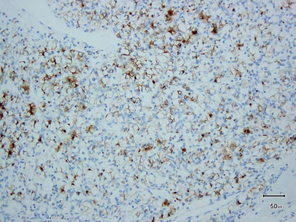
1 CONTROVERSIES IN IMMUNOHISTOCHEMISTRY IN RENAL CELL TUMORS Satish K. Tickoo, M.D. Most of the renal cortical tumors can be easily classified on hematoxylin and eosin evaluation alone. However, there are some cases that need the support of immunohistochemistry and other ancillary tests for their proper classification. Such help is particularly important for the pathologists working in laboratories handling relatively low volumes of renal tumor specimens. The need for good, reliable immunohistochemical stains has also been progressively growing in the recent years, with pathologists increasingly being asked to render diagnosis on limited material (needle core biopsies and fine-needle aspirates). Over the years, a large number of immunohistochemical markers have been reported to show remarkable sensitivity and specificity in discriminating between different renal cortical tumors. After the initial excitement created by the first few publications, very often the subsequent larger studies on wider range of tumors water down the initial claims. One of the common reasons for the initial great versus subsequent not so great results is that in most instances, the initial studies are performed on tumors that are morphologically typical representatives of a tumor type. As a matter of fact, in the day-to-day pathology practice, such cases hardly ever need immunohistochemical support for the diagnosis. When the same antibodies are used on tumors that are morphologically somewhat diverse and atypical, and that really need immunohistochemical support for the diagnosis, the results may be disappointing. As long as the original and subsequent investigators stick to the factualness of their observations, controversies will be very common. However, facts are facts; specific antibodies mark the tumors as reported in the initial publications, as well as in the subsequent papers. Only that, the authors in initial publications often did not investigate the tumors from the perspective that the subsequent authors did. And, non-acceptance of these simple facts makes a CONTROVERSY! While none of them can be regarded as 100% sensitive and specific, in spite of the controversies, a number of antibodies are still useful in the differential diagnosis of renal cell tumors; some more so than others. A few of these are listed and discussed; the list should not be considered all inclusive. Carbonic anhydrase-ix (CA-IX): It is a down-stream marker in the Hypoxia-Inducible Factor (HIF) pathway. HIF is generated in most body cells as a routine. However, in the presence of normal oxygen tension, it immediately gets hydroxylated. Hydroxylated HIF undergoes very quick proteosomic degradation in the presence of normal pvhl. Whenever there is tissue hypoxia, HIF does not get adequately hydroxylated; non-hydroxylated HIF cannot be degraded and is therefore over-expressed. In another scenario, at normal oxygen tension even when HIF gets hydroxylated, and normal pvhl is not present (as is usual in VHL syndrome, and very
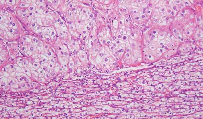
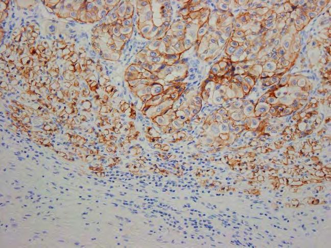
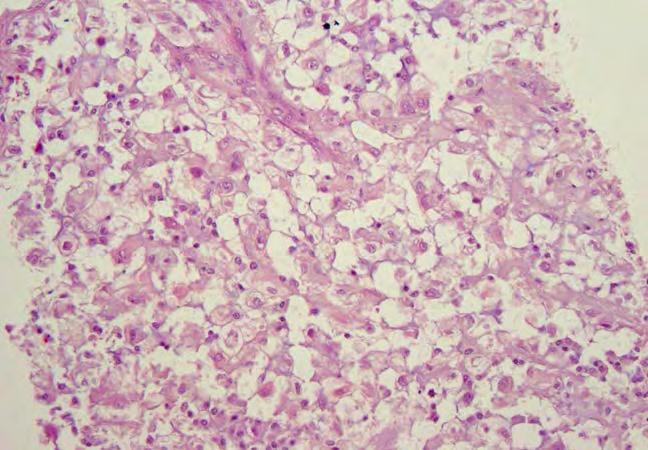
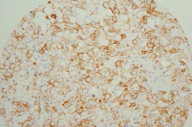
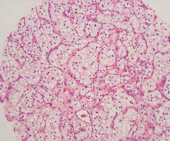
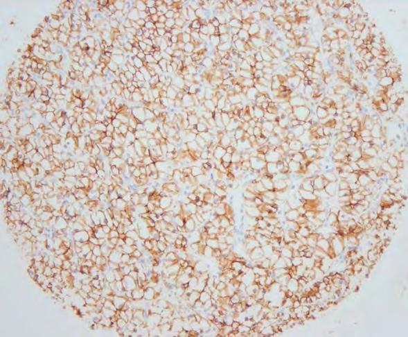
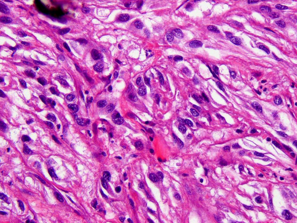
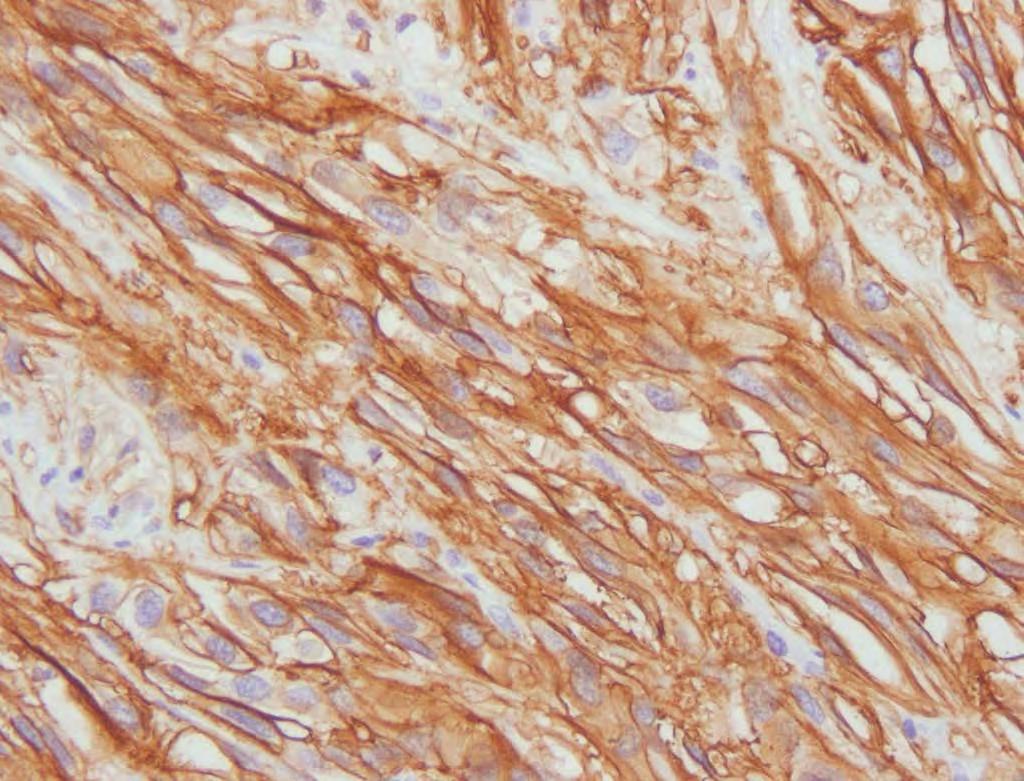
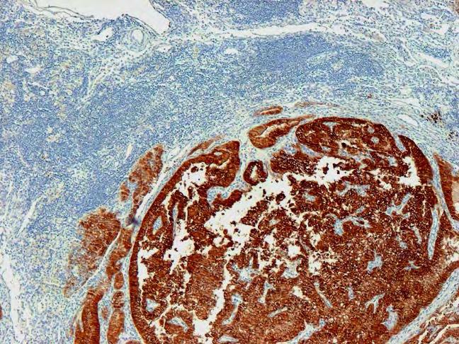
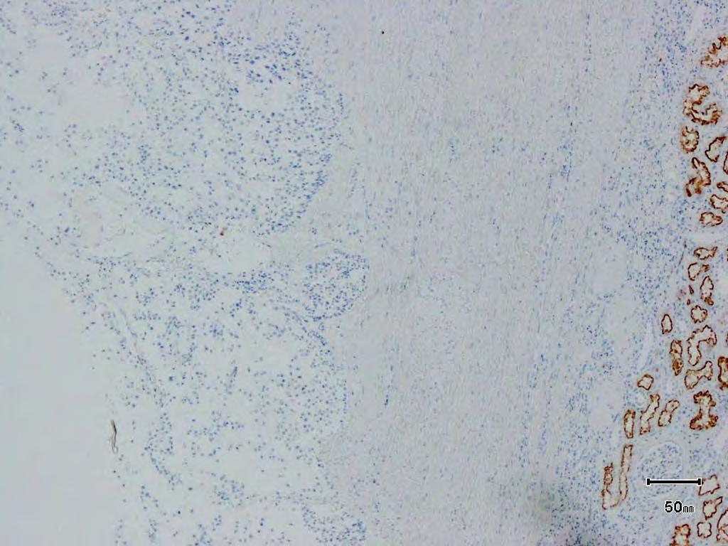
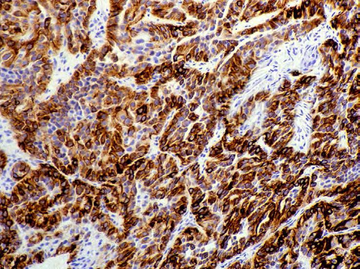
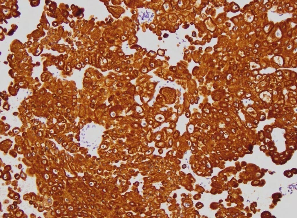
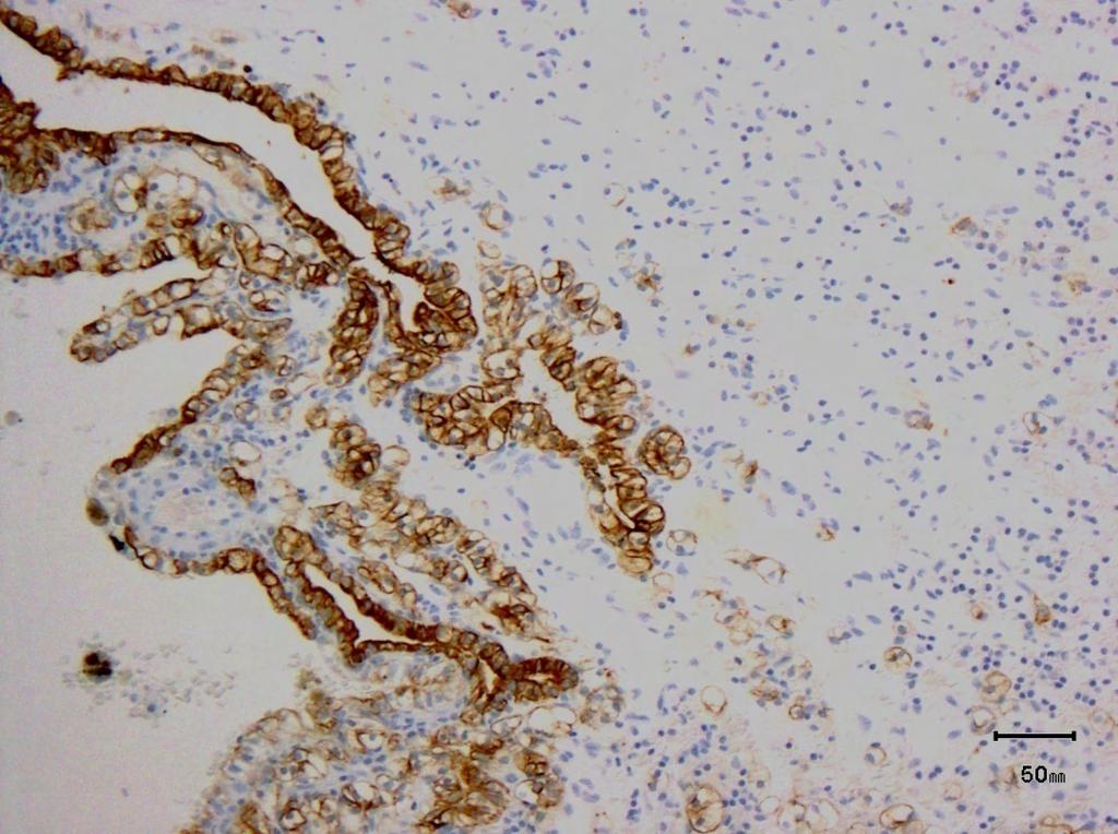
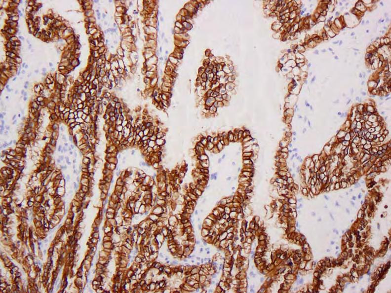
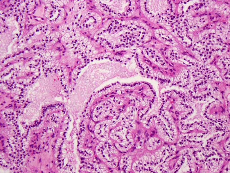
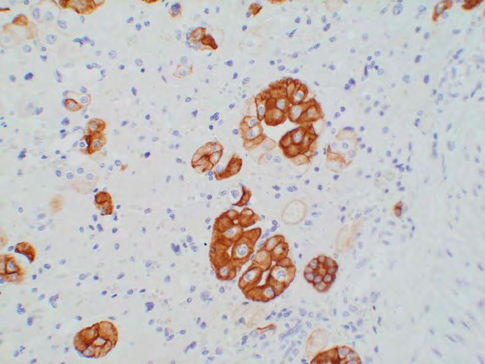
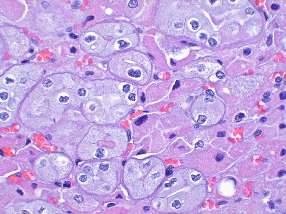
2 common in sporadic clear cell RCCs), the hydroxylated HIF does not get degraded. Thus, it gets over-expressed and activates multiple down-stream proteins, including CA-IX. CA-IX is a trans-membrane protein; therefore, the immunostain is primarily localized to the cell membranes. CA-IX is expressed in overwhelming majority (84-100%) of clear cell RCCs in a diffuse membranous pattern. Its expression has been shown to be retained in metastases from clear cell RCC. The staining is diffuse, irrespective of the nuclear grade of the tumor, although an occasional high grade tumor may show somewhat decreased staining intensity. Unlike any other antibody used for clear cell RCC, CA-IX expression is retained even in sarcomatoid areas in the overwhelming majority of sarcomatoid clear cell RCCs. The antibody is quite useful in the diagnosis of clear cell RCC even on limited material (needle core biopsies and cytological material). Although some pathologists are still concerned about its specificity (see below), the antibody (clone g250) is already being used in clinical trials by radiologists in the diagnosis of clear cell RCC at both the primary and metastatic sites. A recent multicentric phase 3 trial reported a specificity and sensitivity of greater than 86% each, in radiologic scans using a radiolabeled iodine and CA-IX compound in classifying renal tumors as clear cell RCC. CA-IX also shows diffuse membranous immunoreactivity in clear cell papillary RCC, albeit qualitatively a little different from that seen in clear cell RCC. Positive staining has also been reported in bladder urothelial carcinoma, papillary RCC, and a number of other non-renal tumors. However, to the best of our knowledge, even when reported as positive with diffuse staining in only a small proportion of these positive tumors, none of these was reported to show expression in almost 100% of the tumor cells (as is characteristic in clear cell RCC). Such immunoreactivity likely is not related to the absence of pvhl in these tumors, but to the areas of tissue hypoxia. In most tumors other than clear cell RCC, when immunoreactivity is present, it is more prominent around the areas of tumor necrosis, and additionally in the case of papillary RCCs, close to the tips of papillae. Nevertheless, no immunostain can be used as a solitary marker in histo- or cyto-pathological diagnosis. Most of the non-clear cell RCC tumors with some CA-IX positivity will also show immunoreactivity to other markers that are appropriate for those specific tumors. In our experience, only VHL-related syndromic or sporadic tumors (e.g., clear cell papillary cystadenomas of the epididymis, cerebellar hemangioendotheliomas, etc.) show reactivity for CA-IX akin to that seen in clear cell RCC. Another practical caveat in interpreting CA-IX staining is that in limited material (core biopsies, FNAs) that shows extensive tumor necrosis, positivity may be misleading. Most of the positive staining areas can represent peri-necrotic, hypoxic regions in the tumors. Therefore, interpretation of such biopsies requires great caution. Overall, in our opinion, CA-IX is among the best available antibodies for recognizing clear cell RCC; primary, metastatic, high-grade, sarcomatoid, or on limited material. PAX 8/2:
3 Pax8 and Pax2 are members of the paired box family of transcription factors and are very important in the fetal development of the central nervous system, eye, kidney, thyroid gland, and organs of the mesonephric (wolffian) and műllerian duct origin. They show nuclear expression in overwhelming majority of the tumors of renal cell origin, and are, therefore, very useful markers for establishing a renal cell derivation in both the primary and metastatic settings. Pax8 appears to be a more sensitive marker than Pax2, although only Pax2 may be expressed in very rare instances. While evaluating staining for Pax markers, one always needs to remember that both Pax8 and Pax2 are not renal cell-specific, and are known to be expressed in a number of other tumors, including many tumors of the female genital tract, thyroid, a small proportion of upper urinary tract urothelial carcinoma, rare abdominal mesotheliomas, etc. At the same time, both may be absent in some tumors of renal origin, particularly some high grade tumors like collecting duct carcinoma, sarcomatoid RCCs, and tumors with rhabdoid features, as well as many chromophobe RCCs and renal oncocytomas. Thus, interpretation of staining results on Pax 8 or 2 needs be performed in conjunction with other appropriate stains. Neither the presence of staining in isolation is definitive of renal cell origin, nor does absence of staining completely exclude such origin. Renal Cell Carcinoma marker (RCCMa): RCCMa, which is normally expressed in the proximal renal tubules, has been in use as a marker of renal cell origin for many years now. However, it shows sensitivity only in the range of percent among primary renal tumors. At the same time the antibody labels a number of other tumors including lung adenocarcinoma, hepatocellular carcinoma, breast lobular carcinoma, ovarian clear cell carcinoma, parathyroid carcinoma and many others. In a more recent publication by Sharma et al, the antibody was shown to label greater than 40% of non-renal tumors with papillary architecture from various sites. Given the availability of some better antibodies for the diagnosis of renal cell origin, RCCMa appears to have very limited clinical value now. CK7: CK7, a commonly utilized antibody in most surgical pathology laboratories, has multiple utilities in the differentiation of renal cell tumors. CK7 is usually diffusely expressed in type 1 papillary RCC, classical chromophobe RCC and clear cell-papillary RCC. At the 2012 Vancouver ISUP consensus conference, a majority of participants voted for performing immunohistochemical
4 stains (including CK7) to diagnose clear cell-papillary RCC, in addition to taking the morphologic features into consideration. CK7 is usually negative or very focally positive in acquired cystic disease-associated RCC, even when it shows a prominent papillary architecture. CK7 is also generally negative in clear cell RCC, although it may show focal positivity in these tumors around cystic areas. Renal oncocytomas are either negative, or show only rare immunoreactive cells. While it may be useful in distinction of oncocytoma from classical chromophobe RCC, in general CK7 is not very useful in distinction of oncocytoma from eosinophilic variant of chromophobe. Such eosinophilic variants only rarely show diffuse CK7 positivity; the labeling in eosinophilic chromophobes is more often only focal or patchy. Similarly, type 2 papillary RCCs also very often show only limited staining with CK7, unlike the diffuse pattern in type 1 tumors. CK7 remains a useful antibody in the differentiation of many renal tumors. However, too much dependence of the staining results is nor warranted in the distinction of renal oncocytoma from eosinophilic variant of chromophobe RCC, or type 2 papillary RCC from its mimics. CD10: CD10 has been used for many years in the distinction of clear cell RCC from other tumors, particularly in metastatic setting. Only membranous staining can be considered as compatible with clear cell RCC, in an appropriate clinicopathological scenario. On the other hand, cytoplasmic positivity for CD10 is seen in a large number of non-renal and renal tumors. Additionally, membranous immunoreactivity for CD10 is not limited to clear cell RCC, and may be present in papillary RCC, colorectal, pancreatic, prostatic, or some vascular tumors as well. CD10 cannot be used in isolation to diagnose clear cell RCC, either in primary or metastatic setting. Given that better markers for renal cell (like Pax8/2) or clear cell RCC (like CA-IX) origin are now available, the dependence on CD10 may be on the wane. Others: The search for reliable immunohistochemical stains that can consistently distinguish between oncocytic neoplasms (renal oncocytoma, eosinophilic chromophobe RCC, low-grade oncocytic unclassified RCC) has been long and frustrating. A number of immunostains have been proposed in this distinction, but in the long run, none has proven to be as efficient as claimed in the initial publications. The likely reasons for that are discussed above. Multiple antibodies used in this distinction, over the past 1½ to 2 decades include, anti-mitochondrial antibodies, E-cadherin, Kidney-specific cadherin, S100A1, EpCam, etc. Kangai 1 (KAI1/ CD82), product of metastasis
5 suppressor gene, in a few recent publications appears to have some potential. But, more evidence is needed yet. References: 1. Bing Z, Lal P, Lu S, Ziober A, Tomaszewski JE. Role of carbonic anhydrase IX, α-methylacyl coenzyme a racemase, cytokeratin 7, and galectin-3 in the evaluation of renal neoplasms: a tissue microarray immunohistochemical study. Ann Diagn Pathol Feb;17(1): Giménez-Bachs JM, Salinas-Sánchez AS, Serrano-Oviedo L, Nam-Cha SH, Rubio-Del Campo A, Sánchez-Prieto R. Carbonic anhydrase IX as a specific biomarker for clear cell renal cell carcinoma: comparative study of Western blot and immunohistochemistry and implications for diagnosis. Scand J Urol Nephrol Oct;46(5): Shen SS, Truong LD, Scarpelli M, Lopez-Beltran A. Role of immunohistochemistry in diagnosing renal neoplasms: when is it really useful? Arch Pathol Lab Med Apr;136(4): Donato DP, Johnson MT, Yang XJ, Zynger DL. Expression of carbonic anhydrase IX in genitourinary and adrenal tumours. Histopathology Dec;59(6): Al-Ahmadie HA, Alden D, Fine SW, Gopalan A, Touijer KA, Russo P, Reuter VE, Tickoo SK. Role of immunohistochemistry in the evaluation of needle core biopsies in adult renal cortical tumors: an ex vivo study. Am J Surg Pathol Jul;35(7): Sangoi AR, Fujiwara M, West RB, Montgomery KD, Bonventre JV, Higgins JP, Rouse RV, Gokden N, McKenney JK. Immunohistochemical distinction of primary adrenal cortical lesions from metastatic clear cell renal cell carcinoma: a study of 248 cases. Am J Surg Pathol May;35(5): Stillebroer AB, Mulders PF, Boerman OC, Oyen WJ, Oosterwijk E. Carbonic anhydrase IX in renal cell carcinoma: implications for prognosis, diagnosis, and therapy. Eur Urol Jul;58(1): Gupta R, Balzer B, Picken M, Osunkoya AO, Shet T, Alsabeh R, Luthringer D, Paner GP, Amin MB. Diagnostic implications of transcription factor Pax 2 protein and transmembrane enzyme complex carbonic anhydrase IX immunoreactivity in adult renal epithelial neoplasms. Am J Surg Pathol Feb;33(2): Al-Ahmadie HA, Alden D, Qin LX, Olgac S, Fine SW, Gopalan A, Russo P, Motzer RJ, Reuter VE, Tickoo SK. Carbonic anhydrase IX expression in clear cell renal cell carcinoma: an immunohistochemical study comparing 2 antibodies. Am J Surg
6 Pathol Mar;32(3): Tickoo SK, Alden D, Olgac S, Fine SW, Russo P, Kondagunta GV, Motzer RJ, Reuter VE. Immunohistochemical expression of hypoxia inducible factor-1alpha and its downstream molecules in sarcomatoid renal cell carcinoma. J Urol Apr;177(4): Pryma DA, O'Donoghue JA, Humm JL, Jungbluth AA, Old LJ, Larson SM, Divgi CR. Correlation of in vivo and in vitro measures of carbonic anhydrase IX antigen expression in renal masses using antibody 124I-cG250. J Nucl Med Apr;52(4): Divgi CR, Pandit-Taskar N, Jungbluth AA, Reuter VE, Gönen M, Ruan S, Pierre C, Nagel A, Pryma DA, Humm J, Larson SM, Old LJ, Russo P. Preoperative characterisation of clear-cell renal carcinoma using iodine-124-labelled antibody chimeric G250 (124I-cG250) and PET in patients with renal masses: a phase I trial. Lancet Oncol Apr;8(4): Divgi CR, Uzzo RG, Gatsonis C, Bartz R, Treutner S, Yu JQ, Chen D, Carrasquillo JA, Larson S, Bevan P, Russo P. Positron Emission Tomography/Computed Tomography Identification of Clear Cell Renal Cell Carcinoma: Results From the REDECT Trial. J Clin Oncol Jan 10;31(2): Tickoo SK, Milowsky MI, Dhar N, Dudas ME, Gallagher DJ, Al-Ahmadie H, Gopalan A, Fine SW, Ishill N, Bajorin DF, Reuter VE. Hypoxia-inducible factor and mammalian target of rapamycin pathway markers in urothelial carcinoma of the bladder: possible therapeutic implications. BJU Int Mar;107(5): Albadine R, Schultz L, Illei P, Ertoy D, Hicks J, Sharma R, Epstein JI, Netto GJ. PAX8 (+)/p63 (-) immunostaining pattern in renal collecting duct carcinoma (CDC): a useful immunoprofile in the differential diagnosis of CDC versus urothelial carcinoma of upper urinary tract. Am J Surg Pathol Jul;34(7): Ozcan A, Shen SS, Hamilton C, Anjana K, Coffey D, Krishnan B, Truong LD. PAX 8 expression in non-neoplastic tissues, primary tumors, and metastatic tumors: a comprehensive immunohistochemical study. Mod Pathol Jun;24(6): Ordóñez NG. Value of PAX2 immunostaining in tumor diagnosis: a review and update. Adv Anat Pathol Nov;19(6): Bakshi N, Kunju LP, Giordano T, Shah RB. Expression of renal cell carcinoma antigen (RCC) in renal epithelial and nonrenal tumors: diagnostic Implications. Appl Immunohistochem Mol Morphol Sep;15(3): Sharma SG, Gokden M, McKenney JK, Phan DC, Cox RM, Kelly T, Gokden N. The utility of PAX-2 and renal cell carcinoma marker immunohistochemistry in
7 distinguishing papillary renal cell carcinoma from nonrenal cell neoplasms with papillary features. Appl Immunohistochem Mol Morphol Dec;18(6): Liu L, Qian J, Singh H, Meiers I, Zhou X, Bostwick DG. Immunohistochemical analysis of chromophobe renal cell carcinoma, renal oncocytoma, and clear cell carcinoma: an optimal and practical panel for differential diagnosis. Arch Pathol Lab Med Aug;131(8): Avery AK, Beckstead J, Renshaw AA, Corless CL. Use of antibodies to RCC and CD10 in the differential diagnosis of renal neoplasms. Am J Surg Pathol Feb;24(2): Mazal PR, Stichenwirth M, Koller A, Blach S, Haitel A, Susani M. Expression of aquaporins and PAX-2 compared to CD10 and cytokeratin 7 in renal neoplasms: a tissue microarray study. Mod Pathol Apr;18(4): Chu P, Arber DA. Paraffin-section detection of CD10 in 505 nonhematopoietic neoplasms. Frequent expression in renal cell carcinoma and endometrial stromal sarcoma. Am J Clin Pathol Mar;113(3): Weinreb I, Cunningham KS, Perez-Ordoñez B, Hwang DM. CD10 is expressed in most epithelioid hemangioendotheliomas: a potential diagnostic pitfall. Arch Pathol Lab Med Dec;133(12): Notohara K, Hamazaki S, Tsukayama C, Nakamoto S, Kawabata K, Mizobuchi K, Sakamoto K, Okada S. Solid-pseudopapillary tumor of the pancreas: immunohistochemical localization of neuroendocrine markers and CD10. Am J Surg Pathol Oct;24(10): Kuehn A, Paner GP, Skinnider BF, Cohen C, Datta MW, Young AN, Srigley JR, Amin MB. Expression analysis of kidney-specific cadherin in a wide spectrum of traditional and newly recognized renal epithelial neoplasms: diagnostic and histogenetic implications. Am J Surg Pathol Oct;31(10): Ohe C, Kuroda N, Takasu K, Senzaki H, Shikata N, Yamaguchi T, Miyasaka C, Nakano Y, Sakaida N, Uemura Y. Utility of immunohistochemical analysis of KAI1, epithelial-specific antigen, and epithelial-related antigen for distinction of chromophobe renal cell carcinoma, an eosinophilic variant from renal oncocytoma. Med Mol Morphol Jun;45(2): Kauffman EC, Barocas DA, Chen YT, Yang XJ, Scherr DS, Tu JJ. Differential expression of KAI1 metastasis suppressor protein in renal cell tumor histological subtypes. J Urol May;181(5):
8 Controversies in immunohistochemistry in renal cell tumors Satish K. Tickoo
9 Controversy Controversy: Merriam-Webster- discussion marked especially by the expression of opposing views So on controversial issues- what is true Most often all points of view; generally the truth, but often not the whole truth and nothing but the truth o Example; 20 F temperature in NYC- both cold and quite warm Therefore, controversy very often the creation of one s unidirectional thought processes
10 Immunohistochemistry in renal tumors Most stains help in tumors where no help is really needed Some antibodies- put in the controversy list- may actually be useful if used appropriately Carbonic Anhydrase-IX (CA-IX) Pax-8/2 CK7 CD10 RCC Ma
11 Carbonic Anhydrase-IX (CA-IX) CA-IX, a trans-membranous enzyme/protein with functions to regulate intracellular and extracellular ph Expression induced by hypoxic/pseudohypoxic conditions; downstream to HIF protein: overexpression related to hypoxia or lack of pvhl Diffuse membranous positivity in % clear cell RCCs Tu et al. Mod Pathol. 2005;18:169A; Tickoo et al. USCAP, 2007; Al-Ahmadie et al. Am J Surg Pathol. 2011;35:949; Giménez-Bachs et al. Scand J Urol Nephrol. 2012;46:358; Bing et al. Annals Diagnost Pathol. 2013;17:58)
12 CA-IX; Pros Diffuse membranous + in clear cell RCCs Both primary and metastatic (84 vs. 89%) (Tickoo et al. USCAP 2007) Even in high-grade areas with solid, pseudo-papillary, papillary areas (3+ expression; 88 vs. 95%) (Al-Ahmadie et al. Am J Surg Pathol 2008;32:377) 3/4ths of sarcomatoid clear cells in sarcomatoid areas (3+ expression; 73 vs. 86%) (Tickoo et al. J Urol. 2007;177:1224)
13 CA-IX; Pros Highly useful on limited material Biopsies (Al-Ahmadie et al. Am J Surg Pathol 2008;32:377; Donato et al. Histopathology 2011;59:1229) Cell blocks (Personal experience) 124 CA-IX (g250) (chimeric I-cG250) in phase 2 and 3 trials for radio-diagnosis of clear cell RCC (Divgi et al. Lancet Oncol. 2007;8:304; Pryma et al. J Nuc Med. 2011;52:535; Divgi et al. J Clin Oncol. 2013;31:187)
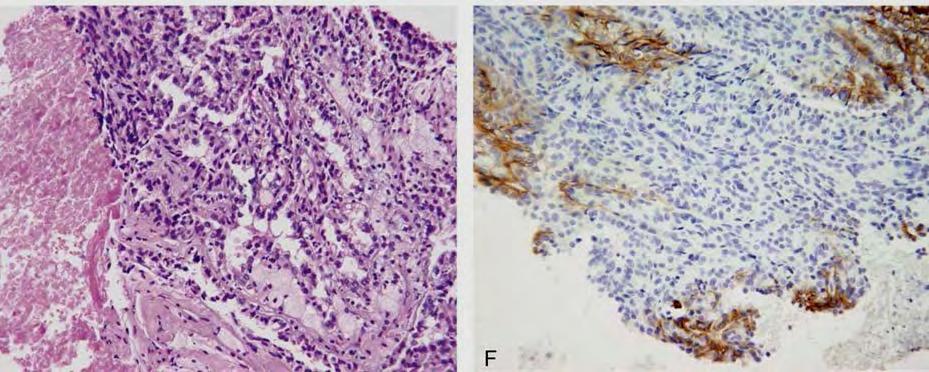
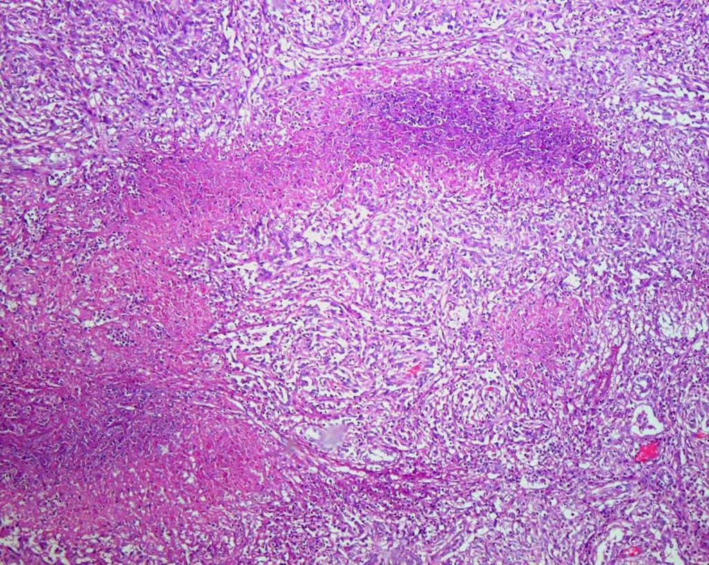
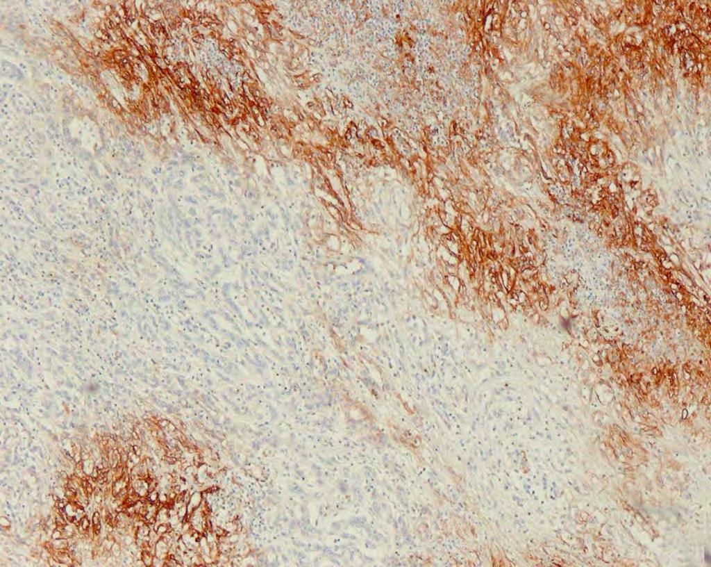
14 CA-IX; Cons Positive in non-renal tumors More concentrated around necrosis; cells away from necrosis, mostly negative Positive in urothelial carcinomas Almost never 100% or close to 100% cells (26% HIF-1, 3+;Tickoo et al. BJU International 2010;107:844; 14-18% CA-IX, 3+; Donato et al Histopathology;59: = >50%) Usually CK7/CK20/HMWCK/p63 positive Reportedly positive in papillary RCC (Gupta et al. Am J Surg Pathol. 2009;33:241-7; Donato et al. Histopathology 2011;59:1229; Al-Ahmadie et al. Am J Surg Pathol. 2011;35:949) Around necrosis and papillary tips; never anywhere close to 100% cells in our experience CK7/AMACR positive
15 CA-IX; Cons Positive in translocation-associated RCC and HLRCC-associated carcinomas Usually absent or patchy staining Clear cell papillary RCC Always positive
16
17

18
19
20
21 CA-IX To date- the best marker in our hands/opinion for clear cell RCC, particularly when used in combination with H&E and other markers
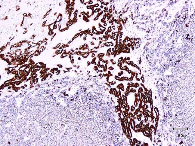
22 Pax 8/2; Pros Overwhelming majority, but not all, of renal tumors positive Clean nuclear stain In most cases both positive, but rarely only one and not the other (Pax 8 generally > Pax 2) If clinically renal tumor a consideration, positivity strongly supports a renal origin
23 Pax8/2; Cons.. Tumors from other organs positive (Female genital tract, thyroid, etc.) May not be positive in high grade tumors of renal cortical origin Positive in a small proportion of upper urinary tract urothelial carcinomas - (Albadine et al. Am J Surg Pathol 2010;34:965)
24 Seminal vesicles 27/27 (100%) Epididymis 17/17 (100%) Ozcan et al. Modern Pathol. 2011;24:751
25 Ordo n ez. Adv Anat Pathol 2012;19:401

26 Pax2 Pax8 Ozcan et al. Arch Pathol Lab Med. 2012;136:1541
27 Pax 8/2 Positivity cannot be used in isolation as diagnostic of renal origin Absent staining does not exclude renal origin, particularly so in high grade tumors


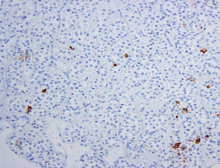
28 Renal Cell Carcinoma marker (RCCMa) an antibody for RCC with reported sensitivity and specificity of approximately 75% Reasonable, but not great sensitivity and specificity Positive in a number of non-renal primary tumors lung adenocarcinomas hepatocellular carcinomas lobular carcinoma breast parathyroid carcinomas ovarian clear cell carcinomas testicular and extra-gonadal seminomas, yolk sac tumor
29 Bakshi et al. Appl Immunohistochem Mol Morphol. 2007;15:310
30 RCC Ma: Cons RCCMa positive in 28/66 (42%) of non-renal papillary neoplasms, including 9/9 (100%) papillary thyroid carcinomas 8/10 (80%) ovarian papillary serous carcinomas 4/9 (44%) uterine papillary serous carcinomas 1/10 (10%) papillary urothelial carcinomas 1/2 (50%) intraductal papillary mucinous carcinomas of the pancreas 3/3 (100%) choroid plexus papillomas 1/1 (100%) pituitary adenoma with papillary features 1/2 (50%) lung adenocarcinomas with papillary features Sharma et al. Appl Immunohistochem Mol Morphol 2010;18:494
31
32 CK7 Is a commonly used antibody in most pathology labs Usually positive in papillary and chromophobe RCC Mostly negative in clear cell RCC Very focally positive in renal oncocytoma
33 CK7; Pros. Very reliable antibody for clear cell papillary RCC- diagnostic utility (ISUP 2012) Quite reliable for papillary RCC, particularly type 1 Often diffusely positive in classical chromophobe RCC Variably positive in collecting duct carcinoma
34 CK7; Cons Clear cell RCCs with cystic components, often positive - (Fine et al. 2012, USCAP) Eosinophilic chromophobe RCC, often very focal or negative Not of much use in distinction from oncocytoma Type 2 papillary RCC, often negative
35 Papillary Chromophobe Oncocytoma Oncocytoma
36
37
38 CK7 Use and interpret with caution in tumors where it is needed the most (oncocytoma vs. eosinophilic chromophobe; high grade papillary)
39 CD10 Common Acute Lymphoblastic Leukemia Antigen (CALLA) Labels clear cell RCC in a membranous pattern (approximately 80%) (Liu et al. Arch Pathol Lab Med. 2007;131:1290; Tickoo et al. 2007, USCAP) Labels papillary RCC (Avery et al Am J Surg Pathol 2000;24:203; Mazal et al. Mod Pathol. 2005;18:535) Usually along the apical membrane Variable expression apical/membranous/ cytoplasmic in chromophobe RCC and oncocytoma (0-35%) (Avery et al Am J Surg Pathol 2000;24:203; Liu et al. Arch Pathol Lab Med. 2007;131:1290)
40 CD10; Pros. In metastatic setting, may differentiate from clear cell adenocarcinoma of GYN tract
41 CD10: Cons.. Apical surface only: well-differentiated colonic, pancreatic and prostatic adenocarcinoma Canalicular pattern in HCC may mimic membranous positivity, particularly in limited tissue Diffuse cytoplasmic or membranous/golgi patterns: poorly differentiated adenocarcinoma, melanoma, urothelial carcinoma, renal cell carcinoma, endometrial stromal sarcoma (Am J Clin Pathol 2000;113:374) Epithelioid hemangioendothelioma can be strong/diffuse CD10+ and mimic metastatic renal cell carcinoma (Arch Pathol Lab Med 2009;133:1965) Solid and pseudopapillary pancreatic tumor is CD10+ (Am J Surg Pathol 2000;24:1361)

42
43 CD10 To support the diagnosis of clear cell RCC, positivity has to be membranous; cytoplasmic positivity is seen in a number of other tumors Cannot depend on CD10 alone
44 Anti-Mito, E-Cad, Ksp-Cad, S100A1, KAI1 Multiple markers claimed to distinguish between oncocytoma and chromophobe RCC Initial publications raise expectations, but ultimately none proven to be great Kidney specific-cadherin: The findings argue against the use of Ksp-cad in differentiating chromophobe renal cell carcinoma and renal oncocytomas (Kuehn et al. Am J Surg Pathol. 2007;31:1528) Currently, KAI1 being touted as potentially useful (Ohe et al. Med Mol Morphol. 2012;45:98)
45 Conclusions When a thing ceases to be a subject of controversy, it ceases to be a subject of interest. William Hazlitt In a controversy the instant we feel anger we have already ceased striving for the truth, and have begun striving for ourselves. Buddha
IMMUNOPROFILES OF THE MAJOR RENAL NEOPLASMS (%staining)
") Stain Clear Cell Papillary IMMUNOPROFILES OF THE MAJOR RENAL NEOPLASMS (%staining) Chromophobe Collecting Duct Carcinom a Sarcomatoid Xp11 Translocat ion Dr Jon Oxley See also www.jonoxley.com Page 1 MTSCC
Stain Clear Cell Papillary IMMUNOPROFILES OF THE MAJOR RENAL NEOPLASMS (%staining) Chromophobe Collecting Duct Carcinom a Sarcomatoid Xp11 Translocat ion Dr Jon Oxley See also www.jonoxley.com Page 1 MTSCC
Diagnostic accuracy of percutaneous renal tumor biopsy May 10 th 2018
 Diagnostic accuracy of percutaneous renal tumor biopsy May 10 th 2018 Dr. Tzahi Neuman Dep.Of Pathology Hadassah Medical Center Jerusalem, Israel, (tneuman@hadassah.org.il) Disclosure: 1 no conflicts of
Disclosure. Relevant Financial Relationship(s) None. Off Label Usage None MFMER slide-1
 None. Off Label Usage None MFMER slide-1") Disclosure Relevant Financial Relationship(s) None Off Label Usage None 2013 MFMER slide-1 Case Presentation A 43 year old male, with partial nephrectomy for a right kidney mass 2013 MFMER slide-2 2013
Disclosure Relevant Financial Relationship(s) None Off Label Usage None 2013 MFMER slide-1 Case Presentation A 43 year old male, with partial nephrectomy for a right kidney mass 2013 MFMER slide-2 2013
Immunohistochemical Diagnosis of Renal Neoplasms. Luan D. Truong, MD; Steven S. Shen, MD, PhD
 Immunohistochemical Diagnosis of Renal Neoplasms Luan D. Truong, MD; Steven S. Shen, MD, PhD N Context. Histologic diagnosis of renal neoplasm is usually straightforward by routine light microscopy. However,
RENAL EPITHELIAL NEOPLASMS: IS THERE A ROLE OF IMMUNOSTAINS IN DIAGNOSIS?
 RENAL EPITHELIAL NEOPLASMS: IS THERE A ROLE OF IMMUNOSTAINS IN DIAGNOSIS? John C. Cheville, M.D. Mayo Clinic and Mayo Foundation Rochester, MN The majority of renal epithelial neoplasms are diagnosed on
Immunoexpression of napsin a in renal neoplasms
 Zhu et al. Diagnostic Pathology (2015) 10:4 DOI 10.1186/s13000-015-0242-z RESEARCH Open Access Immunoexpression of napsin a in renal neoplasms Bing Zhu, Stephen M Rohan and Xiaoqi Lin * Abstract Background:
Coordinate Expression of Cytokeratins 7 and 20 in Prostate Adenocarcinoma and Bladder Urothelial Carcinoma
 Anatomic Pathology / CYTOKERATINS 7 AND 20 IN PROSTATE AND BLADDER CARCINOMAS Coordinate Expression of Cytokeratins 7 and 20 in Prostate Adenocarcinoma and Bladder Urothelial Carcinoma Nader H. Bassily,
2016 WHO CLASSIFICATION OF TUMOURS OF THE PROSTATE. Peter A. Humphrey, MD, PhD Yale University School of Medicine New Haven, CT
 2016 WHO CLASSIFICATION OF TUMOURS OF THE PROSTATE Peter A. Humphrey, MD, PhD Yale University School of Medicine New Haven, CT 2016 WHO CLASSIFICATION OF TUMOURS OF THE PROSTATE AUTHORS : PROSTATE CHAPTER
Renal tumours: use of immunohistochemistry & molecular pathology. Dr Lisa Browning John Radcliffe Hospital Oxford
 Renal tumours: use of immunohistochemistry & molecular pathology Dr Lisa Browning John Radcliffe Hospital Oxford Renal tumours: the use of immunohistochemistry & molecular pathology Classification of RCC
DIAGNOSTIC SLIDE SEMINAR: PART 1 RENAL TUMOUR BIOPSY CASES
 DIAGNOSTIC SLIDE SEMINAR: PART 1 RENAL TUMOUR BIOPSY CASES Dr. Andrew J. Evans MD, PhD, FACP, FRCPC Consultant in Genitourinary Pathology University Health Network, Toronto, ON Case 1 43 year-old female,
performed to help sway the clinician in what the appropriate diagnosis is, which can substantially alter the treatment of management.
 Hello, I am Maura Polansky at the University of Texas MD Anderson Cancer Center. I am a Physician Assistant in the Department of Gastrointestinal Medical Oncology and the Program Director for Physician
Key Words: PAX2; PAX8; renal cell carcinoma; cytology; fine-needle aspiration
 Utility of PAX8 and PAX2 Immunohistochemistry in the Identification of Renal Cell Carcinoma in Diagnostic Cytology Stewart M. Knoepp, M.D., Ph.D., Lakshmi P. Kunju, M.D., and Michael H. Roh, M.D., Ph.D.*
Urinary Bladder: WHO Classification and AJCC Staging Update 2017
 Urinary Bladder: WHO Classification and AJCC Staging Update 2017 Houston Society of Clinical Pathologists 58 th Annual Spring Symposium Houston, TX April 8, 2017 Jesse K. McKenney, MD Classification
Enterprise Interest Nothing to declare
 Enterprise Interest Nothing to declare Biopsy diagnosis of renal tumors. Current applications Ondřej Hes Department of Pathology Charles University and University Hospital Plzeň Czech Republic Dealing
The Panel Approach to Diagnostics. Lauren Hopson International Product Specialist Cell Marque Corporation
 The Panel Approach to Diagnostics Lauren Hopson International Product Specialist Cell Marque Corporation Cell Marque Rocklin, California About Cell Marque: IVD primary antibody manufacturer Distributors
Prostate Immunohistochemistry. Literature Interpretation: Caveats. Must be aware of staining pattern of antibody in the relevant tissue
 IHC Interpretation: General Principles (1) Prostate Immunohistochemistry Murali Varma Cardiff, UK wptmv@cf.ac.uk Sarajevo Nov 2013 Must be aware of staining pattern of antibody in the relevant tissue Nuclear/cytoplasmic/membranous
Jason C Carvalho, 1 Dafydd G Thomas, 1 Jonathan B McHugh, 1 Rajal B Shah 2 & Lakshmi P Kunju 1
 Histopathology 2012, 60, 597 608. DOI: 10.1111/j.1365-2559.2011.04093.x p63, CK7, PAX8 and INI-1: an optimal immunohistochemical panel to distinguish poorly differentiated urothelial cell carcinoma from
Synonyms. Nephrogenic metaplasia Mesonephric adenoma
 Nephrogenic Adenoma Synonyms Nephrogenic metaplasia Mesonephric adenoma Definition Benign epithelial lesion of urinary tract with tubular, glandular, papillary growth pattern Most frequently in the urinary
RENAL EPITHELIAL TUMORS 2009: THE ROLE OF ELECTRON MICROSCOPY IN UNDERSTANDING PATHOGENESIS, DIAGNOSIS, AND CLASSIFICATION.
 RENAL EPITHELIAL TUMORS 2009: THE ROLE OF ELECTRON MICROSCOPY IN UNDERSTANDING PATHOGENESIS, DIAGNOSIS, AND CLASSIFICATION. Guillermo A. Herrera MD Nephrocor, Tempe, Arizona Epithelial renal cell tumors
Carcinoma of unknown primary origin (CUP) is defined
 is defined") REVIEW ARTICLE Metastatic Carcinoma of Unknown Primary: Diagnostic Approach Using Immunohistochemistry James R. Conner, MD, PhD and Jason L. Hornick, MD, PhD Abstract: Carcinoma of unknown primary origin
REVIEW ARTICLE Metastatic Carcinoma of Unknown Primary: Diagnostic Approach Using Immunohistochemistry James R. Conner, MD, PhD and Jason L. Hornick, MD, PhD Abstract: Carcinoma of unknown primary origin
ARTHUR PURDY STOUT SOCIETY COMPANION MEETING: DIFFICULT NEW DIFFERENTIAL DIAGNOSES IN PROSTATE PATHOLOGY. Jonathan I. Epstein.
 1 ARTHUR PURDY STOUT SOCIETY COMPANION MEETING: DIFFICULT NEW DIFFERENTIAL DIAGNOSES IN PROSTATE PATHOLOGY Jonathan I. Epstein Professor Pathology, Urology, Oncology The Reinhard Professor of Urological
Presentation material is for education purposes only. All rights reserved URMC Radiology Page 1 of 98
 Presentation material is for education purposes only. All rights reserved. 2011 URMC Radiology Page 1 of 98 Radiology / Pathology Conference February 2011 Brooke Koltz, Cytopathology Resident Presentation
Unknown Slides Conference
 Unknown Slides Conference Jae Y. Ro, MD, PhD Weill Medical College of Cornell Univ. The Methodist Hospital, and UT MD Anderson Cancer Center Houston, TX November 9, 2013 Amman, Jordan 25 th Congress of
Evening Specialty Conference: Cytopathology
 : Cytopathology N. Paul Ohori, M.D. University of Pittsburgh Medical Center Disclosure of Relevant Financial Relationships Disclosure of Relevant Financial Relationships USCAP requires that all planners
The clinically challenging entity of liver metastasis from tumors of unknown primary
 The clinically challenging entity of liver metastasis from tumors of unknown primary Xuchen Zhang, MD, PhD Associate Professor of Pathology Department of Pathology Yale University School of Medicine Liver
Differential diagnosis of HCC
 Hepatocellular Carcinoma Quest for an Ideal Immunohistochemical Panel Sanjay Kakar, MD UCSF Differential diagnosis of HCC Hepatocellular lesions Adenoma, FNH, HG dysplasia Adenocarcinoma CholangioCA, metastasis
PAX2 and PAX8 Expression in Primary and Metastatic Renal Tumors. A Comprehensive Comparison
 PAX2 and PAX8 Expression in Primary and Metastatic Renal Tumors A Comprehensive Comparison Ayhan Ozcan, MD; Gustavo de la Roza, MD; Jae Y. Ro, MD, PhD; Steven S. Shen, MD, PhD; Luan D. Truong, MD N Context.
CME/SAM. Significant Variation of Immunohistochemical Marker Expression in Paired Primary and Metastatic Clear Cell Renal Cell Carcinomas
 AJCP / Original Article Significant Variation of Immunohistochemical Marker Expression in Paired Primary and Metastatic Clear Cell Renal Cell Carcinomas Zenggang Pan, MD, PhD, 1,2 William Grizzle, MD,
ACCME/Disclosures. Case History 4/13/2016. USCAP GU Specialty Conference Case 3. Ann Arbor, MI
 USCAP GU Specialty Conference Case 3 March 2016 L. Priya Kunju, M.D. University of Michigan Health System Ann Arbor, MI University of Michigan Health System ACCME/Disclosures The USCAP requires that anyone
How to Recognize Gynecologic Cancer Cells from Pelvic Washing and Ascetic Specimens
 How to Recognize Gynecologic Cancer Cells from Pelvic Washing and Ascetic Specimens Wenxin Zheng, M.D. Professor of Pathology and Gynecology University of Arizona zhengw@email.arizona.edu http://www.zheng.gynpath.medicine.arizona.edu/index.html
A 53 year-old woman with a lung mass, right hilar mass and mediastinal adenopathy.
 November 2015 Case of the Month A 53 year-old woman with a lung mass, right hilar mass and mediastinal adenopathy. Contributed by: Rasha Salama, M.D., IU Department of Pathology and Laboratory Medicine
Case 18. M75. Excision of mass on scalp. Clinically SCC. The best diagnosis is:
 Case 18 M75. Excision of mass on scalp. Clinically SCC. The best diagnosis is: A. Pilomatrical carcinoma B. Adnexal carcinoma NOS C. Metastatic squamous cell carcinoma D.Primary squamous cell carcinoma
ACCME/Disclosures. Diagnosing Mesothelioma in Limited Tissue Samples. Papanicolaou Society of Cytopathology Companion Meeting March 12 th, 2016
 Diagnosing Mesothelioma in Limited Tissue Samples Papanicolaou Society of Cytopathology Companion Meeting March 12 th, 2016 Sanja Dacic, MD, PhD University of Pittsburgh ACCME/Disclosures GENERAL RULES
Pitfalls in the diagnosis of well-differentiated hepatocellular lesions
 2013 Colorado Society of Pathology Pitfalls in the diagnosis of well-differentiated hepatocellular lesions Sanjay Kakar, MD University of California, San Francisco Outline Hepatocellular adenoma: new WHO
Abstract. Introduction. Salah Abobaker Ali
 Sensitivity and specificity of combined fine needle aspiration cytology and cell block biopsy versus needle core biopsy in the diagnosis of sonographically detected abdominal masses Salah Abobaker Ali
Select problems in cystic pancreatic lesions
 Disclosure Select problems in cystic pancreatic lesions Five Prime Therapeutics shareholder Adicet Bio shareholder Bristol-Meyer Squibb advisory board grace.kim@ucsf.edu Pancreatic cystic lesions Intraductal
Diagnostic IHC in lung and pleura pathology
 Diagnostic IHC in lung and pleura pathology Mogens Vyberg Professor of Clinical Pathology Director of NordiQC Aalborg University Hospital, Aalborg, Denmark WHO 2004 and Web Malignant mesothelioma Epithelioid
Papillary Lesions of the Breast A Practical Approach to Diagnosis. (Arch Pathol Lab Med. 2016;140: ; doi: /arpa.
 Papillary Lesions of the Breast A Practical Approach to Diagnosis (Arch Pathol Lab Med. 2016;140:1052 1059; doi: 10.5858/arpa.2016-0219-RA) Papillary lesions of the breast Span the spectrum of benign,
Kidney-specific cadherin, a specific marker for the distal portion of the nephron and related renal neoplasms
 & 2005 USCAP, Inc All rights reserved 0893-3952/05 $30.00 www.modernpathology.org Kidney-specific cadherin, a specific marker for the distal portion of the nephron and related renal neoplasms Steven S
Original Articles. PAX-2 Is a Helpful Marker for Diagnosing Metastatic Renal Cell Carcinoma
 Original Articles PAX-2 Is a Helpful Marker for Diagnosing Metastatic Renal Cell Carcinoma Comparison With the Renal Cell Carcinoma Marker Antigen and Kidney-Specific Cadherin Ayhan Ozcan, MD; Qihui Zhai,
Expression of Cytokeratin 5/6 in Epithelial Neoplasms: An Immunohistochemical Study of 509 Cases
 Expression of Cytokeratin 5/6 in Epithelial Neoplasms: An Immunohistochemical Study of 509 Peiguo G. Chu, M.D., Ph.D., Lawrence M. Weiss, M.D. Department of Pathology, City of Hope National Medical Center,
Sustained Response to Temsirolimus in Chromophobe variant of Metastatic Renal Cell Carcinoma
 JOURNAL OF CASE REPORTS 2015;5(1):280-284 Sustained Response to Temsirolimus in Chromophobe variant of Metastatic Renal Cell Carcinoma Chanchal Goswami, Aditi Mandal B. P. Poddar Hospital & Medical Research
New Developments in Immunohistochemistry for Gynecologic Pathology
 New Developments in Immunohistochemistry for Gynecologic Pathology Michael T. Deavers, M.D. Professor, Departments of Pathology and Gynecologic Oncology Immunohistochemistry in Gynecologic Pathology Majority
POORLY DIFFERENTIATED, HIGH GRADE AND ANAPLASTIC CARCINOMAS: WHAT IS EVERYONE TALKING ABOUT?
 POORLY DIFFERENTIATED, HIGH GRADE AND ANAPLASTIC CARCINOMAS: WHAT IS EVERYONE TALKING ABOUT? AGGRESSIVE THYROID CANCERS PAPILLARY CARCINOMA CERTAIN SUBTYPES POORLY DIFFERENTIATED CARCINOMA HIGH GRADE DIFFERENTIATED
4/12/2018. MUSC Pathology Symposium Kiawah Island April 18, Jesse K. McKenney, MD
 MUSC Pathology Symposium Kiawah Island April 18, 2018 Jesse K. McKenney, MD 1 Urothelial Carcinoma with Alternative Differentiation 2 Urothelial Carcinoma with Alternative Differentiation Recognition as
Mesothelioma: diagnostic challenges from a pathological perspective. Naseema Vorajee August 2016
 Mesothelioma: diagnostic challenges from a pathological perspective Naseema Vorajee August 2016 Naseema.vorajee@nhls.ac.za Pleural diseases (whether neoplastic, reactive or infective) may have similar
Cancers of unknown primary : Knowing the unknown. Prof. Ahmed Hossain Professor of Medicine SSMC
 Cancers of unknown primary : Knowing the unknown Prof. Ahmed Hossain Professor of Medicine SSMC Definition Cancers of unknown primary site (CUPs) Represent a heterogeneous group of metastatic tumours,
ROLE OF PROSTATIC BASAL CELL MARKER IN DIAGNOSIS OF PROSTATIC LESIONS
 Original Research Article Pathology International Journal of Pharma and Bio Sciences ISSN 0975-6299 ROLE OF PROSTATIC BASAL CELL MARKER IN DIAGNOSIS OF PROSTATIC LESIONS SUBATHRA K* Department of pathology,
Updates in Urologic Pathology WHO Made Those Changes?! Peyman Tavassoli Pathology Department BC Cancer Agency
 Updates in Urologic Pathology WHO Made Those Changes?! Peyman Tavassoli Pathology Department BC Cancer Agency World Health Organization Available in Feb 2016 Frame work for reporting Major contributing
Breast cancer: IHC classification. Mogens Vyberg Professor of Clinical Pathology Director of NordiQC Aalborg University Hospital, Aalborg, Denmark
 Breast cancer: IHC classification Mogens Vyberg Professor of Clinical Pathology Director of NordiQC Aalborg University Hospital, Aalborg, Denmark http://upload.wikimedia.org/wikipedia/commons/1/1a/breast.svg
Case Report Clear Cell Adenocarcinoma of the Renal Pelvis in a Male Patient
 Case Reports in Pathology Volume 2013, Article ID 494912, 4 pages http://dx.doi.org/10.1155/2013/494912 Case Report Clear Cell Adenocarcinoma of the Renal Pelvis in a Male Patient Sarawut Kongkarnka, 1
Nordic Immunohistochemical Quality Control
 Nordic Immunohistochemical Quality Control Immunohistochemistry in the classifiation of neoplasias of the alimentary tract & External Quality Assurance of Immunohistochemistry for GI cancer markers Mogens
Applications of IHC. Determination of the primary site in metastatic tumors of unknown origin
 Applications of IHC Determination of the primary site in metastatic tumors of unknown origin Classification of tumors that appear 'undifferentiated' by standard light microscopy Precise classification
AMERICAN ASSOCIATION OF NEUROPATHOLOGISTS COMPANION SOCIETY MEETING at the 106 th ANNUAL MEETING OF THE USCAP San Antonio, March 4, 2017
 AMERICAN ASSOCIATION OF NEUROPATHOLOGISTS COMPANION SOCIETY MEETING at the 106 th ANNUAL MEETING OF THE USCAP San Antonio, March 4, 2017 SYLLABUS Papillary Tumor of the Pineal Region and the Differential
CASE year old male with a PET avid nodule in the left adrenal gland
 CASE 1 55 year old male with a PET avid nodule in the left adrenal gland Case 1 Adrenal gland parenchyma partly replaced by a spindle cell tumour with mild nuclear pleomorphism Atypical mitoses present
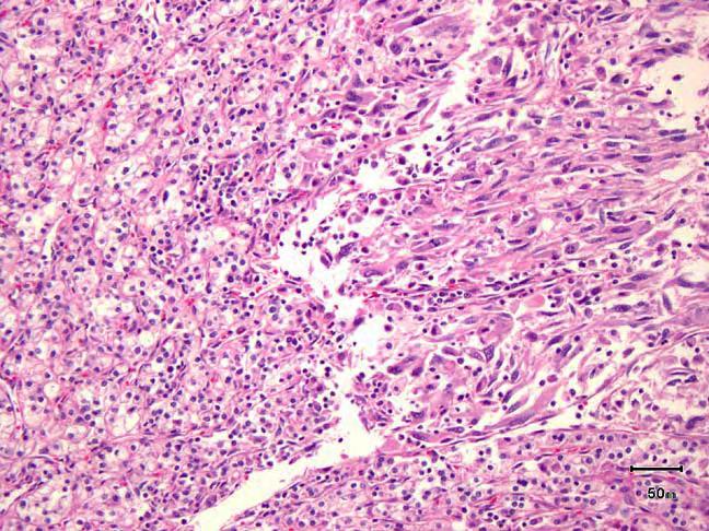
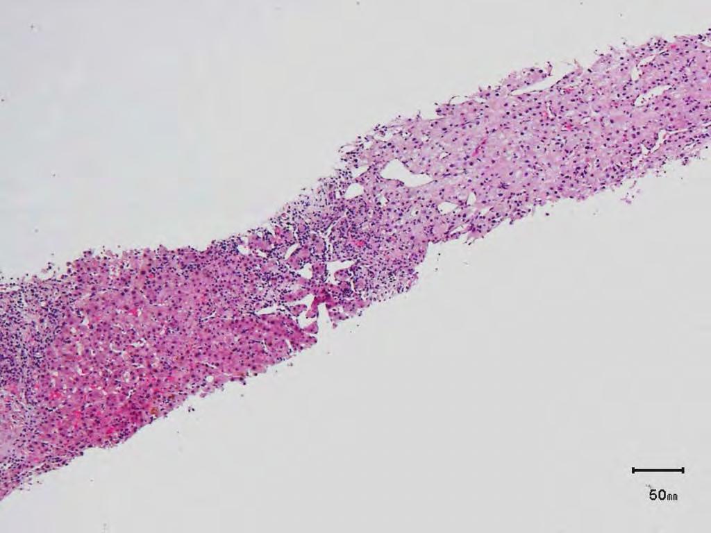
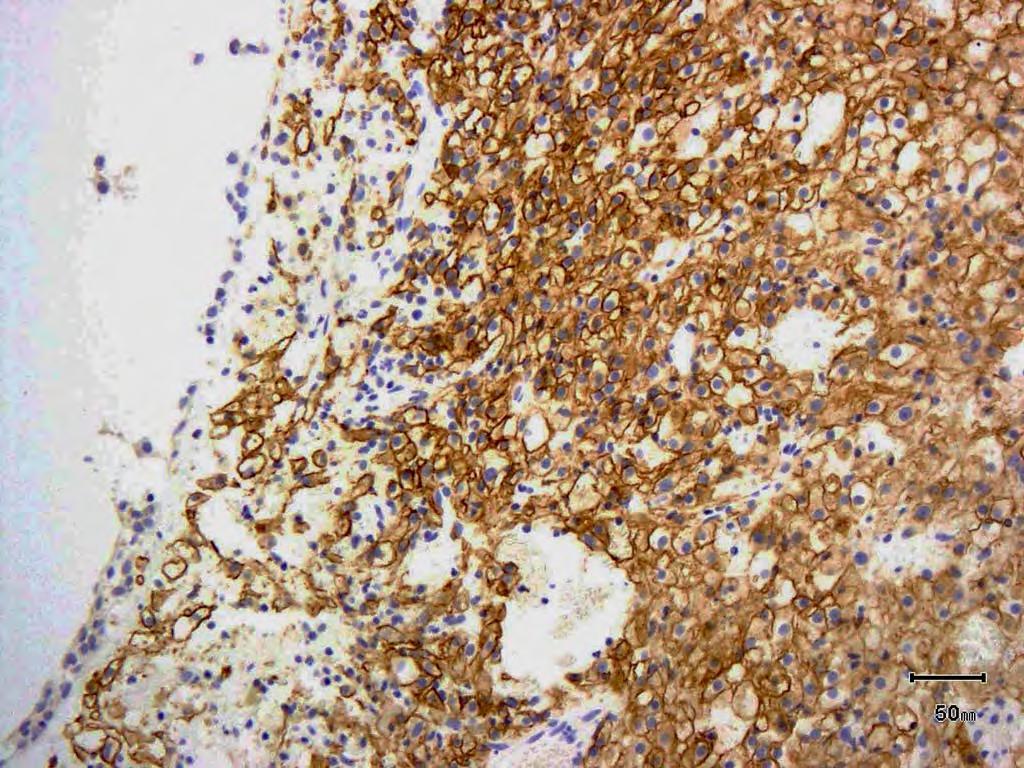
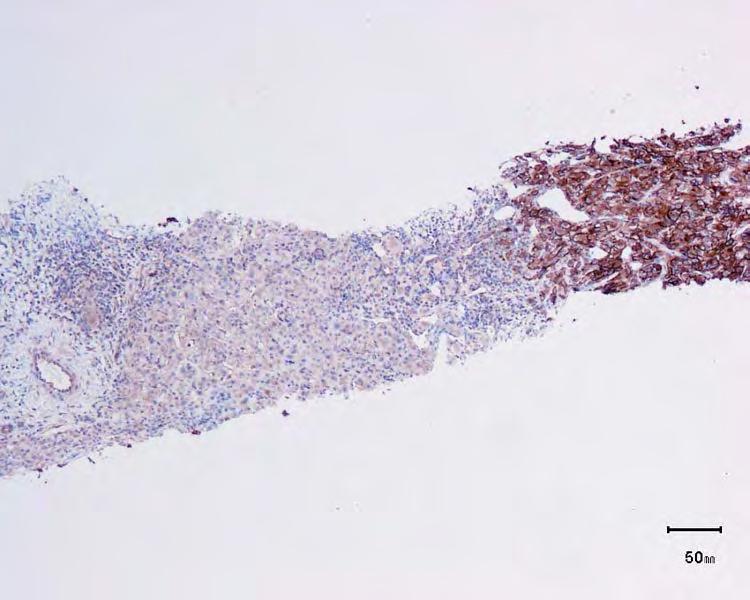
Case: The patient is a 62 year old woman with a history of renal cell carcinoma that was removed years ago. A 2.4 cm liver mass was found on CT
 Case: The patient is a 62 year old woman with a history of renal cell carcinoma that was removed years ago. A 2.4 cm liver mass was found on CT during follow- up. ALT, AST, Alk Phos and bilirubin were
Histopathological diagnosis of CUP
 Histopathological diagnosis of CUP Dr Karin Oien karin.oien@glasgow.ac.uk Disclosure slide Dr Karin Oien has no financial interests in any company mentioned in this presentation. Dr Karin Oien is conducting
PROSTATIC ADENOCARCINOMA: DIAGNOSTIC CRITERIA AND IMPORTANT MIMICKERS PROSTATIC ADENOCARCINOMA: DIAGNOSTIC CRITERIA
 PROSTATIC ADENOCARCINOMA: DIAGNOSTIC CRITERIA AND IMPORTANT MIMICKERS PROSTATIC ADENOCARCINOMA: DIAGNOSTIC CRITERIA 1 A good H & E helps! ADENOCARCINOMA DIAGNOSTIC CRITERIA Relatively uniform proliferation
CASE 01 LA Path Slide Seminar 13 March, 08. Deepti Dhall, MD Department of Pathology and Laboratory Medicine Cedars-Sinai Medical Center
 CASE 01 LA Path Slide Seminar 13 March, 08 Deepti Dhall, MD Department of Pathology and Laboratory Medicine Cedars-Sinai Medical Center Clinical History 60 year old male presented with obstructive jaundice
ACCME/Disclosures. M31078/07 Ondřej Hes 4/13/2016
 M31078/07 Ondřej Hes Department of Pathology Charles University and University Hospital Plzeň Bioptická laboratoř Plzeň Czech Republic ACCME/Disclosures The USCAP requires that anyone in a position to
Prostatic ductal adenocarcinoma is a subtype of
 ORIGINAL ARTICLE High-grade Prostatic Intraepithelial Neoplasialike Ductal Adenocarcinoma of the Prostate: A Clinicopathologic Study of 28 Cases Fabio Tavora, MD* and Jonathan I. Epstein, MD*w z Abstract:
NEW IHC A n t i b o d i e s
 NEW IHC Antibodies TABLE OF CONTENTS NEW IHC ANTIBODIES from Cell Marque CITED1 (5H6).... 1 Claudin 7 (5D10F3).... 1 GATA1 (4F5).... 1 Transgelin (2A10C2).... 1 NEW IHC ANTIBODIES using RabMAb Technology
Pathology of the Prostate. PathoBasic Tatjana Vlajnic
 Pathology of the Prostate PathoBasic 24.01.17 Tatjana Vlajnic Overview Adenocarcinoma of the prostate Grading Special variants Mimickers of prostate adenocarcinoma Atrophy Inflammatory conditions Granulomatous
Update on Thyroid FNA The Bethesda System. Shikha Bose M.D. Associate Professor Cedars Sinai Medical Center
 Update on Thyroid FNA The Bethesda System Shikha Bose M.D. Associate Professor Cedars Sinai Medical Center Thyroid Nodules Frequent occurrence Palpable: 4-7% of adults Ultrasound: 10-31% Majority benign
Disclosure of Relevant Financial Relationships
 Squamous entities of the thyroid: Reactive to Neoplastic Michelle D. Williams Associate Professor Dept of Pathology, Head & Neck Section University of Texas MD Anderson Cancer Center Disclosure of Relevant
Pathology Mystery and Surprise
 Pathology Mystery and Surprise Tim Smith, MD Director Anatomic Pathology Medical University of South Carolina Disclosures No conflicts to declare Some problem cases Kidney tumor Scalp tumor Bladder tumor
Immunohistochemical classification of lung carcinomas and mesotheliomas. Prof. Mogens Vyberg NordiQC Institute of Pathology Aalborg, Denmark
 Immunohistochemical classification of lung carcinomas and mesotheliomas Prof. Mogens Vyberg NordiQC Institute of Pathology Aalborg, Denmark Endobronchial ultrasound guided transbronchial needle biopsy
Cutaneous metastases. Thaddeus Mully. University of California, San Francisco Professor, Departments of Pathology and Dermatology
 Cutaneous metastases Thaddeus Mully University of California, San Francisco Professor, Departments of Pathology and Dermatology DISCLOSURE OF RELATIONSHIPS WITH INDUSTRY Thaddeus Mully Course C005 Essential
3/24/2017. Disclosure of Relevant Financial Relationships. Mixed Epithelial Endometrial Carcinoma. ISGyP Endometrial Cancer Project
 Disclosure of Relevant Financial Relationships USCAP requires that all planners (Education Committee) in a position to influence or control the content of CME disclose any relevant financial relationship
American Journal of. Medical Case Reports. CAM5.2 Expression in Metastatic Tumours of CNS: A Diagnostic Tool
 American Journal of American Journals of Medical Case Reports http://ivyunion.org/index.php/ajmcr/index Medical Case Reports Mathur SK et al. American Journal of Medical Case Reports 2014, 2:1-8 Vol 2,
My personal experience at University of Toronto and recent updates of
 My personal experience at University of Toronto and recent updates of Endocrine Pathology Toshitetsu Hayashi M.D. Ph.D. ¹Department of Diagnostic Pathology, Takamatsu Red Cross Hospital, Japan ²Laboratory
Hemangioblastoma is a benign central nervous system
 ORIGINAL ARTICLE PAX2( )/PAX8( )/Inhibin A(+) Immunoprofile in Hemangioblastoma: A Helpful Combination in the Differential Diagnosis With Metastatic Clear Cell Renal Cell Carcinoma to the Central Nervous
Immunohistochemical Evaluation of Necrotic Malignant Melanomas
 Anatomic Pathology / EVALUATION OF NECROTIC MALIGNANT MELANOMAS Immunohistochemical Evaluation of Necrotic Malignant Melanomas Daisuke Nonaka, MD, Jordan Laser, MD, Rachel Tucker, HTL(ASCP), and Jonathan
04/09/2018. Salivary Gland Pathology in the Molecular Era Old Friends, Old Foes, & New Acquaintances
 Salivary Gland Pathology in the Molecular Era Old Friends, Old Foes, & New Acquaintances Jennifer L. Hunt, MD, MEd Aubrey J. Hough Jr, MD, Endowed Professor of Pathology Chair of Pathology and Laboratory
2018 Surgical Pathology Update: Diagnostic Pearls for the Practicing Pathologist - Volume II
 Release Date: January 1, 15.25 AMA PRA Category 1 Credit(s) TM About This CME Teaching Activity This CME Activity is designed to provide a comprehensive review of soft tissue, gastrointestinal, genitourinary,
DIAGNOSTIC DILEMMA. Case Reports Clinical history. Materials and Methods
 DIAGNOSTIC DILEMMA A Metastatic Renal Carcinoid Tumor Presenting as Breast Mass: A Diagnostic Dilemma Farnaz Hasteh, M.D., 1 Robert Pu, M.D., Ph.D., 2 and Claire W. Michael, M.D. 2 * We present clinicopathological
Various hereditary, acquired and neoplastic conditions can lead to cyst formation in the kidney.
 Dr. Fatima AlAl-Hashimi Hashimi,, MD, FRCPath Salmaniya Medical Complex, Bahrain Various hereditary, acquired and neoplastic conditions can lead to cyst formation in the kidney. The most frequently encountered
Intraductal carcinoma of the prostate on needle biopsy: histologic features and clinical significance
 & 2006 USCAP, Inc All rights reserved 0893-3952/06 $30.00 www.modernpathology.org Intraductal carcinoma of the prostate on needle biopsy: histologic features and clinical significance Charles C Guo 1 and
They Do Look Alike : Mimics of Prostate Cancer in Biopsy Samples
 They Do Look Alike : in Biopsy Samples Gladell P. Paner, MD Departments of Pathology and Surgery (Urology) University of Chicago, IL USA Gladell.paner@uchospitals.edu Benign in Needle Biopsy 1. Benign
Prostate cancer ~ diagnosis and impact of pathology on prognosis ESMO 2017
 Prostate cancer ~ diagnosis and impact of pathology on prognosis ESMO 2017 Dr Puay Hoon Tan Division of Pathology Singapore General Hospital Prostate cancer (acinar adenocarcinoma) Invasive carcinoma composed
A Toriyama et al. 1 / 15 Utility of PAX8 mouse monoclonal antibody in the diagnosis of thyroid, thymic, pleural,
 A Toriyama et al. 1 / 15 Utility of PAX8 mouse monoclonal antibody in the diagnosis of thyroid, thymic, pleural, and lung tumors: a comparison with polyclonal PAX8 antibody Akane Toriyama, 1,3 Taisuke
5/21/2018. Difficulty in Underdiagnosing Prostate Cancer. Diagnosis of Prostate Cancer. Evaluation of Prostate Cancer and Atypical on Needle Biopsy
 Evaluation of Prostate Cancer and Atypical on Needle Biopsy Jonathan I. Epstein Difficulty in Underdiagnosing Prostate Cancer Limited tissue on needle biopsy (1 cm. x
When Immunostains Can Get You in Trouble: Gynecologic Pathology p16: Panacea or Pandora s Box?
 When Immunostains Can Get You in Trouble: Gynecologic Pathology p16: Panacea or Pandora s Box? Teri A. Longacre, MD Stanford Medicine Stanford California pi6 in Gynecologic Pathology: Panacea or Pandora
Although current American Cancer Society guidelines
 ORIGINAL ARTICLE Diffuse Adenosis of the Peripheral Zone in Prostate Needle Biopsy and Prostatectomy Specimens Tamara L. Lotan, MD* and Jonathan I. Epstein, MD*w z Abstract: We have observed a group of
International Journal of Pharma and Bio Sciences CHROMOPHOBE VARIANT OF RENAL CELL CARCINOMA MASQUARDING AS RENAL ONCOCYTOMA ON CYTOLOGY.
 Case Report Pathology International Journal of Pharma and Bio Sciences ISSN 0975-6299 CHROMOPHOBE VARIANT OF RENAL CELL CARCINOMA MASQUARDING AS RENAL ONCOCYTOMA ON CYTOLOGY. DR.MAMATHA K*, DR. ARAKERI
Carcinoma of Unknown Primary (CUP)
") Metasta c Carcinoma of Unknown Primary: Diagnos c Approach Using Immunohistochemistry James R. Conner, MD, PhD Mount Sinai Hospital Toronto, ON Carcinoma of Unknown Primary (CUP) 3-5% of all new malignant
Metasta c Carcinoma of Unknown Primary: Diagnos c Approach Using Immunohistochemistry James R. Conner, MD, PhD Mount Sinai Hospital Toronto, ON Carcinoma of Unknown Primary (CUP) 3-5% of all new malignant
Neoplasia 2018 lecture 11. Dr H Awad FRCPath
 Neoplasia 2018 lecture 11 Dr H Awad FRCPath Clinical aspects of neoplasia Tumors affect patients by: 1. their location 2. hormonal secretions 3. paraneoplastic syndromes 4. cachexia Tumor location Even
Case: The patient is a 24 year- old female who was found to have multiple mural nodules within the antrum. Solid and cystic components were noted on
 Case: The patient is a 24 year- old female who was found to have multiple mural nodules within the antrum. Solid and cystic components were noted on imaging. There is no significant past medical history.
PRELIMINARY CYTOLOGIC DIAGNOSIS: Suspicious for Acinic Cell Carcinoma. Cell Block: Immunohistochemical Studies CYTOLOGIC DIAGNOSIS:
 1 PRELIMINARY CYTOLOGIC DIAGNOSIS: Suspicious for Acinic Cell Carcinoma. Cell Block: Immunohistochemical Studies GCDFP-15 S-100 CYTOLOGIC DIAGNOSIS: Consistent with mammary analogue secretory carcinoma.
Differentiation of Tumors with Specific Red Cell Adherence (SRCA) test
 test") 753 Differentiation of Tumors with Specific Red Cell Adherence (SRCA) test Dr. Abhishek A Mangaonkar *, Dr. A G Valand 1 Intern, Grant Medical College and Sir J.J. Group of Hospitals, Mumbai, India 2 Professor,
753 Differentiation of Tumors with Specific Red Cell Adherence (SRCA) test Dr. Abhishek A Mangaonkar *, Dr. A G Valand 1 Intern, Grant Medical College and Sir J.J. Group of Hospitals, Mumbai, India 2 Professor,
05/07/2018. Types of challenges. Challenging cases in uterine pathology. Case 1 ` 65 year old female Post menopausal bleeding Uterine Polyp
 Types of challenges Challenging cases in uterine pathology Nafisa Wilkinson Gynaecological Pathologist UCLH London Lack of complete history often, NO clinical history at all! Cases from other centres often
Immunohistochemistry and Bladder Tumours
 Immunohistochemistry and Bladder Tumours Dr. Andrew J. Evans MD PhD FRCPC Consultant in Genitourinary Pathology University Health Network Toronto, ON Objec ves Review markers of urothelial differen a on
Renal Mass Biopsy: Needed Now More than Ever
 Renal Mass Biopsy: Needed Now More than Ever Stuart G. Silverman, MD, FACR Professor of Radiology Harvard Medical School Director, Abdominal Imaging and Intervention Brigham and Women s Hospital Boston,
Grading Prostate Cancer: Recent Changes and Refinements
 USPSTF: 2012 Report on serum PSA Screening Recommendation rating of D Reduced screening, Reduced biopsies, reduced incidence Refinements currently occurring in 2017. WHY? Grading Prostate Cancer: Recent
Surgical Pathology Issues of Practical Importance
 Surgical Pathology Issues of Practical Importance Anne Moore, MD Medical Oncology Syed Hoda, MD Surgical Pathology The pathologist is central to the team approach needed to manage the patient with breast
Metachronous anterior urethral metastasis of prostatic ductal adenocarcinoma
 http://dx.doi.org/10.7180/kmj.2016.31.1.66 KMJ Case Report Metachronous anterior urethral metastasis of prostatic ductal adenocarcinoma Jeong Hyun Oh 1, Taek Sang Kim 1, Hyun Yul Rhew 1, Bong Kwon Chun
Cerebral Parenchymal Lesions: I. Metastatic Neoplasms
 Chapter 4 Cerebral Parenchymal Lesions: I. Metastatic Neoplasms After one has reasonably ruled out the possibility of a nonneoplastic diagnosis (see Chap. 3), one is left with considering a diagnosis of
Article begins on next page
 Pseudopapillary Granulosa Cell Tumor: A Case of This Rare Subtype Rutgers University has made this article freely available. Please share how this access benefits you. Your story matters. [https://rucore.libraries.rutgers.edu/rutgers-lib/50622/story/]
Objectives. Salivary Gland FNA: The Milan System. Role of Salivary Gland FNA 04/26/2018
 Salivary Gland FNA: The Milan System Dr. Jennifer Brainard Section Head Cytopathology Cleveland Clinic Objectives Introduce the Milan System for reporting salivary gland cytopathology Define cytologic
5/21/2018. Prostate Adenocarcinoma vs. Urothelial Carcinoma. Common Differential Diagnoses in Urological Pathology. Jonathan I.
 Common Differential Diagnoses in Urological Pathology Jonathan I. Epstein Prostate Adenocarcinoma vs. Urothelial Carcinoma 1 2 NKX3.1 NKX3.1 3 4 5 6 Proposed ISUP Recommendations Option to use PSA as a