History A 89 year old gentleman presenting with a scalp/forehead nodule. Patient had squamous cell carcinoma 18 m at same site, excised. Outside diagn
|
|
|
- Ethelbert Brown
- 6 years ago
- Views:
Transcription
1 Case III
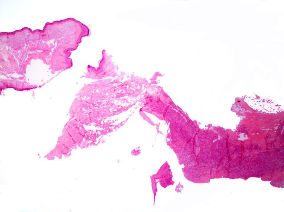
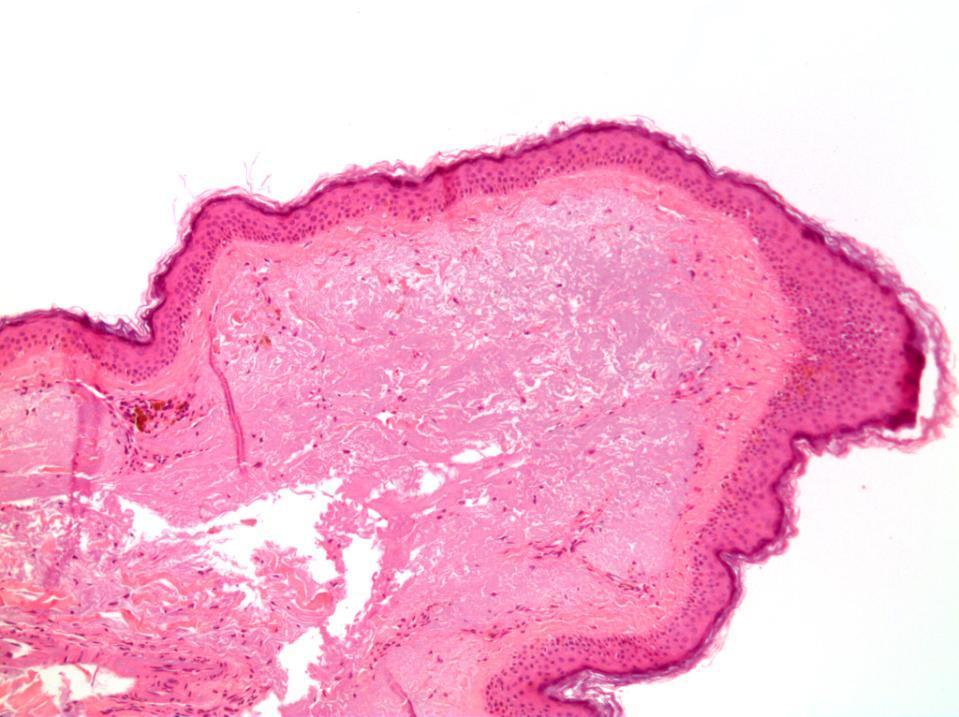
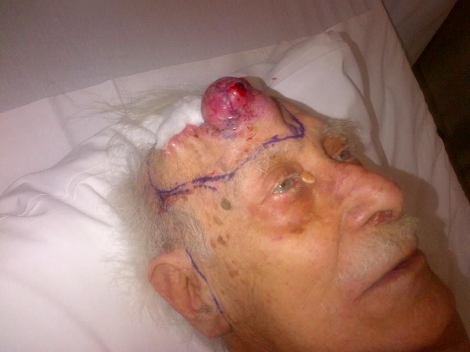
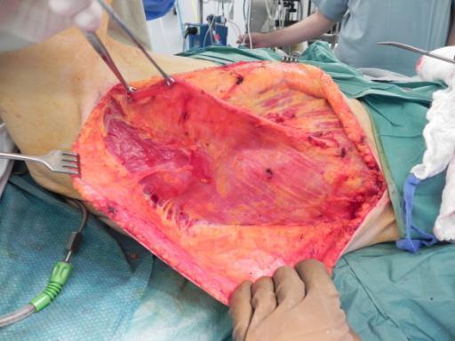
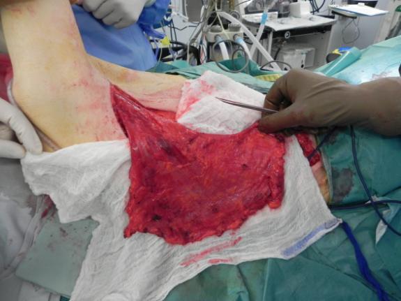
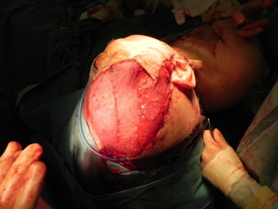
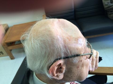
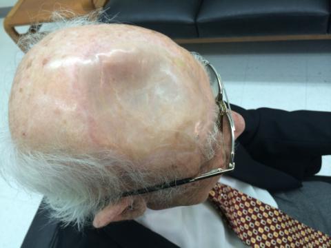
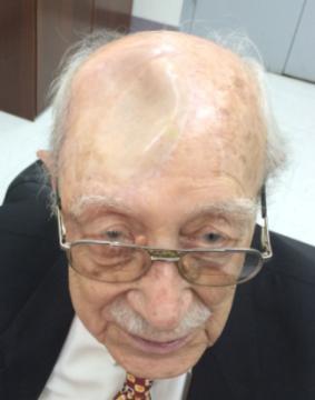
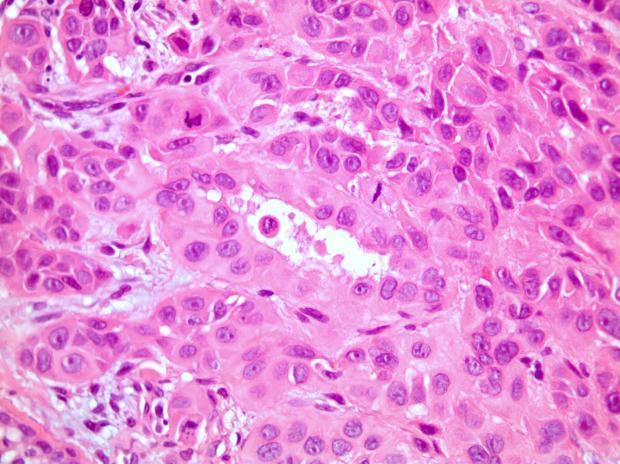
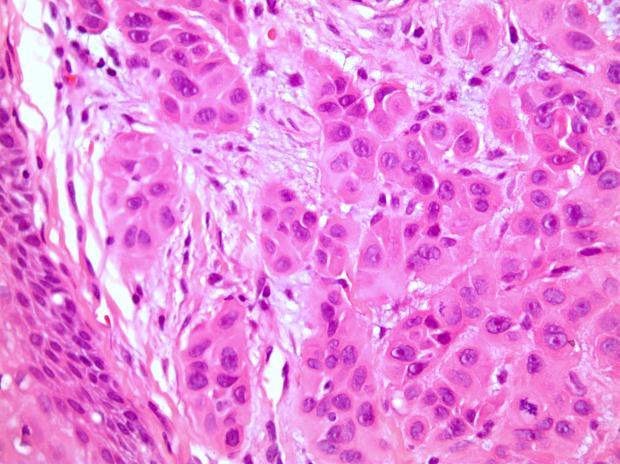
2 History A 89 year old gentleman presenting with a scalp/forehead nodule. Patient had squamous cell carcinoma 18 m at same site, excised. Outside diagnoses: Squamous cell carcinoma. R/O: SCC, Melanoma, others.
3
4
5
6
. D. Spindle cell melanoma. E. Scar and granulation tissue.")
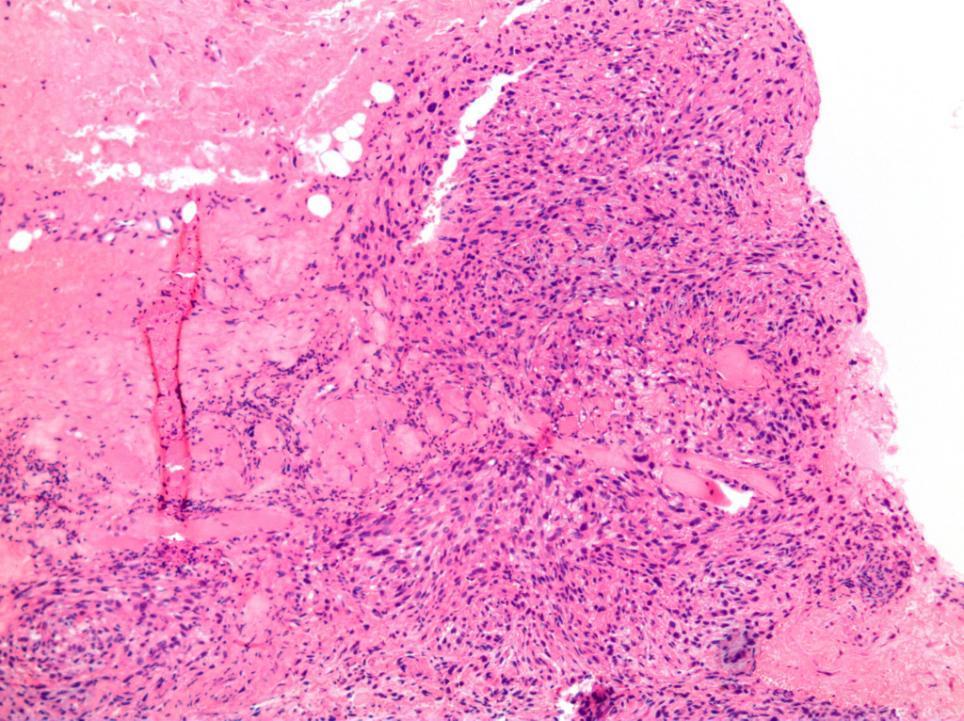
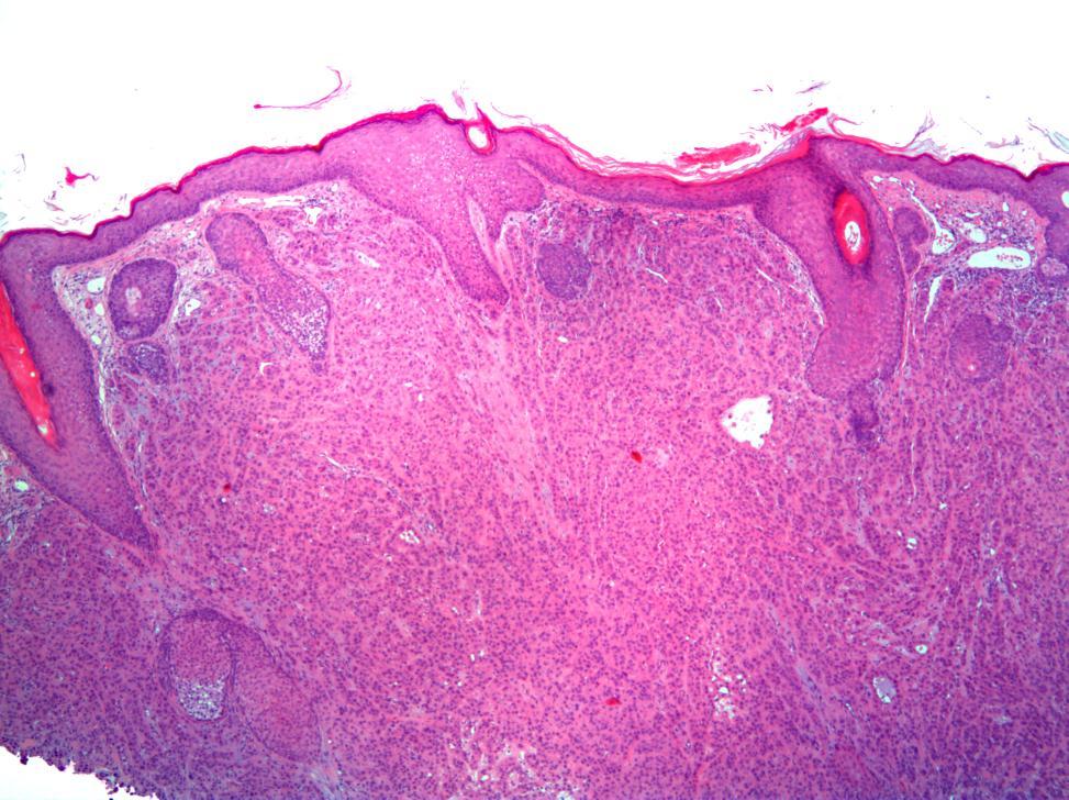
7 What is your Diagnosis? A. Atypical fibroxanthoma. B. Sarcomatoid carcinoma (SCC) C. Pleomorphic sarcoma (MFH). D. Spindle cell melanoma. E. Scar and granulation tissue.
8
9 What is your Diagnosis? A. Atypical fibroxanthoma. B. Sarcomatoid carcinoma (SCC) C. Pleomorphic sarcoma (MFH). D. Spindle cell melanoma. E. Scar and granulation tissue.

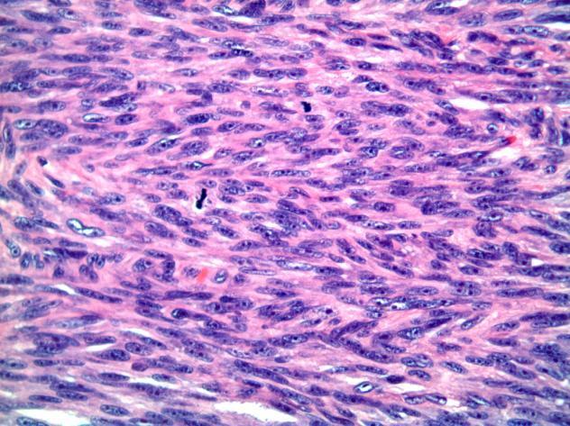
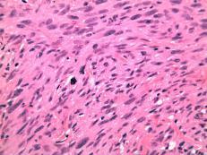
10 Pleomorphic Sarcoma Spindle Cell Melanoma

11 Pleomorphic sarcoma (MFH)
12 Pleomorphic sarcoma Cutaneous PS is very rare years Etiology: - Idiopathic - Rarely post-radiotherapy, scar, chronic ulcer >5cm 5-year survival: 15-30% Superficial and angiomatoid: Better prognosis. Angiomatoid, myxoid, giant-cell & inflammatory.
13 Which of the following is the LEAST accurate statement in describing this entity? A. Usually involves the deep soft tissue and striated muscles. B. Usually involves the proximal part of the ext., particularly the lower. C. Skin is a common site. D. Adults between y are often affected. E. Multiple variants have been described including myxoid, inflammatory, angiomatoid, giant cell, etc.
14 Which of the following is the LEAST accurate statement in describing this entity? A. Usually involves the deep soft tissue and striated muscles. B. Usually involves the proximal part of the ext., particularly the lower. C. Skin is a common site. D. Adults between y are often affected. E. Multiple variants have been described including myxoid, inflammatory, angiomatoid, giant cell, etc.
15 Which of the following is the BEST IHC panel to confirm diagnosis? A. Vimentin +, CD68 + & CKAE1/3 ± B. HMWCK +, CK5/6 +, P63+ & Melan-A - C. S100+, Melan-A + & CKAE1/3- D. CD31 +, CD34 + & S100- E. Desmin +, SMA+, S100+ & CD31-
16 Which of the following is the BEST IHC panel to confirm diagnosis? A. Vimentin +, CD68 + & CKAE1/3 ± B. HMWCK +, CK5/6 +, P63+ & Melan-A - C. S100+, Melan-A + & CKAE1/3- D. CD31 +, CD34 + & S100- E. Desmin +, SMA+, S100+ & CD31-
17


18 Dr. Roger Moukarbil
19
20
21
22 Case IV
23 History A 71 year old gentleman presenting with erythematous plaque over nose. Rule out: SCC vs. Melanoma.
24
25
26 What is your Diagnosis? A. Squamous Cell Carcinoma. B. Nevoid Melanoma. C. Basal cell carcinoma, Metatypical type. D. Eccrine Carcinoma. E. Sebaceous Carcinoma.
27 What is your Diagnosis? A. Squamous Cell Carcinoma. B. Nevoid Melanoma. C. Basal Cell Carcinoma, Metatypical type. D. Eccrine Carcinoma. E. Sebaceous Carcinoma.

28 Nevoid Melanoma

29 Metatypical BCC Nevoid Melanoma
30 Metatypical BCC Basosquamous Carcinoma??? Rare variant of BCC: 1.2% to 2.7%. Recurrence rates: 12-50%. Incidence of metastasis: 5-10% Central face
.")
31 Basal cell carcinoma PCNA: G1-S phases KI-67: G2-M phases Basal layer expression: KI-67>PCNA In BCC: - P53 is negative to focal <10%). - PCNA Ki-67 - Ber-EP4: Positive, diffuse - CK7 & CK18: Positive, diffuse EMA: Negative.
32 Metatypical BCC P53: intense and diffuse staining. Ber-EP4: Positive. PCNA>KI-67. Less expression of CK7 & CK18 EMA: Negative.
33 Which of the following is the LEAST accurate statement in describing this entity? A. It has same prognosis as other variants of this entity. B. Surgery is the main treatment. C. It affects more males than females. D. The most affected site is cervicofacial area. E. Recurrence occurred 10% of cases and metastasis in 5%.
34 Which of the following is the LEAST accurate statement in describing this entity? A. It has same prognosis as other variants of this entity. B. Surgery is the main treatment. C. It affects more males than females. D. The most affected site is cervicofacial area. E. Recurrence occurred 10% of cases and metastasis in 5%.
35 Which of the following is the LEAST accurate statement in describing this entity? A. In comparison to other variants there is less expression of CK17 & CK8. B. It shows diffuse and intense P53 labeling. C. In this tumor, PCNA staining is greater than that of Ki-67 D. The tumor cells express Ber EP4. E. The tumor cells express EMA diffusely.
36 Which of the following is the LEAST accurate statement in describing this entity? A. In comparison to other variants there is less expression of CK17 & CK8. B. It shows diffuse and intense P53 labeling. C. In this tumor, PCNA staining is greater than that of Ki-67 D. The tumor cells express Ber EP4. E. The tumor cells express EMA diffusely.
أملس عضلي غرن = Leiomyosarcoma. Leiomyosarcoma 1 / 5
 Leiomyosarcoma 1 / 5 EPIDEMIOLOGY Exact incidence is unknown, but older studies suggest that leiomyosarcomas comprise approximately 3 percent of soft-tissue sarcomas. Superficial leiomyosarcoma occurs
Update on Cutaneous Mesenchymal Tumors. Thomas Brenn
 Update on Cutaneous Mesenchymal Tumors Thomas Brenn Cutaneous Mesenchymal Tumours Wide morphological and biological spectrum Myofibroblastic, smooth muscle, neural, vascular, apidocytic, undifferentiated;
Post-test Self-assessment Cases
 Post-test Self-assessment Cases Ibrahim Khalifeh, M.D. Associate Professor Department of Pathology American University of Beirut Medical Center Beirut, Lebanon Case I History A 69 year old gentleman presenting
Desmoplastic Melanoma R/O BCC. Clinical Information. 74 y.o. man with lesion on left side of neck r/o BCC
 R/O BCC Sabine Kohler, M.D. Professor of Pathology and Dermatology Dermatopathology Service Stanford University School of Medicine Clinical Information 74 y.o. man with lesion on left side of neck r/o
Maligna Melanoma and Atypical Fibroxanthoma: An Unusual Collision Tumour G Türkcü 1, A Keleş 1, U Alabalık 1, D Uçmak 2, H Büyükbayram 1 ABSTRACT
 Maligna Melanoma and Atypical Fibroxanthoma: An Unusual Collision Tumour G Türkcü 1, A Keleş 1, U Alabalık 1, D Uçmak 2, H Büyükbayram 1 ABSTRACT Two different neoplasia in the same biopsy material called
Special slide seminar
 Special slide seminar Tomáš Rozkoš The Fingerland Department of Pathology Charles University Medical Faculty and Faculty Hospital in Hradec Králové Czech Republic Case history, 33 years old resistance
Immunohistochemistry in Bone and Soft Tissue Tumors. Sahar Rassi Zankoul, MD
 Immunohistochemistry in Bone and Soft Tissue Tumors Sahar Rassi Zankoul, MD Introduction Bone tumors represent a wide variety of tumors of various origins and malignant potentials. These different tumor
Lung Tumor Cases: Common Problems and Helpful Hints
 Lung Tumor Cases: Common Problems and Helpful Hints Brandon T. Larsen, MD, PhD Senior Associate Consultant Department of Laboratory Medicine and Pathology Mayo Clinic Arizona Arizona Society of Pathologists
Spindle Cell Lesions Of The Breast. Emad Rakha Professor of Breast Pathology and Consultant Pathologist
 Spindle Cell Lesions Of The Breast Emad Rakha Professor of Breast Pathology and Consultant Pathologist * SCLs comprise a wide spectrum of diseases, ranging from reactive processes to aggressive malignant
Slide seminar. Asist. Prof. Jože Pižem, MD, PhD Institute of Pathology Medical Faculty, University of Ljubljana
 Slide seminar Asist. Prof. Jože Pižem, MD, PhD Institute of Pathology Medical Faculty, University of Ljubljana Case 5 A 57-year-old man with a dermal/subcutaneous lesion on the scalp, which was interpreted
Cutaneous Adnexal Tumors
 Cutaneous Adnexal Tumors Lesions with Predominant Follicular Differentiation Special Emphasis on Basal Cell Carcinoma 2014-04-01 Prof. Dr. med. Katharina Glatz Pathologie Cutaneous Adnexal Tumors Hair
David B. Troxel, MD. Common Medicolegal Situations: Misdiagnosis of Melanoma
 Common Medicolegal Situations: Misdiagnosis of Melanoma David B. Troxel, MD Medical Director, The Doctors Company, Napa, California Clinical Professor Emeritus, University of California at Berkeley Past
Selected Pseudomalignant Soft Tissue Tumors of the Skin and Subcutis
 Selected Pseudomalignant Soft Tissue Tumors of the Skin and Subcutis Andrew L. Folpe, M.D. Professor of Laboratory Medicine and Pathology Mayo Clinic, Rochester, MN folpe.andrew@mayo.edu 2016 MFMER slide-1
The role of immunohistochemistry in surgical pathology of the uterine corpus and cervix
 The role of immunohistochemistry in surgical pathology of the uterine corpus and cervix Prof. Ben Davidson, MD PhD Department of Pathology, Norwegian Radium Hospital, Oslo University Hospital, Oslo, Norway
Enterprise Interest Nothing to declare
 Enterprise Interest Nothing to declare Diagnoses one would not like to miss in soft tissue pathology early in your career Marta Sbaraglia, MD Department of Pathology Hospital of Treviso University of Padua
Case Report Clear Cell Basal Cell Carcinoma
 SAGE-Hindawi Access to Research Volume 2011, Article ID 386921, 4 pages doi:10.4061/2011/386921 Case Report Clear Cell Basal Cell Carcinoma Deba P. Sarma, 1 Daniel Olson, 1 Jennifer Olivella, 1 Tracey
1/10/2018. Soft Tissue Tumors Showing Melanocytic Differentiation. Overview. Desmoplastic/ Spindle Cell Melanoma
 2016 MFMER slide-1 2016 MFMER slide-2 2016 MFMER slide-3 Soft Tissue Tumors Showing Melanocytic Differentiation Andrew L. Folpe, M.D. Professor of Laboratory Medicine and Pathology Mayo Clinic, Rochester,
Basal cell carcinoma 5/28/2011
 Goal of this Presentation A practical approach to the diagnosis of cutaneous carcinomas and their mimics Thaddeus Mully, MD University of California San Francisco To review common non-melanoma skin cancers
Appendageal skin tumors
 Appendageal skin tumors Ibrahim Khalifeh, M.D. Associate Professor Department of Pathology American University of Beirut Medical Center Beirut, Lebanon Appendageal tumors Neoplasms whose differentiation
CASE year old male with a PET avid nodule in the left adrenal gland
 CASE 1 55 year old male with a PET avid nodule in the left adrenal gland Case 1 Adrenal gland parenchyma partly replaced by a spindle cell tumour with mild nuclear pleomorphism Atypical mitoses present
Financial disclosures
 Mesenchymal Neoplasms with Melanocytic Differentiation By Konstantinos Linos MD, FCAP, FASDP Bone, Soft Tissue and Dermatopathology Assistant Professor of Pathology Dartmouth-Hitchcock Medical Center Geisel
Review and Updates of Immunohistochemistry in Selected Salivary Gland and Head and Neck Tumors
 Review and Updates of Immunohistochemistry in Selected Salivary Gland and Head and Neck Tumors. Monophasic tumors : myoepithelioma, acinic cell carcinoma, and salivary duct carcinoma. Biphasic tumors includes
Diagnoses of Cases 1. Lentigo, other melanosis and the acquired nevus 2. Variations on the acquired nevus 3. Dermal melanocytosis
 Diagnoses of Cases 1. Lentigo, other melanosis and the acquired nevus 1 1A. Lentigo simplex 4 1B. Psoralens and ultraviolet A (PUVA) lentigo 6 1C. Solar lentigo 8 1D. Café au lait macule 10 1E. Ink-spot
Pathology Mystery and Surprise
 Pathology Mystery and Surprise Tim Smith, MD Director Anatomic Pathology Medical University of South Carolina Disclosures No conflicts to declare Some problem cases Kidney tumor Scalp tumor Bladder tumor
Case Presentation. Maha Akkawi, MD, Fatima Obeidat, MD, Tariq Aladily, MD. Department of Pathology Jordan University Hospital Amman, Jordan
 Case Presentation Maha Akkawi, MD, Fatima Obeidat, MD, Tariq Aladily, MD Department of Pathology Jordan University Hospital Amman, Jordan The 25th Annual Congress of the ADIAP The 8/11/2013 1 5th International
Primary Cutaneous CD30-Positive T-cell Lymphoproliferative Disorders
 Primary Cutaneous CD30-Positive T-cell Lymphoproliferative Disorders Definition A spectrum of related conditions originating from transformed or activated CD30-positive T-lymphocytes May coexist in individual
Classification (1) Classification (3) Classification (2) Spindle cell lesions. Spindle cell lesions of bladder (Mills et al.
 Classification (3) Classification (2) Spindle cell lesions. Spindle cell lesions of bladder (Mills et al.") Non-epithelial tumours and nonepithelial tumour-like lesions of the bladder Dr Jonathan H Shanks The Christie NHS Foundation Trust, Manchester, UK Classification (1) Myofibroblastic proliferations and
Non-epithelial tumours and nonepithelial tumour-like lesions of the bladder Dr Jonathan H Shanks The Christie NHS Foundation Trust, Manchester, UK Classification (1) Myofibroblastic proliferations and
American College of Mohs Surgery. Diagnostic Quality Control Program (Review of Answers)
") American College of Mohs Surgery Diagnostic Quality Control Program 2010 (Review of Answers) Question 1 A flesh-colored plaque on the right ala of an otherwise healthy 57 year-old male is referred for
American College of Mohs Surgery Diagnostic Quality Control Program 2010 (Review of Answers) Question 1 A flesh-colored plaque on the right ala of an otherwise healthy 57 year-old male is referred for
Dermatopathology: The tumor is composed of keratinocytes which show atypia, increase mitoses and abnormal mitoses.
 Squamous cell carcinoma (SCC): A common malignant tumor of keratinocytes arising in the epidermis, usually from a precancerous condition: 1- UV induced actinic keratosis, usually of low grade malignancy.
The Relevance of Cytologic Atypia in Cutaneous Neural Tumors
 The Relevance of Cytologic Atypia in Cutaneous Neural Tumors Recent Findings - New Developments New Problems Zsolt B. Argenyi, M.D. Professor of Pathology & Dermatology Director of Dermatopathology Department
Mesothelioma Pathobasic. Lukas Bubendorf Pathology
 Mesothelioma Pathobasic Lukas Bubendorf Pathology Mechanisms of Asbestos Carcinogenesis in Mesothelioma Asprin High-mobility group protein B1 master switch HMGB1 Initiation/ perpetuation of inflamm. response
Cutaneous Malignancies: A Primer COPYRIGHT. Marissa Heller, M.D.
 Cutaneous Malignancies: A Primer Marissa Heller, M.D. Associate Director of Dermatologic Surgery Department of Dermatology Beth Israel Deaconess Medical Center December 10, 2016 Skin Cancer Non-melanoma
PATHOLOGY OF THE SKIN 2. Tumours of the skin
 PATHOLOGY OF THE SKIN 2. Tumours of the skin Máirín E. McMenamin MB MRCPI FRCPath Dip (Dermatopathol) RCPath St. James s Hospital and University of Dublin, Trinity College Tumour (Neoplasia) Benign or
SEBACEOUS NEOPLASMS. Dr. Prachi Saraogi Clinical Fellow in Dermatology
 SEBACEOUS NEOPLASMS Dr. Prachi Saraogi Clinical Fellow in Dermatology Sebaceous neoplasms Sebaceous adenoma (Benign) Sebaceous carcinoma (Malignant) SEBACEOUS ADENOMA Benign tumours composed of incompletely
- Selected Tumors of the Skin Appendages - Primary vs. Metastasis
 - Selected Tumors of the Skin Appendages - Primary vs. Metastasis Napa Valley 2018 Victor G. Prieto, MD, PhD Chair of Pathology UT MD Anderson Cancer Center vprieto@mdanderson.org Napa Valley in May Introduction
Klinisch belang van chromosomale translocatie detectie in sarcomen
 Translocations in sarcomas Klinisch belang van chromosomale translocatie detectie in sarcomen Judith V.M.G. Bovée, M.D., Ph.D. Department of Pathology Leiden University Medical Center RNA binding DNA binding
Whitney A. High, MD, JD, MEng
 ADS Dermatopathology Meeting 2014 Selected Adnexal Tumors Whitney A. High, MD, JD, MEng Associate Professor, Dermatology & Pathology Director of Dermatopathology (Dermatology) University of Colorado School
Rapid Growing Superficial Cutaneous Leiomyosarcoma of the Face
 apid Growing Superficial Leiomyosarcoma Ann Dermatol ol. 25, No. 2, 2013 http://dx.doi.org/10.5021/ad.2013.25.2.237 CASE EPOT apid Growing Superficial Leiomyosarcoma of the Face Kwang Chul Lee 1, Min Sung
3/27/2017. Disclosure of Relevant Financial Relationships
 Ophthalmic Pathology Evening Specialty Conference USCAP 2017 5 th March, 2017 Mukul K. Divatia, MD Assistant Professor Department of Pathology & Genomic Medicine Weill Cornell Medical College Houston Methodist
Differential Diagnosis of Oral Masses. Palatal Lesions
 Differential Diagnosis of Oral Masses Palatal Lesions Palatal Masses Periapical Abscess Torus Palatinus Mucocele Lymphoid Hyperplasia Adenomatous Hyperplasia Benign Salivary Neoplasms Malignant Salivary
Slide Seminar Spanish Society of Pathology
 Slide Seminar Spanish Society of Pathology John R. Goldblum, M.D. Chairman, Department of Anatomic Pathology Cleveland Clinic Professor of Pathology Cleveland Clinic Lerner College of Medicine 1921 Original
Myxo-inflammatory Fibroblastic sarcoma
 AKA Myxo-inflammatory Fibroblastic sarcoma Acral Myxoinflammatory fibroblastic sarcomaam.j.surg.path1998; 22; 911-924 Inflammatory myxoid tumour of soft parts with bizarre giant cells [Pathol.Res.Pract.
Charles Halsey, DVM, PhD, DACVP Pfizer, Inc. IHC Resources
 Charles Halsey, DVM, PhD, DACVP Pfizer, Inc. IHC Resources 1 IHC Identification Targets Specimens Controls 2 Tissue controls Trouble Spots 3 The Key to Description IHC Description 4 Intermediate Filaments
57th Annual HSCP Spring Symposium 4/16/2016
 An Unusual Malignant Spindle Cell Lesion to Involve the Breast Erinn Downs-Kelly, D.O. Associate Professor of Pathology University of Utah & ARUP Laboratories No disclosures Case 39 y/o female with no
Benign versus Cancerous Lesions How to tell the difference FMF 2014 Christie Freeman MD, CCFP, DipPDerm, MSc
 1 Benign versus Cancerous Lesions How to tell the difference FMF 2014 Christie Freeman MD, CCFP, DipPDerm, MSc Benign lesions Seborrheic Keratoses: Warty, stuck-on Genetics and birthdays Can start in late
Cutaneous metastases. Thaddeus Mully. University of California, San Francisco Professor, Departments of Pathology and Dermatology
 Cutaneous metastases Thaddeus Mully University of California, San Francisco Professor, Departments of Pathology and Dermatology DISCLOSURE OF RELATIONSHIPS WITH INDUSTRY Thaddeus Mully Course C005 Essential
Newer soft tissue entities
 Newer soft tissue entities Examples among fibroblastic tumors Turku, May 6, 2010 Markku Miettinen, M.D. AFIP, Washington, DC Fibroblastic neoplasms Solitary fibrous tumor /Hemangiopericytoma Low-grade
Ocular Neoplasia What s Common? What s New? Richard R Dubielzig
 Ocular Neoplasia What s Common? What s New? Richard R Dubielzig Orbit 288 6% Tumors of the globe make up 3225 out of 6110 total neoplasms = 53%. Tumors of the conjunctiva make up 1192 out of 6110 total
Financial disclosures
 Cutaneous Mesenchymal Neoplasms with EWSR1 Rearrangement By Konstantinos Linos MD, FCAP, FASDP Bone, Soft Tissue and Dermatopathology Assistant Professor of Pathology Dartmouth-Hitchc Geisel School of
Diagnostic concordance among dermatopathologists in basal cell carcinoma subtyping: Results of a study in a skin referral hospital in Tehran, Iran
 Original Article Diagnostic concordance among dermatopathologists in basal cell carcinoma subtyping: Results of a study in a skin referral hospital in Azita Nikoo, MD 1 Zahra Naraghi, MD 1 Kambiz Kamyab,
Case Report A Rare Cutaneous Adnexal Tumor: Malignant Proliferating Trichilemmal Tumor
 Case Reports in Medicine Volume 2015, Article ID 742920, 4 pages http://dx.doi.org/10.1155/2015/742920 Case Report A Rare Cutaneous Adnexal Tumor: Malignant Proliferating Trichilemmal Tumor Omer Alici,
Diagnostic Value of Immunohistochemistry in Soft Tissue Tumors
 Original Article DOI: 10.21276/APALM.1637 Diagnostic Value of Immunohistochemistry in Soft Tissue Tumors Sridevi. V*., Susruthan Muralitharan., and Thanka. J Dept of Pathology, SriMuthukumaran Medical
Cutaneous Mesenchymal Neoplasms with EWSR1 Rearrangement
 Cutaneous Mesenchymal Neoplasms with EWSR1 Rearrangement By Konstantinos Linos MD, FCAP, FASDP Bone, Soft Tissue and Dermatopathology Assistant Professor of Pathology Dartmouth-Hitchcock Medical Center
CLEAR CELL BASAL CELL CARCINOMA- AN UNUSUAL ENTITY IN AN INDIAN MALE
 Indian Journal of Medical s ISSN: 2319 3832(Online) CLEAR CELL BASAL CELL CARCINOMA- AN UNUSUAL ENTITY IN AN INDIAN MALE * Cheena Garg 1, Arjun Agarwal 2, Bushra Siddiqui 3, Shahbaz Faridi 4, 1 Department
Living Beyond Cancer Skin Cancer Detection and Prevention
 Living Beyond Cancer Skin Cancer Detection and Prevention Cutaneous Skin Cancers Identification Diagnosis Treatment options Prevention What is the most common cancer in people? What is the most common
An Overview of Genital Stromal Tumors
 An Overview of Genital Stromal Tumors By Konstantinos Linos MD, FCAP, FASDP Bone, Soft Tissue and Dermatopathology Assistant Professor of Pathology Dartmouth-Hitchcock Medical Center Geisel School of Medicine
Surgical Pathology Evening Specialty Conference USCAP 2015
 Surgical Pathology Evening Specialty Conference USCAP 2015 John R. Goldblum, M.D. Chairman, Department of Pathology, Cleveland Clinic Professor of Pathology, Cleveland Clinic Lerner College of Medicine
Diplomate of the American Board of Pathology in Anatomic and Clinical Pathology
 A 33-year-old male with a left lower leg mass. Contributed by Shaoxiong Chen, MD, PhD Assistant Professor Indiana University School of Medicine/ IU Health Partners Department of Pathology and Laboratory
Clinicopathologic Self- Assessment S003 AAD 2017
 Clinicopathologic Self- Assessment S003 AAD 2017 Clay J. Cockerell, M.D. Director, Cockerell Dermatopathology Director, Division of Dermatopathology UT Southwestern Medical Center July 2017 No relevant
What is New in the 2015 WHO Lung Cancer Classification? Zhaolin Xu, MD, FRCPC, FCAP
 What is New in the 2015 WHO Lung Cancer Classification? Zhaolin Xu, MD, FRCPC, FCAP Professor, Dept of Pathology, Dalhousie University, Canada Pulmonary Pathologist and Cytopathologist, QEII HSC Senior
Slide seminar: Soft tissue and bone pathology
 Slide seminar: Soft tissue and bone pathology Unusual tumors of bone and soft tissue or unusual presentations of common ones Gunhild Mechtersheimer Institute of Pathology, Heidelberg/DE (Sylvia Höller,
Keratinocyte tumors. Actinic Keratosis. Squamous cell carcinoma in situ. Squamous Cell Carcinoma. (aka Bowen s disease)
") Actinic Keratosis Keratinocyte tumors Prepared by Kurt Schaberg Precancerous, risk of malignancy ~8-20% per year (progresses to SCC); Due to chronic sun exposure Rough scaly plaque; typically due to sun
Actinic Keratosis Keratinocyte tumors Prepared by Kurt Schaberg Precancerous, risk of malignancy ~8-20% per year (progresses to SCC); Due to chronic sun exposure Rough scaly plaque; typically due to sun
LAC + USC.
 Jeff McDavit,, M.D. LAC + USC mcdavit@usc.edu Clinical History 55 year old male with large, deep, non- tender left thigh mass. Seen at LAC+USC Med Ctr FNA clinic No h/o trauma or radiation Vimentin
Case 18. M75. Excision of mass on scalp. Clinically SCC. The best diagnosis is:
 Case 18 M75. Excision of mass on scalp. Clinically SCC. The best diagnosis is: A. Pilomatrical carcinoma B. Adnexal carcinoma NOS C. Metastatic squamous cell carcinoma D.Primary squamous cell carcinoma
TEST MENU BY SPECIALTY
 1 TEST MENU BY SPECIALTY Breast Pathology Surgical excisions, needle core biopsies, and plastic surgery accepted from all sites Assessment of margins Axillary lymph node dissections Sentinel lymph nodes
CUTANEOUS PSEUDONEOPLASTIC MESENCHYMAL LESIONS ( PSEUDOSARCOMAS )
") CUTANEOUS PSEUDONEOPLASTIC MESENCHYMAL LESIONS ( PSEUDOSARCOMAS ) Mark R. Wick, M.D. University of Virginia Health System Charlottesville, VA www.markwickmd.com; mrw9c@virginia.edu PSEUDONEOPLASMS & PSEUDOTUMORS
CUTANEOUS PSEUDONEOPLASTIC MESENCHYMAL LESIONS ( PSEUDOSARCOMAS ) Mark R. Wick, M.D. University of Virginia Health System Charlottesville, VA www.markwickmd.com; mrw9c@virginia.edu PSEUDONEOPLASMS & PSEUDOTUMORS
Non Small Cell Lung Cancer Histopathology ד"ר יהודית זנדבנק
 Non Small Cell Lung Cancer Histopathology ד"ר יהודית זנדבנק 26.06.09 Lecture outlines WHO histological classification Macro/Micro assessment Early diagnosis Minimal pathology Main subtypes SCC, AdCa, LCLC
Original Article Sarcomatoid variant of anaplastic large cell lymphoma: a diagnostic challenge
 Int J Clin Exp Pathol 2016;9(3):3654-3659 www.ijcep.com /ISSN:1936-2625/IJCEP0021021 Original Article Sarcomatoid variant of anaplastic large cell lymphoma: a diagnostic challenge Hong Yu 1*, Wenchao Wang
USCAP 2011: ASDP companion meeting. Steven D. Billings 1
 USCAP 2011: ASDP companion meeting. Steven D. Billings (billins@ccf.org) 1 Spindle cell tumors that make you say, Oh $*&%! This lecture will focus on examples of cutaneous tumors that present particular
BSD 2015 Case 19. Female 21. Nodule on forehead. The best diagnosis is:
 BSD 2015 Case 19 Female 21. Nodule on forehead. The best diagnosis is: A. mixed tumour of skin B. porocarcinoma C. nodular hidradenoma D. metastatic adenocarcinoma BSD 2015 Case 19 Female 21 Nodule on
Squamous Cell Carcinoma. Basal Cell Carcinoma. Regional Follow-up Guidelines
 West of Scotland Cancer Network Skin Cancer Managed Clinical Network Squamous Cell Carcinoma Basal Cell Carcinoma Regional Follow-up Guidelines Prepared by Dr M Porter, Dr A Matthews Approved by Skin Cancer
New and Emerging Therapies: Non-Melanoma Skin Cancers. David J. Goldberg, MD, JD Skin Laser and Surgery Specialists of NY/NJ
 New and Emerging Therapies: Non-Melanoma Skin Cancers David J. Goldberg, MD, JD Skin Laser and Surgery Specialists of NY/NJ Disclosure Research Grant form Sensus Superficial Radiation Therapy (SRT) Modern
Diffusely Spreading, Superficial Malignant Fibrous Histiocytoma: Case Report with Cyto-and Histomorphologic Features
 ISPUB.COM The Internet Journal of Pathology Volume 6 Number 2 Diffusely Spreading, Superficial Malignant Fibrous Histiocytoma: Case Report with Cyto-and B Wang, C Bewtra, W Hunter Citation B Wang, C Bewtra,
Sarcomatoid (spindle cell) carcinoma of the cricopharynx presenting as dysphagia
 carcinoma of the cricopharynx presenting as dysphagia") Case Report Sarcomatoid (spindle cell) carcinoma of the cricopharynx presenting as dysphagia Jagtap Sunil V. 1, Shukla Dhirajkumar B. 2, Jagtap Swati S. 3, Havle Abhay D. 4 1 Associate Professor, Department
Case Report Sarcomatoid (spindle cell) carcinoma of the cricopharynx presenting as dysphagia Jagtap Sunil V. 1, Shukla Dhirajkumar B. 2, Jagtap Swati S. 3, Havle Abhay D. 4 1 Associate Professor, Department
IT S FUNDAMENTAL MY DEAR WATSON! A SHERLOCKIAN APPROACH TO DERMATOLOGY
 IT S FUNDAMENTAL MY DEAR WATSON! A SHERLOCKIAN APPROACH TO DERMATOLOGY Skin, Bones, and other Private Parts Symposium Dermatology Lectures by Debra Shelby, PhD, DNP, FNP-BC, FADNP, FAANP Debra Shelby,
Diagnostic problems in uterine smooth muscle tumors
 Diagnostic problems in uterine smooth muscle tumors Marina Kos Ljudevit Jurak Clinical Department of Pathology, Clinical Hospital Center Sestre milosrdnice, Zagreb Institute of Pathology, University of
I have a skin lump doc! What s next? 12 th August 2017 Dr. Sue-Ann Ho Ju Ee
 I have a skin lump doc! What s next? 12 th August 2017 Dr. Sue-Ann Ho Ju Ee Some thoughts Is this skin cancer? How common is this? How likely is this in this patient? What happens next if it s something
1. Written information to patient /GP: fax ASAP to GP & offer copy of consultation letter.
 Skin Cancer follow up guidelines If NEW serious diagnosis given: 1. Written information to patient /GP: fax ASAP to GP & offer copy of consultation letter. 2. Free prescription information details. 3.
4/12/2018. MUSC Pathology Symposium Kiawah Island April 18, Jesse K. McKenney, MD
 MUSC Pathology Symposium Kiawah Island April 18, 2018 Jesse K. McKenney, MD 1 Urothelial Carcinoma with Alternative Differentiation 2 Urothelial Carcinoma with Alternative Differentiation Recognition as
From Morphology to Molecular Pathology: A Practical Approach for Cytopathologists Part 1-Cytomorphology. Songlin Zhang, MD, PhD LSUHSC-Shreveport
 From Morphology to Molecular Pathology: A Practical Approach for Cytopathologists Part 1-Cytomorphology Songlin Zhang, MD, PhD LSUHSC-Shreveport I have no Conflict of Interest. FNA on Lymphoproliferative
05/07/2018. Types of challenges. Challenging cases in uterine pathology. Case 1 ` 65 year old female Post menopausal bleeding Uterine Polyp
 Types of challenges Challenging cases in uterine pathology Nafisa Wilkinson Gynaecological Pathologist UCLH London Lack of complete history often, NO clinical history at all! Cases from other centres often
South East England General Histopathology EQA Scheme
 South East England General Round a Final Case Analyses Cases 635 to 646 Circulated January February 2016 131 responses (89.73%) Prepared April 2016 Authorised by: Prof J Schofield Date: 19/4/2016 With
Corporate Medical Policy
 Corporate Medical Policy File Name: Origination: Last CAP Review: Next CAP Review: Last Review: mohs_micrographic_surgery 07/2004 11/2017 11/2018 11/2017 Description of Procedure or Service Mohs Micrographic
Evening Specialty Conference Bone and Soft Tissue Pathology. Diagnostic pitfalls in bone and soft tissue pathology
 Evening Specialty Conference Bone and Soft Tissue Pathology. Case 1 Elizabeth G Demicco, MD, PhD Mount Sinai Hospital, New York Disclosure of Relevant Financial Relationships USCAP requires that all planners
Fluorescence spectroscopy and microscopy of cutaneous tumours correlation between micro- and macro- spectral measurements
 Fluorescence spectroscopy and microscopy of cutaneous tumours correlation between micro- and macro- spectral measurements E. Borisova 1, L. Avramov 1, M. Lomova 2, O. Semyachkina-Glushkovskaya 2, D. Gorin
Nasreen A. Syed, MD F.C. Blodi Eye Pathology Laboratory University of Iowa
 Nasreen A. Syed, MD F.C. Blodi Eye Pathology Laboratory University of Iowa No financial disclosures No discussion of off-label use of medications or unapproved devices 67 year old male referred to Oculoplastics
OBSERVATION. Atypical Fibroxanthoma With Regional Lymph Node Metastasis. Douglas New, MD; Soon Bahrami, MD; Janine Malone, MD; Jeffrey P.
 ONLINE FIRST OBSERVATION Atypical Fibroxanthoma With Regional Lymph Node Metastasis Report of a Case and Review of the Literature Douglas New, MD; Soon Bahrami, MD; Janine Malone, MD; Jeffrey P. Callen,
64 y.o. F with CLL and leg tumour
 64 y.o. F with CLL and leg tumour Case History Excision with split-skin grafting Histology moderately differentiated squamous cell carcinoma with large areas of necrosis and brisk mitotic activity.
C.L. Davis Foundation Descriptive Veterinary Pathology Course
 C.L. Davis Foundation 2015 Descriptive Veterinary Pathology Course IHC Resources IHC Identification Targets Antibodies Antibodies 1 Antibodies Specimens Antigen Retrieval Unmasks antigen epitopes Methods
A Tete a tete of benign, borderline and malignant fibrohistiocytic tumor
 286 Case Report Iran J Pathol. 2016; 11(3): 286-290 http://www.ijp.iranpath.org/ A Tete a tete of benign, borderline and malignant fibrohistiocytic tumor Rashmi. M.V 1, Pavithra. P 1, Shivakumarappa. G.M.
Tumores de células pequeñas, redondas y azules: diagnóstico diferencial cuando el tiempo apremia
 Tumores de células pequeñas, redondas y azules: diagnóstico diferencial cuando el tiempo apremia Sílvia Bagué Servei de Patologia Hospital de Sant Pau Barcelona Soft tissue sarcomas Heterogeneous group
Differential diagnosis of HCC
 Hepatocellular Carcinoma Quest for an Ideal Immunohistochemical Panel Sanjay Kakar, MD UCSF Differential diagnosis of HCC Hepatocellular lesions Adenoma, FNH, HG dysplasia Adenocarcinoma CholangioCA, metastasis
Ocular Neoplasia CL Davis 9/08. Richard R Dubielzig
 Ocular Neoplasia CL Davis 9/08 Richard R Dubielzig 2135/5722 Canine Melanocytic Tumors Outside the Globe: 264 Conjunctival: 159 Eye Lid: 72 Skin: 33 Affecting the Globe: 1871 Anterior Uveal Melanocytoma:
IN THE NAME OF GOD Dr. Kheirandish Oral and maxillofacial pathology
 IN THE NAME OF GOD Dr. Kheirandish Oral and maxillofacial pathology ORAL FOCAL MUCINOSIS Uncommon Tumorlike Cutaneous myxoid cyst Overproduction of hyaluronic acid by firoblasts Young adults Female Gingiva
Dermatopathology. Dr. Rafael Botella Estrada. Hospital La Fe de Valencia
 Dermatopathology Dr. Rafael Botella Estrada. Hospital La Fe de Valencia Melanoma and mimics Dr. Martin Mihm Malignant lesions result from the accumulation of mutations Class I lesions (benign) Class II
Mesothelioma: diagnostic challenges from a pathological perspective. Naseema Vorajee August 2016
 Mesothelioma: diagnostic challenges from a pathological perspective Naseema Vorajee August 2016 Naseema.vorajee@nhls.ac.za Pleural diseases (whether neoplastic, reactive or infective) may have similar
Introduction. Results. Discussion. Histopathologic and immunohistochemical findings. Results. conclusions,
 1/5 2/5 Carcinoma distinctive carcinoma. form erysipeloides (CE), metastasis. which clinically Itfrom has resembles been termed erysipelas, is an uncommon, but may extend It164 toclassically back, presents
Breast - ductal carcinoma CK7 ER PR GATA3 Mammaglobin (50-70%) GCDFP-15 (50-70%) E-cadherin HMWCK CK20 PAX2 ER/PR/HER2 on all newly diagnosed cases
 GCDFP-15 (50-70%) E-cadherin HMWCK CK20 PAX2 ER/PR/HER2 on all newly diagnosed cases") Adrenal cortical carcinoma Inhibin Synap Melan-A Calretinin Vimentin Chromogr CK7 CK20 Breast - ductal carcinoma CK7 ER PR GATA3 Mammaglobin (50-70%) GCDFP-15 (50-70%) E-cadherin HMWCK CK20 PAX2 ER/PR/HER2
Adrenal cortical carcinoma Inhibin Synap Melan-A Calretinin Vimentin Chromogr CK7 CK20 Breast - ductal carcinoma CK7 ER PR GATA3 Mammaglobin (50-70%) GCDFP-15 (50-70%) E-cadherin HMWCK CK20 PAX2 ER/PR/HER2
Periocular Malignancies
 Periocular Malignancies Andrew Gurwood, O.D., F.A.A.O., Dipl. Marc Myers, O.D., F.A.A.O. Drs. Myers and Gurwood have no financial interests to disclose. Course Description Discussion of the most common
Lung Neoplasia II Resection specimens Pathobasic. Lukas Bubendorf Pathology
 Lung Neoplasia II Resection specimens Pathobasic Lukas Bubendorf Pathology Agenda Preneoplastic lesions Histological subtypes of lung cancer Histological patterns of AC Cells of origin and characteristic