ANATOMY OF THE SPINAL CORD. Structure of the spinal cord Tracts of the spinal cord Spinal cord syndromes
|
|
|
- Lillian Patrick
- 6 years ago
- Views:
Transcription
1 SPINAL CORD

2 ANATOMY OF THE SPINAL CORD Structure of the spinal cord Tracts of the spinal cord Spinal cord syndromes

3 The Nervous System Coordinates the activity of muscles, organs, senses, and actions Made up of nervous tissue Has 3 main functions: 1. Receives sensory Input 2. Integration 3. Dictates motor output
4 Spinal Cord
5 Spinal Cord Functions Pathways for nerve impulses within tracts Ascending (sensory). Example: spinothalamic Descending (motor). Example: corticospinal Reflexes: fast, involuntary sequences of actions in response to stimuli Can be simple (withdrawal) or complex (learned sequence such as driving car) Levels Spinal (reflex arc): simple Cranial: more complex Copyright 2010, John Wiley & Sons, Inc.
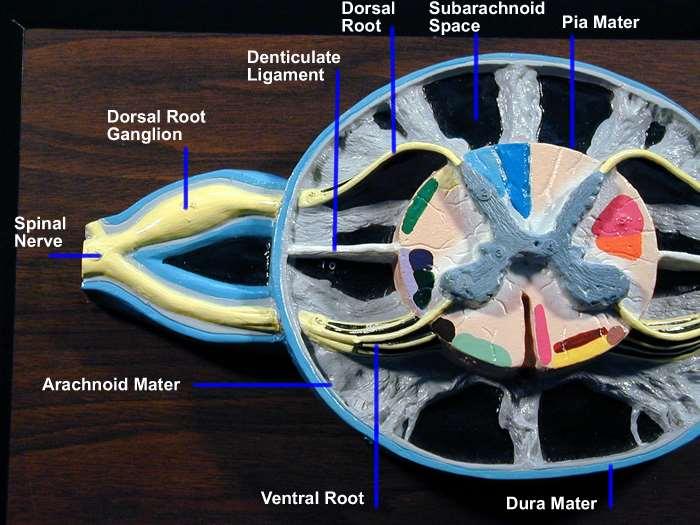
6 Reflex Arc 1. Sensory receptor: responds to stimulus 2. Sensory neuron: through dorsal root ganglion and root posterior horn 3. Integrating center: single synapse between sensory and motor neurons 4. Motor neuron: from anterior horn ventral root spinal nerve 5. Effector: muscle responds
7 Example of Reflex Arc: Patellar Reflex 1. Sensory receptor is stimulated by tap on patellar tendon 2. Sensory neuron: through dorsal root spinal cord 3. Integrating center: single synapse in spinal cord 4. Motor neuron: through ventral root spinal nerve femoral nerve 5. Effector: quads contract, extend leg

8 Example of Reflex Arc: Patellar Reflex
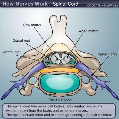
9 General Organization Spinal cord is SMALL! cm long 1 CM wide at widest point Does not extend all the way to the bottom of the spinal column Pattern of grey/white matter is reversed in the cord White matter tracts on outside Grey matter on the inside Staining reverses this!!!
 axons) Grey Matter (cell bodies)")
10 General Organization White matter (tracts of axons) axons) Grey Matter (cell bodies) )

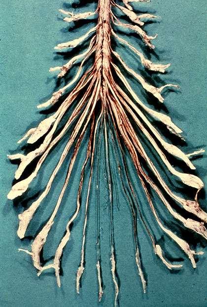
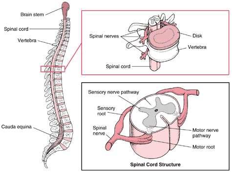
11 General Organization Spinal cord is segmented anatomically Input and output occurs in groups of rootlets arranged in a series longitudinally along the cord Dorsal rootlets -- Input -- carry sensory information Ventral rootlets -- Output -- motor neurons
12 General Organization Each set of rootlets forms a spinal nerve that innervates a corresponding segment of the body, called a dermatome
13 General Organization
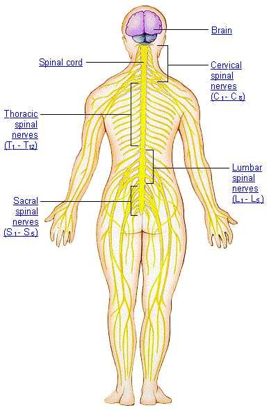
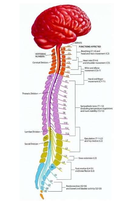
14 General Organization There are 31 segments in the spinal cord: 8 cervical (C1 - C8) 12 Thoracic (T1 - T12) 5 Lumbar (L1 - L5) 5 Sacral (S1 - S5) 1 Coccygeal

15 General Organization The spinal cord is housed within the vertebral column
16 General Organization Each cord segment has a corresponding vertebra of the same name (e.g., C3) Spinal nerves enter/exit underneath their corresponding vertebral segment
17 General Organization
18 Grey and White Matter
19 Grey and White Matter Grey Matter = Cell Body White Matter = Myelinated axon
20 Grey and White Matter Grey matter Cortex Nucleus (CNS) Ganglion (PNS) Exception: Basal Ganglia
21 Grey and White Matter White Matter Nerve (PNS) Tract (CNS) Fasciculus/Funiculus -- Group of fibers with common origin and destination Lemniscus -- Ribbon-like fiber tract Peduncle -- Massive group of fibers -- usually several tracts
22 Grey and White Matter Tracts are named with origin first, then destination Corticospinal tract -- cortex to spinal cord Mammilothalamic tract -- Mammilary bodies to thalamus Spinocerebellar tract -- Spinal cord to cerebellum Corticobulbar tract -- Cortex to brain stem
 Ganglia Nerves Cranial nerves and spinal nerves Communication between regions of body and")
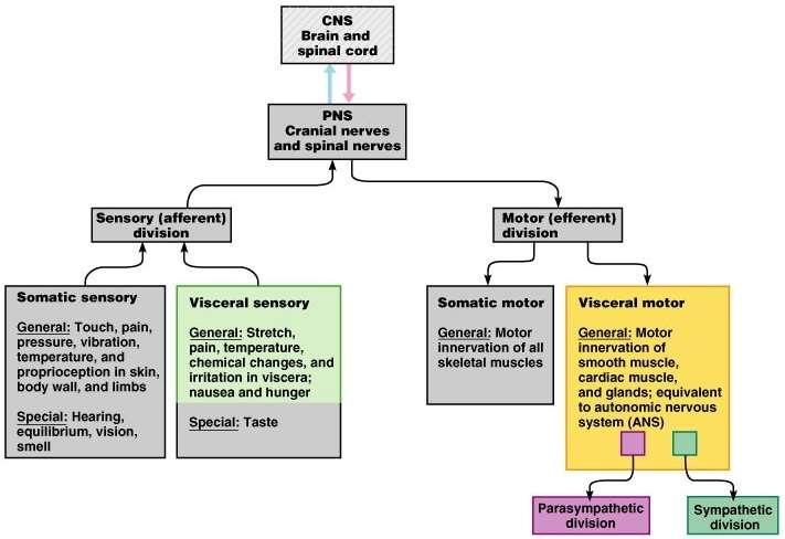
23 DIVISIONS OF THE NERVOUS SYSTEM Central Nervous System (CNS) Brain and spinal cord Interprets incoming sensory signals Dictates motor responses Peripheral Nervous System (PNS) Ganglia Nerves Cranial nerves and spinal nerves Communication between regions of body and CNS
 - Segmental Structure of")
 System of the")
24 Spinal Cord - Spinal Nerve (C8, T12, L5, S5, Cx1) - Segmental Structure of Neural Tube Origin - Comparable to Input-Output (IO) System of the Computer

25 Spinal cord
26
27

28 Spinal cord Internal gray matter: presence of neurons relaying and integrating motor/sensory impulses Fiber tracts: sensory and motor
29

30 Spinal cord Segmental organization derived from neural tube and somites Spinal segments - 31 Spinal nerves: C8, T12, L5, S5, Co1 Comparable to input-output systemof computer Seat of reflexes Origin of ascending and descending projections
31 Somatic PNS ANS Sympathetic Parasymp. Enteric CNS
32
33 PNS Nervous system structures outside the brain and spinal cord Structural components: Sensory receptors Motor endings Nerves and ganglia
34 PNS - Nervous Tissue Made up of 2 cells: Neurons Conduct nervous impulses Supporting cells Surround the neurons Ex. Glial cells
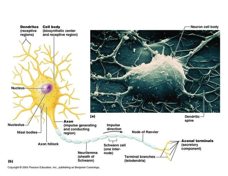
35 35
36 The Neuron Nerve cells Transmit signals in form of nerve impulses Have extreme longevity Do not divide High metabolic rate

37 Neuron-To-Neuron Transmission synapses

38 Neuronal Anatomy Cell body (soma) Most are in CNS Neuron processes Dendrites Toward cell body Axons Transmit away from cell body Synapses Site where neurons communicate
39 Neuronal Anatomy Myelin sheath Fatty sheath that surrounds most nerve fibers Reflex arc Responses to a stimulus Interneuron Nerve cell that lies between a sensory neuron and motor neuron in a reflex arc Confined entirely within the CNS

40 PNS - Sensory and Motor Signals Divided by the body regions they serve: Sensory division Somatic sensory Visceral sensory Motor division Somatic motor Visceral motor
41 The Spinal Cord Foramen magnum to L1 or L2 Runs through the vertebral canal of the vertebral column Functions 1. Sensory and motor innervation of entire body inferior to the head through the spinal nerves 2. Two-way conduction pathway between the body and the brain 3. Major center for reflexes

 http://www.apparelyzed.")
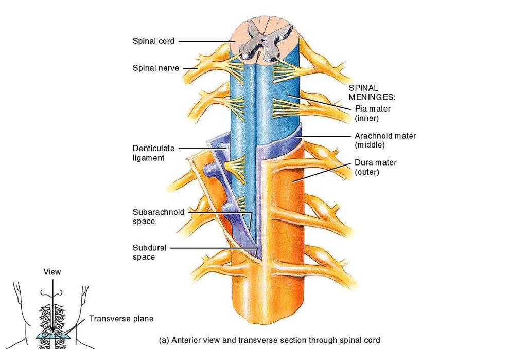
42 Spinal cord Fetal 3 rd month: ends at coccyx Birth: ends at L3 Adult position at approx L1-2 during childhood End: conus medullaris This tapers into filum terminale of connective tissue, tethered to coccyx Spinal cord segments are superior to where their corresponding spinal nerves emerge through intervertebral Denticulate ligaments: lateral shelves of pia mater anchoring to dura (meninges: more later)

43 The Spinal Cord white matter dorsal root grey matter pia mater ventral root arachnoid spinal nerves dura mater


44 The Spinal Cord vertebra spinal cord spinal nerve


45 Nerve Pathways into the Spinal Cord sensory pathway motor pathway
46
47 Autonomic Nervous System Visceral Motor Function Not easily controlled by will Get nervous and sweat Innervate smooth muscle, cardiac muscle, glands Regulate visceral function Heart rate, blood pressure, digestion, urination Has 2 divisions: Parasympathetic Sympathetic
48 ANS Parasympathetic Enables body to unwind and calm down Most active when body at rest Routine maintenance functions Craniosacral division Fibers emerge from brain and sacral spinal cord Sympathetic fight or flight Mobilizes the body during extreme situations Becomes active when extra metabolic effort needed Thoracolumbar division Fibers arise from thoracic and lumbar parts of spinal cord
49 ANS Includes a chain of 2 motor neurons Preganglionic neuron Preganglionic axon Ganglionic neuron Postganglionic axon Autonomic neuron synapses 2 neurons
50 PNS Somatic NS Autonomic NS Sympathetic division Parasympathetic division Enteric division
51 Somatic Nervous System Innervates skeletal muscle Neurons runs from CNS directly to muscle Consists of single neuron plus skeletal muscle cells Voluntary control Running, moving limbs, typing on a computer!
 Provides")
52 CNS Spinal Cord Runs through vertebral canal of the vertebral column Protected by bone, meninges, and cerebrospinal fluid Spinal cord made of a core of gray matter surrounded by white matter 31 pairs of spinal nerves branch off spinal cord through intervertebral foramen Functions in many ways: Involved in sensory and motor innervation of body inferior to the head (through spinal nerves) Provides a 2-way conduction pathway for signals between body and brain Major center for reflexes
53

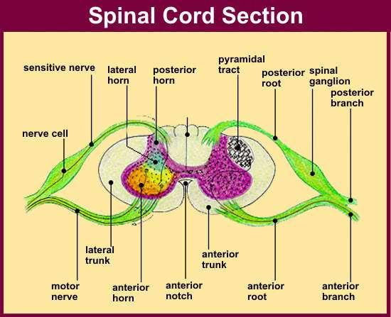
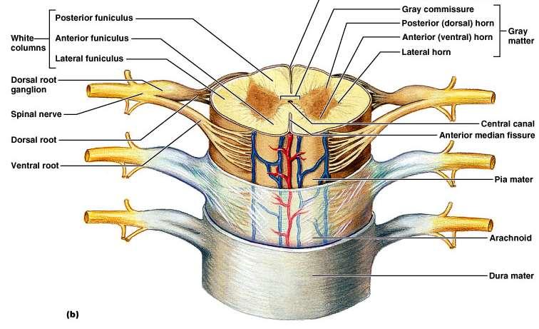
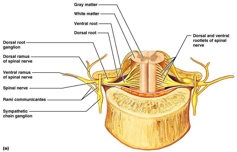
54 Spinal Cord Growth Until 3 rd month of development, does not run to coccyx As vertebral column grows caudally, spinal cord becomes more rostral At birth, ends at L 3 During childhood, terminates at L 1 and L 2 Adults runs from medulla oblongata to L 1

55

56 Regions of the Spinal Cord Cervical Thoracic Lumbar Sacral Coccygeal Cervical + Lumbar enlargements Cauda equina Conus medullaris Filum terminale
 arachnoid mater (middle) pia")

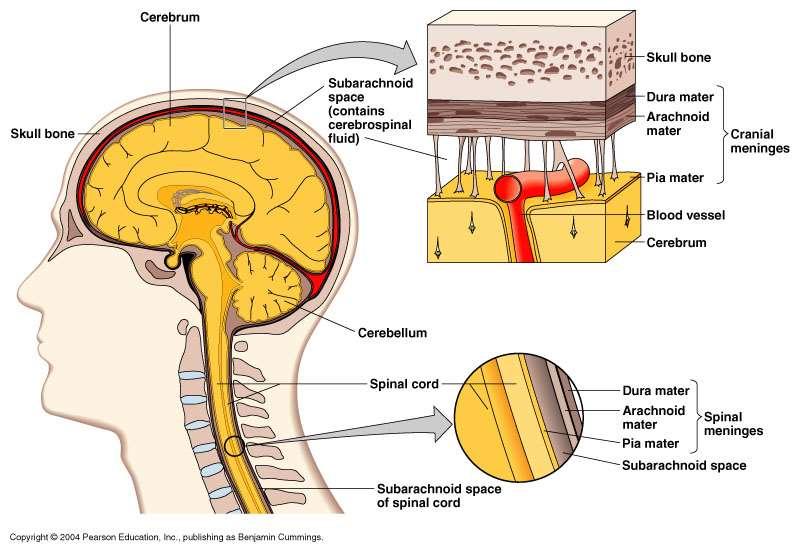
57 Protection: Bone Meninges CSF (cerebrospinal fluid) 3 meninges: dura mater (outer) arachnoid mater (middle) pia mater (inner) 3 potential spaces epidural: outside dura subdural: between dura & arachnoid subarachnoid: deep to arachnoid
58
59 Spinal Meninges and Spaces Epidural space: between vertebrae and dura mater Dura mater- tough,dense connective tissue Extends to vertebra S2 (well beyond spinal cord) Arachnoid mater: resembles spider s web Extends into subarachnoid space Subarachnoid space CSF circulates in this space Pia mater: thin, delicate layer Adheres to surface spinal cord (and brain) Contains blood vessels Copyright 2010, John Wiley & Sons, Inc.
60

61 Spinal Meninges and Spaces Copyright 2010, John Wiley & Sons, Inc.
62 Meninges of Brain and Spinal Cord Dura mater (superficial) Spinal dural sheath Does not attach to bone Epidural space Fat and veins Between dura mater and vertebra Subdural space Between dura mater and arachnoid
 Highly vascular Adheres to brain/spinal cord")
63 Meninges of Brain and Spinal Cord Arachnoid mater (middle) Impermeable layer = barrier Raised off pia mater by rootlets Subarachnoid space Between arachnoid and pia mater Contains CSF Pia mater (deep) Highly vascular Adheres to brain/spinal cord tissue


64 Meninges of the spinal cord Dura mater Arachnoid membrane Pia mater Denticulate ligament - specilization of the pia mater - landmark for cordotomy

65 Meninges of the spinal cord

66 Meninges of the spinal cord
67 Gray Mater Consists of neuron cell bodies, unmyelinated axons, dendrites, and neuroglia Shaped like an H Gray commissure (crossbar) Central canal Posterior horns Anterior horns

68 Gray Mater Posterior horns Consist of interneurons that transmit in from outside spinal cord into it Dorsal root contain sensory fibers Somatic Sensory (SS) Visceral Sensory (VS) Dorsal root ganglia - swelling in dorsal root that these interneurons pass through Anterior horns Cell bodies of motor neurons send info out of spinal cord to muscles and glands Ventral Root contains Motor Fibers Visceral Motor Somatic Motor
69 Arc reflex Arc reflex (fast and involuntary)
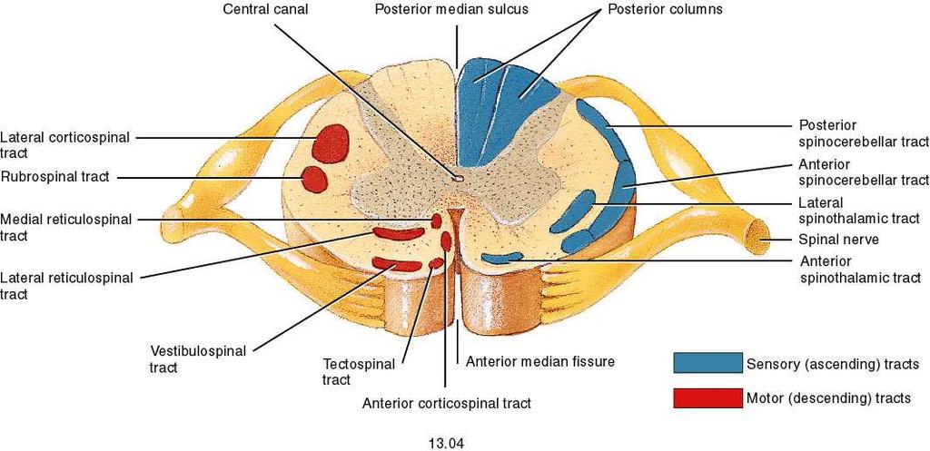
70 White Mater Surrounds gray matter Composed of myelinated and unmyelinated axons Divided into white columns (funiculi) Posterior funiculus Anterior funiculus Lateral funiculus Allow for communication between Parts of the spinal cord Spinal cord and brain

71 White Mater 3 types of nerve fibers: Ascending Carry sensory info from sensory neurons of body to brain touch, pressure, pain, temperature Descending Carry motor instructions from brain to spinal cord Contraction of muscles and secretion of glands control precise, skilled movement = writing, maintain balance, create movement Commissural Cross from one side of cord to the other
 Clusters of cell bodies Remember, in PNS clusters of cell bodies were called ganglia More words: brains stem is caudal (toward tail) to the more rostral (noseward)")
72 Gray and White Matter Like spinal cord but with another layer of gray outside the white Called cortex Cerebrum and cerebellum have Inner gray: brain nuclei (not cell nuclei) Clusters of cell bodies Remember, in PNS clusters of cell bodies were called ganglia More words: brains stem is caudal (toward tail) to the more rostral (noseward) cerebrum
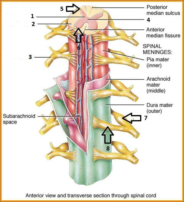
73 Gross Anatomy of Spinal Cord Extends from medulla of brain to L2 vertebra Cauda equina (horse s tail) Extends inferior to end of spinal cord Consists of roots of lumbar, sacral and coccygeal spinal nerves Left and right halves partially separated by Anterior median fissure and posterior median sulcus Small central canal (filled with CSF) in middle Enlargements: cervical and lumbar regions Points of origins of nerves to upper and lower limbs Copyright 2010, John Wiley & Sons, Inc.
74 Gross Anatomy of Spinal Cord Copyright 2010, John Wiley & Sons, Inc.

75 Internal Structure of Spinal Cord Copyright 2010, John Wiley & Sons, Inc.
76 Spinal cord eneral_esi_epidural_space.jpg coverings and spaces Dura mater Arachnoid mater Pia mater
 White matter (yellow here) Gray matter (brown here) p")
77 Spinal cord anatomy Posterior median sulcus ( p ) Anterior median fissure ( a ) White matter (yellow here) Gray matter (brown here) p a
 Dorsal (posterior) white gray Central canal Ventral")
78 Gray/White in spinal cord Hollow central cavity ( central canal ) Gray matter surrounds cavity White matter surrounds gray matter (white: ascending and descending tracts of axons) H shaped on cross section Dorsal half of H : cell bodies of interneurons Ventral half of H : cell bodies of motor neurons No cortex (as in brain) Dorsal (posterior) white gray Central canal Ventral (anterior)
 Anterior (ventral) horns (cell bodies of motor neurons) Lateral horns in thoracic and superior lumbar cord")
79 Spinal cord anatomy Gray commissure with central canal Columns of gray running the length of the spinal cord Posterior (dorsal) horns (cell bodies of interneurons) Anterior (ventral) horns (cell bodies of motor neurons) Lateral horns in thoracic and superior lumbar cord * * *
80 White matter of the spinal cord (myelinated and unmyelinated axons) Ascending fibers: sensory information from sensory neurons of body up to brain Descending fibers: motor instructions from brain to spinal cord Stimulates contraction of body s muscles Stimumulates secretion from body s glands Commissural fibers: white-matter fibers crossing from one side of cord to the other Most pathways cross (or decussate) at some point Most synapse two or three times along the way, e.g. in brain stem, thalamus or other
81 Sensory tracts

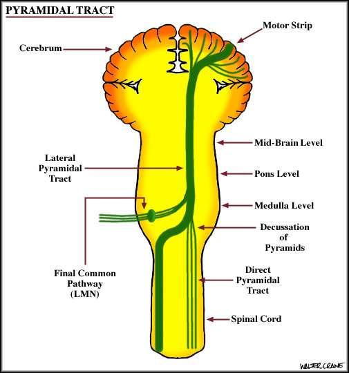
82 Motor tracts

83 The spinal cord provides a vital link between the brain and the rest of the body, and yet it exhibits some functional independence from the brain.
 in")
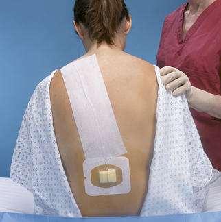
84 The adult spinal cord travels from the foramen magnum and terminates within the vertebral foramen of the first lumbar vertebra (L1) in adults.
. Don t be confused and think that the sacral region of the spinal cord is surrounded by sacral vertebrae. It is NOT!")
85 The spinal cord can be subdivided into five regions: cervical region, thoracic region, lumbar region, sacral region, and coccygeal region (which has only one pair of nerves). Don t be confused and think that the sacral region of the spinal cord is surrounded by sacral vertebrae. It is NOT!

86 The diameter of the spinal cord is the largest in the cervical region and there is a larger proportion of white matter compared to gray matter.
 is the smallest and the proportion of gray matter is largest in the spinal")
87 The diameter of the sacral region of the spinal cord (which is surrounded by the T12/L1 vertebrae) is the smallest and the proportion of gray matter is largest in the spinal cord.

88 The cervical enlargement contains the neurons that innervate the upper limbs The lumbar enlargement contains the neurons that innervate the lower limbs.
89 General Organization Cervical enlargement C5 - T1 Cervical enlargement C5 - T1 Lumbar enlargement L2 - S3

90 The tapering end of the spinal cord is called the conus medullaris. The conus medullaris is surrounded by L1 in and adult and L2 in a child.

91 The adult spinal cord terminates at the level of the first lumbar vertebra (L1) In a developing child, the spinal cord can extend to the level of the second lumbar vertebra (L2)

92 The cauda equina (horse s tail) is composed of nerves that arise from the conus medullaris and extend inferiorly.

93 The filum terminale, which is composed of pia mater, extends from the conus medullaris to the coccyx. Note the subarachnoid space also continues for some distance.

94 There are 31 pairs of spinal nerves that serve defined segments of the human body.
95 There are 8 pairs of cervical spinal nerves. This is possible because the first pair (C1 spinal nerves) exits the spinal column between the occipital bone and the atlas (C1). The remaining 7 pairs (C2- C8 spinal nerves) exit below each of the 7 cervical vertebrae via the intervertebral foramina. All the spinal nerves are mixed nerves.

96 The spinal cord is surrounded by the dura, arachnoid, and pia maters (the meninges)

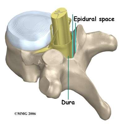
97 The epidural space is between the vertebra and the dura mater
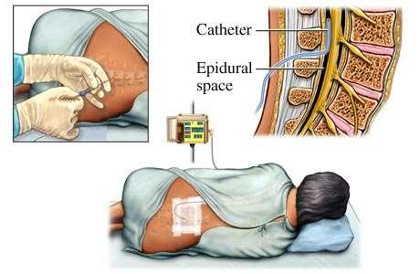
98 Epidural anesthesia

99 LP (lumbar puncure) = spinal tap (needle introduced into subdural space to collect CSF) Lumbar spine needs to be flexed so can go between spinous processes Originally thought to be a narrow fluid-filled interval between the dural and arachnoid; now known to be an artificial space created by the separation of the arachnoid from the dura as the result of trauma or some ongoing pathologic process; in the healthy state, the arachnoid is attached to the dura and a naturally occurring subdural space is not present. Epidural space is external to dura Anesthestics are often injected into epidural space Injection into correct space is vital; mistakes can be lethal
")
100 Lumbar Puncture lumbar (terminal) cistern
 L3 L4 For")


101 Lumbar Puncture (= Spinal Tap) L3 L4 For clinical examination of CSF or administration of radiopaque dyes, drugs and sometimes anesthetics However: mostly epidurals for anesthetics
102 Epidural anesthesia

103 Effect of epidural anesthesia

104 A person who needs an epidural!

105 The dura mater extends along the entire length of the vertebral canal and surrounds the spinal cord. It also extends along the initial portion of the radiating spinal nerves

106 The subarachnoid space is a real space filled with CSF
.")
107 In this midsagittal picture #3 is the dura mater, #5 is the spinal cord, # 4 is the epidural space, and #6 is the subarachnoid space where CSF is located (#1 is an intervertebral disc and #2 is the body of a vertebrae).

108 Needle for spinal tap

109 Spinal taps are done between the third and fourth lumbar vertebrae because there is no spinal cord at that location

110 The tip of the needle is inserted into the subarachnoid space outside the cauda equina and spinal fluid is removed for testing.

111 The entering pressure can be determined when the needle is inserted into the subarachnoid space during a spinal tap.
.")
112 Spinal fluid is normally crystal clear like water. Cloudy spinal fluid, like the specimen shown, is a sign of white blood cells (pus). The most common cause for white blood cells in the spinal fluid is viral or bacterial meningitis.

113 The pia mater directly adheres to the spinal cord

114 The cross-sectional view shows that the gray matter is central and the white matter is peripheral

115 The peripheral white matter contains ascending and descending tracts of nerves traveling to and from the brain. The central gray matter serves as a center for spinal reflexes.
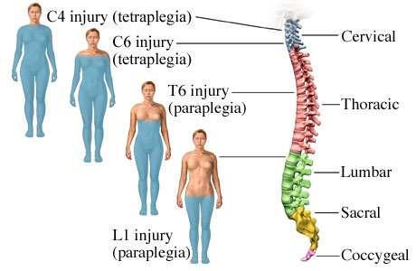
116 The central canal runs the entire length of the spinal cord, is contiguous with the brain and contains cerebrospinal fluid (CSF)

117 The spinal cord develops as 31 segments, each of which gives rise to a pair of spinal nerves that emerge from the cord through the intervertebral foraminae

118 Nerves can be sensory, motor, or mixed (sensory and motor)

119 Mixed nerves carry both types of information and some axons are transmitting impulses in one direction, while other axons are transmitting impulses in the opposite direction. All spinal nerves are mixed nerves.
120 There are 8 pairs of cervical spinal nerves. This is possible because the first pair (C1 spinal nerves) exits the spinal column between the occipital bone and the atlas (C1). The remaining 7 pairs (C2- C8 spinal nerves) exit below each of the 7 cervical vertebrae via the intervertebral foramina. All the spinal nerves are mixed nerves.

121 Most of the spinal nerves are associated with specific dermatomes (an area of skin innervated by all the cutaneous neurons of a certain spinal or cranial nerve).

122 trigeminal Dermatome map. Note the trigeminal nerve has dermatomes on the face.
 are seen on the")
123 Dermatomes of the trigeminal nerve (cranial nerve V) are seen on the face
 are not represented on the surface")
124 Note that the trigeminal nerve has dermatomes on the face (see white area) and that the first pair of cervical spinal nerves (C1 spinal nerves) are not represented on the surface at all.
125 The bony vertebral column (dark line shown) grows faster than the spinal cord. A newborn s spinal cord extends to about the level of L 3. A child s spinal cord may extend to the level of L 2. An adult s spinal cord typically terminates at the level of L 1. Bony vertebral column

126 The inner delicate spinal cord terminates in an adult, as the conus medullaris, at the level of the L 1 vertebra.

127 Damage to the spinal cord can lead to paralysis or death

128 ANTERIOR VIEW OF THE SPINAL CORD CERVICAL CORD (8 nerves) The spinal cord begins at the base of the medulla oblongata and extends to about the 2nd lumbar vertebra. The cord is divided into four regions each of which has branches called spinal nerves. THORACIC CORD (12 nerves) LUMBAR CORD (5 nerves) SACRAL CORD (5 nerves)
 of the spinal cord forming the cauda equina (horse s")
129 SAGITAL SECTION OF LOWER SPINE The inferior, terminal portion of the spinal cord is at the level of the 2nd lumbar vertebra. Branches from the lumbar region pass downward from the cone-shaped tip (conus medullaris) of the spinal cord forming the cauda equina (horse s tail). Cauda equina

130

131 cranial nerves - 12 pr spinal nerves- 31 pr
132
133
134
135

136 Spinal Nerves (31 Pairs) Part of the PNS (Somatic) Lie in intervertebral foramina Send lateral branches to body Named according to their point of issue from the vertebral column 8 pairs of cervical spinal nerves; C 1 -C 8 12 pairs of thoracic spinal nerves; T 1 -T 12 5 pairs of lumbar spinal nerves; L 1 -L 5 5 pairs of sacral spinal nerves; S 1 -S 5 1 pair of coccygeal spinal nerves; C 01


137 Spinal nerves Part of the peripheral nervous system 31 pairs attach through dorsal and ventral nerve roots Lie in intervertebral foramina
138
139
140
141

142 Damage below C3 also results in quadriplegia, but the person can still utilize their diaphragm for breathing via their intact phrenic nerves.
143
144
Human Anatomy. Spinal Cord and Spinal Nerves
 Human Anatomy Spinal Cord and Spinal Nerves 1 The Spinal Cord Link between the brain and the body. Exhibits some functional independence from the brain. The spinal cord and spinal nerves serve two functions:
ANATOMY OF SPINAL CORD. Khaleel Alyahya, PhD, MEd King Saud University School of
 ANATOMY OF SPINAL CORD Khaleel Alyahya, PhD, MEd King Saud University School of Medicine @khaleelya OBJECTIVES At the end of the lecture, students should be able to: Describe the external anatomy of the
Chapter 12b. Overview
 Chapter 12b Spinal Cord Overview Spinal cord gross anatomy Spinal meninges Sectional anatomy Sensory pathways Motor pathways Spinal cord pathologies 1 The Adult Spinal Cord About 18 inches (45 cm) long
The Spinal Cord. The Nervous System. The Spinal Cord. The Spinal Cord 1/2/2016. Continuation of CNS inferior to foramen magnum.
 The Nervous System Spinal Cord Continuation of CNS inferior to foramen magnum Simpler than the brain Conducts impulses to and from brain Two way conduction pathway Reflex actions Passes through vertebral
Spinal Cord Protection. Chapter 13 The Spinal Cord & Spinal Nerves. External Anatomy of Spinal Cord. Structures Covering the Spinal Cord
 Spinal Cord Protection Chapter 13 The Spinal Cord & Spinal Nerves We are only going to cover Pages 420-434 and 447 Together with brain forms the CNS Functions spinal cord reflexes integration (summation
Chapter 13. The Spinal Cord & Spinal Nerves. Spinal Cord. Spinal Cord Protection. Meninges. Together with brain forms the CNS Functions
 Spinal Cord Chapter 13 The Spinal Cord & Spinal Nerves Together with brain forms the CNS Functions spinal cord reflexes integration (summation of inhibitory and excitatory) nerve impulses highway for upward
Note: Please refer to handout Spinal Plexuses and Representative Spinal Nerves for
 Chapter 13 Outline Note: Please refer to handout Spinal Plexuses and Representative Spinal Nerves for what you need to know from Exhibits 13.1 13.4 I. INTRODUCTION A. The spinal cord and spinal nerves
Chapter 13! Chapter 13 Spinal Cord and Spinal Nerves! The Spinal Cord and Spinal Nerves!
 Chapter 13! The Spinal Cord and Spinal Nerves! SECTION 13-1! The brain and spinal cord make up the central nervous system, and the cranial nerves and spinal nerves constitute the peripheral nervous system!
Spinal Cord H. Ruth Clemo, Ph.D.
 Spinal Cord H. Ruth Clemo, Ph.D. OBJECTIVES After studying the material of this lecture, the student should be familiar with: 1. Surface anatomy of the spinal cord. 2. Internal structure and organization
Organization of The Nervous System PROF. SAEED ABUEL MAKAREM
 Organization of The Nervous System PROF. SAEED ABUEL MAKAREM Objectives By the end of the lecture, you should be able to: List the parts of the nervous system. List the function of the nervous system.
The Spinal Cord & Spinal Nerves
 The Spinal Cord & Spinal Nerves Together with brain forms the CNS Functions spinal cord reflexes integration (summation of inhibitory and excitatory) nerve impulses highway for upward and downward travel
Gross Morphology of Spinal Cord
 Gross Morphology of Spinal Cord Lecture Objectives Describe the gross anatomical features of the spinal cord. Describe the level of the different spinal segments compared to the level of their respective
Introduction and Basic structural organization of the nervous system
 Introduction and Basic structural organization of the nervous system **the slides are in bold and the book is in red Done by : razan krishan & marah marahleh INTRODUCTION The nervous system, along with
Gross Morphology of Spinal Cord
 Gross Morphology of Spinal Cord Done By : Rahmeh Alsukkar ** I did my best and sorry for any mistake ** the sheet does not contain pictures, tables and some slides so please be careful and go back to slides
Anatomy of the Nervous System. Brain Components
 Anatomy of the Nervous System Brain Components NERVOUS SYSTEM INTRODUCTION Is the master system of human body, controlling the functions of rest of the body systems Nervous System CLASSIFICATION A. Anatomical
BIOH111. o Cell Module o Tissue Module o Integumentary system o Skeletal system o Muscle system o Nervous system o Endocrine system
 BIOH111 o Cell Module o Tissue Module o Integumentary system o Skeletal system o Muscle system o Nervous system o Endocrine system Endeavour College of Natural Health endeavour.edu.au 1 Textbook and required/recommended
Fig Cervical spinal nerves. Cervical enlargement C7. Dural sheath. Subarachnoid space. Thoracic. Spinal cord Vertebra (cut) spinal nerves
 spinal nerves") Fig. 13.1 C1 Cervical enlargement C7 Cervical spinal nerves Dural sheath Subarachnoid space Thoracic spinal nerves Spinal cord Vertebra (cut) Lumbar enlargement Medullary cone T12 Spinal nerve Spinal nerve
Fig. 13.1 C1 Cervical enlargement C7 Cervical spinal nerves Dural sheath Subarachnoid space Thoracic spinal nerves Spinal cord Vertebra (cut) Lumbar enlargement Medullary cone T12 Spinal nerve Spinal nerve
Chapter 14. The Nervous System. The Spinal Cord and Spinal Nerves. Lecture Presentation by Steven Bassett Southeast Community College
 Chapter 14 The Nervous System The Spinal Cord and Spinal Nerves Lecture Presentation by Steven Bassett Southeast Community College Introduction The Central Nervous System (CNS) consists of: The spinal
Central Nervous System: Part 2
 Central Nervous System: Part 2 1. Meninges 2. CSF 3. Spinal Cord and Spinal Nerves Explain spinal cord anatomy, including gray and white matter and meninges (give the general functions of this organ).
Organization of The Nervous System PROF. MOUSAED ALFAYEZ & DR. SANAA ALSHAARAWY
 Organization of The Nervous System PROF. MOUSAED ALFAYEZ & DR. SANAA ALSHAARAWY Objectives At the end of the lecture, the students should be able to: List the parts of the nervous system. List the function
Lecture 14: The Spinal Cord
 Lecture 14: The Spinal Cord M/O Chapters 16 69. Describe the relationship(s) between the following structures: root, nerve, ramus, plexus, tract, nucleus, and ganglion. 70. Trace the path of information
The Spinal Cord and Spinal Nerves!
 Chapter 13! The Spinal Cord and Spinal Nerves! SECTION 13-1! The brain and spinal cord make up the central nervous system, and the cranial nerves and spinal nerves constitute the peripheral nervous system!
CHAPTER 13 LECTURE OUTLINE
 CHAPTER 13 LECTURE OUTLINE I. INTRODUCTION A. The spinal cord and spinal nerves mediate reactions to environmental changes. B. The spinal cord has several functions. 1. It processes reflexes. 2. It is
THE BACK THE SPINAL CORD
 THE BACK THE SPINAL CORD The structures in the vertebral canal: the spinal cord spinal nerve roots spinal meninges the neurovascular structures THE SPINAL CORD The spinal cord occupies the superior 2/3
Human Anatomy - Problem Drill 11: The Spinal Cord and Spinal Nerves
 Human Anatomy - Problem Drill 11: The Spinal Cord and Spinal Nerves Question No. 1 of 10 Instructions: (1) Read the problem statement and answer choices carefully, (2) Work the problems on paper as needed,
Chapter 13: The Spinal Cord and Spinal Nerves
 Chapter 13: The Spinal Cord and Spinal Nerves Spinal Cord Anatomy Protective structures: Vertebral column and the meninges protect the spinal cord and provide physical stability. a. Dura mater, b. Arachnoid,
ANATOMY & PHYSIOLOGY ONLINE COURSE - SESSION 7 THE NERVOUS SYSTEM
 ANATOMY & PHYSIOLOGY ONLINE COURSE - SESSION 7 THE NERVOUS SYSTEM Introduction The nervous system is the major controlling, regulatory, and communicating system in the body. It is the center of all mental
Neurology study of the nervous system. nervous & endocrine systems work together to maintain homeostasis
 Nervous System Neurology study of the nervous system nervous & endocrine systems work together to maintain homeostasis Nervous System works very fast Uses electrical signals called nerve impulses Short-lived
Spinal Cord- Medulla Spinalis. Cuneyt Mirzanli Istanbul Gelisim University
 Spinal Cord- Medulla Spinalis Cuneyt Mirzanli Istanbul Gelisim University Spinal Column Supports the skull, pectoral girdle, upper limbs and thoracic cage by way of the pelvic girdle. Transmits body weight
Brain Stem. Nervous System (Part A-3) Module 8 -Chapter 14
 Module 8 -Chapter 14") Nervous System (Part A-3) Module 8 -Chapter 14 Overview Susie Turner, M.D. 1/9/13 Cellular structure of the nervous system Neurons Neuroglia Nervous System Divisions Central nervous system Peripheral nervous
Nervous System (Part A-3) Module 8 -Chapter 14 Overview Susie Turner, M.D. 1/9/13 Cellular structure of the nervous system Neurons Neuroglia Nervous System Divisions Central nervous system Peripheral nervous
Cerebral hemisphere. Parietal Frontal Occipital Temporal
 Cerebral hemisphere Sulcus / Fissure Central Precental gyrus Postcentral gyrus Lateral (cerebral) Parieto-occipital Cerebral cortex Frontal lobe Parietal lobe Temporal lobe Insula Amygdala Hippocampus
The neurvous system senses, interprets, and responds to changes in the environment. Two types of cells makes this possible:
 NERVOUS SYSTEM The neurvous system senses, interprets, and responds to changes in the environment. Two types of cells makes this possible: the neuron and the supporting cells ("glial cells"). Neuron Neurons
The Spinal Cord, Spinal Nerves, and Spinal Reflexes
 13 The Spinal Cord, Spinal Nerves, and Spinal Reflexes PowerPoint Lecture Presentations prepared by Jason LaPres Lone Star College North Harris An Introduction to the Spinal Cord, Spinal Nerves, and Spinal
Lesson 33. Objectives: References: Chapter 16: Reading for Next Lesson: Chapter 16:
 Lesson 33 Lesson Outline: Nervous System Structure and Function Neuronal Tissue Supporting Cells Neurons Nerves Functional Classification of Neuronal Tissue Organization of the Nervous System Peripheral
The Nervous System. Functions of the Nervous System input gathering To monitor occurring inside and outside the body Changes =
 The Nervous System Functions of the Nervous System input gathering To monitor occurring inside and outside the body Changes = To process and sensory input and decide if is needed output A response to integrated
Nervous Systems: Diversity & Functional Organization
 Nervous Systems: Diversity & Functional Organization Diversity of Neural Signaling The diversity of neuron structure and function allows neurons to play many roles. 3 basic function of all neurons: Receive
Nervous System: Spinal Cord and Spinal Nerves (Chapter 13)
") Nervous System: Spinal Cord and Spinal Nerves (Chapter 13) Lecture Materials for Amy Warenda Czura, Ph.D. Suffolk County Community College Eastern Campus Primary Sources for figures and content: Marieb,
Nervous System: Spinal Cord and Spinal Nerves (Chapter 13) Lecture Materials for Amy Warenda Czura, Ph.D. Suffolk County Community College Eastern Campus Primary Sources for figures and content: Marieb,
Spinal cord. We have extension of the pia mater below L1-L2 called filum terminale
 Spinal cord Part of the CNS extend from foramen magnum to the level of L1-L2 (it is shorter than the vertebral column) it is covered by spinal meninges. It is cylindrical in shape. It s lower end become
The functional Anatomy of the Nervous System. DR. OKSANA PETRICHKO Department of Human Anatomy
 The functional Anatomy of the Nervous System DR. OKSANA PETRICHKO Department of Human Anatomy Coordination and Regulation of Body Systems Nervous system. Conducts nerve impulses maintaining homeostasis
Chapter 8 Nervous System
 Chapter 8 Nervous System Two message centers: Functions of these systems: 1. * 2. * Overview of the Nervous System Parts: General Functions: Functions Sensory input: Sensation via nerves Integration: interpretation
NERVOUS SYSTEM. Academic Resource Center. Forskellen mellem oscillator og krystal
 NERVOUS SYSTEM Academic Resource Center Forskellen mellem oscillator og krystal Overview of the Nervous System Peripheral nervous system-pns cranial nerves spinal nerves ganglia peripheral nerves enteric
The Nervous System PART A
 7 The Nervous System PART A PowerPoint Lecture Slide Presentation by Jerry L. Cook, Sam Houston University ESSENTIALS OF HUMAN ANATOMY & PHYSIOLOGY EIGHTH EDITION ELAINE N. MARIEB Structural Classification
action potential afferent neuron Weblike; specifically, the weblike middle layer of the three meninges. arachnoid astrocytes autonomic nervous system
 action potential A large transient depolarization event, including polarity reversal, that is conducted along the membrane of a muscle cell or a nerve fiber. afferent neuron Nerve cell that carries impulses
Unit Three. The brain includes: cerebrum, diencephalon, brain stem, & cerebellum. The brain lies within the cranial cavity of the skull.
 Human Anatomy & Physiology 11 Divisions of the Nervous System Karen W. Smith, Instructor Unit Three BRAIN & SPINAL CORD Refer to the following URLs. Be sure to study these along with your book. http://www.sirinet.net/~jgjohnso/nervous.html
Chapter 17 Nervous System
 Chapter 17 Nervous System 1 The Nervous System Two Anatomical Divisions Central Nervous System (CNS) Brain and Spinal Cord Peripheral Nervous System (PNS) Two Types of Cells Neurons Transmit nerve impulses
The Nervous System An overview
 Nervous System The Nervous System An overview Includes Nerve tissue Sense organs Functions to Sense environment Process information it receives Respond to information 1 Copyright 2009 Pearson Education,
Chapter 9 The Nervous System: The Spinal Cord and Spinal Nerves
 Chapter 9 The Nervous System: The Spinal Cord and Spinal Nerves Copyright 2015 Wolters Kluwer Health Lippincott Williams & Wilkins Overview Key Terms acetylcholine motor presynaptic action potential nerve
Nervous System C H A P T E R 2
 Nervous System C H A P T E R 2 Input Output Neuron 3 Nerve cell Allows information to travel throughout the body to various destinations Receptive Segment Cell Body Dendrites: receive message Myelin sheath
Chapter 7 Nervous System
 Chapter 7 Nervous System Two message centers: Functions of these systems: 1. * 2. * Overview of the Nervous System Parts: General Functions: Functions Sensory input: Sensation via nerves Integration: interpretation
Department of Neurology/Division of Anatomical Sciences
 Spinal Cord I Lecture Outline and Objectives CNS/Head and Neck Sequence TOPIC: FACULTY: THE SPINAL CORD AND SPINAL NERVES, Part I Department of Neurology/Division of Anatomical Sciences LECTURE: Monday,
The Nervous System: Sensory and Motor Tracts of the Spinal Cord
 15 The Nervous System: Sensory and Motor Tracts of the Spinal Cord PowerPoint Lecture Presentations prepared by Steven Bassett Southeast Community College Lincoln, Nebraska Introduction Millions of sensory
Human Anatomy. Autonomic Nervous System
 Human Anatomy Autonomic Nervous System 1 Autonomic Nervous System ANS complex system of nerves controls involuntary actions. Works with the somatic nervous system (SNS) regulates body organs maintains
Chapter 9. Nervous System
 Chapter 9 Nervous System Central Nervous System (CNS) vs. Peripheral Nervous System(PNS) CNS Brain Spinal cord PNS Peripheral nerves connecting CNS to the body Cranial nerves Spinal nerves Neurons transmit
With other members of your lab group, discuss the following questions: - The spinal cord connects directly to which part of the brain?
 BIOLOGY 211: HUMAN ANATOMY & PHYSIOLOGY ************************************************************************************************************************* SPINAL CORD, SPINAL NERVES, AND REFLEXES
NERVOUS SYSTEM ANATOMY
 INTRODUCTION to NERVOUS SYSTEM ANATOMY M1 - Gross and Developmental Anatomy Dr. Milton M. Sholley Professor of Anatomy and Neurobiology and Dr. Michael H. Peters Professor of Chemical and Life Science
CHAPTER 13&14: The Central Nervous System. Anatomy of the CNS
 CHAPTER 13&14: The Central Nervous System Anatomy of the CNS in human consists of brain and spinal cord as stated earlier neurons have little support from their extracellular matrix and depend on glial
CHAPTER 11: NERVOUS SYSTEM II: DIVISIONS OF THE NERVOUS SYSTEM. 1. Outline the major divisions of the nervous system.
 CHAPTER 11: NERVOUS II: DIVISIONS OF THE NERVOUS OBJECTIVES: 1. Outline the major divisions of the nervous system. NERVOUS CENTRAL NERVOUS (BRAIN & SPINAL CORD) (INTERNEURONS) PERIPHERAL NERVOUS (CRANIAL
The CNS Part II pg
 The CNS Part II pg. 455-474 Protection of the Brain Objectives Describe how the meninges, cerebrospinal fluid, and the blood brain barrier protect the CNS. Explain how Cerebrospinal fluid is formed, and
Biological Bases of Behavior. 3: Structure of the Nervous System
 Biological Bases of Behavior 3: Structure of the Nervous System Neuroanatomy Terms The neuraxis is an imaginary line drawn through the spinal cord up to the front of the brain Anatomical directions are
The Nervous System: Autonomic Nervous System Pearson Education, Inc.
 17 The Nervous System: Autonomic Nervous System Introduction The autonomic nervous system: Functions outside of our conscious awareness Makes routine adjustments in our body s systems The autonomic nervous
Biology 218 Human Anatomy
 Chapter 20 Adapted form Tortora 10 th ed. LECTURE OUTLINE A. Introduction (p. 632) 1. The autonomic nervous system (ANS) regulates the activity of smooth muscle, cardiac muscle, and certain glands. 2.
Gross Anatomy of Lower Spinal Cord
 Chapter 13 Spinal Cord, Spinal Nerves and Somatic Reflexes Spinal cord Spinal nerves Somatic reflexes Gross Anatomy of Lower Spinal Cord Meninges of Vertebra & Spinal Cord Spina Bifida Congenital defect
3/15/17. Outline. Nervous System - PNS and CNS. Two Parts of the Nervous System
 Nervous System - PNS and CNS Bio 105 Outline I. Central Nervous System vs Peripheral Nervous System II. Peripheral Nervous System A. Autonomic Nervous Systems B. Somatic Nervous Systems III. Autonomic
Nervous System - PNS and CNS. Bio 105
 Nervous System - PNS and CNS Bio 105 Outline I. Central Nervous System vs Peripheral Nervous System II. Peripheral Nervous System A. Autonomic Nervous Systems B. Somatic Nervous Systems III. Autonomic
The Nervous System. Lab Exercise 29. Objectives. Introduction
 Lab Exercise The Nervous System Objectives -You should be able to recognize a neuron and identify its components. - Be able to identify the principal components of the brain and be able to name at least
Somatic Nervous Systems. III. Autonomic Nervous System. Parasympathetic Nervous System. Sympathetic Nervous Systems
 7/21/2014 Outline Nervous System - PNS and CNS I. II. Two Parts of the Nervous System Central Nervous System vs Peripheral Nervous System Peripheral Nervous System A. B. Brain and Spinal Cord III. Autonomic
Principles of Anatomy and Physiology
 Principles of Anatomy and Physiology 14 th Edition CHAPTER 15 The Autonomic Nervous System Comparison of Somatic and Autonomic Nervous Systems The somatic nervous system includes both sensory and motor
NERVOUS SYSTEM ANATOMY
 NTRODUCTON to NERVOUS SYSTEM ANATOMY M1 - Gross and Developmental Anatomy Dr. Milton M. Sholley Professor of Anatomy and Neurobiology and Dr. Michael H. Peters Professor of Chemical and Life Science Engineering
Lecture - Chapter 13: Central Nervous System
 Lecture - Chapter 13: Central Nervous System 1. Describe the following structures of the brain, what is the general function of each: a. Cerebrum b. Diencephalon c. Brain Stem d. Cerebellum 2. What structures
TEST BANK FOR FUNDAMENTALS OF ANATOMY & PHYSIOLOGY 9TH EDITION BY MARTINI
 Link download :https://testbankservice.com/download/test-bank-forfundamentals-of-anatomy-and-physiology-9th-edition-by-martini TEST BANK FOR FUNDAMENTALS OF ANATOMY & PHYSIOLOGY 9TH EDITION BY MARTINI
Spinal Cord and Spinal Nerves. Spinal Cord. Chapter 12
 Chapter 12 Spinal Cord and Spinal Nerves 1 Spinal Cord Extends from foramen magnum to second lumbar vertebra Segmented: Cervical, Thoracic, Lumbar & Sacral Gives rise to 31 pairs of spinal nerves Not uniform
Copyright McGraw-Hill Education. Permission required for reproduction or display. C1. Cervical spinal ner ves. Thor acic. T12 Spinal nerve rootlets
 Fig. 13.1 C1 Cervical enlar gem ent C7 Cervical spinal ner ves Dural sheath Subarachnoi d space Thor acic spinal ner ves Vertebra (cut) Lum bar enlar gem ent Medullar y T12 rootlets cone Posterior median
Cranial Nerves and Spinal Cord Flashcards
 1. Name the cranial nerves and their Roman numeral. 2. What is Cranial Nerve I called, and what does it 3. Scientists who are trying to find a way to make neurons divide to heal nerve injuries often study
Human Anatomy and Physiology I Laboratory
 Human Anatomy and Physiology I Laboratory Histology of Nervous Tissue and The Spinal Cord This lab involves two laboratory exercises: 1) Histology of Nervous Tissue, and 2) Spinal Cord, Spinal Nerves,
THE BACK. Dr. Ali Mohsin. Spinal Cord
 Spinal Cord THE BACK Dr. Ali Mohsin The spinal cord is the elongated caudal part of the CNS. It starts as the inferior continuation of the medulla oblongata at the level of foramen magnum, & ends as an
Lecturer. Prof. Dr. Ali K. Al-Shalchy MBChB/ FIBMS/ MRCS/ FRCS 2014
 Lecturer Prof. Dr. Ali K. Al-Shalchy MBChB/ FIBMS/ MRCS/ FRCS 2014 Dorsal root: The dorsal root carries both myelinated and unmyelinated afferent fibers to the spinal cord. Posterior gray column: Long
Organisation of the nervous system
 Chapter1 Organisation of the nervous system 1. Subdivisions of the nervous system The nervous system is divided: i) Structurally The central nervous system (CNS) composed of the brain and spinal cord.
Chapter 3. Structure and Function of the Nervous System. Copyright (c) Allyn and Bacon 2004
 Allyn and Bacon 2004") Chapter 3 Structure and Function of the Nervous System 1 Basic Features of the Nervous System Neuraxis: An imaginary line drawn through the center of the length of the central nervous system, from the
Chapter 3 Structure and Function of the Nervous System 1 Basic Features of the Nervous System Neuraxis: An imaginary line drawn through the center of the length of the central nervous system, from the
Nervous system. The main regulation mechanism of organism's functions
 Nervous system The main regulation mechanism of organism's functions Questions Neuron The reflex arc The nervous centers Properties of the nervous centers The general principles of coordination Inhibition
Nervous System CHAPTER 9. Copyright 2016 by Elsevier, Inc.
 Nervous System CHAPTER 9 Copyright 2016 by Elsevier, Inc. Neurons and Supporting Cells Copyright 2016 by Elsevier, Inc. 2 Communication and Control Systems nervous system endocrine system uses chemicals
Spinal Cord and Properties of Cerebrospinal Fluid: Options for Drug Delivery. SMA Foundation New York
 Spinal Cord and Properties of Cerebrospinal Fluid: Options for Drug Delivery New York Why Do We Need to Know about the Spinal Cord Anatomy and Properties of Cerebrospinal Fluid? SMA therapeutics need to
Spinal Cord Organization. January 12, 2011
 Spinal Cord Organization January 12, 2011 Spinal Cord 31 segments terminates at L1-L2 special components - conus medullaris - cauda equina no input from the face Spinal Cord, Roots & Nerves Dorsal root
Sympathetic Nervous System
 Sympathetic Nervous System Lecture Objectives Review the subdivisions of the nervous system. Review the general arrangement and compare the sympathetic and parasympathetic parts. Describe the following
Anatomy and Physiology 1 Chapters 12 and 13 self quiz Pro, Dima Darwish,MD.
 Anatomy and Physiology 1 Chapters 12 and 13 self quiz Pro, Dima Darwish,MD. 1) Which of the following is a function of the nervous system? A) sense the internal and external environments B) integrate sensory
Nervous and Endocrine System Exam Review
 Directions: Read each question and complete the statement using the multiple choice responses I. Nervous System 1. The interpretation of olfactory receptor information would fall under which general function
The Nervous System: The
 C h a p t e r 14 The Nervous System: The Spinal Cord and Spinal Nerves PowerPoint Lecture Slides prepared by Jason LaPres North Harris College Houston, Texas Copyright 2009 Pearson Education, Inc., publishing
Meninges. Connective tissue membranes
 Meninges Connective tissue membranes Dura mater: -outermost layer; continuous with epineuriumof the spinal nerves - dense irregular connective tissue - from the level of the foramen magnum to S2 Arachnoid
SPINAL CORD AND PROPERTIES OF CEREBROSPINAL FLUID: OPTIONS FOR DRUG DELIVERY
 SPINAL CORD AND PROPERTIES OF CEREBROSPINAL FLUID: OPTIONS FOR DRUG DELIVERY WHY DO WE NEED TO KNOW ABOUT THE SPINAL CORD ANATOMY AND PROPERTIES OF CEREBROSPINAL FLUID? SMA therapeutics need to reach cells
Chapter 14 Lecture Outline
 Chapter 14 Lecture Outline See separate PowerPoint slides for all figures and tables preinserted into PowerPoint without notes. Copyright McGraw-Hill Education. Permission required for reproduction or
The Nervous System: Neural Tissue Pearson Education, Inc.
 13 The Nervous System: Neural Tissue Introduction Nervous System Characteristics Controls and adjust the activity of the body Provides swift but brief responses The nervous system includes: Central Nervous
NOTES CHAPTER 9 (Brief) The Nervous System LECTURE NOTES
 The Nervous System LECTURE NOTES") NOTES CHAPTER 9 (Brief) The Nervous System LECTURE NOTES I. Divisions of the Nervous System two major divisions A. Central Nervous System (CNS) 1. brain 2. spinal cord B. Peripheral Nervous System (PNS)
NOTES CHAPTER 9 (Brief) The Nervous System LECTURE NOTES I. Divisions of the Nervous System two major divisions A. Central Nervous System (CNS) 1. brain 2. spinal cord B. Peripheral Nervous System (PNS)
WHAT ARE THE FUNCTIONS OF THE NERVOUS SYSTEM?
 THE NERVOUS SYSTEM LEARNING OBJECTIVES To state the function of the Nervous system. To describe the structure and workings of the nervous system. To name the major parts of the nervous system. To describe
Central Nervous System (CNS) -> brain and spinal cord. Major Divisions of the nervous system:
 -> brain and spinal cord. Major Divisions of the nervous system:") Central Nervous System (CNS) -> brain and spinal cord Major Divisions of the nervous system: Afferent (sensory input) -> cell bodies outside of the central nervous system (CNS), carry info into the CNS
Central Nervous System (CNS) -> brain and spinal cord Major Divisions of the nervous system: Afferent (sensory input) -> cell bodies outside of the central nervous system (CNS), carry info into the CNS
Module 5 : Anatomy The nervous system
 Module 5 : Anatomy The nervous system In this module you will learn: The main parts of the nervous system The different sections of the brain and how it functions The structure and function of the spinal
BIO 115 Anatomy & Physiology II Practice Assignment 4: The Nervous System & The Senses This is not a required assignment but it is recommended.
 BIO 115 Anatomy & Physiology II Practice Assignment 4: The Nervous System & The Senses This is not a required assignment but it is recommended. 1. This figure depicts a typical neuron. What structures
Chapter 9 Nervous System
 Chapter 9 Nervous System Introduction: A. The nervous system is composed of neurons and neuroglia. 1. Neurons transmit nerve impulses to other neurons. 2. Nerves are made up of bundles of nerve fibers
Tymaa Al-zaben & Amin Al-ajalouni
 Done by: Tymaa Al-zaben & Amin Al-ajalouni ** Hello SERTONIN! SLIDE 3 note:: the slide included within the sheet but make sure back to slide for pictures The Autonomic Nervous System Function : Regulate
CHAPTER 15 LECTURE OUTLINE
 CHAPTER 15 LECTURE OUTLINE I. INTRODUCTION A. The autonomic nervous system (ANS) regulates the activity of smooth muscle, cardiac muscle, and certain glands. B. Operation of the ANS to maintain homeostasis,
PNS and ANS Flashcards
 1. Name several SOMATIC SENSES Light touch (being touched by a feather), heat, cold, vibration, pressure, pain are SOMATIC SENSES. 2. What are proprioceptors; and how is proprioception tested? PROPRIOCEPTORS