C h a p t e r PowerPoint Lecture Slides prepared by Jason LaPres North Harris College Houston, Texas
|
|
|
- Melvin Lyons
- 6 years ago
- Views:
Transcription

1 C h a p t e r 15 The Nervous System: The Brain and Cranial Nerves PowerPoint Lecture Slides prepared by Jason LaPres North Harris College Houston, Texas Copyright 2009 Pearson Education, Inc., publishing as Pearson Benjamin Cummings
2 Introduction The brain is far more complex than the spinal cord. The brain contains roughly 20 billion neurons. Excitatory and inhibitory interactions among the extensively interconnected neuronal pools ensure that the response can vary to meet changing circumstances.

3 An Introduction to the Organization of the Brain Figure 15.1 Major Divisions of the Brain

4 An Introduction to the Organization of the Brain

5 An Introduction to the Organization of the Brain Figure 15.2a Ventricles of the Brain (Lateral View)

6 An Introduction to the Organization of the Brain Figure 15.2b Ventricles of the Brain (Lateral View)

7 An Introduction to the Organization of the Brain Figure 15.2c Ventricles of the Brain (Anterior View)

8 An Introduction to the Organization of the Brain Figure 15.2d Ventricles of the Brain (Coronal Section)
9 Protection and Support of the Brain Protection, support, and nourishment of the brain involves: Bones of the skull Cranial meninges Dura mater Arachnoid mater Pia mater Cerebrospinal fluid Blood brain barrier Vessels of the cardiovascular system

10 Protection and Support of the Brain Figure 15.3a Relationships among the Brain, Cranium, and Meninges (Lateral View)

11 Protection and Support of the Brain Figure 15.3b Relationships among the Brain, Cranium, and Meninges (Midsagittal View)

12 Protection and Support of the Brain Figure 15.4a The Cranial Meninges

13 Protection and Support of the Brain Figure 15.4b The Cranial Meninges

14 Protection and Support of the Brain Figure 15.4c The Cranial Meninges

15 Protection and Support of the Brain Figure 15.5 The Choroid Plexus and Blood Brain Barrier

16 Protection and Support of the Brain Figure 15.6 Circulation of Cerebrospinal Fluid

17 Protection and Support of the Brain Blood Supply to the Brain Figure 22.13a Arteries of the Neck and Head

18 Protection and Support of the Brain Blood supply to the brain Figure 22.15a Arteries of the Brain (Inferior View)
")
19 Protection and Support of the Brain Blood supply to the brain Figure 22.22a Major Veins of the Head and Neck (Lateral View)

20 Protection and Support of the Brain Blood Supply to the Brain Figure 22.22b Venous Drainage of the Brain (Inferior view)

21 Protection and Support of the Brain Figure 15.8 Hydrocephalus This infant has severe hydrocephalus, a condition usually caused by impaired circulation and removal of cerebrospinal fluid. CSF buildup leads to distortion of the brain and enlargement of the cranium.
22 The Cerebrum The cerebrum is the largest, most superior portion of the human brain. Each cerebral hemisphere receives sensory information from and generates motor commands to the opposite side of the body. The two hemispheres have some functional differences, although anatomically they appear to be identical.

23 The Cerebrum Figure 15.7a The Cerebral Hemispheres, Part I (Superior View)

24 The Cerebrum Figure 15.7b The Cerebral Hemispheres, Part I (Anterior View)

25 The Cerebrum Figure 15.7c The Cerebral Hemispheres, Part I (Posterior View)

26 The Cerebrum Figure 15.9a The Cerebral Hemispheres, Part II (Lateral View of Intact Brain)

27 The Cerebrum Figure 15.9b The Cerebral Hemispheres, Part II (The Left Cerebral Hemisphere)

28 The Cerebrum

29 The Cerebrum Figure 15.10a The Central White Matter (Lateral View)

30 The Cerebrum Figure 15.10b The Central White Matter (Anterior View)

31 The Cerebrum

32 The Cerebrum Figure 15.11a The Basal Nuclei (Lateral View)

33 The Cerebrum Figure 15.11b The Basal Nuclei (Horizonatal View, Dissected)

34 The Cerebrum Figure 15.11d The Basal Nuclei (Frontal Section)

35 The Cerebrum Figure 15.11c The Basal Nuclei (Horizontal Section)

36 The Cerebrum Figure 15.11e The Basal Nuclei (Frontal Section)

37 The Cerebrum

38 The Cerebrum Figure 15.12a The Limbic System

39 The Cerebrum Figure 15.12b The Limbic System

40 The Cerebrum
41 The Diencephalon The diencephalon connects the cerebrum to the brain stem both structurally and functionally. The functions that occur in the diencephalon are almost exclusively subconscious. Epithalamus controls the circadian rhythm Thalamus relays information Hypothalamus coordinates the nervous and endocrine systems

42 The Diencephalon Figure 15.15a Sectional Views of the Brain (Midsagittal Section)

43 The Diencephalon Figure 15.15b Sectional Views of the Brain (Coronal Section)
44 The Diencephalon Figure 15.16a The Diencephalon and Brain Stem (Lateral View)

45 The Diencephalon Figure 15.13a The Thalamus

46 The Diencephalon Figure 15.13b The Thalamus
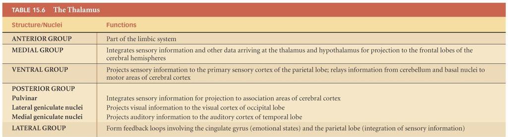
47 The Diencephalon

48 The Diencephalon Figure 15.14a The Hypothalamus (Midsagittal Section)

49 The Diencephalon Figure 15.14b The Hypothalamus
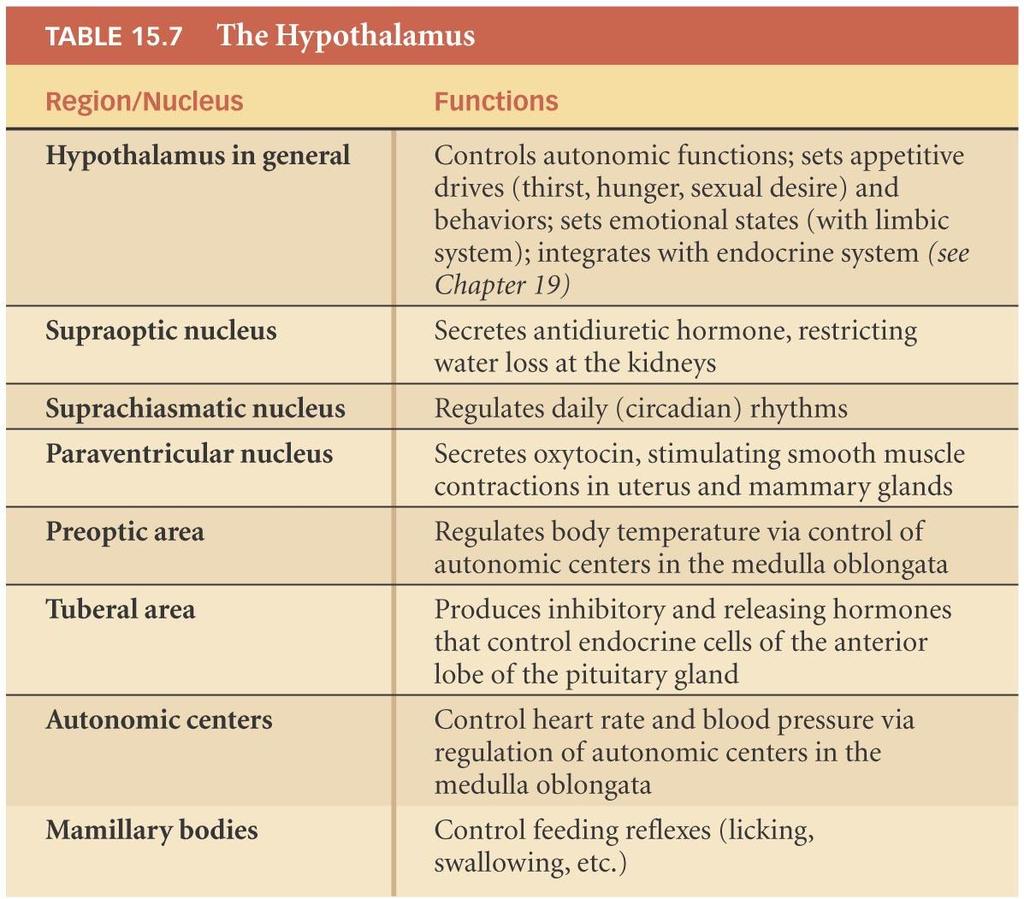
50 The Diencephalon
51 The Mesencephalon The mesencephalon, or midbrain, is the most superior portion of the brain stem. Nuclei coordinate visual and auditory reflexes. Corpora quadregemina Superior colliculi visual Inferior colliculi auditory Limbic system nuclei Coordinate involuntary movements of skeletal muscles Cerebral peduncles Nerve bundles to and from the brain/spinal cord

52 The Mesencephalon Figure 15.16a The Diencephalon and Brain Stem (Lateral View)

53 The Mesencephalon Figure 15.16b The Diencephalon and Brain Stem (Sagittal Section)

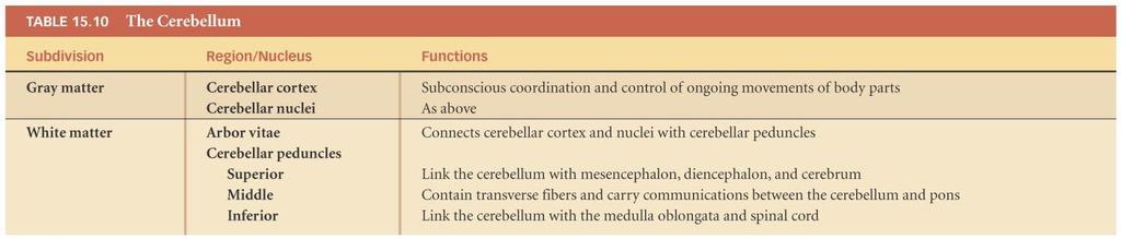
54 The Mesencephalon Figure 15.16c The Diencephalon and Brain Stem (Posterior View)

55 The Mesencephalon Figure 15.16d The Diencephalon and Brain Stem (Posterior View)

56 The Mesencephalon Figure 15.17a The Mesencephalon (Transverse Section, Superior View)

57 The Mesencephalon Figure 15.17b The Mesencephalon (Posterior View)
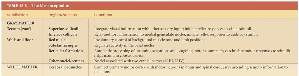
58 The Mesencephalon
59 The Pons The pons mainly functions: As a house for cranial nerve nuclei V, VI, VII, and VIII To help regulate respiration To help coordinate involuntary skeletal muscle movements and muscle tone In relaying information to and from the brain/spinal cord

60 The Pons Figure The Pons
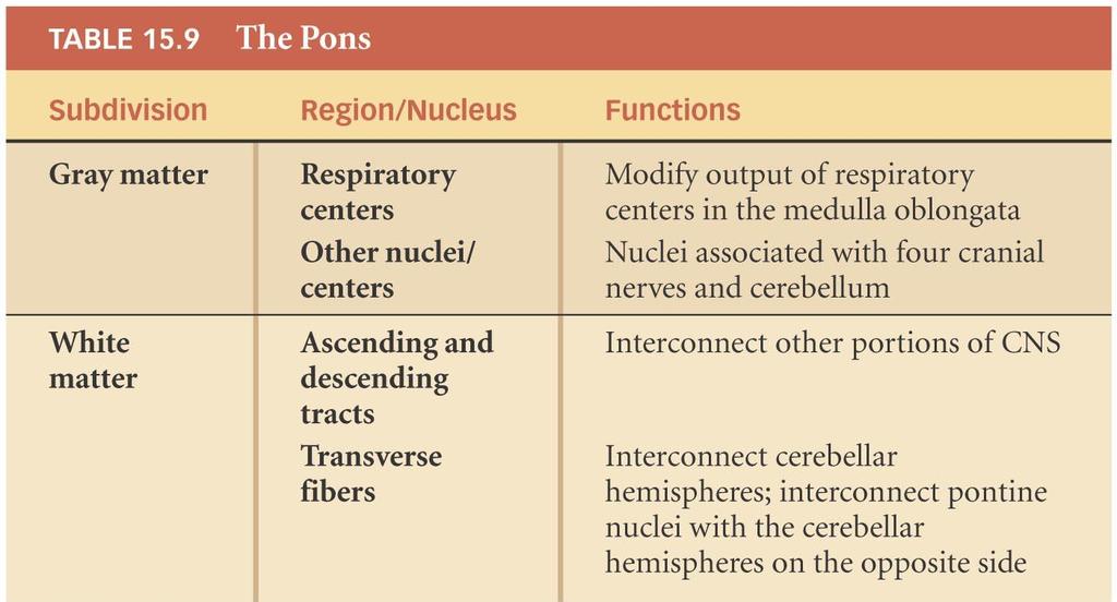
61 The Pons
62 The Cerebellum The cerebellum has two primary functions: Adjusts the postural muscles of the body to maintain balance Programs and fine-tunes voluntary and involuntary movements

63 The Cerebellum Figure 15.19a The Cerebellum (Posterior, Superior Surface)

64 The Cerebellum Figure 15.19b The Cerebellum (Sagittal Section)
65 The Cerebellum
66 The Medulla Oblongata The medulla oblongata physically connects the brain with the spinal cord. It is so important that, if it is severely compromised, the victim will likely die. The medulla oblongata is a relay station, house for cranial nerve nuclei, and most importantly, controls visceral functions like blood pressure, breathing, and heart rate.

67 The Medulla Oblongata Figure The Medulla Oblongata

68 The Medulla Oblongata

69 MBrain Animation Review The Brain
70 The Cranial Nerves Cranial nerves are components of the peripheral nervous system that connect to the brain rather than to the spinal cord. Twelve pairs of cranial nerves Cranial nerves are numbered using Roman numerals Each cranial nerve attaches to the brain near the associated sensory or motor nuclei

71 The Cranial Nerves Figure 15.21a Origins of the Cranial Nerves

72 The Cranial Nerves Figure 15.21b Origins of the Cranial Nerves

73 The Cranial Nerves Figure 15.21c Origins of the Cranial Nerves (Superior View)

74 The Cranial Nerves

75 The Cranial Nerves
76 The Cranial Nerves Olfactory Nerve (N I) Primary function: special sensory (smell) Origin: receptors of olfactory epithelium Passes through: cribriform plate of ethmoid Destination: olfactory bulbs

77 The Cranial Nerves Figure The Olfactory Nerve
78 The Cranial Nerves The Optic Nerve (N II) Primary function: special sensory (vision) Origin: retina of eye Passes through: optic canal of sphenoid Destination: diencephalon by way of the optic chiasm

79 The Cranial Nerves Figure The Optic Nerve
80 The Cranial Nerves The Oculomotor Nerve (N III) Primary function: motor, eye movements Origin: mesencephalon Passes through: superior orbital fissure of sphenoid Destination: Somatic motor: superior, inferior, and medial rectus muscles; the inferior oblique muscle; the levator palpebrae superioris muscle Visceral motor: intrinsic eye muscles
81 The Cranial Nerves Figure Cranial Nerves Controlling the Extra-Ocular Muscles
82 The Cranial Nerves The Trochlear Nerve (N IV) Primary function: motor, eye movements Origin: mesencephalon Passes through: superior orbital fissure of sphenoid Destination: superior oblique muscle
83 The Cranial Nerves Figure Cranial Nerves Controlling the Extra-Ocular Muscles
84 The Cranial Nerves The Trigeminal Nerve (N V) Primary function: Mixed (sensory and motor) Ophthalmic and maxillary branches sensory Mandibular branch mixed Origin: Ophthalmic branch (sensory): orbital structures, nasal cavity, skin of forehead, superior eyelid, eyebrow, and part of the nose Maxillary branch (sensory): inferior eyelid, upper lip, gums, and teeth; cheek; nose, palate, and part of the pharynx Mandibular branch (mixed): sensory from lower gums, teeth, and lips; palate and tongue (part); motor from motor nuclei of pons
85 The Cranial Nerves The Trigeminal Nerve (N V) Passes through: Ophthalmic branch through superior orbital fissure Maxillary branch through foramen rotundum Mandibular branch through foramen ovale Destination: Ophthalmic, maxillary, and mandibular branches to sensory nuclei in the pons Mandibular branch also innervates muscles of mastication

86 The Cranial Nerves Figure The Trigeminal Nerve
87 The Cranial Nerves The Abducens Nerve (N VI) Primary function: motor, eye movements Origin: pons Passes through: superior orbital fissure of sphenoid Destination: lateral rectus muscle
88 The Cranial Nerves Figure Cranial Nerves Controlling the Extra-Ocular Muscles
89 The Cranial Nerves The Facial Nerve (N VII) Primary function: mixed (sensory and motor) Origin: Sensory from taste receptors on anterior two thirds of tongue Motor from motor nuclei of pons Passes through: internal acoustic meatus of temporal bone, along facial canal to reach stylomastoid foramen Destination: Sensory to sensory nuclei of pons Somatic motor: muscles of facial expression Visceral motor: lacrimal (tear) gland and nasal mucous glands via pterygopalatine ganglion; submandibular and sublingual salivary glands via submandibular ganglion

90 The Cranial Nerves Figure 15.26a The Facial Nerve

91 The Cranial Nerves Figure 15.26b The Facial Nerve
92 The Cranial Nerves The Vestibulocochlear Nerve (N VIII) Primary function: special sensory: balance and equilibrium (vestibular branch) and hearing (cochlear branch) Origin: receptors of the inner ear (vestibule and cochlea) Passes through: internal acoustic meatus of the temporal bone Destination: vestibular and cochlear nuclei of pons and medulla oblongata

93 The Cranial Nerves Figure The Vestibulocochlear Nerve
94 The Cranial Nerves The Glossopharyngeal Nerve (N IX) Primary function: mixed (sensory and motor) Origin: Sensory from posterior one third of the tongue, part of the pharynx and palate, the carotid arteries of the neck Motor from motor nuclei of medulla oblongata Passes through: jugular foramen between occipital and temporal bones Destination: Sensory fibers to sensory nuclei of medulla oblongata Somatic motor: pharyngeal muscles involved in swallowing Visceral motor: parotid salivary gland, after synapsing in the otic ganglion

95 The Cranial Nerves Figure The Glossopharyngeal Nerve
96 The Cranial Nerves The Vagus Nerve (N X) Primary function: mixed (sensory and motor) Origin: Visceral sensory from pharynx (part), auricle, external acoustic meatus, diaphragm, and visceral organs in thoracic and abdominopelvic cavities Visceral motor from motor nuclei in the medulla oblongata Passes through: jugular foramen between occipital and temporal bones Destination: Sensory fibers to sensory nuclei and autonomic centers of medulla oblongata Somatic motor to muscles of the palate and pharynx Visceral motor to respiratory, cardiovascular, and digestive organs in the thoracic and abdominal cavities.

97 The Cranial Nerves Figure The Vagus Nerve
98 The Cranial Nerves The Accessory Nerve (N XI) Primary function: motor Origin: motor nuclei of spinal cord and medulla oblongata Passes through: jugular foramen between occipital and temporal bones Destination: Internal branch innervates voluntary muscles of palate, pharynx, and larynx External branch controls sternocleidomastoid and trapezius muscles
99 The Cranial Nerves The Hypoglossal Nerve (XII) Primary function: motor, tongue movements Origin: motor nuclei of the medulla oblongata Passes through: hypoglossal canal of occipital bone Destination: muscles of the tongue

100 The Cranial Nerves Figure The Accessory and Hypoglossal Nerves

101 The Cranial Nerves
ACTIVITY 7: NERVOUS SYSTEM HISTOLOGY, BRAIN, CRANIAL NERVES
 ACTIVITY 7: NERVOUS SYSTEM HISTOLOGY, BRAIN, CRANIAL NERVES LABORATORY OBJECTIVES: 1. Histology: Identify structures indicated on three different slides or images of nervous system tissue. These images
Blood supply to the brain Blood brain barrier isolates neural tissue from general circulation
 The Brain and Cranial Nerves Objectives Name the major regions of the brain and describe their functions. Discuss the formation, circulation, and functions of the CSF. List the main components of the medulla
Cranial nerves.
 Cranial nerves eaglezhyxzy@163.com Key Points of Learning Name Components Passing through Peripheral distribution Central connection Function Cranial nerves Ⅰ olfactory Ⅱ optic Ⅲ occulomotor Ⅳ trochlear
Brain and spinal nerve. By: shirin Kashfi
 Brain and spinal nerve By: shirin Kashfi Nervous system: central nervous system (CNS) peripheral nervous system (PNS) Brain (cranial) nerves Spinal nerves Ganglions (dorsal root ganglions, sympathetic
Principles of Anatomy and Physiology
 Principles of Anatomy and Physiology 14 th Edition CHAPTER 14 The Brain and Cranial Nerves Introduction The purpose of the chapter is to: 1. Understand how the brain is organized, protected, and supplied
Bellringer: The central nervous system is comprised of: What is the name of the outermost layer of the brain? a. Brain. b.
 Bellringer: The central is comprised of: a. Brain b. Spinal cord c. Sensory receptors d. Both a and b What is the name of the outermost layer of the brain? a. Pia mater b. Dura mater c. Arachnoid d. Pons
Biology 323 Human Anatomy for Biology Majors Week 10; Lecture 1; Tuesday Dr. Stuart S. Sumida. Cranial Nerves and Soft Tissues of the Skull
 Biology 323 Human Anatomy for Biology Majors Week 10; Lecture 1; Tuesday Dr. Stuart S. Sumida Cranial Nerves and Soft Tissues of the Skull FOREBRAIN MIDBRAIN HINDBRAIN Forebrain: Cerebrum Perception,
The Nervous System PART B
 7 The Nervous System PART B PowerPoint Lecture Slide Presentation by Jerry L. Cook, Sam Houston University ESSENTIALS OF HUMAN ANATOMY & PHYSIOLOGY EIGHTH EDITION ELAINE N. MARIEB Central Nervous System
Brain and Cranial Nerves (Ch. 15) Human Anatomy lecture. caudal = toward the spinal cord)
 Human Anatomy lecture. caudal = toward the spinal cord)") Insight: Some cranial nerve disorders Brain and Cranial Nerves (Ch. 15) Human Anatomy lecture I. Overview (Directional terms: rostral = toward the forehead caudal = toward the spinal cord) A. 3 Major parts
Insight: Some cranial nerve disorders Brain and Cranial Nerves (Ch. 15) Human Anatomy lecture I. Overview (Directional terms: rostral = toward the forehead caudal = toward the spinal cord) A. 3 Major parts
b. The groove between the two crests is called 2. The neural folds move toward each other & the fuse to create a
 Chapter 13: Brain and Cranial Nerves I. Development of the CNS A. The CNS begins as a flat plate called the B. The process proceeds as: 1. The lateral sides of the become elevated as waves called a. The
ACTIVITY 7: NERVOUS SYSTEM HISTOLOGY, BRAIN, CRANIAL NERVES NERVOUS SYSTEM TISSUES: HISTOLOGY SLIDES
 ACTIVITY 7: NERVOUS SYSTEM HISTOLOGY, BRAIN, CRANIAL NERVES OBJECTIVES: 1) How to get ready: Read Chapter 14 & 15 McKinley et al., Human Anatomy, 4e. All text references are for this textbook. Read dissection
This lab activity is aligned with Visible Body s Human Anatomy Atlas app.
 1 This lab activity is aligned with Visible Body s Human Anatomy Atlas app. Learn more at visiblebody.com/professors We've split our Cranial Nerves lab activity into two parts. Part 1 is pre-lab exercises
Protection of the Brain. Overview of the Brain. Visual Anatomy & Physiology First Edition. Martini & Ober. Chapter 13. Lecture 20
 Visual Anatomy & Physiology First Edition Martini & Ober Chapter 13 Brain and Cranial Nerves Lecture 20 1 Overview of the Brain Functions Major Parts regulates visceral activities cerebrum (two hemispheres)
Cranial Nerves. Steven McLoon Department of Neuroscience University of Minnesota
 Cranial Nerves Steven McLoon Department of Neuroscience University of Minnesota 1 Course News Change in Lab Sequence Week of Oct 2 Lab 5 Week of Oct 9 Lab 4 2 Sensory and Motor Systems Sensory Systems:
PERIPHERAL NERVOUS SYSTEM
 CHAPTER 13 PERIPHERAL NERVOUS SYSTEM Functional division of nervous system = afferent info to the CNS ascending spinal cord = efferent info from CNS descending spinal cord somatic skin, muscles visceral
Introduction to Head and Neck Anatomy
 Introduction to Head and Neck Anatomy Nervous Tissue Controls and integrates all body activities within limits that maintain life Three basic functions 1. sensing changes with sensory receptors 2. interpreting
Peripheral Nervous System
 Peripheral Nervous System Sensory Receptors Motor Endings Cranial Nerves The Four Plexuses Extremities Review of Reflexes Fast, preprogrammed, inborn, automatic responses Occur in the CNS at the spinal
Nervous System. Student Learning Objectives:
 Nervous System Student Learning Objectives: Identify the primary parts of the neuron Identify the major structures of the central nervous system Identify the major structures of the peripheral nervous
Chapter 14: The Brain and Cranial Nerves. Copyright 2009, John Wiley & Sons, Inc.
 Chapter 14: The Brain and Cranial Nerves Development of the Brain Three to four-week embryo: prosencephalon, mesencephalon and rhombencephalon. Five-week embryo: telencephalon (cerebrum), diencephalon
CRANIAL NERVES. Dr. Amani A. Elfaki Associate Professor Department of Anatomy
 CRANIAL NERVES Dr. Amani A. Elfaki Associate Professor Department of Anatomy LEARNING OBJECTIVES Named the cranial nerves Identify the funcunal component of each cranial nerve Identify the effect of each
Ms. K. GOWRI. M.Pharm., Lecturer.
 Ms. K. GOWRI. M.Pharm., Lecturer. CENTRAL NERVOUS SYSTEM (CNS) central nervous system consists of brain and spinal cord membrane covering the brain and spinal cord are surrounded by three membrane Meninges
Unit VIII Problem 3 Neuroanatomy: Brain Stem, Cranial Nerves and Scalp
 Unit VIII Problem 3 Neuroanatomy: Brain Stem, Cranial Nerves and Scalp - Brain stem: It is connected to the cerebellum and cerebral hemispheres. Rostral end of brain stem: diencephalon is the area which
Chapter 10 The Nervous System: The Brain and Cranial Nerves
 Chapter 10 The Nervous System: The Brain and Cranial Nerves Copyright 2015 Wolters Kluwer Health Lippincott Williams & Wilkins Overview Key Terms aphasia corpus callosum meninges basal nuclei diencephalon
Instructor s Review for Final Exams. The Nervous System
 Instructor s Review for Final Exams The Nervous System Divisions of the Central Nervous System? Brain and spinal cord. Key word, central. Divisions of the nervous system Central and Peripheral Coverings
Lab 16: PNS: Nerves and Autonomic NS Hamilton Answers to Pre- Lab Assignments
 Lab 16: PNS: Nerves and Autonomic NS Hamilton Answers to Pre- Lab Assignments Pre-Lab Activity 1: 1. a. olfactory nerve b. optic nerve c. oculomotor nerve d. abducens nerve e. trochlear nerve f. trigeminal
A&P 1 Brain & Cranial Nerves Guide #1 - Pre-Lab Exercises
 A&P 1 Brain & Cranial Nerves Guide #1 - Pre-Lab Exercises In this "Pre-lab Guide", we will be looking at the brain & cranial nerves. This should be done before lab, so we don't waste time in lab! This
The Brain and Cranial Nerves
 14 The Brain and Cranial Nerves PowerPoint Lecture Presentations prepared by Jason LaPres Lone Star College North Harris An Introduction to the Brain and Cranial Nerves The Adult Human Brain Average weight
The Nervous System PART B
 7 The Nervous System PART B PowerPoint Lecture Slide Presentation by Jerry L. Cook, Sam Houston University ESSENTIALS OF HUMAN ANATOMY & PHYSIOLOGY EIGHTH EDITION ELAINE N. MARIEB The Reflex Arc Reflex
The Nervous System PART C. PowerPoint Lecture Slide Presentation by Patty Bostwick-Taylor, Florence-Darlington Technical College
 PowerPoint Lecture Slide Presentation by Patty Bostwick-Taylor, Florence-Darlington Technical College The Nervous System 7 PART C Protection of the Central Nervous System Scalp and skin Skull and vertebral
TRANSVERSE SECTION PLANE Scalp 2. Cranium. 13. Superior sagittal sinus
 TRANSVERSE SECTION PLANE 1 1. Scalp 2. Cranium 3. Superior sagittal sinus 4. Dura mater 5. Falx cerebri 6. Frontal lobes of the cerebrum 7. Middle meningeal artery 8. Cortex, grey matter 9. Cerebral vessels
The Brain and Cranial Nerves Pg. 129
 The Brain and Cranial Nerves Pg. 129 Three Main Regions of the Brain Forebrain Cerbral hemispheres Diencephalon Midbrain Brain stem Hindbrain Pons Cerebellum Medulla oblongata Forebrain Interprets sensory
Read Chapter 14 & 15 McKinley et al
 ACTIVITY 7: NERVOUS SYSTEM HISTOLOGY, BRAIN, CRANIAL NERVES OBJECTIVES: 1) How to get ready: Read Chapter 14 & 15 McKinley et al., Human Anatomy, 5e. All text references are for this textbook. Read dissection
Major Anatomic Components of the Orbit
 Major Anatomic Components of the Orbit 1. Osseous Framework 2. Globe 3. Optic nerve and sheath 4. Extraocular muscles Bony Orbit Seven Bones Frontal bone Zygomatic bone Maxillary bone Ethmoid bone Sphenoid
Nervous System: An Introduction. HAP Susan Chabot Lemon Bay High School
 Nervous System: An Introduction HAP Susan Chabot Lemon Bay High School Function of the Nervous System 3 overlapping functions SENSORY INPUT - Monitor changes inside and outside of the body; these changes
The Brain and Cranial Nerves Pg Three Main Regions of the Brain. Forebrain
 The Brain and Cranial Nerves Pg. 129 Three Main Regions of the Brain Forebrain Cerbral hemispheres Diencephalon Midbrain Brain stem Hindbrain Pons Cerebellum Medulla oblongata Interprets sensory inputs
Organization of The Nervous System PROF. MOUSAED ALFAYEZ & DR. SANAA ALSHAARAWY
 Organization of The Nervous System PROF. MOUSAED ALFAYEZ & DR. SANAA ALSHAARAWY Objectives At the end of the lecture, the students should be able to: List the parts of the nervous system. List the function
Tracing the Cranial Nerves Osteologically
 CN I II III IV V 1 Supra-orbital ethmoidal nn. Ext. nasal V 2 Tracing the Cranial Nerves Osteologically Nucleus of Origin Olfactory tracts of frontal lobe of cerebrum Optic tracts from optic chiasma and
Unit 12a: The Nervous System The Brain. MDL231 Principle of Anatomy
 Unit 12a: The Nervous System The Brain MDL231 Principle of Anatomy The Brain - Overview Cerebrum T PP H midbrain Cerebellum pons m.o. Brain stem medulla oblongata (M.O.) pons midbrain (mesencephalon) Diencephalon
Unit 18: Cranial Cavity and Contents
 Unit 18: Cranial Cavity and Contents Dissection Instructions: The calvaria is to be removed without damage to the dura mater which is attached to the inner surface of the calvaria. Cut through the outer
M555 Medical Neuroscience Lab 1: Gross Anatomy of Brain, Crainal Nerves and Cerebral Blood Vessels
 M555 Medical Neuroscience Lab 1: Gross Anatomy of Brain, Crainal Nerves and Cerebral Blood Vessels Anatomical Directions Terms like dorsal, ventral, and posterior provide a means of locating structures
Nervous System The Brain and Spinal Cord Unit 7b
 Nervous System The Brain and Spinal Cord Unit 7b Chetek High School Mrs. Michaelsen 9.12 Meninges A. Meninges 1. The organs of the CNS are covered by membranes a. The meninges are divided into 3 layers:
14: The Brain and Cranial Nerves
 14: The Brain and Cranial Nerves I. An Introduction to the Organization of the Brain, p. 452 Objectives 1. Name the major regions of the brain and describe their functions 2. Name the three primary brain
Brainstem. Telencephalon Diencephalon Cerebellum Brain stem
 Brainstem Brainstem 脑 脊髓 Brainstem Telencephalon Diencephalon Cerebellum Brain stem Ventral view Lateral view 10 pairs of the cranial nerves are attached to the brain stem The brainstem Midbrain Pons Medulla
4 main parts 1) Cerebrum 2) Diencephalon 3) Brain stem 4) Cerebellum
 Cerebrum 2) Diencephalon 3) Brain stem 4) Cerebellum") 4 main parts 1) Cerebrum 2) Diencephalon 3) Brain stem 4) Cerebellum White Matter = myelinated tracts or nerves Gray Matter = unmyelinated tracts or nerves Brain: gray matter on outside, white matter inside
4 main parts 1) Cerebrum 2) Diencephalon 3) Brain stem 4) Cerebellum White Matter = myelinated tracts or nerves Gray Matter = unmyelinated tracts or nerves Brain: gray matter on outside, white matter inside
Cranial nerve Dept. of Anatomy Zhou Hong Ying
 Cranial nerve Dept. of Anatomy Zhou Hong Ying Key Points of Learning Name Components Passing through Peripheral distribution Central connection Function Cranial nerve Ⅰ olfactory Ⅱ optic Ⅲ occulomotor
Brainstem. By Dr. Bhushan R. Kavimandan
 Brainstem By Dr. Bhushan R. Kavimandan Development Ventricles in brainstem Mesencephalon cerebral aqueduct Metencephalon 4 th ventricle Mylencephalon 4 th ventricle Corpus callosum Posterior commissure
Organization of The Nervous System PROF. SAEED ABUEL MAKAREM
 Organization of The Nervous System PROF. SAEED ABUEL MAKAREM Objectives By the end of the lecture, you should be able to: List the parts of the nervous system. List the function of the nervous system.
human anatomy 2016 lecture fifteen Dr meethak ali ahmed neurosurgeon
 Cranial Nerves Organization of the Cranial Nerves The cranial nerves are named as follows: I. Olfactory II. Optic III. Oculomotor IV. Trochlear V. Trigeminal VI. Abducent VII. Facial VIII. Vestibulocochlear
The Nervous System: Central Nervous System
 The Nervous System: Central Nervous System I. Anatomy of the nervous system A. The CNS & the body by: 1. monitoring of the body 2. & information between parts of the body 3. acting as a to gather, store,
I. Anatomy of the Brain A. Cranial Meninges and Ventricles of the Brain 1. Meninges a. Dura mater 1) Endosteal/Periosteal Layer - Outer 2) Meningeal
 Endosteal/Periosteal Layer - Outer 2) Meningeal") I. Anatomy of the Brain A. Cranial Meninges and Ventricles of the Brain 1. Meninges a. Dura mater 1) Endosteal/Periosteal Layer - Outer 2) Meningeal Layer - Inner 3) Falx cerebri a) Superior sagittal sinus
I. Anatomy of the Brain A. Cranial Meninges and Ventricles of the Brain 1. Meninges a. Dura mater 1) Endosteal/Periosteal Layer - Outer 2) Meningeal Layer - Inner 3) Falx cerebri a) Superior sagittal sinus
Group D: Central nervous system yellow
 Group D: Central nervous system yellow Central nervous system 1. General structure of nervous system (neuron, glia, synapsis, mediators, receptors) Main points: types of neurons and glial cells, synapses,
Cranial Cavity REFERENCES: OBJECTIVES OSTEOLOGY. Stephen A. Gudas, PT, PhD
 Stephen A. Gudas, PT, PhD Cranial Cavity REFERENCES: Moore and Agur, Essential Clinical Anatomy (ECA), 3rd ed., pp. 496 498; 500 507; 512 514 Grant s Atlas 12 th ed., Figs 7.6; 7.19 7.30. Grant s Dissector
Laboratory Manual for Comparative Anatomy and Physiology Figure 15.1 Transparency Master 114
 Neuron Capillary Astrocyte Microglial cell Neuron Fluid-filled cavity Process of oligodendrocyte Ependymal cells Brain or spinal cord tissue Myelin sheath Nerve fibers Figure 15.1 Transparency Master 114
INTRODUCTION: ANATOMY UNDERLYING CLINICAL TESTS OF CRANIAL NERVES
 INTRODUCTION: ANATOMY UNDERLYING CLINICAL TESTS OF CRANIAL NERVES CRANIAL NERVE I - OLFACTORY I - OLFACTORY NERVE - SMELL TEST: SMELL ODORS (note: not ammonia; pain in nasal cavity CN5 DAMAGE: LOSS OF
Note to Self. The Brain and Cranial Nerves. Organization of the Brain
 Note to Self Despite what you think you can get through this chapter in 1 class period, or one with only a few slides left over The Brain and Cranial Nerves Chapter 14 Organization of the Brain The brain
Course: Physical Assessment II Date: October 17, 2008 Doc: Practice Quiz 1
 Course: Physical Assessment II Date: October 17, 2008 Doc: Practice Quiz 1 This is the practice quiz we did in Class 4. The answers are at the end of the quiz should you wish to test yourself. Complete
CHAPTER 11: NERVOUS SYSTEM II: DIVISIONS OF THE NERVOUS SYSTEM. 1. Outline the major divisions of the nervous system.
 CHAPTER 11: NERVOUS II: DIVISIONS OF THE NERVOUS OBJECTIVES: 1. Outline the major divisions of the nervous system. NERVOUS CENTRAL NERVOUS (BRAIN & SPINAL CORD) (INTERNEURONS) PERIPHERAL NERVOUS (CRANIAL
Biological Bases of Behavior. 3: Structure of the Nervous System
 Biological Bases of Behavior 3: Structure of the Nervous System Neuroanatomy Terms The neuraxis is an imaginary line drawn through the spinal cord up to the front of the brain Anatomical directions are
BRAIN PART I (A & B): VENTRICLES & MENINGES
: VENTRICLES & MENINGES") BRAIN PART I (A & B): VENTRICLES & MENINGES Cranial Meninges Cranial meninges are continuous with spinal meninges Dura mater: inner layer (meningeal layer) outer layer (endosteal layer) fused to periosteum
BRAIN PART I (A & B): VENTRICLES & MENINGES Cranial Meninges Cranial meninges are continuous with spinal meninges Dura mater: inner layer (meningeal layer) outer layer (endosteal layer) fused to periosteum
Cranial Nerves and Spinal Cord Flashcards
 1. Name the cranial nerves and their Roman numeral. 2. What is Cranial Nerve I called, and what does it 3. Scientists who are trying to find a way to make neurons divide to heal nerve injuries often study
Peripheral Nervous System Dr. Gary Mumaugh
 Peripheral Nervous System Dr. Gary Mumaugh Spinal Nerves Overview Thirty-one pairs of spinal nerves are connected to the spinal cord No special names; numbered by level of vertebral column at which they
General Sensory Pathways of the Face Area, Taste Pathways and Hearing Pathways
 General Sensory Pathways of the Face Area, Taste Pathways and Hearing Pathways Lecture Objectives Describe pathways for general sensations (pain, temperature, touch and proprioception) from the face area.
Brain ميهاربا لض اف دمح ا د The Meninges 1- Dura Mater of the Brain endosteal layer does not extend meningeal layer falx cerebri tentorium cerebelli
 .احمد د فاضل ابراهيم Lecture 15 Brain The Meninges Three protective membranes or meninges surround the brain in the skull: the dura mater, the arachnoid mater, and the pia mater 1- Dura Mater of the Brain
Lecture - Chapter 13: Central Nervous System
 Lecture - Chapter 13: Central Nervous System 1. Describe the following structures of the brain, what is the general function of each: a. Cerebrum b. Diencephalon c. Brain Stem d. Cerebellum 2. What structures
PTERYGOPALATINE FOSSA
 PTERYGOPALATINE FOSSA Outline Anatomical Structure and Boundaries Foramina and Communications with other spaces and cavities Contents Pterygopalatine Ganglion Especial emphasis on certain arteries and
Nervous and Endocrine System Exam Review
 Directions: Read each question and complete the statement using the multiple choice responses I. Nervous System 1. The interpretation of olfactory receptor information would fall under which general function
Anatomy and Physiology (Bio 220) The Brain Chapter 14 and select portions of Chapter 16
 The Brain Chapter 14 and select portions of Chapter 16") Anatomy and Physiology (Bio 220) The Brain Chapter 14 and select portions of Chapter 16 I. Introduction A. Appearance 1. physical 2. weight 3. relative weight B. Major parts of the brain 1. cerebrum 2.
Anatomy and Physiology (Bio 220) The Brain Chapter 14 and select portions of Chapter 16 I. Introduction A. Appearance 1. physical 2. weight 3. relative weight B. Major parts of the brain 1. cerebrum 2.
The Nervous System 7PART B. PowerPoint Lecture Slide Presentation by Patty Bostwick-Taylor, Florence-Darlington Technical College
 PowerPoint Lecture Slide Presentation by Patty Bostwick-Taylor, Florence-Darlington Technical College The Nervous System 7PART B What is a reflex? What is a reflex? What is meant by the statement that
Biology 340 Comparative Embryology Lecture 11 Dr. Stuart Sumida. Overview of Embryology of the Vertebrate Skull. Emphasis on Amniota
 Biology 340 Comparative Embryology Lecture 11 Dr. Stuart Sumida Overview of Embryology of the Vertebrate Skull Emphasis on Amniota Initial introduction to components parts of a vertebrate head. This lecture
Structure Location Function
 Frontal Bone Cranium forms the forehead and roof of the orbits Occipital Bone Cranium forms posterior and inferior portions of the cranium Temporal Bone Cranium inferior to the parietal bone forms the
PHYSIOLOHY OF BRAIN STEM
 PHYSIOLOHY OF BRAIN STEM Learning Objectives The brain stem is the lower part of the brain. It is adjoining and structurally continuous with the spinal cord. 1 Mid Brain 2 Pons 3 Medulla Oblongata The
Anatomy and Physiology. Bones, Sutures, Teeth, Processes and Foramina of the Human Skull
 Anatomy and Physiology Chapter 6 DRO Bones, Sutures, Teeth, Processes and Foramina of the Human Skull Name: Period: Bones of the Human Skull Bones of the Cranium: Frontal bone: forms the forehead and the
Brainstem and Cranial Nerves II. Nerves covered in other lectures. A reminder about embryology. Prof. Stuart Bunt
 Brainstem and Cranial Nerves II Prof. Stuart Bunt Nerves covered in other lectures 1 Olfactory 2 Optic 3,4,6 Extraocular eye muscles 8 Vestibulo-cochlear 5 Motor and Sensory to the face and muscles of
Ch 13: Central Nervous System Part 1: The Brain p 374
 Ch 13: Central Nervous System Part 1: The Brain p 374 Discuss the organization of the brain, including the major structures and how they relate to one another! Review the meninges of the spinal cord and
By : Prof Saeed Abuel Makarem & Dr.Sanaa Alshaarawi
 By : Prof Saeed Abuel Makarem & Dr.Sanaa Alshaarawi OBJECTIVES By the end of the lecture, students shouldbe able to: List the nuclei of the deep origin of the trigeminal and facial nerves in the brain
HEAD AND NECK ANATOMY PRACTICE QUESTIONS
 HEAD AND NECK ANATOMY PRACTICE QUESTIONS 1. A patient complains that he has lost sensation on his face and that the skin of his face feels numb. The physician tests tactile acuity by touching the forehead
The Brain and Cranial Nerves Student Objectives
 The Brain and Cranial Nerves Student Objectives Chapter 14 Textbook and Laboratory Manual Name the major regions of the brain and describe their functions Name the ventricles of the brain and describe
Sheep Brain Dissection
 Sheep Brain Dissection Mammalian brains have many features in common. Human brains may not be available, so sheep brains often are dissected as an aid to understanding the mammalian brain since he general
Lab 10: Muscle Tissue and Selected Muscles
 117 Ex. 10: Histology of Muscle Muscle Tissue Lab 10: Muscle Tissue and Selected Muscles Skeletal muscle: Cardiac muscle: Smooth muscle MUSCLE LIST Ex. 11: Gross Anatomy of Muscle Locate these muscles
Chapter 14: Nervous System Guided Notes (A-day)
") Chapter 14: Nervous System Guided Notes (A-day) Nervous System Overview Major Function: Control the body's and. Divided into the Nervous System (CNS=Brain and Spinal Cord) and the Nervous System (PNS=Cranial
Chapter 14: Nervous System Guided Notes (A-day) Nervous System Overview Major Function: Control the body's and. Divided into the Nervous System (CNS=Brain and Spinal Cord) and the Nervous System (PNS=Cranial
Cranial Nerve VII - Facial Nerve. The facial nerve has 3 main components with distinct functions
 Cranial Nerve VII - Facial Nerve The facial nerve has 3 main components with distinct functions Somatic motor efferent Supplies the muscles of facial expression; posterior belly of digastric muscle; stylohyoid,
Dr. Sami Zaqout, IUG Medical School
 The skull The skull is composed of several separate bones united at immobile joints called sutures. Exceptions? Frontal bone Occipital bone Vault Cranium Sphenoid bone Zygomatic bones Base Ethmoid bone
Chapter 3. Structure and Function of the Nervous System. Copyright (c) Allyn and Bacon 2004
 Allyn and Bacon 2004") Chapter 3 Structure and Function of the Nervous System 1 Basic Features of the Nervous System Neuraxis: An imaginary line drawn through the center of the length of the central nervous system, from the
Chapter 3 Structure and Function of the Nervous System 1 Basic Features of the Nervous System Neuraxis: An imaginary line drawn through the center of the length of the central nervous system, from the
Neural Integration I: Sensory Pathways and the Somatic Nervous System
 C h a p t e r 15 Neural Integration I: Sensory Pathways and the Somatic Nervous System PowerPoint Lecture Slides prepared by Jason LaPres Lone Star College - North Harris Copyright 2009 Pearson Education,
Nervous System - PNS and CNS. Bio 105
 Nervous System - PNS and CNS Bio 105 Outline I. Central Nervous System vs Peripheral Nervous System II. Peripheral Nervous System A. Autonomic Nervous Systems B. Somatic Nervous Systems III. Autonomic
meninges Outermost layer of the meninge dura mater arachnoid mater pia mater membranes located between bone and soft tissue of the nervous system
 membranes located between bone and soft tissue of the nervous system meninges Outermost layer of the meninge dura mater middle layer of the meninges, contains no blood vessels arachnoid mater Innermost
3/15/17. Outline. Nervous System - PNS and CNS. Two Parts of the Nervous System
 Nervous System - PNS and CNS Bio 105 Outline I. Central Nervous System vs Peripheral Nervous System II. Peripheral Nervous System A. Autonomic Nervous Systems B. Somatic Nervous Systems III. Autonomic
Model 3-50B or 3-88 III VIII. Olfactory Nerve. Optic Nerve. Oculomotor Nerve. Trochlear Nerve. Trigeminal Nerve. Abducens Nerve.
 Model 3-50B or 3-88 I Olfactory Nerve II Optic Nerve Oculomotor Nerve III IV Trochlear Nerve Trigeminal Nerve V VI Abducens Nerve Glossopharyngeal Nerve IX VII Facial Nerve VIII Vestibocochlear Nerve or
stored information, making decisions, and taking action. 1. It is also the center for intellect, emotions, behavior, and memory.
 Chapter 14 - Outline I. INTRODUCTION A. The brain is the center for registering sensations, correlating them with one another and with stored information, making decisions, and taking action. 1. It is
Biology 218 Human Anatomy
 Chapter 19 Adapted form Tortora 10 th ed. LECTURE OUTLINE A. Overview of Brain Organization (p. 584) I. Principal Parts of the Brain: 1. The adult brain consists of about 100 billion neurons and 10-50
The Nervous System: Sensory and Motor Tracts of the Spinal Cord
 15 The Nervous System: Sensory and Motor Tracts of the Spinal Cord PowerPoint Lecture Presentations prepared by Steven Bassett Southeast Community College Lincoln, Nebraska Introduction Millions of sensory
Neural Integration I: Sensory Pathways and the Somatic Nervous System
 15 Neural Integration I: Sensory Pathways and the Somatic Nervous System PowerPoint Lecture Presentations prepared by Jason LaPres Lone Star College North Harris An Introduction to Sensory Pathways and
CHAPTER 13&14: The Central Nervous System. Anatomy of the CNS
 CHAPTER 13&14: The Central Nervous System Anatomy of the CNS in human consists of brain and spinal cord as stated earlier neurons have little support from their extracellular matrix and depend on glial
Stanley Pruisinger 1980's
 Neuroanatomy Prion disease cerebellum chapter b/c cerebellar ataxia here as a warning for obvious reasons. Creutzfeldt - Jakob Disease (CJD) "Spongiform" (brain turns to sponge) Jews in Lybia who ate
Lab Activity 19 & 20. Cranial Nerves General Senses. Portland Community College BI 232
 Lab Activity 19 & 20 Cranial Nerves General Senses Portland Community College BI 232 Cranial Nerves Nerves that originate from the brain rather than the spinal cord Part of the peripheral nervous system
Human Nervous System:
 OLLI Brain: Making Sense of Our World: Lecture 3 Human Nervous System: The Motor & Sensory Divisions Copyright 2004 Pearson Education, Inc., publishing as Benjamin Cummings Organization of the Nervous
Nervous System. The Peripheral Nervous System Agenda Review of CNS v. PNS PNS Basics Cranial Nerves Spinal Nerves Reflexes Pathways
 Nervous System Agenda Review of CNS v. PNS PNS Basics Cranial Nerves Spinal Nerves Sensory Motor Review of CNS v. PNS Central nervous system (CNS) Brain Spinal cord Peripheral nervous system (PNS) All
Chapter 13: The Peripheral Nervous System
 Chapter 13: The Peripheral Nervous System Objectives: 1. Define peripheral nervous system and list its components. 2. Classify general sensory receptors by structure, stimulus detected, and body location.
Faculty of Dental Medicine and Surgery. Sem 4 Cranial Nerves Dr. Abbas Garib Alla
 Faculty of Dental Medicine and Surgery Sem 4 Cranial Nerves Dr. Abbas Garib Alla Cranial Nerves I through XII FUNCTIPONAL CLSSIFICATION OF THE CN parasympathetic nerves 1973 PHARYNGEAL ARCHES nerves 1975
Bones of the skull & face
 Bones of the skull & face Cranium= brain case or helmet Copyright The McGraw-Hill Companies, Inc. Permission required for reproduction or display. The cranium is composed of eight bones : frontal Occipital