PATHOLOGY OF LIVER TUMORS
|
|
|
- Joseph Jefferson
- 6 years ago
- Views:
Transcription
1 PATHOLOGY OF LIVER TUMORS Pathobasic,
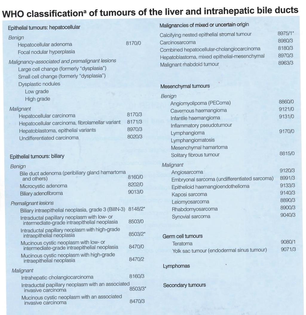
2 WHO Classification
3 Approach to a Liver Mass Lesion in a patient with chronic liver disease? Lesion in a patient without chronic liver disease?
4 Malignant Neoplasms in the Liver - Site of Origin Stom ach 4% Pancreas 2% Colon 4% Other 6% Lung 7% Liver 77% Patients with cirrhosis Melato M et al, Cancer, 1989
5 Malignant Neoplasms in the Liver - Site of Origin Other 27% Liver 2% Lung 23% Stom ach 10% Colon 13% Breast 7% Pancreas 18% No Liver Disease Melato M et al, Cancer, 1989

6 Global cause of death

7 Incidence Llovet et al Nat Rev Dis Prim 2016

8 Risk factors

9 Histopathological progression and molecular features of HCC Farazi PA, Nat Rev Cancer, 2006
10 Macroscopy 1. Expansive (nodular) 2. Infiltrative 3. Mixed Expansive and Infiltrative 4. Diffuse 5. Pedunculated

11 Expansive Macroscopy The most common type. Typically seen in association with cirrhosis

12 Infiltrative Macroscopy The lesion is poorly circumscribed with ill-defined invasive borders. This pattern is seen more commonly in non-cirrhotic livers
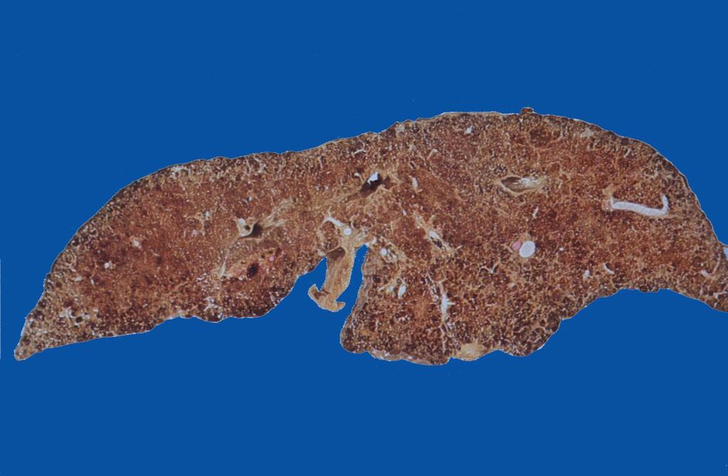
13 Diffuse HCC/cirrhotomimetic
14 Microscopy The outstanding histological feature is the resemblance of the tumor cells to normal hepatocytes 1. Trabecular (sinusoidal) pattern 2. Pseudoglandular (acinar) pattern 3. Compact (solid) pattern

15 HCC Trabecular with trabecular pattern pattern Tumor cells grow in cords of variable thickness separated by prominent sinusoids lined by flat endothelial cells

16 Pseudoglandular pattern HCC with pseudoglandular pattern A variety of gland-like structures may be seen.

17 Pseudoglandular pattern HCC with pseudoglandular pattern Bile production: the most specific marker of hepatocellular differentiation!

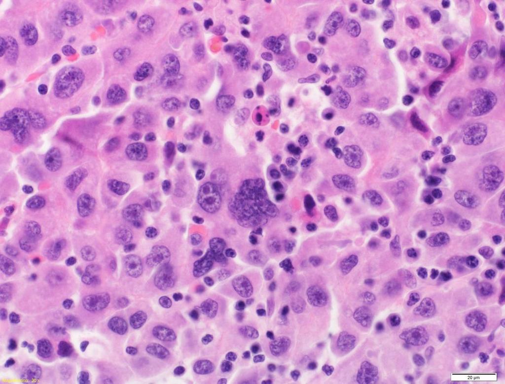
18 Solid pattern HCC with solid pattern The pattern is basically trabecular but the tumor cells apparently grow in solid masses and the sinusoids are rendered inconspicuous by compression.
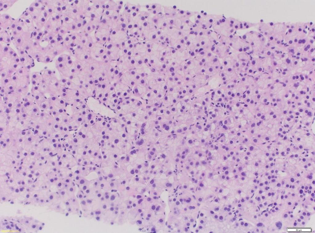
19 Histology Grading WHO Well differentiated Most common in small early stage tumors of < 2cm, mild atypia, increased nucleus-to-cytoplasma ratio Moderately differentiated Most common in tumors of > 3cm, mainly trabecular growth. Tumor cells with abundant eosinophilic cytoplasma and round nuclei. Poorly differentiated Rare in small tumours, mainly solid pattern, marked pleomorphism.
20 Histology Grading Edmondson Grade I: Best differentiated, simulates normal liver plates Grade II: Larger nucleus, prominent nuclei, eosinophilic and granular cytoplasma Grade III: Nuclei more enlarged and hyperchromatic, angulated nuclei, less abundant cytoplasm Grade IV: Marked pleomorphism, hyperchromasia, scanty cytoplasm, anaplasia loss of trabecular pattern Edmondson HA and Steiner PE, Cancer, 1954

21 Grade I Grade II Grade III Grade IV
22 Diagnosis of HCC 1) HE 2) Reticulin stain 3) Immunohistochemistry 4) (Molecular pathology)
, but the tumor shows substantial loss of")
23 Reticulin The reticulin framework is preserved in the non-neoplastic liver (left part of image), but the tumor shows substantial loss of reticulin
24 Immunohistochemistry


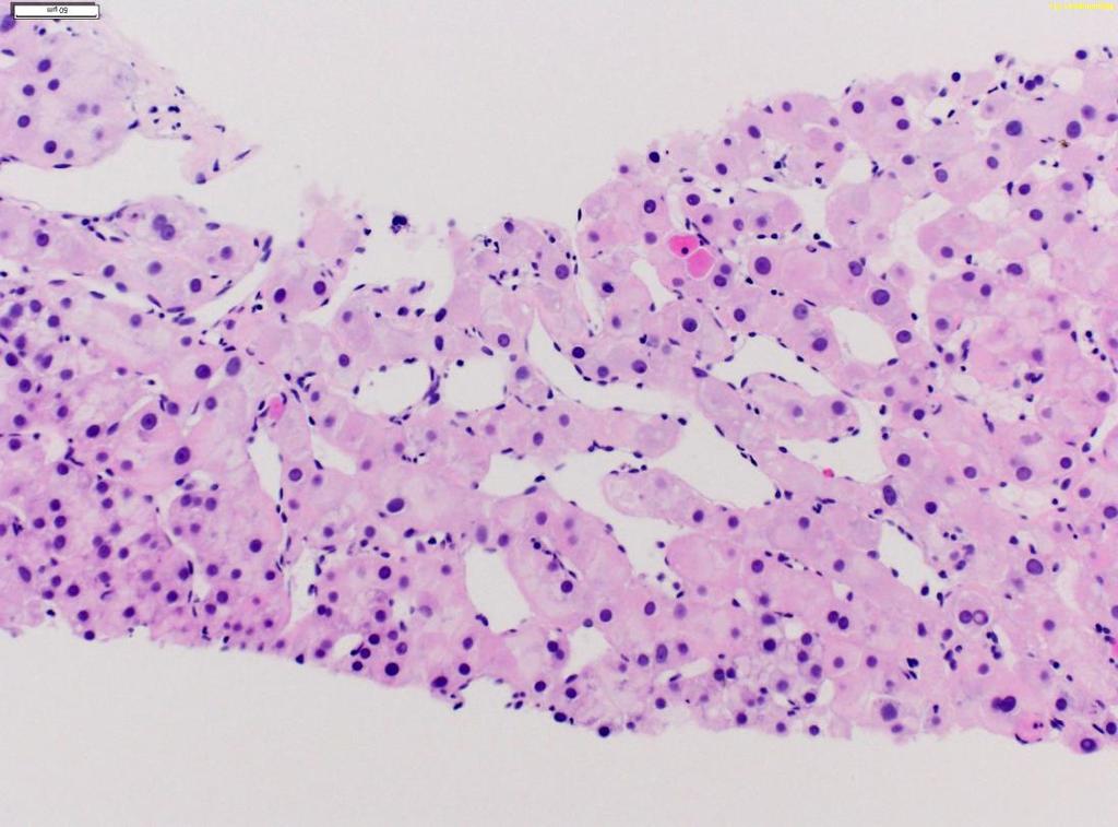
25 Immunohistochemistry hepatic origin Hep Par 1 Arginase 1 Polyclonal CEA Cave: Poorly-differentiated tumours often have aberrant staining patterns Edmondson grade: grade I-II 100% grade III 84% grade IV 50%
26 Differential Diagnosis Differential diagnosis: - Adrenocortical carcinoma - Angiomyolipom - Renal cell carcinoma - Clear cell carcinoma - Melanoma - Large cell neuroendocrine carcinoma
27 Special Types of HCC Fibrolamellar carcinoma Scirrhous HCC Undifferentiated carcinoma Lymphoepithelioma-like carcinoma Sarcomatoid carcinoma
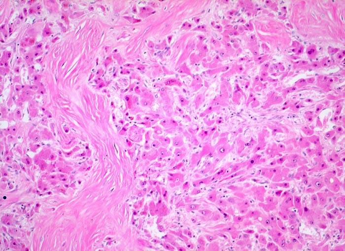
28 Fibrolamellar HCC Mainly in children and young adults (Ø 25 years old) 0.5%-9.0% of primary liver cancers Arises in non-cirrhotic liver Etiology and risk factors are unknown Large polygonal cells with abundant eosinophilic (oncocytic) cytoplasm, large vesicular nuclei and large nucleoli Lamellar fibrosis
29 Fibrolamellar HCC

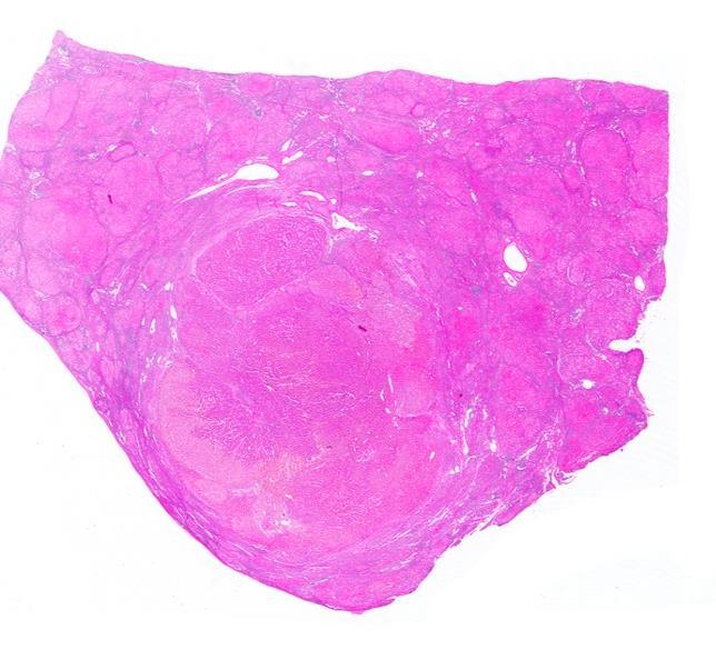
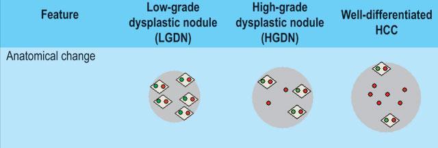
30 Precursor lesions for HCC Macrogenerative Nodule LGDN HGDN Early HCC
31 Precursor lesions for HCC Macroregenerative Nodule/Low grade dysplastic nodule Tumor-like hepatocellular mass Well demarcated and surrounded by condensed connective tissue Clonal cell population without architectural atypia Portal tracts (artery and bile duct), ductular reaction
, Some aberrant unpaired arterioles Ductular reaction Tommaso L et")
32 High grade dysplastic nodule Clonal cel population with architectural atypia, No stromal invasion Increased cell density Some portal tracts (artery and bile duct), Some aberrant unpaired arterioles Ductular reaction Tommaso L et al 2013

33 Early HCC <2cm Ø Slowly growing tumor of vaguely nodular appearance Higher cell density Often fatty changes Stromal invasion Increased cell density Tommaso L et al 2013
34 HGDN versus ehcc Glypican-3 Genes/Proteins Upregulated in Early HCC Heparan sulphate proteoglycan Promotes growth of HCC by stimulating Wnt signalling Immunohistochemistry (criteria for positivity) Cytoplasmic/membranous (> 5-10% of cells) HSP 70 Chaperone stress protein Potent anti-apoptotic survival factor Cytoplasmic/nuclear (> 5-10% of cells) Glutamine Synthetase Target gene for beta- catenin, GS overexpressed with activation/ mutation of beta-catenin, Involved with hepatocyte regeneration & proliferation Cytoplasmic (diffuse > 50%, unrelated to vessels)

35 IHC for early HCC Di Tommaso L et al, 2009
36 IHC for early HCC Sensitivity Specificity Heat shock protein 70 Glutamine synthetase 78% 95% 59% 86% Glypican 3 69% 91% When 2 of them are positive 72% 100% Di Tommaso L, Hepatology, 2007 Bruix J, Hepatology, 2011

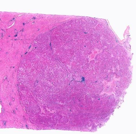
37 Molecular pathology of HCC Llovet et al Nat Rev Dis Prim 2016
38 Hepatocellular Adenoma Rare, benign liver neoplasm Strongly associated with oral contraceptive use and androgen steroid therapy, also in patients with glycogen storage disease, diabetes mellitus 3-4 per in long term OC users Can become symptomatic and lead to bleeding A small subset have the potential for malignant transformation

39 Hepatocellular Adenoma, Macroscopy Solitary, with size to 30cm Soft white to brown and delineated with little or no fibrous capsule, heterogenous areas of necrosis or hemorrhage
40 Molecular classification of HCA - HNF1a inactivated (TCF1 gene mutated) (30%-35%) b-catenin mutated (10%-15%) - Inflammatory (50%) - Unclassified (10%)

41 HNF1a inactivated L-FABP

42 b-catenin mutated b-catenin glutamine synthetase

43 Inflammatory/teleangiectatic HCA Serum amyloid precursor protein

44 IHC for HCA subtypes
45 HCA vs HCC Favoring HCC: thickened cell plates, loss of reticulin network, cytological atypia, mitosis, pseudoacinar architecture
46 Focal nodular hyperplasia (FNH) 10 times more common than HCA Women between years of age Develop in the context of hepatic venous outflow obstruction including Budd-Chiari syndrome Macroscopy shows solitary, discrete, rounded mass, pale, with well-deliniation from background normal liver Frequently with central scar

47 Macroscopy FNH Solitary, discrete, rounded mass, pale, with well-deliniation from background normal liver, frequently with central scar


48 Microscopy FNH Fibrous septa contain often large, dystrophic arteries with thick walls Benign hepatocellular nodules arranged in plates no more than two cell thick Maplike pattern of glutamine synthetase
49 References Advances precancerous lesions in the liver Tommaso L et al., Best Practice and Research Clinical Gastroenterology, 2013 Pathologic Diagnosis of Early Hepatocellular Carcinoma: A Report of the International Consensus Group of Hepatocellular Neoplasia Hepatology 2008 Hepatocellular Carcinoma Llovet et al. Nature Review Disease Primers 2016
Pathological Classification of Hepatocellular Carcinoma
 3 rd APASL Single Topic Conference: HCC in 3D Pathological Classification of Hepatocellular Carcinoma Glenda Lyn Y. Pua, M.D. HCC Primary liver cancer is the 2 nd most common cancer in Asia HCC is the
Alastair Burt Newcastle University
 Alastair Burt Newcastle University Benign Hepatocellular adenoma 8170/0 Focal nodular hyperplasia Malignancy-associated and premalignant lesions Large cell change (formerly dysplasia ) Small cell change
Pitfalls in the diagnosis of well-differentiated hepatocellular lesions
 2013 Colorado Society of Pathology Pitfalls in the diagnosis of well-differentiated hepatocellular lesions Sanjay Kakar, MD University of California, San Francisco Outline Hepatocellular adenoma: new WHO
Evaluation of Liver Mass Lesions. American College of Gastroenterology 2013 Regional Postgraduate Course
 Evaluation of Liver Mass Lesions American College of Gastroenterology 2013 Regional Postgraduate Course Lewis R. Roberts, MB ChB, PhD Division of Gastroenterology and Hepatology Mayo Clinic College of
Liver Specialty Evening Conference. Matthew M. Yeh, MD, PhD Professor of Pathology Adjunct Professor of Medicine University of Washington, Seattle
 Liver Specialty Evening Conference Matthew M. Yeh, MD, PhD Professor of Pathology Adjunct Professor of Medicine University of Washington, Seattle Case History A 65 year-old man presents with abdominal
O Farrell Legacy UPDATE ON WHO NOMENCLATURE. World Health Organization, 2010 DISCLOSURES WITH EMPHASIS ON PROBLEM HEPATOCELLULAR TUMORS
 O Farrell Legacy UPDATE ON WHO NOMENCLATURE WITH EMPHASIS ON PROBLEM HEPATOCELLULAR TUMORS Linda Ferrell, MD University of California San Francisco Vice Chair, Director of Surgical Pathology World Health
Outline. Hepatocellular Carcinoma Histologic variants. HCC: Histologic variants
 2018 Park City AP Update Hepatocellular Carcinoma Histologic variants Sanjay Kakar, MD University of California, San Francisco Outline Histologic variants of HCC Morphologic and Immunohistochemical pitfalls
Liver Tumors. Prof. Dr. Ahmed El - Samongy
 Liver Tumors Prof. Dr. Ahmed El - Samongy Objective 1. Identify the most important features of common benign liver tumors 2. Know the risk factors, diagnosis, and management of hepatocellular carcinoma
Atypical Well differentiated Hepatocellular Neoplasms Cruising through the maze of criteria, terminology and risk assessment
 2016 Hans Popper Companion Meeting Atypical Well differentiated Hepatocellular Neoplasms Cruising through the maze of criteria, terminology and risk assessment Disclosure Dr. Kakar has nothing to Disclose
Intrahepatic cholangiocarcinoma Histologic spectrum, novel markers and molecular assays
 2018 Current Issues in Surgical Pathology Summary (not actual lecture) Intrahepatic cholangiocarcinoma Histologic spectrum, novel markers and molecular assays Sanjay Kakar, MD University of California,
HEPATOCYTE SPECIFIC CONTRAST MEDIA: WHERE DO WE STAND?
 HEPATOCYTE SPECIFIC CONTRAST MEDIA: WHERE DO WE STAND? Andrew T. Trout, MD @AndrewTroutMD Disclosures No relevant disclosures Outline Review of hepatocyte specific contrast media Review of hepatocellular
XIII. Tumours of the liver and biliary system
 XIII. Tumours of the liver and biliary system V. PONOMARKOV 1 & L. J. MACKEY 2 In this histological classification of liver and gall bladder tumours the tumour types largely correspond to those found in
Histopathologic Features of Hepatocellular Carcinoma
 REVIEW REVIEW Histopathologic Features of Hepatocellular Carcinoma Elizabeth M. Brunt, M.D. Paradoxically, with the recognized increase in hepatocellular carcinoma, liver biopsy is used less frequently
Mesenchymal Tumors MESENCHYMAL TUMORS OF THE LIVER: WHAT S NEW AND UNUSUAL (MY PERSPECTIVE)
") MESENCHYMAL TUMORS OF THE LIVER: WHAT S NEW AND UNUSUAL (MY PERSPECTIVE) CURRENT ISSUES IN ANATOMIC PATHOLOGY MAY 23, 2014 Linda Ferrell, MD, UCSF Mesenchymal Tumors Focus on Vascular Tumors Benign and
MESENCHYMAL TUMORS OF THE LIVER: WHAT S NEW AND UNUSUAL (MY PERSPECTIVE) CURRENT ISSUES IN ANATOMIC PATHOLOGY MAY 23, 2014 Linda Ferrell, MD, UCSF Mesenchymal Tumors Focus on Vascular Tumors Benign and
Jesse Civan, M.D. Medical Director, Jefferson Liver Tumor Center
 Liver Tumors Jesse Civan, M.D. Medical Director, Jefferson Liver Tumor Center Differential Diagnosis Malignant Metastatic from non-hepatic primary Hepatocellular carcinoma Cholangiocarcinoma Biliary cystcarcinoma
Mesenchymal Tumors. Cavernous Hemangioma (CH) VASCULAR TUMORS MESENCHYMAL TUMORS OF THE LIVER: WHAT S NEW AND UNUSUAL (MY PERSPECTIVE)
 VASCULAR TUMORS MESENCHYMAL TUMORS OF THE LIVER: WHAT S NEW AND UNUSUAL (MY PERSPECTIVE)") Mesenchymal Tumors MESENCHYMAL TUMORS OF THE LIVER: WHAT S NEW AND UNUSUAL (MY PERSPECTIVE) CURRENT ISSUES IN ANATOMIC PATHOLOGY MAY 23, 2014 Linda Ferrell, MD, UCSF Focus on Vascular Tumors Benign and
Mesenchymal Tumors MESENCHYMAL TUMORS OF THE LIVER: WHAT S NEW AND UNUSUAL (MY PERSPECTIVE) CURRENT ISSUES IN ANATOMIC PATHOLOGY MAY 23, 2014 Linda Ferrell, MD, UCSF Focus on Vascular Tumors Benign and
Disclosures. Parathyroid Pathology. Objectives. The normal parathyroid 11/10/2012
 Disclosures Parathyroid Pathology I have nothing to disclose Annemieke van Zante MD/PhD Assistant Professor of Clinical Pathology Associate Chief of Cytopathology Objectives 1. Review the pathologic features
Detection and Characterization of Hepatocellular Carcinoma by Imaging
 CLINICAL GASTROENTEROLOGY AND HEPATOLOGY 2005;3:S136 S140 Detection and Characterization of Hepatocellular Carcinoma by Imaging OSAMU MATSUI Department of Imaging Diagnosis and Interventional Radiology,
Proliferative Epithelial lesions of the Breast. Sami Shousha, MD, FRCPath Charing Cross Hospital & Imperial College, London
 Proliferative Epithelial lesions of the Breast Sami Shousha, MD, FRCPath Charing Cross Hospital & Imperial College, London Amman, November2013 Proliferative Epithelial Lesions of the Breast Usual type
Neoplasms of the Canine, Feline and Lemur Liver:
 Neoplasms of the Canine, Feline and Lemur Liver: Classification and Prognosis Annual Seminar of the French Society of Veterinary Pathology John M. Cullen VMD PhD DACVP North Carolina State University Primary
Differential diagnosis of HCC
 Hepatocellular Carcinoma Quest for an Ideal Immunohistochemical Panel Sanjay Kakar, MD UCSF Differential diagnosis of HCC Hepatocellular lesions Adenoma, FNH, HG dysplasia Adenocarcinoma CholangioCA, metastasis
NEOPLASMS AND TUMOR-LIKE CONDITIONS OF LIVER
 NEOPLASMS AND TUMOR-LIKE CONDITIONS OF LIVER Epithelial Tumors Focal nodular hyperplasia Focal nodular hyperplasia is a localized hyperplasic overgrowth of hepatocytes around a vascular anomaly, particularly
Neoplasia 2018 Lecture 2. Dr Heyam Awad MD, FRCPath
 Neoplasia 2018 Lecture 2 Dr Heyam Awad MD, FRCPath ILOS 1. List the differences between benign and malignant tumors. 2. Recognize the histological features of malignancy. 3. Define dysplasia and understand
number Done by Corrected by Doctor Maha Shomaf
 number 16 Done by Waseem Abo-Obeida Corrected by Zeina Assaf Doctor Maha Shomaf MALIGNANT NEOPLASMS The four fundamental features by which benign and malignant tumors can be distinguished are: 1- differentiation
G3.02 The malignant potential of the neoplasm should be recorded. CG3.02a
 G3.02 The malignant potential of the neoplasm should be recorded. CG3.02a Conventional adrenocortical neoplasm. Each of the below parameters is scored 0 when absent and 1 when present. 3 or more of these
CTA/MRA of Pediatric Hepatic Masses Radiology-Pathology Correlation
 Acta Radiológica Portuguesa, Vol.XVIII, nº70, pág. 41-50, Abr.-Jun., 2006 CTA/MRA of Pediatric Hepatic Masses Radiology-Pathology Correlation Marilyn J. Siegel Mallinckrodt Institute of Radiology, Washington
Interesting Cases from Liver Tumor Board. Jeffrey C. Weinreb, M.D.,FACR Yale University School of Medicine
 Interesting Cases from Liver Tumor Board Jeffrey C. Weinreb, M.D.,FACR Yale University School of Medicine jeffrey.weinreb@yale.edu Common Liver Diseases Hemangioma Cyst FNH Focal Fat/Sparing THID Non-Cirrhotic
Video Microscopy Tutorial 8
 Video Microscopy Tutorial 8 Common and Uncommon Lesions of the Liver Gladwyn Leiman, MD There are no disclosures necessary. Common and Uncommon Lesions in Liver FNA Gladwyn Leiman University of Vermont
Raga Ramachandran, MD, PhD Assistant Professor and Director of Medical Education, UCSF Pathology
 Variants of Hepatocellular Carcinoma: Practical Issues Raga Ramachandran, MD, PhD Assistant Professor and Director of Medical Education, UCSF Pathology raga.ramachandran@ucsf.edu A full copy of the presentation
Biliary tract tumors
 Short Course 2010 Annual Fall Meeting of the Korean Society for Pathologists Biliary tract tumors Joon Hyuk Choi, M.D., Ph.D. Professor, Department of Pathology, Yeungnam Univ. College of Medicine, Daegu,
activated hepatocellular adenoma
 pissn 2287-2728 eissn 2287-285X Liver Pathology Clinical and Molecular Hepatology 2013;19:185-189 5.2 3.6 cm sized, homogeneous, green colored hepatic mass was observed on the cut section (Fig. 1). The
Papillary Lesions of the Breast A Practical Approach to Diagnosis. (Arch Pathol Lab Med. 2016;140: ; doi: /arpa.
 Papillary Lesions of the Breast A Practical Approach to Diagnosis (Arch Pathol Lab Med. 2016;140:1052 1059; doi: 10.5858/arpa.2016-0219-RA) Papillary lesions of the breast Span the spectrum of benign,
MRI OF FOCAL LESIONS IN
 Introduction MRI OF FOCAL LESIONS IN THE NON-CIRRHOTIC LIVER Ivan Pedrosa M.D. Ph.D. Associate Professor of Radiology and Advanced Imaging Research Center University of Texas Southwestern. Dallas, TX Incidental
Hepatocellular neoplasia - Recent developments
 22 November 2018 Hepatocellular neoplasia - Recent developments Dina G. Tiniakos Institute of Cellular Medicine, Faculty of Medical Sciences, Newcastle University, UK Dept of Pathology, Aretaieion Hospital,
Papillary Lesions of the breast
 Papillary Lesions of the breast Emad Rakha Professor of Breast Pathology The University of Nottingham Papillary lesions of the breast are a heterogeneous group of disease, which are characterised by neoplastic
8 years later! Next Generation Sequencing. Pathogenic Findings: HNF1A c.864delinscc, p.g292rfs*25 (NM_ ) (VAF: 59%) HNF1A Loss
 (VAF: 59%) HNF1A Loss") 8 years later! Next Generation Sequencing Pathogenic Findings: HNF1A c.864delinscc, p.g292rfs*25 (NM_000545.6) (VAF: 59%) HNF1A Loss Interpretation HNF1A c.864delinscc, p.g292rfs*25 (NM_000545.6) This
8 years later! Next Generation Sequencing Pathogenic Findings: HNF1A c.864delinscc, p.g292rfs*25 (NM_000545.6) (VAF: 59%) HNF1A Loss Interpretation HNF1A c.864delinscc, p.g292rfs*25 (NM_000545.6) This
CT & MRI of Benign Liver Neoplasms Srinivasa R Prasad
 CT & MRI of Benign Liver Neoplasms Srinivasa R Prasad No financial disclosures Acknowledgements Many thanks to Drs. Heiken, Narra & Menias (MIR) Dr. Sahani (MGH) for sharing images Benign Liver Tumors:
Pitfalls in thyroid tumor pathology. Prof.Valdi Pešutić-Pisac MD, PhD
 Pitfalls in thyroid tumor pathology Prof.Valdi Pešutić-Pisac MD, PhD Too many or... Tumour herniation through a torn capsule simulating capsular invasion fibrous capsule with a sharp discontinuity, suggestive
Endometrial Stromal Tumors
 Endometrial Stromal Tumors WHO Categories: Endometrial Stromal Nodule (ESN) Endometrial Stromal Sarcoma, low grade (LGESS) Endometrial Stromal Sarcoma, high grade (HGESS) Undifferentiated Uterine Sarcoma
Hepatocellular Adenomas: Genetics & Imaging Update 2017
 No financial disclosures Hepatocellular Adenomas: Genetics & Imaging Update 2017 Srinivasa Prasad MD The UT MD Anderson Cancer Center Aims & Objectives To provide a current update on genetics & molecular
Dysplastic Nodules. Department of Pathology, Chonbuk National University Medical School. Woo Sung Moon. Introduction
 Dysplastic Nodules Department of Pathology, Chonbuk National University Medical School Woo Sung Moon 만성간질환에발생하는간의결절병변에는재생결절, 형성이상결절및간세포암종이있다. 여러인자에의해손상받은간세포는괴사와재생과정을반복하며재생결절, 형성이상결절을형성하는동안유전자의변이와 epigenetic
Salivary Glands 3/7/2017
 Salivary Glands 3/7/2017 Goals and objectives Focus on the entities unique to H&N Common board type facts Information for your future practice Salivary Glands Salivary Glands Major gland. Paratid. Submandibular.
Hepatocelluar nodules in liver cirrhosis: hemodynamic evaluation (angiographyassisted CT) with special reference to multi-step hepatocarcinogenesis
 with special reference to multi-step hepatocarcinogenesis") Abdominal Imaging ª The Author(s) 2011. This article is published with open access at Springerlink.com Published online: 26 January 2011 Abdom Imaging (2011) 36:264 272 DOI: 10.1007/s00261-011-9685-1 INVITED
Abdominal Imaging ª The Author(s) 2011. This article is published with open access at Springerlink.com Published online: 26 January 2011 Abdom Imaging (2011) 36:264 272 DOI: 10.1007/s00261-011-9685-1 INVITED
Primary and Metastatic Tumours of the Liver: Expanding Scope of Morphological and Immunohistochemical Details in the Biopsy
 Chapter 7 Primary and Metastatic Tumours of the Liver: Expanding Scope of Morphological and Immunohistochemical Details in the Biopsy Ilze Strumfa, Janis Vilmanis, Andrejs Vanags, Ervins Vasko, Dzeina
Neoplasia part I. Dr. Mohsen Dashti. Clinical Medicine & Pathology nd Lecture
 Neoplasia part I By Dr. Mohsen Dashti Clinical Medicine & Pathology 316 2 nd Lecture Lecture outline Review of structure & function. Basic definitions. Classification of neoplasms. Morphologic features.
LIVER PATHOLOGY(3) Prof. Andras Kiss. M.D., Ph.D., D.Sc. Semmelweis University 2 nd Department of Pathology Budapest February 26.
 Prof. Andras Kiss. M.D., Ph.D., D.Sc. Semmelweis University 2 nd Department of Pathology Budapest February 26.") LIVER PATHOLOGY(3) Prof. Andras Kiss. M.D., Ph.D., D.Sc. Semmelweis University 2 nd Department of Pathology Budapest February 26. 2018 Vascular disorders Inflow A.hepatica thromb., embolia infarctus V.portae
LIVER PATHOLOGY(3) Prof. Andras Kiss. M.D., Ph.D., D.Sc. Semmelweis University 2 nd Department of Pathology Budapest February 26. 2018 Vascular disorders Inflow A.hepatica thromb., embolia infarctus V.portae
Demystifying Endometrial Hyperplasia
 Demystifying Endometrial Hyperplasia A review from Diagnostic Histopathology 19:7 Dr R Hadden ST5 Histopathology Derriford Hospital Plymouth Endometrium Target for sex-steroid hormones Glands Stroma Proliferate
Pancreatitis: A Potential Pitfall in Endoscopic Ultrasound Guided Pancreatic FNA
 Pancreatitis: A Potential Pitfall in Endoscopic Ultrasound Guided Pancreatic FNA Jack Yang, MD Department of Pathology, Medical University of South Carolina Objectives Understand the indication of EUS
Diseases of the breast (1 of 2)
") Diseases of the breast (1 of 2) Introduction A histology introduction Normal ducts and lobules of the breast are lined by two layers of cells a layer of luminal cells overlying a second layer of myoepithelial
Diseases of the breast (1 of 2) Introduction A histology introduction Normal ducts and lobules of the breast are lined by two layers of cells a layer of luminal cells overlying a second layer of myoepithelial
Thyroid follicular neoplasms in cytology. Ulrika Klopčič Institute of Oncology, Department of Cytopathology, Ljubljana, Slovenia
 Thyroid follicular neoplasms in cytology Ulrika Klopčič Institute of Oncology, Department of Cytopathology, Ljubljana, Slovenia Lecture overview importance of FNAB in assessing thyroid lesions follicular
Case: The patient is a 62 year old woman with a history of renal cell carcinoma that was removed years ago. A 2.4 cm liver mass was found on CT
 Case: The patient is a 62 year old woman with a history of renal cell carcinoma that was removed years ago. A 2.4 cm liver mass was found on CT during follow- up. ALT, AST, Alk Phos and bilirubin were
Outline 11/2/2017. Pancreatic EUS-FNA general aspects. Cytomorphologic features of solid neoplasms/lesions of the pancreas
 ENDOSCOPIC ULTRASOUND GUIDED-FINE NEEDLE ASPIRATION CYTOLOGY OF PANCREAS Khalid Amin M.D. Assistant Professor Department of Laboratory Medicine and Pathology University of Minnesota Outline Pancreatic
Section 8 Liver and Gallbladder
 General and Systemic Histopathology C601 and C602 Section 8 As we will see in this unit, the liver is subject to many types of injury. Additionally, many systemic diseases have a liver component and sometimes
CINtec p16 INK4a Staining Atlas
 CINtec p16 INK4a Staining Atlas Rating Rating Positive The rating positive will be assigned if the p16 INK4a -stained slide shows a continuous staining of cells of the basal and parabasal cell layers of
CONTINUING EDUCATION IN TOXICOLOGIC PATHOLOGY REPRODUCTIVE SYSTEM
 CONTINUING EDUCATION IN TOXICOLOGIC PATHOLOGY REPRODUCTIVE SYSTEM ORGANIZED BY SOCIETY FOR TOXICOLOGIC PATHOLOGY IN INDIA (STPI) OCTOBER 29-31, 2010 The Atria Hotel, # 1, Palace Road, Bangalore - 560 001
Malignant Focal Liver Lesions
 Malignant Focal Liver Lesions Other Than HCC Pablo R. Ros, MD, MPH, PhD Departments of Radiology and Pathology University Hospitals Cleveland Medical Center Case Western Reserve University Pablo.Ros@UHhospitals.org
Prepared By Jocelyn Palao and Layla Faqih
 Prepared By Jocelyn Palao and Layla Faqih The structure of the suspected atypical cell should always be compared to the structure of other similar, benign, cells which are present in the smears. The diagnosis
ARTHUR PURDY STOUT SOCIETY COMPANION MEETING: DIFFICULT NEW DIFFERENTIAL DIAGNOSES IN PROSTATE PATHOLOGY. Jonathan I. Epstein.
 1 ARTHUR PURDY STOUT SOCIETY COMPANION MEETING: DIFFICULT NEW DIFFERENTIAL DIAGNOSES IN PROSTATE PATHOLOGY Jonathan I. Epstein Professor Pathology, Urology, Oncology The Reinhard Professor of Urological
SESSION 1: GENERAL (BASIC) PATHOLOGY CONCEPTS Thursday, October 16, :30am - 11:30am FACULTY COPY
 PATHOLOGY CONCEPTS Thursday, October 16, :30am - 11:30am FACULTY COPY") SESSION 1: GENERAL (BASIC) PATHOLOGY CONCEPTS Thursday, October 16, 2008 9:30am - 11:30am FACULTY COPY GOAL: Describe the basic morphologic (structural) changes which occur in various pathologic conditions.
SESSION 1: GENERAL (BASIC) PATHOLOGY CONCEPTS Thursday, October 16, 2008 9:30am - 11:30am FACULTY COPY GOAL: Describe the basic morphologic (structural) changes which occur in various pathologic conditions.
British Liver Transplant Group Pathology meeting September Leeds cases
 British Liver Transplant Group Pathology meeting September 2014 Leeds cases Leeds Case 1 Male 61 years Liver transplant for HCV cirrhosis with HCC in January 2014. Now raised ALT and bilirubin,? acute
Neoplasia literally means "new growth.
 NEOPLASIA Neoplasia literally means "new growth. A neoplasm, defined as "an abnormal mass of tissue the growth of which exceeds and is uncoordinated with that of the normal tissues and persists in the
Select problems in cystic pancreatic lesions
 Disclosure Select problems in cystic pancreatic lesions Five Prime Therapeutics shareholder Adicet Bio shareholder Bristol-Meyer Squibb advisory board grace.kim@ucsf.edu Pancreatic cystic lesions Intraductal
Invited Re vie W. Analytical histopathological diagnosis of small hepatocellular nodules in chronic liver diseases
 Histol Histopathol (1 998) 13: 1077-1 087 http://www.ehu.es/histoi-histopathol Histology and Histopathology Invited Re vie W Analytical histopathological diagnosis of small hepatocellular nodules in chronic
The Relevance of Cytologic Atypia in Cutaneous Neural Tumors
 The Relevance of Cytologic Atypia in Cutaneous Neural Tumors Recent Findings - New Developments New Problems Zsolt B. Argenyi, M.D. Professor of Pathology & Dermatology Director of Dermatopathology Department
Respiratory Tract Cytology
 Respiratory Tract Cytology 40 th European Congress of Cytology Liverpool, UK Momin T. Siddiqui M.D. Professor of Pathology and Laboratory Medicine Director of Cytopathology Emory University Hospital, Atlanta,
Disorders of Cell Growth & Neoplasia. Histopathology Lab
 Disorders of Cell Growth & Neoplasia Histopathology Lab Paul Hanna April 2010 Case #84 Clinical History: 5 yr-old, West Highland White terrier. skin mass from axillary region. has been present for the
Mammary Nodular Hyperplasia in Intact R hesus Monkeys
 Vet. Path. 10: 130-134 (1973) Mammary Nodular Hyperplasia in Intact R hesus Monkeys L. W NELSON and L. D. SHOTT Department of Pathology and Toxicology, Mead Johnson Research Center, Evansville, Ind., and
Multistep hepatocarcinogenesis is characterized by the following
 Early hepatocellular carcinoma with high-grade atypia in small vaguely nodular lesions Hidenori Ojima, 1 Yohei Masugi, 1 Hanako Tsujikawa, 1 Katsura Emoto, 1 Yoko Fujii-Nishimura, 1 Mami Hatano, 1 Miho
Histologic Growth Patterns of Metastatic Carcinomas of the Liver
 Histologic Growth Patterns of Metastatic Carcinomas of the Liver Noboru Terayama, Tadashi Terada and Yasuni Nakanuma Second Department of Pathology, Kanazawa University School of Medicine, Kanazawa One
ACRIN 6690 PATHOLOGY MANUAL
 ACRIN 6690 PATHOLOGY MANUAL ACRIN 6690 Prepared by the American College of Radiology Imaging Network Original: December 15, 2010 Version Date: May 3, 2012 American College of Radiology Imaging Network
2 to 3% of All New Visceral Cancers Peak Incidence is 6th Decade M:F = 2:1 Grossly is a Bright Yellow, Necrotic Mass with a Pseudocapsule
 GENITOURINARY PATHOLOGY Kathleen M. O Toole, M.D. Renal Cell Carcinoma 2 to 3% of All New Visceral Cancers Peak Incidence is 6th Decade M:F = 2:1 Grossly is a Bright Yellow Necrotic Mass Grossly is a Bright
TUMOR,NEOPLASM. Pathology Department, Zhejiang University School of Medicine,
 TUMOR,NEOPLASM Pathology Department, Zhejiang University School of Medicine, 马丽琴,maliqin198@zju.edu.cn The points in this chapter What is a neoplasm (conception) Morphology of neoplasm Macroscopy of Neoplasm
Fig. 59 Malignant phaeochromocytoma, hepatic metastasis.
 Fig. 59 Malignant phaeochromocytoma, hepatic metastasis. X 120 Hyperte nsion Fig. 60 Malignant sympathetic paraganglioma, lymph node metastasis Primary in bladder. x 1 20 Hypertension Fig. 61 Malignant
Recently role of non-invasive diagnostics methods
 CERTAIN ASPECTS OF NLS-DIAGNOSTICS OF LIVER FOCAL PATHOLOGY A.Y. Shvack, V.I. Nesterov, N.L. Ogluzdina This article contains information about NLS-graphy application in diagnostics of liver focal affections:
WSC , Conference 9, Case 1. Tissue from a nyala.
 WSC 2009-2010, Conference 9, Case 1. Tissue from a nyala. MICROSCOPIC DESCRIPTION: Heart, atrium (1 pt.): Approximately 40% of the atrial myocardium is replaced by areas of fibrous connective tissue (1
Diagnostic accuracy of percutaneous renal tumor biopsy May 10 th 2018
 Diagnostic accuracy of percutaneous renal tumor biopsy May 10 th 2018 Dr. Tzahi Neuman Dep.Of Pathology Hadassah Medical Center Jerusalem, Israel, (tneuman@hadassah.org.il) Disclosure: 1 no conflicts of
Benign and malignant epithelial lesions: Seborrheic keratosis: A common benign pigmented epidermal tumor occur in middle-aged or older persons more
 Benign and malignant epithelial lesions: Seborrheic keratosis: A common benign pigmented epidermal tumor occur in middle-aged or older persons more common on the trunk; but extremities, head and neck are
CNS pathology Third year medical students. Dr Heyam Awad 2018 Lecture 12: CNS tumours 2/3
 CNS pathology Third year medical students Dr Heyam Awad 2018 Lecture 12: CNS tumours 2/3 Pilocytic astrocytoma Relatively benign ( WHO grade 1) Occurs in children and young adults Mostly: in the cerebellum
Liver Pathology in the 0bese
 Liver Pathology in the 0bese Rob Goldin Centre for Pathology, Imperial College r.goldin@imperial.ac.uk Ludwig et al. Non-alcoholic steatohepatitis: Mayo Clinic experiences with a hitherto unnamed disease.
Financial Disclosure
 Benign Liver Masses Adil Abdalla, MBBS Creighton University-CHI Health August 25, 2018 Financial Disclosure Nothing to disclose Financial Disclosure 1 Objectives To assess patients with benign liver tumors
Mesothelioma: diagnostic challenges from a pathological perspective. Naseema Vorajee August 2016
 Mesothelioma: diagnostic challenges from a pathological perspective Naseema Vorajee August 2016 Naseema.vorajee@nhls.ac.za Pleural diseases (whether neoplastic, reactive or infective) may have similar
Malignant neoplasms of the gastrointestinal (GI) tract,
 tract,") Special Section First Chinese American Pathologists Association Diagnostic Pathology Course, Part II Practical Immunohistochemistry in Neoplastic Pathology of the Gastrointestinal Tract, Liver, Biliary
Special Section First Chinese American Pathologists Association Diagnostic Pathology Course, Part II Practical Immunohistochemistry in Neoplastic Pathology of the Gastrointestinal Tract, Liver, Biliary
Benign liver tumors : Diagnosis and management
 4th International Hepatology Conference 2016 HEPATOLOGY SOCIETY, DHAKA, BANGLADESH Benign liver tumors : Diagnosis and management Pr Laurence Chiche Hepato biliary surgery and transplantation Bordeaux,
Review Article Assessment of Stromal Invasion for Correct Histological Diagnosis of Early Hepatocellular Carcinoma
 SAGE-Hindawi Access to Research International Hepatology Volume 2011, Article ID 241652, 7 pages doi:10.4061/2011/241652 Review Article Assessment of Stromal Invasion for Correct Histological Diagnosis
1 NORMAL HISTOLOGY AND METAPLASIAS
 1 NORMAL HISTOLOGY AND METAPLASIAS, MD Anatomy and Histology 1 Metaplasias 2 ANATOMY AND HISTOLOGY The female breast is composed of a branching duct system, which begins at the nipple with the major lactiferous
Pathological Analysis of Small Hepatocellular Carcinoma with Poor Prognosis
 Pathological Analysis of Small Hepatocellular Carcinoma with Poor Prognosis Haeryoung Kim, M.D., Ph.D. Department of Pathology Seoul National University Bundang Hospital Small HCC Definition: HCC < 2cm
HISTOPATHOLOGY. Shannon Martinson
 HISTOPATHOLOGY Shannon Martinson March 2013 Case #1 History: 8 year old beagle Neck pain for the past couple of weeks Paresis, followed by paralysis developed over the past few days Gross Description courtesy
NODULAR CYSTIC HIDRADENOMA OVER THE GLUTEAL REGION: A RARE CYTOMORPHOLOGICAL DIAGNOSIS
 NODULAR CYSTIC HIDRADENOMA OVER THE GLUTEAL REGION: A RARE CYTOMORPHOLOGICAL DIAGNOSIS Abstract: The primary as well as metastatic tumours of the skin can be diagnosed by fine needle aspiration cytology
AN AUTOPSY CASE OF PARATHYROID CARC. Matsumoto, Koji; Ito, Masahiro; Sek. Citation Acta medica Nagasakiensia. 1989, 34
 NAOSITE: Nagasaki University's Ac Title Author(s) AN AUTOPSY CASE OF PARATHYROID CARC Hsu, Chao-Tien; Naito, Shinji; Shik Matsumoto, Koji; Ito, Masahiro; Sek Citation Acta medica Nagasakiensia. 1989, 34
FNA of Thyroid. Toward a Uniform Terminology With Management Guidelines. NCI NCI Thyroid FNA State of the Science Conference
 FNA of Thyroid NCI NCI Thyroid FNA State of the Science Conference Toward a Uniform Terminology With Management Guidelines Thyroid Thyroid FNA Cytomorphology NCI Thyroid FNA State of the Science Conference
Essentials of Clinical MR, 2 nd edition. 65. Benign Hepatic Masses
 65. Benign Hepatic Masses Pulse sequences acquired for abdominal MRI typically consist of fast acquisition schemes such as single-shot turbo spin echo (i.e. HASTE) and gradient echo schemes such as FLASH
MVST BOD & NST PART IB Thurs. 2 nd & Fri. 3 rd March 2017 Pathology Practical Class 23
 MVST BOD & NST PART IB Thurs. 2 nd & Fri. 3 rd March 2017 Pathology Practical Class 23 Neoplasia I Neoplasia I: Benign and malignant neoplasms in glandular epithelium and mesenchyme 1.0. Aims 1. To understand
A 53 year-old woman with a lung mass, right hilar mass and mediastinal adenopathy.
 November 2015 Case of the Month A 53 year-old woman with a lung mass, right hilar mass and mediastinal adenopathy. Contributed by: Rasha Salama, M.D., IU Department of Pathology and Laboratory Medicine
Basic patterns of liver damage what information can a liver biopsy provide and what clinical information does the pathologist need?
 Basic patterns of liver damage what information can a liver biopsy provide and what clinical information does the pathologist need? Rob Goldin r.goldin@imperial.ac.uk Fatty liver disease Is there fatty
Hepatocellular carcinoma Cholangiocarcinoma. Jewels of hepatobiliary cancer imaging : what to look for? Imaging characteristics of HCC.
 Outline : Imaging Jewels Jewels of hepatobiliary cancer imaging : what to look for? Hepatocellular carcinoma Cholangiocarcinoma Surachate Siripongsakun, M.D. Chulabhorn Cancer Center Imaging characteristics
What s New in Adrenal Gland Pathology. Marina Scarpelli
 What s New in Adrenal Gland Pathology Marina Scarpelli Background Histological criteria for adrenocortical proliferative lesions Immunohistochemical markers Molecular markers Histological Criteria for
BREAST PATHOLOGY. Fibrocystic Changes
 BREAST PATHOLOGY Lesions of the breast are very common, and they present as palpable, sometimes painful, nodules or masses. Most of these lesions are benign. Breast cancer is the 2 nd most common cause
Note: The cause of testicular neoplasms remains unknown
 - In the 15- to 34-year-old age group, they are the most common tumors of men. - Tumors of the testis are a heterogeneous group of neoplasms that include: I. Germ cell tumors : 95%; all are malignant.
Lesions Mimicking Adenoid Cystic Carcinoma. Diagnostic Problems in Salivary Gland Pathology An Update 5/29/2009
 Diagnostic Problems in Salivary Gland Pathology An Update Lesions Mimicking Adenoid Cystic Carcinoma Stacey E. Mills, M.D. W.S. Royster Professor of Pathology Director of Surgical and Cytopathology University