Pre-surgical planning for brain tumor resection using functional MRI
|
|
|
- Kristopher Thomas
- 6 years ago
- Views:
Transcription
1 June 2011 Divya S Bolar, HMSIV Pre-surgical planning for brain tumor resection using functional MRI Divya S. Bolar,, HMS IV 1
2 Our patient: clinical history 85-year year-old right-handed handed woman presents s/p fall with occipital head strike Denies LOC or pre-syncopal symptoms. Endorses eight month history of falls secondary to progressive left-sided weakness and loss of balance. PMHx: : CAD, DM2, HTN, RCC s/p nephrectomy in 1999 Physical exam: notable for intact CN, diffuse left-sided weakness, unsteady gait. No sensory deficits. Labs unremarkable. 2

3 Our patient: Right parietal lesion on CT Central focus of hyper-attenuation CT head C- 3
4 Our patient: Surrounding edema Central focus of hyper-attenuation Substantial surrounding edema 4
5 Our patient: Sulci effacement Central focus of hyper-attenuation Substantial surrounding edema Mass effect with sulci effacement 5
6 Our patient: Other findings Central focus of hyper-attenuation Substantial surrounding edema Mass effect with sulci effacement Minimal midline shift No fractures or other acute findings 6
7 Our patient: Differential diagnosis Differential Dx: 1. Hemorrhagic infarct 2. Hemorrhage into underlying mass lesion Well-circumscribed lesion, chronicity of symptoms => suspect hemorrhage into mass 7
8 Our patient: Intracranial hemorrhage on C- C MRI Mild degree of T1 hyperintensity MRI head T1/ C- 8

9 Our patient: Intracranial hemorrhage on C+ MRI MRI head T1/ C+ Moderate degree of lesion enhancement Uncharacteristic of hemorrhagic infarct Consistent with contrast uptake by abnormal tumor vasculature 9
10 Our patient: Approach to management Additional body imaging with CT: Masses found in lung & breast No masses found in remaining kidney or at prior surgical site Etiology of brain tumor unclear Metastasis from occult RCC? New primary brain tumor? Metastasis from other site? Neurosurgical resection was recommended to decompress, reduce edema, improve local control, and biopsy 10
11 Frontoparietal tumors Neurosurgical resection often indicated, but carries risk of injury to: Primary motor cortex in precentral gyrus and/or descending corticospinal tract Frontal language regions if lesion is in dominant hemisphere (Broca( Broca s s area in orpeculum) Chief concern is paralysis and loss of speech and language Preoperative mapping of these areas could assist in surgical planning and reduce risk of injury 11
12 Pre-surgical planning: What does the neurosurgeon want to know? Distance between tumor margin and essential functional areas Golden rule says minimum distance 10 mm to preserve function Trajectory to tumor that avoids functional area (if one exists) With this information surgeon can: 1. Determine if tumor is amenable for resection 2. Decide if intraoperative cortical stimulation is needed 3. Better navigate surgical procedure itself So-called functional MRI is a noninvasive approach that can safely identify essential functional areas in advance of surgical intervention 12
13 What is functional MRI (fmri( fmri)? fmri is a technique for determining which parts of the brain are activated by different types of physical sensation or activity, such as sight, sound or the movement of a subject's fingers. - Steve Smith, FMRIB, Oxford 13
14 Approach to fmri Subjects are continuously imaged while performing specifically timed tasks Tasks are chosen based on neural system we wish to interrogate. Examples include: Motor => Finger or foot-tapping tapping Visual => Viewing flashing checkerboards Analysis of images allows creation of statistical maps that localize task-based neural activity to corresponding brain regions 14
15 How is neural activity reflected in the MRI signal? (i.e. how does fmri work!!)

16 Contents of brain MRI voxel: Parenchyma Parenchyma Van Zijl et al, Nat Med, 1998 Voxel = volumetric picture element 16
17 Contents of brain MRI voxel: Microvasculature 2008 Sinauer Associates, Inc. Van Zijl et al, Nat Med, 1998 Voxel = volumetric picture element 17
18 From neural activity to MRI signal: Microvasculature in brain voxel Arterial circulation Capillary Bed Venous circulation 2008 Sinauer Associates, Inc. 18
19 From neural activity to MRI signal: Oxygen delivery via cerebral blood flow I O 2 At baseline, cerebral blood flow (CBF) supplies oxygen-rich blood to capillary bed O Sinauer Associates, Inc. 19
20 From neural activity to MRI signal: Oxygen delivery via cerebral blood flow II RBC with HbO 2 O 2 O 2 Chief oxygen-carrier is macromolecule hemoglobin in red blood cell (RBC) Oxygenated hemoglobin is called oxyhemoglobin (HbO 2 ) 2008 Sinauer Associates, Inc. 20
21 From neural activity to MRI signal: Oxygen extraction and consumption RBC with HbO 2 O 2 As blood traverses capillary bed, oxygen is extracted from HbO 2 into tissue and consumed O Sinauer Associates, Inc. 21
22 From neural activity to MRI signal: Venous deoxygenated hemoglobin (dhb( dhb) RBC with HbO 2 Deoxygenated hemoglobin (dhb) subsequently results on venous side RBC with dhb 2008 Sinauer Associates, Inc. 22
23 From neural activity to MRI signal: Effects of dhb on MRI signal RBC with HbO 2 dhb is paramagnetic and perturbs the magnetic field Decreases baseline MR signal RBC with dhb 2008 Sinauer Associates, Inc. 23
24 From neural activity to MRI signal: Stimulation increases neuronal activity During stimulation (e.g. from a task) neuronal activity increases 2008 Sinauer Associates, Inc. 24
25 From neural activity to MRI signal: Secondary increase in CBF flushes out dhb O 2 A secondary increase in CBF follows Increased CBF delivers more HbO 2 and flushes out venous dhb O Sinauer Associates, Inc. 25
26 From neural activity to MRI signal: Reduction in dhb increases MR signal O 2 Decreased dhb results in a smaller field perturbation and an increase in MR signal O Sinauer Associates, Inc. 26
27 MR signal intensity: Baseline State Divya S Bolar, HMSIV 2008 Sinauer Associates, Inc. [dhb] = 40% 2011 IMAIOS 27
28 MR signal intensity: Activated State Divya S Bolar, HMSIV Slight intensity increase in corresponding region 2008 Sinauer Associates, Inc. [dhb] = 20% This is the blood oxygen level dependent (BOLD) effect and is the basis for fmri! 2011 IMAIOS 28

29 Sample block block-design fmri task: Right handed finger-tapping Divya S Bolar, HMSIV REST 1 min TAP! X 3 1 min Acquire low-resolution MR images every two seconds MR signal from left motor cortex 29
30 Generation of statistical map shows activated voxels Map overlaid on high-resolution T1-weighted anatomical 30
31 Central voxel with highly significant activation 31
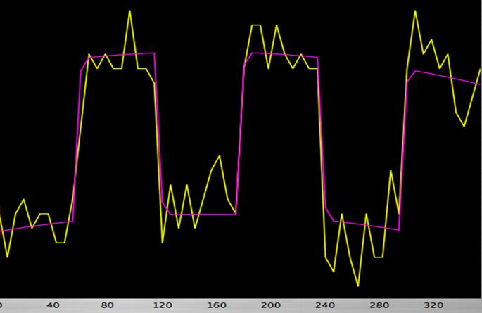
32 Peripheral voxel with less significant activation 32
33 Our patient: fmri protocol Use similar block-design fmri paradigm Include: Motor task: LH finger-tapping Language: Verb repetition 33
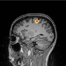
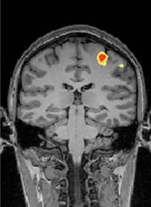
34 Our patient: Activation from LH finger-tapping Divya S Bolar, HMSIV 34
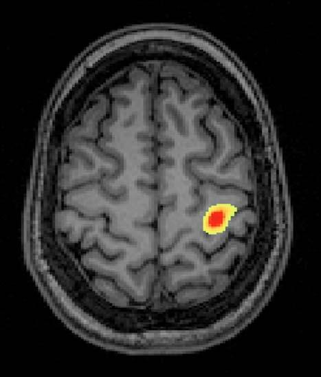
35 Our patient: Primary motor cortex Primary motor cortex 35
36 Our patient: Supplementary motor cortex(?) Supplementary motor cortex(?) 36

37 Our patient: Activation from Verb repetition Operculum frontale Activation seen in operculum frontale; location consistent with Broca s s Area Suggests patient has left hemispheric dominance Reduced risk of language impairment with resection of right-sided lesion. 37
38 Our patient: Surgical planning Tumor margin 5 mm away from motor activation strip Unobscured oblique trajectory available for direct approach 5 mm 38

39 Our patient: Pre-post op comparison PRE POST 39

40 Our patient: Post-operative changes T1 hyperintensity consistent with post- operative blood and proteinaceous material Thin rim of enhancement could be post-op op change, but residual tumor cannot be excluded 40
41 Our patient: Outcome Patient initially had increased left-sided weakness post-operatively operatively Not surprising given proximity of tumor to motor cortex Improved over time Preliminary pathology suggested metastasis from renal cell carcinoma She was discharged and will see oncology to discuss chemotherapy options 41
42 Summary Functional MRI can be useful tool for preoperative planning and assessment for brain tumor resection fmri creates activation maps which correlate to neural activity patterns Link between neural activity and MRI signal arises from increased blood flow flushing out paramagnetic dhb during stimulation Use of maps allow surgeons to: 1. Assess resectability of tumors near essential functional areas 2. Decide if intraoperative cortical stimulation is needed 3. Better navigate surgical procedure 42
43 References Buxton RB. Introduction to Functional Magnetic Resonance Imaging. Second Edition. New York, NY: Cambridge University Press; Hoa D. Functional MRI of the Brain. MRI/Functional-MRI/introduction, Accessed 6/16/2011. Holodny AI. Functional Neuroimaging: A Clinical Approach. New York, NY: Informa Healthcare; Purves, Augustine, Fitzpatric, Hall, LaMantia, McNamara, White. Neuroscience. Fourth Edition. Accessed 6/16/2011. Smith S. Brief Introduction to FMRI /fmri_smith_1998.pdf. Accessed 6/16/2011. Sunaert S. Presurgical planning for tumor resectioning. JMRI. 2006; 23: van Zijl PC, Eleff SM, Ulatowski JA, Oja JM, Ulug AM, Traystman RJ, Kauppinen RA. Quantitative assessment of blood flow, blood volume and blood oxygenation effects in functional magnetic resonance imaging. Nat Med 1998;4: Brugge WR, Van Dam J. 43
44 Thanks to: Acknowledgements Rafael Rajos MD, Neuroradiology, BIDMC Ted Brewer MD, Neuroradiology, BIDMC Gillian Lieberman MD, Radiology, BIDMC Emily Hanson, Radiology, BIDMC 44
Define functional MRI. Briefly describe fmri image acquisition. Discuss relative functional neuroanatomy. Review clinical applications.
 Dr. Peter J. Fiester November 14, 2012 Define functional MRI. Briefly describe fmri image acquisition. Discuss relative functional neuroanatomy. Review clinical applications. Briefly discuss a few examples
Seamless pre-surgical fmri and DTI mapping
 Seamless pre-surgical fmri and DTI mapping Newest release Achieva 3.0T X-series and Eloquence enable efficient, real-time fmri for brain activity mapping in clinical practice at Nebraska Medical Center
Functional MRI and Diffusion Tensor Imaging
 Functional MRI and Diffusion Tensor Imaging Andrew Steven March 23, 2018 Ochsner Neuroscience Symposium None Disclosure 1 Objectives Review basic principles of BOLD fmri and DTI. Discuss indications and
functional MRI everything you always wanted to know, but never dared to MD PhD
 functional MRI everything you always wanted to know, but never dared to ask @MarionSmits, MD PhD Associate Professor of Neuroradiology Dept. of Radiology, Erasmus MC, Rotterdam (NL) Honorary Consultant
HST.583 Functional Magnetic Resonance Imaging: Data Acquisition and Analysis Fall 2008
 MIT OpenCourseWare http://ocw.mit.edu HST.583 Functional Magnetic Resonance Imaging: Data Acquisition and Analysis Fall 2008 For information about citing these materials or our Terms of Use, visit: http://ocw.mit.edu/terms.
Advances in Clinical Neuroimaging
 Advances in Clinical Neuroimaging Joseph I. Tracy 1, PhD, ABPP/CN; Gaelle Doucet 2, PhD; Xaiosong He 2, PhD; Dorian Pustina 2, PhD; Karol Osipowicz 2, PhD 1 Department of Radiology, Thomas Jefferson University,
SURGICAL MANAGEMENT OF BRAIN TUMORS
 SURGICAL MANAGEMENT OF BRAIN TUMORS LIGIA TATARANU, MD, Ph D NEUROSURGICAL CLINIC, BAGDASAR ARSENI CLINICAL HOSPITAL BUCHAREST, ROMANIA SURGICAL INDICATIONS CONFIRMING HISTOLOGIC DIAGNOSIS REDUCING TUMOR
fmri: Interpretation, Limits and Potential Pitfalls
 fmri: Interpretation, Limits and Potential Pitfalls Seong-Gi Kim kimsg@pitt.edu www.kimlab.pitt.edu Mapping Brain Functions Stimulation/Task Functional Map (MRI) Pre-synaptic activity Post-synaptic activity
fmri and Pre-operative Planning for CNS tumors
 fmri and Pre-operative Planning for CNS tumors Miami Brain Symposium Andrei I. Holodny, M.D. Chief of Neuroradiology Director of the fmri Lab Memorial Sloan-Kettering Cancer Center Disclosures: 1. NIH-NIBIB
The neurolinguistic toolbox Jonathan R. Brennan. Introduction to Neurolinguistics, LSA2017 1
 The neurolinguistic toolbox Jonathan R. Brennan Introduction to Neurolinguistics, LSA2017 1 Psycholinguistics / Neurolinguistics Happy Hour!!! Tuesdays 7/11, 7/18, 7/25 5:30-6:30 PM @ the Boone Center
Homework Week 2. PreLab 2 HW #2 Synapses (Page 1 in the HW Section)
") Homework Week 2 Due in Lab PreLab 2 HW #2 Synapses (Page 1 in the HW Section) Reminders No class next Monday Quiz 1 is @ 5:30pm on Tuesday, 1/22/13 Study guide posted under Study Aids section of website
Homework Week 2 Due in Lab PreLab 2 HW #2 Synapses (Page 1 in the HW Section) Reminders No class next Monday Quiz 1 is @ 5:30pm on Tuesday, 1/22/13 Study guide posted under Study Aids section of website
1 year of experiences with fmri, DTI in neuronavigation
 1 year of experiences with fmri, DTI in neuronavigation Poster No.: C-1604 Congress: ECR 2012 Type: Scientific Paper Authors: J. Luxemburgova, M. Kaiser ; Jablonec nad Nisou/CZ, Liberec/ CZ Keywords: Neuroradiology
Hemodynamics and fmri Signals
 Cerebral Blood Flow and Brain Activation UCLA NITP July 2011 Hemodynamics and fmri Signals Richard B. Buxton University of California, San Diego rbuxton@ucsd.edu... The subject to be observed lay on a
with susceptibility-weighted imaging and computed tomography perfusion abnormalities in diagnosis of classic migraine
 Emerg Radiol (2012) 19:565 569 DOI 10.1007/s10140-012-1051-2 CASE REPORT Susceptibility-weighted imaging and computed tomography perfusion abnormalities in diagnosis of classic migraine Christopher Miller
Cerebro-vascular stroke
 Cerebro-vascular stroke CT Terminology Hypodense lesion = lesion of lower density than the normal brain tissue Hyperdense lesion = lesion of higher density than normal brain tissue Isodense lesion = lesion
PSYC& 100: Biological Psychology (Lilienfeld Chap 3) 1
 1") PSYC& 100: Biological Psychology (Lilienfeld Chap 3) 1 1 What is a neuron? 2 Name and describe the functions of the three main parts of the neuron. 3 What do glial cells do? 4 Describe the three basic
PSYC& 100: Biological Psychology (Lilienfeld Chap 3) 1 1 What is a neuron? 2 Name and describe the functions of the three main parts of the neuron. 3 What do glial cells do? 4 Describe the three basic
Titelmaster The physics of functional magnetic resonance imaging (fmri)
") Titelmaster The physics of functional magnetic resonance imaging (fmri) Outline 1.Introduction 2.The fmri experiment 2 3.The physics basis of fmri 4.Application Outline 3 1.Introduction Introduction Phrenology
Titelmaster The physics of functional magnetic resonance imaging (fmri) Outline 1.Introduction 2.The fmri experiment 2 3.The physics basis of fmri 4.Application Outline 3 1.Introduction Introduction Phrenology
Cerebral Cortex 1. Sarah Heilbronner
 Cerebral Cortex 1 Sarah Heilbronner heilb028@umn.edu Want to meet? Coffee hour 10-11am Tuesday 11/27 Surdyk s Overview and organization of the cerebral cortex What is the cerebral cortex? Where is each
Neuroradiology of AIDS
 Neuroradiology of AIDS Frank Minja,, HMS IV Gillian Lieberman MD September 2002 AIDS 90% of HIV patients have CNS involvement 1 10% of AIDS patients present first with neurological symptoms 2 73-80% of
Stuttering Research. Vincent Gracco, PhD Haskins Laboratories
 Stuttering Research Vincent Gracco, PhD Haskins Laboratories Stuttering Developmental disorder occurs in 5% of children Spontaneous remission in approximately 70% of cases Approximately 1% of adults with
1. Processes nutrients and provides energy for the neuron to function; contains the cell's nucleus; also called the soma.
 1. Base of brainstem; controls heartbeat and breathing 2. tissue destruction; a brain lesion is a naturally or experimentally caused destruction of brain tissue 3. A thick band of axons that connects the
Announcements. Exam 1. VII. Imaging techniques of the brain. Anatomical/Structural Scans. Structural Scans: CT. Structural Scans: CT 2/17/2014
 Exam 1 None at the moment! Announcements Mean 78.0% Median 80% Mode 86% Min 26% Max 98% Std Dev 12.6% VII. Imaging techniques of the brain A. CT: anatomical B. MRI: anatomical C. fmri: functional D. SPECT
Exam 1. Mean 78.0% Median 80% Mode 86% Min 26% Max 98% Std Dev 12.6%
 Exam 1 Mean 78.0% Median 80% Mode 86% Min 26% Max 98% Std Dev 12.6% None at the moment! Announcements VII. Imaging techniques of the brain A. CT: anatomical B. MRI: anatomical C. fmri: functional D. SPECT
Hemodynamics and fmri Signals
 Cerebral Blood Flow and Brain Activation UCLA NITP July 2010 Hemodynamics and fmri Signals Richard B. Buxton University of California, San Diego rbuxton@ucsd.edu... The subject to be observed lay on a
P. Hitchcock, Ph.D. Department of Cell and Developmental Biology Kellogg Eye Center. Wednesday, 16 March 2009, 1:00p.m. 2:00p.m.
 Normal CNS, Special Senses, Head and Neck TOPIC: CEREBRAL HEMISPHERES FACULTY: LECTURE: READING: P. Hitchcock, Ph.D. Department of Cell and Developmental Biology Kellogg Eye Center Wednesday, 16 March
Subject: Functional Brain MRI
 Subject: Functional Brain MRI Guidance Number: MCG-136 Revision Date(s): Original Effective Date: 6/26/13 Medical Coverage Guidance Approval Date: 6/26/13 PREFACE This Medical Guidance is intended to facilitate
Pearls and Pitfalls of MR Diffusion in Clinical Neurology
 Pearls and Pitfalls of MR Diffusion in Clinical Neurology Dr. Alberto Bizzi Neuroradiology Unit Fondazione IRCCS Istituto Neurologico Carlo Besta Milan, Italy Email: alberto_bizzi@fastwebnet.it Diffusion
Masses of the Corpus Callosum
 Masses of the Corpus Callosum Kesav Raghavan, HMS Year III Dr. Agenda Corpus Callosum Development and Anatomy Our Patient: Clinical Presentation Differential Diagnosis of Masses in the Corpus Callosum
Gross Organization I The Brain. Reading: BCP Chapter 7
 Gross Organization I The Brain Reading: BCP Chapter 7 Layout of the Nervous System Central Nervous System (CNS) Located inside of bone Includes the brain (in the skull) and the spinal cord (in the backbone)
Title:Atypical language organization in temporal lobe epilepsy revealed by a passive semantic paradigm
 Author's response to reviews Title:Atypical language organization in temporal lobe epilepsy revealed by a passive semantic paradigm Authors: Julia Miro (juliamirollado@gmail.com) Pablo Ripollès (pablo.ripolles.vidal@gmail.com)
Clinical Policy: Functional MRI Reference Number: CP.MP.43
 Clinical Policy: Reference Number: CP.MP.43 Effective Date: 09/09 Last Review Date: 10/17 Coding Implications Revision Log See Important Reminder at the end of this policy for important regulatory and
shows syntax in his language. has a large neocortex, which explains his language abilities. shows remarkable cognitive abilities. all of the above.
 Section: Chapter 14: Multiple Choice 1. Alex the parrot: pp.529-530 shows syntax in his language. has a large neocortex, which explains his language abilities. shows remarkable cognitive abilities. all
MEDICAL REVIEW Functional Magnetic Resonance Imaging: From Acquisition to Application
 Functional Magnetic Resonance Imaging: From Acquisition to Application Gail Yarmish * and Michael L. Lipton *, Departments of Radiology *, and Neuroscience Albert Einstein College of Medicine Bronx, New
PHYSICS OF MRI ACQUISITION. Alternatives to BOLD for fmri
 PHYSICS OF MRI ACQUISITION Quick Review for fmri HST-583, Fall 2002 HST.583: Functional Magnetic Resonance Imaging: Data Acquisition and Analysis Harvard-MIT Division of Health Sciences and Technology
Neural Correlates of Human Cognitive Function:
 Neural Correlates of Human Cognitive Function: A Comparison of Electrophysiological and Other Neuroimaging Approaches Leun J. Otten Institute of Cognitive Neuroscience & Department of Psychology University
fmri and Tractography in Preoperative Neurosurgical Planning Ronald L. Wolf, M.D., Ph.D. University of Pennsylvania Medical Center
 fmri and Tractography in Preoperative Neurosurgical Planning Ronald L. Wolf, M.D., Ph.D. University of Pennsylvania Medical Center Acknowledgements/Disclosures Radiology Ragini Verma Birkan Tunc Sumei
P2 Visual - Perception
 P2 Visual - Perception 2014 SOSE Neuroimaging of high-level visual functions gyula.kovacs@uni-jena.de 11/09/06 Functional magnetic resonance imaging (fmri) The very basics What is fmri? What is MRI? The
Surgery Within and Around Critical White Matter Tracts
 Surgery Within and Around Critical White Matter Tracts Kaisorn L. Chaichana, M.D. Assistant Professor of Neurosurgery, Oncology, and Otolaryngology-Head & Neck Surgery Mayo Clinic Florida, Jacksonville,
Intraoperative Monitoring: Role in Epilepsy Based Tumor Surgery December 2, 2012
 Intraoperative Monitoring: Role in Epilepsy Based Tumor Surgery December 2, 2012 Aatif M. Husain, M.D. Duke University and Veterans Affairs Medical Centers, Durham, NC American Epilepsy Society Annual
Functional Magnetic Resonance Imaging with Arterial Spin Labeling: Techniques and Potential Clinical and Research Applications
 pissn 2384-1095 eissn 2384-1109 imri 2017;21:91-96 https://doi.org/10.13104/imri.2017.21.2.91 Functional Magnetic Resonance Imaging with Arterial Spin Labeling: Techniques and Potential Clinical and Research
Automated Identification of Neoplasia in Diagnostic Imaging text reports
 Automated Identification of Neoplasia in Diagnostic Imaging text reports "This work has been funded in whole or in part with Federal funds from the National Cancer Institute, National Institutes of Health,
Visualization strategies for major white matter tracts identified by diffusion tensor imaging for intraoperative use
 International Congress Series 1281 (2005) 793 797 www.ics-elsevier.com Visualization strategies for major white matter tracts identified by diffusion tensor imaging for intraoperative use Ch. Nimsky a,b,
Biennial SPM course The BOLD signal. Cyril Pernet. Centre for Clinical Brain Sciences (CCBS) Neuroimaging Sciences
 Neuroimaging Sciences") Biennial SPM course 2017 The BOLD signal Cyril Pernet Centre for Clinical Brain Sciences (CCBS) Neuroimaging Sciences Overview 1. MRI physics 2. Neurovascular coupling 3. Neural activity and BOLD 4. Experimental
Biennial SPM course 2017 The BOLD signal Cyril Pernet Centre for Clinical Brain Sciences (CCBS) Neuroimaging Sciences Overview 1. MRI physics 2. Neurovascular coupling 3. Neural activity and BOLD 4. Experimental
Psy /16 Human Communication. By Joseline
 Psy-302 11/16 Human Communication By Joseline Lateralization Left Hemisphere dominance in speech production in 95% of right handed and 70% of left handed people Left -> Timing, Sequence of events Right
HEAD AND NECK IMAGING. James Chen (MS IV)
") HEAD AND NECK IMAGING James Chen (MS IV) Anatomy Course Johns Hopkins School of Medicine Sept. 27, 2011 OBJECTIVES Introduce cross sectional imaging of head and neck Computed tomography (CT) Review head
HEAD AND NECK IMAGING James Chen (MS IV) Anatomy Course Johns Hopkins School of Medicine Sept. 27, 2011 OBJECTIVES Introduce cross sectional imaging of head and neck Computed tomography (CT) Review head
Neuroimaging and Assessment Methods
 Psych 2200, Lecture 5 Experimental Design and Brain Imaging Methods Tues Sept 15, 2015 Revised TA office hours (Sam), today 4-5p, and wed 11:30-1:30. I will not have office hours this thurs but you should
BOLD signal dependence on blood flow and metabolism. Outline
 BOLD signal dependence on blood flow and metabolism R. Hoge, MGH NMR Center Outline physiological events accompanying neuronal activation factors affecting BOLD signal sensitivity BOLD response dynamics
Neurovascular Physiology and Pathophysiology
 Neurovascular Physiology and Pathophysiology The physiological questions aim at understanding the molecular and biochemical mechanisms, by which the brain adapts local blood flow to neuronal activity and
Automated detection of abnormal changes in cortical thickness: A tool to help diagnosis in neocortical focal epilepsy
 Automated detection of abnormal changes in cortical thickness: A tool to help diagnosis in neocortical focal epilepsy 1. Introduction Epilepsy is a common neurological disorder, which affects about 1 %
Diffusion Tensor Imaging in brain tumours
 Diffusion Tensor Imaging in brain tumours @MarionSmits, MD PhD Associate Professor of Neuroradiology Dept. of Radiology, Erasmus MC, Rotterdam (NL) Honorary Consultant and Reader UCLH National Hospital
Prof. Greg Francis 5/23/08
 Brain parts The brain IIE 269: Cognitive Psychology Greg Francis Lecture 02 The source of cognition (consider transplant!) Weighs about 3 pounds Damage to some parts result in immediate death or disability
INTRO TO BOLD FMRI FRANZ JOSEPH GALL ( ) OUTLINE. MRI & Fast MRI Observations Models Statistical Detection
 OUTLINE. MRI & Fast MRI Observations Models Statistical Detection") INTRO TO BOLD FMRI 2014 M.S. Cohen all rights reserved mscohen@g.ucla.edu OUTLINE FRANZ JOSEPH GALL (1758-1828) MRI & Fast MRI Observations Models Statistical Detection PAUL BROCA (1824-1880) WILLIAM JAMES
INTRO TO BOLD FMRI 2014 M.S. Cohen all rights reserved mscohen@g.ucla.edu OUTLINE FRANZ JOSEPH GALL (1758-1828) MRI & Fast MRI Observations Models Statistical Detection PAUL BROCA (1824-1880) WILLIAM JAMES
Brain anatomy tutorial. Dr. Michal Ben-Shachar 459 Neurolinguistics
 Brain anatomy tutorial Dr. Michal Ben-Shachar 459 Neurolinguistics The human brain Left hemisphere Right hemisphere http://www.brainmuseum.org/ Zoom out Zoom in Types of Brain Tissue Gray Matter: Cell
NEURO IMAGING 2. Dr. Said Huwaijah Chairman of radiology Dep, Damascus Univercity
 NEURO IMAGING 2 Dr. Said Huwaijah Chairman of radiology Dep, Damascus Univercity I. EPIDURAL HEMATOMA (EDH) LOCATION Seventy to seventy-five percent occur in temporoparietal region. CAUSE Most likely caused
NEURORADIOLOGY DIL part 4
 NEURORADIOLOGY DIL part 4 Strokes and infarcts K. Agyem MD, G. Hall MD, D. Palathinkal MD, Alexandre Menard March/April 2015 OVERVIEW Introduction to Neuroimaging - DIL part 1 Basic Brain Anatomy - DIL
Anatomy Lab (1) Theoretical Part. Page (2 A) Page (2B)
 Theoretical Part. Page (2 A) Page (2B)") Anatomy Lab (1) This sheet only includes the extra notes for the lab handout regarding the theoretical part, as for the practical part it includes everything the doctor mentioned. Theoretical Part Page
Anatomy Lab (1) This sheet only includes the extra notes for the lab handout regarding the theoretical part, as for the practical part it includes everything the doctor mentioned. Theoretical Part Page
Supplemental Information. Direct Electrical Stimulation in the Human Brain. Disrupts Melody Processing
 Current Biology, Volume 27 Supplemental Information Direct Electrical Stimulation in the Human Brain Disrupts Melody Processing Frank E. Garcea, Benjamin L. Chernoff, Bram Diamond, Wesley Lewis, Maxwell
Functional Magnetic Resonance Imaging of the Brain
 Page: 1 of 9 Last Review Status/Date: December 2016 Description Functional magnetic resonance imaging (fmri) is a noninvasive method for localizing areas of brain function and has been used for the presurgical
Medical Neuroscience Tutorial Notes
 Medical Neuroscience Tutorial Notes Finding the Central Sulcus MAP TO NEUROSCIENCE CORE CONCEPTS 1 NCC1. The brain is the body's most complex organ. LEARNING OBJECTIVES After study of the assigned learning
XIXth Century: Localization of Functions to Different Parts of the Brain
 XIXth Century: Localization of Functions to Different Parts of the Brain Studies by Bell and Magendie initiated an extremely important scientific procedure,, where a specific part of the nervous system
STRUCTURAL ORGANIZATION OF THE NERVOUS SYSTEM
 STRUCTURAL ORGANIZATION OF THE NERVOUS SYSTEM STRUCTURAL ORGANIZATION OF THE BRAIN The central nervous system (CNS), consisting of the brain and spinal cord, receives input from sensory neurons and directs
The Brain and Behavior
 PNS Chapter 1 The Brain and Behavior 18-698 / 42-632 Neural Signal Processing Spring 2017 Prof. Byron Yu Roadmap Introduction to neuroscience Chapter 1 The brain and behavior Chapter 2 Nerve cells and
Myers Psychology for AP*
 Myers Psychology for AP* David G. Myers PowerPoint Presentation Slides by Kent Korek Germantown High School Worth Publishers, 2010 *AP is a trademark registered and/or owned by the College Board, which
Announcement. Danny to schedule a time if you are interested.
 Announcement If you need more experiments to participate in, contact Danny Sanchez (dsanchez@ucsd.edu) make sure to tell him that you are from LIGN171, so he will let me know about your credit (1 point).
Imaging ischemic strokes: Correlating radiological findings with the pathophysiological evolution of an infarct
 Imaging ischemic strokes: Correlating radiological findings with the pathophysiological evolution of an infarct Jay Chyung,, PhD, HMS III Patient A: history 91 y.o. woman Acute onset R sided weakness and
Exam 1 PSYC Fall 1998
 Exam 1 PSYC 2022 Fall 1998 (2 points) Briefly describe the difference between a dualistic and a materialistic explanation of brain-mind relationships. (1 point) True or False. George Berkely was a monist.
Invasive Evaluation for Epilepsy Surgery Lesional Cases NO DISCLOSURES. Mr. Johnson. Seizures at 29 Years of Age. Dileep Nair, MD Juan Bulacio, MD
 Invasive Evaluation for Epilepsy Surgery Lesional Cases NO DISCLOSURES Dileep Nair, MD Juan Bulacio, MD Mr. Johnson Seizures at 29 Years of Age Onset of seizures at 16 years of age bed wetting episodes
Endocrine MR. Jan 30, 2015 Michael LaFata, MD
 Endocrine MR Jan 30, 2015 Michael LaFata, MD Brief case 55-year-old female in ED PMH: HTN, DM2, HLD, GERD CC: Epigastric/LUQ abdominal pain, N/V x2 days AF, HR 103, BP 155/85, room air CMP: Na 133, K 3.6,
FUNCTIONAL MRI IN EPILEPSY December 6 th 2013
 FUNCTIONAL MRI IN EPILEPSY December 6 th 2013 Matthias J Koepp, MD, PhD UCL Institute of Neurology National Hospital for Neurology and Neurosurgery London, UK American Epilepsy Society Annual Meeting Disclosure
Presurgical Language Mapping Kathleen B. McDermott, 1 Jason M. Watson, 1 and Jeffrey G. Ojemann 2
 CURRENT DIRECTIONS IN PSYCHOLOGICAL SCIENCE Presurgical Language Mapping Kathleen B. McDermott, 1 Jason M. Watson, 1 and Jeffrey G. Ojemann 2 1 Washington University in St. Louis and 2 University of Washington
XIXth Century: Localization of Functions to Different Parts of the Brain
 XIXth Century: Localization of Functions to Different Parts of the Brain Studies by Bell and Magendie initiated an extremely important scientific procedure,, where a specific part of the nervous system
APPLICATION OF PHOTOGRAMMETRY TO BRAIN ANATOMY
 http://medifitbiologicals.com/central-nervous-system-cns/ 25/06/2017 PSBB17 ISPRS International Workshop APPLICATION OF PHOTOGRAMMETRY TO BRAIN ANATOMY E. Nocerino, F. Menna, F. Remondino, S. Sarubbo,
Principles Arteries & Veins of the CNS LO14
 Principles Arteries & Veins of the CNS LO14 14. Identify (on cadaver specimens, models and diagrams) and name the principal arteries and veins of the CNS: Why is it important to understand blood supply
Reorganization of Language Areas in Patient with a Frontal Lobe Low Grade Glioma fmri Case Study
 Signature: Pol J Radiol, 2015; 80: 290-295 DOI: 10.12659/PJR.893897 CASE REPORT Received: 2015.02.18 Accepted: 2015.03.05 Published: 2015.06.04 Authors Contribution: A Study Design B Data Collection C
Essentials of Clinical MR, 2 nd edition. 14. Ischemia and Infarction II
 14. Ischemia and Infarction II Lacunar infarcts are small deep parenchymal lesions involving the basal ganglia, internal capsule, thalamus, and brainstem. The vascular supply of these areas includes the
 BRAIN AND ITS VITAL FUNCTIONS 1 Brain and Its Vital Functions Student s Name Institution Name Professor s Name Course Title BRAIN AND ITS VITAL FUNCTIONS 2 The brain is the integral organism and all its
fmri (functional MRI)
") Lesion fmri (functional MRI) Electroencephalogram (EEG) Brainstem CT (computed tomography) Scan Medulla PET (positron emission tomography) Scan Reticular Formation MRI (magnetic resonance imaging) Thalamus
Lesion fmri (functional MRI) Electroencephalogram (EEG) Brainstem CT (computed tomography) Scan Medulla PET (positron emission tomography) Scan Reticular Formation MRI (magnetic resonance imaging) Thalamus
Explainer: This is your brain
 Explainer: This is your brain By The Conversation, adapted by Newsela staff on 03.24.17 Word Count 803 TOP: There are many different parts of the brain with their own specific function. There are times
Stroke School for Internists Part 1
 Stroke School for Internists Part 1 November 4, 2017 Dr. Albert Jin Dr. Gurpreet Jaswal Disclosures I receive a stipend for my role as Medical Director of the Stroke Network of SEO I have no commercial
The Tools: Imaging the Living Brain
 The Tools: Imaging the Living Brain I believe the study of neuroimaging has supported the localization of mental operations within the human brain. -Michael I. Posner, 2003 Neuroimaging methods Since Descarte
Clinically Available Optical Topography System
 Clinically Available Optical Topography System Clinically Available Optical Topography System 18 Fumio Kawaguchi Noriyoshi Ichikawa Noriyuki Fujiwara Yûichi Yamashita Shingo Kawasaki OVERVIEW: Progress
Hemodynamic patterns of status epilepticus detected by susceptibility weighted imaging (SWI)
") Hemodynamic patterns of status epilepticus detected by susceptibility weighted imaging (SWI) Poster No.: C-1086 Congress: ECR 014 Type: Scientific Exhibit Authors: J. AELLEN, E. Abela, R. Kottke, E. Springer,
Hemodynamic patterns of status epilepticus detected by susceptibility weighted imaging (SWI) Poster No.: C-1086 Congress: ECR 014 Type: Scientific Exhibit Authors: J. AELLEN, E. Abela, R. Kottke, E. Springer,
Daniel Bulte. Centre for Functional Magnetic Resonance Imaging of the Brain. University of Oxford
 Daniel Bulte Centre for Functional Magnetic Resonance Imaging of the Brain University of Oxford Overview Signal Sources BOLD Contrast Mechanism of MR signal change FMRI Modelling Scan design details Factors
Studying structure-function relationships in the human brain. Lesley Fellows
 Studying structure-function relationships in the human brain Lesley Fellows lesley.fellows@mcgill.ca Studying structure-function relationships in the human brain Historical background Experimental design
HST.583 Functional Magnetic Resonance Imaging: Data Acquisition and Analysis Fall 2008
 MIT OpenCourseWare http://ocw.mit.edu HST.583 Functional Magnetic Resonance Imaging: Data Acquisition and Analysis Fall 2008 For information about citing these materials or our Terms of Use, visit: http://ocw.mit.edu/terms.
Concurrent near-infrared spectroscopy (NIRS) and functional magnetic resonance imaging (fmri) of the brain
 and functional magnetic resonance imaging (fmri) of the brain") Motor cortex activation fmri Near-infrared imaging Concurrent near-infrared spectroscopy (NIRS) and functional magnetic resonance imaging (fmri) of the brain Sergio Fantini s group, Department of Biomedical
Motor cortex activation fmri Near-infrared imaging Concurrent near-infrared spectroscopy (NIRS) and functional magnetic resonance imaging (fmri) of the brain Sergio Fantini s group, Department of Biomedical
Resting-State functional Connectivity MRI (fcmri) NeuroImaging
 NeuroImaging") Resting-State functional Connectivity MRI (fcmri) NeuroImaging Randy L. Buckner et. at., The Brain s Default Network: Anatomy, Function, and Relevance to Disease, Ann. N. Y. Acad. Sci. 1124: 1-38 (2008)
Resting-State functional Connectivity MRI (fcmri) NeuroImaging Randy L. Buckner et. at., The Brain s Default Network: Anatomy, Function, and Relevance to Disease, Ann. N. Y. Acad. Sci. 1124: 1-38 (2008)
How to Think like a Neurologist Review of Exam Process and Assessment Findings
 Lehigh Valley Health Network LVHN Scholarly Works Neurology Update for the Non-Neurologist 2013 Neurology Update for the Non-Neurologist Feb 20th, 5:10 PM - 5:40 PM How to Think like a Neurologist Review
Marc Norman, Ph.D. - Do Not Use without Permission 1. Cerebrovascular Accidents. Marc Norman, Ph.D. Department of Psychiatry
 Cerebrovascular Accidents Marc Norman, Ph.D. Department of Psychiatry Neuropsychiatry and Behavioral Medicine Neuropsychology Clinical Training Seminar 1 5 http://www.nlm.nih.gov/medlineplus/ency/images/ency/fullsize/18009.jpg
Functional aspects of anatomical imaging techniques
 Functional aspects of anatomical imaging techniques Nilendu Purandare Associate Professor & Consultant Radiologist Tata Memorial Centre Functional/metabolic/molecular imaging (radioisotope scanning) PET
biological psychology, p. 40 The study of the nervous system, especially the brain. neuroscience, p. 40
 biological psychology, p. 40 The specialized branch of psychology that studies the relationship between behavior and bodily processes and system; also called biopsychology or psychobiology. neuroscience,
-Zeina Assaf. -Omar Odeh. - Maha Beltagy
 -3 -Zeina Assaf -Omar Odeh - Maha Beltagy 1 P a g e The Inferior Surface Of The Brain The inferior surface of the brain is divide by the stem of the lateral fissure into 2 parts : The orbital surface and
MRI and CT of the CNS
 MRI and CT of the CNS Dr.Maha ELBeltagy Assistant Professor of Anatomy Faculty of Medicine The University of Jordan 2018 Computed Tomography CT is used for the detection of intracranial lesions. CT relies
The dura is sensitive to stretching, which produces the sensation of headache.
 Dural Nerve Supply Branches of the trigeminal, vagus, and first three cervical nerves and branches from the sympathetic system pass to the dura. Numerous sensory endings are in the dura. The dura is sensitive
Introduction to the Nervous System. Code: HMP 100/ UPC 103/ VNP 100. Course: Medical Physiology. Level 1 MBChB/BDS/BPharm
 Introduction to the Nervous System. Code: HMP 100/ UPC 103/ VNP 100. Course: Medical Physiology Level 1 MBChB/BDS/BPharm Lecture 2. Functional Organisation of the Nervous System Lecture Outline 1.1 Introduction
3/1/18. Overview of the Talk. Important Aspects of Neuroimaging Technology
 3/1/18 Considerations for the Use of Neuroimaging for Predicting Recovery of Speech and Language in Aphasia Linda I. Shuster, Ph.D., CCC-SLP Overview of the Talk Important aspects of neuroimaging technology
Brain and behaviour (Wk 6 + 7)
") Brain and behaviour (Wk 6 + 7) What is a neuron? What is the cell body? What is the axon? The basic building block of the nervous system, the individual nerve cell that receives, processes and transmits
Brain and behaviour (Wk 6 + 7) What is a neuron? What is the cell body? What is the axon? The basic building block of the nervous system, the individual nerve cell that receives, processes and transmits
Medical Neuroscience Tutorial Notes
 Medical Neuroscience Tutorial Notes Blood Supply to the Brain MAP TO NEUROSCIENCE CORE CONCEPTS 1 NCC1. The brain is the body's most complex organ. LEARNING OBJECTIVES After study of the assigned learning
Diagnostic improvement from average image in acute ischemic stroke
 Diagnostic improvement from average image in acute ischemic stroke N. Magne (1), E.Tollard (1), O. Ozkul- Wermester (2), V. Macaigne (1), J.-N. Dacher (1), E. Gerardin (1) (1) Department of Radiology,