Controversies and Problematic Issues in Core Needle Biopsies (To excise or not to excise)
|
|
|
- Adele Nelson
- 6 years ago
- Views:
Transcription
1 Controversies and Problematic Issues in Core Needle Biopsies (To excise or not to excise) Laura C. Collins, M.D. Beth Israel Deaconess Medical Center and Harvard Medical School Boston, MA
2 Schematic Representation of Percutaneous Biopsy Techniques Microcalcification Core biopsy CNB 14G VACB 11 or 8 G IPEX or En Bloc Adapted from Wong et al, Adv Anat Pathol 2000;7:26-35

3 Comparison of Specimen Size 8G 14G
4 Upgrade Rates are Dependent on Needle Size Bx Device Underestimation 14g Gun 45% 14g DVA 25% 11g DVA 18% 8g DVA <10%
5 Diagnoses On Core Needle Biopsies Specific diagnoses Invasive cancer DCIS LCIS Atypical hyperplasias Fibroadenoma Non-specific diagnoses Cysts Fibrosis Fibrocystic changes Normal breast tissue
6 CNB Diagnoses may be Specific but not Definitive Atypical ductal hyperplasia -14G carcinoma in 50-60% (2/3-3/4 DCIS; remainder invasive) -11G (and 9G) DVA carcinoma in ~20% Ductal carcinoma in situ -14 G invasive carcinoma in ~20% -11G DVA invasive carcinoma in ~ 10%
7 Likelihood of Specific Diagnosis Related to Presence of Calcifications on Specimen Radiograph (Liberman, 1994) Specific Diagnosis Ca ++ present 118/146 (81%) Ca ++ absent 81/215 (38%)
8 Likelihood of Malignant Diagnosis Related to Presence of Calcifications on Specimen Radiograph (Margolin, 2004) Malignant Diagnosis Ca ++ present 98/116 (84%) Ca ++ absent 82/116 (71%) p=0.02
9 Likelihood of Missed Malignant Diagnosis Related to Absence of Calcifications on Specimen Radiograph (Margolin, 2004) Missed Malignant Diagnosis Ca ++ present 1/116 (1%) Ca ++ absent 13/116 (11%) P<0.001
10 Pathologist Agreement: Local vs. Central Dx Collins, Am J Surg Pathol, 2004 CNB (n=2002) Overall 96% Benign 99% Invasive 97% DCIS 84% ADH 64% ALH/LCIS 56% Open p (n=596) 93% % ns 98% ns 92% ns 58% ns 67% ns
11 Mammographic-Pathologic Correlation The pathologic diagnosis on a core biopsy must be concordant with the impression from imaging studies Discordant diagnoses must be reconciled; may require repeat core biopsies or surgical excision
12 Examples of Discordance Imaging Spiculated mass Pathology any benign dx (?except RS/CSL) Circumscribed mass benign, non-specific dx Malignant Ca ++ any benign dx, even if Ca ++ present
13 Diagnostic Problems Similar to those encountered in open surgical biopsies: ADH vs. DCIS Identifying foci of invasion in association with DCIS DCIS vs. LCIS Tubular carcinoma vs. benign sclerosing lesions Papillary lesions Mucocele-like lesion vs. mucinous carcinoma Fibroepithelial lesions
14 Diagnostic Problems Err on the conservative side Avoid overdiagnosis when findings are equivocal
15 Management Problems To Excise or Not to Excise? ADH Lobular neoplasia (ALH, LCIS) Papillary lesions Radial scars Fibroepithelial lesions Columnar cell lesions
16 Management Problems To Excise or Not to Excise? ADH Lobular neoplasia (ALH, LCIS) Papillary lesions Radial scars Fibroepithelial lesions Columnar cell lesions
17 ADH on CNB Conventional Wisdom: ADH on CNB requires surgical excision to exclude carcinoma (DCIS + invasion)
18 ADH on CNB Likelihood of Carcinoma on Excision Related to: Technical aspects: Gauge of needle Lesion targeted (calcs vs. mass) Completeness of removal Pathologic aspects: Extent of ADH on core Histologic features of ADH
19 Attempts at Pathologic Stratification Extent of ADH on CNB # of foci of ADH Features of ADH on CNB Micropapillary pattern of ADH Marked ADH Cytologic features bordering on DCIS Features of Microcalcifications Linear, branching vs. fine, rounded calcifications Ely, 2001; Sneige, 2003; Dalton, 2003; Eby, 2008; Hoang, 2008; Wagoner, 2009; VandenBussche, 2013
20 Attempts at Stratification Forgeard, AJS 2008
21 ADH Diagnosed at 11G VA Breast Biopsy Villa, AJR 2011

22 AJSP, 2013

23 MADH involving a large duct significantly more likely to show DCIS on excision (p<0.01) AJSP, 2013
 AJSP, 2013")
24 MADH arising in association with CCL significantly more likely to show ADH or benign findings on excision (p<0.05) AJSP, 2013
25 Attempts at Stratification We may be getting closer to identifying a subset of patients with ADH on CNB who can safely be spared excision Larger gauge needles Multiple cores No residual calcifications Limited ADH on histology
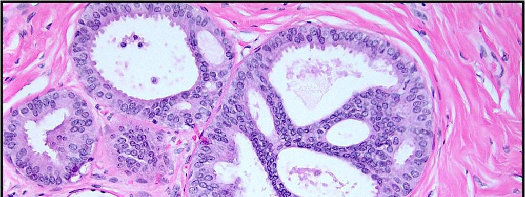
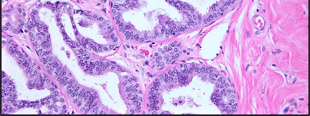
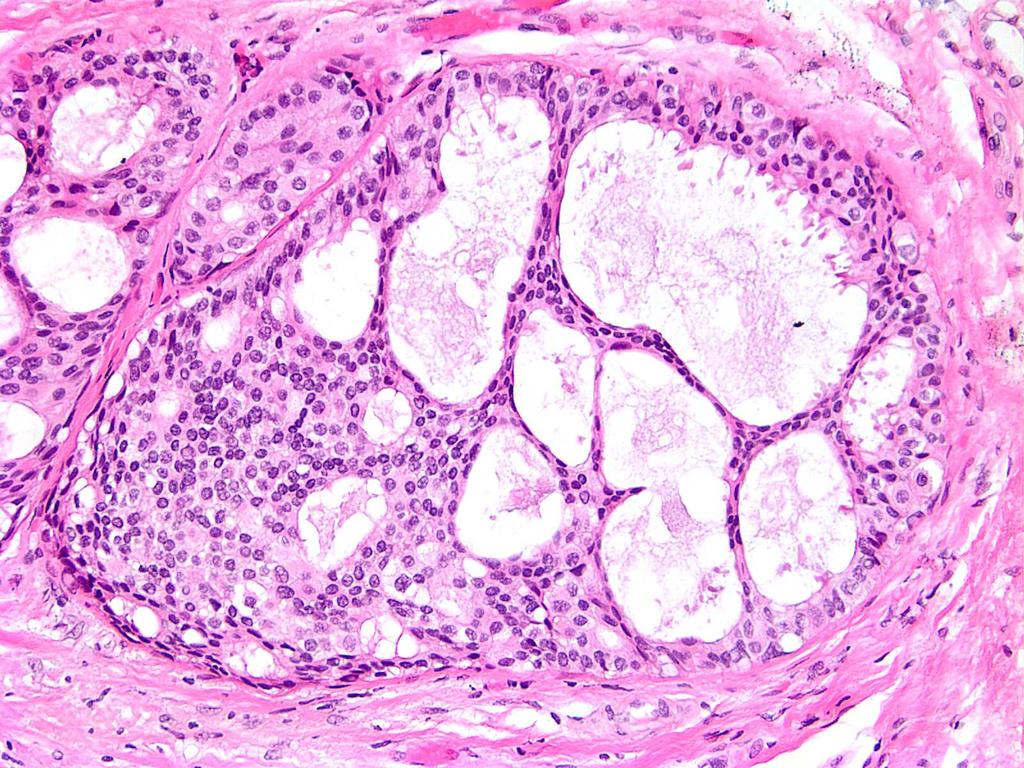
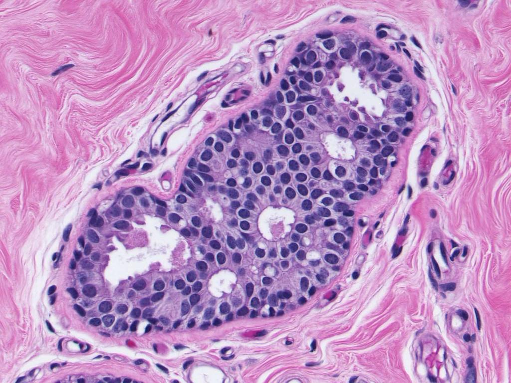
26 Distinction between ADH and DCIS (on CNB) ADH is composed of the same population of atypical epithelial cells as LG DCIS Incompletely filling the space Some features of UDH Comprises 2 spaces or less or 2 mm or less May be difficult to be definitive on CNB
27
28
29
30 Management of ADH vs. DCIS on CNB On CNB, determination of the extent is not possible, it may be more prudent to classify a lesion as ADH bordering on low grade DCIS or severely atypical intraductal proliferation bordering on low grade DCIS Both ADH and DCIS are managed with excisional biopsy As such it may be more appropriate to classify a lesion as ADH rather than labeling a patient with DCIS on a limited amount of tissue

31 Our current practice is to perform excision for patients with ADH diagnosed on CNB
32 Management Problems To Excise or Not to Excise? ADH Lobular neoplasia (ALH, LCIS) Papillary lesions Radial scars Fibroepithelial lesions Columnar cell lesions
33 Management Problems To Excise or Not to Excise? ADH Lobular neoplasia (ALH, LCIS) Papillary lesions Radial scars Fibroepithelial lesions Columnar cell lesions
34 Intraductal Papilloma on CNB Issues of Concern Papilloma vs. papillary DCIS may be difficult, especially with limited material? Representative of lesion as whole - otherwise benign papillomas may harbor foci of ADH or DCIS Limited data available
35 Benign Papilloma on CNB with Excision Follow-up Author # with excision f/u CA on Philpots 6 1 (17%) Liberman 4 0 Ivan 6 0 Renshaw 18 0 Mercado 36 2 (6%) Kil 76 6 (8%) Bernik (36%) Tseng 24 7 (29%) Rizzo (2012) (9%) Linda (2012) 64 4 (6%) Lu (2012) 66 4 (6%) Fu (2012) (6%) Li (2012) (2%) Swapp (2013) 77 0
36 Intraductal Papilloma on CNB Rizzo, J Am Coll Surg, with IDP only 21/234 (9%) upgraded to DCIS or IDC Among many clinical and radiologic variables analyzed, only older age was predictive Recommend excision due to lack of predictors for upgrade
37 Solitary Intraductal Papilloma on CNB Swapp, Ann Surg Oncol, with IDP only and excision 100 with no excision and stable f/u No upgrades to atypia or malignancy Recommend imaging f/u rather than excision for solitary intraductal papillomas with no atypia and radiologic concordance
38 34 studies 2,236 non malignant papillary lesions 346 upgraded to malignant Pooled underestimation rate of 15.7% Rate for benign papillomas =7.0% ( %) Rate for atypical papillomas =36.9% ( %) Ann Surg Oncol 2013

39 Micropapillomas
40 Microscopic Incidental Intraductal Papillomas on CNB Jaffer, Breast J excisions for incidental papilloma 8 fibrocystic change, 5/6 incidental papillomas 1 alteration to targeted papilloma No upgrades to atypia Lee AJR microscopic papillomas Could not determine if incidental or associated with imaging target No upgrades to malignancy BIDMC experience 10% of papillomas (12/121) on CNB represent incidental findings 50% underwent excision with no upgrades to malignancy

41 Targeted benign papilloma on CNB requires excision
42 Management Problems To Excise or Not to Excise? ADH Lobular neoplasia (ALH, LCIS) Papillary lesions Radial scars Fibroepithelial lesions Columnar cell lesions
43 Radial Scar on CNB 7 studies ( ) 7/113 pts (6.1%) with RS on CNB had carcinoma on excision Largest study to date (Brenner, 2002) 157 cases with RS on CNB with either surgical excision (n=102) or 24 month follow-up Malignancy in 13 cases (8.3%) No malignancy if No associated AH CNB >12 specimens Mammotome used
44 Radial Scar on CNB Linda, AJR, women with radial scar diagnosed on US or MRI guided core needle biopsy underwent excision 2 upgrades (3.7%) 1 ILC 1 incidental low grade DCIS Suggest that with negative MRI, may be able to observe patients with radial scar
45 Performance of MRI Linda, ACR, 2012

46 All Patients with Radial Scar on CNB Undergo Excision
47 Management Problems To Excise or Not to Excise? ADH Lobular neoplasia (ALH, LCIS) Papillary lesions Radial scars Fibroepithelial lesions Columnar cell lesions

48 Fibroepithelial Lesions on CNB Dx of fibroadenoma usually readily made on CNB; excision not required.
49 Fibroepithelial Lesions of the Breast Issues for Core Needle Biopsy Predictors of phyllodes tumors on core needle biopsy
50 Fibroepithelial Lesions of the Breast Issues for Core Needle Biopsy Several studies have attempted to stratify cellular fibroepithelial lesions Jacobs et al. (Am J Clin Pathol, 2005) Evaluated 29 FELCS on CNB (16 FA and 12 PT) Lee et al. (Histopathol, 2007) Evaluated 36 PT and 38 FA with prior CNB Jara-Lazaro et al. (Histopathol, 2010) Evaluated 261 CNB of FEL
51 Fibroepithelial Lesions of the Breast Issues for Core Needle Biopsy Jacobs et al. (Am J Clin Pathol, 2005) Evaluated 29 FELCS on CNB (16 FA and 12 PT) Features assessed included: Stromal cellularity (mild, moderate, marked) Stromal cell nuclear atypia Stromal mitotic count Stromal to epithelial ratio (%) Stromal overgrowth (4x field) Infiltrative edge Epithelial hyperplasia Stromal condensation Growth pattern (peri-, intracanalicular, mixed) Leaf-like pattern Multinucleated stromal giant cells
52 FELCS on CNB Jacobs et al. (Am J Clin Pathol, 2005) 80 % of cases Mildly Increased Moderately Increased Markedly Increased Stromal Cellularity
53 Fibroepithelial Lesions of the Breast Issues for Core Needle Biopsy Lee et al. (Histopathol, 2007) Evaluated 36 PT and 38 FA with prior CNB Features assessed included: Stromal cellularity Stromal condensation Stromal cell pleomorphism Stromal overgrowth (10x and 20x) Percentage of lesion composed of stroma Lesion edge (circumscribed, infiltrative, NA) Stromal mitoses Specimen fragmentation Intracanalicular pattern (present vs. absent) Clefting Stromal adipose tissue or heterologous elements
54 Fibroepithelial Lesions of the Breast Issues for Core Needle Biopsy Jara-Lazaro et al. (Histopathol, 2010) Evaluated 261 CNB of FEL (21 FA and 36 PT on excision included in the histologic assessment) Features assessed included: Lesional edge Stromal cellularity Stromal overgrowth (4x) Stromal distribution (condensation) Stromal nuclear atypia Stromal mitoses Ratio of epithelial to stromal elements Leaf-like pattern Epithelial hyperplasia Presence of PASH
55 Fibroepithelial Lesions of the Breast Issues for Core Needle Biopsy Predictors of phyllodes tumors on core needle biopsy Immunohistochemistry
 Evaluated Ki-67, topoisomerase II and")
56 Fibroepithelial Lesions of the Breast Immunohistochemistry Jacobs et al. (Am J Clin Pathol, 2005) Evaluated Ki-67, topoisomerase II and p53
57 Fibroepithelial Lesions of the Breast Immunohistochemistry Jara-Lazaro et al. (Histopathol, 2010) Evaluated Bcl-2, CD117, Ki-67, topoisomerase II and CD34
58 Fibroepithelial Lesions of the Breast Immunohistochemistry Jara-Lazaro et al. (Histopathol, 2010)
59 Fibroepithelial Lesions of the Breast Issues for Core Needle Biopsy Management recommendations include: Excision with margin of normal tissue if two or more features present Less extensive excision if only one feature is present Possibly observation BUT..given sampling issues excision recommended for all cellular fibroepithelial lesions Core needle biopsy diagnosis of fibroadenoma does not completely exclude phyllodes tumor Jacobs, 2005 Lee, 2007 Jara-Lazaro, 2010 Resetkova, 2010
60 Management Problems To Excise or Not to Excise? ADH Lobular neoplasia (ALH, LCIS) Papillary lesions Radial scars Fibroepithelial lesions Columnar cell lesions

61 Columnar Cell Change/Hyperplasia Found on excision: No further treatment Found on CNB: No excision necessary No additional levels obtained
62 Flat Epithelial Atypia FEA on core biopsy Excision required Upgraded in 0-30% of cases Bianchi, Virchow Arch, 2012 Peres, BCRT, 2012 De Mascarel, Mod Pathol, 2011 Lee, Breast Journal, 2010 Ingegnoli, Breast Journal, 2010 Tomasino, J Cell Physiol, 2009 Chivukula, Am J Clin Pathol, 2009 Senetta, Mod Pathol, 2009 Piubello, Am J Surg Pathol, 2009 Kunju, Hum Pathol, 2007


63 Flat Epithelial Atypia FEA on core biopsy Upgraded in 0-30% of cases But need for excision remains uncertain Rad-path correlation required WHO, 2012
64 Management Problems To Excise or Not to Excise? A major role of CNB is to spare patients with probably benign lesions open surgical excision where possible Threshold for recommending open biopsy should be low If there is doubt, take it out
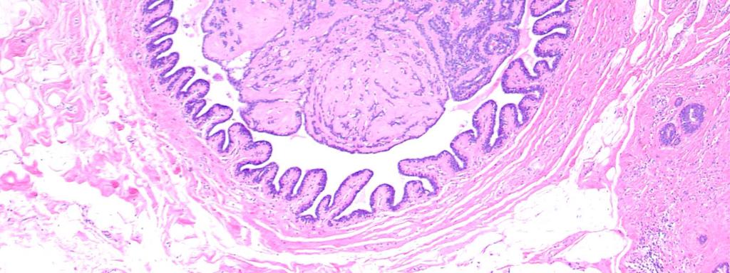
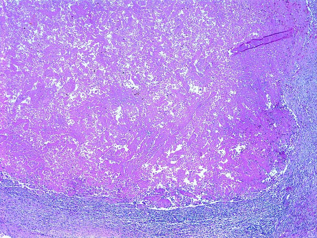
65 Consequences, Complications and Artifacts Related to Core Needle Biopsies Hemorrhage, granulation tissue, scarring and bx site Infarction Epidermoid cysts The missing cancer Epithelial displacement
66 Consequences, Complications and Artifacts Related to Core Needle Biopsies Hemorrhage, granulation tissue, scarring and bx site Infarction Epidermoid cysts The missing cancer Epithelial displacement
67 Consequences, Complications and Artifacts Related to Core Needle Biopsies Hemorrhage, granulation tissue, scarring and bx site Infarction Epidermoid cysts The missing cancer Epithelial displacement
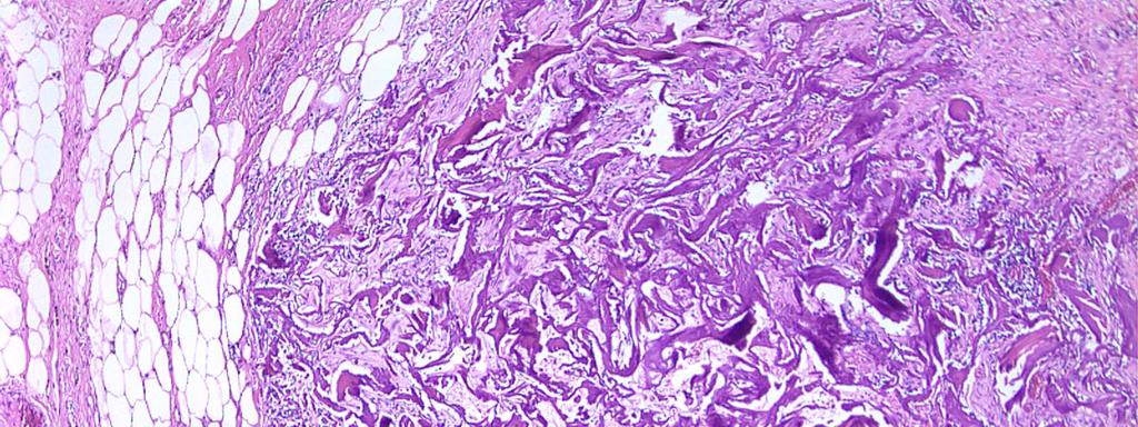
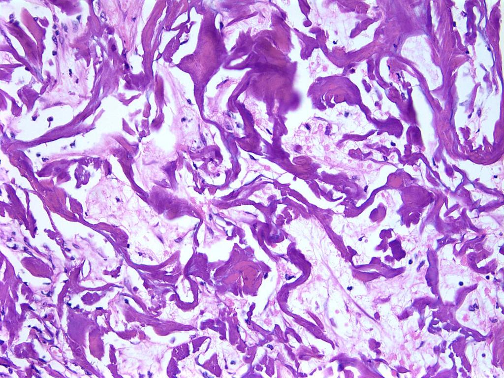
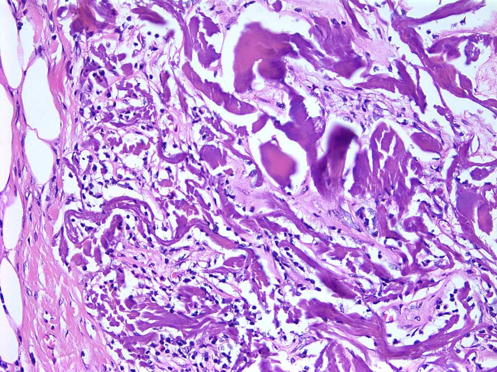
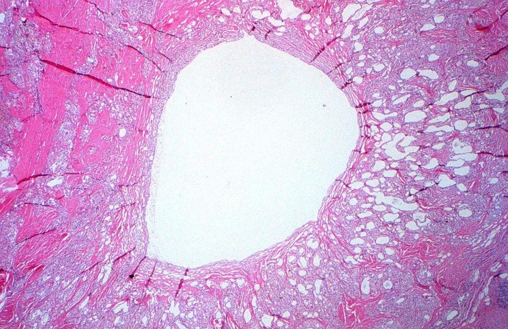
68 Biopsy Site Marking Devices Guarda et al, AJSP 2005 Two major types Pellets of resorbable copolymer of polylactic acid/polyglycolic acid Cell poor fibrotic reaction around empty spaces followed by FBGCR Plug of bovine collagen Eosinophilic hyalinized acellular material with lymphocytic infiltrate Degradation of plug associated with deposition of native collagen Absence of significant FBGCR

69
70
71
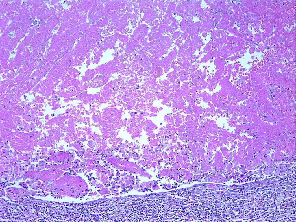
72 Consequences, Complications and Artifacts Related to Core Needle Biopsies Hemorrhage, granulation tissue, scarring and bx site Infarction Epidermoid cysts The missing cancer Epithelial displacement
73
74
75 Consequences, Complications and Artifacts Related to Core Needle Biopsies Hemorrhage, granulation tissue, scarring and bx site Infarction Epidermoid cysts The missing cancer Epithelial displacement

76

77
78 Consequences, Complications and Artifacts Related to Core Needle Biopsies Hemorrhage, granulation tissue, scarring and bx site Infarction Epidermoid cysts The missing cancer Epithelial displacement
79
80 The Missing Cancer Cancer in core; no cancer in surgical specimen Uncommon Likely to be seen increasingly with larger gauge needles and use of mammotome
81 Schematic Representation of Percutaneous Biopsy Techniques Microcalcification Core biopsy CNB 14G VACB 11 or 8 G ABBI or En Bloc Adapted from Wong et al, Adv Anat Pathol 2000;7:26-35
82 The Missing Cancer Patient misidentification False positive CNB Biopsy site not excised Inadequate sampling Lesion entirely removed by CNB Obliteration of residual cancer by healing process
83 Contents of the Surgical Pathology Report Sufficient information to permit radiologic-pathologic correlation and to get patient into appropriate therapeutic algorithm Optimal amount of information to include for cases of invasive cancer controversial Inclusion of too much information can be problematic
84 Which features should be noted in pathology reports of CNB specimens containing an invasive cancer?
85 Assessment of Standard Prognostic Factors on CNB Sharifi, 1999 Factor Agreement Between CNB and Excision Size 21% Histologic type 81% (72-82%)* Histologic grade 75% (62-83%)* Rakha, J Clin Pathol, 2007 Park, Am J Surg, 2009 *range reported in literature
86 Assessment of Standard Prognostic Factors on CNB If size, special histologic type, and/or grade are reported, should have a caveat in this limited sample for size: this represents a minimum size
87 Assessment of Prognostic Factor and Predictive Markers Jacobs, 1998 Agreement Between CNB and Excision ER 100% HER2 100% p53 100% bcl2 100% Rakha, J Clin Pathol, 2007 Park, Am J Surg, 2009 Sutela, Acta Oncol, 2008
88 Assessment of Prognostic and Predictive Markers False negative ER on CNB (Lee, J Clin Pathol, 2008) Intratumor heterogeneity for HER2 (Striebel, Am J Clin Pathol, 2008; Chivukula, Mod Pathol, 2008) Our practice is to perform markers on all CNBs and not on excisions Repeat on excision for aberrant results, negative results or cases of neoadjuvant therapy
89 Conclusions Core needle biopsies represent an important advance in the evaluation of non-palpable breast lesions Pathologists, radiologists, and clinicians need to understand limitations Pathologists need to be aware of the artifacts associated with CNB Radiologic-pathologic correlation is essential in every case
Columnar Cell Lesions
 Columnar Cell Lesions Laura C. Collins, M.D. Department of Pathology Beth Israel Deaconess Medical Center and Harvard Medical School Boston, MA Question? Columnar cell lesions are: a) Annoying lesions
3/27/2017. Disclosure of Relevant Financial Relationships. Papilloma???
 Management of Papillary Lesions Diagnosed at Rad Path Concordant Core Biopsy (CNB) Disclosure of Relevant Financial Relationships USCAP requires that all planners (Education Committee) in a position to
Papillary Lesions of the Breast
 Papillary Lesions of the Breast Texas Society of Pathologists 2013 Laura C. Collins, M.D. Associate Professor of Pathology Associate Director, Division of Anatomic Pathology Beth Israel Deaconess Medical
CNB vs Surgical Excision
 Update on Core Needle Biopsy of Non-palpable Breast Lesions Nour Sneige, M.D. UT MD Anderson Cancer Center Houston, Tx Image-Guided CNB of Breast Lesions An alternative to surgical biospy CNB vs Surgical
Columnar Cell Lesions and Flat Epithelial Atypia
 Columnar Cell Lesions and Flat Epithelial Atypia Laura C. Collins, M.D. Department of Pathology Beth Israel Deaconess Medical Center and Harvard Medical School, Boston, MA Terminology for Columnar Cell
Breast: Difficulties in Core Biopsies
 Breast: Difficulties in Core Biopsies Anna Marie Mulligan, MB, MSc, FRCPath University Health Network and University of Toronto E-mail: annamarie.mulligan@uhn.ca No conflicts of interest Role of Core Needle
Treatment options for the precancerous Atypical Breast lesions. Prof. YOUNG-JIN SUH The Catholic University of Korea
 Treatment options for the precancerous Atypical Breast lesions Prof. YOUNG-JIN SUH The Catholic University of Korea Not so benign lesions? Imaging abnormalities(10% recall) lead to diagnostic evaluation,
04/10/2018 HIGH RISK BREAST LESIONS. Pathology Perspectives of High Risk Breast Lesions ELEVATED RISK OF BREAST CANCER HISTORICAL PERSPECTIVES
 Pathology Perspectives of High Risk Breast Lesions Savitri Krishnamurthy MD Professor of Pathology Deputy Division Head Director of Clinical Trials, Research and Development The University of Texas MD
Enterprise Interest None
 Enterprise Interest None B3 lesions of the breast What are they at surgery? Case 4 Edi Brogi MD PhD Attending Pathologist - Director of Breast Pathology Memorial Sloan Kettering Cancer Center New York
Breast pathology. 2nd Department of Pathology Semmelweis University
 Breast pathology 2nd Department of Pathology Semmelweis University Breast pathology - Summary - Benign lesions - Acute mastitis - Plasma cell mastitis / duct ectasia - Fat necrosis - Fibrocystic change/
Proliferative Breast Disease: implications of core biopsy diagnosis. Proliferative Breast Disease
 Proliferative Breast Disease: implications of core biopsy diagnosis Jean F. Simpson, M.D. Breast Pathology Consultants, Inc. Nashville, TN Proliferative Breast Disease Must be interpreted in clinical and
Flat Epithelial Atypia
 Flat Epithelial Atypia Richard Owings, M.D. University of Arkansas for Medical Sciences Department of Pathology Flat epithelial atypia can be a difficult lesion May be a subtle diagnosis Lots of changes
Columnar Cell Lesions. Columnar Cell Lesions and Flat Epithelial Atypia
 Columnar Cell Lesions and Stuart J. Schnitt, M.D. Beth Israel Deaconess Medical Center and Harvard Medical School Boston, MA, USA Columnar Cell Lesions Lesions characterized by columnar epithelial cells
Ductal Carcinoma in Situ. Laura C. Collins, M.D. Department of Pathology Beth Israel Deaconess Medical Center and Harvard Medical School Boston, MA
 Ductal Carcinoma in Situ Laura C. Collins, M.D. Department of Pathology Beth Israel Deaconess Medical Center and Harvard Medical School Boston, MA Definition of DCIS WHO 2012 A neoplastic proliferation
OUTLINE FIBROADENOMA FIBROADENOMA. FIBROEPITHELIAL LESIONS OF THE BREAST UCSF Current Issues in Anatomic Pathology 2015 FIBROADENOMA PHYLLODES TUMOR
 OUTLINE FIBROADENOMA FIBROEPITHELIAL LESIONS OF THE BREAST UCSF Current Issues in Anatomic Pathology 2015 Gregor Krings, MD PhD Assistant Professor PHYLLODES TUMOR DIFFERENTIAL DIAGNOSIS CELLULAR FIBROEPITHELIAL
Epithelial Columnar Breast Lesions: Histopathology and Molecular Markers
 29th Annual International Conference Advances in the Application of Monoclonal Antibodies in Clinical Oncology and Symposium on Cancer Stem Cells 25 th -27t h June, 2012, Mykonos, Greece Epithelial Columnar
Papillary Lesions of the Breast
 Papillary Lesions of the Breast Laura C. Collins, M.D. Associate Professor of Pathology Associate Director, Division of Anatomic Pathology Beth Israel Deaconess Medical Center and Harvard Medical School
Image guided core biopsies:
 Recommendations on the Surgical, Radiologic and Pathologic Approaches to Breast Disease: Using best practices based on multidisciplinary methodologies developed through the Allina Breast Committee. Image
Atypical Ductal Hyperplasia and Papillomas: A Comparison of Ultrasound Guided Breast Biopsy and Stereotactic Guided Breast Biopsy
 Atypical Ductal Hyperplasia and Papillomas: A Comparison of Ultrasound Guided Breast Biopsy and Stereotactic Guided Breast Biopsy Breast Cancer is the most common cancer diagnosed in women in the United
IBCM 2, April 2009, Sarajevo, Bosnia and Herzegovina
 Preoperative diagnosis and treatment planning in breast cancer The pathologist s perspective L. Mazzucchelli Istituto Cantonale di Patologia Locarno, Switzerland IBCM 2, 23-25 April 2009, Sarajevo, Bosnia
Atypical proliferative lesions diagnosed on core biopsy - 6 year review
 Atypical proliferative lesions diagnosed on core biopsy - 6 year review Dr Angela Harris, Dr Julie Weigner & Dr Ricardo Vilain NSW Health Pathology Pathology North, Hunter Anatomical Pathology & Cytology
Management of B3 lesions
 Management of B3 lesions Pathological view Abeer Shaaban Queen Elizabeth Hospital Birmingham FEA AIDP B3 lesions In situ Lobular neoplasia Papilloma Radial scar Fibroaepithelial lesion Mucocoele like lesion
Disclosures 5/27/2012. Outline of Talk. Outline of Talk. When Is LCIS Clinically Significant? Classic LCIS. Classic LCIS
 When Is LCIS Clinically Significant? Disclosures I have nothing to disclose Yunn-Yi Chen, MD, PhD Professor Outline of Talk Outline of Talk Classic LCIS Classic LCIS Definition of lobular differentiation
Benign Breast Disease and Breast Cancer Risk
 Benign Breast Disease and Breast Cancer Risk Jean F. Simpson, M.D. Vanderbilt University Nashville, Tennessee December 1, 2011 Nashville Nashville Lebanon 1 Cedars of Lebanon State Park The American University
Excisional biopsy or long term follow-up results in breast high-risk lesions diagnosed at core needle biopsy
 Excisional biopsy or long term follow-up results in breast high-risk lesions diagnosed at core needle biopsy Poster No.: C-2515 Congress: ECR 2015 Type: Authors: Scientific Exhibit Ö. S. Okcu 1, A. Oktay
Papillary Lesions of the breast
 Papillary Lesions of the breast Emad Rakha Professor of Breast Pathology The University of Nottingham Papillary lesions of the breast are a heterogeneous group of disease, which are characterised by neoplastic
Guidance on the management of B3 lesions
 Guidance on the management of B3 lesions Lesion diagnosed on 14g or vacuumassisted biopsy (VAB) Risk of upgrade Recommended investigation Suggested approach for follow-up if no malignancy on VAE awaiting
Benign Mimics of Malignancy in Breast Pathology
 Arthur Purdy Stout Society of Surgical Pathologists Companion Meeting Benign Mimics of Malignancy in Breast Pathology Stuart J. Schnitt, M.D. Beth Israel Deaconess Medical Center and Harvard Medical School,
Incidence of ductal lesions
 Ductal Proliferative Lesions of the Breast: From FEA to ADH to DCIS Incidence of ductal lesions Pre-mammography: DCIS < 3% of breast cancers, large palpable masses, with invasion Mammography: DCIS 25%
Papillary Lesions of the Breast A Practical Approach to Diagnosis. (Arch Pathol Lab Med. 2016;140: ; doi: /arpa.
 Papillary Lesions of the Breast A Practical Approach to Diagnosis (Arch Pathol Lab Med. 2016;140:1052 1059; doi: 10.5858/arpa.2016-0219-RA) Papillary lesions of the breast Span the spectrum of benign,
Pathology of Lobular & Ductal Preneoplasia. Syed A Hoda, MD Weill-Cornell, New York, NY
 Pathology of Lobular & Ductal Preneoplasia Syed A Hoda, MD Weill-Cornell, New York, NY Proliferative Epithelial Changes in Breast A wide range of proliferative epithelial changes occur in the breast There
Advocating Nonsurgical Management of Patients With Small, Incidental Radial Scars at the Time of Needle Core Biopsy. A Study of 77 Cases
 Advocating Nonsurgical Management of Patients With Small, Incidental Radial Scars at the Time of Needle Core Biopsy A Study of 77 Cases Cathleen Matrai, MD; Timothy M. D Alfonso, MD; Lindsay Pharmer, MD;
Interpretation of Breast Pathology in the Era of Minimally Invasive Procedures
 Shahla Masood, M.D. Professor and Chair Department of Pathology and Laboratory Medicine University of Florida College of Medicine Jacksonville Medical Director, UF Health Breast Center Chief of Pathology
Controversies on the Management of High Risk Breast Lesions on Core Biopsy: An Update on the Literature
 Controversies on the Management of High Risk Breast Lesions on Core Biopsy: An Update on the Literature Dianne Georgian- Smith MD Brigham and Women s Hospital Associate Professor of Radiology, Harvard
High risk lesions of the breast : Review of the current diagnostic and management strategies
 High risk lesions of the breast : Review of the current diagnostic and management strategies Poster No.: C-1204 Congress: ECR 2016 Type: Educational Exhibit Authors: P. Jagmohan, F. J. Pool, P. G. Pillay,
Surgical Pathology Issues of Practical Importance
 Surgical Pathology Issues of Practical Importance Anne Moore, MD Medical Oncology Syed Hoda, MD Surgical Pathology The pathologist is central to the team approach needed to manage the patient with breast
Proliferative Epithelial lesions of the Breast. Sami Shousha, MD, FRCPath Charing Cross Hospital & Imperial College, London
 Proliferative Epithelial lesions of the Breast Sami Shousha, MD, FRCPath Charing Cross Hospital & Imperial College, London Amman, November2013 Proliferative Epithelial Lesions of the Breast Usual type
The Hot Topic for today is a biopsy from a 58-year-old woman who had worrisome mammographic calcifications on screening.
 The Hot Topic for today is a biopsy from a 58-year-old woman who had worrisome mammographic calcifications on screening. 1 My name is Dan Visscher; I am a consultant in the Division of Anatomic Pathology
A Practical Approach to the Evaluation of Fibroepithelial Lesions. Edi Brogi MD PhD Attending Pathologist Director of Breast Pathology
 A Practical Approach to the Evaluation of Fibroepithelial Lesions Edi Brogi MD PhD Attending Pathologist Director of Breast Pathology Overview Fibroadenomas (FAs) Phyllodes Tumors (PTs) Morphology and
Papillary Lesions of the Breast: WHO Update
 Papillary Lesions of the Breast: WHO Update Stuart J. Schnitt, M.D. Department of Pathology Beth Israel Deaconess Medical Center and Harvard Medical School Boston, MA, USA Papillary Lesions of the Breast
Breast Pathology. Breast Development
 Breast Pathology Lecturer: Hanina Hibshoosh, M.D. Reading: Kumar, Cotran, Robbins, Basic Pathology, 6th Edition, pages 623-635 Breast Development 5th week - thickening of the epidermis - milk line 5th
Anatomic Pathology / Mucocele-like Lesions on Breast Core Biopsy. Mucocele-like Lesions Diagnosed on Breast Core Biopsy
 Anatomic Pathology / Mucocele-like Lesions on Breast Core Biopsy Mucocele-like Lesions Diagnosed on Breast Core Biopsy Assessment of Upgrade Rate and Need for Surgical Excision Brian Sutton, MD, 1 Simone
Non-mass Enhancement on Breast MRI. Aditi A. Desai, MD Margaret Ann Mays, MD
 Non-mass Enhancement on Breast MRI Aditi A. Desai, MD Margaret Ann Mays, MD Breast MRI Important screening and diagnostic tool, given its high sensitivity for breast cancer detection Breast MRI - Indications
RSNA, /radiol Appendix E1. Methods
 RSNA, 2016 10.1148/radiol.2016151097 Appendix E1 Methods US and Near-infrared Data Acquisition Four optical wavelengths (740 nm, 780 nm, 808 nm, and 830 nm) were used to sequentially deliver the light
Quality ID #263: Preoperative Diagnosis of Breast Cancer National Quality Strategy Domain: Effective Clinical Care
 Quality ID #263: Preoperative Diagnosis of Breast Cancer National Quality Strategy Domain: Effective Clinical Care 2018 OPTIONS FOR INDIVIDUAL MEASURES: REGISTRY ONLY MEASURE TYPE: Process DESCRIPTION:
Mammographic features and correlation with biopsy findings using 11-gauge stereotactic vacuum-assisted breast biopsy (SVABB)
") Original article Annals of Oncology 14: 450 454, 2003 DOI: 10.1093/annonc/mdh088 Mammographic features and correlation with biopsy findings using 11-gauge stereotactic vacuum-assisted breast biopsy (SVABB)
Original article Annals of Oncology 14: 450 454, 2003 DOI: 10.1093/annonc/mdh088 Mammographic features and correlation with biopsy findings using 11-gauge stereotactic vacuum-assisted breast biopsy (SVABB)
CLINICAL SIGNIFICANCE OF BENIGN EPITHELIAL CHANGES
 Papillomas. Papillomas are composed of multiple branching fibrovascular cores, each having a connective tissue axis lined by luminal and myoepithelial cells ( Fig. 23-11 ). Growth occurs within a dilated
Utility of Adequate Core Biopsy Samples from Ultrasound Biopsies Needed for Today s Breast Pathology
 Utility of Adequate Core Biopsy Samples from Ultrasound Biopsies Needed for Today s Breast Pathology Ugur Ozerdem, M.D. 1 Abstract Background: There is a paradigm shift in breast biopsy philosophy. In
The management of B3 lesions with emphasis on lobular neoplasia
 The management of B3 lesions with emphasis on lobular neoplasia Abeer Shaaban Queen Elizabeth Hospital Birmingham NHSBSP core biopsy categories B1 - Normal B2 - Benign B3 Uncertain malignant potential
Emad A Rakha, Bernard Chi-Shern Ho, Veena K Naik, Soumadri Sen, Lisa Hamilton, Zsolt Hodi, Ian Ellis, Andrew Hs Lee
 Outcome of breast lesions diagnosed as lesion of uncertain malignant potential (B3) or suspicious of malignancy (B4) on needle core biopsy including detailed review of epithelial atypia Emad A Rakha, Bernard
6/3/2010. Outline of Talk. Lobular Breast Cancer: Definition of lobular differentiation. Common Problems in Diagnosing LCIS in Core Biopsies
 Outline of Talk Lobular Breast Cancer: Common Problems in Diagnosing LCIS in Core Biopsies Definition of lobular differentiation Variants of LCIS that: carry risk for unsampled invasive cancer mimic DCIS
Atypical papillary lesions after core needle biopsy and subsequent breast carcinoma
 Asian Biomedicine Vol. 5 No. 2 April 2011; 243-248 DOI: 10.5372/1905-7415.0502.031 Original article Atypical papillary lesions after core needle biopsy and subsequent breast carcinoma Tuenchit Khamapirad
Women s Imaging Original Research
 Women s Imaging Original Research Villa et al. Biopsy of Atypical Ductal Hyperplasia Women s Imaging Original Research WOMEN S IMAGING Alessandro Villa 1 Alberto Tagliafico 2 Fabio Chiesa 1 Maurizio Chiaramondia
04/10/2018. Intraductal Papillary Neoplasms Of Breast INTRADUCTAL PAPILLOMA
 Intraductal Papillary Neoplasms Of Breast Savitri Krishnamurthy MD Professor of Pathology Deputy Division Head The University of Texas MD Anderson Cancer Center 25 th Annual Seminar in Pathology Pittsburgh,
Original Report. Mucocele-Like Tumors of the Breast: Mammographic and Sonographic Appearances. Katrina Glazebrook 1 Carol Reynolds 2
 Katrina Glazebrook 1 Carol Reynolds 2 Received January 2, 2002; accepted after revision August 28, 2002. 1 Department of Radiology, Mayo Clinic, 200 First St. S.W., Rochester, MN 55905. Address correspondence
1 NORMAL HISTOLOGY AND METAPLASIAS
 1 NORMAL HISTOLOGY AND METAPLASIAS, MD Anatomy and Histology 1 Metaplasias 2 ANATOMY AND HISTOLOGY The female breast is composed of a branching duct system, which begins at the nipple with the major lactiferous
Diseases of the breast (1 of 2)
") Diseases of the breast (1 of 2) Introduction A histology introduction Normal ducts and lobules of the breast are lined by two layers of cells a layer of luminal cells overlying a second layer of myoepithelial
Diseases of the breast (1 of 2) Introduction A histology introduction Normal ducts and lobules of the breast are lined by two layers of cells a layer of luminal cells overlying a second layer of myoepithelial
Breast Intraductal Papillomas Without Atypia in Radiologic- Pathologic Concordant Core-Needle Biopsies: Rate of Upgrade to Carcinoma at Excision
 Breast Intraductal Papillomas Without Atypia in Radiologic- Pathologic Concordant Core-Needle Biopsies: Rate of Upgrade to Carcinoma at Excision Fresia Pareja, MD, PhD 1 ; Adriana D. Corben, MD 1 ; Sandra
LYMPHATIC DRAINAGE AXILLARY (MOSTLY) INTERNAL MAMMARY SUPRACLAVICULAR
 INTERNAL MAMMARY SUPRACLAVICULAR") BREAST LYMPHATIC DRAINAGE AXILLARY (MOSTLY) INTERNAL MAMMARY SUPRACLAVICULAR HISTOLOGY LOBE: (10 in whole breast) LOBULE: (many per lobe) ACINUS/I, aka ALVEOLUS/I: (many per lobule) DUCT(S): INTRA- or
BREAST LYMPHATIC DRAINAGE AXILLARY (MOSTLY) INTERNAL MAMMARY SUPRACLAVICULAR HISTOLOGY LOBE: (10 in whole breast) LOBULE: (many per lobe) ACINUS/I, aka ALVEOLUS/I: (many per lobule) DUCT(S): INTRA- or
Lesion Imaging Characteristics Mass, Favoring Benign Circumscribed Margins Intramammary Lymph Node
 Lesion Imaging Characteristics Mass, Favoring Benign Circumscribed Margins Intramammary Lymph Node Oil Cyst Mass, Intermediate Concern Microlobulated Margins Obscured Margins Mass, Favoring Malignant Indistinct
Ductal Proliferations of the Breast: The Good, the Bad, and the Ugly
 Ductal Proliferations of the Breast: The Good, the Bad, and the Ugly Melinda F. Lerwill, MD CRITERIA FOR DISTINGUISHING LOW-GRADE DUCTAL CARCINOMA IN SITU FROM USUAL DUCTAL HYPERPLASIA CYTOLOGY Low-grade
Cytyc Corporation - Case Presentation Archive - March 2002
 FirstCyte Ductal Lavage History: 68 Year Old Female Gail Index: Unknown Clinical History: Negative Mammogram in 1995 6 yrs. later presents with bloody nipple discharge Subsequent suspicious mammogram Suspicious
Diagnostic benefits of ultrasound-guided. CNB) versus mammograph-guided biopsy for suspicious microcalcifications. without definite breast mass
 versus mammograph-guided biopsy for suspicious microcalcifications. without definite breast mass") Volume 118 No. 19 2018, 531-543 ISSN: 1311-8080 (printed version); ISSN: 1314-3395 (on-line version) url: http://www.ijpam.eu ijpam.eu Diagnostic benefits of ultrasound-guided biopsy versus mammography-guided
Volume 118 No. 19 2018, 531-543 ISSN: 1311-8080 (printed version); ISSN: 1314-3395 (on-line version) url: http://www.ijpam.eu ijpam.eu Diagnostic benefits of ultrasound-guided biopsy versus mammography-guided
Underestimation of cancer in case of diagnosis of atypical ductal hyperplasia (ADH) by vacuum assisted core needle biopsy
 by vacuum assisted core needle biopsy") reports of practical oncology and radiotherapy 1 7 (2 0 1 2) 129 133 Available online at www.sciencedirect.com journal homepage: http://www.elsevier.com/locate/rpor Original research article Underestimation
reports of practical oncology and radiotherapy 1 7 (2 0 1 2) 129 133 Available online at www.sciencedirect.com journal homepage: http://www.elsevier.com/locate/rpor Original research article Underestimation
FIBROEPITHELIAL LESIONS
 DEFINITIONS FIBROEPITHELIAL LESIONS Suzanne Moore FIBROADENOMA- A discrete benign tumour showing evidence of connective tissue and epithelial proliferation- WHO Fibrous stromal element of these tumours
Case study 1. Rie Horii, M.D., Ph.D. Division of Pathology Cancer Institute Hospital, Japanese Foundation for Cancer Research
 NCCN/JCCNB Seminar in Japan April 15, 2012 Case study 1 Rie Horii, M.D., Ph.D. Division of Pathology Cancer Institute Hospital, Japanese Foundation for Cancer Research Present illness: A 50y.o.premenopausal
Breast core needle biopsy: issues and controversies
 S36 & 2010 USCAP, Inc. All rights reserved 0893-3952/10 $32.00 Breast core needle biopsy: issues and controversies Michael Bilous Institute of Clinical Pathology and Medical Research, Westmead Hospital
Surgical Management of High- Risk Breast Lesions
 Surgical Management of High- Risk Breast Lesions Amy C. Degnim, MD a, Tari A. King, MD b, * KEYWORDS High-risk lesion Atypical hyperplasia Lobular carcinoma in situ Percutaneous breast biopsy Breast cancer
Journal of Breast Cancer
 Journal of Breast Cancer ORIGINAL ARTICLE J Breast Cancer 2012 December; 15(4): 407-411 Validation of a Scoring System for Predicting Malignancy in Patients Diagnosed with Atypical Ductal Hyperplasia Using
Diagnostic accuracy of ultrasonography-guided core needle biopsy for breast lesions
 Singapore Med J 01; 5(1) 40 Diagnostic accuracy of ultrasonography-guided core needle biopsy for breast lesions Wiratkapun Cl, MD, Treesit T1, MD, Wibulpolprasert E1, MD, Lertsithichai P, MD, MSc INTRODUCTION
COMMON BENIGN DISORDERS AND DISEASES OF THE BREAST
 COMMON BENIGN DISORDERS AND DISEASES OF THE BREAST Aberrations of Normal Development and Involution (ANDI). The basic principles underlying the aberrations of normal development and involution (ANDI) classification
Mousa. Israa Ayed. Abdullah AlZibdeh. 0 P a g e
 1 Mousa Israa Ayed Abdullah AlZibdeh 0 P a g e Breast pathology The basic histological units of the breast are called lobules, which are composed of glandular epithelial cells (luminal cells) resting on
Stereotactic 11-Gauge Vacuum- Assisted Breast Biopsy: A Validation Study
 Georg Pfarl 1 Thomas H. Helbich 1 Christopher C. Riedl 1 Teresa Wagner 2 Michael Gnant 3 Margaretha Rudas 4 Laura Liberman 5 Received March 11, 2002; accepted after revision May 17, 2002. 1 Department
Benign, Reactive and Inflammatory Lesions of the Breast
 Benign, Reactive and Inflammatory Lesions of the Breast Marilin Rosa, MD Associate Member Section Head of Breast Pathology Department of Anatomic Pathology Program Director, Breast Pathology Fellowship
Pitfalls and Limitations of Breast MRI. Susan Orel Roth, MD Professor of Radiology University of Pennsylvania
 Pitfalls and Limitations of Breast MRI Susan Orel Roth, MD Professor of Radiology University of Pennsylvania Objectives Review the etiologies of false negative breast MRI examinations Discuss the limitations
A E K Ibrahim, A C Bateman, J M Theaker, J L Low, B Addis, P Tidbury, C Rubin, M Briley, G T Royle
 J Clin Pathol 2001;54:121 125 121 Departments of Histopathology and Cytopathology,, Tremona Road,, SO16 6YO, UK A E K Ibrahim A C Bateman J M Theaker B Addis P Tidbury Medical Statistics and Computing,
Atypical ductal hyperplasia diagnosed at ultrasound guided biopsy of breast mass
 Atypical ductal hyperplasia diagnosed at ultrasound guided biopsy of breast mass Poster No.: C-1483 Congress: ECR 2014 Type: Authors: Keywords: DOI: Scientific Exhibit J. Cho, J. Chung, E. S. Cha, J. E.
Ultrasound of the Breast BASICS FOR THE ORDERING CLINICIAN
 Ultrasound of the Breast BASICS FOR THE ORDERING CLINICIAN Breast Ultrasound Anatomy Skin Breast Parenchyma Pectoralis Fascia Pectoralis Breast Ultrasound Anatomy Indications for Breast Ultrasound Palpable
Imaging-Guided Core Needle Biopsy of Papillary Lesions of the Breast
 Eric L. Rosen 1 Rex C. Bentley 2 Jay A. Baker 1 Mary Scott Soo 1 Received January 30, 2002; accepted after revision April 12, 2002. 1 Department of Radiology, Breast Imaging Division, Duke University Medical
ACRIN 6666 Therapeutic Surgery Form
 S1 ACRIN 6666 Therapeutic Surgery Form 6666 Instructions: Complete a separate S1 form for each separate area of each breast excised with the intent to treat a cancer (e.g. each lumpectomy or mastectomy).
Papillary lesions of the breast - Imaging findings and diagnostic challenges
 Papillary lesions of the breast - Imaging findings and diagnostic challenges Poster No.: R-0146 Congress: RANZCR-AOCR 2012 Type: Educational Exhibit Authors: P. Jagmohan, F. J. Pool Keywords: Breast, Mammography,
BREAST PATHOLOGY. Fibrocystic Changes
 BREAST PATHOLOGY Lesions of the breast are very common, and they present as palpable, sometimes painful, nodules or masses. Most of these lesions are benign. Breast cancer is the 2 nd most common cause
Disclosures. Premalignant Lesions of the Breast: What Clinicians Want and Why. NY Times: Prone to Error: Earliest Steps to Find Cancer.
 Disclosures Premalignant Lesions of the Breast: What Clinicians Want and Why I have nothing to disclose Rick Baehner, MD Assistant Professor, UCSF Pathology NY Times: Prone to Error: Earliest Steps to
Breast Lesion Excision System-Intact (BLES): A Stereotactic Method of Biopsy of Suspicius Non-Palpable Mammographic Lesions.
: A Stereotactic Method of Biopsy of Suspicius Non-Palpable Mammographic Lesions.") Breast Lesion Excision System-Intact (BLES): A Stereotactic Method of Biopsy of Suspicius Non-Palpable Mammographic Lesions. Poster No.: C-1595 Congress: ECR 2014 Type: Authors: Scientific Exhibit I. Georgiou
Breast Lesion Excision System-Intact (BLES): A Stereotactic Method of Biopsy of Suspicius Non-Palpable Mammographic Lesions. Poster No.: C-1595 Congress: ECR 2014 Type: Authors: Scientific Exhibit I. Georgiou
BI-RADS CATEGORIZATION AND BREAST BIOPSY categorization in the selection of appropriate breast biopsy technique is also discussed. Patients and method
 Original Article Positive Predictive Value of BI-RADS Categorization in an Asian Population Yah-Yuen Tan, Siew-Bock Wee, Mona P.C. Tan and Bee-Kiang Chong, 1 Departments of General Surgery and 1Diagnostic
Diagnosis of Fibroepithelial and Mesenchymal Lesions on Core Needle Biopsy
 Diagnosis of Fibroepithelial and Mesenchymal Lesions on Core Needle Biopsy Emmanuel Agosto-Arroyo, MD Assistant Member Department of Anatomic Pathology 3/3/2018 Disclosure There are no conflicts of interest.
Diagnostic Problems in Breast Pathology How to avoid the pitfalls
 FORUM OF PATHOLOGY Diagnostic Problems in Breast Pathology How to avoid the pitfalls Professor C W Elston City Hospital Nottingham, United Kingdom Introduction Almost any breast lesion may produce diagnostic
Low-grade Adenosquamous Carcinoma Coexisting with Sclerosing Adenosis of the Breast: A Case Report
 31 Case Report J. St. Marianna Univ. Vol. 8, pp. 31 35, 2017 Low-grade Adenosquamous Carcinoma Coexisting with Sclerosing Adenosis of the Breast: A Case Report Ryoko Oi 1, 2, Ichiro Maeda 1, Yoshio Aida
Short Term Cancer Risk
 in a Cohort of 2312 Women with High Risk Breast Lesions Kubat E 1, Puligandla B 2, Collins L 3, Jiang SF 4, Callahan M 5, Kutner S 6, Habel L 4, Shim V 1 1 Surgery, Kaiser Permanente, Oakland, CA; 2 Pathology,
Breast Intraductal Papillomas without Atypia in Radiologic-Pathologic Concordant. Core Needle Biopsies: Predictors of Upgrade to Carcinoma at Excision
 Page 1 of 28 Breast Intraductal Papillomas without Atypia in Radiologic-Pathologic Concordant Core Needle Biopsies: Predictors of Upgrade to Carcinoma at Excision Fresia Pareja MD PhD 1, *, Adriana Corben
Underestimation of Atypical Ductal Hyperplasia at Sonographically Guided Core Biopsy of the Breast
 Women s Imaging Original Research Jang et al. Sonographic Breast Biopsy Women s Imaging Original Research WOMEN S IMAGING Underestimation of Atypical Ductal Hyperplasia at Sonographically Guided Core Biopsy
Radiologic and pathologic correlation of non-mass like breast lesions on US and MRI: Benign, high risk, versus malignant
 Radiologic and pathologic correlation of non-mass like breast lesions on US and MRI: Benign, high risk, versus malignant Poster No.: C-1161 Congress: ECR 2013 Type: Educational Exhibit Authors: J. Kwak,
In situ lobular neoplasia of the breast with marked myoepithelial proliferation
 In situ lobular neoplasia of the breast with marked myoepithelial proliferation Sami Shousha To cite this version: Sami Shousha. In situ lobular neoplasia of the breast with marked myoepithelial proliferation.
Index. C Calcifications fat necrosis 1, 61 fat necrosis 4, 69 nipple/peri-areolar involvement 1, 165
 A ADH. See Atypical ductal hyperplasia (ADH) American College of Radiology (ACR), BI-RADS background parenchymal enhancement, 8, 9, 81, 82 fibroglandular tissue guidelines, 6 American Joint Committee on
PURPOSE IMAGE-GUIDANCE MODALITIES IMAGE-GUIDED BREAST BIOPSY. US-Techniques. Ultrasound. US guided NLOBB. TH. Helbich
 IMAGE-GUIDED BREAST BIOPSY PURPOSE TH. Helbich Department of Radiology Division of Molecular & Gender Imaging Medical University of Vienna Imaging techniques Interventional procedures Quality management
Abid Irshad, MD Director Breast Imaging. Medical University of South Carolina Charleston
 Abid Irshad, MD Director Breast Imaging Medical University of South Carolina Charleston Cases Financial disclosure: I or my family have no financial interest related to the material discussed in this presentation
Classic lobular neoplasia on core biopsy: a clinical and radio-pathologic correlation study with follow-up excision biopsy
 762 & 2013 USCAP, Inc All rights reserved 0893-3952/13 $32.00 Classic lobular neoplasia on core biopsy: a clinical and radio-pathologic correlation study with follow-up excision biopsy Shweta Chaudhary
Clinician s Guide to Imaging and Pathologic Findings in Benign Breast Disease. Lonzetta Neal, MD; Cindy L. Tortorelli, MD; and Aziza Nassar, MD
 CONCISE REVIEW FOR CLINICIANS GUIDE TO IMAGING AND PATHOLOGIC FINDINGS IN BENIGN BREAST DISEASE Clinician s Guide to Imaging and Pathologic Findings in Benign Breast Disease Lonzetta Neal, MD; Cindy L.
CURRICULUM FOR THE BREAST PATHOLOGY ROTATION UNIVERSITY OF FLORIDA DEPARTMENT OF PATHOLOGY
 CURRICULUM FOR THE BREAST PATHOLOGY ROTATION UNIVERSITY OF FLORIDA DEPARTMENT OF PATHOLOGY JULY, 2003 The following is a conceptual curriculum and set of guidelines for Pathology Residents on the Breast