Model 3-50B or 3-88 III VIII. Olfactory Nerve. Optic Nerve. Oculomotor Nerve. Trochlear Nerve. Trigeminal Nerve. Abducens Nerve.
|
|
|
- Phebe Neal
- 6 years ago
- Views:
Transcription
1

2 Model 3-50B or 3-88 I Olfactory Nerve II Optic Nerve Oculomotor Nerve III IV Trochlear Nerve Trigeminal Nerve V VI Abducens Nerve Glossopharyngeal Nerve IX VII Facial Nerve VIII Vestibocochlear Nerve or Auditory Nerve XII Hypoglossal Nerve Vagus Nerve X Accessory Nerve XI
 Primary Gustatory")
 Motor Speech")
3 Model 3-59 Primary Sensory Area (Postcentral Gyrus) Primary Gustatory Area Primary Auditory Area Primary Motor Area (Precentral Gyrus) Motor Speech Primary Visual Area

4 Brain from Torso Model 3-1 Parietal Lobe Central Sulcus Frontal Lobe Occipital Lobe Lateral Fissure Cerebellum Temporal Lobe
 Motor Speech Area Primary Auditory Area Frontal Lobe Occipital Lobe Primary Visual Area Temporal")
5 Brain from Torso Model 3-44 Parietal Lobe Primary Gustatory Area Primary Sensory Area (Postcentral Gyrus) Primary Motor Area (Precentral Gyrus) Motor Speech Area Primary Auditory Area Frontal Lobe Occipital Lobe Primary Visual Area Temporal Lobe

 Axon Nissl Bodies")
6 Schwann Cell Motor Neuron Model Unlabeled Axoplasm Axolemma Neurolemma Schwann Cell Membrane Endoneurium Myelin Sheath Nodes of Ranvier Dendrite Synaptic knob Neuron Cell Body Axon Hillock Trigger Zone Nucleolus (in Nucleus) Axon Nissl Bodies Neurofibrils

7 Model 3-84 Posterior Horn Arachnoid Mater Posterior Column Dura Mater Posterior Median Sulcus Pia Mater Central Canal Dorsal root White or Gray? White Lateral Column Dorsal root ganglion Ventral Root Lateral Horn White or Gray? Gray Anterior Column Anterior median fissure Ventral Horn

8 Myelinated Model 1-14 Myelinated or Unmyelinated? Dorsal root ganglion Subarachnoid Space Posterior Horn Arachnoid Mater Blue Posterior Column Gray Dura Mater Posterior Median Sulcus Central Canal Dorsal root Lateral Column Spinal Nerve Ventral Root Lateral Horn Ventral Horn Pia Mater Pink Anterior Column Anterior median fissure Myelinated or Unmyelinated? Unmyelinated


9 Model 3-66 Dorsal root ganglion Posterior Column Posterior Median Sulcus Central Canal Posterior Horn Dorsal root Lateral Column Ventral Root Lateral Horn Ventral Horn Anterior median fissure Anterior Column


10 Model 3-75 Posterior Median Sulcus Dorsal root ganglion Dorsal root Sensory or Motor? Sensory Posterior Column Ventral Root Sensory or Motor? Motor Posterior Horn Central Canal Lateral Column Lateral Horn Ventral Horn Anterior Column Anterior median fissure
 Sulci (pl.")
 Gyri (pl.")
11 Cerebral Cortex Sulcus (sing.) Sulci (pl.) Valley Gyrus (sing.) Gyri (pl.) Hill Cerebral Medulla

12 Cerebral Peduncles Lateral & 3 rd Ventricals
 Superior Colliculus Inferior Colliculus Arbor vitae Central Canal Spinal Cord Model")
13 Septum pellucidum Corpus Callosum Lateral Ventricle Intermediate mass Thalamus Hypothalamus Cerebral Peduncles Cerebral Aqueduct Pons Medulla Forth Ventricle Fornix Choriod plexus Third Ventricle Pineal body (gland) Superior Colliculus Inferior Colliculus Arbor vitae Central Canal Spinal Cord Model 3-54
 Fornix Hypothalamus Optic Chiasma Mamillary Body 4 th Ventricle Pons Spinal Cord Medulla")
14 Model 3-50B or 3-88 Intermediate Mass Cerebellum Thalamus Pineal Gland Superior Colliculus Inferior Colliculus Cerebral Peduncles Cerebral Aqueduct Arbor Vitae of Cerebellum Corpus Callosum Septum Pellucidum (membrane) Fornix Hypothalamus Optic Chiasma Mamillary Body 4 th Ventricle Pons Spinal Cord Medulla Infundibulum


15 Lateral Ventricles Foramen of Monro 3 rd Ventricle Foramen of Monro Lateral Ventricle Foramen Of Menro Choroid Plexus Pink Forth Ventricle

16 Sheep Brain Optic Chiasm Olfactory Bulb Cerebral Hemisphere Optic Nerve Mammillary Body Cerebral Peduncle Pons Cerebellum Medulla Spinal Cord

17 Cerebellum w/ Arbor Vitae Massa Intermedia Pineal Body Thalamus Superior Colliculus Fornix Corpus Callosum Inferior Colliculus Central Canal Forth Ventricle Lateral Ventricle Arbor Vitae Olfactory bulb Spinal Cord Medulla Cerebral peduncles Third Ventricle Hypothalamus Optic Chiasma Pons

18 Sheep Brain (Inferior View) Olfactory Bulb (nerve) Optic Nerve Optic Chiasm Mammillary Body Cerebral Pedeuncles Pons Cerebellum Medulla Spinal Cord
19 Sheep Brain (Superior View) Cerebrum (Cerebral Hemispheres) Sulcus (Valley) Longitudinal Fissure Gyrus (Hill) Cerebellum Spinal Cord
 Cerebrum (Cerebral")
 Longitudinal Fissure")
20 Dura Mater Sheep Brain (Superior View) Cerebrum (Cerebral Hemispheres) Gyrus (Hill) Sulcus (Valley) Longitudinal Fissure Cerebellum Spinal Cord


21 Association Neuron Sensory Axon Sensory Nerve Cell Body Receptor Sensory Dendrite Motor Axon Effector Motor Neuron

22 Sensory Nerve Cell Body Sensory Axon Reflex Arc (Model 3-90) Association Neuron Motor Neuron Motor Axon Sensory Dendrite Effector Receptor green dot
23 Endocrine Review

24 Midsagittal Head Model (3-54) Hypothalamus Pineal Gland Pituitary Gland
 Hypothalamus Pituitary Gland")
25 Pineal Gland Brain Model (3-50B or 3-83) Hypothalamus Pituitary Gland
 Anterior Pituitary (Dark")
26 Posterior Pituitary (Light Stain) Anterior Pituitary (Dark Stain)
 Anterior Pituitary (Dark")
27 Posterior Pituitary (Light Stain) Anterior Pituitary (Dark Stain)


28 Thyroid Gland Thyroid Gland Adrenal Gland


29 Thyroid Gland Orange Yellow Dots On Back? Parathyroid Glands

30 Thyroid Gland Slide

31 Thyroid Gland Slide

32 Pancreas
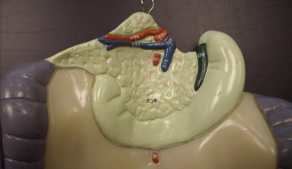
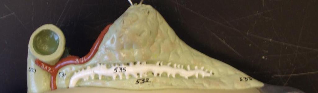
33 Pancreas

34 Islets of Langerhans (Pancreas) Endocrine Portion of Pancreas Called Islets of Langerhans Exocrine Portion of Pancreas
 Can you see the")
35 Islets of Langerhans (pancreas) Can you see the Islet?

36 Islets of Langerhans (Pancreas) Which hormone from these structures lowers blood glucose levels? Which hormone from these structures raises blood glucose levels? Insulin Glucagon
 Capsule Adrenal Medulla Zona")
37 Adrenal Gland Slide Zona Reticularis (Cortex) Zona Fasciculata (Cortex) Capsule Adrenal Medulla Zona Glomerulus (Cortex)
 Zona Fasciculata (Cortex) Zona Glomerulosa")
38 Adrenal Gland Slide Adrenal Medulla Capsule Zona Reticulans (Cortex) Zona Fasciculata (Cortex) Zona Glomerulosa (Cortex)
 Zona Fasciculata (Cortex) Adrenal")
39 Adrenal Gland Slide Zona Glomerulus (Cortex) Capsule Zona Reticularis (Cortex) Zona Fasciculata (Cortex) Adrenal Medulla
Laboratory Manual for Comparative Anatomy and Physiology Figure 15.1 Transparency Master 114
 Neuron Capillary Astrocyte Microglial cell Neuron Fluid-filled cavity Process of oligodendrocyte Ependymal cells Brain or spinal cord tissue Myelin sheath Nerve fibers Figure 15.1 Transparency Master 114
ACTIVITY 7: NERVOUS SYSTEM HISTOLOGY, BRAIN, CRANIAL NERVES
 ACTIVITY 7: NERVOUS SYSTEM HISTOLOGY, BRAIN, CRANIAL NERVES LABORATORY OBJECTIVES: 1. Histology: Identify structures indicated on three different slides or images of nervous system tissue. These images
Blood supply to the brain Blood brain barrier isolates neural tissue from general circulation
 The Brain and Cranial Nerves Objectives Name the major regions of the brain and describe their functions. Discuss the formation, circulation, and functions of the CSF. List the main components of the medulla
ACTIVITY 7: NERVOUS SYSTEM HISTOLOGY, BRAIN, CRANIAL NERVES NERVOUS SYSTEM TISSUES: HISTOLOGY SLIDES
 ACTIVITY 7: NERVOUS SYSTEM HISTOLOGY, BRAIN, CRANIAL NERVES OBJECTIVES: 1) How to get ready: Read Chapter 14 & 15 McKinley et al., Human Anatomy, 4e. All text references are for this textbook. Read dissection
Student Lab #: Date. Lab: Gross Anatomy of Brain Sheep Brain Dissection Organ System: Nervous Subdivision: CNS (Central Nervous System)
") Lab: Gross Anatomy of Brain Sheep Brain Dissection Organ System: Nervous Subdivision: CNS (Central Nervous System) Student Lab #: Date 1 Objectives: 1. Learn the main components making up a motor neuron.
Lab: Gross Anatomy of Brain Sheep Brain Dissection Organ System: Nervous Subdivision: CNS (Central Nervous System) Student Lab #: Date 1 Objectives: 1. Learn the main components making up a motor neuron.
Sheep Brain Dissection
 Sheep Brain Dissection Mammalian brains have many features in common. Human brains may not be available, so sheep brains often are dissected as an aid to understanding the mammalian brain since he general
The Brain and Cranial Nerves Pg. 129
 The Brain and Cranial Nerves Pg. 129 Three Main Regions of the Brain Forebrain Cerbral hemispheres Diencephalon Midbrain Brain stem Hindbrain Pons Cerebellum Medulla oblongata Forebrain Interprets sensory
The Brain and Cranial Nerves Pg Three Main Regions of the Brain. Forebrain
 The Brain and Cranial Nerves Pg. 129 Three Main Regions of the Brain Forebrain Cerbral hemispheres Diencephalon Midbrain Brain stem Hindbrain Pons Cerebellum Medulla oblongata Interprets sensory inputs
I. Anatomy of the Brain A. Cranial Meninges and Ventricles of the Brain 1. Meninges a. Dura mater 1) Endosteal/Periosteal Layer - Outer 2) Meningeal
 Endosteal/Periosteal Layer - Outer 2) Meningeal") I. Anatomy of the Brain A. Cranial Meninges and Ventricles of the Brain 1. Meninges a. Dura mater 1) Endosteal/Periosteal Layer - Outer 2) Meningeal Layer - Inner 3) Falx cerebri a) Superior sagittal sinus
I. Anatomy of the Brain A. Cranial Meninges and Ventricles of the Brain 1. Meninges a. Dura mater 1) Endosteal/Periosteal Layer - Outer 2) Meningeal Layer - Inner 3) Falx cerebri a) Superior sagittal sinus
Read Chapter 14 & 15 McKinley et al
 ACTIVITY 7: NERVOUS SYSTEM HISTOLOGY, BRAIN, CRANIAL NERVES OBJECTIVES: 1) How to get ready: Read Chapter 14 & 15 McKinley et al., Human Anatomy, 5e. All text references are for this textbook. Read dissection
Bellringer: The central nervous system is comprised of: What is the name of the outermost layer of the brain? a. Brain. b.
 Bellringer: The central is comprised of: a. Brain b. Spinal cord c. Sensory receptors d. Both a and b What is the name of the outermost layer of the brain? a. Pia mater b. Dura mater c. Arachnoid d. Pons
Brain, Cranial Nerves, and Spinal Cord
 Bio101 Laboratory 13 Neuron/Spinal Cord Histology Brain Anatomy Ear & Eye Anatomy 1 Brain, Cranial Nerves, and Spinal Cord Objectives for today s lab Become familiar with the gross anatomy of the brain
b. The groove between the two crests is called 2. The neural folds move toward each other & the fuse to create a
 Chapter 13: Brain and Cranial Nerves I. Development of the CNS A. The CNS begins as a flat plate called the B. The process proceeds as: 1. The lateral sides of the become elevated as waves called a. The
Nervous and Endocrine System Exam Review
 Directions: Read each question and complete the statement using the multiple choice responses I. Nervous System 1. The interpretation of olfactory receptor information would fall under which general function
M555 Medical Neuroscience Lab 1: Gross Anatomy of Brain, Crainal Nerves and Cerebral Blood Vessels
 M555 Medical Neuroscience Lab 1: Gross Anatomy of Brain, Crainal Nerves and Cerebral Blood Vessels Anatomical Directions Terms like dorsal, ventral, and posterior provide a means of locating structures
Anatomy and Physiology (Bio 220) The Brain Chapter 14 and select portions of Chapter 16
 The Brain Chapter 14 and select portions of Chapter 16") Anatomy and Physiology (Bio 220) The Brain Chapter 14 and select portions of Chapter 16 I. Introduction A. Appearance 1. physical 2. weight 3. relative weight B. Major parts of the brain 1. cerebrum 2.
Anatomy and Physiology (Bio 220) The Brain Chapter 14 and select portions of Chapter 16 I. Introduction A. Appearance 1. physical 2. weight 3. relative weight B. Major parts of the brain 1. cerebrum 2.
The Brain and Cranial Nerves Student Objectives
 The Brain and Cranial Nerves Student Objectives Chapter 14 Textbook and Laboratory Manual Name the major regions of the brain and describe their functions Name the ventricles of the brain and describe
The neurvous system senses, interprets, and responds to changes in the environment. Two types of cells makes this possible:
 NERVOUS SYSTEM The neurvous system senses, interprets, and responds to changes in the environment. Two types of cells makes this possible: the neuron and the supporting cells ("glial cells"). Neuron Neurons
Lecture - Chapter 13: Central Nervous System
 Lecture - Chapter 13: Central Nervous System 1. Describe the following structures of the brain, what is the general function of each: a. Cerebrum b. Diencephalon c. Brain Stem d. Cerebellum 2. What structures
Principles of Anatomy and Physiology
 Principles of Anatomy and Physiology 14 th Edition CHAPTER 14 The Brain and Cranial Nerves Introduction The purpose of the chapter is to: 1. Understand how the brain is organized, protected, and supplied
Chapter 14: The Brain and Cranial Nerves. Copyright 2009, John Wiley & Sons, Inc.
 Chapter 14: The Brain and Cranial Nerves Development of the Brain Three to four-week embryo: prosencephalon, mesencephalon and rhombencephalon. Five-week embryo: telencephalon (cerebrum), diencephalon
Dissection of the Sheep Brain
 Dissection of the Sheep Brain Laboratory Objectives After completing this lab, you should be able to: 1. Identify the main structures in the sheep brain and to compare them with those of the human brain.
BRAIN PART I (A & B): VENTRICLES & MENINGES
: VENTRICLES & MENINGES") BRAIN PART I (A & B): VENTRICLES & MENINGES Cranial Meninges Cranial meninges are continuous with spinal meninges Dura mater: inner layer (meningeal layer) outer layer (endosteal layer) fused to periosteum
BRAIN PART I (A & B): VENTRICLES & MENINGES Cranial Meninges Cranial meninges are continuous with spinal meninges Dura mater: inner layer (meningeal layer) outer layer (endosteal layer) fused to periosteum
Nervous System. Student Learning Objectives:
 Nervous System Student Learning Objectives: Identify the primary parts of the neuron Identify the major structures of the central nervous system Identify the major structures of the peripheral nervous
BIO 210 CHAPTER 13. The Central Nervous System SUPPLEMENT 2. PowerPoint by John McGill Supplemental Notes by Beth Wyatt CEREBELLUM
 BIO 210 CHAPTER 13 The Central Nervous System SUPPLEMENT 2 PowerPoint by John McGill Supplemental Notes by Beth Wyatt CEREBELLUM Second Largest Division of the Brain Lies Below the Posterior Portion of
Brain and Cranial Nerves (Ch. 15) Human Anatomy lecture. caudal = toward the spinal cord)
 Human Anatomy lecture. caudal = toward the spinal cord)") Insight: Some cranial nerve disorders Brain and Cranial Nerves (Ch. 15) Human Anatomy lecture I. Overview (Directional terms: rostral = toward the forehead caudal = toward the spinal cord) A. 3 Major parts
Insight: Some cranial nerve disorders Brain and Cranial Nerves (Ch. 15) Human Anatomy lecture I. Overview (Directional terms: rostral = toward the forehead caudal = toward the spinal cord) A. 3 Major parts
Ch 13: Central Nervous System Part 1: The Brain p 374
 Ch 13: Central Nervous System Part 1: The Brain p 374 Discuss the organization of the brain, including the major structures and how they relate to one another! Review the meninges of the spinal cord and
The Nervous System PART B
 7 The Nervous System PART B PowerPoint Lecture Slide Presentation by Jerry L. Cook, Sam Houston University ESSENTIALS OF HUMAN ANATOMY & PHYSIOLOGY EIGHTH EDITION ELAINE N. MARIEB Central Nervous System
DISSECTION OF THE SHEEP'S BRAIN
 Sheep Brain Dissection Guide Page 1 DISSECTION OF THE SHEEP'S BRAIN Introduction The purpose of the sheep brain dissection is to familiarize you with the threedimensional structure of the brain and teach
BIOL Dissection of the Sheep and Human Brain
 BIOL 2401 Dissection of the Sheep and Human Brain Laboratory Objectives After completing this lab, you should be able to: Identify the main structures in the sheep brain and to compare them with those
Central N.S. Peripheral N.S. 2) List the functional subdivisions. 1) List the anatomical subdivisions.
 List the functional subdivisions. 1) List the anatomical subdivisions.") S T U D Y G U I D E 8 1. Divisions of the Nervous System 1) List the anatomical subdivisions. Central N.S. Peripheral N.S. 2) List the functional subdivisions. Somatic N.S. Autonomic N.S. 2. Nerve Tissue
S T U D Y G U I D E 8 1. Divisions of the Nervous System 1) List the anatomical subdivisions. Central N.S. Peripheral N.S. 2) List the functional subdivisions. Somatic N.S. Autonomic N.S. 2. Nerve Tissue
Introduction to the Central Nervous System: Internal Structure
 Introduction to the Central Nervous System: Internal Structure Objective To understand, in general terms, the internal organization of the brain and spinal cord. To understand the 3-dimensional organization
This lab activity is aligned with Visible Body s Human Anatomy Atlas app.
 1 This lab activity is aligned with Visible Body s Human Anatomy Atlas app. Learn more at visiblebody.com/professors We've split our Cranial Nerves lab activity into two parts. Part 1 is pre-lab exercises
A&P 1 Brain & Cranial Nerves Guide - Lab Exercises
 A&P 1 Brain & Cranial Nerves Guide - Lab Exercises Please make sure you read the entire set of instructions on Dissection the Sheep Brain before beginning to cut. Also, please do not forget to go over
Anatomy & Physiology Central Nervous System Worksheet
 1. What are the two parts of the CNS? 2. What are the four functions of the CNS Anatomy & Physiology Central Nervous System Worksheet 3. What are the four functions of the meninges? (p430) 4. Starting
TRANSVERSE SECTION PLANE Scalp 2. Cranium. 13. Superior sagittal sinus
 TRANSVERSE SECTION PLANE 1 1. Scalp 2. Cranium 3. Superior sagittal sinus 4. Dura mater 5. Falx cerebri 6. Frontal lobes of the cerebrum 7. Middle meningeal artery 8. Cortex, grey matter 9. Cerebral vessels
PSY 302: CHAPTER 3 NOTES THE BRAIN (PART II) - 9/5/17. By: Joseline
 - 9/5/17. By: Joseline") PSY 302: CHAPTER 3 NOTES THE BRAIN (PART II) - 9/5/17 By: Joseline Left 3 MAJOR FISSURES : 2HEMISPHERES Right Lateral Ventricle Central Fissure Third Ventricle Sulcus Lateral Fissure Gyros Fissure- Fissures
PSY 302: CHAPTER 3 NOTES THE BRAIN (PART II) - 9/5/17 By: Joseline Left 3 MAJOR FISSURES : 2HEMISPHERES Right Lateral Ventricle Central Fissure Third Ventricle Sulcus Lateral Fissure Gyros Fissure- Fissures
Protection of the Brain. Overview of the Brain. Visual Anatomy & Physiology First Edition. Martini & Ober. Chapter 13. Lecture 20
 Visual Anatomy & Physiology First Edition Martini & Ober Chapter 13 Brain and Cranial Nerves Lecture 20 1 Overview of the Brain Functions Major Parts regulates visceral activities cerebrum (two hemispheres)
Stanley Pruisinger 1980's
 Neuroanatomy Prion disease cerebellum chapter b/c cerebellar ataxia here as a warning for obvious reasons. Creutzfeldt - Jakob Disease (CJD) "Spongiform" (brain turns to sponge) Jews in Lybia who ate
Neurology study of the nervous system. nervous & endocrine systems work together to maintain homeostasis
 Nervous System Neurology study of the nervous system nervous & endocrine systems work together to maintain homeostasis Nervous System works very fast Uses electrical signals called nerve impulses Short-lived
Lab Photo Review Sheet
 9 8 0. Posterior Median Sulcus. Central Canal. Dorsal (Posterior) Horn. Ventral (Anterior) Horn. Grey Matter. White Matter. Anterior Median Fissure 8. Ventral (Anterior) Root (ramus) 9. Dorsal (Posterior)
ANATOMY & PHYSIOLOGY DISSECTION OF THE SHEEP BRAIN LAB GROUP:
 ANATOMY & PHYSIOLOGY DISSECTION OF THE SHEEP BRAIN LAB GROUP: Introduction The purpose of the sheep brain dissection is to familiarize you with the three dimensional structure of the brain and teach you
Lab 10: Muscle Tissue and Selected Muscles
 117 Ex. 10: Histology of Muscle Muscle Tissue Lab 10: Muscle Tissue and Selected Muscles Skeletal muscle: Cardiac muscle: Smooth muscle MUSCLE LIST Ex. 11: Gross Anatomy of Muscle Locate these muscles
The Nervous System. Lab Exercise 29. Objectives. Introduction
 Lab Exercise The Nervous System Objectives -You should be able to recognize a neuron and identify its components. - Be able to identify the principal components of the brain and be able to name at least
Chapter 9. Nervous System
 Chapter 9 Nervous System Central Nervous System (CNS) vs. Peripheral Nervous System(PNS) CNS Brain Spinal cord PNS Peripheral nerves connecting CNS to the body Cranial nerves Spinal nerves Neurons transmit
Embryonic Brain Development
 Chapter 14 The Brain and Cranial Nerves Largest organ in the body? Brain functions in sensations, memory, emotions, decision making, behavior 19-1 19-2 Embryonic Brain Development Principal Parts of the
BIOL2010 Huaman A&P I -- Exam XX -- Form A
 BIOL2010 Huaman A&P I -- Exam 5 -- 20XX -- Form A Name: 1. Axons A. have a distal portion that branches to form the presynaptic terminals. B. are numerous extensions from each neuron. C. do not have a
The Brain. Brain. Spinal Cord. Cauda Equina
 The Brain Brain Spinal Cord Cauda Equina The Brain Ventricles- cavities in the brain filled with cerebrospinal fluid connected to the subarachnoid space- fluid filled space surrounding the brain Brain
Unit 7: The Nervous System
 Unit 7: The Nervous System I. Functions of the Nervous System A. Sensory input gathering information 1. To monitor changes occurring inside and outside the body 2. Changes = stimuli B. Integration 1. To
Nervous System The Brain and Spinal Cord Unit 7b
 Nervous System The Brain and Spinal Cord Unit 7b Chetek High School Mrs. Michaelsen 9.12 Meninges A. Meninges 1. The organs of the CNS are covered by membranes a. The meninges are divided into 3 layers:
Brainstem. By Dr. Bhushan R. Kavimandan
 Brainstem By Dr. Bhushan R. Kavimandan Development Ventricles in brainstem Mesencephalon cerebral aqueduct Metencephalon 4 th ventricle Mylencephalon 4 th ventricle Corpus callosum Posterior commissure
The human brain weighs roughly 1.5 kg and has an average volume of 1130 cm 3. A sheep s brain weighs in however at kg.
 Sheep Brain Dissection Objectives: 1. List and describe the principal structures of the sheep brain 2. Identify important parts of the sheep brain in a preserved specimen Materials: Dissection tools, lab
NOTES CHAPTER 9 (Brief) The Nervous System LECTURE NOTES
 The Nervous System LECTURE NOTES") NOTES CHAPTER 9 (Brief) The Nervous System LECTURE NOTES I. Divisions of the Nervous System two major divisions A. Central Nervous System (CNS) 1. brain 2. spinal cord B. Peripheral Nervous System (PNS)
NOTES CHAPTER 9 (Brief) The Nervous System LECTURE NOTES I. Divisions of the Nervous System two major divisions A. Central Nervous System (CNS) 1. brain 2. spinal cord B. Peripheral Nervous System (PNS)
Chapter 10 The Nervous System: The Brain and Cranial Nerves
 Chapter 10 The Nervous System: The Brain and Cranial Nerves Copyright 2015 Wolters Kluwer Health Lippincott Williams & Wilkins Overview Key Terms aphasia corpus callosum meninges basal nuclei diencephalon
Dispose of debris Nervous Tissue: Support Cells Ependymal cells Line cavities of the brain and spinal cord Circulate cerebrospinal fluid Nervous
 The Nervous System Functions of the Nervous System Sensory input gathering information To monitor changes occurring inside and outside the body Changes = stimuli Integration To process and interpret sensory
C h a p t e r PowerPoint Lecture Slides prepared by Jason LaPres North Harris College Houston, Texas
 C h a p t e r 15 The Nervous System: The Brain and Cranial Nerves PowerPoint Lecture Slides prepared by Jason LaPres North Harris College Houston, Texas Copyright 2009 Pearson Education, Inc., publishing
Week 7 and 8 Master Worksheet
 The Nervous System Week 7 and 8 Master Worksheet 1. Complete the chart regarding the 3 functions of the nervous system: Sensory input What does it do? Integration Motor output 2. Complete the chart: Component
Unit 7 - The Nervous System 1
 Unit 7 - The Nervous System 1 I. Unit 7: The Nervous System A. Functions of the Nervous System 1. Sensory input - gathering information a) To monitor changes occurring inside and outside the body b) Changes
CHAPTER 11: NERVOUS SYSTEM II: DIVISIONS OF THE NERVOUS SYSTEM. 1. Outline the major divisions of the nervous system.
 CHAPTER 11: NERVOUS II: DIVISIONS OF THE NERVOUS OBJECTIVES: 1. Outline the major divisions of the nervous system. NERVOUS CENTRAL NERVOUS (BRAIN & SPINAL CORD) (INTERNEURONS) PERIPHERAL NERVOUS (CRANIAL
Nervous System. 1. What N.S. division controls skeletal muscles? 3. What kind of neuroglia myelinates axons in the PNS?
 . What N.S. division controls skeletal muscles? Nervous System SRS Review %. Central nervous system %. Peripheral nervous system %. Afferent division %. Somatic division %. Autonomic division %. Sympathetic
The Nervous System PART B
 7 The Nervous System PART B PowerPoint Lecture Slide Presentation by Jerry L. Cook, Sam Houston University ESSENTIALS OF HUMAN ANATOMY & PHYSIOLOGY EIGHTH EDITION ELAINE N. MARIEB The Reflex Arc Reflex
If I Only Had a Brain
 If I Only Had a Brain A Heart. (The Nerve!) Regions of the Brain Cerebral hemisphere Diencephalon Cerebellum (b) Adult brain Brain stem Regions of the Brain: Cerebrum Precentral gyrus Frontal lobe Central
Note to Self. The Brain and Cranial Nerves. Organization of the Brain
 Note to Self Despite what you think you can get through this chapter in 1 class period, or one with only a few slides left over The Brain and Cranial Nerves Chapter 14 Organization of the Brain The brain
Unit 12a: The Nervous System The Brain. MDL231 Principle of Anatomy
 Unit 12a: The Nervous System The Brain MDL231 Principle of Anatomy The Brain - Overview Cerebrum T PP H midbrain Cerebellum pons m.o. Brain stem medulla oblongata (M.O.) pons midbrain (mesencephalon) Diencephalon
Nervous System. Lecture 4
 Nervous System Lecture 4 Neurons Functional unit of the nervous system Also called the nerve cell Soma or body Axon Dendrites Neuroglial cells support cells Schwann cells produce myelin in PNS Oligodendrocytes
Cranial Nerves. Steven McLoon Department of Neuroscience University of Minnesota
 Cranial Nerves Steven McLoon Department of Neuroscience University of Minnesota 1 Course News Change in Lab Sequence Week of Oct 2 Lab 5 Week of Oct 9 Lab 4 2 Sensory and Motor Systems Sensory Systems:
3 rd week ectoderm thickens to form neural plate, which is later flanked by neural folds This neural groove deepens, forming a neural tube by 4 th
 3 rd week ectoderm thickens to form neural plate, which is later flanked by neural folds This neural groove deepens, forming a neural tube by 4 th week differentiates into the CNS = brain development begins
The Nervous System. PowerPoint Lecture Slides C H A P T E R 7. Prepared by Patty Bostwick-Taylor, Florence-Darlington Technical College
 PowerPoint Lecture Slides Prepared by Patty Bostwick-Taylor, Florence-Darlington Technical College C H A P T E R 7 The Nervous System Functions of the Nervous System Sensory input gathering information
Cerebral hemisphere. Parietal Frontal Occipital Temporal
 Cerebral hemisphere Sulcus / Fissure Central Precental gyrus Postcentral gyrus Lateral (cerebral) Parieto-occipital Cerebral cortex Frontal lobe Parietal lobe Temporal lobe Insula Amygdala Hippocampus
Human Brain and Senses October 13, 2008 Page 1. Examination of the Human Brain
 Human Brain and Senses October 13, 2008 Page 1 Examination of the Human Brain With only a few hours today we can only begin to scratch the surface of a complex subject like neuroanatomy. The purpose of
Name this muscle. Name this muscle
 this muscle this muscle Pectoralis Major Pectoralis Minor Serratus anterior Pectoralis minor Serratus anterior this muscle Deltoid: The major abductor of the upper limb this muscle this muscle this muscle
Gross Morphology of the Brain
 Gross Morphology of the Brain Done by : Marah Marahleh & Razan Krishan *slides in bold Principal Parts of the Brain Cerebrum : largest part of the brain Diencephalon Thalamus & hypothalamus Cerebellum
Dura mater is the outermost layer next to the inside of the cranial cavity and forms the internal periosteum. In some regions, it extends inward
 11.1: Introduction The central nervous system (CNS) consists of the brain and spinal cord. The brainstem connects the brain to the spinal cord. Communication to the peripheral nervous system (PNS) is by
Chapter 13 Brain and Cranial Nerves
 Chapter 13 Brain and Cranial Nerves 13-1 Brain and Cranial Nerves Brain Part of CNS contained in cranial cavity Control center for many of body s functions Much like a complex computer but more Parts of
CEREBRUM & CEREBRAL CORTEX
 CEREBRUM & CEREBRAL CORTEX Seonghan Kim Dept. of Anatomy Inje University, College of Medicine THE BRAIN ANATOMICAL REGIONS A. Cerebrum B. Diencephalon Thalamus Hypothalamus C. Brain Stem Midbrain Pons
Anatomy & Physiology. Chapter 7 Notes Nervous System. Monitor/collecting stimuli occurring inside and outside the body
 Anatomy & Physiology Chapter 7 Notes Nervous System Functions of the Nervous System 1) Sensory input 2) Integration Monitor/collecting stimuli occurring inside and outside the body To process and interpret
Good Morning! Take out your notes and vocab 1-10! Copyright 2003 Pearson Education, Inc. publishing as Benjamin Cummings
 Good Morning! Take out your notes and vocab 1-10! Functions of the Nervous System 1. Sensory input gathering information To monitor changes occurring inside and outside the body (changes = stimuli) 2.
PowerPoint Lecture Slides prepared by Meg Flemming Austin Community College C H A P T E R 8. The Nervous System Pearson Education, Inc.
 PowerPoint Lecture Slides prepared by Meg Flemming Austin Community College C H A P T E R 8 The Nervous System Chapter 8 Learning Outcomes 8-1 8-2 8-3 8-4 8-5 Describe the anatomical and functional divisions
The Nervous System: Central Nervous System
 The Nervous System: Central Nervous System I. Anatomy of the nervous system A. The CNS & the body by: 1. monitoring of the body 2. & information between parts of the body 3. acting as a to gather, store,
Dendrites Receive impulse from the axon of other neurons through synaptic connection. Conduct impulse towards the cell body Axon
 Dendrites Receive impulse from the axon of other neurons through synaptic connection. Conduct impulse towards the cell body Axon Page 22 of 237 Conduct impulses away from cell body Impulses arise from
Introduction to the Organization of the Brain 98% of the body s neural tissue is in the brain
 Introduction to the Organization of the Brain 98% of the body s neural tissue is in the brain An avg. human brain weighs 1.4 kg (3lbs.) and has a volume of 1200 cc brains average about 10% bigger than
The Brain and Cranial Nerves
 14 The Brain and Cranial Nerves PowerPoint Lecture Presentations prepared by Jason LaPres Lone Star College North Harris An Introduction to the Brain and Cranial Nerves The Adult Human Brain Average weight
Chapter 7. The Nervous System. Lecture Presentation by Patty Bostwick-Taylor Florence-Darlington Technical College Pearson Education, Inc.
 Chapter 7 The Nervous System Lecture Presentation by Patty Bostwick-Taylor Florence-Darlington Technical College Functions of the Nervous System 1. Sensory input gathering information To monitor Δs occurring
Lab #10 Nervous System I
 Page1 Nervous System I: Histology, Brain, & Reflexes Lab #10 Nervous System I Histology, Brain, & Reflexes Objectives: View slides of nervous tissue Observe the external and internal anatomy of the brain
meninges Outermost layer of the meninge dura mater arachnoid mater pia mater membranes located between bone and soft tissue of the nervous system
 membranes located between bone and soft tissue of the nervous system meninges Outermost layer of the meninge dura mater middle layer of the meninges, contains no blood vessels arachnoid mater Innermost
Cranial Nerves and Spinal Cord Flashcards
 1. Name the cranial nerves and their Roman numeral. 2. What is Cranial Nerve I called, and what does it 3. Scientists who are trying to find a way to make neurons divide to heal nerve injuries often study
The Central Nervous System I. Chapter 12
 The Central Nervous System I Chapter 12 The Central Nervous System The Brain and Spinal Cord Contained within the Axial Skeleton Brain Regions and Organization Medical Scheme (4 regions) 1. Cerebral Hemispheres
Regional and Lobe Parcellation Rhesus Monkey Brain Atlas. Manual Tracing for Parcellation Template
 Regional and Lobe Parcellation Rhesus Monkey Brain Atlas Manual Tracing for Parcellation Template Overview of Tracing Guidelines A) Traces are performed in a systematic order they, allowing the more easily
Chapter 9. Nervous System
 Chapter 9 Nervous System 1 9.1 Introduction A. The nervous system is composed of neurons and neuroglia. 1. Neurons transmit nerve impulses along nerve fibers to other neurons. 2. Neurons typically have
Chapter 9 Nervous System
 Chapter 9 Nervous System Introduction: A. The nervous system is composed of neurons and neuroglia. 1. Neurons transmit nerve impulses to other neurons. 2. Nerves are made up of bundles of nerve fibers
The Nervous System PART C. PowerPoint Lecture Slide Presentation by Patty Bostwick-Taylor, Florence-Darlington Technical College
 PowerPoint Lecture Slide Presentation by Patty Bostwick-Taylor, Florence-Darlington Technical College The Nervous System 7 PART C Protection of the Central Nervous System Scalp and skin Skull and vertebral
A&P 1 Brain & Cranial Nerves Guide #1 - Pre-Lab Exercises
 A&P 1 Brain & Cranial Nerves Guide #1 - Pre-Lab Exercises In this "Pre-lab Guide", we will be looking at the brain & cranial nerves. This should be done before lab, so we don't waste time in lab! This
SHORT ANSWER. Write the word or phrase that best completes each statement or answers the question.
 Exam Name 1) A change in the conditions in the synaptic terminal can influence the soma as a result of axoplasmic transport. 2) The nervous system is composed of the brain and spinal cord. A) efferent
Chapter 7 The Nervous System
 Chapter 7 The Nervous System Fxns of the Nervous System 1. Sensory input gathering information To monitor Δs occurring inside and outside the body (Δs = stimuli) 2. Integration to process and interpret
LEARNING OBJECTIVES: PART 3 ASC171 NERVOUS SYSTEM MODULE 5 MAJOR SUBDIVISIONS AND ANATOMICAL LANDMARKS OF THE BRAIN: LO8 PROTECTION OF THE CNS
 LEARNING OBJECTIVES: PART 3 ASC171 NERVOUS SYSTEM MODULE 5 Part 3 Part 4 8. List the major subdivisions and anatomical landmarks of the brain 9. List the meninges & the order in which they are found around
Chapter 3. Structure and Function of the Nervous System. Copyright (c) Allyn and Bacon 2004
 Allyn and Bacon 2004") Chapter 3 Structure and Function of the Nervous System 1 Basic Features of the Nervous System Neuraxis: An imaginary line drawn through the center of the length of the central nervous system, from the
Chapter 3 Structure and Function of the Nervous System 1 Basic Features of the Nervous System Neuraxis: An imaginary line drawn through the center of the length of the central nervous system, from the
8.3 The Central Nervous System. SBI4U Ms. Ho-Lau
 8.3 The Central Nervous System SBI4U Ms. Ho-Lau The Central Nervous System the structural and functional centre for the entire nervous system the site of neural integration and processing The Central
Divisions of the Nervous System
 Marieb s Human Anatomy and Physiology Marieb Hoehn Chapter 12 The Central Nervous System Lecture 19 1 Divisions of the Nervous System You are here CNS PNS 3 Brain Embryology & Overview Table & Figure From:
4 main parts 1) Cerebrum 2) Diencephalon 3) Brain stem 4) Cerebellum
 Cerebrum 2) Diencephalon 3) Brain stem 4) Cerebellum") 4 main parts 1) Cerebrum 2) Diencephalon 3) Brain stem 4) Cerebellum White Matter = myelinated tracts or nerves Gray Matter = unmyelinated tracts or nerves Brain: gray matter on outside, white matter inside
4 main parts 1) Cerebrum 2) Diencephalon 3) Brain stem 4) Cerebellum White Matter = myelinated tracts or nerves Gray Matter = unmyelinated tracts or nerves Brain: gray matter on outside, white matter inside