Functional MRI and Diffusion Tensor Imaging
|
|
|
- Charleen Mosley
- 6 years ago
- Views:
Transcription

1 Functional MRI and Diffusion Tensor Imaging Andrew Steven March 23, 2018 Ochsner Neuroscience Symposium None Disclosure 1
2 Objectives Review basic principles of BOLD fmri and DTI. Discuss indications and clinical applications. Share image-rich examples of the techniques in practice. Stimulate discussion and foster interdepartmental collaboration. Disclaimer Not comprehensive Multiple applications focus on pre-surgical evaluation Techniques and images vendor specific fmri is not currently available Acknowledge: Wilson Altmeyer, MD 2
3 BOLD fmri What is BOLD? A) New scent of AXE body spray. B) The opposite of most radiology reports. C) Blood Oxygen Level Dependent contrast. -Predicated on neurovascular coupling -Dynamic task based assessment -Identify eloquent cerebral cortex 3

 Dilution of deoxyhemoglobin")
4 Neurovascular Coupling BOLD fmri Neuronal Acvity O 2 extraction Cerebral blood flow Deoxyhemoglobin (T2* dark signal) Oxyhemoglobin (T2* bright signal) Dilution of deoxyhemoglobin T2* Signal 4


5 BOLD fmri REST ACTIVATION Functional Neuroradiology 2011, Faro et.al. Hemodynamic Response Curve 2 to 5 seconds MRI T2* Signal Neuronal Activity CBF dominates O2 extracon Time CBF normalizes CBV still elevated 5
6 Typical fmri experiment (paradigm). MRI T2* Signal 20 seconds duration 2 sec T2* sequence 10 brain volumes acquired ACTIVATION 20 seconds REST 20 seconds Repeat: 8 times TOTAL: 320 seconds ~6 minutes per paradigm ~160 brain acquisitions Time 6


7 Voxel by voxel correlation coefficient analysis Random noise T2* signal time No signal True correlation T2* signal time signal Results: 7
8 Post-processing p =7 x p = 6 x p = 0.5 Threshold 8


9 Paradigm Selection Right hand motor activity 9


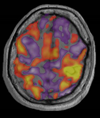
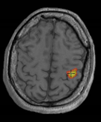
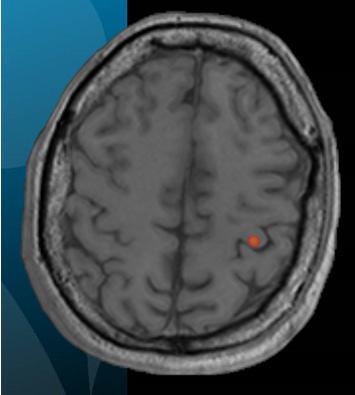
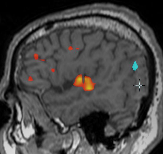
10 Left foot motor activity produces activations at the right paracentral lobule. Little Bo Peep lost her. 10
11 A + 11


12 fmri of Vision X fmri auditory task 12

13 Uses Presurgical Evaluation 1. Determine hemispheric language dominance 2. Define relationship between lesion and eloquent cerebral cortex Surgical Navigation 1. Activation maps can be imported to navigation system to aid in surgical guidance 2. May speed awake electrocortical stimulation mapping Low Grade Glioma Neurographics. Volume 5, Issue 4, pages


14 Anterior Displacement PMC Finger Tapping Low Grade Glioma Neurographics. Volume 5, Issue 4, pages


15 Left hemisphere language dominance Epilepsy patient, normal MRI 15


16 Left hemisphere language dominance Word Generation Sentence Completion AVM 16

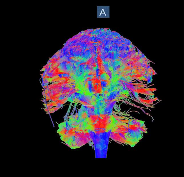
17 Spetzler Martin Grade I Diffusion Tensor Imaging and Tractography 17

18 Diffusion imaging is based on the inherent Brownian motion of water molecules. Any alteration in the diffusivity of water can create contrast. Diffusion imaging is based on the inherent Brownian motion of water molecules. Any alteration in the diffusivity of water can create contrast. 18


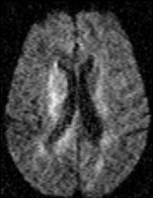
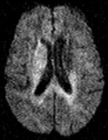
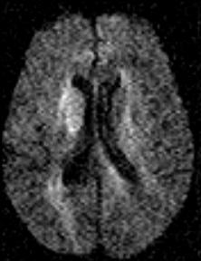
19 R MCA infarct b = 560 b = 560 Diffusion imaging is sensitive to the motion of water molecules that are aligned with the motion-probing gradients. 19
20 X Diffusion-Weighting Y Diffusion-Weighting Z Diffusion-Weighting x y z phase read slice RF Diffusion Ellipsoid v 1, λ 1 v 2, λ 2 v 3, λ 3 20


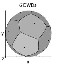
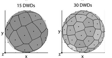
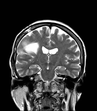
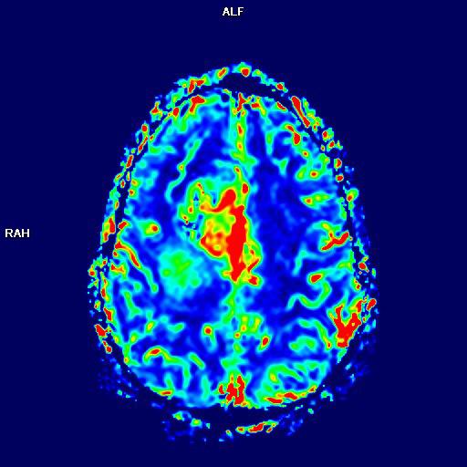
21 When diffusion is random, or equal in all directions the tensor is spherical. FA is low (0). When influenced by organized structures, as in white matter tracts, the ellipsoid becomes elongated. FA is high(1). Fractional Anisotropy Note low signal intensity in gray matter structures. (Basal ganglia) FA = 0 And high signal along white matter tracts (Corpus callosum) FA = 1 21


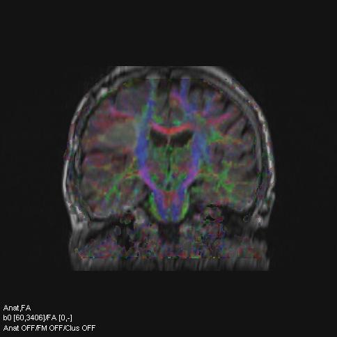
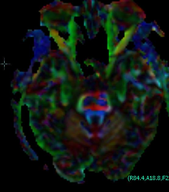
22 Quantitative FA Principle Direction Color FA x = Color FA map. Blue=CC Red=TV Green=AP 22


23 3D fusion Jellison B J et al. AJNR Am J Neuroradiol 2004;25:
24 Hypertensive microhemorrhage Tractography 24
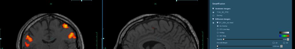
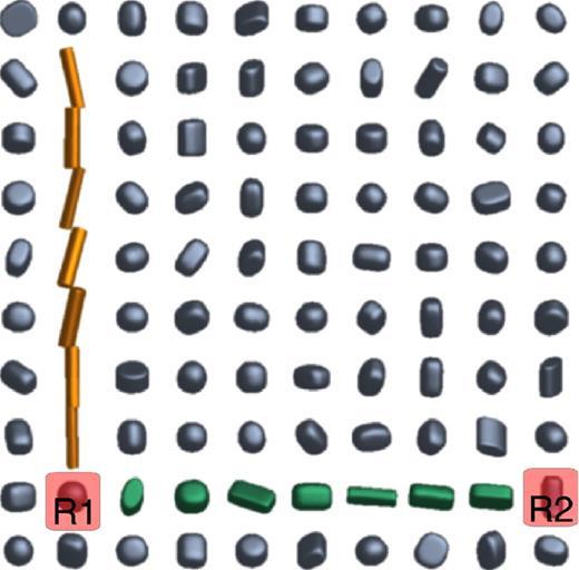
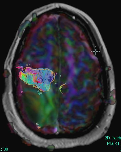
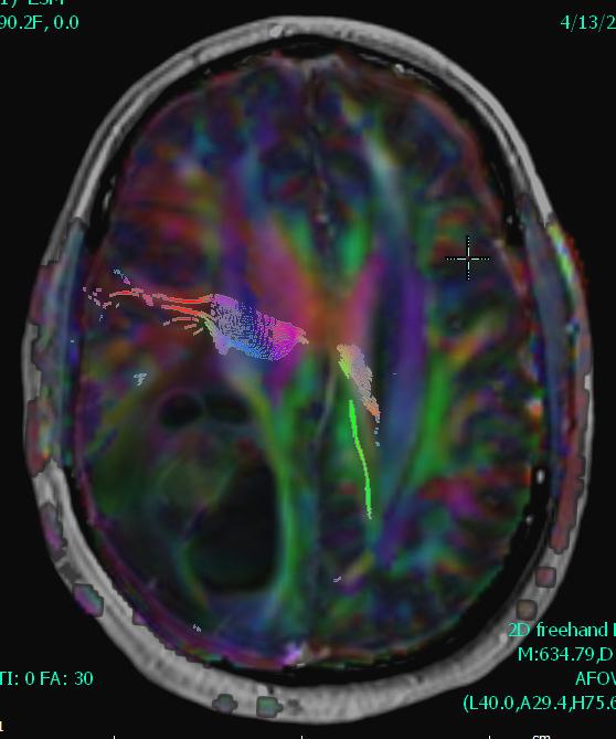
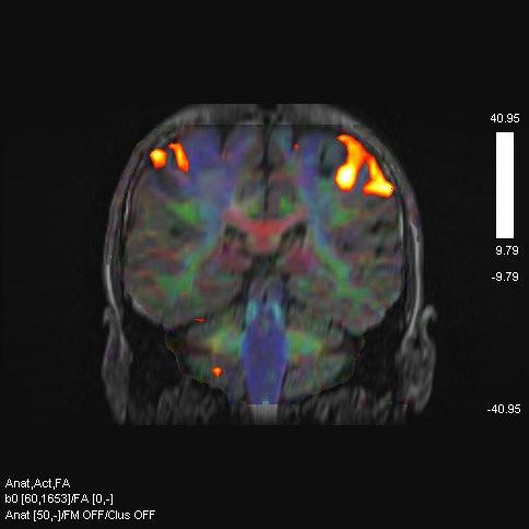
25 Fused fmri, color FA, and T1 post contrast Seeds Typically seeds are drawn in a region of interest. In this case the internal capsule. The computer estimates tracts by connecting high FA voxels with similarly strongly oriented voxels. 25

26 The placement of additional seeds can help isolate specific tracts. Subcortical white matter of the precentral gyrus. Seed 2 Connecting the two regions of interest isolates the corticospinal tract. Seed 2 Seed 1 26


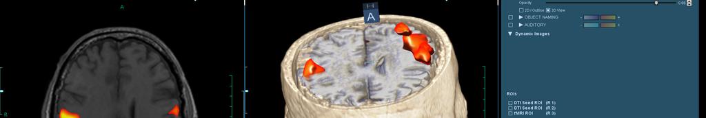
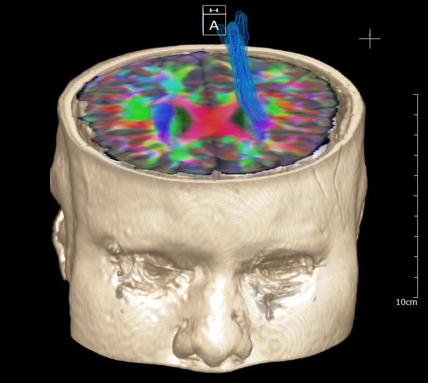
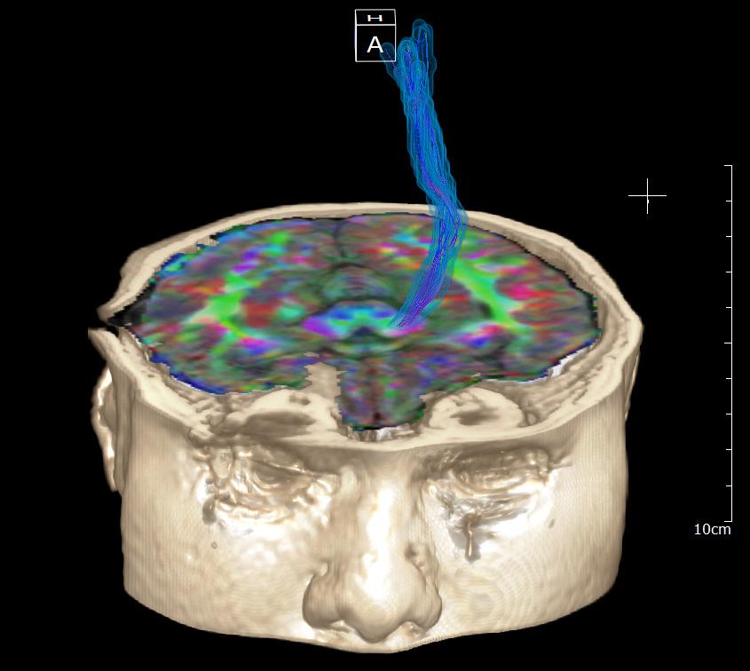
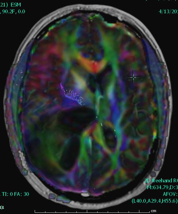
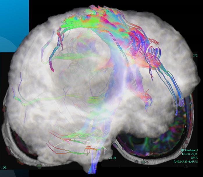
27 The tracts can be displayed in a variety of manners including 3D models Low Grade Glioma 27
 approaches the CST at the")

28 T2 FA CST intact T2/color FA fusion Large circumscribed T2 hyperintense lesion (low grade glioma) approaches the CST at the level of the internal capsule DTI exhibits minimal displacement of the CST, but preserved FA suggesting a lack of tumor infiltration High Grade Glioma 28
29 Glioblastoma 29

30 Finger Tapping Sentence Completion 30
31 Successful resection No permanent deficit. Conclusions Complimentary techniques: BOLD fmri used to assess eloquent cortex DTI and tractography interrogate white matter tracts Individualized experiments requires communication and collaboration Not just pretty pictures or science fiction 31

32 Questions, Comments, Cases? 32
Diffusion Tensor Imaging in brain tumours
 Diffusion Tensor Imaging in brain tumours @MarionSmits, MD PhD Associate Professor of Neuroradiology Dept. of Radiology, Erasmus MC, Rotterdam (NL) Honorary Consultant and Reader UCLH National Hospital
Pearls and Pitfalls of MR Diffusion in Clinical Neurology
 Pearls and Pitfalls of MR Diffusion in Clinical Neurology Dr. Alberto Bizzi Neuroradiology Unit Fondazione IRCCS Istituto Neurologico Carlo Besta Milan, Italy Email: alberto_bizzi@fastwebnet.it Diffusion
Overview. Fundamentals of functional MRI. Task related versus resting state functional imaging for sensorimotor mapping
 Functional MRI and the Sensorimotor System in MS Nancy Sicotte, MD, FAAN Professor and Vice Chair Director, Multiple Sclerosis Program Director, Neurology Residency Program Cedars-Sinai Medical Center
Define functional MRI. Briefly describe fmri image acquisition. Discuss relative functional neuroanatomy. Review clinical applications.
 Dr. Peter J. Fiester November 14, 2012 Define functional MRI. Briefly describe fmri image acquisition. Discuss relative functional neuroanatomy. Review clinical applications. Briefly discuss a few examples
functional MRI everything you always wanted to know, but never dared to MD PhD
 functional MRI everything you always wanted to know, but never dared to ask @MarionSmits, MD PhD Associate Professor of Neuroradiology Dept. of Radiology, Erasmus MC, Rotterdam (NL) Honorary Consultant
Pre-surgical planning for brain tumor resection using functional MRI
 June 2011 Divya S Bolar, HMSIV Pre-surgical planning for brain tumor resection using functional MRI Divya S. Bolar,, HMS IV 1 Our patient: clinical history 85-year year-old right-handed handed woman presents
fmri and Tractography in Preoperative Neurosurgical Planning Ronald L. Wolf, M.D., Ph.D. University of Pennsylvania Medical Center
 fmri and Tractography in Preoperative Neurosurgical Planning Ronald L. Wolf, M.D., Ph.D. University of Pennsylvania Medical Center Acknowledgements/Disclosures Radiology Ragini Verma Birkan Tunc Sumei
Seamless pre-surgical fmri and DTI mapping
 Seamless pre-surgical fmri and DTI mapping Newest release Achieva 3.0T X-series and Eloquence enable efficient, real-time fmri for brain activity mapping in clinical practice at Nebraska Medical Center
Visualization strategies for major white matter tracts identified by diffusion tensor imaging for intraoperative use
 International Congress Series 1281 (2005) 793 797 www.ics-elsevier.com Visualization strategies for major white matter tracts identified by diffusion tensor imaging for intraoperative use Ch. Nimsky a,b,
Diffusion tensor imaging of the infant brain: From technical problems to neuroscientific breakthroughs Jessica Dubois
 Diffusion tensor imaging of the infant brain: From technical problems to neuroscientific breakthroughs Jessica Dubois L. Hertz-Pannier, G. Dehaene-Lambertz, J.F. Mangin, D. Le Bihan Inserm U56, U663; NeuroSpin
Patterns of Brain Tumor Recurrence Predicted From DTI Tractography
 Patterns of Brain Tumor Recurrence Predicted From DTI Tractography Anitha Priya Krishnan 1, Isaac Asher 2, Dave Fuller 2, Delphine Davis 3, Paul Okunieff 2, Walter O Dell 1,2 Department of Biomedical Engineering
10/3/2016. T1 Anatomical structures are clearly identified, white matter (which has a high fat content) appears bright.
 appears bright.") H2O -2 atoms of Hydrogen, 1 of Oxygen Hydrogen just has one single proton and orbited by one single electron Proton has a magnetic moment similar to the earths magnetic pole Also similar to earth in that
H2O -2 atoms of Hydrogen, 1 of Oxygen Hydrogen just has one single proton and orbited by one single electron Proton has a magnetic moment similar to the earths magnetic pole Also similar to earth in that
Speed, Comfort and Quality with NeuroDrive
 Speed, Comfort and Quality with NeuroDrive Echelon Oval provides a broad range of capabilities supporting fast, accurate diagnosis of brain conditions and injuries. From anatomical depiction to vascular
Stereotactic Diffusion Tensor Tractography For Gamma Knife Stereotactic Radiosurgery
 Disclosures The authors of this study declare that they have no commercial or other interests in the presentation of this study. This study does not contain any use of offlabel devices or treatments. Stereotactic
Post Stroke Brain Plasticity
 Post Stroke Brain Plasticity François CHOLLET MD, PhD Neurology Department: Stroke Unit Toulouse University Hospital (CHU) Neurosciences Institute of Toulouse CNRS, INSERM, University, CHU Versailles le
Stuttering Research. Vincent Gracco, PhD Haskins Laboratories
 Stuttering Research Vincent Gracco, PhD Haskins Laboratories Stuttering Developmental disorder occurs in 5% of children Spontaneous remission in approximately 70% of cases Approximately 1% of adults with
Hemodynamics and fmri Signals
 Cerebral Blood Flow and Brain Activation UCLA NITP July 2011 Hemodynamics and fmri Signals Richard B. Buxton University of California, San Diego rbuxton@ucsd.edu... The subject to be observed lay on a
Diffusion Tensor Imaging 12/06/2013
 12/06/2013 Beate Diehl, MD PhD FRCP University College London National Hospital for Neurology and Neurosurgery Queen Square London, UK American Epilepsy Society Annual Meeting Disclosure None Learning
Surgery Within and Around Critical White Matter Tracts
 Surgery Within and Around Critical White Matter Tracts Kaisorn L. Chaichana, M.D. Assistant Professor of Neurosurgery, Oncology, and Otolaryngology-Head & Neck Surgery Mayo Clinic Florida, Jacksonville,
Titelmaster The physics of functional magnetic resonance imaging (fmri)
") Titelmaster The physics of functional magnetic resonance imaging (fmri) Outline 1.Introduction 2.The fmri experiment 2 3.The physics basis of fmri 4.Application Outline 3 1.Introduction Introduction Phrenology
Titelmaster The physics of functional magnetic resonance imaging (fmri) Outline 1.Introduction 2.The fmri experiment 2 3.The physics basis of fmri 4.Application Outline 3 1.Introduction Introduction Phrenology
Intraoperative Monitoring: Role in Epilepsy Based Tumor Surgery December 2, 2012
 Intraoperative Monitoring: Role in Epilepsy Based Tumor Surgery December 2, 2012 Aatif M. Husain, M.D. Duke University and Veterans Affairs Medical Centers, Durham, NC American Epilepsy Society Annual
Outline. Neuroradiology. Diffusion Imaging in. Clinical Applications of. Basics of Diffusion Imaging. Basics of Diffusion Imaging
 Clinical Applications of Diffusion Imaging in Neuroradiology No disclosures Stephen F. Kralik Assistant Professor of Radiology Indiana University School of Medicine Department of Radiology and Imaging
Diffusion Tensor Tractography in the Presurgical Assessment of Cerebral Gliomas
 The Neuroradiology Journal 27: 75-84, 2014 - doi: 10.15274/NRJ-2014-10008 www.centauro.it Diffusion Tensor Tractography in the Presurgical Assessment of Cerebral Gliomas ZAHRA FARSHIDFAR 1, FARIBORZ FAEGHI
PHYSICS OF MRI ACQUISITION. Alternatives to BOLD for fmri
 PHYSICS OF MRI ACQUISITION Quick Review for fmri HST-583, Fall 2002 HST.583: Functional Magnetic Resonance Imaging: Data Acquisition and Analysis Harvard-MIT Division of Health Sciences and Technology
Advances in Clinical Neuroimaging
 Advances in Clinical Neuroimaging Joseph I. Tracy 1, PhD, ABPP/CN; Gaelle Doucet 2, PhD; Xaiosong He 2, PhD; Dorian Pustina 2, PhD; Karol Osipowicz 2, PhD 1 Department of Radiology, Thomas Jefferson University,
Introduction to Brain Imaging
 Introduction to Brain Imaging Human Brain Imaging NEUR 570 & BIC lecture series September 9, 2013 Petra Schweinhardt, MD PhD Montreal Neurological Institute McGill University Montreal, Canada Various techniques
1 year of experiences with fmri, DTI in neuronavigation
 1 year of experiences with fmri, DTI in neuronavigation Poster No.: C-1604 Congress: ECR 2012 Type: Scientific Paper Authors: J. Luxemburgova, M. Kaiser ; Jablonec nad Nisou/CZ, Liberec/ CZ Keywords: Neuroradiology
Exploring Peritumoral White Matter Fibers for Neurosurgical Planning
 Exploring Peritumoral White Matter Fibers for Sonia Pujol, Ph.D. Ron Kikinis, M.D. Surgical Planning Laboratory Harvard University Clinical Goal Diffusion Tensor Imaging (DTI) Tractography has the potential
Biophysical and physiological bases of fmri signals: challenges of interpretation and methodological concerns
 Biophysical and physiological bases of fmri signals: challenges of interpretation and methodological concerns Antonio Ferretti aferretti@itab.unich.it Institute for Advanced Biomedical Technologies, University
HST.583 Functional Magnetic Resonance Imaging: Data Acquisition and Analysis Fall 2008
 MIT OpenCourseWare http://ocw.mit.edu HST.583 Functional Magnetic Resonance Imaging: Data Acquisition and Analysis Fall 2008 For information about citing these materials or our Terms of Use, visit: http://ocw.mit.edu/terms.
Brain Structure and Function in Nephropathic Cystinosis
 Brain Structure and Function in Nephropathic Cystinosis Doris A. Trauner M.D. Professor, Depts. of Neurosciences and Pediatrics University of California San Diego School of Medicine La Jolla, CA USA Cystinosis
Early experiences with diffusion tensor imaging and magnetic resonance tractography in stroke patients
 O r i g i n a l A r t i c l e Singapore Med Med J 2006; J 2006; 47(3) 47(3) : 198 : 1 Early experiences with diffusion tensor imaging and magnetic resonance tractography in stroke patients Parmar H, Golay
High spatial resolution reveals excellent detail in pediatric neuro imaging
 Publication for the Philips MRI Community Issue 46 2012/2 High spatial resolution reveals excellent detail in pediatric neuro imaging Achieva 3.0T with 32-channel SENSE Head coil has become the system
FUNCTIONAL MAGNETIC RESONANCE IMAGING IN FOLLOW-UP OF CEREBRAL GLIAL TUMORS
 Anvita Bieza FUNCTIONAL MAGNETIC RESONANCE IMAGING IN FOLLOW-UP OF CEREBRAL GLIAL TUMORS Summary of Doctoral Thesis to obtain PhD degree in medicine Specialty Diagnostic Radiology Riga, 2013 Doctoral thesis
Experimental Assessment of Infarct Lesion Growth in Mice using Time-Resolved T2* MR Image Sequences
 Experimental Assessment of Infarct Lesion Growth in Mice using Time-Resolved T2* MR Image Sequences Nils Daniel Forkert 1, Dennis Säring 1, Andrea Eisenbeis 2, Frank Leypoldt 3, Jens Fiehler 2, Heinz Handels
International Conference on Biological Sciences and Technology (BST 2016)
") International Conference on Biological Sciences and Technology (BST 2016) A Better Characterization of Brain Damage in Carbon Monoxide Intoxication Assessed in Vivo Using Diffusion Kurtosis Imaging Wen-Yao
International Conference on Biological Sciences and Technology (BST 2016) A Better Characterization of Brain Damage in Carbon Monoxide Intoxication Assessed in Vivo Using Diffusion Kurtosis Imaging Wen-Yao
Hemodynamics and fmri Signals
 Cerebral Blood Flow and Brain Activation UCLA NITP July 2010 Hemodynamics and fmri Signals Richard B. Buxton University of California, San Diego rbuxton@ucsd.edu... The subject to be observed lay on a
Functional MRI Mapping Cognition
 Outline Functional MRI Mapping Cognition Michael A. Yassa, B.A. Division of Psychiatric Neuro-imaging Psychiatry and Behavioral Sciences Johns Hopkins School of Medicine Why fmri? fmri - How it works Research
BOLD signal dependence on blood flow and metabolism. Outline
 BOLD signal dependence on blood flow and metabolism R. Hoge, MGH NMR Center Outline physiological events accompanying neuronal activation factors affecting BOLD signal sensitivity BOLD response dynamics
Leah Militello, class of 2018
 Leah Militello, class of 2018 Objectives 1. Describe the general organization of cerebral hemispheres. 2. Describe the locations and features of the different functional areas of cortex. 3. Understand
SURGICAL MANAGEMENT OF BRAIN TUMORS
 SURGICAL MANAGEMENT OF BRAIN TUMORS LIGIA TATARANU, MD, Ph D NEUROSURGICAL CLINIC, BAGDASAR ARSENI CLINICAL HOSPITAL BUCHAREST, ROMANIA SURGICAL INDICATIONS CONFIRMING HISTOLOGIC DIAGNOSIS REDUCING TUMOR
fmri and Pre-operative Planning for CNS tumors
 fmri and Pre-operative Planning for CNS tumors Miami Brain Symposium Andrei I. Holodny, M.D. Chief of Neuroradiology Director of the fmri Lab Memorial Sloan-Kettering Cancer Center Disclosures: 1. NIH-NIBIB
During human aging, the brain exhibits both macro- and
 Published November 5, 2009 as 10.3174/ajnr.A1862 ORIGINAL RESEARCH Q. Wang X. Xu M. Zhang Normal Aging in the Basal Ganglia Evaluated by Eigenvalues of Diffusion Tensor Imaging BACKGROUND AND PURPOSE:
Diffusion-weighted magnetic resonance imaging (MRI) allows for tissue
 allows for tissue") MAGNETIC RESONANCE IMAGING / IMAGERIE PAR RÉSONANCE MAGNÉTIQUE Nonischemic causes of hyperintense signals on diffusion-weighted magnetic resonance images: a pictorial essay Jeffrey M. Hinman, MD; James
MAGNETIC RESONANCE IMAGING / IMAGERIE PAR RÉSONANCE MAGNÉTIQUE Nonischemic causes of hyperintense signals on diffusion-weighted magnetic resonance images: a pictorial essay Jeffrey M. Hinman, MD; James
DWI assessment of ischemic changes in the fetal brain
 DWI assessment of ischemic changes in the fetal brain Dafi Bergman, 4 th year Medical student in the 4-year program, Sackler school of medicine B.Sc Life and Medical Sciences, Tel Aviv University Supervised
Language comprehension in young people with severe cerebral palsy in relation to language tracts: a diffusion tensor imaging study
 Language comprehension in young people with severe cerebral palsy in relation to language tracts: a diffusion tensor imaging study Laurike Harlaar, Petra J. Pouwels, Joke J. Geytenbeek, Kim J. Oostrom,
Lecture X. Brain Pathways: Movement!
 Bio 3411 Readings (background only) Bio 3411 Monday Neuroscience 4 th ed Page(s) Feature 423-451Upper motor control of Brain Stem and Spinal Cord The Brain Atlas 3 rd ed Page(s) Feature 198-199 Vestibular
fmri: Interpretation, Limits and Potential Pitfalls
 fmri: Interpretation, Limits and Potential Pitfalls Seong-Gi Kim kimsg@pitt.edu www.kimlab.pitt.edu Mapping Brain Functions Stimulation/Task Functional Map (MRI) Pre-synaptic activity Post-synaptic activity
Resting-State functional Connectivity MRI (fcmri) NeuroImaging
 NeuroImaging") Resting-State functional Connectivity MRI (fcmri) NeuroImaging Randy L. Buckner et. at., The Brain s Default Network: Anatomy, Function, and Relevance to Disease, Ann. N. Y. Acad. Sci. 1124: 1-38 (2008)
Resting-State functional Connectivity MRI (fcmri) NeuroImaging Randy L. Buckner et. at., The Brain s Default Network: Anatomy, Function, and Relevance to Disease, Ann. N. Y. Acad. Sci. 1124: 1-38 (2008)
Lecture X. Brain Pathways: Movement!
 Bio 3411 Monday 1 Readings (background only) Neuroscience 5 th ed Page(s) Feature 353-398Upper motor control of Brain Stem and Spinal Cord Neuroscience 4 th ed Page(s) Feature 423-451Upper motor control
Title:Atypical language organization in temporal lobe epilepsy revealed by a passive semantic paradigm
 Author's response to reviews Title:Atypical language organization in temporal lobe epilepsy revealed by a passive semantic paradigm Authors: Julia Miro (juliamirollado@gmail.com) Pablo Ripollès (pablo.ripolles.vidal@gmail.com)
The neurolinguistic toolbox Jonathan R. Brennan. Introduction to Neurolinguistics, LSA2017 1
 The neurolinguistic toolbox Jonathan R. Brennan Introduction to Neurolinguistics, LSA2017 1 Psycholinguistics / Neurolinguistics Happy Hour!!! Tuesdays 7/11, 7/18, 7/25 5:30-6:30 PM @ the Boone Center
Injuries of neural tracts in a patient with CADASIL: a diffusion tensor imaging study
 Jang and Seo BMC Neurology (2015) 15:176 DOI 10.1186/s12883-015-0434-x CASE REPORT Open Access Injuries of neural tracts in a patient with CADASIL: a diffusion tensor imaging study Sung Ho Jang and You
Advanced magnetic resonance imaging for monitoring brain development and injury
 Advanced magnetic resonance imaging for monitoring brain development and injury Stéphane Sizonenko, MD-PhD Division of Development and Growth Department of Child and Adolescent Medicine Geneva University
Note: Waxman is very sketchy on today s pathways and nonexistent on the Trigeminal.
 Dental Neuroanatomy Thursday, February 3, 2011 Suzanne Stensaas, PhD Note: Waxman is very sketchy on today s pathways and nonexistent on the Trigeminal. Resources: Pathway Quiz for HyperBrain Ch. 5 and
Diffusion MRI explores new indications
 DECEMBER 2001 Diffusion MRI finds new indications Neuroimaging expands with functional MRI 3-tesla MRI bests 1.5-tesla in body and brain Diffusion MRI explores new indications Diffusion tensor imaging
Announcements. Final Exam will be a take-home exam. Format similar to the short assignment (no multiple choice, etc.)
") Announcements Final Exam will be a take-home exam Format similar to the short assignment (no multiple choice, etc.) Will be handed out at end of last class period (Thursday June 5 th ) Due by 6 pm June
Announcements Final Exam will be a take-home exam Format similar to the short assignment (no multiple choice, etc.) Will be handed out at end of last class period (Thursday June 5 th ) Due by 6 pm June
Methods for assessing the brain basis of developmental disorders
 Announcements LIGN171: Child Language Acquisition http://ling.ucsd.edu/courses/lign171 Final Exam will be a take-home exam Format similar to the short assignment (no multiple choice, etc.) Will be handed
Functional Magnetic Resonance Imaging with Arterial Spin Labeling: Techniques and Potential Clinical and Research Applications
 pissn 2384-1095 eissn 2384-1109 imri 2017;21:91-96 https://doi.org/10.13104/imri.2017.21.2.91 Functional Magnetic Resonance Imaging with Arterial Spin Labeling: Techniques and Potential Clinical and Research
Diffusion-Weighted and Conventional MR Imaging Findings of Neuroaxonal Dystrophy
 AJNR Am J Neuroradiol 25:1269 1273, August 2004 Diffusion-Weighted and Conventional MR Imaging Findings of Neuroaxonal Dystrophy R. Nuri Sener BACKGROUND AND PURPOSE: Neuroaxonal dystrophy is a rare progressive
CISC 3250 Systems Neuroscience
 CISC 3250 Systems Neuroscience Levels of organization Central Nervous System 1m 10 11 neurons Neural systems and neuroanatomy Systems 10cm Networks 1mm Neurons 100μm 10 8 neurons Professor Daniel Leeds
Research Article Corticospinal Tract Change during Motor Recovery in Patients with Medulla Infarct: A Diffusion Tensor Imaging Study
 BioMed Research International, Article ID 524096, 5 pages http://dx.doi.org/10.1155/2014/524096 Research Article Corticospinal Tract Change during Motor Recovery in Patients with Medulla Infarct: A Diffusion
Psy /16 Human Communication. By Joseline
 Psy-302 11/16 Human Communication By Joseline Lateralization Left Hemisphere dominance in speech production in 95% of right handed and 70% of left handed people Left -> Timing, Sequence of events Right
PDFlib PLOP: PDF Linearization, Optimization, Protection. Page inserted by evaluation version
 PDFlib PLOP: PDF Linearization, Optimization, Protection Page inserted by evaluation version www.pdflib.com sales@pdflib.com Principal Diffusion Direction in Peritumoral Fiber Tracts Color Map Patterns
The supplementary motor area (SMA) is an eloquent. FOCUS Neurosurg Focus 44 (6):E3, 2018
 is an eloquent. FOCUS Neurosurg Focus 44 (6):E3, 2018") NEUROSURGICAL FOCUS Neurosurg Focus 44 (6):E3, 2018 Prediction of recovery from supplementary area syndrome after brain tumor surgery: preoperative diffusion tensor tractography analysis and postoperative
NEUROSURGICAL FOCUS Neurosurg Focus 44 (6):E3, 2018 Prediction of recovery from supplementary area syndrome after brain tumor surgery: preoperative diffusion tensor tractography analysis and postoperative
Progress Report. Author: Dr Joseph Yuan-Mou Yang Qualification: PhD Institution: Royal Children s Hospital Date: October 2017
 Author: Dr Joseph Yuan-Mou Qualification: PhD Institution: Royal Children s Hospital Date: October 2017 Progress Report Title of Project: Brain structural and motor function correlations in childhood arterial
1) Diffusion weighted imaging DWI is a term used to describe moving molecules due to random thermal motion. This motion is restricted by boundaries
 Diffusion weighted imaging DWI is a term used to describe moving molecules due to random thermal motion. This motion is restricted by boundaries") 1) Diffusion weighted imaging DWI is a term used to describe moving molecules due to random thermal motion. This motion is restricted by boundaries such as ligaments, membranes and macro molecules. Diffusion
1) Diffusion weighted imaging DWI is a term used to describe moving molecules due to random thermal motion. This motion is restricted by boundaries such as ligaments, membranes and macro molecules. Diffusion
Internal capsule: The homunculus distribution in the posterior limb
 Received: 9 March 2016 Revised: 1 December 2016 Accepted: 2 December 2016 DOI: 10.1002/brb3.629 ORIGINAL RESEARCH Internal capsule: The homunculus distribution in the posterior limb Cheng Qian Fei Tan
Diffusion-tensor imaging in brain tumors
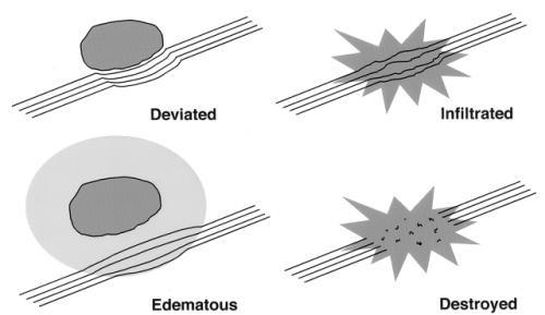
 REVIEW Diffusion-tensor imaging in brain tumors Diffusion-tensor imaging (DTI) is the only novel imaging technique that is able to demonstrate white matter tracts and their structural changes related to
The Effect of Local Fiber Model On Population Studies
 The Effect of Local Fiber Model On Population Studies James G. Malcolm Marek Kubicki Martha E. Shenton Yogesh Rathi Psychiatry Neuroimaging Laboratory, Harvard Medical School, Boston, MA VA Boston Healthcare
Prof. Greg Francis 1/2/19
 Brain scans PSY 200 Greg Francis Lecture 03 How to study the brain without killing someone. Scanning Technology provides insight into brain processes w EEG recordings w MRI w Non-invasive Maps of brain
Diagnostic improvement from average image in acute ischemic stroke
 Diagnostic improvement from average image in acute ischemic stroke N. Magne (1), E.Tollard (1), O. Ozkul- Wermester (2), V. Macaigne (1), J.-N. Dacher (1), E. Gerardin (1) (1) Department of Radiology,
fmri Acquisition: Temporal Effects
 Functional MRI Data Acquisition: Temporal fmri Acquisition: Temporal Effects Session length Repetition time Fixed vs. distributed temporal sampling Sparse temporal sampling Noise source recording Prospective
Disclosure Information AACPDM 68 th Annual Meeting September 10-13, 2014 Diffusion Tensor Imaging: Analysis options in pediatric neuroimaging research
 Disclosure Information AACPDM 68 th Annual Meeting September 10-13, 2014 Diffusion Tensor Imaging: Analysis options in pediatric neuroimaging research Andrea Poretti, MD Research Associate Section of Pediatric
P2 Visual - Perception
 P2 Visual - Perception 2014 SOSE Neuroimaging of high-level visual functions gyula.kovacs@uni-jena.de 11/09/06 Functional magnetic resonance imaging (fmri) The very basics What is fmri? What is MRI? The
Case 9511 Hypertensive microangiopathy
 Case 9511 Hypertensive microangiopathy Schepers S, Barthels C Section: Neuroradiology Published: 2011, Nov. 3 Patient: 67 year(s), male Authors' Institution Department of Radiology, Jessa ziekenhuis campus
The Optimal Trackability Threshold of Fractional Anisotropy for DiŠusion Tensor Tractography of the Corticospinal Tract
 Magnetic Resonance in Medical Sciences, Vol. 3, No. 1, p. 11 17, 2004 MAJOR PAPER The Optimal Trackability Threshold of Fractional Anisotropy for DiŠusion Tensor Tractography of the Corticospinal Tract
Clinically focused workflow with unique ability to integrate fmri, DTI, fiber tracks and perfusion in a single, multi-layered 3D rendering
 Clinically focused workflow with unique ability to integrate fmri, DTI, fiber tracks and perfusion in a single, multi-layered 3D rendering Neurosurgeons are demanding more from neuroradiologists and increasingly
P. Hitchcock, Ph.D. Department of Cell and Developmental Biology Kellogg Eye Center. Wednesday, 16 March 2009, 1:00p.m. 2:00p.m.
 Normal CNS, Special Senses, Head and Neck TOPIC: CEREBRAL HEMISPHERES FACULTY: LECTURE: READING: P. Hitchcock, Ph.D. Department of Cell and Developmental Biology Kellogg Eye Center Wednesday, 16 March
Key Words aphasia; brain tumor; diffusion tensor imaging; fiber tracking; interhemispheric connectivity; transcranial magnetic stimulation; oncology
 clinical article J Neurosurg 126:222 233, 2017 Interhemispheric connectivity revealed by diffusion tensor imaging fiber tracking derived from navigated transcranial magnetic stimulation maps as a sign
Summary of findings from the previous meta-analyses of DTI studies in MDD patients. SDM (39) 221 Left superior longitudinal
 221 Left superior longitudinal") Supplemental Data Table S1 Summary of findings from the previous meta-analyses of DTI studies in MDD patients Study Analysis Method Included studies, n MDD (medicated) HC Results (MDDHC)
Supplemental Data Table S1 Summary of findings from the previous meta-analyses of DTI studies in MDD patients Study Analysis Method Included studies, n MDD (medicated) HC Results (MDDHC)
Diffusion Tensor Eigenvector Directional Color Imaging Patterns in the Evaluation of Cerebral White Matter Tracts Altered by Tumor
 JOURNAL OF MAGNETIC RESONANCE IMAGING 20:555 562 (2004) Original Research Diffusion Tensor Eigenvector Directional Color Imaging Patterns in the Evaluation of Cerebral White Matter Tracts Altered by Tumor
Announcements. Exam 1. VII. Imaging techniques of the brain. Anatomical/Structural Scans. Structural Scans: CT. Structural Scans: CT 2/17/2014
 Exam 1 None at the moment! Announcements Mean 78.0% Median 80% Mode 86% Min 26% Max 98% Std Dev 12.6% VII. Imaging techniques of the brain A. CT: anatomical B. MRI: anatomical C. fmri: functional D. SPECT
Exam 1. Mean 78.0% Median 80% Mode 86% Min 26% Max 98% Std Dev 12.6%
 Exam 1 Mean 78.0% Median 80% Mode 86% Min 26% Max 98% Std Dev 12.6% None at the moment! Announcements VII. Imaging techniques of the brain A. CT: anatomical B. MRI: anatomical C. fmri: functional D. SPECT
INTRO TO BOLD FMRI FRANZ JOSEPH GALL ( ) OUTLINE. MRI & Fast MRI Observations Models Statistical Detection
 OUTLINE. MRI & Fast MRI Observations Models Statistical Detection") INTRO TO BOLD FMRI 2014 M.S. Cohen all rights reserved mscohen@g.ucla.edu OUTLINE FRANZ JOSEPH GALL (1758-1828) MRI & Fast MRI Observations Models Statistical Detection PAUL BROCA (1824-1880) WILLIAM JAMES
INTRO TO BOLD FMRI 2014 M.S. Cohen all rights reserved mscohen@g.ucla.edu OUTLINE FRANZ JOSEPH GALL (1758-1828) MRI & Fast MRI Observations Models Statistical Detection PAUL BROCA (1824-1880) WILLIAM JAMES
Cerebral Cortex 1. Sarah Heilbronner
 Cerebral Cortex 1 Sarah Heilbronner heilb028@umn.edu Want to meet? Coffee hour 10-11am Tuesday 11/27 Surdyk s Overview and organization of the cerebral cortex What is the cerebral cortex? Where is each
Final Report. Title of Project: Quantifying and measuring cortical reorganisation and excitability with post-stroke Wii-based Movement Therapy
 Final Report Author: Dr Penelope McNulty Qualification: PhD Institution: Neuroscience Research Australia Date: 26 th August, 2015 Title of Project: Quantifying and measuring cortical reorganisation and
Gross Organization I The Brain. Reading: BCP Chapter 7
 Gross Organization I The Brain Reading: BCP Chapter 7 Layout of the Nervous System Central Nervous System (CNS) Located inside of bone Includes the brain (in the skull) and the spinal cord (in the backbone)
HST.583 Functional Magnetic Resonance Imaging: Data Acquisition and Analysis Fall 2008
 MIT OpenCourseWare http://ocw.mit.edu HST.583 Functional Magnetic Resonance Imaging: Data Acquisition and Analysis Fall 2008 For information about citing these materials or our Terms of Use, visit: http://ocw.mit.edu/terms.
Neuroimaging and Psychiatry
 Neuroimaging and Psychiatry Chris Gale Otago Regional Psychiatry Training Programme Feb 12, 2011 In psychosis. Decrease volume frontal, parietal, hippocampus, corpus collosum. Increase CSF volume Functional
Cerebrum-Cerebral Hemispheres. Cuneyt Mirzanli Istanbul Gelisim University
 Cerebrum-Cerebral Hemispheres Cuneyt Mirzanli Istanbul Gelisim University The largest part of the brain. Ovoid shape. Two incompletely separated cerebral hemispheres. The outer surface of the cerebral
PETER PAZMANY CATHOLIC UNIVERSITY Consortium members SEMMELWEIS UNIVERSITY, DIALOG CAMPUS PUBLISHER
 PETER PAZMANY CATHOLIC UNIVERSITY SEMMELWEIS UNIVERSITY Development of Complex Curricula for Molecular Bionics and Infobionics Programs within a consortial* framework** Consortium leader PETER PAZMANY
Paul Moes, Professor of Psychology - Neuroscience and Education Group - Christian Perspectives in Science - Education Program
 Paul Moes, Professor of Psychology - Neuroscience and Education Group - Christian Perspectives in Science - Education Program Thesis: Secret to the brain is Unity The problem: focused on parts A sample
FUNCTIONAL MRI IN EPILEPSY December 6 th 2013
 FUNCTIONAL MRI IN EPILEPSY December 6 th 2013 Matthias J Koepp, MD, PhD UCL Institute of Neurology National Hospital for Neurology and Neurosurgery London, UK American Epilepsy Society Annual Meeting Disclosure
Brain diffusion tensor imaging changes in cerebrotendinous xanthomatosis reversed with
 Brain diffusion tensor imaging changes in cerebrotendinous xanthomatosis reversed with treatment Claudia B. Catarino, MD, PhD, 1*, Christian Vollmar, MD, PhD, 2,3* Clemens Küpper, MD, 1,4 Klaus Seelos,
Outline of the next three lectures
 Outline of the next three lectures Lecture 35 Anatomy of the human cerebral cortex gross and microscopic cell types connections Vascular supply of the cerebral cortex Disorders involving the cerebral cortex
A characteristic feature of acute haematomas in the brain on echo-planar diffusion-weighted imaging
 Neuroradiology (2002) 44: 907 911 DOI 10.1007/s00234-002-0860-5 DIAGNOSTIC NEURORADIOLOGY N. Morita M. Harada K. Yoneda H. Nishitani M. Uno A characteristic feature of acute haematomas in the brain on
Functional Magnetic Resonance Imaging of the Brain
 Page: 1 of 9 Last Review Status/Date: December 2016 Description Functional magnetic resonance imaging (fmri) is a noninvasive method for localizing areas of brain function and has been used for the presurgical
ASSUMPTION OF COGNITIVE UNIFORMITY
 The Human Brain cerebral hemispheres: two most important divisions of the brain, separated by the longitudinal fissure corpus callosum: a large bundle of axons that constitutes the major connection between
UNIVERSITY OF CALGARY. A New Method for Assessing Tissue Alignment using Clinical MRI in Multiple Sclerosis. Shrushrita Sharma A THESIS
 UNIVERSITY OF CALGARY A New Method for Assessing Tissue Alignment using Clinical MRI in Multiple Sclerosis by Shrushrita Sharma A THESIS SUBMITTED TO THE FACULTY OF GRADUATE STUDIES IN PARTIAL FULFILMENT