INTERDISCIPLINARY DISCUSSIONS IN LOCALISED RCC DIAGNOSIS AND SURGICAL STRATEGIES FOR ATYPICAL RENAL CYSTIC LESIONS. Maria Cova
|
|
|
- Margery Farmer
- 6 years ago
- Views:
Transcription
1 INTERDISCIPLINARY DISCUSSIONS IN LOCALISED RCC DIAGNOSIS AND SURGICAL STRATEGIES FOR ATYPICAL RENAL CYSTIC LESIONS Maria Cova Radiology Department University of Trieste (IT) Eleventh European International Kidney Cancer Symposium April 2016, Barcelona, Spain


2 SIMPLE RENAL CYST Commonest focal renal lesion More often identified incidentally at US Incidence varies with age, sex, renal function and other factors
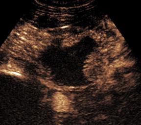
3 SIMPLE RENAL CYST Fully characterized on US, no further imaging necessary - Thin wall - Anechoic content - Back enhancement - (Lateral acoustic shadows)

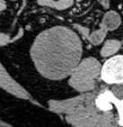
4 SIMPLE RENAL CYST Fully characterized also on CT - Thin wall, no calcifications - Water content - Sharp interface with adjacent renal parenchyma - No enhancement


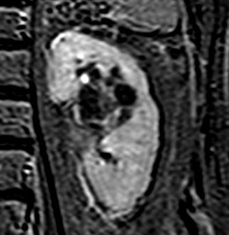
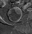
5 SIMPLE RENAL CYST Fully characterized also on CT and on MR T2 T1 T1 + Gd Subtraction

")
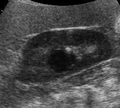
6 COMPLEX RENAL CYST Full characterization often impossible on grey-scale US only Other imaging modalities are required (CT/MR/CEUS) - Septa and calcifications - Echogenic content (hemorrhage, debris) - Inflammatory changes
7 QUESTIONS Is it a cyst? Is it a benign or malignant cyst?


8 QUESTION 1 Is it a cyst? Unenhanced CT scan 20 HU or less: benign renal cyst HU: indeterminate Homogeneous renal mass >70 HU: >99.9% chance of representing a high attenuation benign renal cyst Jonisch AI, Radiology 2007 O Connor SD, AJR 2011


9 QUESTION 1 Is it a cyst? Lesions not characterized on CT single phase Unenhanced CT scan


10 QUESTION 1 Is it a cyst? Equivocal enhancement on CT 30 HU 36 HU 45 HU In our opinion, a renal mass that enhances HU is indeterminate and needs further evaluation for definitive characterization Israel GM, Radiology 2005


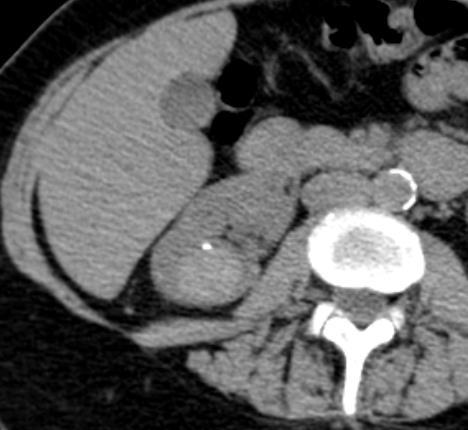
11 QUESTION 1 Is it a cyst? 49 HU 65 HU


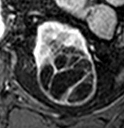
12 QUESTION 2 Is it benign or malignant cyst? Bosniak classification First introduced by Bosniak in 1986 for CT Applied also to MRI and CEUS Widely used to differentiate surgical from non-surgical complex cystic lesions Five categories, increasing risk of malignancy


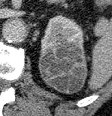
13 Bosniak classification - CT Category II Minimally complicated benign cysts Few septa, perceivable enhancement Thin calcifications Homogeneous attenuation (<3cm)


14 Bosniak classification - CT Category IIF Probably benign cysts that have some suspicious feature and need follow-up to detect any change in character 16 months after


15 Bosniak classification - CT Category III Indeterminate cystic masses Uniform wall thickening, or wall and septal irregularities Enhancing wall and septa


16 Bosniak classification - CT Category IV I likely malignant cystic masses Non-uniform enhancing thick wall and septations Enhancing nodules and solid components


17 Bosniak classification - CT Remember The Bosniak classification is clinically oriented, and clinic is important! Follow-up Follow-up Category III Category III
 56 lesions (81%): Similar findings 8 lesions (12%) more septa at MRI 7")
 IIF>III (n=3)")
18 Bosniak classification - MRI Successfully used also for MRI CT vs. MRI (69 complex cysts) 56 lesions (81%): Similar findings 8 lesions (12%) more septa at MRI 7 lesions (10%) increased wall and/or septa thickness at MRI 2 lesions (3%) different enhancement MRI does not identify calcifications Upgrade at MRI in 7 lesions II>IIF (n=2) IIF>III (n=3) IIF>IV (n=2) Israel GM, Radiology 2004

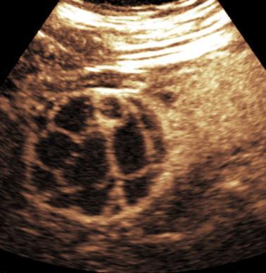
19 Bosniak classification - CEUS CEUS is at least as effective as CT in the characterization of complex renal cysts Reference procedure: helical CT Ascenti, Radiology 2007 Park, Eur J Radiol 2007 Quaia, AJR 2008 Clevert, Clin Hemorheol Microcirc 2008

 No need")

20 Bosniak classification - CEUS Advantages Very high sensitivity to flow (hypovascular lesions) No need for nephrotoxic contrast agents No radiation no need to limit the number of examinations

 Very large")
 Staging of")
21 Bosniak classification - CEUS Limitations Calcifications (can mask cystic content) Very large cysts (insufficient panoramicity) Staging of cystic tumours impossible
 -")


22 Bosniak classification Which imaging? CT: widest experience (since 1986) - Radiation, nephrotoxic contrast agents MRI: limited experience (only few investigations) - Cannot be performed in all patients CEUS: More robust than MRI (more investigations)


23 Bosniak classification Which imaging? Comparative studies: results are equivalent, but not equal on a per-patient basis. Differences exist Bosniak MA, Radiology 1986 Bosniak MA,Urol Radiol 1991 Israel GM, AJR 2003 Israel GM, Radiology 2004 Ascenti G, Radiology 2007 Park BK, Eur J Rad 2007 Quaia E, AJR 2008
24 IIF III III
25 IIF III III II IIF IIF


26 IIF III III II IIF IIF IV IV IV

 MR in heavily calcified")
 More than one technique")
27 Bosniak classification Which imaging? CEUS preferable (inexpensive, no radiation, no nephrotoxic contrast agents) MR in heavily calcified lesions CT for staging (more panoramic) More than one technique in difficult cases
28 THANK YOU FOR YOUR ATTENTION
The Incidental Renal lesion
 The Incidental Renal lesion BACKGROUND Increase in abdominal CT/US in last 15 years Resulted in detection of many (small) renal lesions 50% > 50yrs has at least 1 lesion majority simple cysts Renal lesions
Contrast-enhanced ultrasound (CEUS) in the evaluation and characterization of complex renal cysts
 in the evaluation and characterization of complex renal cysts") Contrast-enhanced ultrasound (CEUS) in the evaluation and characterization of complex renal cysts Poster No.: C-2812 Congress: ECR 2018 Type: Educational Exhibit Authors: J. A. Torres de Abreu Macedo,
Contrast-enhanced ultrasound (CEUS) in the evaluation and characterization of complex renal cysts Poster No.: C-2812 Congress: ECR 2018 Type: Educational Exhibit Authors: J. A. Torres de Abreu Macedo,
Caveat sonologist Mistakes to avoid in Kidney Ultrasound
 Caveat sonologist Mistakes to avoid in Kidney Ultrasound Simon Freeman Derriford Hospital, Plymouth simonfreeman@nhs.net Bear trap 1 Report: There is a 4cm solid mass arising from the left kidney likely
Role of imaging in RCC. Ultrasonography. Solid lesion. Cystic RCC. Solid RCC 31/08/60. From Diagnosis to Treatment: the Radiologist Perspective
 Role of imaging in RCC From Diagnosis to Treatment: the Radiologist Perspective Diagnosis Staging Follow up Imaging modalities Limitations and pitfalls Duangkamon Prapruttam, MD Department of Therapeutic
Renal masses - the role of diagnostic imaging
 Renal masses - the role of diagnostic imaging Poster No.: C-2471 Congress: ECR 2015 Type: Educational Exhibit Authors: V. Rai#; Bjelovar/HR Keywords: Cysts, Cancer, Structured reporting, Ultrasound, MR,
Measure #405: Appropriate Follow-up Imaging for Incidental Abdominal Lesions National Quality Strategy Domain: Effective Clinical Care
 Measure #405: Appropriate Follow-up Imaging for Incidental Abdominal Lesions National Quality Strategy Domain: Effective Clinical Care 2016 PQRS OPTIONS FOR INDIVIDUAL MEASURES: CLAIMS, REGISTRY DESCRIPTION:
CME Article Clinics in diagnostic imaging (135)
") Medical Education Singapore Med J 2011; 52(5) : 384 CME Article Clinics in diagnostic imaging (135) Pojchamarnwiputh S, Muttarak M, Sriplakich S H 1a 1b 1c 1d Fig. 1 (a) Axial unenhanced; (b & c) delayed
Medical Education Singapore Med J 2011; 52(5) : 384 CME Article Clinics in diagnostic imaging (135) Pojchamarnwiputh S, Muttarak M, Sriplakich S H 1a 1b 1c 1d Fig. 1 (a) Axial unenhanced; (b & c) delayed
Management of the Incidental Renal Mass on CT: A White Paper of the ACR Incidental Findings Committee
 ORIGINAL ARTICLE Management of the Incidental Renal Mass on CT: A White Paper of the ACR Incidental Findings Committee Brian R. Herts, MD a, Stuart G. Silverman, MD b, Nicole M. Hindman, MD c, Robert G.
Renal Masses With Equivocal Enhancement at CT: Characterization With Contrast- Enhanced Ultrasound
 Genitourinary Imaging Original Research Bertolotto et al. Use of Contrast-Enhanced Ultrasound to Characterize Renal Masses Genitourinary Imaging Original Research Michele Bertolotto 1 Calogero Cicero 2
THYROID NODULES: THE ROLE OF ULTRASOUND
 THYROID NODULES: THE ROLE OF ULTRASOUND NOVEMBER 2017 DR. DEAN DURANT DEFINITION Thyroid nodule: Focal area within the thyroid gland with echogenicity different from surrounding parenchyma. THYROID NODULES
How To Approach Renal Masses? - Differential Diagnosis On Image
 How To Approach Renal Masses? - Differential Diagnosis On Image Poster No.: C-1646 Congress: ECR 2015 Type: Educational Exhibit Authors: A. E. A. G. Costa, A. Gomes, A. Duarte, I. Távora; Lisbon/PT Keywords:
Imaging of liver and pancreas
 Imaging of liver and pancreas.. Disease of the liver Focal liver disease Diffusion liver disease Focal liver disease Benign Cyst Abscess Hemangioma FNH Hepatic adenoma HCC Malignant Fibrolamellar carcinoma
Case-based discussion:
 Case-based discussion: Pailin Kongmebhol, M.D. Department of Radiology Faculty of Medicine Chiang Mai University There are many guidelines for managing thyroid nodules Two important guidelines: 2015 American
ESUR 2018, Sept. 13 th.-16 th., 2018 Barcelona, Spain
 ESUR 2018, Sept. 13 th.-16 th., 2018 Barcelona, Spain OUR APPROACH Incidental adrenal nodule/mass Isaac R Francis, M.B;B.S University of Michigan, Ann Arbor, Michigan Disclosures None (in memory) M Korobkin,
Radiologic Findings of Mucocele-like Tumors of the breast: Can we differentiate pure benign from associated with high risk lesions?
 Radiologic Findings of Mucocele-like Tumors of the breast: Can we differentiate pure benign from associated with high risk lesions? Poster No.: C-0332 Congress: ECR 2014 Type: Educational Exhibit Authors:
11/1/2014. Radiologic incidentalomas Ordering pitfalls Newer technology and applications
 Bilal Tahir, MD Gitasree Borthakur, MD Indiana University School of Medicine Department of Radiology & Imaging Sciences October 31, 2014 ACP 2014 Dr. V. Aaron Nuclear (vaaron@iupui.edu) Dr. S. Westphal
IN THE NAME OF GOD POV: CYSTIC OVARIAN LESION
 IN THE NAME OF GOD POV: CYSTIC OVARIAN LESION CASE 1 20 years old girl with AUB and pelvic pain from 2 weeks ago Impression :Simple unilocular 6 cm ovarian cyst Next step? Almost certainly benign so FU
Traumatic and Non Traumatic Adrenal Emergencies
 Traumatic and Non Traumatic Adrenal Emergencies Michael N. Patlas, MD, FRCPC (1), Christine O. Menias, MD (2), Douglas S. Katz, MD, FACR (3), Ania Z. Kielar, MD, FRCPC (4), Alla M. Rozenblit, MD (5), Jorge
LUNG NODULES: MODERN MANAGEMENT STRATEGIES
 Department of Radiology LUNG NODULES: MODERN MANAGEMENT STRATEGIES Christian J. Herold M.D. Department of Biomedical Imaging and Image-guided Therapy Medical University of Vienna Vienna, Austria Pulmonary
Renal Cell Carcinoma: Attenuation Values on Unenhanced CT
 Genitourinary Imaging Original Research Pooler et al. Attenuation Values of Unenhanced CT of Renal Cell Carcinoma Genitourinary Imaging Original Research B. Dustin Pooler 1 Perry J. Pickhardt 1 Stacy D.
Small renal mass: differential diagnosis on image
 Small renal mass: differential diagnosis on image Poster No.: R-0166 Congress: RANZCR-AOCR 2012 Type: Educational Exhibit Authors: H. Lee, K. S. Lee, M. J. Kim; Anyang/KR Keywords: Cysts, Cancer, Staging,
USING R-SCAN TO IMPROVE ADHERENCE TO CHOOSING WISELY RECOMMENDATIONS FOR SIMPLE ADNEXAL CYSTS
 USING R-SCAN TO IMPROVE ADHERENCE TO CHOOSING WISELY RECOMMENDATIONS FOR SIMPLE ADNEXAL CYSTS JUAN J. JIMENEZ, M.D. CARLE FOUNDATION HOSPITAL-URBANA, ILLINOIS UNIVERSITY OF ILLINOIS COLLEGE OF MEDICINE
US in non-traumatic acute abdomen. Lalita, M.D. Radiologist Department of radiology Faculty of Medicine ChiangMai university
 US in non-traumatic acute abdomen Lalita, M.D. Radiologist Department of radiology Faculty of Medicine ChiangMai university Sagittal Orientation Transverse (Axial) Orientation Coronal Orientation Intercostal
Multislice computed tomography/contrastenhanced ultrasound image fusion as a tool for evaluating unclear renal cysts
 Multislice computed tomography/contrastenhanced ultrasound image fusion as a tool for evaluating unclear renal cysts Johannes Rübenthaler 1, Stephanie Wilson 2, Dirk-ndré Clevert 1 1 Department of Clinical
Thyroid in a Nutshell Dublin Catherine Kirkpatrick Consultant Sonographer ULHT
 Thyroid in a Nutshell Dublin 2017 Catherine Kirkpatrick Consultant Sonographer ULHT Acknowledgements Dr. Steve Colley Dr. Rhodri Evans Dr. Rhian Rhys Dr. Andrew McQueen Aims Anatomy & Physiology Incidence
Do Incidental Hyperechoic Renal Lesions Measuring Up to 1 cm Warrant Further Imaging? Outcomes of 161 Lesions
 Genitourinary Imaging Original Research Genitourinary Imaging Original Research Ankur M. Doshi 1 Abimbola Ayoola Andrew B. Rosenkrantz Doshi AM, Ayoola A, Rosenkrantz AB Keywords: angiomyolipoma, hyperechoic
Normal Sonographic Anatomy
 hapter 2:The Liver DUNSTAN ABRAHAM Normal Sonographic Anatomy Homogeneous, echogenic texture (Figure 2-1) Measures approximately 15 cm in length and 10 12.5 cm anterior to posterior; measurement taken
MANAGEMENT RECOMMENDATIONS
 1 MANAGEMENT RECOMMENDATIONS 1. Adrenal masses!!!!!!! page 2 2. Liver Masses!!!!!!! page 3 3. Obstetric US Soft Markers for Aneuploidy!! pages 4-6 4. Ovarian and Adnexal Cysts!!!!! pages 7-10 5. Pancreatic
X-Ray Corner. Imaging Approach to Cystic Liver Lesions. Pantongrag-Brown L. Solitary cystic liver lesions. Hepatic simple cyst (Figure 1)
") THAI J 136 Imaging Approach to Cystic Liver Lesions GASTROENTEROL 2013 X-Ray Corner Imaging Approach to Cystic Liver Lesions Pantongrag-Brown L Cystic liver lesions are common findings in daily practice
THAI J 136 Imaging Approach to Cystic Liver Lesions GASTROENTEROL 2013 X-Ray Corner Imaging Approach to Cystic Liver Lesions Pantongrag-Brown L Cystic liver lesions are common findings in daily practice
Leonard M. Glassman MD
 BI-RADS The New BI-RADS Leonard M. Glassman MD FACR Former Chief of Breast Imaging American Institute for Radiologic Pathology Washington Radiology Associates, PC Breast Imaging Reporting and Data System
8/3/2016. Consultant for / research support from: Astellas Bayer Bracco GE Healthcare Guerbet Medrad Siemens Healthcare. Single Energy.
 U. Joseph Schoepf, MD Prof. (h.c.), FAHA, FSCBT-MR, FNASCI, FSCCT Professor of Radiology, Medicine, and Pediatrics Director, Division of Cardiovascular Imaging Consultant for / research support from: Astellas
Contrast Enhanced Ultrasound of Parenchymal Masses in Children
 Contrast Enhanced Ultrasound of Parenchymal Masses in Children Sue C Kaste, DO On behalf of Beth McCarville, MD St. Jude Children s Research Hospital Memphis, TN Overview Share St. Jude experience with
Autosomal Dominant Polycystic Kidney Disease
 Case Studies [1] July 01, 2014 By Amar Udare, MBBS [2] Case History: 45-year-old female with vague pain in the abdomen. Case History: A 45-year-old female presented with vague pain in the abdomen. A USG
Category Term Definition Comments 1 Major Categories 1a
 Working Lexicon Categories, Terms & Definitions Category Term Definition Comments 1 Major Categories 1a Physiologic Category (consistent with normal ovarian physiology) Follicle Simple 3 cm in premenopausal
Detecting Lung Nodules: Challenges and Solutions. Geoffrey D. Rubin, MD, MBA, FACR, FSCBTMR
 Detecting Lung Nodules: Challenges and Solutions Geoffrey D. Rubin, MD, MBA, FACR, FSCBTMR Learning Objectives Awareness of lung nodule detection performance Apply best practice for detection of lung nodules
Various kinds of cystic tumor or tumor-like lesions in the kidney :radiologic-pathologic correlation.
 Various kinds of cystic tumor or tumor-like lesions in the kidney :radiologic-pathologic correlation. Poster No.: C-0299 Congress: ECR 2014 Type: Educational Exhibit Authors: S.-J. Lee, J.-H. Yoon, Y.
Incidental Radiology Findings
 Incidental Radiology Findings D Arcy Little MD CCFP FRCPC Radiologist-in-Chief Orillia Soldiers Memorial Hospital 1 Incidental findings in imaging diagnostic tests: a systematic review Review Article British
The role of Bosniak classification in malignant tumor diagnosis: A single institution experience
 Original Article - Urological Oncology http://dx.doi.org/10.4111/icu.2016.57.2.100 pissn 2466-0493 eissn 2466-054X The role of Bosniak classification in malignant tumor diagnosis: A single institution
Policies, Standards, and Guidelines. Guidelines on Breast Ultrasound Examination and Reporting
 Policies, Standards, and Guidelines Guidelines on Breast Ultrasound Examination and Reporting Approved by Council June 2018 Approved: June 2018 Guidelines on Breast Ultrasound Examination and Reporting
Sulfur hexafluoride-filled microbubbles SonoVue 3-7microns diameter Blood pool agent
 Sulfur hexafluoride-filled microbubbles SonoVue 3-7microns diameter Blood pool agent Extremely good tolerance in clinical practice - No nephrotoxicity, - No thyroid interaction - No need of Blood test
Interpreting the Thyroid Ultrasound Report
 Interpreting the Thyroid Ultrasound Report Michael Neuman, MD Radiology Specialists of the Northwest February 2, 2018 Goals Review indications for thyroid ultrasound Review the role of ultrasound in evaluation
Analysis of Changes in Attenuation of Proven Renal Cysts on Different Scanning Phases of Triphasic MDCT
 Eugene P. Chung 1 Brian R. Herts 1,2 Grant Linnell 1 Andrew C. Novick 2 Nancy Obuchowski 1,3 Deirdre M. Coll 1,4 Mark E. Baker 1 Received June 24, 2003; accepted after revision August 28, 2003. Presented
Ultrasound of the Breast BASICS FOR THE ORDERING CLINICIAN
 Ultrasound of the Breast BASICS FOR THE ORDERING CLINICIAN Breast Ultrasound Anatomy Skin Breast Parenchyma Pectoralis Fascia Pectoralis Breast Ultrasound Anatomy Indications for Breast Ultrasound Palpable
HEPATO-BILIARY IMAGING
 HEPATO-BILIARY IMAGING BY MAMDOUH MAHFOUZ MD PROF.OF RADIOLOGY CAIRO UNIVERSITY mamdouh.m5@gmail.com www.ssregypt.com CT ABDOMEN Indications Patient preparation Patient position Scanogram Fasting 4-6 hours
The Adnexal Mass. Handout NCUS 3/18/2017 Suzanne Dixon, MD
 The Adnexal Mass Handout NCUS 3/18/2017 Suzanne Dixon, MD Objectives: Pelvic mass differential Characteristics of the normal ovary Standard terminology for ovarian masses Benign vs. malignant features
STANDARDIZED MANAGEMENT RECOMMENDATIONS FOR ADRENAL NODULES: EVIDENCE-BASED CONSENSUS POWERSCRIBE MACROS FROM AN ACADEMIC/PRIVATE PRACTICE
 STANDARDIZED MANAGEMENT RECOMMENDATIONS FOR ADRENAL NODULES: EVIDENCE-BASED CONSENSUS POWERSCRIBE MACROS FROM AN ACADEMIC/PRIVATE PRACTICE COLLABORATIVE Pamela Johnson 1, Darcy Wolfman 2, Upma Rawal 3,
Low-dose CT Lung Cancer Screening Guidelines for Pulmonary Nodules Management Version 2
 Low-dose CT Lung Cancer Screening Guidelines for Pulmonary Nodules Management Version 2 The Committee for Management of CT-screening-detected Pulmonary Nodules 2009-2011 The Japanese Society of CT Screening
Renal Masses in Patients with Known Extrarenal Primary Primary Cancer Primary Primary n Met Mets s RCC Beni L mphoma Lung Breast Others
 The Importance of Stuart G. Silverman, MD, FACR Professor of Radiology Harvard ard Medical School Director, Abdominal Imaging and Intervention Brigham and Women s Hospital Boston, MA The Importance of
Imaging in breast cancer. Mammography and Ultrasound Donya Farrokh.MD Radiologist Mashhad University of Medical Since
 Imaging in breast cancer Mammography and Ultrasound Donya Farrokh.MD Radiologist Mashhad University of Medical Since A mammogram report is a key component of the breast cancer diagnostic process. A mammogram
Characterization of Renal Cell Carcinoma Using Agent Detection Imaging: Comparison with Gray-Scale US
 Characterization of Renal Cell Carcinoma Using Agent Detection Imaging: Comparison with Gray-Scale US Byung Kwan Park, MD 1, 2 Seung Hyup Kim, MD 1 Hyuck Jae Choi, MD 1 Index terms: Contrast media Ultrasound
Diagnostic imaging of lymphatic malformations
 Diagnostic imaging of lymphatic malformations Poster No.: C-1440 Congress: ECR 2016 Type: Educational Exhibit Authors: M. M. Coman 1, M. T. A. Buzan 2, S. Manole 3, S. M. Dudea 3 ; 1 2 3 Campia Turzii/RO,
The Sonographic Findings and Differing Clinical Implications of Simple, Complicated, and Complex Breast Cysts
 1101 The Sonographic Findings and Differing Clinical Implications of Simple, Complicated, and Complex Breast Cysts John G. Huff, MD, Nashville, Tennessee Key Words Fibrocystic condition, fibrocystic change,
Neckmasses in infancy and childhood: Clinical and radiological classification and imaging approaches M. Mearadji
 Neckmasses in infancy and childhood: Clinical and radiological classification and imaging approaches M. Mearadji International Foundation for Pediatric Imaging Aid Introduction Neck masses are a frequent
Thyroid Nodules and Ultrasound. Patrick Vos Department of Radiology St. Paul s Hospital Vancouver, BC
 Thyroid Nodules and Ultrasound Patrick Vos Department of Radiology St. Paul s Hospital Vancouver, BC No Financial Disclosures Patrick Vos Department of Radiology St. Paul s Hospital Vancouver, BC Acknowledgements
ADRENAL LESIONS 10/09/2012. Adrenal + lesion. Introduction. Common causes. Anatomy. Financial disclosure. Dr. Boraiah Sreeharsha. Nothing to declare
 ADRENAL LESIONS Financial disclosure Nothing to declare Dr. Boraiah Sreeharsha MBBS;FRCR;FRCPSC Introduction Adrenal + lesion Adrenal lesions are common 9% of the population Increase in the detection rate
International Journal of Current Medical Sciences- Vol. 6, Issue,, pp , June, 2016 A B S T R A C T
 ISSN: 2320-8147 International Journal of Current Medical Sciences- Vol. 6, Issue,, pp. 122-126, June, 2016 COMPUTED TOMOGRAPHY IN HEPATIC METASTASES Ananthakumar P and Adaikkappan M., Available online
Thyroid Nodules: US Risk Stratification. Alex Tessnow, MD, FACE, ECNU University of Texas Southwestern Associate Professor of Medicine Dallas, Texas
 Thyroid Nodules: US Risk Stratification Alex Tessnow, MD, FACE, ECNU University of Texas Southwestern Associate Professor of Medicine Dallas, Texas Which of the following is true? A. All echogenic foci
Cystic Lymphangioma of the Adrenal Gland: a rare case report
 J Radiol Sci 2013; 38: 59-64 Cystic Lymphangioma of the Adrenal Gland: a rare case report Xiang-Jun Lin Chun-Chao Huang She-Meng Cheng Department of Radiology, Mackay Memorial Hospital and Mackay Medical
ACRIN 6666 IM Additional Evaluation: Additional Views/Targeted US
 Additional Evaluation: Additional Views/Targeted US For revised or corrected form check box and fax to 215-717-0936. Instructions: The form is completed based on recommendations (from ID form) for additional
of Thyroid Lesions Comet Tail Crystals
 2 Ultrasound Features of Thyroid Lesions There are many different features indicating a certain benign or malignant tumor type, but many of these are overlapping signs. Combining several features is considered
Guidelines for the Management of Pulmonary Nodules Detected by Low-dose CT Lung Cancer Screening
 Guidelines for the Management of Pulmonary Nodules Detected by Low-dose CT Lung Cancer Screening 1. Introduction In January 2005, the Committee for Preparation of Clinical Practice Guidelines for the Management
Chapter 3. Sonographic Image Interpretation
 Chapter 3 Sonographic Image Interpretation Sonograms are two-dimensional gray-scale images that allow assessment and diagnosis of many anatomic and pathologic changes that can occur in the human body.
The diagnostic criteria of multilocular renal cysts
 Case Report 772 Multilocular Renal Cysts with Renal Cell Carcinoma: Report of Four Cases Chia-Hsi Chen, MD; Cheng-Keng Chuang, MD, PhD; Chun-Te Wu, MD; Kwai-Fong Ng 1, MD; Shuen-Kuei Liao 2, PhD According
Intracystic papillary carcinoma of the breast
 Intracystic papillary carcinoma of the breast Poster No.: C-1932 Congress: ECR 2011 Type: Educational Exhibit Authors: V. Dimarelos, F. TZIKOS, N. Kotziamani, G. Rodokalakis, 1 2 3 1 1 1 2 T. MALKOTSI
How to Analyse Difficult Chest CT
 How to Analyse Difficult Chest CT Complex diseases are:- - Large lesion - Unusual or atypical pattern - Multiple discordant findings Diffuse diseases are:- - Numerous findings in both sides 3 basic steps
Fusion Ultrasound: Characterization of Abdominal Masses with MR, CT, PET, and Contrast Ultrasound
 Fusion Ultrasound: Characterization of Abdominal Masses with MR, CT, PET, and Contrast Ultrasound Mollie Rashid, MD Corinne Deurdulian, MD Hisham Tchelepi, MD Keck School of Medicine, University of Southern
Introduction UROGENITAL
 Eur Radiol (2017) 27:2239 2247 DOI 10.1007/s00330-016-4631-9 UROGENITAL Malignancy rates and diagnostic performance of the Bosniak classification for the diagnosis of cystic renal lesions in computed tomography
Review Article Radiologic Evaluation of Small Renal Masses (I): Pretreatment Management
: Pretreatment Management") Hindawi Publishing Corporation Advances in Urology Volume 2008, Article ID 415848, 16 pages doi:10.1155/2008/415848 Review Article Radiologic Evaluation of Small Renal Masses (I): Pretreatment Management
Hindawi Publishing Corporation Advances in Urology Volume 2008, Article ID 415848, 16 pages doi:10.1155/2008/415848 Review Article Radiologic Evaluation of Small Renal Masses (I): Pretreatment Management
Learning Objectives. 1. Identify which patients meet criteria for annual lung cancer screening
 Disclosure I, Taylor Rowlett, DO NOT have a financial interest /arrangement or affiliation with one or more organizations that could be perceived as a real or apparent conflict of interest in the context
Chief Complaint. Retroperitoneal cystic mass incidentally found at health examination center.
 Personal Information Age: 34 y/o Sex: female Past history: major systemic medical history(-) surgical history(-), family history(-) Denied food or drug allergy Chief Complaint Retroperitoneal cystic mass
Role of FDG PET/CT in imaging of renal lesions
 Journal of Medical Imaging and Radiation Oncology 54 (2010) 347 357 PICTORIAL ESSAY ara_2181 347..357 Role of FDG PET/CT in imaging of renal lesions R Kochhar, 1 RK Brown, 2 CO Wong, 3 NR Dunnick, 2 KA
Thyroid Nodule Risk Stratification and FNA Guidelines
 Thyroid Nodule Risk Stratification and FNA Guidelines Mark A. Lupo, MD, FACE, ECNU Thyroid & Endocrine Center of Florida Assistant Clinical Professor of Medicine Florida State University, College of Medicine
Is Ultrasound Useful for Further Evaluation of Homogeneously Hyperattenuating Renal Lesions Detected on CT?
 Genitourinary Imaging Original Research Genitourinary Imaging Original Research Mahadevaswamy Siddaiah 1 Satheesh Krishna Matthew D. F. McInnes Jeffrey S. Quon Wael M. Shabana Demetri Papadatos Nicola
Acute flank pain in children: Imaging considerations
 Acute flank pain in children: Imaging considerations Carlos J. Sivit MD Rainbow Babies and Children s Hospital Case Western Reserve School of Medicine Flank pain Results from distention of ureter or renal
Sonographic Features of Thyroid Nodules & Guidelines for Management
 Sonographic Features of Thyroid Nodules & Guidelines for Management Mark A. Lupo, MD, FACE, ECNU Thyroid & Endocrine Center of Florida Assistant Clinical Professor of Medicine Florida State University,
What is Ultrasound? What is Ultrasound? B A. Basic Principles of Ultrasound. Basic Principles of Ultrasound. Basic Principles of Ultrasound
 Introduction to Ultrasound Principles Mani Montazemi, RDMS Baylor College of Medicine Division of Maternal-Fetal Medicine Department of Obstetrics and Gynecology Manager, Maternal Fetal Center Imaging
Ultrasound of soft-tissue vascular anomalies
 Ultrasound of soft-tissue vascular anomalies Oscar M. Navarro Associate Professor, University of Toronto Dept. of Diagnostic Imaging, The Hospital for Sick Children Toronto, Canada Declaration of Disclosure
Hyperechoic renal masses
 Hyperechoic renal masses Jean-Yves Meuwly, MD Department of Diagnostic and Interventional Radiology, University Hospital Lausanne, Switzerland Department of Diagnostic and Interventional Radiology Renal
ACG Clinical Guideline: Diagnosis and Management of Focal Liver Lesions
 ACG Clinical Guideline: Diagnosis and Management of Focal Liver Lesions Jorge A. Marrero, MD, 1 Joseph Ahn, MD, FACG, 2 K. Rajender Reddy, MD, FACG 3 1 University of Texas at Southwestern, Dallas, Texas,
Atypical kidney tumors and pseudotumors: Imaging features in 44 patients.
 Atypical kidney tumors and pseudotumors: Imaging features in 44 patients. Poster No.: C-1472 Congress: ECR 2011 Type: Scientific Exhibit Authors: M. Kasbi, Y. kallel, M. basly, Z. fitouri, K. Nouira, Y.
석회성건염 한양의대재활의학교실 이규훈
 석회성건염 한양의대재활의학교실 이규훈 Definition Calcifying tendinitis Acute or chronically painful condition that is caused by inflammation around calcium deposits located in or around the tendons Vascularized, viable
Hepatic Imaging: What Every Practitioner Should Know
 Hepatic Imaging: What Every Practitioner Should Know Shuchi K. Rodgers, MD Section Chief, Abdominal Imaging Director of Ultrasound Department of Radiology Einstein Medical Center rodgerss@einstein.edu
A Practical Approach to Adnexal Masses
 A Practical Approach to Adnexal Masses Darcy J. Wolfman, MD Section Chief of Genitourinary Imaging American Institute for Radiologic Pathology Clinical Associate Johns Hopkins Community Radiology Division
Ultrasound screening of soft tissue masses in the trunk and extremity - a BSG guide for ultrasonographers and primary care
 Ultrasound screening of soft tissue masses in the trunk and extremity - a BSG guide for ultrasonographers and primary care Introduction Soft tissue masses in the trunk and extremity are common and most
The role for contrast-enhanced ultrasonography outside of focal liver lesions
 The role for contrast-enhanced ultrasonography outside of focal liver lesions Paul S. Sidhu King s College Hospital, London, UK Introduction Contrast-enhanced ultrasonography (US) of focal liver lesions
AB MR Interpretation Overview
 AB MR Interpretation Overview Goal of AB MR interpretation is to maintain high sensitivity and specificity In order to minimize false positives and short term follow ups, it is fundamental to focus only
US LI-RADS v2017 CORE
 US LI-RADS v2017 CORE Screening or surveillance US in patient at high risk for HCC US category US-1 US-2 US-3 Negative Subthreshold Positive Category Concept Definition US-1 Negative US-2 Subthreshold
Original Report. Mucocele-Like Tumors of the Breast: Mammographic and Sonographic Appearances. Katrina Glazebrook 1 Carol Reynolds 2
 Katrina Glazebrook 1 Carol Reynolds 2 Received January 2, 2002; accepted after revision August 28, 2002. 1 Department of Radiology, Mayo Clinic, 200 First St. S.W., Rochester, MN 55905. Address correspondence
Fat Necrosis: A Grand Imposter
 Fat Necrosis: A Grand Imposter Poster No.: C-0751 Congress: ECR 2015 Type: Educational Exhibit Authors: L. C. Flores Salinas, Y. A. Ramirez Galvan, A. Garza Báez, C. M. Ferrara Chapa; Monterrey/MX Keywords:
Evaluation of Incidental Lesions Discovered at Imaging
 Evaluation of Incidental Lesions Discovered at Imaging Radiology Associates of Indianapolis Richard L Scales MD Indeterminate Lesions Current Discussion Future Discussion Thyroid nodule Adrenal nodule
Imaging of Kidney Cancer
 119 Imaging of Kidney Cancer RADIOLOGIC CLINICS OF NORTH AMERICA Radiol Clin N Am 45 (2007) 119 147 Jingbo Zhang, MD*, Robert A. Lefkowitz, MD, Ariadne Bach, MD - Detection and diagnosis - CT scan Solid
Financial Disclosure
 Benign Liver Masses Adil Abdalla, MBBS Creighton University-CHI Health August 25, 2018 Financial Disclosure Nothing to disclose Financial Disclosure 1 Objectives To assess patients with benign liver tumors
MEDICAL IMAGING AND BREAST DISEASE HOW CAN WE HELP YOU?
 MEDICAL IMAGING AND BREAST DISEASE HOW CAN WE HELP YOU? Barbara M. Preston, M.D. SCREENING MAMMOGRAPHY AVERAGE RISK PATIENTS KAISER RECOMMENDATION: ALL WOMEN (INCLUDING TRANSGENDER FEMALES) Every 1-21
ACG Clinical Guideline: Diagnosis and Management of Pancreatic Cysts
 ACG Clinical Guideline: Diagnosis and Management of Pancreatic Cysts Grace H. Elta, MD, FACG 1, Brintha K. Enestvedt, MD, MBA 2, Bryan G. Sauer, MD, MSc, FACG (GRADE Methodologist) 3 and Anne Marie Lennon,
Pediatric Hepatobiliary, Pancreatic & Splenic US
 Pediatric Hepatobiliary, Pancreatic & Splenic US Susan J. Back, MD Department of Radiology, The Children s Hospital of Philadelphia No Disclosures Objectives Normal Abnormal: cases and US advances Objectives
ACUTE PANCREATITIS: NEW CLASSIFICATION OF AN OLD FOE. T Barrow, A Nasrullah, S Liong, V Rudralingam, S A Sukumar
 ACUTE PANCREATITIS: NEW CLASSIFICATION OF AN OLD FOE T Barrow, A Nasrullah, S Liong, V Rudralingam, S A Sukumar LEARNING OBJECTIVES q Through a series of cases illustrate the updated Atlanta symposium
Interesting Cases from Liver Tumor Board. Jeffrey C. Weinreb, M.D.,FACR Yale University School of Medicine
 Interesting Cases from Liver Tumor Board Jeffrey C. Weinreb, M.D.,FACR Yale University School of Medicine jeffrey.weinreb@yale.edu Common Liver Diseases Hemangioma Cyst FNH Focal Fat/Sparing THID Non-Cirrhotic
ID data. Sex: female Age: 46y/o Birthday: 1955/10/13
 ID data Sex: female Age: 46y/o Birthday: 1955/10/13 Chief Complain Right upper quadrate abdominal tenderness for one month. Present illness (1) This 46 years old female patient was in a healthy condition
Acknowledgements. Update of Focal Liver Lesions Goals. Focal Liver Lesions. Imaging Choices For Liver Lesions. Focal Liver Lesions
 Acknowledgements Update of Focal Liver Lesions 2012 Giles Boland Massachusetts General Hospital Harvard Medical School No disclosures Dushyant Sahani Mukesh Harisinghani Goals Focal liver lesions Imaging