ORTHOPAEDIC TUMOURS DJM FRANTZEN 2012 AUGUST TUMOURS 1
|
|
|
- Maurice Bridges
- 6 years ago
- Views:
Transcription


1 ORTHOPAEDIC TUMOURS DJM FRANTZEN 2012 AUGUST TUMOURS 1

2 PRINCIPLES STAGING WORKUP RADIOLOGY BIOPSY PROCEDURES CHEMOTHERAPY RADIOTHERAPY 2012 AUGUST TUMOURS 2
3 STAGING ENNEKING'S SURGICAL STAGES (ENNEKING) STAGE GRADE SITE METASTASES 1A Low(G1) Intracompartmental(T1) None(M0) 1B Low(G1) Extracompartmental(T2) None(M0) 2A High(G2) Intracompartmental(T1) None(M0) 2B High(G2) Extracompartmental(T2) None(M0) 3 Low(G1) or High(G2) Intracompartmental(T1) or Extracompartmental(T2) Yes(M1) 2012 AUGUST TUMOURS 3
4 GRADING G0= Histologically benign ( well differentiated and low cell to matrix ratio ) G1= Low grade malignant (few mitoses, moderate differentiation and local spread only ) G3= High grade malignant (frequent mitoses, poorly differentiated, high risk for metastases ) 2012 AUGUST TUMOURS 4

5 Features of aggressive tumours Cellular atypia Frequent mitoses Extensive necroses Significant vascularity Small amounts of immature matrix 2012 AUGUST TUMOURS 5


6 EXAMPLES LOW (G1) Parosteal osteosarcoma Secondary osteosarcoma Myxoid liposarcoma HIGH (G2) Classic osteosarcoma Primary osteosarcoma Pleomorphic liposarcoma 2012 AUGUST TUMOURS 6
7 SITE T1 = INTRA- COMPARTMENTAL T2 = EXTRA- COMPARTMENTAL 2012 AUGUST TUMOURS 7
8 Intracompartmental intraosseous intra-articular Extracompartmental soft tissue extension deep fascial extension Intrafascial compartments: ray of hand or foot posterior or anterior leg ant, med, post thigh buttocks volar or dorsal forearm anterior or posterior arm pericapsular Extrafascial planes/spaces: (neurovascular containing spaces) mid & hind foot / mid hand popliteal fossa groin-femoral triangle intra-pelvic antecubital fossa axilla paraspinal 2012 AUGUST TUMOURS 8


9 METASTASES (Nodal or bloodborn spread) MO = No evidence of regional or distant metastases M1 = Regional or distant metastases present 2012 AUGUST TUMOURS 9
10 TUMOUR WORKUP Clinical examination Bloods Urinalysis CXR Abdominal sonar Bonescan MRI CT (lesion/chest) Angiography Biopsy 2012 AUGUST TUMOURS 10
11 CLINICAL EXAMINATION (age,sex,site,past history) Breasts Thyroid Chest Liver Kidney Rectal 2012 AUGUST TUMOURS 11

12 AGE Probability of OS in yrs vs Mets in > 50 yrs Chondroblastoma vs GCT yrs vs yrs 2012 AUGUST TUMOURS 12
13 ROUND CELL TUMOURS < 6 YRS : Metastatic neuroblastoma 6-15 yrs : Single lesion = Ewing s Multiples = Lymphoma / Leukaemia yrs : Lymphoma / Leukaemia > 35 yrs : Myeloma / Lymphoma / Metastatic melanoma 2012 AUGUST TUMOURS 13
14 HISTORY Presenting complaint Pain and / or swelling Character / duration of symptoms (distinguish benign / malignant clinically) Past and family history Loss of weight Other 2012 AUGUST TUMOURS 14
15 PHYSICAL EXAMINATION General health Anemia, wasting, spleen? Skin lesions Precocious puberty Hypogonadism Optical abnormalities Exophthalmos ( EG, FD ) 2012 AUGUST TUMOURS 15
16 LOCAL EXAMINATION Location ( epi, meta, diaphysis ) Tumor size Consistency ( bone or soft tissue ) Fixed or mobile Solitary or multiple N/V status Lymph nodes? 2012 AUGUST TUMOURS 16
17 LABORATORY Blood / urine samples seldom helpful Multiple myeloma Metastatic disease Pagets Infection 2012 AUGUST TUMOURS 17
18 LABORATORY examinations FBC ESR Biochemistry Acid-phosphatase PSA Thyroid function test Serum protein electrophoresis 2012 AUGUST TUMOURS 18
19 ALKALINE PHOSPHATASE ELEVATED IN ANY ENTITY WITH OSTEOBLASTIC ACTIVITY BONEFORMING OS BLASTIC METS PAGETS ETC 2012 AUGUST TUMOURS 19
20 NEUTROPHILIA Osteomyelitis, sometimes Ewing s sarcoma 2012 AUGUST TUMOURS 20
21 ESR ELEVATED IN : INFECTION EOSINOPHILIC GRANULOMA MYELOMA 2012 AUGUST TUMOURS 21
22 ANEMIA LYMPHOMA / LEUKEMIA CHRONIC DISEASE EWINGS ETC 2012 AUGUST TUMOURS 22
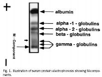
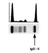
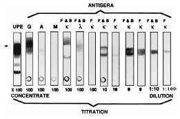
23 PROTEIN ELECTROPHORESIS MYELOMA 2012 AUGUST TUMOURS 23

24 Tc 99 CT-scans Angiography MRI RADIOLOGICAL EXAMINATIONS 2012 AUGUST TUMOURS 24


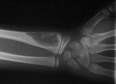
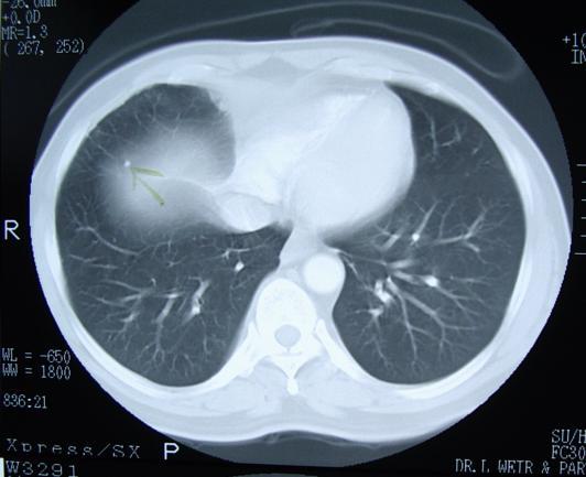
25 RADIOGRAPHIC INTERPRETATION X-RAYS IN 2 PLANES CT : BONE FORMATION, CALCIFIED LESIONS INTEGRETY CORTEX LUNG METS 2012 AUGUST TUMOURS 25


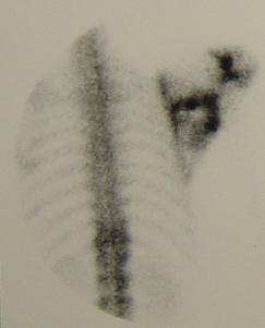
26 Tc- BONESCAN : - Detects skeletal mets - presence of multiple lesions, enchondromas, osteochondromas,etc. May be false negative in multiple myeloma 2012 AUGUST TUMOURS 26
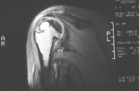
27 MRI STUDY of choice to determine anatomical setting of both bone and soft tissue tumors Tumour relationship to vital structures like blood vessels and nerves Evaluation of lesion on Tc scan, not yet visible on X-rays 2012 AUGUST TUMOURS 27
28 ULTRASOUND FOR SYSTIC LESIONS / SOFT TISSUE MASS 2012 AUGUST TUMOURS 28
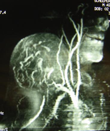
29 ARTERIOGRAPHY SELDOM USED EMBOLIZATION OF APPROPRIATE LESIONS 2012 AUGUST TUMOURS 29

30 RADIOGRAPHIC FEATURES TUMORS AND TUMOR- LIKE LESIONS OF BONE 2012 AUGUST TUMOURS 30
31 SITE Epiphysis : GCT, Chondroblastoma Metaphysis : Osteosarcoma, osteoblastoma, Chondromyxoid fibroma, NOF osteochondroma Diaphysis : Metastatic carcinoma,ewing s, Chondrosarcoma 2012 AUGUST TUMOURS 31

32 RADIOGRAPHIC FEATURES BORDERS - SLOW GROWING.. BENIGN - AGGRESSIVE.. MALIGNANT 2012 AUGUST TUMOURS 32

33 TYPE OF BONY DESTRUCTION RADIOGRAPHIC FEATURES 2012 AUGUST TUMOURS 33

34 CARTILAGE VS BONE 2012 AUGUST TUMOURS 34

35 PERIOSTEAL REACTION 2012 AUGUST TUMOURS 35
36 PERIOSTEAL REACTIONS Codman : Triangular cuff of reactive periosteal bone at the edge of a lesion. ( OS, osteomyelitis, ABC, infection) Onion-skinning : Due to episodic or pulsatile tumour growth ( Ewing s, Infection, OS of shaft ) Sunburst : Rapid continuous periosteal lifting and stretching ( bone next to Sharpy fibres ) 2012 AUGUST TUMOURS 36

37 SOFT TISSUE INVOLVEMENT PRIMARY SOFT TISSUE TUMOUR VS PRIMARY BONE TUMOUR 2012 AUGUST TUMOURS 37
38 RADIOGRAPHIC FEATURES BENIGN VS MALIGNANT 2012 AUGUST TUMOURS 38
39 BIOPSY OF BONE TUMOURS? Diagnosis,? Stage Same surgeon as final procedure Biopsy tract location important Meticulous haemostasis Samples for micro & histo 2012 AUGUST TUMOURS 39


40 OPEN BIOPSY EXCISIONAL : When possible in benign lesions INCISIONAL : Preferable in malignant lesions 2012 AUGUST TUMOURS 40

41 PRINCIPLES Longitudinal incision Sharp dissection Limited anatomic exposure Avoid neurovascular exposure Sample reactive tissue, pseudo capsule, capsule, tumour Small bone window if necessary 2012 AUGUST TUMOURS 41
42 PRINCIPLES (CONT) Haemostasis Subcutaneous stitch Drain in line with incision If procedure follows biopsy new instruments and drapes 2012 AUGUST TUMOURS 42
43 NEEDLE BIOPSY As for open biopsy Biopsy tract where can be excised Fine needle biopsy Core needle biopsy Disadvantage = tissue biopsy possibility being non-representative (eg. Necrosis or reactive) 2012 AUGUST TUMOURS 43


44 FROZEN SECTION Determine if specimen adequate or reactive Lesion inflammatory/ needs mcs? Need for further investigations? Immediate diagnosis possible 2012 AUGUST TUMOURS 44


45 SURGICAL PROCEDURES GOAL REMOVE LESION WITH MINIMAL RISK OF LOCAL RECURRENCE 2012 AUGUST TUMOURS 45


46 LIMB SALVAGE CRITERIA : 1. Local control = amputation 2. Saved limb must be functional 2012 AUGUST TUMOURS 46

47 LIMB SALVAGE VARIOUS METHODS: 1. Endoprosthesis 2. Allograft 3. Composite 4. Arthrodesis 2012 AUGUST TUMOURS 47


48 Contra indications: LIMB SALVAGE 1. Pathological fractures 2. Skeletal immaturity 3. Anatomical site Distant metastases is not a Contra-indication 2012 AUGUST TUMOURS 48
49 SURGICAL MARGINS 1. INTRA-LESIONAL 2. MARGINAL 3. WIDE 4. RADICAL 5. AMPUTATION 2012 AUGUST TUMOURS 49
50 INTRA-LESIONAL Dissection passes through the tumour Leaves macroscopic tumour Not therapeutic 2012 AUGUST TUMOURS 50
51 MARGINAL Through pseudo-capsule of tumour / reactive zone Controls non-invasive benign tumours Recurrence in malignant lesions = 25-50% 2012 AUGUST TUMOURS 51
52 WIDE Dissection entirely through normal tissue at a distance from the lesion Skip lesions / microscopic satellites may be left Recurrence of malignant lesions = < 10% 2012 AUGUST TUMOURS 52
53 RADICAL Removal of entire compartment Distant metastases left 2012 AUGUST TUMOURS 53
54 AMPUTATION Intra-capsular Marginal Wide Radical 2012 AUGUST TUMOURS 54
55 ADJUVANT CHEMOTHERAPY Reduces mass and vascularity of tumour Time for operative planning Want 90% kill rate Localized disease = 60-70% long-term disease free survival 2012 AUGUST TUMOURS 55
56 COMPLICATIONS Stunting of growth Osteoporosis AVN Cisplatinum nephro & oto toxicity Adriamycin Cardiotoxic Vincristine Neurotoxicity Chemotherapeutic induced malignancy usually blood forming, eg Leukaemias 2012 AUGUST TUMOURS 56
57 RADIOTHERAPY Absorption by complex molecules rupture of chemical bonds Indirect DNA changes stop cell reproduction & specific cell Fx Destruction of small blood vessels Radiosensitivity = mitotic activity x= degree differentiation 2012 AUGUST TUMOURS 57
58 ADVERSE EFFECTS Joint stiffness / function loss Subcutaneous fibrosis Premature growthplate closure Irradiation induced sarcoma Enteritis, diarrhoea, obstruction and bleeding Cystitis and hepatitis 2012 AUGUST TUMOURS 58
59 ADVERSE EFFECTS Muscle atrophy and fibrosis Erythema and hyperpigmentation Hair loss and skin flaking Lymphoedema 2012 AUGUST TUMOURS 59
60 DEFINITIONS Rad (radiation absorbed dose) =Energy imparted to matter by ionising radiation per unit mass Grays (Gr) = 1 Joule of energy absorbed by a mass of 1 kg 2012 AUGUST TUMOURS 60
Bone Tumors Clues and Cues
 William Herring, M.D. 2002 Bone Tumors Clues and Cues In Slide Show mode, advance the slides by pressing the spacebar All Photos Retain the Copyright of their Authors Clues by Appearance of Lesion Patterns
STAGING, BIOPSY AND NATURAL HISTORY OF TUMORS SCOTT D WEINER MD
 STAGING, BIOPSY AND NATURAL HISTORY OF TUMORS SCOTT D WEINER MD WHAT DO YOU DO WHEN THIS SHOWS UP IN YOUR OFFICE? besides panicking KEY PRINCIPLE!!! Reactive zone is the edema, neovascularity and inflammation
Malignant bone tumors. Incidence Myeloma 45% Osteosarcoma 24% Chondrosarcoma 12% Lyphoma 8% Ewing s Sarcoma 7%
 Malignant bone tumors Incidence Myeloma 45% Osteosarcoma 24% Chondrosarcoma 12% Lyphoma 8% Ewing s Sarcoma 7% Commonest primary bone sarcoma is osteosarcoma X ray Questions to ask 1. Solitary or Multiple
Primary bone tumors > metastases from other sites Primary bone tumors widely range -from benign to malignant. Classified according to the normal cell
 Primary bone tumors > metastases from other sites Primary bone tumors widely range -from benign to malignant. Classified according to the normal cell counterpart and line of differentiation. Among the
The Radiology Assistant : Bone tumor - ill defined osteolytic tumors and tumor-like lesions
 Bone tumor - ill defined osteolytic tumors and tumor-like lesions Henk Jan van der Woude and Robin Smithuis Radiology department of the Onze Lieve Vrouwe Gasthuis, Amsterdam and the Rijnland hospital,
Introduction to Musculoskeletal Tumors. James C. Wittig, MD Orthopedic Oncologist Sarcoma Surgeon
 Introduction to Musculoskeletal Tumors James C. Wittig, MD Orthopedic Oncologist Sarcoma Surgeon www.tumorsurgery.org Definitions Primary Bone / Soft tissue tumors Mesenchymally derived tumors (Mesodermal)
APMA 2018 Radiology Track Bone Tumors When to say Gulp!
 APMA 2018 Radiology Track Bone Tumors When to say Gulp! DANIEL P. EVANS, DPM, FACFAOM Professor, Department of Podiatric Medicine and Radiology Dr. Wm. Scholl College of Podiatric Medicine Conflict of
Immunohistochemistry in Bone and Soft Tissue Tumors. Sahar Rassi Zankoul, MD
 Immunohistochemistry in Bone and Soft Tissue Tumors Sahar Rassi Zankoul, MD Introduction Bone tumors represent a wide variety of tumors of various origins and malignant potentials. These different tumor
Bone Tumours - a synopsis. Dr Zena Slim SpR in Histopathology QAH 2009
 Bone Tumours - a synopsis Dr Zena Slim SpR in Histopathology QAH 2009 Aims General approach to diagnosis Common entities.and not so common ones. Mini quiz Challenge of bone tumour diagnosis Bone tumours
IAEA Pediatric Radiation Oncology Training Dr Laskar Version 1 June SOFT TISSUE SARCOMA (Non Rhabdomyosarcoma)
") SOFT TISSUE SARCOMA (Non Rhabdomyosarcoma) Soft Tissue structures Fat, Muscles, Fibrous tissue, Blood vessels, Supporting cells of peripheral nervous system Soft Tissue Sarcomas:- embryologically arise
SOFT TISSUE SARCOMA (Non Rhabdomyosarcoma) Soft Tissue structures Fat, Muscles, Fibrous tissue, Blood vessels, Supporting cells of peripheral nervous system Soft Tissue Sarcomas:- embryologically arise
The Radiology Assistant : Bone tumor - well-defined osteolytic tumors and tumor-like lesions
 Bone tumor - well-defined osteolytic tumors and tumor-like lesions Henk Jan van der Woude and Robin Smithuis Radiology department of the Onze Lieve Vrouwe Gasthuis, Amsterdam and the Rijnland hospital,
Musculoskeletal Sarcomas
 Musculoskeletal Sarcomas Robert C. Orth, M.D., Ph.D. Edward B. Singleton Department of Pediatric Radiology Texas Children s Hospital Page 0 xxx00.#####.ppt 9/23/2012 9:01:18 AM No disclosures Page 1 xxx00.#####.ppt
Department of Radiology, University of Szeged. Imaging of the skeleton
 Imaging of the skeleton Methods of examination: plain x-ray (radiography, densitometry) x-ray with contrast material (fistulography, angiography) ultrasound (b-mode, Doppler, color, duplex) computed tomography
Effective local and systemic therapy is necessary for the cure of Ewing tumor Most chemotherapy regimens are a combination of cyclophosphamide,
 Ewing Tumor Perez Ewing tumor is the second most common primary tumor of bone in childhood, and also occurs in soft tissues Ewing tumor is uncommon before 8 years of age and after 25 years of age In the
Case 8 Soft tissue swelling
 Case 8 Soft tissue swelling 26-year-old female presented with a swelling on the back of the left knee joint since the last 6 months and chronic pain in the calf and foot since the last 2 months. Pain in
Endovascular and surgical treatment of giant pelvic tumor
 Endovascular and surgical treatment of giant pelvic tumor Mitrev Z., MD FETCS; Anguseva T., MD; Milev I., MD; Zafiroski G., PhD MD Center for Cardiosurgery, Filip the II, Skopje, Macedonia Background Giant
A 76 year old male presented with sudden increase of dyspnoea on 15 November 2014, following a biopsy. A previous CXR was reviewed.
 Question 1 A 76 year old male presented with sudden increase of dyspnoea on 15 November 2014, following a biopsy. A previous CXR was reviewed. Imaging A CXR was performed on 28 May 2014. A CT of the chest
BONES & JOINTS INFECTION BONE TUMOURS
 BONES & JOINTS INFECTION BONE TUMOURS IMPORTANT SERIOUS CONSEQUENCE PLEASE DON T MISS!! EARLY DIAGNOSIS & PROPER TREATMENT HOW?? AWARE of THEIR EXISTENCE (Knowledge) PREPARE for THEIR OCCURRENCE A HIGH
Grading of Bone Tumors
 Grading of Bone Tumors Joon Hyuk Choi, M.D. Department of Pathology College of Medicine, Yeungnam University Introduction to grading system of bone tumor used at Mayo Clinic WHO Histologic Classification
LAC + USC.
 Jeff McDavit,, M.D. LAC + USC mcdavit@usc.edu Clinical History 55 year old male with large, deep, non- tender left thigh mass. Seen at LAC+USC Med Ctr FNA clinic No h/o trauma or radiation Vimentin
University Journal of Surgery and Surgical Specialities
 University Journal of Surgery and Surgical Specialities Volume 1 Issue 1 2015 EXTRA SKELETAL MESENCHYMAL CHONDROSARCOMA :A CASE REPORT Rajaraman R Subbiah S Navin Naushad Kilpaulk Medical College Abstract:
Update on Sarcomas of the Head and Neck. Kevin Harrington
 Update on Sarcomas of the Head and Neck Kevin Harrington Overview Classification and incidence of sarcomas Clinical presentation Challenges to treatment Management approaches Prognostic factors Radiation-induced
GIANT CELL-RICH OSTEOSARCOMA: A CASE REPORT
 Nagoya J. Med. Sci. 59. 151-157, 1996 CASE REPORTS GIANT CELL-RICH OSTEOSARCOMA: A CASE REPORT KEIJI SATO!, SHIGEKI YAMAMURA!, HISASHI IWATA!, HIDESHI SUGIURA 2, NOBUO NAKASHIMA 3 and TETSURO NAGASAKA
Primary Bone Tumors: Spine Surgery Live -Video Techniques Mobile Spine
 Primary Bone Tumors: Spine Surgery Live -Video Techniques Mobile Spine Christopher Ames MD Professor of Neurosurgery and Orthopedic Surgery Director of Spine Tumor And Deformity Surgery UCSF Department
Disclosures. Giant Cell Rich Tumors of Bone. Outline. The osteoclast. Giant cell rich tumors 5/21/11
 Disclosures Giant Cell Rich Tumors of Bone Andrew Horvai, MD, PhD Associate Clinical Professor, Pathology This lecture discusses "off label" uses of a number of pharmaceutical agents. The speaker is describing
Carcinoma of Unknown Primary site (CUP) in HEAD & NECK SURGERY
 in HEAD & NECK SURGERY") Carcinoma of Unknown Primary site (CUP) in HEAD & NECK SURGERY SEARCHING FOR THE PRIMARY? P r o f J P P r e t o r i u s H e a d : C l i n i c a l U n i t C r i t i c a l C a r e U n i v e r s i t y O f
Carcinoma of Unknown Primary site (CUP) in HEAD & NECK SURGERY SEARCHING FOR THE PRIMARY? P r o f J P P r e t o r i u s H e a d : C l i n i c a l U n i t C r i t i c a l C a r e U n i v e r s i t y O f
MUSCLE - INVASIVE AND METASTATIC BLADDER CANCER
 10 MUSCLE - INVASIVE AND METASTATIC BLADDER CANCER Recommendations from the EAU Working Party on Muscle Invasive and Metastatic Bladder Cancer G. Jakse (chairman), F. Algaba, S. Fossa, A. Stenzl, C. Sternberg
Malignant Bone Tumors - Part I: a brief revision of diagnostic aspects with conventional radiology
 Malignant Bone Tumors - Part I: a brief revision of diagnostic aspects with conventional radiology Poster No.: C-2473 Congress: ECR 2013 Type: Educational Exhibit Authors: I. Candelaria, L. B. Barbosa,
Malignant Primary Bone Tumours
 Volume 05 / Issue 04 / December 2017 boa.ac.uk Page 62 JTO Subspecialty Section Malignant Primary one Tumours Paul Cool Malignant primary tumours of bone are a diverse group of tumours. Overall, the incidence
Osteosarcoma (Canine)
") Osteosarcoma (Canine) Answering Your Questions About Osteosarcoma In Dogs What Is Osteosarcoma? Usual Sites for Osteosarcoma Development Osteosarcoma is by far the most common bone tumor of the dog, usually
Osteosarcoma (Canine) Answering Your Questions About Osteosarcoma In Dogs What Is Osteosarcoma? Usual Sites for Osteosarcoma Development Osteosarcoma is by far the most common bone tumor of the dog, usually
is time consuming and expensive. An intra-operative assessment is not going to be helpful if there is no more tissue that can be taken to improve the
 My name is Barry Feig. I am a Professor of Surgical Oncology at The University of Texas MD Anderson Cancer Center in Houston, Texas. I am going to talk to you today about the role for surgery in the treatment
Biopsy. DR. K.B.PRABHUDEV Consultant Orthopedic surgeon Bone And Soft Tissue Tumor Services Sparsh Hospital. Bone Bangalore
 Biopsy DR. K.B.PRABHUDEV Consultant Orthopedic surgeon Bone And Soft Tissue Tumor Services Sparsh Hospital Biopsy Biopsy is a critical procedure in the treatment of muskuloskeletal tumors. Biopsy is a
Bone and Joint Part 2. Leslie G Dodd, MD
 Bone and Joint Part 2 Leslie G Dodd, MD Relative rates of cancer Sarcomas are relatively uncommon tumors New cancer cases 2007 All sites 1.4 million prostate 218,890 lung 213,380 breast 180,510 Soft tissue
The Scandinavian Sarcoma Group annual report on extremity and trunk wall soft tissue and bone sarcomas
 The Scandinavian Sarcoma Group annual report on extremity and trunk wall soft tissue and bone sarcomas 2012-2016 1 The SSG annual report on extremity and trunk wall soft tissue and bone sarcomas. The Scandinavian
Hsin-Nung Shih M.D. Soft Tissue Tumor
 Soft Tissue Tumor Hsin-Nung Shih M.D. PROFESSOR DIVISION OF JOINT RECONSTRUCTION DEPARTMENT OF ORTHOPEADIC CHANG GUNG MEMORIAL HOSPITAL CHANG GUNG UNIVERSITY,COLLEGE OF MEDICINE TAIWAN Soft Tissue Tumor
Malignant Bone Tumours. PathoBasic, Daniel Baumhoer
 Malignant Bone Tumours PathoBasic, 20.03.18 Daniel Baumhoer FNCLCC Grading The differentiation score is defined as the extent to which a tumor resembles adult mesenchymal tissue (score 1), the extent to
Pathology of Sarcoma ELEANOR CHEN, MD, PHD, ASSISTANT PROFESSOR DEPARTMENT OF PATHOLOGY UNIVERSITY OF WASHINGTON
 Pathology of Sarcoma ELEANOR CHEN, MD, PHD, ASSISTANT PROFESSOR DEPARTMENT OF PATHOLOGY UNIVERSITY OF WASHINGTON Presentation outline Background and epidemiology of sarcomas Sarcoma classification Sarcoma
RESEARCH INFORMATION AWARENESS SUPPORT PRIMARY BONE CANCER CHONDROSARCOMA. Visit bcrt.org.uk for more information
 RESEARCH INFORMATION AWARENESS SUPPORT PRIMARY BONE CANCER CHONDROSARCOMA Visit bcrt.org.uk for more information CONTENTS What is it? Who does it affect? Symptoms Types of Chondrosarcoma Cause and Risk
What is Cancer? Petra Ketterl, MD Medical Oncology and Functional Medicine
 What is Cancer? Petra Ketterl, MD Medical Oncology and Functional Medicine What is Cancer? Layman s terms: cancer starts when cells grow out of control (in any place in the body) and crowd out normal cells
Radiology-Pathology Conference
 July 31, 2009 Radiology-Pathology Conference Daniel T Ginat, M.D., M.S. Sharlin Johnykutty,, M.D. Presentation material is for education purposes only. All rights reserved. 2009 URMC Radiology Page 1 of
CASE REPORT PLEOMORPHIC LIPOSARCOMA OF PECTORALIS MAJOR MUSCLE IN ELDERLY MAN- CASE REPORT & REVIEW OF LITERATURE.
 PLEOMORPHIC LIPOSARCOMA OF PECTORALIS MAJOR MUSCLE IN ELDERLY MAN- CASE REPORT & REVIEW OF LITERATURE. M. Madan 1, K. Nischal 2, Sharan Basavaraj. C. J 3. HOW TO CITE THIS ARTICLE: M. Madan, K. Nischal,
Da Costa was the first to coin the term. Marjolin s Ulcer: A Case Report and Literature Review. Case Report. Introduction
 E-Da Medical Journal 2016;3(2):24-28 Case Report Marjolin s Ulcer: A Case Report and Literature Review Yue-Chiu Su 1, Li-Ren Chang 2 Marjolin s ulcer is an aggressive cutaneous malignancy, which is common
PET IMAGING (POSITRON EMISSION TOMOGRAPY) FACT SHEET
 FACT SHEET") Positron Emission Tomography (PET) When calling Anthem (1-800-533-1120) or using the Point of Care authorization system for a Health Service Review, the following clinical information may be needed to
Positron Emission Tomography (PET) When calling Anthem (1-800-533-1120) or using the Point of Care authorization system for a Health Service Review, the following clinical information may be needed to
Review Course «Musculoskeletal Oncology» October 6, 2011 UNIKLINIK BALGRIST. Imaging of Bone and Soft Tissue. Tumors
 Imaging of Bone and Soft Tissue Tumors Approach from a radiologist s point of view Florian Buck Radiology Radio- Radio- Oncologist Oncologist Orthopedist Orthopedist Patient Management Oncologist Oncologist
Surgery for Breast Cancer
 Surgery for Breast Cancer 1750 Mastectomy - Petit 1894 Radical mastectomy Halsted Extended, Super radical mastectomy 1948 Modified radical mastectomy Patey 1950-60 WLE & RT Baclesse, Mustakallio 1981-85
History. 33 y/o F with hx of palpable anterior tibial mass x 2 years, only painful with palpation
 History 33 y/o F with hx of palpable anterior tibial mass x 2 years, only painful with palpation Imaging Photo Album Patient also had a smaller lesion 1 cm proximal to this lesion, not seen radiographically.
Louisa Fleure. Advanced Prostate Cancer Clinical Nurse Specialist. Guys and St Thomas NHS Trust
 Louisa Fleure Advanced Prostate Cancer Clinical Nurse Specialist Guys and St Thomas NHS Trust The classification of advanced prostate cancer The incidence of patients presenting with, or developing advanced
COPYRIGHT 2004 BY THE JOURNAL OF BONE AND JOINT SURGERY, INCORPORATED
 84 COPYRIGHT 2004 BY THE JOURNAL BONE AND JOINT SURGERY, INCORPORATED Radiographic Evaluation of Pathological Bone Lesions: Current Spectrum of Disease and Approach to Diagnosis BY BENJAMIN G. DOMB, MD,
* I have no disclosures or any
 Howard Rosenthal, M.D. Associate Professor of Orthopedic Surgery University of Kansas Sarcoma Center I have no disclosures or any conflicts related to the content of this presentation. Objectives 1. Describe
ORTHOPAEDIC ONCOLOGY OITE REVIEW COURSE
 ORTHOPAEDIC ONCOLOGY OITE REVIEW COURSE Richard D. Lackman, MD FACS Director, Orthopaedic Oncology Center Cancer Institute Introduction In the evaluation of a patient with a bone tumor, there are several
Radiology Pathology Conference
 Radiology Pathology Conference Sharlin Johnykutty,, MD, Cytopathology Fellow Sara Majewski, MD, Radiology Resident Friday, August 28, 2009 Presentation material is for education purposes only. All rights
Multidisciplinary management of retroperitoneal sarcomas
 Multidisciplinary management of retroperitoneal sarcomas Eric K. Nakakura, MD UCSF Department of Surgery UCSF Comprehensive Cancer Center San Francisco, CA 7 th Annual Clinical Cancer Update North Lake
Vertebral and Paravertebral Diseases
 Department of Radiology University of California San Diego Vertebral and Paravertebral Diseases John R. Hesselink, M.D. Vertebral / Paravertebral Disease (Extradural) Metastatic disease Primary bone tumors
Different Types of Cancer
 Different Types of Cancer Cancer can originate almost anywhere in the body. Sarcomas (connective tissue) Ø arise from cells found in the supporting tissues of the body such as bone, cartilage, fat, connective
Louisa Fleure. Advanced Prostate Cancer Clinical Nurse Specialist. Guys and St Thomas NHS Trust
 Louisa Fleure Advanced Prostate Cancer Clinical Nurse Specialist Guys and St Thomas NHS Trust The classification of advanced prostate cancer The incidence of patients presenting with, or developing advanced
Oncology General Principles L A U R I E S I M A R D B R E A S T S U R G I C A L O N C O L O G Y F E L L O W D E C E M B E R
 Oncology General Principles L A U R I E S I M A R D B R E A S T S U R G I C A L O N C O L O G Y F E L L O W D E C E M B E R 2 0 1 2 Objectives Discuss Diagnostic and staging strategies in oncology Know
MRI XR, CT, NM. Principal Modality (2): Case Report # 2. Date accepted: 15 March 2013
: Case Report # 2. Date accepted: 15 March 2013") Radiological Category: Musculoskeletal Principal Modality (1): Principal Modality (2): MRI XR, CT, NM Case Report # 2 Submitted by: Hannah Safia Elamir, D.O. Faculty reviewer: Naga R. Chinapuvvula, M.D.
Radiological Category: Musculoskeletal Principal Modality (1): Principal Modality (2): MRI XR, CT, NM Case Report # 2 Submitted by: Hannah Safia Elamir, D.O. Faculty reviewer: Naga R. Chinapuvvula, M.D.
Essential Dermatopathology. Jinah Kim, MD, PhD Department of Pathology and Dermatology Stanford University Medical Center
 Essential Dermatopathology Jinah Kim, MD, PhD Department of Pathology and Dermatology Stanford University Medical Center OBJECTIVES Review clinical, pathologic and molecular aspects of bone and fat tumors
Soft Tissue Sarcomas: Questions and Answers
 Soft Tissue Sarcomas: Questions and Answers 1. What is soft tissue? The term soft tissue refers to tissues that connect, support, or surround other structures and organs of the body. Soft tissue includes
Recommendations for cross-sectional imaging in cancer management, Second edition
 www.rcr.ac.uk Recommendations for cross-sectional imaging in cancer management, Second edition Musculoskeletal tumours Faculty of Clinical Radiology www.rcr.ac.uk Contents Primary bone tumours 3 Clinical
DISORDERS OF THE SALIVARY GLANDS Neoplasms Dr.M.Baskaran Selvapathy S IV
 DISORDERS OF THE SALIVARY GLANDS Neoplasms Dr.M.Baskaran Selvapathy S IV NEOPLASMS A) Epithelial I. Benign Pleomorphic adenoma( Mixed tumour) Adenolymphoma (Warthin s tumour) Oxyphil adenoma (Oncocytoma)
Soft Tissue Sarcoma. Presley Regional Trauma Center Department of Surgery University of Tennessee Health Science Center Memphis, Tennessee
 Soft Tissue Sarcoma Presley Regional Trauma Center Department of Surgery University of Tennessee Health Science Center Memphis, Tennessee Soft Tissue Sarcoma Collective term for an unusual and diverse
Typical skeletal location and differential diagnosis of bone tumors.
 Typical skeletal location and differential diagnosis of bone tumors. Poster No.: C-2418 Congress: ECR 2015 Type: Educational Exhibit Authors: M. Barros, L. A. Ferreira, Y. Costa, P. J. V. Coelho, F. Caseiro
Dr Rosalie Stephens. Mr Richard Martin. Medical Oncologist Auckland City Hospital Auckland
 Dr Rosalie Stephens Medical Oncologist Auckland City Hospital Auckland Mr Richard Martin General Surgeon Melanoma Unit Team Waitemata District Health Board Auckland 8:30-9:25 WS #99: Interactive Case Studies
performed to help sway the clinician in what the appropriate diagnosis is, which can substantially alter the treatment of management.
 Hello, I am Maura Polansky at the University of Texas MD Anderson Cancer Center. I am a Physician Assistant in the Department of Gastrointestinal Medical Oncology and the Program Director for Physician
Guideline for the Management of Vulval Cancer
 Version History Guideline for the Management of Vulval Cancer Version Date Brief Summary of Change Issued 2.0 20.02.08 Endorsed by the Governance Committee 2.1 19.11.10 Circulated at NSSG meeting 2.2 13.04.11
Bone tumors. RMG: jan
 Bone tumors RMG: jan 217. @Kijohs KIZZA JOHN KIJOHS Diseases arising in bone Lipoma Fibrous cortical defects Non-ossifying fibroma Bone island Benign simple cysts Enchondroma Osteochondroma Osteoid osteoma
Advertisement. Osteochondroma
 Advertisement Osteochondroma An osteochondroma is a benign (noncancerous) tumor that develops during childhood or adolescence. It is an abnormal growth that forms on the surface of a bone near the growth
Overview of Surgical Resection of Space Sarcomas
 13282_ON-33.qxd 3/31/09 4:50 PM Page 1 Chapter 33 Overview of Surgical Resection of Space Sarcomas Amir Sternheim, Tamir Pritsch, and Martin M. Malawer BACKGROUND The three main extracompartmental spaces
Definition Prostate cancer
 Prostate cancer 61 Definition Prostate cancer is a malignant neoplasm that arises from the prostate gland and the most common form of cancer in men. localized prostate cancer is curable by surgery or radiation
The breast advice for managing radiotherapy induced skin reactions
 15/05/2016 The breast advice for managing radiotherapy induced skin reactions Margaret Hjorth Nurse Unit Manager Epworth Radiation Oncology 1 15/05/2016 What is Radiotherapy? Use of high energy radiation
Monophasic Synovial Carcinoma of knee joint- A Case Report and Review of Literature
 IOSR Journal of Dental and Medical Sciences (IOSR-JDMS) e-issn: 2279-0853, p-issn: 2279-0861.Volume 17, Issue 3 Ver.5 March. (2018), PP 13-17 www.iosrjournals.org Monophasic Synovial Carcinoma of knee
What s new in bone and soft tissue sarcoma Treatment and Guidelines 2012? Rob Grimer
 What s new in bone and soft tissue sarcoma Treatment and Guidelines 2012? Rob Grimer ESMO conference 2012 Top Oncologists in world (~ 400) Lots of sarcoma basic science key messages: 40% of STS diagnoses
Pre-operative Ultrasound of Lymph Nodes in Thyroid Cancer
 Pre-operative Ultrasound of Lymph Nodes in Thyroid Cancer AACE - Advances in Medical and Surgical Management of Thyroid Cancer - 2018 Robert A. Levine, MD, FACE, ECNU Thyroid Center of New Hampshire Geisel
What is Cancer? Understanding the basics of
 What is Cancer? Understanding the basics of A process of physical and biological changes that occur when normal healthy cells are transformed into cancer cells What is Cancer? Cancer is an umbrella term
1. Written information to patient /GP: fax ASAP to GP & offer copy of consultation letter.
 Skin Cancer follow up guidelines If NEW serious diagnosis given: 1. Written information to patient /GP: fax ASAP to GP & offer copy of consultation letter. 2. Free prescription information details. 3.
Q&A. Fabulous Prizes. Collecting Cancer Data: Bone and Soft Tissue 1/10/113. NAACCR Webinar Series
 Collecting Cancer Data Bone & Soft Tissue NAACCR 2012 2013 Webinar Series Q&A Please submit all questions concerning webinar content through the Q&A panel. Reminder: If you have participants watching this
Ultrasound for Pre-operative Evaluation of Well Differentiated Thyroid Cancer
 Ultrasound for Pre-operative Evaluation of Well Differentiated Thyroid Cancer Its Not Just About the Nodes AACE Advances in Medical and Surgical Management of Thyroid Cancer - 2017 Robert A. Levine, MD,
Radiology Pathology Conference
 Radiology Pathology Conference Nadia F. Yusaf, M.D. PGY-3 1/29/2010 Presentation material is for education purposes only. All rights reserved. 2010 URMC Radiology Page 1 of 90 Case 1 60 year- old man presents
Vaginal intraepithelial neoplasia
 Vaginal intraepithelial neoplasia The terminology and pathology of VAIN are analogous to those of CIN (VAIN I-III). The main difference is that vaginal epithelium does not normally have crypts, so the
Case Scenario 1 Worksheet. Primary Site C44.4 Morphology 8743/3 Laterality 0 Stage/ Prognostic Factors
 CASE SCENARIO 1 9/10/13 HISTORY: Patient is a 67-year-old white male and presents with lesion located 4-5cm above his right ear. The lesion has been present for years. No lymphadenopathy. 9/10/13 anterior
Cervical cancer presentation
 Carcinoma of the cervix: Carcinoma of the cervix is the second commonest cancer among women worldwide, with only breast cancer occurring more commonly. Worldwide, cervical cancer accounts for about 500,000
Case Report Solitary Osteolytic Skull Metastasis in a Case of Unknown Primary Being latter Diagnosed as Carcinoma of Gall Bladder
 Cronicon OPEN ACCESS CANCER Case Report Solitary Osteolytic Skull Metastasis in a Case of Unknown Primary Being latter Diagnosed as Carcinoma of Gall Kartik Mittal 1, Rajaram Sharma 1, Amit Dey 1, Meet
ARTICLE. MR imaging in staging of bone tumors
 Cancer Imaging (2006) 6, 158 162 DOI: 10.1102/1470-7330.2006.0026 CI ARTICLE MR imaging in staging of bone tumors Shigeru Ehara Department of Radiology, Iwate Medical University School of Medicine, Morioka
SMALL ROUND BLUE CELL LESION OF BONE
 DISCLOSURE SMALL ROUND BLUE CELL LESION OF BONE Dr. Alistair Jordan University of South Alabama No financial support or endorsement OBJECTIVES Describe the more common small round cell lesions of bone
Case Report Intramedullary Chondrosarcoma of Proximal Humerus
 Hindawi Publishing Corporation Case Reports in Radiology Volume 2012, Article ID 642062, 7 pages doi:10.1155/2012/642062 Case Report Intramedullary Chondrosarcoma of Proximal Humerus Pratiksha Yadav, Dolly
Prostate Case Scenario 1
 Prostate Case Scenario 1 H&P 5/12/16: A 57-year-old Hispanic male presents with frequency of micturition, urinary urgency, and hesitancy associated with a weak stream. Over the past several weeks, he has
Intrarenal Extension. sinus
 Intrarenal Extension into sinus Document Capsular Penetration sinus 16 Pediatric Renal Tumor Staging Stage I Limited to Kidney & Completely Resected Intact Renal Capsule No Previous Rupture or Biopsy Renal
Take Home Quiz 1 Please complete the quiz below prior to the session. Use the Multiple Primary and Histology Rules
 Take Home Quiz 1 Please complete the quiz below prior to the session. Use the Multiple Primary and Histology Rules Case 1 72 year old white female presents with a nodular thyroid. This was biopsied in
Case Scenario 1: Thyroid
 Case Scenario 1: Thyroid History and Physical Patient is an otherwise healthy 80 year old female with the complaint of a neck mass first noticed two weeks ago. The mass has increased in size and is palpable.
Associated Terms: Osteosarcoma, Bone Cancer, Limb Salvage, Appendicular Osteosarcoma, Pathologic Fracture, Chondrosarcoma
 1 of 9 9/29/2014 8:25 PM Associated Terms: Osteosarcoma, Bone Cancer, Limb Salvage, Appendicular Osteosarcoma, Pathologic Fracture, Chondrosarcoma The term "ACVS Diplomate" refers to a veterinarian who
Melanoma Case Scenario 1
 Melanoma Case Scenario 1 History and physical 11/5/16 Patient is a single, 48-year-old male in good health who presented to his primary physician for a yearly physical exam during which a 3.4 x 2.8 x 1.5
Unusual location of bone sarcoma in children
 Unusual location of bone sarcoma in children Poster No.: C-1517 Congress: ECR 2014 Type: Educational Exhibit Authors: S. JERBI, A. Khalfalli, G. Abid, O. Bradai, N. chouchane, H. HAMZA; Mahdia/TN Keywords:
CODING PRIMARY SITE. Nadya Dimitrova
 CODING PRIMARY SITE Nadya Dimitrova OUTLINE What is coding and why do we need it? ICD-10 and ICD-O ICD-O-3 Topography coding rules ICD-O-3 online WHAT IS CODING AND WHY DO WE NEED IT? Coding: to assign
Specialist Referral Service Willows Information Sheets. Cancer in cats and dogs: Assessment of the patient
 Specialist Referral Service Willows Information Sheets Cancer in cats and dogs: Assessment of the patient Cancer in cats and dogs: Assessment of the patient Cancer is common in human and veterinary medicine.
5.1 Breast, Anatomy. 70
 Chapter 5 Breast 5.1 Breast, Anatomy Breasts, also called Mamma are mammary glands, subcutaneously placed on the ventral side of the trunk in mammalian species, and develop for the sole purpose of secreting
Cervical Cancer 3/25/2019. Abnormal vaginal bleeding
 Cervical Cancer Abnormal vaginal bleeding Postcoital, intermenstrual or postmenopausal Vaginal discharge Pelvic pain or pressure Asymptomatic In most patients who are not sexually active due to symptoms
CNS TUMORS. D r. Ali Eltayb ( U. of Omdurman. I ). M. Path (U. of Alexandria)
. M. Path (U. of Alexandria)") CNS TUMORS D r. Ali Eltayb ( U. of Omdurman. I ). M. Path (U. of Alexandria) CNS TUMORS The annual incidence of intracranial tumors of the CNS ISmore than intraspinal tumors May be Primary or Secondary
CNS TUMORS D r. Ali Eltayb ( U. of Omdurman. I ). M. Path (U. of Alexandria) CNS TUMORS The annual incidence of intracranial tumors of the CNS ISmore than intraspinal tumors May be Primary or Secondary
Principles of Surgical Oncology. Winnie Achilles Tierklinik Hollabrunn Lastenstrasse Hollabrunn
 Principles of Surgical Oncology Winnie Achilles Tierklinik Hollabrunn Lastenstrasse 2 2020 Hollabrunn boexi@gmx.de The first surgery provides the best chance for a cure in an animal with a tumor Clinical