CT Radiation Dosimetry Study Using Monte Carlo Simulation and. Computational Anthropomorphic Phantoms
|
|
|
- Warren Newton
- 6 years ago
- Views:
Transcription
1 CT Radiation Dosimetry Study Using Monte Carlo Simulation and Computational Anthropomorphic Phantoms by Yakun Zhang Graduate Program in Medical Physics Duke University Date: Approved: Ehsan Samei, Supervisor W. Paul Segars Robert Reiman Thesis submitted in partial fulfillment of the requirements for the degree of Master of Science in the Graduate Program in Medical Physics in the Graduate School of Duke University 2012
2 ABSTRACT CT Radiation Dosimetry Study Using Monte Carlo Simulation and Computational Anthropomorphic Phantoms by Yakun Zhang Graduate Program in Medical Physics Duke University Date: Approved: Ehsan Samei, Supervisor W. Paul Segars Robert Reiman An abstract of a thesis submitted in partial fulfillment of the requirements for the degree of Master of Science in the Graduate Program in Medical Physics in the Graduate School of Duke University 2012
3 Copyright by Yakun Zhang 2012
4 Abstract There are three main x-ray based modalities for imaging the thorax: radiography, tomosynthesis, and computed tomography (CT). CT perhaps provides the highest level of feature resolution but at notably higher radiation dose, which has increased the concern among radiation protection professionals. Being able to accurately assess the radiation dose patients receive during CT procedures is a crucial step in the management of CT dose. To identify the best imaging modality for patients, the American College of Radiology published the guiding principle of The right exam, for the right reason, at the right time. To implement this principle in making an appropriate choice between standard chest projection imaging, tomosynthesis, and CT, the organ and effective dose for each modality should be accurately known. This thesis work attempted to explain the effect on dose results when choosing different types of computational phantoms used in CT dosimetry; this work also compared radiation dose across three main x-ray based modalities on one common platform for different body shape adults. The first part of this thesis compared organ doses, effective doses, and risk indices from 13 representative adult CT protocols using four types of reference phantoms (XCAT, ICRP 110, ImPACT, and CT-Expo). Despite closely-matched organ mass, total body weight, and height, large differences in organ dose exist due to iv
5 variation in organ location, spatial distribution, and dose approximation method. Dose differences for fully irradiated radiosensitive organs were much smaller than those for partially irradiated organs. Weighted dosimetry quantities including effective dose, male risk indices, k factors, and male q factors agreed well across phantoms. The female risk indices and q factors varied considerably across phantoms. The second part of this thesis estimated organ doses, effective doses and risk indices for the three clinical X-ray imaging techniques (chest radiography, tomosynthesis, and CT) using 59 anatomically variable voxelized phantoms and Monte Carlo simulation methods. The average effective dose of the chest posteroanterior examination was found to be 0.04 msv, which was 1.3% that of the chest CT examination. The average effective dose of the chest tomosynthesis examination was found to be about ten times that of the chest posteroanterior examination and about 12% that of the chest CT examination. With increasing patient average chest diameter, both the effective dose and risk index for CT increased considerably in an exponential fashion, while these two dose metrics only increased slightly for radiographic modalities and for chest tomosynthesis. Effective and organ doses normalized to mas all illustrated an exponential decrease with increasing patient size. In conclusion, patient body size has a much greater impact on radiation dose of chest CT examinations than chest radiography and tomosynthesis. Patients of different sizes should be considered differently when choosing the best thoracic imaging modality. v
6 Contents Abstract... iv List of Tables... ix List of Figures... x 1. Introduction Organ doses, effective doses, and risk indices in adult CT: Comparison of four types of reference phantoms across different examination protocols Introduction Methods Computational Phantoms XCAT hybrid phantoms ICRP 110 voxelized phantoms ImPACT stylized phantom CT-Expo stylized phantoms CT examination categories Organ doses simulations XCAT and ICRP 110 phantoms ImPACT phantoms CT-Expo phantoms Effective dose and cancer risk index calculations Results Comparison of organ doses vi
7 2.3.2 Comparison of effective doses and risk indices Comparison of k and q factors Discussion Conclusion Comparison of patient specific dose metrics between chest radiography, tomosynthesis, and CT for adult patients of wide ranging body habitus Introduction Methods Patients and XCAT Computational Phantoms Organ dose simulation Chest x-ray radiography configuration Tomosysthesis configuration Radiographic and tomosynthesis techniques CT system simulation and scan techniques Effective dose and risk index calculation Data analysis Resutls Average effective dose across imaging modalities Effect of patient size on radiation dose metrics Correlation of patient size with effective dose per mas or 100 mas Correlation of patient size with organ dose per mas or 100 mas Effect of tomosynthesis angles on radiation dose vii
8 3.4 Discussion Effective dose across imaging modalities Effect of body size on radiation dose Effect of tomosynthesis angles on radiation dose Limitations and future work Conclusion References viii
9 List of Tables Table 1: Organ masses for the four types of reference phantoms employed in this study. Reference organ masses published in ICRP 89 are also listed for comparison. Note that the CT-Expo male and female phantoms were matched to ICRP publication Table 2: CT examination categories investigated in this study Table 3: Scan parameters for body and neurological examination categories Table 4: Coefficients of variation (COV) and average differences of fully irradiated organs for all 13 examination categories. The average COV was obtained by first calculating COV for each organ across four phantoms, and then averaged over all fully irradiated organs. The average difference was calculated using: DXCAT/DICRP110-1, DImPACT/DICRP110-1, and DCT-Expo/DICRP110-1, and then averaged over all fully irradiated organs. The CT-Expo male phantom does not have any breasts, so breasts were excluded in the calculation for male. The female only organs were bracketed in the organ list. Note that for the adrenals, kidneys, and neck examinations there was no fully radiosensitive organs Table 5: Average differences and coefficients of variation (COV) of partially irradiated organs for all 13 examination categories Table 6: k and q factor over three types of phantoms for the 13 examination categories. 38 Table 7: Comparison of k factors obtained using ImPACT and CT-Expo phantoms in this study and from other 3 research groups. All the k factors here were calculated using ICRP 103 tissue weighting factors Table 8: Imaging techniques for all modalities. PA = Posteroanterior. AP = Anteroposterior. LAT = Lateral Table 9: Exponential relationship between effective dose and body size Table 10: Exponential relationship of organ dose with body size ix
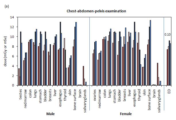
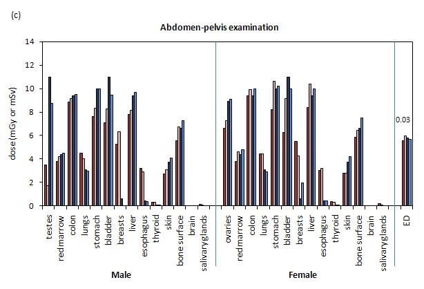
10 List of Figures Figure 1: Flow chart of the methodology used in this study. RI: risk index; ED: effective dose Figure 2: Lateral and frontal views of the four types of phantoms used in this study. (a) and (b): XCAT reference male and female hybrid phantoms after voxelization. (c) and (d): ICRP 110 reference male and female voxelized phantoms. The organs were relabeled to be consistent with the XCAT phantoms. (e): ImPACT stylized/mathematical hermaphrodite phantom. (f) and (g): CT-Expo male and female stylized/mathematical phantoms Figure 3: Comparison between different types of phantoms in terms of organ doses and effective doses (ED) in 13 examination categories: (a) chest-abdomen-pelvis; (b) chest; (c) abdomen-pelvis; (d) abdomen; (e) pelvis; (f) adrenals; (g) liver; (h) kidneys; (i) liver-tokidneys; (j) kidneys-to-bladder; (k) head; (l) neck; (m) head-and-neck. Note that doses for ImPACT are the same for male and female except for testes and ovaries. Doses are normalized to 100 mas. The quantitative differences were tabulated in Tables 4 and 5. The coefficient of variation for ED is displayed above the bars for each examination Figure 4: Comparison of risk index (RI) over four types of phantoms for the 13 examination categories for male (a) and female (b) phantoms. The coefficient of variation for each examination is displayed above the bars Figure 5: Comparison of k factor among four types of phantoms for 13 examination categories. The k factor for XCAT, ICRP 110, and CT-Expo phantoms used gender averaged effective dose and DLP. The tabulated k values for 6 protocol classes from the AAPM were also included for comparison in this graph. The coefficient of variation for each examination is displayed above the bars Figure 6: Comparison of q factor over four types of phantoms for the 13 examination categories for male (a) and female (b) phantoms. The coefficient of variation for each examination is displayed above the bars Figure 7: Schematic illustration of the tomosynthesis acquisition geometry (a) and the CT acquisition geometry (b) Figure 8: Average effective dose across imaging modalities. ED = Effective dose. PA = Posteroanterior. AP = Anteroposterior. LAT = Lateral x
11 Figure 9: (a) Distribution of effective dose with respect to patient size for all modalities; (b) effective dose ratio between chest CT and tomosynthesis and between chest tomosynthesis and conventional two-view chest x-ray. Average chest diameter is defined as the square root of anteroposterior multiplying lateral thickness at the beam center. ED = Effective dose. PA = Posteroanterior. AP = Anteroposterior. LAT = Lateral Figure 10: (a) & (c) Distribution of risk index with respect to patient size for all modalities; (b) & (d) risk index ratio between chest CT and tomosynthesis and between chest tomosynthesis and conventional two-view chest x-ray. Average chest diameter is defined as the square root of anteroposterior multiplying lateral thickness at the beam center. ED = Effective dose. RI = Risk index. PA = Posteroanterior. AP = Anteroposterior. LAT = Lateral Figure 11: Effective dose per mas or 100 mas as a function of body dimension for all patients and modalities: (a) Chest PA and Chest AP; (c) Chest left lateral (Left LAT); (d) Chest tomosynthesis; (e) Chest CT. For (a), (b), and (c), patient s anteroposterior thickness at the beam center is used for the x-axis; for (c), patient s lateral thickness at the beam center is used for the x-axis; for (e), average diameter is used for the x-axis. + or x are the effective dose values calculated from the organ dose of individual patients. Lines are the exponential fit ED (d) = exp (αedd +βed) with parameters αed and βed tabulated in Table 9. ED = Effective dose. PA = Posteroanterior. AP = Anteroposterior. LAT = Lateral Figure 12: Lungs, breasts, and liver doses as a function of body dimension. Lines are the exponential fit: Organ dose (d) = exp (αorgand + βorgan) with the fitting parameters tabulated in Table 10 for each organ. Average chest diameter is defined as the square root of anteroposterior multiplying lateral thickness at the beam center. PA = Posteroanterior. AP = Anteroposterior. LAT = Lateral Figure 13: (a) Effective dose at each acquisition angle of chest tomosynthesis for the reference phantoms; (b) Doses to lungs, breasts, and liver at each acquisition angle for the reference female phantom. The effective dose was averaged across male and female reference phantoms as defined in ICRP publication 103. Doses to the three organs for both reference phantoms showed the same pattern, so only female data was plotted here. ED = Effective dose xi
12 1. Introduction Compared to the overlaying projected anatomy from conventional radiography, computed tomography (CT) has provided great diagnostic accuracy to the medical imaging world. However, it also introduces much more radiation exposure to the public, which has gained increased attention from the general public and health care providers. Therefore, it is important to be able to accurately estimate the radiation dose that patients receive during CT procedures so that any potential risk can be estimated and minimized. Computational anthropomorphic phantoms combined with Monte Carlo simulation of radiation transport have proven to be an accurate and reliable method to estimate organ doses during CT examinations. Such phantoms have evolved from the original stylized phantoms, to voxelized phantoms, and to more advanced hybrid phantoms. Some comparisons between hybrid and stylized phantoms used in CT dosimetry have been made, but these comparisons included only limited examination types, leaving out some other important and routinely used CT examination, such as liver examinations. Part one of this thesis was to extend the earlier comparison efforts to four types of widely used reference phantoms, to a wide range of body and neurological examination types, and to dose and risk conversion coefficients. One lower dose imaging technique tomosynthesis, which can also provide sliceby-slice anatomical information, was re-introduced to the medical imaging world for an 1
13 alternative to CT. Application of tomosynthesis on screening and detection of lung nodules had been proposed because of its lower dose to radiosensitive organs lungs and breasts as well as its reasonable detectability. It is desirable to compare the radiation dose between chest CT and tomosynthesis on one common simulation platform across different patient body shapes, which has not been done before, to allow health care professionals to make informative decision on the best imaging modality for individual patient. Part two of this thesis compared organ doses, effective doses, and risk index between three x-ray based modalities for imaging the thorax: radiography, tomosynthesis, and CT. 2
14 2. Organ doses, effective doses, and risk indices in adult CT: Comparison of four types of reference phantoms across different examination protocols Introduction Over the past few decades, technological advances in x ray computed tomography (CT) have made CT an essential diagnostic imaging tool. While this technology is bringing convenience and high quality tomography to the medical imaging world, it also introduces radiation exposure to the public. 1 From the data published in 2006, the use of CT has increased at a rate of 8 to 15% per year for the last 7 to 10 years. 2 In 2006, nearly half of the total medical radiation exposure was from CT. 2 This has increased concern about CT imaging among radiation protection professionals as well as the general public. 3-5 Therefore, it is important to be able to accurately estimate the radiation dose that patients receive during CT procedures so that any potential risk can be estimated and minimized. Computational anthropomorphic phantoms combined with Monte Carlo simulation of radiation transport have proven to be an accurate and reliable method to estimate organ doses during CT examinations Such phantoms have evolved from the original stylized phantoms, 20 to voxelized phantoms, 21, 22 and to more advanced hybrid phantoms. 11, 23, 24 Stylized phantoms define the organs and structures in the human body 1 This chapter is based on a manuscript with the same title published in the journal Medical Physics 3
15 through mathematical equations and simple geometric primitives. While highly flexible, stylized phantoms are limited in their ability to model the complexities of the human anatomy accurately. 25, 26 To overcome the unrealistic nature of the stylized phantoms, voxelized phantoms have been developed over the last 20 years. These phantoms are based directly on segmented patient data from high resolution MRI or CT whole body scans, and have a high degree of realism. However, voxelized phantoms do not have the flexibility of stylized phantoms and cannot be easily used to model anatomical variations. To overcome this limitation, more advanced phantoms, hybrids of stylized and voxelized phantoms seeking to combine the advantages of both have been developed by several research groups. 23, 24, 27 Hybrid phantoms are also based on segmented tomographic data of real patients. However, the segmented data are further used to generate polygon mesh surfaces, which are then fitted to sophisticated mathematical descriptors such as non-uniform rational B-splines (NURBS). They represent anatomy realistically since they are based on tomographic data from real patients, while at the same time they offer the flexibility to model variations in anatomical geometry. Some comparisons between hybrid and stylized phantoms used in CT dosimetry have been made by Liu et al and Lee et al. 10, 11, 15 However, earlier studies included only limited examination types, leaving out some other important and routinely used CT examinations, such as liver examinations. Furthermore, in the clinical environment, the 4
16 effective dose is often estimated by a dose length product (DLP) to effective dose conversion coefficient, called the k factor. These earlier studies did not examine the choice of phantoms on the resulting dose conversion coefficients. In addition, comparisons between anatomically-realistic reference phantoms developed by different research groups have not yet been explored. The purpose of this study was to extend the earlier comparison efforts to four types of widely used reference phantoms, to a wide range of body and neurological examination types, and to dose and risk conversion coefficients. 2.2 Methods Figure 1 demonstrates the methodology used in this study. Four types of reference phantoms were employed: male and female hybrid phantoms in the XCAT family, 24 male and female voxelized phantoms provided in ICRP publication 110, 21 the hermaphrodite stylized phantom used in the ImPACT CT dose spreadsheet, 28 and male and female stylized phantoms used in the CT-Expo dose spreadsheet. For the XCAT 24 and ICRP 110 phantoms, 21 a validated Monte Carlo program modeling a 64-slice CT scanner (LightSpeed VCT, GE Healthcare, Waukesha, WI) was employed to simulate organ doses, which were then used to calculate effective doses, risk indices, and conversion coefficients from DLP to effective dose and risk index (k and q factors). For the ImPACT and CT-Expo phantoms, organ doses were obtained directly from their 5

17 associated dosimetry spreadsheets for the same CT scanner model, which were then used in the effective dose and risk index calculations. Figure 1: Flow chart of the methodology used in this study. RI: risk index; ED: effective dose. Figure 2: Lateral and frontal views of the four types of phantoms used in this study. (a) and (b): XCAT reference male and female hybrid phantoms after voxelization. (c) and (d): ICRP 110 reference male and female voxelized phantoms. The organs were 6
18 relabeled to be consistent with the XCAT phantoms. (e): ImPACT stylized/mathematical hermaphrodite phantom. (f) and (g): CT-Expo male and female stylized/mathematical phantoms Computational Phantoms The four types of reference phantoms used in this study are detailed below. Their organ masses are tabulated in Table XCAT hybrid phantoms The first set of phantoms was the reference male and female extended cardiactorso (XCAT) phantoms (Figures 2a and 2b). 24 The XCAT reference phantoms were initially created from the Visible Human anatomical data distributed by the National Library of Medicine (NLM). 29 Segmentation of anatomical structures was applied semiautomatically by using a tablet PC with the help of a custom graphical application IMAGESEGMENT (RAI Laboratories, Duke University, Durham, NC, USA). The resulting segmented structure was converted to a 3D polygon model, and later fitted to cubic NURBS surfaces for a more compact mathematical description of the objects. Brains were modeled separately using MRI data of a normal male subject. After segmentation and conversion to a polygon model of the brain, subdivision surfaces replaced NURBS surfaces in order to more accurately represent the arbitrary topological structure in the brain. 24 7
19 The NURBS based phantoms developed from the Visible Human data were subsequently modified to match ICRP reference values. The body measurements and organ volumes were first matched to a 50 th percentile (height and weight) male and female with desired data obtained using the survey data of US adults from the PEOPLESIZE program ( The organ masses were then matched to ICRP publication 89 values. Thus, the resulting phantoms were named reference male and female phantoms in the XCAT phantoms family. In the voxelized versions of the XCAT phantoms, skin was modeled as a single voxel layer on the surface, which caused the skin mass to be higher than the ICRP 89 reference mass. 8
20 Table 1: Organ masses for the four types of reference phantoms employed in this study. Reference organ masses published in ICRP 89 are also listed for comparison. Note that the CT-Expo male and female phantoms were matched to ICRP publication 23. Mass (g) Organ/structure XCAT reference male XCAT reference female ICRP110 reference male ICRP110 reference female 9 ImPACT hermaphrodite CT- Expo male CT- Expo female ICRP 89 male ICRP 89 female breast soft tissue a heart kidneys lungs b liver gall bladder c spleen stomach d bladder e large intestine f pancreas prostate adrenals thyroid thymus larynx, pharynx trach and bronchi testes eyes small intestine g esophagus brain skin ovaries uterus skeleton h total body
21 weight height (cm) a Soft tissue included skeletal muscle, adipose tissue, cartilage, blood, lymphatic tissues, and connective tissues. For ICRP 110 phantoms, teeth, tongue, tonsils, and ureters were all set to soft tissue for comparison with the XCAT phantoms. b Lungs were compressed in the ICRP 110 phantoms, but dilated in the XCAT reference phantoms. The densities of the lungs were set such that the total mass of the lungs match the reference lung mass. c,d,f,g These GI track organs included both wall and contents for XCAT reference phantoms but only wall for ICRP 110 reference phantoms. The ICRP89 reference values were organ walls. e Bladder included only wall. h Skeleton included cortical bone, trabecular bone, yellow marrow, red marrow, and various connective tissues. 10
22 ICRP 110 voxelized phantoms The second set of phantoms was the reference male and female phantoms (Figures 2c and 2d) provided in ICRP publication The phantoms were constructed from tomographic data of individuals whose body heights closely matched to the reference values defined in ICRP publication 89. Initially, radiosensitive organs were directly segmented from the tomographic data using commercial image processing software by applying CT number thresholding. The resulting male and female voxelized phantoms, referred to as Golem and Laura, were then modified to match the following reference body parameters: height, organ mass, and total body mass. Specifically, the voxels were scaled along the body long axis to match the reference height and then adjusted again to match the skeletal mass according to ICRP publication 70, ICRP publication 89, and ICRU report Subsequently, individual organ volume was modified to match the reference mass. Finally, additional tissues and different body regions were modified to match the total reference mass. 21 The resulting voxel sizes for the reference male phantom were 8 mm along the body long axis and mm in the transverse plane. The respective values for the female phantom were 4.84 mm and mm. Similar to the XCAT phantoms, the skin was also modeled as a single voxel layer on the surface, causing the skin mass to be higher than the ICRP 89 reference mass. 11
23 ImPACT stylized phantom The third type of phantom (Figure 2e) was a stylized/mathematical phantom the ImPACT hermaphrodite phantom which is available commercially from the ImPACT website (ImPACT CT Patient Dosimetry Calculator, version 1.0.3; ImPACT, London, England). 28 This phantom is composed of 208 contiguous 5 mm thick slabs extending from upper legs to head. It was first designed for a UK national survey of doses to patients from conventional X-ray examinations. 33 Definition of individual organs and cross-section data can be found in NRPB-R CT-Expo stylized phantoms The fourth set of phantoms was stylized/mathematical phantoms (Figure 2f and 2g) based on the design characteristics of the MIRD-5 phantom. 35 The two sex-specific adult phantoms ADAM and EVA were originally created in the German National Research Center for Environment and Health (GSF) based on reference male and female organ masses given by ICRP publication 23. The ratios of reference female to male organ masses were analyzed, yielding an average value of 0.89 and the whole body mass ratio of As the MIRD-5 phantom had similar dimensions of the male reference adult, the female phantom was derived to a first approximation by shrinking all relevant volumes of the MIRD-5 phantom with the total whole body mass ratio of Then, female organ masses were modified to match the reference ratios. Finally, sex-specific organs including testes, ovaries, uterus and breasts were added to the phantoms. The resulting 12
24 phantoms consisted of an elliptical cylinder for the trunk and arms, two truncated circular cones for the legs, and a circular cylinder with an elliptical cylinder capped by half an ellipsoid for the neck and head. 35 Organ masses, total body mass and height was tabulated in Table CT examination categories Body and neurological CT protocols routinely used at our institution were reviewed. Thirteen examination categories were selected for this study, consisting of 10 body categories and 3 neurological categories (Table 2). Each examination category represented a class of protocols that cover the same anatomical region and share the same scan parameters, including tube voltage, pitch, beam collimation, and the type of scan field-of-view (Table 3). These scan parameters were explicitly modeled in the Monte Carlo simulations. Before simulating a given protocol, relevant organ locations were computed along the longitudinal body axis to locate the starting and ending anatomical landmarks (Table 2). For helical scans, over-ranging (necessary for data interpolation in helical reconstruction) was added to the total scan length. For the helical scan parameters in Table 3, the total over-ranging distance (a sum of the over-ranging distances at the beginning and the end of the scan) was estimated to be 6.4 cm. 36 This value was used for all but the CT-Expo phantoms; an over-ranging distance of 5.55 cm was automatically considered by the CT-Expo dosimetry spreadsheet. 13
25 Table 2: CT examination categories investigated in this study. simulated image coverage examination categories start (1 cm above) end (1 cm below) Body chest-abdomen-pelvis lung apex inferior ischium chest lung apex below lung base abdomen-pelvis superior liver below inferior ischium abdomen superior liver below superior iliac crest pelvis superior iliac creast below inferior ischium adrenals superior adrenals inferior adrenals liver superior liver inferior liver kidneys superior kidney inferior kidney liver to kidneys superior liver inferior kidney kidneys to bladder superior kidney inferior bladder Neurological head scalp top scalp bottom neck C1 C7 head and neck scalp top C7 14
26 Table 3: Scan parameters for body and neurological examination categories. tube voltage pitch beam collimation scan field-of-view Body examination categories 120 kvp mm large body Neurological examination categories 120 kvp 1 20 mm head 15
27 2.2.3 Organ doses simulations XCAT and ICRP 110 phantoms For XCAT and ICRP 110 phantoms, a Monte Carlo program previously developed in our laboratory was used to simulate organ doses (Figure 1). 13 This program was based on a benchmarked Monte Carlo subroutine package (PENELOPE, version 2006, Universitat de Barcelona, Spain) specialized for photon, electron and positron transport. 37, 38 This program explicitly simulates the geometry of the GE LightSpeed VCT system (GE Healthcare, Waukesha, WI), including the complex 3D bowtie filters as well as the trajectories of X-ray tube motion during axial and helical scans. The accuracy of the simulated dose was previously validated in a cylindrical phantom and two anthropomorphic phantoms in both axial and helical scanning modes. Simulations agreed with measurements to within 1-11% on average and 5-17% maximum. 13 Arms of XCAT reference phantoms were positioned up for body examinations and down for neurological examinations; arms of ICRP 110 phantoms were set to vacuum for body examinations and remained unchanged for neurological examinations. Organ labels and densities for ICRP 110 phantoms were adapted in order to run the phantoms through the Monte Carlo simulation. Energy deposited in organs and tissues was tallied for the dose computation. 14 To estimate dose to the red bone marrow, volume-averaged photon fluence spectrum was tallied individually at each skeletal site and used to calculated dose to the red bone 16
28 marrow via fluence-to-dose conversion coefficients. 20 A single active marrow dose was then calculated as its skeletal average using the age-dependent fractional distribution of active marrow tabulated in ICRP Dose to the bone surface was approximated by the mass-weighted average of dose to the homogenous bones. 39 Doses to organs that were not explicitly modeled were approximated by doses to neighboring organs. 14 A total of histories were simulated to achieve relative errors of less than 1% for organs inside the scan coverage ImPACT phantoms For the ImPACT phantom, the ImPACT CT dosimetry spreadsheet provides organ doses based on 23 Monte Carlo data sets from National Radiological Protection Board (NRPB) s SR250 report. 40 The Monte Carlo techniques were initially developed at the NRPB in support of a UK national survey of doses to patients from conventional X- ray examinations. 33, 34 Modifications, including rotating fan beam spectral x-ray source and bow-tie shaped filter, were accommodated to the Monte Carlo program to simulate commercial CT scanners. Doses generated by this program were compared to doses from GSF Monte Carlo code using a female GSF phantom. Good agreements among doses were found between these two programs for the same input conditions. 40 The data set for one model of CT scanner included doses received when each of 208 contiguous 5 mm thick slabs of the hermaphrodite phantom is individually irradiated. The dose was expressed as absorbed dose in the organ relative to the dose on 17
29 the axis of rotation of the scanner in the absence of the phantom, i.e. free-in-air axial dose. In order to use the normalized organ dose data to estimate patient dose for a specific CT examination on a specific scanner, several steps were employed by the spreadsheet. These steps involved obtaining scan volume, total normalized organ doses ( ), packing factor (p), and CTDIn. Doses to organs were calculated by the multiplication of, p, and CTDIn. Over-ranging was also added to the total scan region when defining the start and end position of the scan CT-Expo phantoms For the CT-Expo male and female phantoms, similar to the ImPACT dosimetry spreadsheet, the CT-Expo dosimetry spreadsheet provides organ doses. The spreadsheet is based on computational methods for evaluating the data collected in two German surveys on CT exposure practice in 1999 and The methods used Monte Carlo simulation to transport radiation in the phantoms. Doses were calculated as conversion factors (f) of individual mean organ doses per axial free-in-air kerma for single slices at positions varying contiguously from 10 cm below the bottom of the trunk up to the top of the head with slice thickness of 1 cm. Mean organ doses can then be obtained by summing up the values of f (organ,z) over the scanned region as D (organ) = Kair, where zl and zu indicate the lower and upper boundaries of the scanned region and Kair is the axial free-in-air kerma. 18
30 2.2.4 Effective dose and cancer risk index calculations Effective dose was calculated from the obtained organ doses using the tissue weighting factors defined in ICRP publication Effective dose for the ImPACT and the CT-Expo phantoms were conveniently provided by the dosimetry spreadsheets. For XCAT and ICRP 110 reference phantoms, which were run through the validated Monte Carlo simulation code, effective dose was calculated as follows. Organ doses obtained from the Monte Carlo method were first averaged over male and female for each type of phantoms. Dose to the testes or ovaries was averaged to obtain the dose to gonads. Following the recommendations of ICRP 103, dose to the remainder tissues was first calculated separately for each gender, including gender-specific reproductive organ (male: prostate; female: uterus/cervix). The results were then averaged across genders to obtain gender-averaged dose to the remainder tissues. These gender-averaged organ doses were then weighted by the tissue weighting factors and summed to obtain effective dose, as, (1) where wt is the tissue weighting factor defined by ICRP publication 103 and HT is the dose for organ T. DLP was calculated as the product of volume-weighted CT dose index (CTDIvol) and total scan length, where the CTDIvol was obtained from the technical manual of the GE LightSpeed VCT scanner simulated in this study. After calculation of DLP for each anthropomorphic phantom, DLPs for XCAT phantoms and ICRP
31 phantoms were averaged over their male and female phantoms. Gender-averaged k factor was determined by dividing the obtained effective dose over the gender-averaged DLP. In order to estimate cancer risk, risk index (RI) was obtained as 14 (2) where rt is the gender-, age-, and tissue-specific risk coefficient with the unit of cases/100,000 exposed to 0.1 Gy and HT is the equivalent dose to organ T. The values of rt were tabulated in BEIR VII report for lifetime attributable risk of cancer incidence. 42 In this study, all phantoms were assumed to be 20 years old for a conservative risk estimation. For males, values of rt are available for leukemia and for cancers of stomach, colon, liver, lung, prostate, bladder, and thyroid. For females, values of rt are available for leukemia and for cancers of stomach, colon, liver, lung, breast, uterus, ovary, bladder, and thyroid. Cancers of other radiosensitive organs share a collective risk coefficient rother, also provided in BEIR VII report. 42 rother was applied to a weighted average of the dose to other radiosensitive organs as described by Li et al. 14 For the XCAT, the ICRP 110, and the CT-Expo phantoms, risk index was calculated for each gender separately. For the ImPACT hermaphrodite phantom, since only one set of organ doses was provided for each examination category, that set of data was used to calculated risk index with different rt values for male and female. Risk index values were then normalized over their corresponding DLPs to obtain q factors (Figure 1). 20
32 2.3 Results Comparison of organ doses Figure 3 shows the organ doses estimated using the four types of phantoms for the 13 examination categories. Some organs received noticeably different doses between phantoms for the same examination. For example, in the chest-abdomen-pelvis and abdomen-pelvis examinations, doses to the testes for the XCAT and the ICRP 110 male phantoms were less than half of the doses to the testes for the ImPACT and the CT-Expo male phantoms. In the pelvis examination, doses to the testes for the ICRP 110 phantoms differed by four-fold from the XCAT phantom and even more from the ImPACT and the CT-Expo phantoms. In the abdomen-pelvis, abdomen, liver, and liver-to-kidneys examinations, dose to breasts for the ImPACT phantom was much smaller compared to doses for the other three female phantoms. Esophageal doses for the two voxelized phantoms (XCAT and ICRP 110) were smaller than the two stylized phantoms (ImPACT and CT-Expo) in the chest-abdomen-pelvis and chest examinations, but were larger in the abdomen-pelvis, abdomen, liver, and liver-to-kidneys examinations. The quantitative differences between phantoms are summarized in Tables 4 and 5 for fully-irradiated organs (organs directly irradiated and completely within the scan coverage for all four types of phantoms) and partially-irradiated organs (organs directly irradiated in four types of phantoms and partially inside the scan coverage for at least one type of phantoms). Two comparison methods were used: comparisons across all 21
33 four types of phantoms using coefficient of variation (COV) and the difference between the ICRP 110 phantoms used as the reference standard and the other three types of phantoms. It is obvious that the average COV for fully irradiated organs were smaller than the COV for partially irradiated organs. In general, the average differences between each type of phantoms and ICRP 110 phantoms were also smaller for fully irradiated organs than those for partially irradiated organs (Tables 4 and 5). For male phantoms, the average differences between the XCAT and the ICRP 110 phantoms (ranging from 3% to 22% for fully irradiated and 6% to 21% for partially irradiated organs) were overall smaller than those between the ImPACT and the ICRP 110 phantoms (ranging from 13% to 37% for fully irradiated and 12% to 72% for partially irradiated organs) or between the CT-Expo and the ICRP 110 phantoms (ranging from 8% to 38% for fully irradiated and 16% to 66% for partially irradiated). This trend was not demonstrated as prominently in the female phantoms. For fully irradiated female organs, the average differences between the CT-Expo and the ICRP 110 phantoms appeared to be the smallest (ranging from 3% to 20%), and the average differences between the XCAT and the ICRP 110 phantoms for partially irradiated female organs were the smallest (ranging from 8% to 56%). 22
34 23
35 24
36 25
37 26
38 27
39 28
40 Figure 3: Comparison between different types of phantoms in terms of organ doses and effective doses (ED) in 13 examination categories: (a) chest-abdomen-pelvis; (b) chest; (c) abdomen-pelvis; (d) abdomen; (e) pelvis; (f) adrenals; (g) liver; (h) kidneys; (i) liver-to-kidneys; (j) kidneys-to-bladder; (k) head; (l) neck; (m) head-and-neck. Note that doses for ImPACT are the same for male and female except for testes and ovaries. Doses are normalized to 100 mas. The quantitative differences were tabulated in Tables 4 and 5. The coefficient of variation for ED is displayed above the bars for each examination. 29
41 Table 4: Coefficients of variation (COV) and average differences of fully irradiated organs for all 13 examination categories. The average COV was obtained by first calculating COV for each organ across four phantoms, and then averaged over all fully irradiated organs. The average difference was calculated using: DXCAT/DICRP110-1, DImPACT/DICRP110-1, and DCT-Expo/DICRP110-1, and then averaged over all fully irradiated organs. The CT-Expo male phantom does not have any breasts, so breasts were excluded in the calculation for male. The female only organs were bracketed in the organ list. Note that for the adrenals, kidneys, and neck examinations there was no fully radiosensitive organs. average COV across four phantoms Fully irradiated average difference between XCAT and ICRP 110 average difference between ImPACT and ICRP 110 average difference between CT- Expo and ICRP 110 fully irradiated radiosensitive organs male female male female male female male female chest- colon, lungs, abdomen- pelvis stomach, bladder, liver, (ovaries, breasts) chest lungs, (breasts) abdomenpelvis colon, stomach, bladder, liver, (ovaries) abdomen stomach, liver pelvis bladder, (ovaries) adrenals liver stomach, liver kidneys liver to kidneys kidneys to bladder head neck head and neck stomach, liver colon, bladder, (ovaries) brain brain, salivary glands
42 Table 5: Average differences and coefficients of variation (COV) of partially irradiated organs for all 13 examination categories. Table 4b. Average differences and COV of partially irradiated organs for all 13 examination categories. chestabdomenpelvis chest abdomenpelvis abdomen pelvis adrenals liver kidneys liver to kidneys kidneys to bladder head neck head and neck average COV across four phantoms Partially irradiated average difference between XCAT and ICRP 110 average difference between ImPACT and ICRP 110 average difference between CT- Expo and ICRP 110 partially irradiated radiosensitive organs male female male female male female male female marrow, esophagus, thyroid, skin, bones marrow, stomach, liver, esophagus, thyroid, skin, bones marrow, lungs, skin, bones, (breasts) marrow, colon, lungs, skin, bones marrow, colon, skin, bones marrow, lungs, stomach, liver, skin, bones marrow, colon, lungs, skin, bones marrow, colon, stomach, liver, skin, bones marrow, colon, lungs, skin, bones marrow, stomach, liver, skin, bones marrow, skin, bones marrow, thyroid, skin, bones marrow, thyroid, skin, bones
43 2.3.2 Comparison of effective doses and risk indices The differences between effective doses of the four types of phantoms (Figure 3) were not as dramatic as the differences in organ doses. COV of effective doses across all four types of phantoms was calculated for each examination category and displayed in Figure 3. The effective dose COV for all the examinations other than pelvis (0.21) and head (0.23) examinations, were less than 0.2, with the lowest value being 0.03 (abdomenpelvis examination). Percent differences for effective doses using ICRP 110 phantoms as the reference standard showed that the XCAT and the ICRP 110 were very close; the percent differences between the two were under 10% for most examination categories except the head examination, which had a percent difference of 20%. In contrast, the maximum percent difference between the ImPACT and the ICRP 110 phantoms was 42% (pelvis examination) and between the CT-Expo and the ICRP 110 phantoms was 34% (pelvis examination). Figure 4 illustrates the risk indices for the 13 examination categories with the COV of all four phantoms displayed above the bars. The COV for the male phantoms were all under 0.20 except in the pelvis (0.28) and head (0.30) examinations, indicating the risk indices agreed well across male phantoms. The COV for female phantoms were larger than for male phantoms with maximum value of 0.49 (neck examination). The risk indices also fluctuated more across examinations for females. 32
44 Figure 4: Comparison of risk index (RI) over four types of phantoms for the 13 examination categories for male (a) and female (b) phantoms. The coefficient of variation for each examination is displayed above the bars. 33
45 2.3.3 Comparison of k and q factors Figure 5 and Table 6 show DLP to effective dose conversion coefficient (k factor) for each examination category on all four types of phantoms. The k factors for each examination agreed well across phantoms, providing that the COV were all equal to or less than 0.20, with the exception of the head examination (0.30). Average difference with respect to the ICRP 110 phantoms showed that k factors from the XCAT phantoms were the closest to the ICRP 110 phantoms (average difference of 11% across 13 examination categories). The k factors from the ImPACT and CT-Expo phantoms had similar average differences relative to ICRP 110 phantoms (20% for ImPACT and 21% for CT-Expo). As expected, the k factors for body examinations were significantly higher than those of neurological examinations. For body examinations, k factors for ICRP 110 phantoms were the largest among all four types of phantoms except for the pelvis examination. The k factors from AAPM publication 96 are displayed in the same graph for reference. These values are smaller than any of the values calculated for body examinations in this study, but no apparent trend was observed for neurological examinations. Figure 6 and Table 6 show the DLP to risk index conversion coefficient (q factor) for all four types of phantoms. The q factors from the four male phantoms agreed well, having COV all equal to or less than 0.20, with the exception of head examinations (0.36). For females, q factors showed more variance across different types of phantoms with the 34
46 maximum COV of 0.54 from the head-and-neck examination. For both males and females, q factors calculated from the XCAT and the ICRP 110 phantoms agreed well, with an average difference of 10% for males and 13% for females across all examination categories. The q factors from CT-Expo phantoms had a higher percent difference from ICRP 110 phantoms, with an average difference of 17% for males and 22% for females. The q factors from the ImPACT phantom had the highest percent difference from ICRP 110, with an average difference of 20% for males and 31% for females. The q factors showed drastic variation between male and female. For males, the q factors were small and consistent across body examinations (COV of 0.11 across all body examinations and male phantoms), while for females, the q factors showed a high degree of variability. 35

47 Figure 5: Comparison of k factor among four types of phantoms for 13 examination categories. The k factor for XCAT, ICRP 110, and CT-Expo phantoms used gender averaged effective dose and DLP. The tabulated k values for 6 protocol classes from the AAPM were also included for comparison in this graph. The coefficient of variation for each examination is displayed above the bars. 36
48 Figure 6: Comparison of q factor over four types of phantoms for the 13 examination categories for male (a) and female (b) phantoms. The coefficient of variation for each examination is displayed above the bars. 37
49 38 Table 6: k and q factor over three types of phantoms for the 13 examination categories. chest-abdomenpelvis k (msv/mgy-cm) XCAT ICRP110 ImPACT Male CT- Expo COV XCAT ICRP110 ImPACT q (cases/10,000 exposed/100 mgy-cm) Female CT- Expo COV XCAT ICRP110 ImPACT chest abdomen-pelvis abdomen pelvis adrenals liver kidneys liver to kidneys kidneys to bladder head neck head and neck CT- Expo COV
50 Table 7: Comparison of k factors obtained using ImPACT and CT-Expo phantoms in this study and from other 3 research groups. All the k factors here were calculated using ICRP 103 tissue weighting factors. scan region ImPACT from this study DLP to ED conversion coefficient - k (msv/mgy-cm) CT-Expo from this study Huda et al. Christner et al. Deak et al. head neck chest abdomen pelvis abdomen & pelvis liver
51 2.4 Discussion In this study, we compared organ doses, effective doses, risk indices, k factors, and q factors across four types of phantoms. Several comparison studies between voxelized phantoms and stylized phantoms in CT have been conducted by two other research groups. 10, 11, 15 These studies were focused on only one type of voxelized phantom and only examined a small number of protocols. In this study, two types of voxelized phantoms were studied, the voxelized version of the hybrid XCAT reference phantoms and the voxelized ICRP 110 reference phantoms. Monte Carlo simulated organ doses were compared for 13 routinely used body and neurological CT examination categories. DLP to effective dose conversion coefficient (k factor) was reported for each category to expand on the limited amount of published data available for various anatomical regions. Recognizing the limitations of the effective dose, we further estimated cancer risk using the risk index concept 14, 36 and calculated DLP-to-risk index conversion coefficient (q factor). Our study showed that sizable differences in organ dose exist among different types of reference phantoms despite their closely-matched organ masses, total body weight, and height. One major cause of the organ dose differences is the difference in organ locations. For example, the locations of testes in the two stylized male phantoms (ImPACT and CT-Expo) are superior to the corresponding locations in the two voxelized male phantoms (XCAT and ICRP 110). In the chest-abdomen-pelvis and abdomen-pelvis 40
52 examinations, where the scans both end at 1 cm below inferior ischium, testes in the four male phantoms were irradiated differently. The percentages of the testes length that were irradiated for the ImPACT, the CT-Expo, the XCAT, and the ICRP 110 male phantoms were 100%, 82%, 47%, and 0% respectively. This resulted in the dose to the testes being highest for ImPACT, second highest for CT-Expo, third highest for XCAT, and minimal for ICRP 110. Another example is the colon. In the XCAT female phantom, the transverse colon was superior in the body in comparison to the location of the transverse colon in the ICRP 110 female phantom. Therefore, during the chest examination, part of the colon was irradiated in the XCAT phantom, while the colon in the ICRP 110 phantom was completely outside of the scan field. This resulted in an 18- fold difference in colon dose. In addition to organ location, the spatial distribution (the spread) of an organ also plays an important role. The breasts of the ImPACT hermaphrodite phantom have a length of 4.1 cm, which is much shorter than the female breasts in the XCAT (15.2 cm), the ICRP 110 (13.6 cm) and the CT-Expo (11.6 cm) phantoms. While the breasts were out of the scan coverage for abdomen-pelvis, abdomen, liver, and liver-to-kidneys examinations on the ImPACT hermaphrodite phantom, they were fully irradiated on the ICRP 110 female phantom and partially irradiated on the XCAT and the CT-Expo female phantoms for these examinations. This caused the breast dose in the ImPACT phantom to be much lower than in the other three female phantoms. 41
53 Because no phantoms have all the radiosensitive organs, approximation methods had to be used, which also resulted in organ dose differences between phantoms. For example, in the ImPACT and the CT-Expo phantoms, dose to the esophagus was approximated by dose to the thymus, which was much shorter than a real esophagus, causing the esophageal dose in the stylized phantoms to be very different from that in the voxelized phantoms. Thus, in the stylized phantoms, dose to esophagus was high for upper body examinations where thymus was fully irradiated, as in chest and chestabdomen-pelvis exams, but really low for other examinations compared to the voxelized phantoms. Another example of this effect is the salivary glands. For the Impact phantom, dose to the salivary glands was approximated by dose to the brain, while for the XCAT and the ICRP 110 phantoms it was approximated by dose to the larynx-pharynx. In the head examination, the brain was fully irradiated, but the larynx-pharynx was partially irradiated. Therefore, dose to the salivary glands in the ImPACT phantom was much higher than those in the XCAT or the ICRP 110 phantoms. The location of the larynxpharynx in XCAT phantoms are greatly inferior to that of the ICRP 110 phantoms, resulting in even smaller dose in the XCAT phantoms. The opposite trend was found in the neck examination, where the brain was out of the scan coverage and the larynxpharynx was mostly within the scan coverage. As mentioned earlier (section II.A), the organ masses in the XCAT reference, the ICRP 110 reference, and the ImPACT reference phantoms in this study were matched to 42
54 the organ masses recommended in the ICRP publication 89; the organ masses in the CT- Expo reference phantoms were matched to the ICRP publication 23. Nevertheless, the masses of the organs (Table 1) were very similar for all four types of phantoms, but the organ doses received can be considerably different. This indicated that the anatomical locations and spatial distributions of organs in a phantom affected radiation dose much more than the masses of the organs. When comparing phantoms using ICRP 110 phantoms as the reference standard, XCAT male was the closest match to ICRP 110 male for both fully and partially irradiated organs. For female, XCAT was not the closest match to the ICRP 110 female for fully irradiated organs. Average diameters for the XCAT and the ICRP 110 phantoms were calculated for chest and abdomen using the following method. The body volume was first calculated by multiplying the voxel volume by the total number of voxels for chest and abdomen. The total volume was then divided by the z-axis length of each part to obtain the average axial plane area. This area was used to calculate the average diameters for chest and abdomen assuming the axial plane to be a circle. The average diameters for the XCAT female phantom are cm for chest and cm for abdomen, while for ICRP 110 these values are 27.1 cm and cm respectively. The average diameters for the XCAT female are 2.22 cm and 1.79 cm larger than the diameters for the ICRP 110 female. The larger diameter in the XCAT female was partially due to single voxel layer skin. Both phantoms used one single voxel layer skin, 43
55 but the transverse plane voxel size for the XCAT female (3.45 mm) was slightly larger than the voxel size for the ICRP 110 female (1.775 mm). The average diameter difference caused major radiosensitive organs in the chest and abdomen regions (lungs, stomach, and liver) to receive less doses in the XCAT female phantom when these organs were fully irradiated. The effect of body diameters on radiation dose has been explored in the literature. 16, 36 The variability in effective doses of the four types of phantoms (Figure 3) was not as dramatic as the variability in organ doses because effective dose was weighted and summed over all radiosensitive organs. The organs which received small doses tended to show higher differences in dose between phantoms, but were not as important in the summation. The same argument can be applied to risk indices for males. However, risk indices for females varied considerably across phantoms due to the large variance of lung and breast doses, which were the two highest risk organs for females. In a clinical environment, estimating effective dose using Monte Carlo simulation is not practical. An easy and fast way to estimate effective dose is to use DLP to effective dose conversion coefficients, also known as k factors. The accuracy of k factors is therefore very important. In this study, we examined how k factors would vary when obtained from different types of phantoms, and compared our results with the commonly used k factors published in AAPM The AAPM values were only available for a small number of examinations, which did not include some routinely 44
56 used CT examinations that were incorporated in this study. We showed that the k factors for the unreported scan regions were rather different from the body regions reported in AAPM 96. Take the ICRP 110 phantoms for example, the k factor of the adrenal examination is msv/mgy-cm, about 1.5 times the value of chest-abdomen-pelvis examination. The result is that the effective dose calculated using the chest-abdomenpelvis examination k factor represents a gross underestimation. Therefore, the k factor reported here is of great utility for clinical effective dose estimation. The k factor for each examination agreed well across phantoms, which can be explained using the same argument as effective dose. For the head examination, the COV for k factors was much higher than any other examination. This was due to the significant difference in doses to the salivary glands, mentioned earlier in this section. Note that the k factors in this study were all calculated using the tissue weighting factors from the ICRP publication 103, while the commonly used k factors in AAPM 96 were calculated based on weighting factors from the ICRP publication 60. The k factors from AAPM 96 were smaller than the corresponding k factors calculated with the four types of reference phantoms for all examinations and the head-and-neck examination. In contrast, the k factor for the neck examination published in AAPM 96 report is larger than any of the k factors obtained in this study. Given the sizable differences, the published k factors should be updated to reflect the new tissue weighting factors. 45
57 The effect of new tissue weighting factors on conversion coefficients has also been reported by other researchers Table 7 shows the results from three other groups and compares this data with the data from the ImPACT and the CT-Expo phantoms. These two types of phantoms were chosen because the three groups all used stylized phantoms in their studies. Since the same phantom and software were used, the results from Huda et al 49 and Christner et al 47 were very similar to the data from the ImPACT phantom in this study. The only examination that showed a noticeable difference was the head exam. The inconsistency was caused by a variation in scan region. The data from Deak et al 48 showed more variance than other groups but closer match to the data from the CT-Expo phantoms in this study. This was expected because the CT-Expo phantoms were derived from the ORNL phantoms which were used in Deak et al s study. However, the two set of data still differed considerably because of the differences in scan length, anatomic variations, and Monte Carlo codes implemented in computer simulation. Since the effective dose is based on tissue weighting factors that have been calculated from an extremely broad pool of data, 41 it is not appropriate to use this concept on patients with specific age and gender. The effective dose provides dosimetry information on radiobiological risk associated detriment, but it is meant to be used in a general sense. In order to gain better understanding of dose received by different genders, the risk index was calculated for male and female separately in this study. The 46
58 q factors for each examination agreed well across male phantoms. The q factors were also consistent across body examinations with the COV of 0.11 across all body examinations, indicating the possibility of using one q factor for all male body examinations. 12 The female q factors have a high degree of fluctuation across various phantoms and examinations for several reasons. The primary reason is the high risk of lung and breast cancer incidences. For female abdomen, liver and liver-to-kidneys examinations, the q factors of the ImPACT and the CT-EXPO phantoms were much smaller than q factors obtained from the other two phantoms. This can be explained by the relatively small dose to the breasts in the two stylized phantoms. Although gender specific effective dose has been proposed, the real gender difference cannot be represented by the effective dose unless the gender specific tissue weighting factors are used. One limitation of this study was that only one CT scanner model was explored. The dose values for other scanners may be different from what we obtained here. However, when normalized to CTDI and DLP, doses from various scanner models show similar results. 16 Furthermore, the major finding, i.e., organ dose differences across reference phantoms were mainly introduced by the locations, spatial distributions, and dose approximation methods, should still be applicable to other scanners. Additionally, the risk data used in the cancer risk is based on studies of atomic bomb survivors and other limited research studies involving occupational exposures, and thus suffers from large uncertainty. Finally, this study only addressed k and q factors across reference size 47
59 adult phantoms. The effect of patient size requires a presentation of its own. Again, the main purpose of this study was to provide insight into the effects that different types of reference phantoms have on CT dosimetry, and thus lead to a better understand of radiation dose received by patients undergoing CT examinations. 2.5 Conclusion In this study, the XCAT reference male and female, the ICRP 110 reference male and female, the ImPACT reference hermaphrodite, and the CT-Expo reference male and female phantoms were compared for CT dosimetry. Dose differences for fully irradiated radiosensitive organs were much smaller than those for partially irradiated organs. The observed differences were mainly introduced by variation in organ location (e.g. testes in the pelvis examination; colon in the female chest examinations), spatial distribution (e.g. female breasts in abdomen-pelvis, abdomen, liver, and liver-to-kidneys examinations), as well as the approximation method implemented (e.g. esophagus in chest and chest-abdomen-pelvis examinations; salivary glands in head and neck examinations). The weighted dosimetry quantities including effective doses, male risk indices, k factors, and male q factors agreed well across phantoms, because organs which received small doses tended to show higher variation in dose between phantoms, but were not as important in the summation. The female risk indices varied considerably across phantoms due to high risk of lung and breast cancer incidence. 48
60 3. Comparison of patient specific dose metrics between chest radiography, tomosynthesis, and CT for adult patients of wide ranging body habitus Introduction There are three main x-ray based modalities for imaging the thorax: radiography, tomosynthesis, and CT. Chest radiography remains the most commonly performed diagnostic imaging test overall, especially for the diagnosis of many pulmonary diseases. 50 The advantages of chest radiography include high accessibility, low cost and minimal radiation dose to lungs and breasts. However, the detectability of pathologies in radiography is very limited by quantum noise, spatial resolution, and overlaying projected anatomy. 50 The introduction of computed tomography (CT) in the 1970 s has provided a great solution for limitations of the overlaying anatomy in radiography to image the inner depth of the body, slice by slice. 51 The high level of feature resolution in CT brings with it much higher radiation exposure to the public, which has increased concern about CT imaging among health care professionals. 1, 52, 53 Tomosynthesis, which can also eliminate overlaying structures and provide depth information, was introduced to the medical imaging world as a possible low dose alternative to CT. 54 It is certain that each of the three imaging modalities carries its own value and is beneficial to patients, but the ratio of benefit to radiation risk should be well assessed before deciding on the 1 This chapter is based on a manuscript with the same title submitted to the journal Medical Physics 49
61 appropriate modality to implement. Therefore, it is important to have the ability to accurately estimate radiation doses that patients receive during these procedures to place them in perspective with diagnostic quantity. There are many radiation dose studies focused on a specific procedure as well as several studies comparing radiation doses between the three chest imaging modalities. 66, 67 Both Sabol and Bath s study simulated the chest radiography and tomosynthesis, however, previous comparison studies across the three modalities have not been based on one common platform. 66, 67 Limited to one generic phantom, patient specific doses have not been previously compared. This study fills this gap by estimating organ doses, effective doses and risk indices for each of the three modalities. This study used a common Monte Carlo simulation platform to enable direct comparison of the results. Furthermore, this study was based on 59 voxelized phantoms emulating patient variability that exists in a clinical practice. As such, this study aimed to offer a comparison of the three modalities using patient specific methods. 3.2 Methods Patients and XCAT Computational Phantoms A total of 59 adult patients (35 male and 24 female) were studied. These patients were chosen retrospectively from our CT data such that they represent a wide range of body types and ages of adult males and females. Their BMIs ranged from kg/m 2 to 50
62 36.11 kg/m 2 for male and from kg/m 2 to kg/m 2 for female; their ages ranged from 18 to 78 for male and from 27 to 75 for female. An experienced radiologist examined the chest-abdomen-pelvis (CAP) CT data of these patients and ensured that their anatomies were normal. The CAP data were then used in the first step of phantom building, as outlined below. 24, 68 These computational phantoms were created at Duke RAI Laboratories based on a four step process. 68 First, high resolution chest-abdomen-pelvis CT data were collected such that the chosen patients followed a good BMI distribution. These CT data were then segmented semi-manually by in-house software ImageSegment (RAI Laboratories, Duke University, Durham, NC) on a tablet. Second, the obtained segmentation data were imported to a processing software (Rhinoceros, to build 3D NURBS models. The surfaces of segmented organs were smoothed and fit to 3D polygon mesh surfaces. After the trunk was built, the length of arms and legs were determined according to a linear relationship between trunk height and body height, and then attached to the trunk. Third, the large deformation diffeomorphic metric mapping (LDDMM) was applied to obtain the patient s framework. 69 Lastly, the transformed phantom was finalized by careful virtual examination. All the phantoms were isotropically voxelized with 3.45 mm voxels for the implementation of Monte Carlo simulations. During the CT simulation, patients were positioned on a table to mimic the real clinical setting (Figure 13b). 51
 and the CT acquisition geometry (b). 3.2.")
63 Among all the phantoms, one male and one female phantoms organ weights were adjusted to match the reference values published in ICRP Thus, they are named reference phantoms in the XCAT phantom family. Figure 7: Schematic illustration of the tomosynthesis acquisition geometry (a) and the CT acquisition geometry (b) Organ dose simulation The Monte Carlo simulation software PENELOPE (version 2006, Universitat de Barcelona, Spain) was used to calculate the organ dose for all modalities. Energy deposition in organs and tissues was tallied for each modality and used later for the dose computation. A total of histories were simulated for each examination to achieve relative errors of less than 1% for organs inside the field-of-view Chest x-ray radiography configuration A clinical radiography system with the capability of conducting chest radiography and tomosynthesis (Definium 8000, GE Healthcare) was modeled. To accurately generate the x-ray spectrum, the half-value-layer (HVL) was measured to be 6.9 mm Al at the chest x-ray clinical setting (120 kvp with 2 mm Al mm Cu 52
State of the art and future development for standardized estimation of organ doses in CT
 State of the art and future development for standardized estimation of organ doses in CT March 2015 William J. O Connel, Dr. Ph, Senior Medical Physicist Imagination at work. Agenda Introduction Duke Florida
Measurement of organ dose in abdomen-pelvis CT exam as a function of ma, KV and scanner type by Monte Carlo method
 Iran. J. Radiat. Res., 2004; 1(4): 187-194 Measurement of organ dose in abdomen-pelvis CT exam as a function of ma, KV and scanner type by Monte Carlo method M.R. Ay 1, M. Shahriari 2, S. Sarkar 3, P.
Assessment of effective dose in paediatric CT examinations
 Assessment of effective dose in paediatric CT examinations E. Dougeni 1,2 CL. Chapple 1, J. Willis 1, G. Panayiotakis 2 1 Regional Medical Physics Department, Freeman Hospital, Freeman Road, Newcastle
Comparison of organ doses estimations in radiology with PCXMC application based on MIRD phantoms and CALDose-X application based on voxel phantoms
 Comparison of organ doses estimations in radiology with PCXMC application based on MIRD phantoms and CALDose-X application based on voxel phantoms G. Gialousis 1, Z. Pappouli 2, A. Dimitriadis 2, E. Karavassilis
CT Dose Estimation. John M. Boone, Ph.D., FAAPM, FSBI, FACR Professor and Vice Chair of Radiology. University of California Davis Medical Center
 CT Dose Estimation John M. Boone, Ph.D., FAAPM, FSBI, FACR Professor and Vice Chair of Radiology 1 University of California Davis Medical Center CT Dose Estimation Introduction The CTDI Family of Metrics
CURRENT CT DOSE METRICS: MAKING CTDI SIZE-SPECIFIC
 CURRENT CT DOSE METRICS: MAKING CTDI SIZE-SPECIFIC Keith Strauss, MSc, FAAPM, FACR Cincinnati Children s Hospital University of Cincinnati College of Medicine Acknowledgments John Boone, PhD Michael McNitt-Grey,
FAX, A FEMALE ADULT VOXEL PHANTOM FOR RADIATION PROTECTION DOSIMETRY
 EFFECTIVE DOSE RATIOS FOR THE TOMOGRAPHIC MAX AND FAX PHANTOMS ******************************************************** EFFECTIVE DOSE RATIOS FOR TOMOGRAPHIC AND STYLIZED MODELS FROM > EXTERNAL EXPOSURE
Estimating Patient Radiation Dose from Computed Tomography
 Estimating Patient Radiation Dose from Computed Tomography C. Cagnon, J. DeMarco, E. Angel, M. McNitt-Gray UCLA David Geffen School of Medicine 1 Patient Dose from CT Advances in Technology... Helical
Radiation Dosimetry for CT Protocols
 Radiation Dosimetry for CT Protocols This document contains radiation dosimetry information from CT scans and can be used by investigators to estimate the dosimetry information required by the JRSC or
Doses from pediatric CT examinations in Norway Are pediatric scan protocols developed and in daily use?
 Doses from pediatric CT examinations in Norway Are pediatric scan protocols developed and in daily use? Eva Godske Friberg * Norwegian Radiation Protection Authority, P.O. Box, Østerås, Norway Abstract.
ESTABLISHING DRLs in PEDIATRIC CT. Keith Strauss, MSc, FAAPM, FACR Cincinnati Children s Hospital University of Cincinnati College of Medicine
 ESTABLISHING DRLs in PEDIATRIC CT Keith Strauss, MSc, FAAPM, FACR Cincinnati Children s Hospital University of Cincinnati College of Medicine CT Dose Indices CTDI INTRODUCTION CTDI 100, CTDI w, CTDI vol
Accounting for Imaging Dose
 Accounting for Imaging Dose High Profile Over-exposures Lead to Growing Concern FDA issues warning in October 2009-209 patients exposed to 8 times typical dose for CT brain perfusion scan (3-4 Gy) - Some
A more accurate method to estimate patient dose during body CT examinations with tube current modulation
 A more accurate method to estimate patient dose during body CT examinations with tube current modulation Poster No.: C-0738 Congress: ECR 2014 Type: Scientific Exhibit Authors: A. Kawaguchi 1, Y. Matsunaga
Skyscan 1076 in vivo scanning: X-ray dosimetry
 Skyscan 1076 in vivo scanning: X-ray dosimetry DOSIMETRY OF HIGH RESOLUTION IN VIVO RODENT MICRO-CT IMAGING WITH THE SKYSCAN 1076 An important distinction is drawn between local tissue absorbed dose in
Tracking Doses in the Pediatric Population
 Tracking Doses in the Pediatric Population Frederic H. Fahey DSc Boston Children s Hospital Harvard Medical School frederic.fahey@childrens.harvard.edu Disclosures Sadly, none that pay me any money! SNMMI
Estimated Radiation Dose Associated With Low-Dose Chest CT of Average-Size Participants in the National Lung Screening Trial
 Medical Physics and Informatics Original Research Larke et al. Estimated Radiation Dose for Low-Dose Chest CT Medical Physics and Informatics Original Research Frederick J. Larke 1 Randell L. Kruger 2
Machine Learning Powered Automatic Organ Classification for Patient Specific Organ Dose Estimation
 Machine Learning Powered Automatic Organ Classification for Patient Specific Organ Dose Estimation Junghwan Cho, Eunmi Lee, Hyunkwang Lee, Bob Liu, Xinhua Li, Shahein Tajmir, Dushyant Sahani, and Synho
Acknowledgments. A Specific Diagnostic Task: Lung Nodule Detection. A Specific Diagnostic Task: Chest CT Protocols. Chest CT Protocols
 Personalization of Pediatric Imaging in Terms of Needed Indication-Based Quality Per Dose Acknowledgments Duke University Medical Center Ehsan Samei, PhD Donald Frush, MD Xiang Li PhD DABR Cleveland Clinic
Impact of ICRP-89 Based Models on Dose Estimates for Radiopharmaceuticals and CT Exams. Stabin MG, Kost SD, Clark JH, Pickens DR, Price RR, Carver DE
 Impact of ICRP-89 Based Models on Dose Estimates for Radiopharmaceuticals and CT Exams Stabin MG, Kost SD, Clark JH, Pickens DR, Price RR, Carver DE Vanderbilt University, Nashville, TN, USA Abstract New
Patient / Organ Dose in CT
 Patient / Organ Dose in CT Patient specific and organ dose estimation H.D. Nagel Dr. HD Nagel, Science & Technology for Radiology Buchholz / Germany www.sascrad.com 1 Topics CTDI & patient dose SSDE Organ
Cone Beam CT Protocol Optimisation for Prostate Imaging with the Varian Radiotherapy OBI imaging system. Dr Craig Moore & Dr Tim Wood
 Cone Beam CT Protocol Optimisation for Prostate Imaging with the Varian Radiotherapy OBI imaging system Dr Craig Moore & Dr Tim Wood Background With the increasing use of CBCT imaging alongside complex
An Update of VirtualDose Software Used for Assessing Patient Organ Doses from CT Examinations
 An Update of VirtualDose Software Used for Assessing Patient Organ Doses from CT Examinations Aiping Ding, X. George Xu Rensselaer Polytechnic Institute Troy, NY USA http://rrmdg.rpi.edu 1 Acknowledgements
A Real-Time Monte Carlo System for Internal Dose Evaluation Using an Anthropomorphic Phantom with Different Shapes of Tumors Inserted
 A Real-Time Monte Carlo System for Internal Dose Evaluation Using an Anthropomorphic Phantom with Different Shapes of Tumors Inserted J. Wu, S. J. Chang, B. J. Chang, I. J. Chen, J. H. Chiu Health Physics
Medical Physics and Informatics Original Research
 Medical Physics and Informatics Original Research Christner et al. Estimating Effective Dose for CT Medical Physics and Informatics Original Research FOCUS ON: Jodie A. Christner 1 James M. Kofler Cynthia
Impact of ICRP-89 Based Models on Dose Estimates for Radiopharmaceuticals and CT Exams
 Impact of ICRP-89 Based Models on Dose Estimates for Radiopharmaceuticals and CT Exams Stabin MG, Kost SD, Clark JH, Pickens DR, Price RR, Carver DE Vanderbilt University Nashville, TN, USA 13th International
Posture-specific phantoms representing female and male adults in Monte Carlo-based simulations for radiological protection
 Posture-specific phantoms representing female and male adults in Monte Carlo-based simulations for radiological protection V F Cassola, R Kramer, C A B de Oliveira Lira and H J Khoury Department of Nuclear
Computed tomography Acceptance testing and dose measurements
 Computed tomography Acceptance testing and dose measurements Jonas Andersson Medical Physicist, Ph.D. Department of Radiation Sciences University Hospital of Norrland, Umeå Sweden Contents The Computed
CT Radiation Risks and Dose Reduction
 CT Radiation Risks and Dose Reduction Walter L. Robinson, M.S. D.A.B.S.N.M., D.A.B.M.P., D.A.B.R. Consultant Certified Medical Radiation Health & Diagnostic Imaging Physicist Medical Radiation and Children
Calculation of Normalised Organ and Effective Doses to Adult Reference Computational Phantoms from Contemporary Computed Tomography Scanners
 Progress in NUCLEAR SCIENCE and TECHNOLOGY, Vol. 2, pp.165-171 (2011) ARTICLE Calculation of Normalised Organ and Effective Doses to Adult Reference Computational Phantoms from Contemporary Computed Tomography
Thoracic examinations with 16, 64, 128 and 256 slices CT: comparison of exposure doses measured with an anthropomorphic phantom and TLD dosimeters
 Thoracic examinations with 16, 64, 128 and 256 slices CT: comparison of exposure doses measured with an anthropomorphic phantom and TLD dosimeters Poster No.: C-2584 Congress: ECR 2015 Type: Scientific
Managing the imaging dose during Image-guided Radiotherapy. Martin J Murphy PhD Department of Radiation Oncology Virginia Commonwealth University
 Managing the imaging dose during Image-guided Radiotherapy Martin J Murphy PhD Department of Radiation Oncology Virginia Commonwealth University Radiographic image guidance has emerged as the new paradigm
8/18/2011. Acknowledgements. Managing Pediatric CT Patient Doses INTRODUCTION
 Managing Pediatric CT Patient Doses Keith J. Strauss, MSc, FAAPM, FACR President X-Ray Computations, Inc. Boston, Massachusetts Acknowledgements Marilyn Goske, MD John Boone, PhD Cynthia McCollough, PhD
X-Ray & CT Physics / Clinical CT
 Computed Tomography-Basic Principles and Good Practice X-Ray & CT Physics / Clinical CT INSTRUCTORS: Dane Franklin, MBA, RT (R) (CT) Office hours will be Tuesdays from 5pm to 6pm CLASSROOM: TIME: REQUIRED
Toshiba Aquillion 64 CT Scanner. Phantom Center Periphery Center Periphery Center Periphery
 Comparison of radiation dose and imaging performance for the standard Varian x-ray tube and the Richardson Healthcare ALTA750 replacement tube for the Toshiba Aquillion CT scanners. by Robert L. Dixon,
Why is CT Dose of Interest?
 Why is CT Dose of Interest? CT usage has increased rapidly in the past decade Compared to other medical imaging CT produces a larger radiation dose. There is direct epidemiological evidence for a an increase
Introduction and Background
 CT Lung Cancer Screening and the Medical Physicist: Background, Findings and Participant Dosimetry Summary of the National Lung Screening Trial (NLST) Randell Kruger, PhD, DABR Medical Physics Section
CT Dosimetry in the Clinical Environment: Methods and Analysis
 CT Dosimetry in the Clinical Environment: Methods and Analysis Manuel Arreola, Ph.D. DABR Associate Chair of Radiology Director, Medical Physics Graduate Program Department of Radiology University of Florida
National Cancer Institute
 National Cancer Institute A Dosimetry Summary of CT Participants in the National Lung Screening Trial (NLST) U.S. DEPARTMENT OF HEALTH AND HUMAN SERVICES National Institutes of Health AAPM 2015 Anaheim,
Automatic Patient Centering for MDCT: Effect on Radiation Dose
 Patient Centering for MDCT CT Imaging Original Research Jianhai Li 1 Unni K. Udayasankar 1 Thomas L. Toth 2 John Seamans 2 William C. Small 1 Mannudeep K. Kalra 1,3 Li J, Udayasankar UK, Toth TL, Seamans
Dosimetric Consideration in Diagnostic Radiology
 Dosimetric Consideration in Diagnostic Radiology Prof. Ng Kwan-Hoong Department of Biomedical Imaging University of Malaya ngkh@um.edu.my Radiation Dosimetry Workshop, 28-29 March 2014 2 Why do we measure
Calculation of Effective Doses for Radiotherapy Cone-Beam CT and Nuclear Medicine Hawkeye CT Laura Sawyer
 Calculation of Effective Doses for Radiotherapy Cone-Beam CT and Nuclear Medicine Hawkeye CT Laura Sawyer Department of Medical Physics and Bioengineering, Royal United Hospital, Bath Overview Varian Acuity
DANIEL J. LONG UNIVERSITY OF FLORIDA
 MONTE CARLO CALCULATIONS OF PATIENT ORGAN DOSES IN TOSHIBA COMPUTED TOMOGRAPHY EXAMINATIONS WITH AUTOMATIC TUBE CURRENT MODULATION: A FEASIBILITY STUDY By DANIEL J. LONG A DISSERTATION PRESENTED TO THE
Dianna Cody, PhD, DABR, FAAPM Professor & Clinical Operations Director Imaging Physics U.T. M.D. Anderson Cancer Center Houston, TX
 Dianna Cody, PhD, DABR, FAAPM Professor & Clinical Operations Director Imaging Physics U.T. M.D. Anderson Cancer Center Houston, TX Learning Objectives: Limitations for estimating patient dose for CT Methods
Austin Radiological Association Ga-68 NETSPOT (Ga-68 dotatate)
") Austin Radiological Association Ga-68 NETSPOT (Ga-68 dotatate) Overview Ga-68 dotatate binds to somatostatin receptors, with highest affinity for subtype 2 receptors (sstr2). It binds to cells that express
Austin Radiological Association Ga-68 NETSPOT (Ga-68 dotatate) Overview Ga-68 dotatate binds to somatostatin receptors, with highest affinity for subtype 2 receptors (sstr2). It binds to cells that express
Bone Densitometry Radiation dose: what you need to know
 Bone Densitometry Radiation dose: what you need to know John Damilakis, PhD Associate Professor and Chairman University of Crete, Iraklion, Crete, GREECE Estimation of bone status using X-rays Assessment
Austin Radiological Association Nuclear Medicine Procedure PET SODIUM FLUORIDE BONE SCAN (F-18 NaF)
") Austin Radiological Association Nuclear Medicine Procedure PET SODIUM FLUORIDE BONE SCAN (F-18 NaF) Overview Indication Sodium Fluoride F18 injection is a radioactive diagnostic agent for positron emission
Austin Radiological Association Nuclear Medicine Procedure PET SODIUM FLUORIDE BONE SCAN (F-18 NaF) Overview Indication Sodium Fluoride F18 injection is a radioactive diagnostic agent for positron emission
Radiation Dose Reduction Strategies in Coronary CT Angiography
 Radiation Dose Reduction Strategies in Coronary CT Angiography Noor Diyana Osman, PhD noordiyana@usm.my Contents: Introduction Radiation dosimetry in CT Radiation risk associated with coronary CT angiography
Combined Anatomical and Functional Imaging with Revolution * CT
 GE Healthcare Case studies Combined Anatomical and Functional Imaging with Revolution * CT Jean-Louis Sablayrolles, M.D. Centre Cardiologique du Nord, Saint-Denis, France Case 1 Whole Brain Perfusion and
ORGAN DOSES FOR PATIENTS UNDERGOING COMPUTED TOMOGRAPHY EXAMINATIONS: VALIDATION OF MONTE CARLO CALCULATIONS USING ANTHROPOMORPHIC PHANTOMS
 ORGAN DOSES FOR PATIENTS UNDERGOING COMPUTED TOMOGRAPHY EXAMINATIONS: VALIDATION OF MONTE CARLO CALCULATIONS USING ANTHROPOMORPHIC PHANTOMS By DANIEL J. LONG A THESIS PRESENTED TO THE GRADUATE SCHOOL OF
An Assessment of Organ and Effective Dose of Patients who Undertake CT Examinations in two Teaching Hospitals of Mashhad&Isfahan
 An Assessment of Organ and Effective Dose of Patients who Undertake CT Examinations in two Teaching Hospitals of Mashhad&Isfahan *M.T.Bahreyni Toossi, **S.Mohandes Dastgherdi Medical Physics Dep., Faculty
Managing Patient Dose in Computed Tomography (CT) INTERNATIONAL COMMISSION ON RADIOLOGICAL PROTECTION
 INTERNATIONAL COMMISSION ON RADIOLOGICAL PROTECTION") Managing Patient Dose in Computed Tomography (CT) International Commission on Radiological Protection Information abstracted from ICRP Publication 87 Available at www.icrp.org Task Group: M.M. Rehani,
Managing Patient Dose in Computed Tomography (CT) International Commission on Radiological Protection Information abstracted from ICRP Publication 87 Available at www.icrp.org Task Group: M.M. Rehani,
Assessment of radiation risk to pediatric patients undergoing conventional X-ray examinations
 Radioprotection 50(1), 19-25 (2015) c EDP Sciences 2015 DOI: 10.1051/radiopro/2014023 Available online at: www.radioprotection.org Article Assessment of radiation risk to pediatric patients undergoing
Introduction. Modalities used in imaging guidance. Flat panel detector. X-ray Imaging Dose to Patients in the Era of Image-Guided Radiation Therapy
 X-ray Imaging Dose to Patients in the Era of Image-Guided Radiation Therapy George Ding, Ron Price, Charles Coffey Vanderbilt-Ingram Cancer Center Vanderbilt University Medical Center, Nashville, TN Introduction
Prof. Dr. Doğan BOR Ankara University Institute of Nuclear Science
 PATIENT DOSIMETRY IN DIAGNOSTIC RADIOLOGY MODALITIES Prof. Dr. Doğan BOR Ankara University Institute of Nuclear Science Ankara University Institute of Nuclear Science USE OF RADIATION! INCREASING? Natural
Dosimetry of recently introduced CBCT Units for Oral and Maxillofacial Radiology
 Dosimetry of recently introduced CBCT Units for Oral and Maxillofacial Radiology John B Ludlow, Laura E Davies-Ludlow, André Mol University of North Carolina, Chapel Hill, NC Background CBCT is seeing
AAPM Task Group 180 Image Guidance Doses Delivered During Radiotherapy: Quantification, Management, and Reduction
 AAPM Task Group 180 Image Guidance Doses Delivered During Radiotherapy: Quantification, Management, and Reduction Parham Alaei, Ph.D. Department of Radiation Oncology University of Minnesota NCCAAPM Fall
MONTE CARLO SIMULATIONS FOR HOMELAND SECURITY USING ANTHROPOMORPHIC PHANTOMS. A Thesis Presented to The Academic Faculty.
 MONTE CARLO SIMULATIONS FOR HOMELAND SECURITY USING ANTHROPOMORPHIC PHANTOMS A Thesis Presented to The Academic Faculty by Kimberly A Burns In Partial Fulfillment of the Requirements for the Degree Master
Recent Progress in Radiation Dosimetry for Epidemiology and Radiological Protection. John Harrison ICRP Committee 2
 Recent Progress in Radiation Dosimetry for Epidemiology and Radiological Protection John Harrison ICRP Committee 2 Joint ICRP-RERF-JHPS Workshop: Tokyo, December 2017 Task Group 79 : Use of Effective Dose
FDG-18 PET/CT - radiation dose and dose-reduction strategy
 FDG-18 PET/CT - radiation dose and dose-reduction strategy Poster No.: C-1856 Congress: ECR 2014 Type: Authors: Keywords: DOI: Scientific Exhibit P. Nicholson, S. McSweeney, K. O'Regan; Cork/IE Radiation
The UF family of reference hybrid phantoms for computational radiation dosimetry
 Physics in Medicine & Biology The UF family of reference hybrid phantoms for computational radiation dosimetry To cite this article: Choonsik Lee et al 2010 Phys. Med. Biol. 55 339 View the article online
Estimating Testicular Cancer specific Mortality by Using the Surveillance Epidemiology and End Results Registry
 Appendix E1 Estimating Testicular Cancer specific Mortality by Using the Surveillance Epidemiology and End Results Registry To estimate cancer-specific mortality for 33-year-old men with stage I testicular
SOMATOM Drive System Owner Manual Dosimetry and imaging performance report
 www.siemens.com/healthcare SOMATOM Drive System Owner Manual Dosimetry and imaging performance report Table of contents 1 Dosimetry and imaging performance report 5 1.1 Dose information 5 1.1.1 General
Radiation exposure of the Yazd population from medical conventional X-ray examinations
 Iran. J. Radiat. Res., 2007; 4 (4): 195-200 Radiation exposure of the Yazd population from medical conventional X-ray examinations F. Bouzarjomehri 1*, M.H. Dashti 2, M.H. Zare 1 1 Department of Medical
Conventional and spiral CT dose indices in Yazd general hospitals, Iran
 Iran. J. Radiat. Res., 2006; 3 (4): 183-189 Conventional and spiral CT dose indices in Yazd general hospitals, Iran F. Bouzarjomehri 1*,M.H.Zare 2, D. Shahbazi 2 1 Department of Medical Physics, Shahid
Patient Dosimetry in Mammography and Tomosynthesis:
 2013 ICTP/IAEA Training Course on Radiation Protection of Patients Trieste Patient Dosimetry in Mammography and Tomosynthesis: What to measure, why and how John M. Boone, Ph.D., FAAPM, FSBI, FACR Professor
A comparison of radiation doses from modern multi-slice Computed Tomography angiography and conventional diagnostic Angiography:
 A comparison of radiation doses from modern multi-slice Computed Tomography angiography and conventional diagnostic Angiography: Rob Loader Oliver Gosling Introduction Approached by Dr Oliver Gosling (Research
Increased Radiation Dose to Overweight and Obese Patients from Radiographic Examinations
 Increased Radiation Dose to Overweight and Obese Patients from Radiographic Examinations The MIT Faculty has made this article openly available. Please share how this access benefits you. Your story matters.
PACS: ERGONOMIC CONSIDERATIONS 1
 RADIOLOGY RESEARCH Radiographic Tomosynthesis: Acquisition Parameters Michael J. Flynn, PhD Henry Ford Health System Detroit, MI Learning Objectives Learn.. 1. Appreciate the importance of scan direction,
Survey of patients CT radiation dose in Jiangsu Province
 Original Article Page 1 of 6 Survey of patients CT radiation dose in Jiangsu Province Yuanyuan Zhou 1, Chunyong Yang 1, Xingjiang Cao 1, Xiang Du 1, Ningle Yu 1, Xianfeng Zhou 2, Baoli Zhu 1, Jin Wang
Managing Radiation Risk in Pediatric CT Imaging
 Managing Radiation Risk in Pediatric CT Imaging Mahadevappa Mahesh, MS, PhD, FAAPM, FACR, FACMP, FSCCT. Professor of Radiology and Cardiology Johns Hopkins University School of Medicine Chief Physicist
Organ Dose Variability with Gender, Age and BMI
 Organ Dose Variability with Gender, Age and BMI Dr Ted Lazo CRPPH Scientific Secretariat Article 31 Meeting, 17 May 2017, Luxembourg Background The ICRP system uses a generalised, gender and age averaged
Knowledge-Based IMRT Treatment Planning for Prostate Cancer: Experience with 101. Cases from Duke Clinic. Deon Martina Dick
 Knowledge-Based IMRT Treatment Planning for Prostate Cancer: Experience with 101 Cases from Duke Clinic by Deon Martina Dick Department of Medical Physics Duke University Date: Approved: Joseph Lo, Chair
CT Low Dose Lung Cancer Screening. Part I. Journey to LDCT LCS Program
 CT Low Dose Lung Cancer Screening Part I Journey to LDCT LCS Program Paul Johnson, M.S., DABHP, DABR Cleveland Clinic September 26, 2015 Lung Caner is No. 1 In Cancer Related Death In The United States
ALTERNATIVES TO THE EFFECTIVE DOSE FOR STOCHASTIC RISK ASSESSMENT IN MEDICAL IMAGING
 ALTERNATIVES TO THE EFFECTIVE DOSE FOR STOCHASTIC RISK ASSESSMENT IN MEDICAL IMAGING By ANDRES F. ABADIA A THESIS PRESENTED TO THE GRADUATE SCHOOL OF THE UNIVERSITY OF FLORIDA IN PARTIAL FULFILLMENT OF
Ask EuroSafe Imaging Tips & Tricks. CT Working Group
 Ask EuroSafe Imaging Tips & Tricks CT Working Group Organ Based Tube Current Modulation to Reduce Radiation Dose to Superficial Radiosensitive Organs Eileen Kelly (Galway University Hospitals, IE) Matthias
Modern Computational Phantoms and Their Applications
 Modern Computational Phantoms and Their Applications Joint ICRP-RERF-JHPS Workshop Recent Progress on Radiation Dosimetry for Epidemiology and Radiological Protection Sunday, December 2, 2017 Wesley Bolch,
Breast CT and Dosimetry
 2013 ICTP/IAEA Training Course on Radiation Protection of Patients Trieste Breast CT and Dosimetry John M. Boone, Ph.D., FAAPM, FSBI, FACR Professor and Vice Chair (Research) of Radiology Professor of
Managing the imaging dose during image-guided radiation therapy
 Managing the imaging dose during image-guided radiation therapy Martin J Murphy PhD Department of Radiation Oncology Virginia Commonwealth University Richmond VA Imaging during radiotherapy Radiographic
Radiation Doses in Radiology: Influence of Standards and Regulations
 Radiation Doses in Radiology: Influence of Standards and Regulations Beebe Symposium National Academy of Sciences December 9, 2009 Washington D.C. Orhan H Suleiman MS PhD, FAAPM Senior Science Policy Adviser
CT Optimisation for Paediatric SPECT/CT Examinations. Sarah Bell
 CT Optimisation for Paediatric SPECT/CT Examinations Sarah Bell Sarah.bell14@nhs.net Outline 1. Introduction 2. Aims and Objectives 3. Methods 4. Results 5. Discussion 6. Conclusions 7. References Introduction
Application of 3D Printing to Molecular Radiotherapy Phantoms. Nick Calvert Nuclear Medicine Group The Christie NHS Foundation Trust, Manchester
 Application of 3D Printing to Molecular Radiotherapy Phantoms Nick Calvert Nuclear Medicine Group The Christie NHS Foundation Trust, Manchester Molecular Radiotherapy Radionuclide administered to patient
Human Anatomy Key Points Unit 1/ Study Guide
 Human Anatomy Key Points Unit 1/ Study Guide I. Anatomy and Physiology a. Anatomy 1. Means cutting apart (dissection) 2. Study of the body and the relationships of its parts to each other. 3. Dissection
Amira K. Brown, Ph.D. Molecular Imaging Branch, NIMH Bldg. 1 Rm. B3-10
 Whole-body biodistribution and radiation dosimetry estimates for the metabotropic glutamate receptor subtype 5 (mglur5) radioligand [ 18 F]SP203 in nonhuman primates Amira K. Brown, Ph.D. Molecular Imaging
Non-target dose from radiotherapy: Magnitude, Evaluation, and Impact. Stephen F. Kry, Ph.D., D.ABR.
 Non-target dose from radiotherapy: Magnitude, Evaluation, and Impact Stephen F. Kry, Ph.D., D.ABR. Goals Compare out-of-field doses from various techniques Methods to reduce out-of-field doses Impact of
Radiation Dose Reduction: Should You Use a Bismuth Breast Shield?
 Radiation Dose Reduction: Should You Use a Bismuth Breast Shield? Lincoln L. Berland, M.D., F.A.C.R. Michael V. Yester, Ph.D. University of Alabama at Birmingham Breast Radiation on CT Use of chest CT
Fetal Dose Calculations and Impact on Patient Care
 Fetal Dose Calculations and Impact on Patient Care Matt Hough, MS, DABR, DABMP Florida Hospital Diagnostic Medical Physics and Radiation Safety Resource ACR-SPR Practice Parameter for Imaging Pregnant
Chapter 3 General Anatomy and Radiographic Positioning Terminology General Anatomy
 Chapter 3 General Anatomy and Radiographic Positioning Terminology General Anatomy Definition of Terms Anatomy- term applied to the science of the structure of the body Physiology- study of the function
JHM-IRB Guidelines for Radiation Statements
 JHM-IRB Guidelines for Radiation Statements When a participant in a research study is subjected to ionizing radiation exposure (other than that which is incidental for the standard medical management of
Standardization of Radiopharmaceutical Dosimetry
 Standardization of Radiopharmaceutical Dosimetry Jonathon A. Nye, PhD Department of Radiology and Imaging Sciences Emory University SEAAPM 2011 Myrtle Beach, SC Review of Dosimetry Nomenclature Dose Gray
AN ABSTRACT OF THE THESIS OF
 AN ABSTRACT OF THE THESIS OF Michael D. Petrocchi for the degree of Master of Science in Radiation Health Physics presented on June 7, 2010. Title: Relative Effective Dose Risk Based on Medical Diagnostic
BIO 137 Human Anatomy & Physiology I. Laboratory Manual. Laboratory #1: Measurements, Body Organization and Anatomical Systems
 BIO 137 Human Anatomy & Physiology I Laboratory Manual Laboratory #1: Measurements, Body Organization and Anatomical Systems Lab Exercise 1 Measurements Body Organization Body Systems What you need to
3 rd International Symposium on the System of Radiological Protection Seoul, October John Harrison
 3 rd International Symposium on the System of Radiological Protection Seoul, October 2015 John Harrison UK Task Group 79 : Use of Effective Dose as a Risk-related Radiological Protection Quantity John
Estimating the Radiation-Induced Cancer Risks in Pediatric Computed Tomography
 Iranian Journal of Medical Physics Vol. 13, No. 4, December 2016, 218-227 Received: August 20, 2016; Accepted: November 04, 2016 Original Article Estimating the Radiation-Induced Cancer Risks in Pediatric
The Management of Imaging Procedure Dose Nuclear Medicine Dose Indices
 The Management of Imaging Procedure Dose Nuclear Medicine Dose Indices Wesley E. Bolch, PhD, PE, DABHP, FHPS, FAAPM Director, Advanced Laboratory for Radiation Dosimetry Studies Department of Biomedical
Chapter Overview. Chapter 1. Anatomy. Physiology
 Chapter Overview Chapter 1 An Introduction to the Human Body Define Anatomy and Physiology Levels of Organization Characteristics of Living Things Homeostasis Anatomical Terminology 1 2 Anatomy Describes
Low-dose CT Lung Cancer Screening Guidelines for Pulmonary Nodules Management Version 2
 Low-dose CT Lung Cancer Screening Guidelines for Pulmonary Nodules Management Version 2 The Committee for Management of CT-screening-detected Pulmonary Nodules 2009-2011 The Japanese Society of CT Screening
Measurements of Air Kerma Index in Computed Tomography: A comparison among methodologies
 Measurements of Air Kerma Index in Computed Tomography: A comparison among methodologies Thêssa C. Alonso 1, 2, Arnaldo P. Mourão 1, 3, Teógenes A. Da Silva 1, 2 1 Program of Nuclear Science and Techniques
EFFECTIVE DOSE ESTIMATION AND CANCER RISK ASSESSMENT DUE TO SOME SELECTED CT EXAMINATIONS AT SWEDEN GHANA MEDICAL CENTRE (SGMC)
") EFFECTIVE DOSE ESTIMATION AND CANCER RISK ASSESSMENT DUE TO SOME SELECTED CT EXAMINATIONS AT SWEDEN GHANA MEDICAL CENTRE (SGMC) BY KOFI OKYERE AKYEA-LARBI 10442894 THIS THESIS IS SUBMITTED TO THE MEDICAL
EFFECTIVE DOSE ESTIMATION AND CANCER RISK ASSESSMENT DUE TO SOME SELECTED CT EXAMINATIONS AT SWEDEN GHANA MEDICAL CENTRE (SGMC) BY KOFI OKYERE AKYEA-LARBI 10442894 THIS THESIS IS SUBMITTED TO THE MEDICAL
Outline. Lifetime Attributable Risk 10 mgy in 100,000 exposed persons (BEIR VII 2006) SPECT/CT and PET/CT Dosimetry
 SPECT/CT and PET/CT Dosimetry") SPECT/CT and PET/CT Dosimetry Lifetime Attributable Risk 10 mgy in 100,000 exposed persons (BEIR VII 2006) All Solid Tumors Leukemia Frederic H. Fahey DSc Male Female Male Female Children s Hospital Boston
SPECT/CT and PET/CT Dosimetry Lifetime Attributable Risk 10 mgy in 100,000 exposed persons (BEIR VII 2006) All Solid Tumors Leukemia Frederic H. Fahey DSc Male Female Male Female Children s Hospital Boston
Doses from Cervical Spine Computed Tomography (CT) examinations in the UK. John Holroyd and Sue Edyvean
 examinations in the UK. John Holroyd and Sue Edyvean") Doses from Cervical Spine Computed Tomography (CT) examinations in the UK John Holroyd and Sue Edyvean Why a new dose survey? Number of enquires received concerning the current NDRL Concern that could
Doses from Cervical Spine Computed Tomography (CT) examinations in the UK John Holroyd and Sue Edyvean Why a new dose survey? Number of enquires received concerning the current NDRL Concern that could