Case 1. Clinical history
|
|
|
- Evangeline Preston
- 6 years ago
- Views:
Transcription
1 Case 1
2 Case 1 Clinical history 17-month-old boy with a kidney tumor found during routine childhood care program. CT scan showed a solid mass. Chemotherapy was given for 4 weeks using actinomycin D and vincrsitin followed by nephrectomy.

3
4
5
6
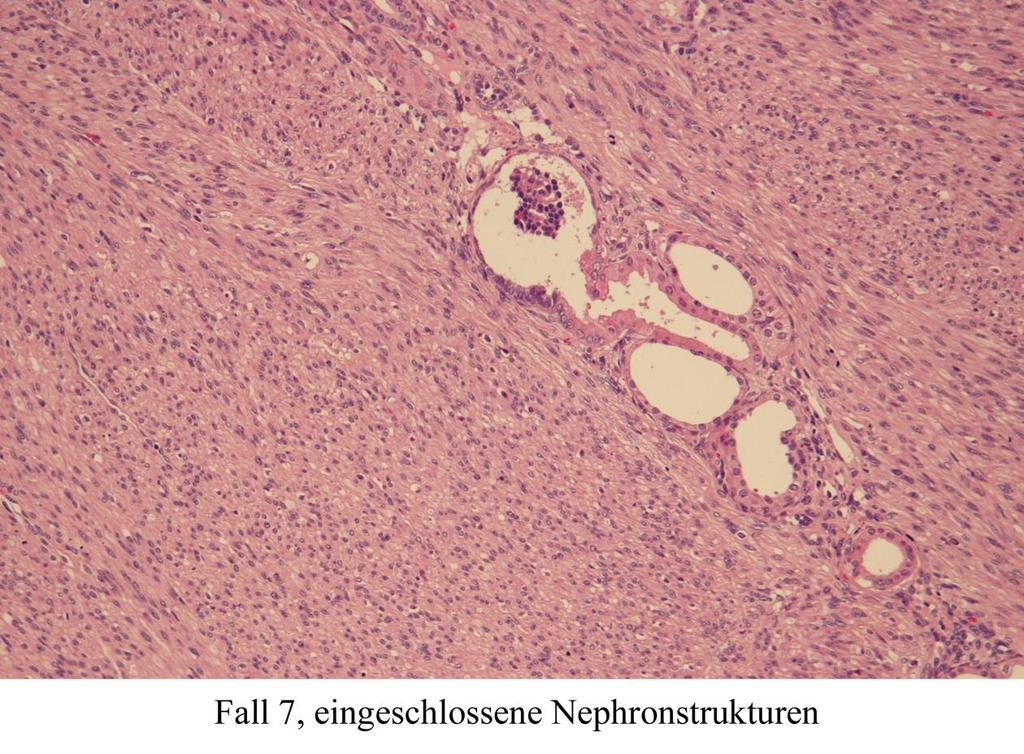
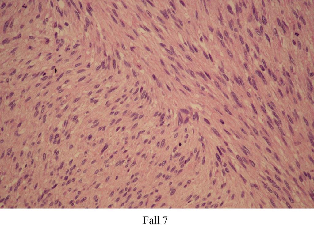
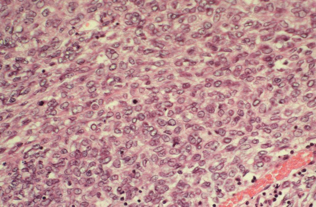
7
8
9
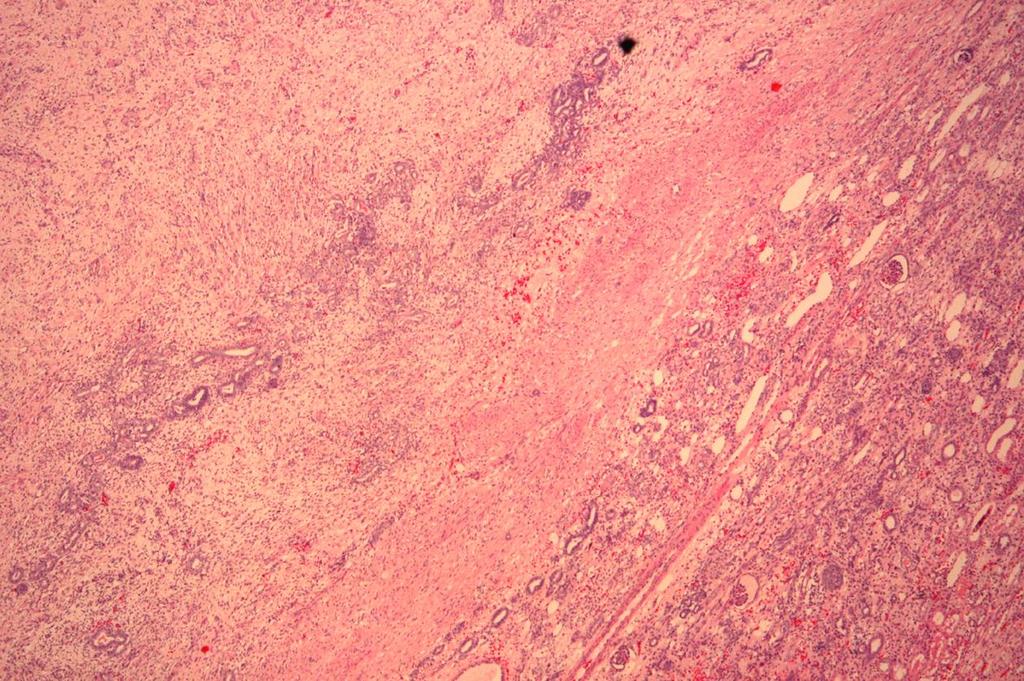
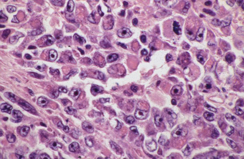
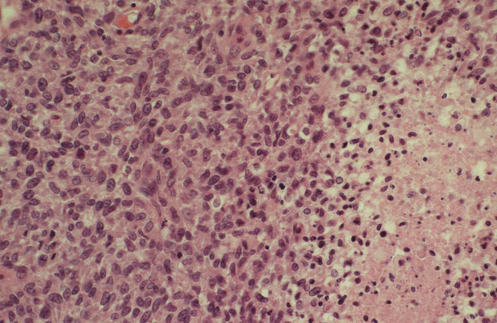
10 Case 1 Microscopy Encapsulated tumor Mixture of small cells grown in nests, spindle cells and tubular structures Focally cells with broad eosinophilic cytoplasm Regressive changes
11 Case 1 Nephroblastoma, mixed type intermediate risk (SIOP)
12 Case 1 Nephroblastoma Synonymous: Wilms tumor Most common malignant kidney tumor in childhood Inzidence (Germany): 0,9 cases/ chidren/year
13 Case 1 Nephroblastoma Epidemiology Most common between the 2. and 3. year of life Very rare in children below 6 months and in adolescents above 16 years of age Almost equal sex distribution Site About 95 percent unilateral About 5 percent bilateral
14 Treatment protocols for pediatric renal tumors Children's Oncology Group (COG) Operation first, if possible -> followed by chemotherapy (and radiotherapy) depending on tumor type and stage International Society of Paediatric Oncology (SIOP) Chemotherapy first (in patients between 6 months and 16 years of age) -> followd by operation and a second chemotherapy (and radiotherapy) depending on tumor type and stage
15 International Society of Paediatric Oncology (SIOP) Advantages of the SIOP strategy smaller tumors after pre-operative chemotherapy lower incidence of intraoperative tumor ruptur lower incidence of radiotherapy lower tumor stages -> less chemotherapy necessary response to chemotherapy can be measured Survival rate of COG and SIOP studies are similar!
16 Treatment of pediatric renal tumors in Africa French African Pediatric Oncology Group GFAOP NEPHRO 229 patients treatment based on the SIOP 2001 protocol 7.5 percent tumor rupture Two-year and 5-year disease-free survival: 72.7 and 71.6 percent Moreira et al., Pediatr Bool cancer (2012) 58: 37-42
17 Parameters necessary for treatment of pediatric renal tumors (SIOP) Risk group of tumor Tumor stage
18 Classification of pediatric renal tumors (SIOP) Tumors primarily operated Tumors after pre-operative Chemotherapy I Low risk tumors - connatal mesoblastic Nephroma - cystic partial diff. Nephroblastoma I Low risk tumors - connatal mesoblastic Nephroma - cystic partial diff. Nephroblastoma - completely necrotic Nephroblastoma II Intermediate risk tumors - Nephroblastoma non-anaplastic - Nephroblastoma - focal anaplasia II Intermediate risk tumors - Nephroblastoma epithelial type - Nephroblastoma stromal type - Nephroblastoma - regressive type - Nephroblastoma - mixed type - Nephroblastoma - focal anaplasia III High risk tumors - Nephroblastoma - diffuse anaplasia - Clear cell sarcoma of kidney - Mal. rhabdoid tumor of kidney III High risk tumors - Nephroblastoma blastemal type - Nephroblastoma - diffuse anaplasia - Clear cell sarcoma of kidney - Mal. rhabdoid tumor of kidney
19 Classification of pediatric renal tumors (SIOP) Tumors primarily operated Tumors after pre-operative Chemotherapy I Low risk tumors - connatal mesoblastic Nephroma - cystic partial diff. Nephroblastoma I Low risk tumors - connatal mesoblastic Nephroma - cystic partial diff. Nephroblastoma - completely necrotic Nephroblastoma II Intermediate risk tumors - Nephroblastoma non-anaplastic - Nephroblastoma - focal anaplasia II Intermediate risk tumors - Nephroblastoma epithelial type - Nephroblastoma stromal type - Nephroblastoma - regressive type - Nephroblastoma - mixed type - Nephroblastoma - focal anaplasia III High risk tumors - Nephroblastoma - diffuse anaplasia - Clear cell sarcoma of kidney - Mal. rhabdoid tumor of kidney III High risk tumors - Nephroblastoma blastemal type - Nephroblastoma - diffuse anaplasia - Clear cell sarcoma of kidney - Mal. rhabdoid tumor of kidney
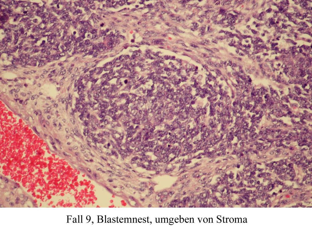
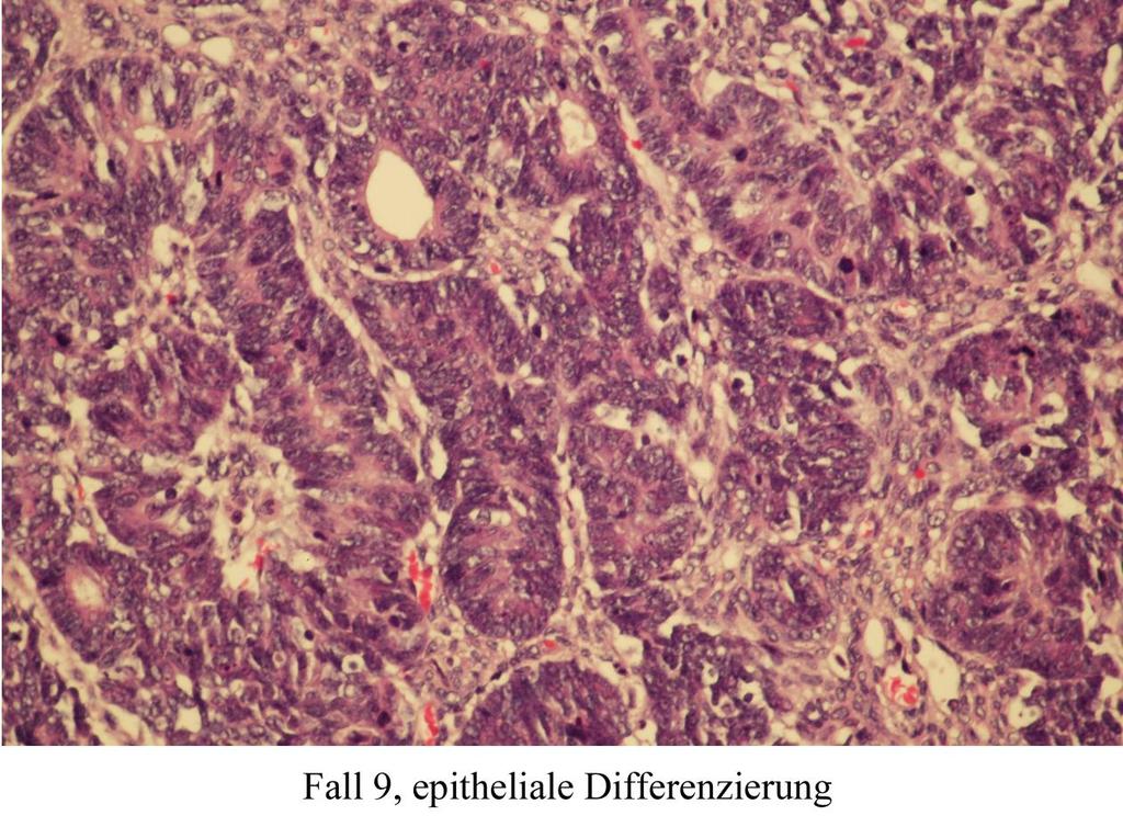
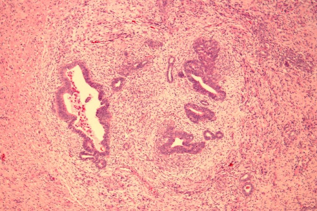
20 Case 1 Nephroblastoma Three tumor components Blastema Epithelial structures (tubules, glomerular bodies) Stromal cells (Spindle cells, myogenic cells, cartilage) Large variability in composition of the components

21 Classification is based on histological differentiation in nephroblastomas
22 Classification of pediatric renal tumors (SIOP) Tumors primarily operated Tumors after pre-operative Chemotherapy I Low risk tumors - connatal mesoblastic Nephroma - cystic partial diff. Nephroblastoma I Low risk tumors - connatal mesoblastic Nephroma - cystic partial diff. Nephroblastoma - completely necrotic Nephroblastoma II Intermediate risk tumors - Nephroblastoma non-anaplastic - Nephroblastoma - focal anaplasia II Intermediate risk tumors - Nephroblastoma epithelial type - Nephroblastoma stromal type - Nephroblastoma - regressive type - Nephroblastoma - mixed type - Nephroblastoma - focal anaplasia III High risk tumors - Nephroblastoma - diffuse anaplasia - Clear cell sarcoma of kidney - Mal. rhabdoid tumor of kidney III High risk tumors - Nephroblastoma blastemal type - Nephroblastoma - diffuse anaplasia - Clear cell sarcoma of kidney - Mal. rhabdoid tumor of kidney
23 Classification of pediatric renal tumors (SIOP) Tumors primarily operated Tumors after pre-operative Chemotherapy I Low risk tumors - connatal mesoblastic Nephroma - cystic partial diff. Nephroblastoma I Low risk tumors - connatal mesoblastic Nephroma - cystic partial diff. Nephroblastoma - completely necrotic Nephroblastoma II Intermediate risk tumors - Nephroblastoma non-anaplastic - Nephroblastoma - focal anaplasia II Intermediate risk tumors - Nephroblastoma epithelial type - Nephroblastoma stromal type - Nephroblastoma - regressive type - Nephroblastoma - mixed type - Nephroblastoma - focal anaplasia III High risk tumors - Nephroblastoma - diffuse anaplasia - Clear cell sarcoma of kidney - Mal. rhabdoid tumor of kidney III High risk tumors - Nephroblastoma blastemal type - Nephroblastoma - diffuse anaplasia - Clear cell sarcoma of kidney - Mal. rhabdoid tumor of kidney
24
25 Classification of pediatric renal tumors (SIOP) Tumors primarily operated Tumors after pre-operative Chemotherapy I Low risk tumors - connatal mesoblastic Nephroma - cystic partial diff. Nephroblastoma I Low risk tumors - connatal mesoblastic Nephroma - cystic partial diff. Nephroblastoma - completely necrotic Nephroblastoma II Intermediate risk tumors - Nephroblastoma non-anaplastic - Nephroblastoma - focal anaplasia II Intermediate risk tumors - Nephroblastoma epithelial type - Nephroblastoma stromal type - Nephroblastoma - regressive type - Nephroblastoma - mixed type - Nephroblastoma - focal anaplasia III High risk tumors - Nephroblastoma - diffuse anaplasia - Clear cell sarcoma of kidney - Mal. rhabdoid tumor of kidney III High risk tumors - Nephroblastoma blastemal type - Nephroblastoma - diffuse anaplasia - Clear cell sarcoma of kidney - Mal. rhabdoid tumor of kidney
26 Anaplasia in Nephroblastoma focal anaplasia not in LK not in metastases not extrarenal Bloc guide neccessary!
27 Classification of pediatric renal tumors (SIOP) Tumors primarily operated Tumors after pre-operative Chemotherapy I Low risk tumors - connatal mesoblastic Nephroma - cystic partial diff. Nephroblastoma I Low risk tumors - connatal mesoblastic Nephroma - cystic partial diff. Nephroblastoma - completely necrotic Nephroblastoma II Intermediate risk tumors - Nephroblastoma non-anaplastic - Nephroblastoma - focal anaplasia II Intermediate risk tumors - Nephroblastoma epithelial type - Nephroblastoma stromal type - Nephroblastoma - regressive type - Nephroblastoma - mixed type - Nephroblastoma - focal anaplasia III High risk tumors - Nephroblastoma - diffuse anaplasia - Clear cell sarcoma of kidney - Mal. rhabdoid tumor of kidney III High risk tumors - Nephroblastoma blastemal type - Nephroblastoma - diffuse anaplasia - Clear cell sarcoma of kidney - Mal. rhabdoid tumor of kidney
28 Classification of pediatric renal tumors (SIOP) Tumors primarily operated Tumors after pre-operative Chemotherapy I Low risk tumors - connatal mesoblastic Nephroma - cystic partial diff. Nephroblastoma I Low risk tumors - connatal mesoblastic Nephroma - cystic partial diff. Nephroblastoma - completely necrotic Nephroblastoma II Intermediate risk tumors - Nephroblastoma non-anaplastic - Nephroblastoma - focal anaplasia II Intermediate risk tumors - Nephroblastoma epithelial type - Nephroblastoma stromal type - Nephroblastoma - regressive type - Nephroblastoma - mixed type - Nephroblastoma - focal anaplasia III High risk tumors - Nephroblastoma - diffuse anaplasia - Clear cell sarcoma of kidney - Mal. rhabdoid tumor of kidney III High risk tumors - Nephroblastoma blastemal type - Nephroblastoma - diffuse anaplasia - Clear cell sarcoma of kidney - Mal. rhabdoid tumor of kidney

29 cystic-part. diff. nephroblastoma

30 Cystic nephroma
31 Classification of pediatric renal tumors (SIOP) Tumors primarily operated Tumors after pre-operative Chemotherapy I Low risk tumors - connatal mesoblastic Nephroma - cystic partial diff. Nephroblastoma I Low risk tumors - connatal mesoblastic Nephroma - cystic partial diff. Nephroblastoma - completely necrotic Nephroblastoma II Intermediate risk tumors - Nephroblastoma non-anaplastic - Nephroblastoma - focal anaplasia II Intermediate risk tumors - Nephroblastoma epithelial type - Nephroblastoma stromal type - Nephroblastoma - regressive type - Nephroblastoma - mixed type - Nephroblastoma - focal anaplasia III High risk tumors - Nephroblastoma - diffuse anaplasia - Clear cell sarcoma of kidney - Mal. rhabdoid tumor of kidney III High risk tumors - Nephroblastoma blastemal type - Nephroblastoma - diffuse anaplasia - Clear cell sarcoma of kidney - Mal. rhabdoid tumor of kidney

32 A 1 A 2 complete embedding of at least one tumor slice


 WT - complete")
 WT diffuse anaplasia")
 WT focal anaplasia")
33 Histolocal classification of nephroblastoma (SIOP) diffuse anaplasia ANAPLASIA? Quantification of regression no or focal anaplasia regression < 66% => subtyping of different tumour components regression 66% - 99% regression 100% WT - regressive type (intermediate risk) WT - complete necrotic (low risk) WT mixed type (intermediate risk) WT blastemal type (high risk) WT diffuse anaplasia (high risk) WT epithelial type (intermediate risk) WT stromal type (intermediate risk) WT focal anaplasia (intermediate risk) blastemal regressive mixed epithelial stromal
34 SIOP Prognosis SIOP-RTSG, Interim Report - Update April 2010
35 Parameters necessary for treatment of pediatric renal tumors (SIOP) Risk group of tumor Nephroblastomas (pre-operative treatment) viable tumor -> amount of blastema! Anaplasia Tumor stage
36 SIOP und 2001 Patients with unilateral nephroblastomas percentage of blastema SIOP-RTSG, Brainstorm Meetings 2009 and 2010
37 Parameters necessary for treatment of pediatric renal tumors (SIOP) Risk group of tumor Nephroblastomas (pre-operative treatment) Viable tumor Regression should be assessed on gross specimen Histological types of components will be assessed on H&E slides

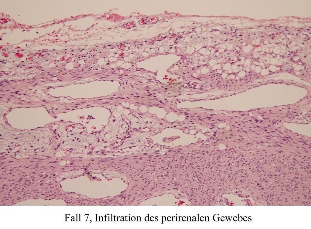
38
39

40 Parameters necessary for treatment of pediatric renal tumors (SIOP) Regression should be assessed on gross specimen If not a complete slice can be prosessed, take representative blocs of viable tumor and make a drawing as bloc guide

41 Parameters necessary for treatment of pediatric renal tumors (SIOP) Histological types of components will be assessed on H&E slides Quantification of amount of blastema (semi-quantitativ in slides)
42 Classification of pediatric renal tumors (SIOP) Tumors primarily operated Tumors after pre-operative Chemotherapy I Low risk tumors - connatal mesoblastic Nephroma - cystic partial diff. Nephroblastoma I Low risk tumors - connatal mesoblastic Nephroma - cystic partial diff. Nephroblastoma - completely necrotic Nephroblastoma II Intermediate risk tumors - Nephroblastoma non-anaplastic - Nephroblastoma - focal anaplasia II Intermediate risk tumors - Nephroblastoma epithelial type - Nephroblastoma stromal type - Nephroblastoma - regressive type - Nephroblastoma - mixed type - Nephroblastoma - focal anaplasia III High risk tumors - Nephroblastoma - diffuse anaplasia - Clear cell sarcoma of kidney - Mal. rhabdoid tumor of kidney III High risk tumors - Nephroblastoma blastemal type - Nephroblastoma - diffuse anaplasia - Clear cell sarcoma of kidney - Mal. rhabdoid tumor of kidney
43 Classification of pediatric renal tumors (SIOP) Tumors primarily operated Tumors after pre-operative Chemotherapy I Low risk tumors - connatal mesoblastic Nephroma - cystic partial diff. Nephroblastoma I Low risk tumors - connatal mesoblastic Nephroma - cystic partial diff. Nephroblastoma - completely necrotic Nephroblastoma II Intermediate risk tumors - Nephroblastoma non-anaplastic - Nephroblastoma - focal anaplasia II Intermediate risk tumors - Nephroblastoma epithelial type - Nephroblastoma stromal type - Nephroblastoma - regressive type - Nephroblastoma - mixed type - Nephroblastoma - focal anaplasia III High risk tumors - Nephroblastoma - diffuse anaplasia - Clear cell sarcoma of kidney - Mal. rhabdoid tumor of kidney III High risk tumors - Nephroblastoma blastemal type - Nephroblastoma - diffuse anaplasia - Clear cell sarcoma of kidney - Mal. rhabdoid tumor of kidney
44 Classification of pediatric renal tumors (SIOP) Tumors primarily operated Tumors after pre-operative Chemotherapy I Low risk tumors - connatal mesoblastic Nephroma - cystic partial diff. Nephroblastoma I Low risk tumors - connatal mesoblastic Nephroma - cystic partial diff. Nephroblastoma - completely necrotic Nephroblastoma II Intermediate risk tumors - Nephroblastoma non-anaplastic - Nephroblastoma - focal anaplasia II Intermediate risk tumors - Nephroblastoma epithelial type - Nephroblastoma stromal type - Nephroblastoma - regressive type - Nephroblastoma - mixed type - Nephroblastoma - focal anaplasia III High risk tumors - Nephroblastoma - diffuse anaplasia - Clear cell sarcoma of kidney - Mal. rhabdoid tumor of kidney III High risk tumors - Nephroblastoma blastemal type - Nephroblastoma - diffuse anaplasia - Clear cell sarcoma of kidney - Mal. rhabdoid tumor of kidney
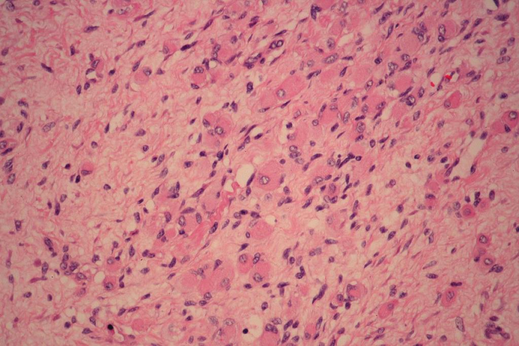
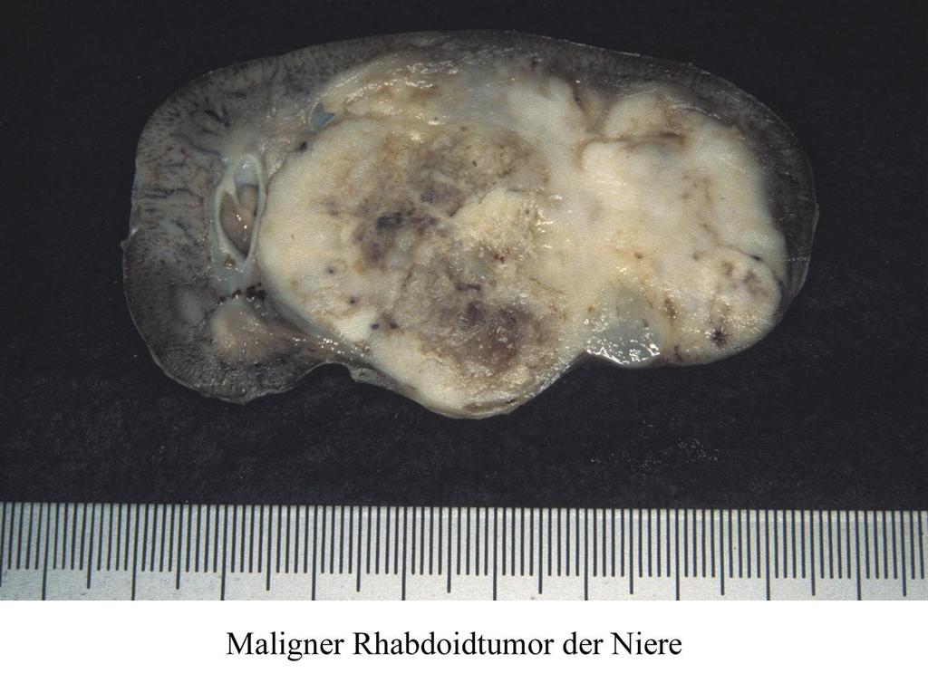
45 Classification of pediatric renal tumors (SIOP) Tumors primarily operated Tumors after pre-operative Chemotherapy I Low risk tumors - connatal mesoblastic Nephroma - cystic partial diff. Nephroblastoma I Low risk tumors - connatal mesoblastic Nephroma - cystic partial diff. Nephroblastoma - completely necrotic Nephroblastoma II Intermediate risk tumors - Nephroblastoma non-anaplastic - Nephroblastoma - focal anaplasia II Intermediate risk tumors - Nephroblastoma epithelial type - Nephroblastoma stromal type - Nephroblastoma - regressive type - Nephroblastoma - mixed type - Nephroblastoma - focal anaplasia III High risk tumors - Nephroblastoma - diffuse anaplasia - Clear cell sarcoma of kidney - Mal. rhabdoid tumor of kidney III High risk tumors - Nephroblastoma blastemal type - Nephroblastoma - diffuse anaplasia - Clear cell sarcoma of kidney - Mal. rhabdoid tumor of kidney
46 Classification of pediatric renal tumors (SIOP) Tumors primarily operated Tumors after pre-operative Chemotherapy I Low risk tumors - connatal mesoblastic Nephroma - cystic partial diff. Nephroblastoma I Low risk tumors - connatal mesoblastic Nephroma - cystic partial diff. Nephroblastoma - completely necrotic Nephroblastoma II Intermediate risk tumors - Nephroblastoma non-anaplastic - Nephroblastoma - focal anaplasia II Intermediate risk tumors - Nephroblastoma epithelial type - Nephroblastoma stromal type - Nephroblastoma - regressive type - Nephroblastoma - mixed type - Nephroblastoma - focal anaplasia III High risk tumors - Nephroblastoma - diffuse anaplasia - Clear cell sarcoma of kidney - Mal. rhabdoid tumor of kidney III High risk tumors - Nephroblastoma blastemal type - Nephroblastoma - diffuse anaplasia - Clear cell sarcoma of kidney - Mal. rhabdoid tumor of kidney
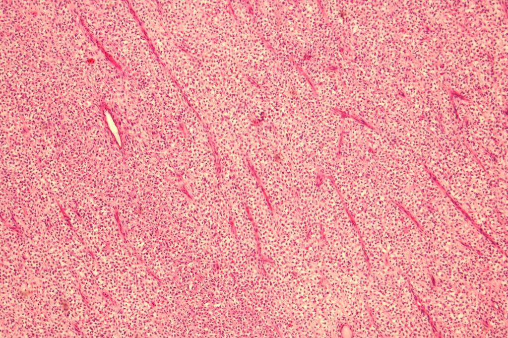
47 Parameters necessary for treatment of pediatric renal tumors (SIOP) Risk group of tumor Tumor stage post-operative chemotherapy stage I -> 2 drugs, 4 weeks stage II -> 2 drugs, 26 weeks! stage III -> 3 drugs and radiotherapy
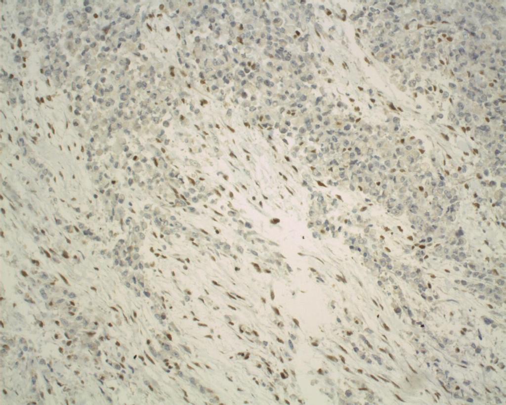
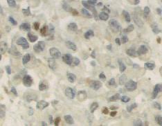
48 Parameters necessary for treatment of pediatric renal tumors (SIOP) Tumor stage stage II only true viable infiltration of Sinus Hilar area Perirenal fat Viable tumor thrombi don't chase for minimal infiltration

49 Viable infiltration of perirenal fat

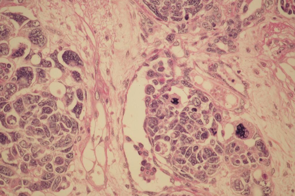
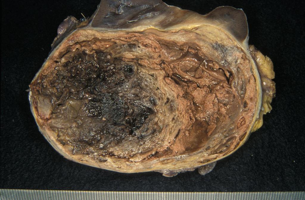
50 Regressive thrombus -> not stage II

51 Perilobar nephroblastomatosis in hilar area -> not stage II
52 Parameters necessary for treatment of pediatric renal tumors (SIOP) Tumor stage stage III viable or regressive tumor at resection margin viable or regressive tumor in lymph nodes don't chase for minimal changes

53
54 Differential diagnosis
55 Malignant Rhabdoid Tumor of kidney Epidemiology Germany: 1 percent of all malignant pediatric renal tumors High risk tumor according to the SIOP classification
56

57 MRT: Diffuse infiltration of renal parenchyma
58 INI-Protein
59 Malignant Rhabdoid Tumor of kidney Molecular genetics Various changes on chromosome 22q hsnf4/ini1 gene (SMARCB1-Gen) Deletions, mutations
60 Clear cell sarcoma of the kidney Epidemiology Germany: 1 percent of all malignant pediatric renal tumors High risk tumor according to the SIOP classification
61 Clear cell sarcoma of the kidney Age at diagnosis percent Mo 4-6 Mo 0-3 Mo >1-2 >2-3 >3-4 >4-5 >5-6 >6-7 >7-8 >8-9 >9-10 >10-11 >11-12 >12-13 >13-14 Years From: AFIP 2004

62 CCSK: mucoid tumor tissue on cut surface
63 Clear cell sarcoma of the kidney various morphological pattern: 'classical' pattern epitheloid pattern spindel cell pattern sclerosing pattern etc.
64

65 CCSK: bland cytology; cave: entrapped renal tubuli
66 Clear cell sarcoma of the kidney Diagnosis can usually be made on H&E stain: typical bland cytology typical vascular pattern
67 Clear cell sarcoma of the kidney Important differential diagnosis Nephroblastoma - blastemal type also high risk tumor! Cellular mesoblastic nephroma low risk tumor! molecular genetics might be necessary t(12;15) translocation
68 Mesoblastic nephroma Most common kidney with the first 6 months of life Often already diagnosed in utero
69 Mesoblastic nephroma Epidemiology Age distribution: 90 percent of cases within the first year of life Mean age 2 month Extremely rare in children above 2 years-of-age Sex distribution: almost 1:1 (AFIP 2004) Site almost unilateral
70 Mesoblastic nephroma Macroscopy classial type: Firm storiforme cut surface cellular type: Cysts and hemorrhages are common
71 Mesoblastic nephroma Microscopy classical type: Elongated spindle cells in bundles Finger-like projection into the renal parenchyma low mitotic activity

72
73
74
75
76 Mesoblastic nephroma Microscopy cellular type: Ovoide to round cells pushing border against the renal parenchyma increased mitotic activity necrosis and tumor cysts are common
77
78
79 Mesoblastic nephroma Mixed type: Combination of both growth pattern
80 Mesoblastic nephroma Molecular genetics t(12;15)(p13;q25) translocation in the cellular type ETV6 gene on chromosome 12 NTRK3 gene on chromosome 15 Identical translocation as in infantile fibrosarcoma No prognostic impact!

81 Thank you for your attention!
PITFALLS AND TRAPS IN THE DIAGNOSIS AND STAGING OF RENAL TUMOURS OF CHILDHOOD. Gordan M. Vujanić Cardiff, U.K.
 PITFALLS AND TRAPS IN THE DIAGNOSIS AND STAGING OF RENAL TUMOURS OF CHILDHOOD Gordan M. Vujanić Cardiff, U.K. RENAL TUMOURS OF CHILDHOOD - CLASSIFICATION (2016) Nephroblastic tumours Mesenchymal tumours
Intrarenal Extension. sinus
 Intrarenal Extension into sinus Document Capsular Penetration sinus 16 Pediatric Renal Tumor Staging Stage I Limited to Kidney & Completely Resected Intact Renal Capsule No Previous Rupture or Biopsy Renal
Protocol applies to specimens from patients with Wilms tumor (nephroblastoma) or other renal tumors of childhood.
 or other renal tumors of childhood.") Wilms Tumor Protocol applies to specimens from patients with Wilms tumor (nephroblastoma) or other renal tumors of childhood. Procedures Cytology (No Accompanying Checklist) Incisional Biopsy (Needle or
Wilms Tumor Protocol applies to specimens from patients with Wilms tumor (nephroblastoma) or other renal tumors of childhood. Procedures Cytology (No Accompanying Checklist) Incisional Biopsy (Needle or
Kidney Case 1 SURGICAL PATHOLOGY REPORT
 Kidney Case 1 Surgical Pathology Report February 9, 2007 Clinical History: This 45 year old woman was found to have a left renal mass. CT urography with reconstruction revealed a 2 cm medial mass which
Standards and datasets for reporting cancers. Dataset for histopathological reporting of renal tumours in childhood. November 2018
 Standards and datasets for reporting cancers Dataset for histopathological reporting of renal tumours in childhood November 2018 Authors: Professor Gordan M Vujanić, Sidra Medicine, Doha, Qatar Professor
SIOP PODC WILMS TUMOUR-1 Protocol
 SIOP PODC WILMS TUMOUR-1 Protocol A SIOP PODC Level 1 protocol and guideline for the treatment of Wilms Tumour in Children Adapted for Papua New Guinea A/Professor MICHAEL SULLIVAN FRACP PhD Head Solid
Wilms Tumour A brief note for the parents
 Wilms Tumour A brief note for the parents By Dr. Abid Qazi!1 PARENT S GUIDE FACTSHEET Cancer is not an incurable disease any more in many circumstances. The key for cure is early diagnosis. However, it
Pediatric Retroperitoneal Masses Radiologic-Pathologic Correlation
 Acta Radiológica Portuguesa, Vol.XVIII, nº 70, pág. 61-70, Abr.-Jun., 2006 Pediatric Retroperitoneal Masses Radiologic-Pathologic Correlation Marilyn J. Siegel Mallinckrodt Institute of Radiology, Washington
Protocol for the Examination of Specimens From Patients With Wilms and Other Pediatric Renal Tumors
 Protocol for the Examination of Specimens From Patients With Wilms and Other Pediatric Renal Tumors Version: Protocol Posting Date: August 2016 Includes the Children s Oncology Group staging system For
DIAGNOSTIC SLIDE SEMINAR: PART 1 RENAL TUMOUR BIOPSY CASES
 DIAGNOSTIC SLIDE SEMINAR: PART 1 RENAL TUMOUR BIOPSY CASES Dr. Andrew J. Evans MD, PhD, FACP, FRCPC Consultant in Genitourinary Pathology University Health Network, Toronto, ON Case 1 43 year-old female,
CYSTIC TUMORS OF THE KIDNEY JOHN N. EBLE, M.D. CYSTIC NEPHROMA
 Page 1 CYSTIC TUMORS OF THE KIDNEY JOHN N. EBLE, M.D. Department of Pathology & Laboratory Medicine Phone (317) 274-4806 Medical Science A-128 FAX: (317) 278-2018 635 Barnhill Drive jeble @iupui.edu Indianapolis,
WILMS TUMOR. Click to edit Master subtitle style. Dr.S.G.RAMANAN M.D,D.M MCCF, CRRT
 WILMS TUMOR Click to edit Master subtitle style Dr.S.G.RAMANAN M.D,D.M MCCF, CRRT HISTORY German Surgeon Childhood renal tumors Burns. tuberculosis Radiation Co-author of Surgical text book Died taking
Diagnostically Challenging Cases in Gynecologic Pathology
 Diagnostically Challenging Cases in Gynecologic Pathology Eric C. Huang, M.D., Ph.D. Department of Pathology and Laboratory Medicine University of California, Davis Medical Center Case 1 Presentation 38
RENAL CELL CARCINOMA 2 to 3% of All New Visceral Cancers Peak Incidence is 6th Decade M:F = 2:1 Grossly is a Bright Yellow, Necrotic Mass with a Pseud
 GENITOURINARY PATHOLOGY Kathleen M. O Toole Toole, M.D. RENAL CELL CARCINOMA 2 to 3% of All New Visceral Cancers Peak Incidence is 6th Decade M:F = 2:1 Grossly is a Bright Yellow, Necrotic Mass with a
Whole-tumor apparent diffusion coefficient measurements in nephroblastoma: Can it identify blastemal predominance? Abstract Purpose To explore the
 Whole-tumor apparent diffusion coefficient measurements in nephroblastoma: Can it identify blastemal predominance? Abstract Purpose To explore the potential relation between whole-tumor apparent diffusion
MR Tumor Staging for Treatment Decision in Case of Wilms Tumor
 MR Tumor Staging for Treatment Decision in Case of Wilms Tumor G. Schneider, M.D., Ph.D.; P. Fries, M.D. Dept. of Diagnostic and Interventional Radiology, Saarland University Hospital, Homburg/Saar, Germany
Protocol for the Examination of Biopsy Specimens From Patients With Wilms and Other Pediatric Renal Tumors
 Protocol for the Examination of Specimens From Patients With Wilms and Other Pediatric Renal Tumors Version: Wilms Tumor 4.0.0.0 Protocol Posting Date: February 2019 Accreditation Requirements The use
2 to 3% of All New Visceral Cancers Peak Incidence is 6th Decade M:F = 2:1 Grossly is a Bright Yellow, Necrotic Mass with a Pseudocapsule
 GENITOURINARY PATHOLOGY Kathleen M. O Toole, M.D. Renal Cell Carcinoma 2 to 3% of All New Visceral Cancers Peak Incidence is 6th Decade M:F = 2:1 Grossly is a Bright Yellow Necrotic Mass Grossly is a Bright
PDF created with pdffactory Pro trial version
 Neuroblastoma Tumor derived from neural crest cell that form the sympathetic ganglia&adrenal medulla. Causes *unknown. *familial neuroblastoma has been reported but is rare. * The incidence is 1:100,000
Wilms' tumour and renal dysplasia: an hypothesis
 J Clin Pathol 1982;35:1069-1073 Wilms' tumour and renal dysplasia: an hypothesis HB MARSDEN, W LAWLER* From the Royal Manchester Children's Hospital, Manchester M27 I HA and the *Department of Pathology,
Protocol for the Examination of Specimens From Pediatric Patients With Wilms Tumors
 Protocol for the Examination of Specimens From Pediatric Patients With Wilms Tumors Protocol applies to all renal tumors of childhood except renal cell carcinoma. Use the adult kidney protocol for renal
All India Institute of Medical Sciences, New Delhi, INDIA. Department of Pediatric Surgery, Medical Oncology, and Radiology
 All India Institute of Medical Sciences, New Delhi, INDIA Department of Pediatric Surgery, Medical Oncology, and Radiology Clear cell sarcoma of the kidney- rare renal neoplasm second most common renal
the urinary system pathology Dr. Fairoz A Eltorgman
 the urinary system pathology Dr. Fairoz A Eltorgman Tumors of the renal pelvis & kidney Benign tumors of the renal pelvis: Hemangioma Leiomyoma Malignant tumors: Transitional cell carcinoma Squamous cell
Università di Roma La Sapienza
 Università di Roma La Sapienza U.O.C. Chirurgia Pediatrica - Policlinico Umberto I Direttore: Prof Francesco Cozzi Chirurgia nephron-sparing per tumori renali primitivi in età pediatrica. Prof. Denis A.
The College of American Pathologists offers these protocols
 Protocol for the Examination of Specimens From Patients With Wilms Tumor (Nephroblastoma) or Other Renal Tumors of Childhood Stephen J. Qualman, MD; Jay Bowen, MS; Mahul B. Amin, MD; John R. Srigley, MD;
Disclosure. Relevant Financial Relationship(s) None. Off Label Usage None MFMER slide-1
 None. Off Label Usage None MFMER slide-1") Disclosure Relevant Financial Relationship(s) None Off Label Usage None 2013 MFMER slide-1 Case Presentation A 43 year old male, with partial nephrectomy for a right kidney mass 2013 MFMER slide-2 2013
Disclosure Relevant Financial Relationship(s) None Off Label Usage None 2013 MFMER slide-1 Case Presentation A 43 year old male, with partial nephrectomy for a right kidney mass 2013 MFMER slide-2 2013
04/09/2018. Salivary Gland Pathology in the Molecular Era Old Friends, Old Foes, & New Acquaintances
 Salivary Gland Pathology in the Molecular Era Old Friends, Old Foes, & New Acquaintances Jennifer L. Hunt, MD, MEd Aubrey J. Hough Jr, MD, Endowed Professor of Pathology Chair of Pathology and Laboratory
The Child With An Abdominal Mass
 The Child With An Abdominal Mass Today we are going to talk about pediatric surgery, the abdominal masses in children. Firstly we have to take a full history and make a general, local and rectal examination
Selected Pseudomalignant Soft Tissue Tumors of the Skin and Subcutis
 Selected Pseudomalignant Soft Tissue Tumors of the Skin and Subcutis Andrew L. Folpe, M.D. Professor of Laboratory Medicine and Pathology Mayo Clinic, Rochester, MN folpe.andrew@mayo.edu 2016 MFMER slide-1
CNS pathology Third year medical students. Dr Heyam Awad 2018 Lecture 12: CNS tumours 2/3
 CNS pathology Third year medical students Dr Heyam Awad 2018 Lecture 12: CNS tumours 2/3 Pilocytic astrocytoma Relatively benign ( WHO grade 1) Occurs in children and young adults Mostly: in the cerebellum
Various hereditary, acquired and neoplastic conditions can lead to cyst formation in the kidney.
 Dr. Fatima AlAl-Hashimi Hashimi,, MD, FRCPath Salmaniya Medical Complex, Bahrain Various hereditary, acquired and neoplastic conditions can lead to cyst formation in the kidney. The most frequently encountered
Tumors of kidney and urinary bladder
 Tumors of kidney and urinary bladder Overview of kidney tumors Benign and malignant Of the benign: papillary adenoma -cortical -small (0.5cm) -in 40% of population -clinically insignificant The most common
(CYLINDROMA) ATLAS OF HEAD AND NECK PATHOLOGY ADENOID CYSTIC CARCINOMA
 ATLAS OF HEAD AND NECK PATHOLOGY ADENOID CYSTIC CARCINOMA") (CYLINDROMA) This malignant tumor is poorly encapsulated and while seemingly well defined within the affected gland, there is usually infiltration of surrounding tissue on closer examination. The cut surface
(CYLINDROMA) This malignant tumor is poorly encapsulated and while seemingly well defined within the affected gland, there is usually infiltration of surrounding tissue on closer examination. The cut surface
3. Guidelines for Reporting Bladder Cancer, Prostate Cancer and Renal Tumours
 60 3. Guidelines for Reporting Bladder Cancer, Prostate Cancer and Renal Tumours Compilation and editing and of this volume: Prof. Chandu de Silva (Consultant Histopathologist) List of contributors Consultant
Wilms Tumor and Neuroblastoma
 Wilms Tumor and Neuroblastoma Wilm s Tumor AKA: Nephroblastoma the most common intra-abdominal cancer in children. peak incidence is 2 to 3 years of age Biology somatic mutations restricted to tumor tissue
Part 1. Slides 1-38, Rita Alaggio Soft tissue tumors Trondheim 14. mars 2013
 Part 1 Slides 1-38, Rita Alaggio Soft tissue tumors Trondheim 14. mars 2013 Pediatric Pathology Soft Tissue Tumors AN UPDATE Rita Alaggio Azienda Ospedaliera Università di Padova Soft Tissue Tumors More
Wilms Tumor Outcomes at a Single Institution and Review of Current Management Recommendations
 Wilms Tumor Outcomes at a Single Institution and Review of Current Management Recommendations Background: The Duval County Medical Society (DCMS) is proud to provide its members with free continuing medical
The management of bilateral Wilms tumor
 Wilms Tumor The management of bilateral Wilms tumor Derya Özyörük 1, Suna Emir 2 1 Pediatric Oncologist, Department of Pediatric Hematology Oncology, 2 Associate Professor of Pediatrics, Department of
Enterprise Interest Nothing to declare
 Enterprise Interest Nothing to declare Sarcoma with CIC-DUX4 gene fusion: case report of kidney tumor location in a 12-year-old boy. Perret Cécile 1, Pierron Gaelle 2, Anne Mc Leer 3, Piolat Christian
Case Presentation. Maha Akkawi, MD, Fatima Obeidat, MD, Tariq Aladily, MD. Department of Pathology Jordan University Hospital Amman, Jordan
 Case Presentation Maha Akkawi, MD, Fatima Obeidat, MD, Tariq Aladily, MD Department of Pathology Jordan University Hospital Amman, Jordan The 25th Annual Congress of the ADIAP The 8/11/2013 1 5th International
Kidney, Bladder and Prostate Neoplasia. David Bingham MD
 Kidney, Bladder and Prostate Neoplasia David Bingham MD typical malignant cytology of bladder washings 1 benign 2 malignant typical malignant cytology of bladder washings b Bladder tumor Non invasive papillary
ESS: Pathologic Insights
 GEIS XVI INTERNATIONAL SYMPOSIUM Seville 4th October 2018 ESS: Pathologic Insights Sílvia Bagué The Royal Marsden Hospital London (United Kingdom) I have no conflicts of interest Endometrial stromal sarcoma
JMSCR Vol 06 Issue 02 Page February 2018
 www.jmscr.igmpublication.org Impact Factor (SJIF): 6.379 Index Copernicus Value: 71.58 ISSN (e)-2347-176x ISSN (p) 2455-0450 DOI: https://dx.doi.org/10.18535/jmscr/v6i2.08 Pattern of Renal Tumors: A Tertiary
Case Presentation. Gordon Callender M.D. Surgical Resident
 Case Presentation Gordon Callender M.D. Surgical Resident Retroperitoneal Sarcomas Sarcomas Heterogeneous group of rare tumors that arise predominantly from the embryonic mesoderm. Expected incidence for
Renal tumours: use of immunohistochemistry & molecular pathology. Dr Lisa Browning John Radcliffe Hospital Oxford
 Renal tumours: use of immunohistochemistry & molecular pathology Dr Lisa Browning John Radcliffe Hospital Oxford Renal tumours: the use of immunohistochemistry & molecular pathology Classification of RCC
RARE TUMORS OF INFANCY. RAJKUMAR VENKATRAMANI, MD, MS Director, Rare Tumors Program, Texas Children s Hospital
 RARE TUMORS OF INFANCY RAJKUMAR VENKATRAMANI, MD, MS Director, Rare Tumors Program, Texas Children s Hospital OBJECTIVES Review the epidemiology and clinical presentation of soft tissue sarcomas in infancy.
Case Scenario 1: Thyroid
 Case Scenario 1: Thyroid History and Physical Patient is an otherwise healthy 80 year old female with the complaint of a neck mass first noticed two weeks ago. The mass has increased in size and is palpable.
Diplomate of the American Board of Pathology in Anatomic and Clinical Pathology
 A 33-year-old male with a left lower leg mass. Contributed by Shaoxiong Chen, MD, PhD Assistant Professor Indiana University School of Medicine/ IU Health Partners Department of Pathology and Laboratory
Etiopathological and management profile of Wilms tumour A single centre experience
 IOSR Journal of Dental and Medical Sciences (IOSR-JDMS) e-issn: 2279-0853, p-issn: 2279-0861.Volume 17, Issue 10 Ver. 10 (October. 2018), PP 48-53 www.iosrjournals.org Etiopathological and management profile
Synonyms. Nephrogenic metaplasia Mesonephric adenoma
 Nephrogenic Adenoma Synonyms Nephrogenic metaplasia Mesonephric adenoma Definition Benign epithelial lesion of urinary tract with tubular, glandular, papillary growth pattern Most frequently in the urinary
Note: The cause of testicular neoplasms remains unknown
 - In the 15- to 34-year-old age group, they are the most common tumors of men. - Tumors of the testis are a heterogeneous group of neoplasms that include: I. Germ cell tumors : 95%; all are malignant.
CASE REPORT. CELLULAR MESOBLASTIC NEPHROMA: REPORT OF 3 CASES M. Ramani, C. Sandhya Rani, K. Geetha, K. Ramesh Reddy.
 CELLULAR MESOBLASTIC NEPHROMA: REPORT OF 3 CASES M. Ramani, C. Sandhya Rani, K. Geetha, K. Ramesh Reddy. 1. Professor, Department. of Pathology, Niloufer Hospital for Women and Children. 2. Post Graduate,
!! 2 to 3% of All New Visceral Cancers.!! Peak Incidence is 6th Decade!! M:F = 2:1
 !! Kathleen M. O Toole, M.D.!! 2 to 3% of All New Visceral Cancers!! Peak Incidence is 6th Decade!! M:F = 2:1!! Grossly is a Bright Yellow, Necrotic Mass with a Pseudocapsule 1 !!Conventional RCC! Clear
Case 2. Dr. Sathima Natarajan M.D. Kaiser Permanente Medical Center Sunset
 Case 2 Dr. Sathima Natarajan M.D. Kaiser Permanente Medical Center Sunset History 24 year old male presented with a 3 day history of right flank pain, sharp in nature Denies fever, chills, hematuria or
AGGRESSIVE VARIANTS OF PAPILLARY THYROID CARCINOMA DIAGNOSIS AND PROGNOSIS
 AGGRESSIVE VARIANTS OF PAPILLARY THYROID CARCINOMA DIAGNOSIS AND PROGNOSIS PAPILLARY THYROID CARCINOMA Clinical Any age Microscopic to large Female: Male= 2-4:1 Radiation history Lymph nodes Prognosis
A 25 year old female with a palpable mass in the right lower quadrant of her abdomen
 May 2016 A 25 year old female with a palpable mass in the right lower quadrant of her abdomen Contributed by: Paul Ndekwe, MD, Resident Physician, Indiana University School of Department of Pathology and
Renal tumors of adults
 Renal tumors of adults Urinary Tract Tumors 2%-3% of all cancers in adults. The most common malignant tumor of the kidney is renal cell carcinoma. Tumors of the lower urinary tract are twice as common
Pediatric Abdominal Masses. Andrew Phelps MD Assistant Professor of Pediatric Radiology UCSF Benioff Children's Hospital
 Pediatric Abdominal Masses Andrew Phelps MD Assistant Professor of Pediatric Radiology UCSF Benioff Children's Hospital No Disclosures Take Home Message All you need to remember are the 5 common masses
Breast pathology. 2nd Department of Pathology Semmelweis University
 Breast pathology 2nd Department of Pathology Semmelweis University Breast pathology - Summary - Benign lesions - Acute mastitis - Plasma cell mastitis / duct ectasia - Fat necrosis - Fibrocystic change/
Study of Paediatric Small Blue Round Cell Tumors with Immunohistochemical Correlation
 IOSR Journal of Dental and Medical Sciences (IOSR-JDMS) e-issn: 2279-0853, p-issn: 2279-0861.Volume 15, Issue 6 Ver. XIII (June. 2016), PP 65-70 www.iosrjournals.org Study of Paediatric Small Blue Round
Pediatric Pathology Grossing Guidelines
 When you get a case that should/could be for Pediatric Pathology, show it to the gross room supervisor or page Dr. Goldstein at 31418 before you cut it in. If it is the weekend or late at night, page the
Spectrum of Preneoplastic and Neoplastic Cystic Lesions of the Kidney in Adult. by dr. Banan Burhan Mohammed Lecturer in Pathology Department
 Spectrum of Preneoplastic and Neoplastic Cystic Lesions of the Kidney in Adult by dr. Banan Burhan Mohammed Lecturer in Pathology Department Various hereditary, acquired, and neoplastic conditions can
Enterprise Interest Nothing to declare
 Enterprise Interest Nothing to declare Diagnoses one would not like to miss in soft tissue pathology early in your career Marta Sbaraglia, MD Department of Pathology Hospital of Treviso University of Padua
Presentation material is for education purposes only. All rights reserved URMC Radiology Page 1 of 98
 Presentation material is for education purposes only. All rights reserved. 2011 URMC Radiology Page 1 of 98 Radiology / Pathology Conference February 2011 Brooke Koltz, Cytopathology Resident Presentation
Dr Marty Campbell Paediatric Oncologist Royal Children's Hospital, Melbourne. FRACP lecture series Oct 2014
 Dr Marty Campbell Paediatric Oncologist Royal Children's Hospital, Melbourne FRACP lecture series Oct 2014 Background Neuroblastoma (NB) is the most common solid tumour outside the CNS in children 6-10%
Pathology of the Thyroid
 Pathology of the Thyroid Thyroid Carcinoma Arising from Follicular Cells 2015-01-19 Prof. Dr. med. Katharina Glatz Pathologie Carcinomas Arising from Follicular Cells Differentiated Carcinoma Papillary
Mammary analogue secretory carcinoma of salivary gland A case report of new entity
 Case Report Mammary analogue secretory carcinoma of salivary gland A case report of new entity Vaibhav Bhika Bari 1*, Sandhya Unmesh Bholay 2 1 Assistant Professor, 2 Associate Professor Rajiv Gandhi Medical
University Journal of Pre and Para Clinical Sciences
 ISSN 2455 2879 Volume 2 Issue 1 2016 Metaplastic carcinoma breast a rare case report Abstract : Metaplastic carcinoma of the breast is a rare malignancy with two distinct cell lines described as a breast
Salivary Glands 3/7/2017
 Salivary Glands 3/7/2017 Goals and objectives Focus on the entities unique to H&N Common board type facts Information for your future practice Salivary Glands Salivary Glands Major gland. Paratid. Submandibular.
General: Brain tumors are lesions that have mass effect distorting the normal tissue and often result in increased intracranial pressure.
 1 Lecture Objectives Know the histologic features of the most common tumors of the CNS. Know the differences in behavior of the different tumor types. Be aware of the treatment modalities in the various
Goals and Objectives. Historical Background. Historical Background. Incidence. Epidemiology 7/1/2015 ROLE OF RADIOTHERAPY IN WILMS TUMOR
 Goals and Objectives ROLE OF RADIOTHERAPY IN WILMS TUMOR Arnold C. Paulino, M.D. Professor of Radiation Oncology MD Anderson Cancer Center To gain an understanding of presentation and work-up of Wilms
ACCME/Disclosures ALK FUSION-POSITIVE MESENCHYMAL TUMORS. Tumor types with ALK rearrangements. Anaplastic Lymphoma Kinase. Jason L.
 Companion Meeting of the International Society of Bone and Soft Tissue Pathology The Evolving Concept of Mesenchymal Tumors ALK FUSION-POSITIVE MESENCHYMAL TUMORS Jason L. Hornick, MD, PhD March 13, 2016
Children s Cancer and Leukaemia Group CLINICAL MANAGEMENT GUIDELINES WILMS TUMOUR. To be used in conjunction with IMPORT study protocol
 Children s Cancer and Leukaemia Group CLINICAL MANAGEMENT GUIDELINES WILMS TUMOUR To be used in conjunction with IMPORT study protocol Produced by The CCLG Renal Tumour Interest Group January 2015 Contributors:
GUT-C 11/30/2017. Debasmita Das, M.D. PGY-1 Danbury Hospital
 GUT-C 11/30/2017 Debasmita Das, M.D. PGY-1 Danbury Hospital CLINICAL SUMMARY 8/2017 59 year old female Presented to the ED with 1 month history of general malaise, fever and weight loss PMH: Significant
Special slide seminar
 Special slide seminar Tomáš Rozkoš The Fingerland Department of Pathology Charles University Medical Faculty and Faculty Hospital in Hradec Králové Czech Republic Case history, 33 years old resistance
Wilms Tumor with Long-delayed Recurrence: 25 Years after Initial Treatment
 www.kjurology.org http://dx.doi.org/10.4111/kju.2012.53.4.288 Case Report Wilms Tumor with Long-delayed Recurrence: 25 Years after Initial Treatment So-Young Lee 1, Kyu-Rae Kim 1, Jung-Yeol Park 2, Jae
SPECIAL SLIDE SEMINAR CASE 3
 SPECIAL SLIDE SEMINAR CASE 3 Tihana Džombeta, MD Leo Pažanin, MD, PhD Department of Pathology, School of Medicine, University of Zagreb Department of Pathology, Clinical Hospital Centre Sestre milosrdnice
Renal Parenchymal Neoplasms
 Renal Parenchymal Neoplasms د. BENIGN TUMORS : Benign renal tumors include adenoma, oncocytoma, angiomyolipoma, leiomyoma, lipoma, hemangioma, and juxtaglomerular tumors. Renal Adenomas : The adenoma is
Malignant rhabdoid t~rmour of the kidney: report of a case showing focal glomeruloid differentiation
 Muluy.siun.I Pathol 1994; lh(1): 83-87 Malignant rhabdoid t~rmour of the kidney: report of a case showing focal glomeruloid differentiation Gita JAYARAM, MD, MlAC and Lai-Meng L001, FRCPath, FRCPA Department
Clinicopathologic Analysis of Wilms Tumor: A Retrospective Study of 35 Cases Over 10 Years
 Original Article DOI: 0.2276/APALM.606 Clinicopathologic Analysis of Wilms Tumor: A Retrospective Study of 35 Cases Over 0 Years Ankita Shashikant Shende* and Pragati Aditya Sathe Dept of Pathology, Seth
Pre-operative Ultrasound of Lymph Nodes in Thyroid Cancer
 Pre-operative Ultrasound of Lymph Nodes in Thyroid Cancer AACE - Advances in Medical and Surgical Management of Thyroid Cancer - 2018 Robert A. Levine, MD, FACE, ECNU Thyroid Center of New Hampshire Geisel
CONSULTATION DURING SURGERY / NOT A FINAL DIAGNOSIS. FROZEN SECTION DIAGNOSIS: - A. High grade sarcoma. Wait for paraffin sections results.
 Pathology Report Date: 3/5/02 A, B. Biopsy right distal femur- high grade spindle cell sarcoma Immunohistochemistry studies are pending to further classify the nature of the tumor. CONSULTATION DURING
IMMUNOPROFILES OF THE MAJOR RENAL NEOPLASMS (%staining)
") Stain Clear Cell Papillary IMMUNOPROFILES OF THE MAJOR RENAL NEOPLASMS (%staining) Chromophobe Collecting Duct Carcinom a Sarcomatoid Xp11 Translocat ion Dr Jon Oxley See also www.jonoxley.com Page 1 MTSCC
Stain Clear Cell Papillary IMMUNOPROFILES OF THE MAJOR RENAL NEOPLASMS (%staining) Chromophobe Collecting Duct Carcinom a Sarcomatoid Xp11 Translocat ion Dr Jon Oxley See also www.jonoxley.com Page 1 MTSCC
Nephroblastoma: Does The Decrease In Tumour Volume Under Preoperative Chemotherapy Predict The Lymph Nodes Status At Surgery?
 Nephroblastoma: Does The Decrease In Tumour Volume Under Preoperative Chemotherapy Predict The Lymph Nodes Status At Surgery? Jan Godzinski,, Harm van Tinteren,, Jan de Kraker,, Norbert Graf, Christophe
NAACCR Webinar Series 1
 NAACCR 2009 2010 Webinar Series Collecting Cancer Data: Kidney 1 Questions Please use the Q&A panel to submit your questions Send questions to All Panelist 2 Fabulous Prizes 3 NAACCR 2009 2010 Webinar
Invasive Papillary Breast Carcinoma
 410 This is an Open Access article licensed under the terms of the Creative Commons Attribution- NonCommercial-NoDerivs 3.0 License (www.karger.com/oa-license), applicable to the online version of the
Renal masses - the role of diagnostic imaging
 Renal masses - the role of diagnostic imaging Poster No.: C-2471 Congress: ECR 2015 Type: Educational Exhibit Authors: V. Rai#; Bjelovar/HR Keywords: Cysts, Cancer, Structured reporting, Ultrasound, MR,
BILATERAL TERATOID WILMS TUMOR IN A CHILD: A CASE REPORT AND REVIEW OF LITERATURE ABSTRACT
 77 Amezene Tadesse, Tatek Girma, Jakob Schneider. Ethiop Med J, Vol. 56, No. 1 CASE REPORT BILATERAL TERATOID WILMS TUMOR IN A CHILD: A CASE REPORT AND REVIEW OF LITERATURE Amezene Tadesse, MD 1*, Tatek
Index. Note: Page numbers of article titles are in boldface type.
 Magn Reson Imaging Clin N Am 12 (2004) 587 591 Index Note: Page numbers of article titles are in boldface type. A Adenoma(s), adrenal, gadolinium-enhanced MR imaging in, 533 534 hyperfunctioning versus
Evening Specialty Conference: Cytopathology
 : Cytopathology N. Paul Ohori, M.D. University of Pittsburgh Medical Center Disclosure of Relevant Financial Relationships Disclosure of Relevant Financial Relationships USCAP requires that all planners
Pathological Classification of Hepatocellular Carcinoma
 3 rd APASL Single Topic Conference: HCC in 3D Pathological Classification of Hepatocellular Carcinoma Glenda Lyn Y. Pua, M.D. HCC Primary liver cancer is the 2 nd most common cancer in Asia HCC is the
A 20-Year Prospective Study of Wilms Tumor and Other Kidney Tumors: A Report From Hong Kong Pediatric Hematology and Oncology Study Group
 ORIGINAL ARTICLE A 20-Year Prospective Study of Wilms Tumor and Other Kidney Tumors: A Report From Hong Kong Pediatric Hematology and Oncology Study Group Ching Ching Chan, MRCP,* Ka Fai To, FHKAM,w Hui
SESSION 1: GENERAL (BASIC) PATHOLOGY CONCEPTS Thursday, October 16, :30am - 11:30am FACULTY COPY
 PATHOLOGY CONCEPTS Thursday, October 16, :30am - 11:30am FACULTY COPY") SESSION 1: GENERAL (BASIC) PATHOLOGY CONCEPTS Thursday, October 16, 2008 9:30am - 11:30am FACULTY COPY GOAL: Describe the basic morphologic (structural) changes which occur in various pathologic conditions.
SESSION 1: GENERAL (BASIC) PATHOLOGY CONCEPTS Thursday, October 16, 2008 9:30am - 11:30am FACULTY COPY GOAL: Describe the basic morphologic (structural) changes which occur in various pathologic conditions.
Principles of Surgical Oncology. Winnie Achilles Tierklinik Hollabrunn Lastenstrasse Hollabrunn
 Principles of Surgical Oncology Winnie Achilles Tierklinik Hollabrunn Lastenstrasse 2 2020 Hollabrunn boexi@gmx.de The first surgery provides the best chance for a cure in an animal with a tumor Clinical
Neoplasia literally means "new growth.
 NEOPLASIA Neoplasia literally means "new growth. A neoplasm, defined as "an abnormal mass of tissue the growth of which exceeds and is uncoordinated with that of the normal tissues and persists in the
Effective local and systemic therapy is necessary for the cure of Ewing tumor Most chemotherapy regimens are a combination of cyclophosphamide,
 Ewing Tumor Perez Ewing tumor is the second most common primary tumor of bone in childhood, and also occurs in soft tissues Ewing tumor is uncommon before 8 years of age and after 25 years of age In the
Corporate Medical Policy
 Corporate Medical Policy Hematopoietic Stem-Cell Transplantation for Solid Tumors of File Name: Origination: Last CAP Review: Next CAP Review: Last Review: hematopoietic_stem-cell_transplantation_for_solid_tumors_childhood
1 Department of Epidemiology and Cancer Control, St. Jude Children s. 4 Midwest Children s Cancer Center at Children s Hospital of
 Pediatr Blood Cancer 2014;61:134 139 Outcome of Patients With Stage II/Favorable Histology Wilms Tumor With and Without Local Tumor Spill: A Report From the National Wilms Tumor Study Group Daniel M. Green,
04/10/2018. Intraductal Papillary Neoplasms Of Breast INTRADUCTAL PAPILLOMA
 Intraductal Papillary Neoplasms Of Breast Savitri Krishnamurthy MD Professor of Pathology Deputy Division Head The University of Texas MD Anderson Cancer Center 25 th Annual Seminar in Pathology Pittsburgh,
Normal thyroid tissue
 Thyroid Pathology Overview Normal thyroid tissue Normal thyroid tissue with follicles filled with colloid. Thyroid cells form follicles, spheres of epithelial cells (always single layered in health, usually