Malignant Focal Liver Lesions
|
|
|
- Easter Cross
- 5 years ago
- Views:
Transcription


1 Malignant Focal Liver Lesions Other Than HCC Pablo R. Ros, MD, MPH, PhD Departments of Radiology and Pathology University Hospitals Cleveland Medical Center Case Western Reserve University

2
3 Malignant Focal Liver Lesions Other than HCC HEPATOCELLULAR ORIGIN Fibrolamellar carcinoma (FLC) CHOLANGIOCELLULAR ORIGIN Cholangiocarcinoma (I-CAC) Cystadenocarcinoma
4 Malignant Focal Liver Lesions Other than HCC MESENCHYMAL ORIGIN Angiosarcoma Epithelioid Hemangioendothelioma Kaposi sarcoma Primary lymphoma METASTASIS
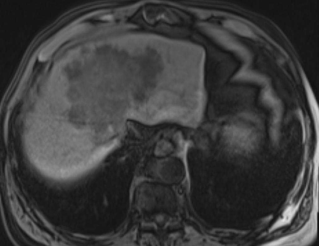
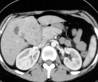
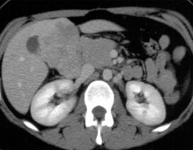
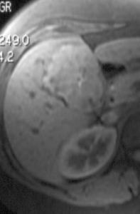
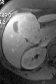
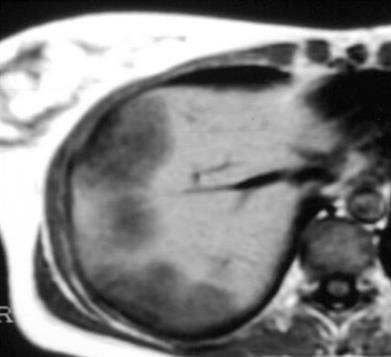
5 Malignant Focal Liver Lesions Unknown Case 56 yo man with jaundice and abdominal pain


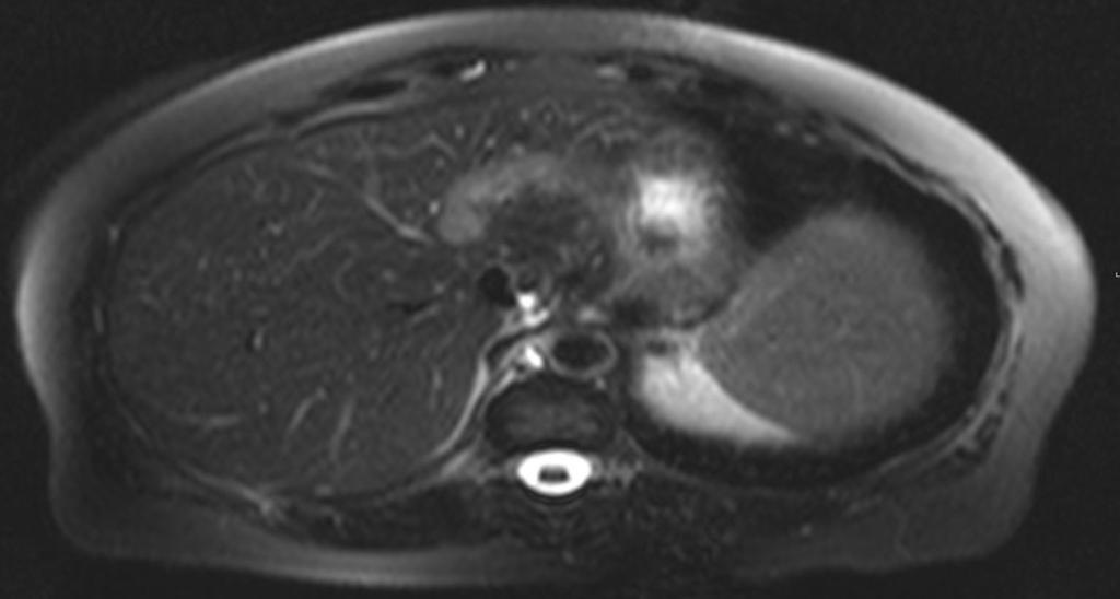
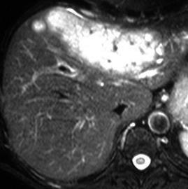
6 T2 FS T1 FS arterial T1 FS delayed
7 What is the most likely diagnosis? A. Intrahepatic Cholangiocarcinoma B. Epithelioid Hemangioendothelioma C. Fibrolamellar carcinoma D. Metastasis E. Angiosarcoma
8 What is the most likely diagnosis? A. Intrahepatic Cholangiocarcinoma B. Epithelioid Hemangioendothelioma C. Fibrolamellar carcinoma D. Metastasis E. Ansgiosarcoma
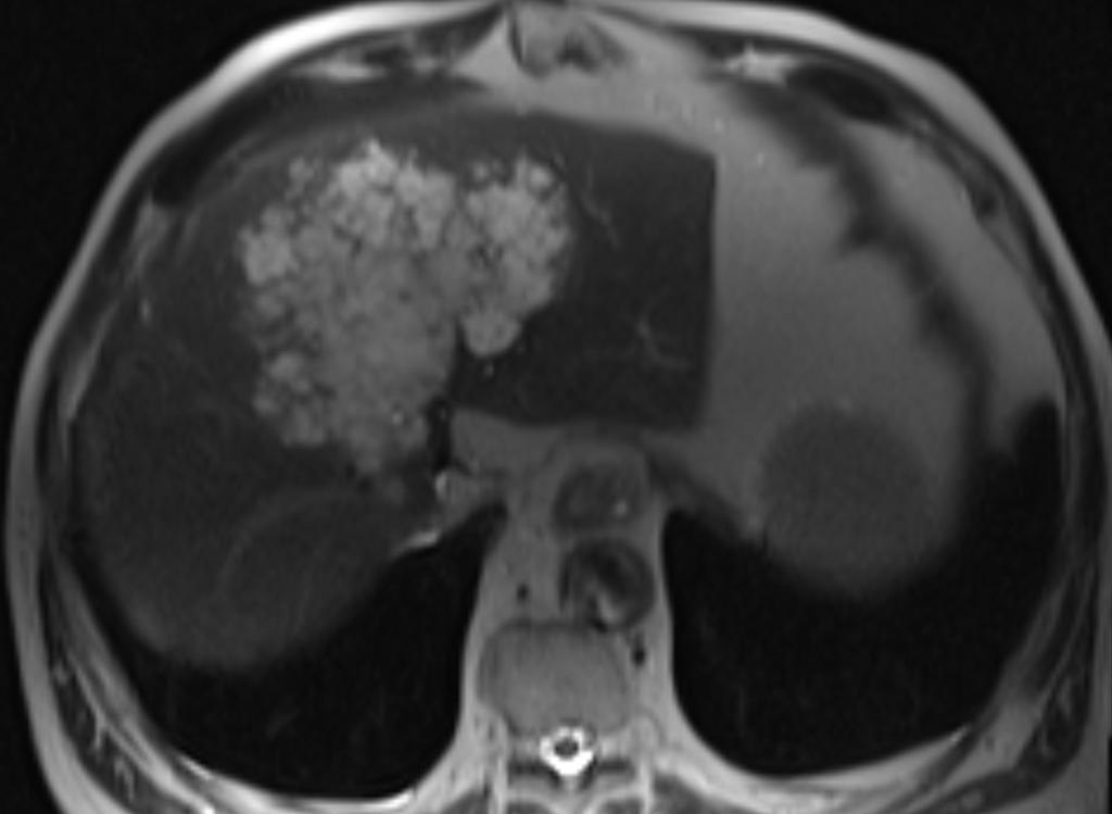
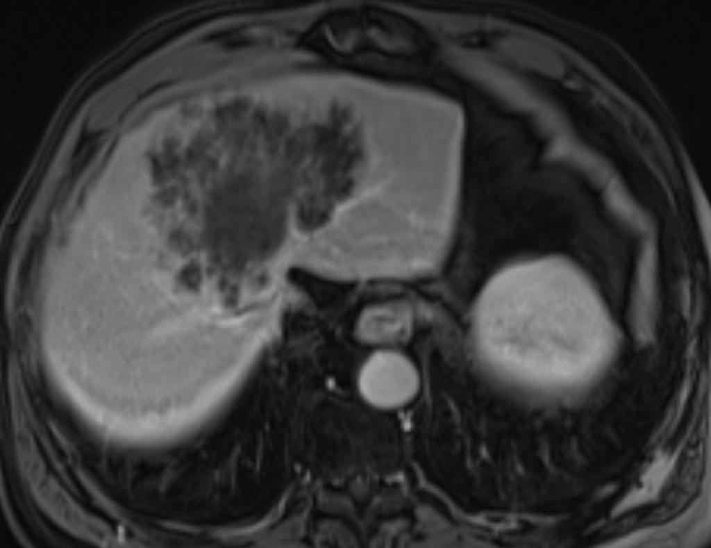
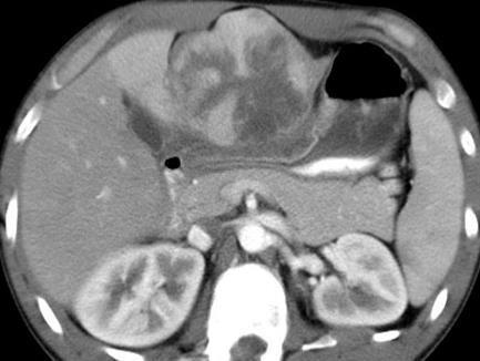
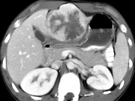
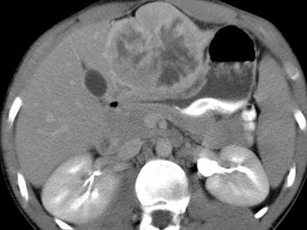
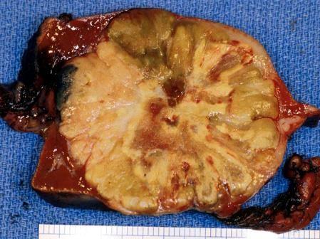
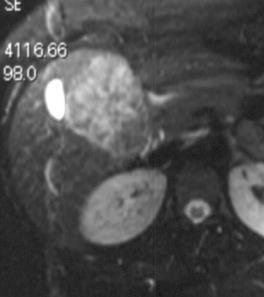
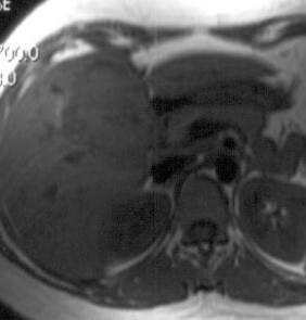
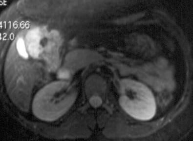
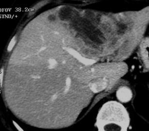
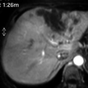
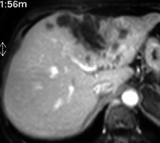
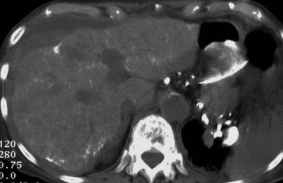
9 Malignant Focal Liver Lesions Unknown Case 19 yo man with complex medical history, now with elevated liver function tests and palpable mid abdominal mass


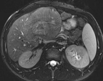
10 T2 FS T1 FS T1 FS 2 min - Eovist T1 FS 20 min - Eovist
11
12 What is the most likely diagnosis? A. Intrahepatic Cholangiocarcinoma B. Epithelioid Hemangioendothelioma C. Fibrolamellar carcinoma D. Metastasis E. Cystadenocarcinoma
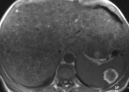
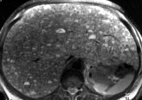
13 What is the most likely diagnosis? A. Intrahepatic Cholangiocarcinoma B. Epithelioid Hemangioendothelioma C. Fibrolamellar carcinoma D. Metastasis E. Cystadenocarcinoma
14 What is the key finding? A.Size B. Lobular contour C. Small calcifications D.Hypodense
15 What is the key CT finding? A.Size B. Lobular contour C. Small calcifications D.Hypodense
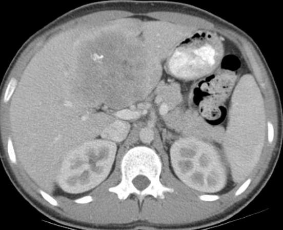
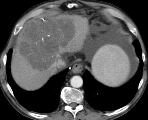
16 Malignant Focal Liver Lesions Unknown Case 61 yo woman Intermittent abdominal pain and distension RUQ ultrasound, followed by CT Second abdominal CT scan, 8 months after initial one
17
18 What is the most likely diagnosis? A. Intrahepatic Cholangiocarcinoma B. Epithelioid Hemangioendothelioma C. Fibrolamellar carcinoma D. Metastasis E. Angisarcoma
19 What is the most likely diagnosis? A. Intrahepatic Cholangiocarcinoma B. Epithelioid Hemangioendothelioma C. Fibrolamellar carcinoma D. Metastasis E. Angiosarcoma
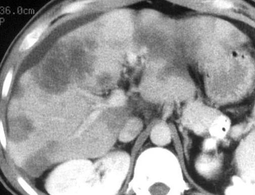
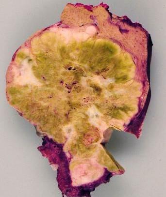
20 Malignant Focal Liver Lesions Unknown Case 54 yo woman Abdominal pain Bloating Her two dogs died recently due to chronic arsenic intoxication
21
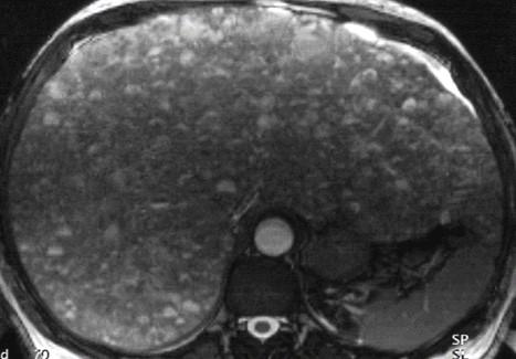
22 What is the most likely diagnosis? A. Intrahepatic Cholangiocarcinoma B. Epithelioid Hemangioendothelioma C. Fibrolamellar carcinoma D. Metastasis E. Angisarcoma
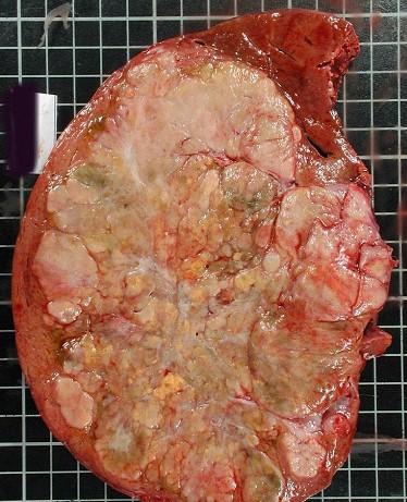
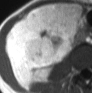
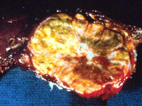
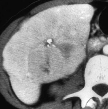
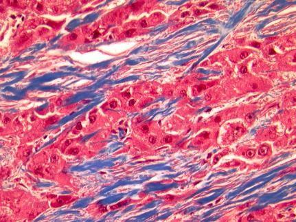
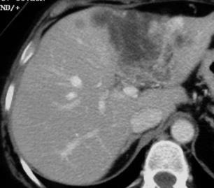
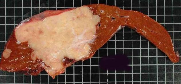
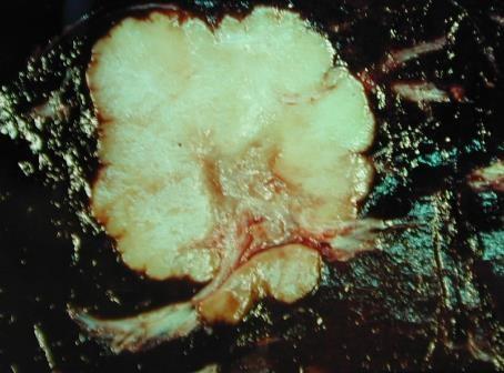
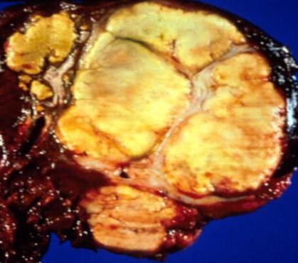
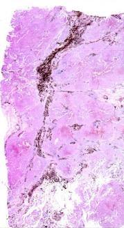
23 What is the most likely diagnosis? A. Intrahepatic Cholangiocarcinoma B. Epithelioid Hemangioendothelioma C. Fibrolamellar carcinoma D. Metastasis E. Angisarcoma
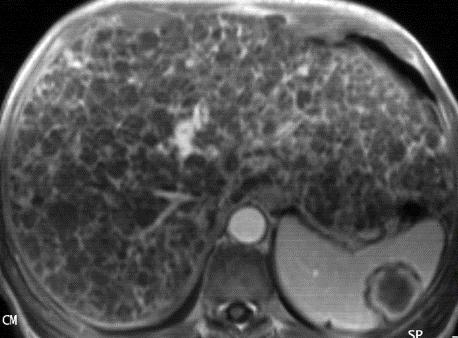
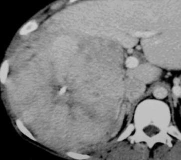
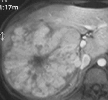
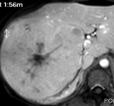
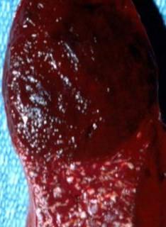
24 Malignant Focal Liver Lesions Unknown Case 55 yo woman with liver mass on ultrasound
25
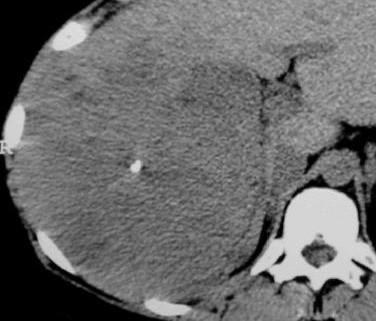
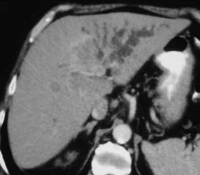
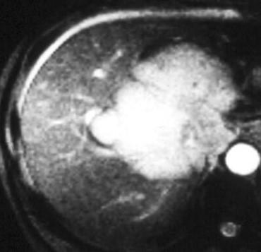
26
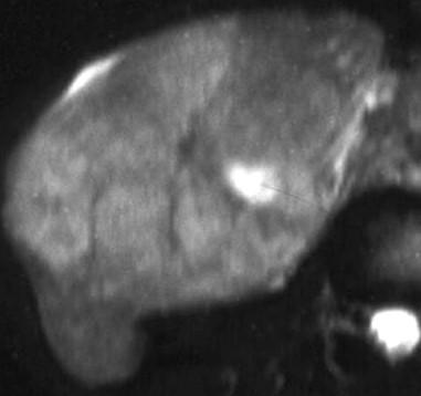
27 What is the most likely diagnosis? A. Intrahepatic Cholangiocarcinoma B. Epithelioid Hemangioendothelioma C. Fibrolamellar carcinoma D. Metastasis E. Angiosarcoma
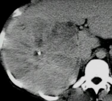
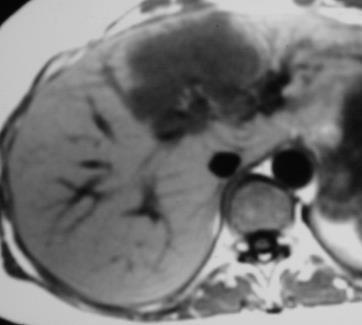
28 What is the most likely diagnosis? A. Intrahepatic Cholangiocarcinoma B. Epithelioid Hemangioendothelioma C. Fibrolamellar carcinoma D. Metastasis E. Cystadenocarcinoma
29 Metastasis 10 times more frequent than primary malignant liver neoplams Multiple If solitary, likely from Colon Adenocarcinoma HCC, 10 times more frequent than other primary liver tumors


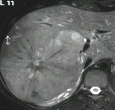
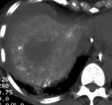
30 Fibrolamellar Carcinoma - Malignant Hepatocytes - Lamellar fibrosis - Confluent centrally - AFP negative


31 Fibrolamellar Carcinoma - Poorer vascularity, Bile stained - Central scar, Radiating Septa - No vessels in scar - Calcification
32 Fibrolamellar Carcinoma
33 Fibrolamellar Carcinoma: Scar - Low signal in T2 - No enhancement - Calcification


34 Fibrolamellar Carcinoma - Heterogeneous - Poor vascularity - Ill defined scar

35 Fibrolamellar Carcinoma Heterogeneous No Calcifications No FDG activity
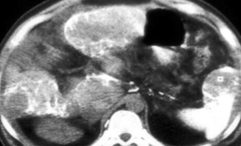
36 Fibrolamellar Carcinoma

37 Fibrolamellar Carcinoma Homogeneous, but Calcifications
38 Fibrolamellar Carcinoma
39 MALIGNANT HEPATIC NEOPLASMS CHOLANGIOCELLULAR ORIGIN Cholangiocarcinoma (I-CAC) Cystadenocarcinoma
")

40 Intrahepatic Cholangiocarcinoma (I-CAC) Microscopy Adenocarcinoma: mucin rich, no bile Abundant fibrous stroma Calcification
 - Fibrous,")
 - Local extension, no")
41 I-CAC - Large (5-20 cm), - Solid (Hemorrhage/necrosis rare) - Fibrous, scar, calcifications - Satellite nodules (20%) - Local extension, no tumor thrombus

42 I-CAC Pathogenesis/Associations Thorotrast Hepatolithiasis (5% - 20%) Clonorchis sinensis Sclerosing Cholangitis Caroli Disease Congenital Hepatic Fibrosis
43 I-CAC Hypodense, homogeneous, irregular borders Capsular Retraction Satellite nodules No Cirrhosis

44 I-CAC Biliary dilatation, segmental
45 I-CAC


46 I-CAC T1: Hypointense Early: Peripheral enhancement

47 I-CAC Late: central enhancement in scar


48 I-CAC Vascular encasement, no invasion Extrahepatic local extension
49 MALIGNANT HEPATIC NEOPLASMS MESENCHYMAL ORIGIN Angiosarcoma Epithelioid Hemangioendothelioma Kaposi sarcoma Primary lymphoma

50 Risk Factors: Thorotrast, Vinyl chloride, Arsenicals


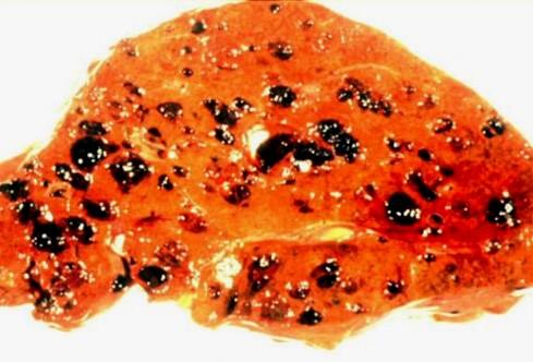
51 Angiosarcoma Pathologic Findings Foci of malignant endothelial cells Along vascular spaces Thorotrast granules displaced peripherally
 - Solitary")

52 Angiosarcoma Pathologic Findings - Multinodular (70%) - Solitary - Thorotrast peripherally displaced reticulated surface fibrosis


53 Angiosarcoma - Thorotrast, liver, spleen, lymph nodes peripherally displaced
54 Angiosarcoma
55 Angiosarcoma
56 Angiosarcoma
57 Angiosarcoma

 growth")
58 Epithelioid Hemangioendothelioma Slow, peripheral (subcapsular) growth Hypertrophy of uninvolved liver

59 Epithelioid Hemangioendothelioma
60 Epithelioid Hemangioendothelioma Nonspecific symptoms or asymptomatic (20%) Slow, peripheral (subcapsular) growth Hypertrophy of uninvolved liver


61 Epithelioid Hemangioendothelioma Peripheral, globular enhancement Intratumoral shunting Invasive
62 Regarding Primary Hepatic Lymphoma? A. The liver is commonly involved B. Its contour is ill defined C. It is typically Hodgkin Disease D. Typically has calcifications E. Solitary, multiple and diffuse forms
63 Regarding Primary Hepatic Lymphoma? A. The liver is commonly involved B. Its contour is ill defined C. It is typically Hodgkin Disease D. Typically has calcifications E. Solitary, multiple and diffuse forms
64 Lymphoma Primary lymphoma, extremely rare NHL, diffuse large cell type Immunocompromised (transplant, IDS, ) Solitary, Multiple, Diffuse Low attenuation, well defined MRI preferred for diffuse form


65 Lymphoma NHL, Hemosiderosis Multiple, well defined nodules Hypodense, No enhancement


66 Lymphoma Diffuse Geographic involvement Spleen deposit
67 Malignant Focal Liver Lesions Other than HCC HEPATOCELLULAR ORIGIN Fibrolamellar carcinoma (FLC) CHOLANGIOCELLULAR ORIGIN Cholangiocarcinoma (I-CAC) Cystadenocarcinoma
68 Malignant Focal Liver Lesions Other than HCC MESENCHYMAL ORIGIN Angiosarcoma Epithelioid Hemangioendothelioma Kaposi sarcoma Primary lymphoma METASTASIS
69 Malignant Focal Liver Lesions More than Metastasis and HCC Gamut of primary malignant neoplasms Different cellular lines and appearances Imaging: Benign vs. Malignant Primary vs. Secondary Resectable vs. Unresectable Imaging guided Biopsy Percutaneous therapy
70 Surgical vs. Nonsurgical Lesions Cyst Focal Fat Flow Phenomenon Abscess Hemangioma FNH Unresectable metastasis
71
Liver Tumors. Prof. Dr. Ahmed El - Samongy
 Liver Tumors Prof. Dr. Ahmed El - Samongy Objective 1. Identify the most important features of common benign liver tumors 2. Know the risk factors, diagnosis, and management of hepatocellular carcinoma
Interesting Cases from Liver Tumor Board. Jeffrey C. Weinreb, M.D.,FACR Yale University School of Medicine
 Interesting Cases from Liver Tumor Board Jeffrey C. Weinreb, M.D.,FACR Yale University School of Medicine jeffrey.weinreb@yale.edu Common Liver Diseases Hemangioma Cyst FNH Focal Fat/Sparing THID Non-Cirrhotic
HEPATO-BILIARY IMAGING
 HEPATO-BILIARY IMAGING BY MAMDOUH MAHFOUZ MD PROF.OF RADIOLOGY CAIRO UNIVERSITY mamdouh.m5@gmail.com www.ssregypt.com CT ABDOMEN Indications Patient preparation Patient position Scanogram Fasting 4-6 hours
CTA/MRA of Pediatric Hepatic Masses Radiology-Pathology Correlation
 Acta Radiológica Portuguesa, Vol.XVIII, nº70, pág. 41-50, Abr.-Jun., 2006 CTA/MRA of Pediatric Hepatic Masses Radiology-Pathology Correlation Marilyn J. Siegel Mallinckrodt Institute of Radiology, Washington
Evaluation of Liver Mass Lesions. American College of Gastroenterology 2013 Regional Postgraduate Course
 Evaluation of Liver Mass Lesions American College of Gastroenterology 2013 Regional Postgraduate Course Lewis R. Roberts, MB ChB, PhD Division of Gastroenterology and Hepatology Mayo Clinic College of
Financial Disclosure
 Benign Liver Masses Adil Abdalla, MBBS Creighton University-CHI Health August 25, 2018 Financial Disclosure Nothing to disclose Financial Disclosure 1 Objectives To assess patients with benign liver tumors
Management of Rare Liver Tumours
 Gian Luca Grazi Hepato-Biliary-Pancreatic Surgery National Cancer Institute Regina Elena Rome Fibrolamellar Carcinoma Mixed Hepato Cholangiocellular Carcinoma Hepatoblastoma Carcinosarcoma Primary Hepatic
Rare primary liver tumors - MRI pictorial review
 Rare primary liver tumors - MRI pictorial review Poster No.: C-2293 Congress: ECR 2017 Type: Educational Exhibit Authors: R. Lameiras, J. Cruz, J. Felício Costa, F. D. Figueiredo, A. 1 1 1 1 2 1 1 1 2
We are IntechOpen, the world s leading publisher of Open Access books Built by scientists, for scientists. International authors and editors
 We are IntechOpen, the world s leading publisher of Open Access books Built by scientists, for scientists 3,350 108,000 1.7 M Open access books available International authors and editors Downloads Our
Interesting case. Vikas Kundra, M.D., Ph.D. October Vikas Kundra, M.D., Ph.D.
 Interesting case October 2012 Disclosure Information Vikas Kundra, M.D, Ph.D. I have no financial relationships to disclose. I WILL NOT include discussion of investigational or off-label use of a product
Hepatocellular carcinoma Cholangiocarcinoma. Jewels of hepatobiliary cancer imaging : what to look for? Imaging characteristics of HCC.
 Outline : Imaging Jewels Jewels of hepatobiliary cancer imaging : what to look for? Hepatocellular carcinoma Cholangiocarcinoma Surachate Siripongsakun, M.D. Chulabhorn Cancer Center Imaging characteristics
Imaging of liver and pancreas
 Imaging of liver and pancreas.. Disease of the liver Focal liver disease Diffusion liver disease Focal liver disease Benign Cyst Abscess Hemangioma FNH Hepatic adenoma HCC Malignant Fibrolamellar carcinoma
Primary Hepatic Undifferentiated Pleomorphic Sarcoma: CT and angiographic findings in two cases
 J Radiol Sci 2013; 38: 15-19 Primary Hepatic Undifferentiated Pleomorphic Sarcoma: CT and angiographic findings in two cases Jan-Wen Ku Ying-Chi Tseng Kuo-Luon Kung Hsien-Chang Shen Yen-Lin Huang Chi-Jen
CT 101 :Pancreas and Spleen
 CT 101 :Pancreas and Spleen Shikha Khullar,, MD, MPH Division of Radiology University of South Alabama The Pancreas Normal Pancreas 3 Phase Pancreatic CT Non contrast Arterial phase : 30-35 35 second
CT & MRI of Benign Liver Neoplasms Srinivasa R Prasad
 CT & MRI of Benign Liver Neoplasms Srinivasa R Prasad No financial disclosures Acknowledgements Many thanks to Drs. Heiken, Narra & Menias (MIR) Dr. Sahani (MGH) for sharing images Benign Liver Tumors:
Essentials of Clinical MR, 2 nd edition. 65. Benign Hepatic Masses
 65. Benign Hepatic Masses Pulse sequences acquired for abdominal MRI typically consist of fast acquisition schemes such as single-shot turbo spin echo (i.e. HASTE) and gradient echo schemes such as FLASH
MRI OF FOCAL LESIONS IN
 Introduction MRI OF FOCAL LESIONS IN THE NON-CIRRHOTIC LIVER Ivan Pedrosa M.D. Ph.D. Associate Professor of Radiology and Advanced Imaging Research Center University of Texas Southwestern. Dallas, TX Incidental
objectives Pitfalls and Pearls in PET/CT imaging Kevin Robinson, DO Assistant Professor Department of Radiology Michigan State University
 objectives Pitfalls and Pearls in PET/CT imaging Kevin Robinson, DO Assistant Professor Department of Radiology Michigan State University To determine the regions of physiologic activity To understand
Radiology of hepatobiliary diseases
 GI cycle - Lecture 14 436 Teams Radiology of hepatobiliary diseases Objectives 1. To Interpret plan x-ray radiograph of abdomen with common pathologies. 2. To know the common pathologies presentation.
Jesse Civan, M.D. Medical Director, Jefferson Liver Tumor Center
 Liver Tumors Jesse Civan, M.D. Medical Director, Jefferson Liver Tumor Center Differential Diagnosis Malignant Metastatic from non-hepatic primary Hepatocellular carcinoma Cholangiocarcinoma Biliary cystcarcinoma
NEOPLASMS AND TUMOR-LIKE CONDITIONS OF LIVER
 NEOPLASMS AND TUMOR-LIKE CONDITIONS OF LIVER Epithelial Tumors Focal nodular hyperplasia Focal nodular hyperplasia is a localized hyperplasic overgrowth of hepatocytes around a vascular anomaly, particularly
The Focal Hepatic Lesion: Radiologic Assessment
 The Focal Hepatic Lesion: Radiologic Assessment Kevin Kuo, Harvard Medical School Year III Our Patient: PS 67 y/o female w/ long history of alcohol use Drinking since age 18, up to one bottle of wine/day
Approach to Liver Lesions. Anjana A. Pillai, MD Associate Professor of Medicine Director, Liver Tumor Clinic The University of Chicago Medical Center
 Approach to Liver Lesions Anjana A. Pillai, MD Associate Professor of Medicine Director, Liver Tumor Clinic The University of Chicago Medical Center Objectives Identify common clinical features and imaging
Complete Summary GUIDELINE TITLE. Liver lesion characterization. BIBLIOGRAPHIC SOURCE(S)
") Complete Summary GUIDELINE TITLE Liver lesion characterization. BIBLIOGRAPHIC SOURCE(S) Foley WD, Bree RL, Gay SB, Glick SN, Heiken JP, Huprich JE, Levine MS, Ros PR, Rosen MP, Shuman WP, Greene FL, Rockey
Complete Summary GUIDELINE TITLE Liver lesion characterization. BIBLIOGRAPHIC SOURCE(S) Foley WD, Bree RL, Gay SB, Glick SN, Heiken JP, Huprich JE, Levine MS, Ros PR, Rosen MP, Shuman WP, Greene FL, Rockey
Liver Tumors. Patient Education. Treatment options 8 4A. About the Liver. Surgical Specialties
 Patient Education Treatment options This handout describes different kinds of tumors that form in the liver and how they are treated. About the Liver Your liver is the largest organ in your abdomen. It
LIVER IMAGING TIPS IN VARIOUS MODALITIES. M.Vlychou, MD, PhD Assoc. Professor of Radiology University of Thessaly
 LIVER IMAGING TIPS IN VARIOUS MODALITIES M.Vlychou, MD, PhD Assoc. Professor of Radiology University of Thessaly Hepatocellular carcinoma is a common malignancy for which prevention, screening, diagnosis,
IT 의료융합 1 차임상세미나 복부질환초음파 이재영
 IT 의료융합 1 차임상세미나 2013-4-3 복부질환초음파 이재영 나는오늘누구를위하여 종을울리나? 전통적의료 의사 공학설계자 의사 최첨단진단장비들 USG, CT, MRI 환자 환자 현대의료 사용자중심의사고 US in the Abdomen Detection DDx Look Behavior Response by external stimuli Guiding Tool
Acknowledgements. Update of Focal Liver Lesions Goals. Focal Liver Lesions. Imaging Choices For Liver Lesions. Focal Liver Lesions
 Acknowledgements Update of Focal Liver Lesions 2012 Giles Boland Massachusetts General Hospital Harvard Medical School No disclosures Dushyant Sahani Mukesh Harisinghani Goals Focal liver lesions Imaging
Radiological Reasoning: Incidentally Discovered Liver Mass
 AJR Integrative Imaging LIFELONG LEARNING FOR RADIOLOGY This Radiological Reasoning article is available for SAM credit and CME credits when completed with the additional educational material provided
Workup of a Solid Liver Lesion
 Workup of a Solid Liver Lesion Joseph B. Cofer MD FACS Chief Quality Officer Erlanger Health System Affiliate Professor of Surgery UTHSC-Chattanooga I have no financial or other relationships with any
What is Liver Cancer? About the Liver
 Your liver is important and it has many functions. The top three are that it cleans your blood of toxins, gives you energy and produces bile for digestion. What is Liver Cancer? Cancer starts when cells
ACG Clinical Guideline: Diagnosis and Management of Focal Liver Lesions
 ACG Clinical Guideline: Diagnosis and Management of Focal Liver Lesions Jorge A. Marrero, MD, 1 Joseph Ahn, MD, FACG, 2 K. Rajender Reddy, MD, FACG 3 1 University of Texas at Southwestern, Dallas, Texas,
With the widespread use of hepatic imaging, liver masses
 2B: Liver Assessment of the Liver Mass: What Do You Need to Know? With the widespread use of hepatic imaging, liver masses are detected either unexpectedly or in the course of screening for liver cancer
Enhancements in Hepatobiliary Imaging:
 Enhancements in Hepatobiliary Imaging: S. Channual 1, MD; A. Pahwa 2, MD; S. Raman 1, MD. 1 UCLA Medical Center, Department of Radiologic Sciences 2 Olive-View UCLA Medical Center, Department of Radiology
Centrally-located Sclerosing Hepatocellular Carcinoma: A Case Report with Imaging Findings before and after Transcatheter Arterial Chemoembolization
 Chin J Radiol 2001; 26: 147-152 147 CASE REPORT Centrally-located Sclerosing Hepatocellular Carcinoma: A Case Report with Imaging Findings before and after Transcatheter Arterial Chemoembolization TUN-MEI
Lewis R. Roberts, MB, ChB, PhD, FACG
 2B: Hot Topics in Liver Disease Evaluation of Liver Mass Lesions Lewis R. Roberts, MB, ChB, PhD, FACG Clinical Classification of Liver Mass Lesions It is helpful to subclassify lesions into three clinical
Liver Cancer (Hepatocellular Carcinoma or HCC) Overview
 Overview") Liver Cancer (Hepatocellular Carcinoma or HCC) Overview Recent advances in liver cancer care seek to address the rising incidence of liver cancer, which has steadily increased over the past three decades.
Liver Cancer (Hepatocellular Carcinoma or HCC) Overview Recent advances in liver cancer care seek to address the rising incidence of liver cancer, which has steadily increased over the past three decades.
Alice Fung, MD Oregon Health and Science University
 Alice Fung, MD Oregon Health and Science University Disclosure Comments The speaker Alice Fung, MD Has relevant financial relationships to disclose. Received honorarium from (Guerbet). This individual
Cholangiocarcinoma: appearances and mimics
 Cholangiocarcinoma: appearances and mimics Poster No.: C-1572 Congress: ECR 2011 Type: Educational Exhibit Authors: C. Cardenas Valencia, J. Fernandez Jara, J. Cubero Carralero, B. Corral Ramos, P. Perez
Video Microscopy Tutorial 8
 Video Microscopy Tutorial 8 Common and Uncommon Lesions of the Liver Gladwyn Leiman, MD There are no disclosures necessary. Common and Uncommon Lesions in Liver FNA Gladwyn Leiman University of Vermont
State of the Art Imaging for Hepatic Malignancy: My Assignment
 State of the Art Imaging for Hepatic Malignancy: My Assignment CT vs MR vs MRCP Which one to choose for HCC vs Cholangiocarcinoma What special protocols to use for liver tumors Role of PET and Duplex US
Hepatobiliary and Pancreatic Malignancies
 Hepatobiliary and Pancreatic Malignancies Gareth Eeson MD MSc FRCSC Surgical Oncologist and General Surgeon Kelowna General Hospital Interior Health Consultant, Surgical Oncology BC Cancer Agency Centre
Neoplasms of the Canine, Feline and Lemur Liver:
 Neoplasms of the Canine, Feline and Lemur Liver: Classification and Prognosis Annual Seminar of the French Society of Veterinary Pathology John M. Cullen VMD PhD DACVP North Carolina State University Primary
GASTROINTESTINAL IMAGING STUDY GUIDE
 GASTROINTESTINAL IMAGING STUDY GUIDE Pharynx Diverticula Foreign bodies Trauma o Motility Disorders Esophagus Diverticula Trauma Esophagitis Barrett esophagus Rings, webs, and strictures Varices Benign
Cholangiocarcinoma (Bile Duct Cancer)
") Cholangiocarcinoma (Bile Duct Cancer) The Bile Duct System (Biliary Tract) A network of bile ducts (tubes) connects the liver and the gallbladder to the small intestine. This network begins in the liver
Cholangiocarcinoma (Bile Duct Cancer) The Bile Duct System (Biliary Tract) A network of bile ducts (tubes) connects the liver and the gallbladder to the small intestine. This network begins in the liver
R.Sotoudehmanesh, MD Professor of Gastroenterology Digestive Disease Research Institute Tehran University of Medical Sciences Pancreatobiliary
 R.Sotoudehmanesh, MD Professor of Gastroenterology Digestive Disease Research Institute Tehran University of Medical Sciences Pancreatobiliary /Advanced Endoscopy group Most common biliary malignancy and
Case Study: #3: Gallbladder Carcinoma?
 Case Study: #3: Gallbladder Carcinoma? By: Megan Wyatt K. SON Wyatt 225 2B1 RDMS, RVT Patient: Male 85 YOA Caucasian Indication: Elevated Alkaline Phosphatase History Annual physical showed elevated alkaline
X-Ray Corner. Imaging Approach to Cystic Liver Lesions. Pantongrag-Brown L. Solitary cystic liver lesions. Hepatic simple cyst (Figure 1)
") THAI J 136 Imaging Approach to Cystic Liver Lesions GASTROENTEROL 2013 X-Ray Corner Imaging Approach to Cystic Liver Lesions Pantongrag-Brown L Cystic liver lesions are common findings in daily practice
THAI J 136 Imaging Approach to Cystic Liver Lesions GASTROENTEROL 2013 X-Ray Corner Imaging Approach to Cystic Liver Lesions Pantongrag-Brown L Cystic liver lesions are common findings in daily practice
Recently role of non-invasive diagnostics methods
 CERTAIN ASPECTS OF NLS-DIAGNOSTICS OF LIVER FOCAL PATHOLOGY A.Y. Shvack, V.I. Nesterov, N.L. Ogluzdina This article contains information about NLS-graphy application in diagnostics of liver focal affections:
Hematologic Malignancies of the Liver : Spectrum of Disease. Zhou Jian
 Hematologic Malignancies of the Liver : Spectrum of Disease Zhou Jian 2015-7-8 Hematologic malignancies include a wide spectrum of lymphoproliferative and myeloproliferative disorders with nodal and extranodal
Liver Cancer And Tumours
 Liver Cancer And Tumours What causes liver cancer? Many factors may play a role in the development of cancer. Because the liver filters blood from all parts of the body, cancer cells from elsewhere can
Navigating the Biliary Tract with CT & MR: An Imaging Approach to Bile Duct Obstruction
 Navigating the Biliary Tract with CT & MR: An Imaging Approach to Bile Duct Obstruction Ann S. Fulcher, MD Medical College of Virginia Virginia Commonwealth University Richmond, Virginia Objectives To
Biliary cancers: imaging diagnosis. Study of 30 cases
 Biliary cancers: imaging diagnosis. Study of 30 cases N Hammoune, S Semlali, M Eddarai, T. Amil, M Zentar, S. El Kandri,, M Benameur,, S Chaouir. Radiology Department. Mohamed V Military Hospital. Rabat-
Multiple Primary Quiz
 Multiple Primary Quiz Case 1 A 72 year old man was found to have a 12 mm solid lesion in the pancreatic tail by computed tomography carried out during a routine follow up study of this patient with adult
Imaging of Gastrointestinal Stromal Tumors (GIST) Amir Reza Radmard, MD Assistant Professor Shariati hospital Tehran University of Medical Sciences
 Amir Reza Radmard, MD Assistant Professor Shariati hospital Tehran University of Medical Sciences") Imaging of Gastrointestinal Stromal Tumors (GIST) Amir Reza Radmard, MD Assistant Professor Shariati hospital Tehran University of Medical Sciences Describe the typical imaging findings of GIST at initial
Imaging of Gastrointestinal Stromal Tumors (GIST) Amir Reza Radmard, MD Assistant Professor Shariati hospital Tehran University of Medical Sciences Describe the typical imaging findings of GIST at initial
MALIGNANT HEPATIC NEOPLASMS: USING ULTRASONOGRAPHY AS A MEANS OF DEFINING HEPATIC LESIONS. 1.5 Contact Hours. Presented by: CEU Professor 7
 MALIGNANT HEPATIC NEOPLASMS: USING ULTRASONOGRAPHY AS A MEANS OF DEFINING HEPATIC LESIONS 1.5 Contact Hours Presented by: CEU Professor 7 www.ceuprofessoronline.com Copyright 8 2007 The Magellan Group,
Benign liver tumors : Diagnosis and management
 4th International Hepatology Conference 2016 HEPATOLOGY SOCIETY, DHAKA, BANGLADESH Benign liver tumors : Diagnosis and management Pr Laurence Chiche Hepato biliary surgery and transplantation Bordeaux,
Alpha-fetoprotein
 Other Names/Abbreviations AFP 190.25 - Alpha-fetoprotein Alpha-fetoprotein (AFP) is a polysaccharide found in some carcinomas. It is effective as a biochemical marker for monitoring the response of certain
Liver nodules mimicking metastatic disease
 Liver nodules mimicking metastatic disease Poster No.: C-1703 Congress: ECR 2011 Type: Educational Exhibit Authors: F. Vandenbroucke, B. Ilsen, B. Op de Beeck, J. de Mey ; 1 1 2 2 3 2 3 Brussels/BE, Brussel/BE,
CHOLANGIOCARCINOMA (CCA)
") CHOLANGIOCARCINOMA (CCA) Deepak Hariharan MD (Research), FRCS, Locum Consultant HPB Surgeon AIM Outline essential facts & principles Present 4 cases Discuss Challenges /Controversies INTRODUCTION Most
CHOLANGIOCARCINOMA (CCA) Deepak Hariharan MD (Research), FRCS, Locum Consultant HPB Surgeon AIM Outline essential facts & principles Present 4 cases Discuss Challenges /Controversies INTRODUCTION Most
Chief Complain. Liver lesion found in routine health check 41 days ago
 Chief Complain Liver lesion found in routine health check 41 days ago Present Illness On 2005-7-26 at 台北署立醫院 he underwent a health check for the first time. Abdominal US showed suspicious of a 6*5 cm hepatoma,
CT and MRI of Hepatic Contour Abnormalities
 Hepatobiliary Imaging Pictorial Essay CT and MRI of Jafi A. Lipson 1, Aliya Qayyum 1, David E. Avrin 2, Antonio Westphalen 1, Benjamin M. Yeh 1, Fergus V. Coakley 1 Fig. 1. 38-year-old woman imaged for
Hepatocellular Carcinoma: Diagnosis and Management
 Hepatocellular Carcinoma: Diagnosis and Management Nizar A. Mukhtar, MD Co-director, SMC Liver Tumor Board April 30, 2016 1 Objectives Review screening/surveillance guidelines Discuss diagnostic algorithm
Pediatric Hepatobiliary, Pancreatic & Splenic US
 Pediatric Hepatobiliary, Pancreatic & Splenic US Susan J. Back, MD Department of Radiology, The Children s Hospital of Philadelphia No Disclosures Objectives Normal Abnormal: cases and US advances Objectives
Pathological Classification of Hepatocellular Carcinoma
 3 rd APASL Single Topic Conference: HCC in 3D Pathological Classification of Hepatocellular Carcinoma Glenda Lyn Y. Pua, M.D. HCC Primary liver cancer is the 2 nd most common cancer in Asia HCC is the
TUMOR AND TUMOR-LIKE CONDITIONS OF THE PERITONEUM AND OMENTUM/MESENTERY 40 th. Annual Meeting SCBTMR September 9-13, 2017, Nashville, Tennessee
 TUMOR AND TUMOR-LIKE CONDITIONS OF THE PERITONEUM AND OMENTUM/MESENTERY 40 th. Annual Meeting SCBTMR September 9-13, 2017, Nashville, Tennessee Isaac R Francis University of Michigan Department of Radiology
Liver Specialty Evening Conference. Matthew M. Yeh, MD, PhD Professor of Pathology Adjunct Professor of Medicine University of Washington, Seattle
 Liver Specialty Evening Conference Matthew M. Yeh, MD, PhD Professor of Pathology Adjunct Professor of Medicine University of Washington, Seattle Case History A 65 year-old man presents with abdominal
Personal Profile. Name: 劉 XX Gender: Female Age: 53-y/o Past history. Hepatitis B carrier
 Personal Profile Name: 劉 XX Gender: Female Age: 53-y/o Past history Hepatitis B carrier Chief complaint Fever on and off for 2 days Present illness 94.10.14 Sudden onset of epigastric pain 94.10.15 Fever
Primary Hepatic Neoplasms. estimated 560,000 new cases per year. There is tremendous regional variation in incidence of
 Primary Hepatic Neoplasms Hepatocellular Carcinoma Incidence and Epidemiology Worldwide, hepatocellular carcinoma is the 3 rd most common causes of cancer death with an estimated 560,000 new cases per
Diagnostic Studies Then. It s important to be able to distinguish. Diagnostic Studies Now
 Jonathan S. Fisher, MD, FACS It s important to be able to distinguish Diagnostic Studies Then Diagnostic Studies Then History Biopsy Diagnostic Studies Now History Biopsy Serum markers (AFP, CA19 9, CEA)
Common and unusual CT and MRI manifestations of pancreatic adenocarcinoma: a pictorial review
 Review Article Common and unusual CT and MRI manifestations of pancreatic adenocarcinoma: a pictorial review Min-Jie Yang, Su Li, Yong-Guang Liu, Na Jiao, Jing-Shan Gong Department of Radiology, Shenzhen
Liver imaging takes a step forward with Ingenia
 Publication for the Philips MRI Community ISSUE 49 2013 / 2 Liver imaging takes a step forward with Ingenia Lyon South Hospital strives to move from several studies first CT, then MR or PET to using just
CLINICAL MANAGEMENT. Asymptomatic Liver Mass. Clinical Case. Background
 GASTROENTEROLOGY 2006;131:619 623 CLINICAL MANAGEMENT Loren Laine, Section Editor Asymptomatic Liver Mass ROBERT S. BROWN, Jr Department of Medicine and Department of Surgery, Columbia University College
Chief Complaint. Retroperitoneal cystic mass incidentally found at health examination center.
 Personal Information Age: 34 y/o Sex: female Past history: major systemic medical history(-) surgical history(-), family history(-) Denied food or drug allergy Chief Complaint Retroperitoneal cystic mass
Imaging in gastric cancer
 Imaging in gastric cancer Gastric cancer remains a deadly disease because of late diagnosis. Adenocarcinoma represents 90% of malignant tumors. Diagnosis is based on endoscopic examination with biopsies.
performed to help sway the clinician in what the appropriate diagnosis is, which can substantially alter the treatment of management.
 Hello, I am Maura Polansky at the University of Texas MD Anderson Cancer Center. I am a Physician Assistant in the Department of Gastrointestinal Medical Oncology and the Program Director for Physician
Pediatric Retroperitoneal Masses Radiologic-Pathologic Correlation
 Acta Radiológica Portuguesa, Vol.XVIII, nº 70, pág. 61-70, Abr.-Jun., 2006 Pediatric Retroperitoneal Masses Radiologic-Pathologic Correlation Marilyn J. Siegel Mallinckrodt Institute of Radiology, Washington
간암의조직검사 : 언제, 어떻게? 계명대학교의과대학내과학교실 정우진
 간암의조직검사 : 언제, 어떻게? 계명대학교의과대학내과학교실 정우진 간생검한다 vs 안한다? M/81 Alcoholic LC, albumin 4.0, bil 0.6, Cr 1.06, glucose 141, afp 2.2, CA19-9 12.41 CT: R/O HCC in S8, R/O CC M/69 HBV(-), HCV(-), social alcoholics
Cystic lesions of the liver
 Cystic lesions of the liver Poster No.: C-0408 Congress: ECR 2014 Type: Educational Exhibit Authors: E. Rosado, J. Pereira, S. El Bouchaibi, M. A. A. Bali ; 1 1 2 2 3 3 4 4 Amadora/PT, Lisboa/PT, Bruxelles/BE,
Objectives. HCC Incidence and Mortality. Disclosure Statement HCC. Imaging of Hepatocellular Carcinoma. Treatment of Hepatocellular Carcinoma
 Imaging of Hepatocellular Carcinoma and the use of LI RADS Treatment of Hepatocellular Carcinoma Aaron D. Anderson, D.O. AOCR April 2015 Objectives Show how the use of LI RADS can simplify the diagnosis
Imaging in breast cancer. Mammography and Ultrasound Donya Farrokh.MD Radiologist Mashhad University of Medical Since
 Imaging in breast cancer Mammography and Ultrasound Donya Farrokh.MD Radiologist Mashhad University of Medical Since A mammogram report is a key component of the breast cancer diagnostic process. A mammogram
Essentials of Clinical MR, 2 nd edition. 73. Urinary Bladder and Male Pelvis
 73. Urinary Bladder and Male Pelvis Urinary bladder carcinoma is best locally staged with MRI. It is important however to note that a thickened wall (> 5 mm) is a non-specific finding seen in an underfilled
NAACCR Webinar Series 1
 Collecting Cancer Data: Liver 2013 2014 NAACCR Webinar Series June 5, 2014 Q&A Please submit all questions concerning webinar content through the Q&A panel. Reminder: If you have participants watching
Imaging of common diseases of hepatobiliary and GI system
 Imaging of common diseases of hepatobiliary and GI system Natthaporn Tanpowpong, M.D. Diagnostic radiology Faculty of Medicine, Chulalongkorn University Normal plain radiograph A = Common bile duct
Dr Claire Smith, Consultant Radiologist St James University Hospital Leeds
 Dr Claire Smith, Consultant Radiologist St James University Hospital Leeds Imaging in jaundice and 2ww pathway Image protocol Staging Limitations Pancreatic cancer 1.2.4 Refer people using a suspected
The role for contrast-enhanced ultrasonography outside of focal liver lesions
 The role for contrast-enhanced ultrasonography outside of focal liver lesions Paul S. Sidhu King s College Hospital, London, UK Introduction Contrast-enhanced ultrasonography (US) of focal liver lesions
Alpha-fetoprotein
 Other Names/Abbreviations AFP 190.25 - Alpha-fetoprotein Alpha-fetoprotein (AFP) is a polysaccharide found in some carcinomas. It is effective as a biochemical marker for monitoring the response of certain
Innovations in HCC Imaging: MDCT/MRI
 Innovations in HCC Imaging: MDCT/MRI Anthony E. Cheng, M.D. Cardinal MRI Center Cardinal Santos Medical Center, Wilson Street, San Juan Innovations in HCC Imaging: Goals/Objectives MDCT/MRI Learn the diagnostic
HCC e CEUS. Prof. A. Giorgio. Direttore IX UOC di Malattie Infettive ad Indirizzo Ecointerventistico
 HCC e CEUS Prof. A. Giorgio Direttore IX UOC di Malattie Infettive ad Indirizzo Ecointerventistico The natural history of compensated cirrhosis due to hepatitis C virus: a 17 year cohort study of 214 patients
Alpha-fetoprotein
 Other Names/Abbreviations AFP 190.25 - Alpha-fetoprotein Alpha-fetoprotein (AFP) is a polysaccharide found in some carcinomas. It is effective as a biochemical marker for monitoring the response of certain
Liver Ultrasound - Beyond the Basics. Pamela Parker Lead Sonographer
 Liver Ultrasound - Beyond the Basics Pamela Parker Lead Sonographer Aims Review what we know about the liver Reasons for imaging Focal lesions Diffuse disease Can we do more? The Liver The Liver The Liver
Approach to the Patient with Liver Disease
 Approach to the Patient with Liver Disease Diagnosis of liver disease Careful history taking Physical examination Laboratory tests Radiologic examination and imaging studies Liver biopsy Liver diseases
Case Scenarios. 12/28/12 MRI Liver: multiple focal arterially enhancing liver lesions, indeterminate. Repeat MRI in 4 months.
 Case Scenarios Case Scenario 1 A 63 year old black female presented with a history of liver lesions present for the past three years being monitored by serial scans. The patient has a history of esophageal
ADRENAL MR: PEARLS AND PITFALLS
 ADRENAL MR: PEARLS AND PITFALLS Frank Miller, M.D. Lee F. Rogers MD Professor of Medical Education Chief, Body Imaging Section and Fellowship Medical Director, MR Imaging Professor of Radiology Northwestern
I LOVE Immunostains. Two Types of Pitfalls 3/23/2017. Disclosure of Relevant Financial Relationships
 There Are No Magic Bullets: When Immunostains Can Get You into Trouble in Hepatic & Gastrointestinal Pathology John Hart, M.D. Sections of Surgical Pathology & Hepatology University of Chicago Medical
Two Case Reports and a Literature Review of Liver Transplantation for Epithelioid Hemangioendothelioma
 Case report Two Case Reports and a Literature Review of Liver Transplantation for Epithelioid Hemangioendothelioma Juan Ignacio Marín, MD, 1 Óscar Santos, MD, 1 Octavio Muñoz, MD, 1 Germán Osorio, MD,
Pediatric Abdominal Masses. Andrew Phelps MD Assistant Professor of Pediatric Radiology UCSF Benioff Children's Hospital
 Pediatric Abdominal Masses Andrew Phelps MD Assistant Professor of Pediatric Radiology UCSF Benioff Children's Hospital No Disclosures Take Home Message All you need to remember are the 5 common masses
Normal Sonographic Anatomy
 hapter 2:The Liver DUNSTAN ABRAHAM Normal Sonographic Anatomy Homogeneous, echogenic texture (Figure 2-1) Measures approximately 15 cm in length and 10 12.5 cm anterior to posterior; measurement taken
Armed Forces Institute of Pathology.
 Armed Forces Institute of Pathology www.radpath.com Armed Forces Institute of Pathology Breast Disease www.radpath.org Armed Forces Institute of Pathology Interpretation of Breast MRI Leonard M. Glassman