The Relevance of Cytologic Atypia in Cutaneous Neural Tumors
|
|
|
- Chad Richardson
- 5 years ago
- Views:
Transcription
1 The Relevance of Cytologic Atypia in Cutaneous Neural Tumors Recent Findings - New Developments New Problems Zsolt B. Argenyi, M.D. Professor of Pathology & Dermatology Director of Dermatopathology Department of Pathology University of Washington Seattle, WA, USA
2 Biologic Spectrum of Cutaneous Neural Tumors Atypical Benign Malignant De novo MPNST Ex-plexiform neurofibromas Diffuse neurofibromas Schwannomas Granular cell tumors
3 Cardinal Features of Malignant Peripheral Nerve Sheath Tumors Size Necrosis Mitotic rate Cellularity Atypia
4 Growing Number of Cases With Atypia Alone What is the relevance of cytologic atypia in these tumors? borderline tumors? reactive changes? low grade malignancy? Very limited studies to determine outcomes
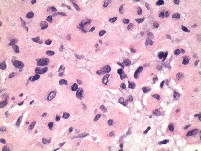
5 Growing Spectrum of Atypical Cutaneous Neural Tumors What constitutes atypia in these tumors? 1. Cytologic deviation from normal cell types epitheliod pleomorphic multinucleated cells 2. Nuclear atypia 3. Mitotic atypia 4. Cellularity 5. Growth pattern
6 The Most Common Cutaneous Neural Tumors With Atypia 1. Neurofibromas 2. Schwannomas 3. Neurothekeomas
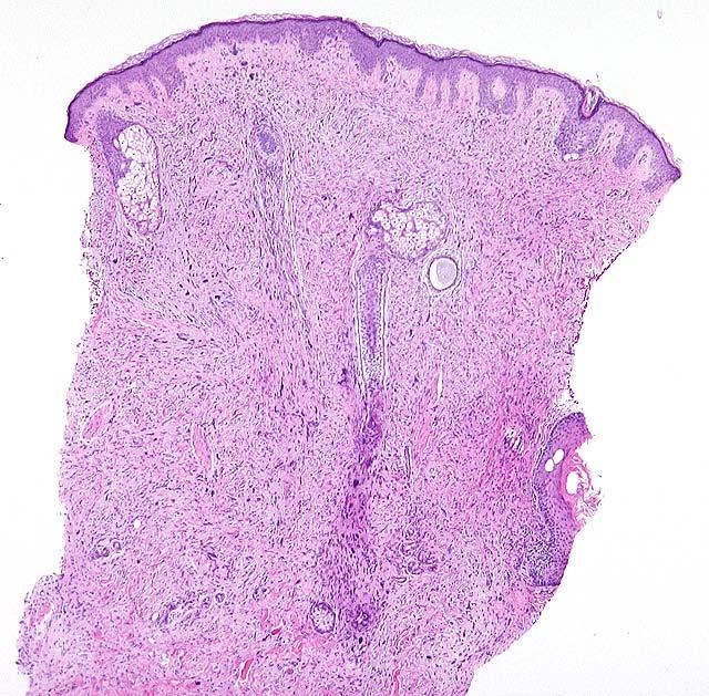
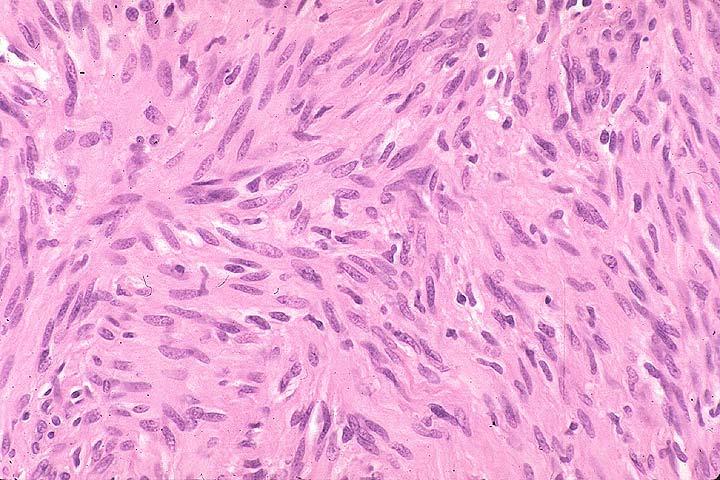
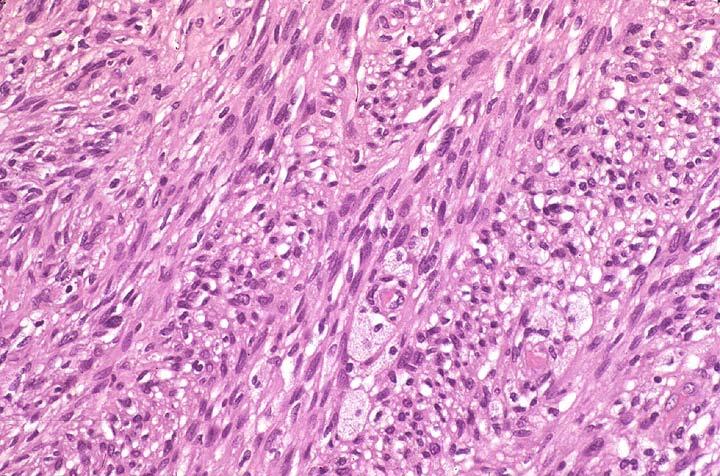
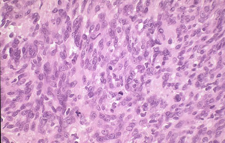
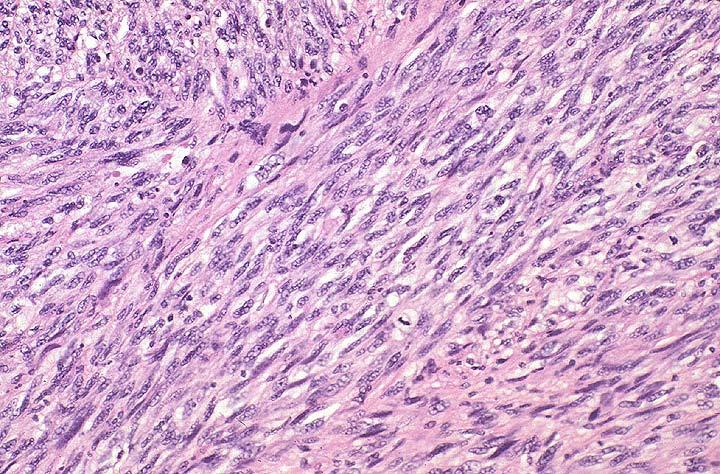
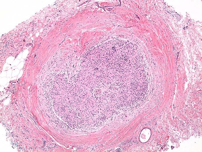
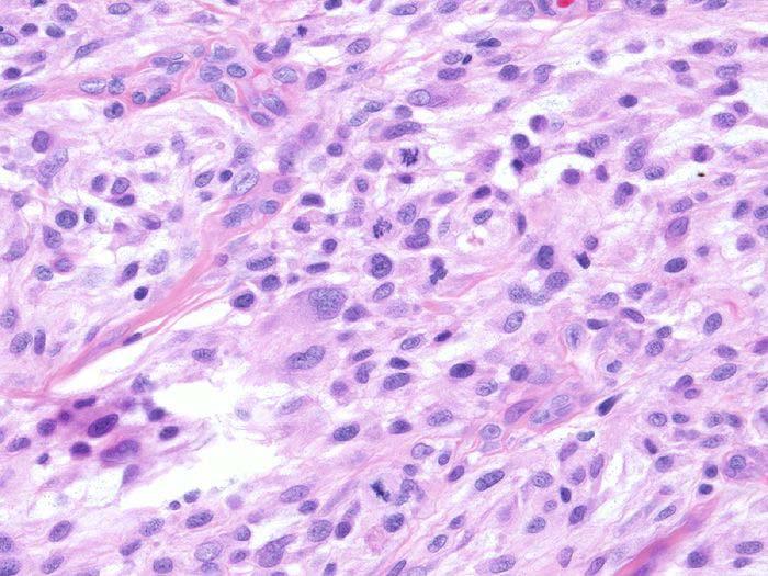
7 Atypia in Neurofibromas
8
9
10 Atypical Neurofibroma Differential Diagnosis Malignant peripheral nerve sheath tumor Pleomorphic liposarcoma Pleomorphic lipoma Myxoid MFH Cellular Schwannoma Cellular dermatofibroma
11 Relevance of Atypia Cutaneous Neurofibromatosis Important distinctions: Plexiform type high association with NF-1 Solitary, sporadic type; benign subtype or malignant? Prior studies: Lin BT-Y, Weiss L, Medeiros LJ. Neurofibroma and cellular neurofibroma with atypia: a report of 14 tumors. Am J Surg Pathol 1997; 21: 1443 predominantly superficial soft tissues
12 Neurofibroma and Cellular Neurofibroma With Atypia Clinicopathologic study by Lin BT et al. Am J Surg Pathol 21(12): , cases of 6 patients, mean age 40, F:M = 2:1 Head (5), trunk (5) and extremities (4) 3 patients with type I neurofibromatosis Benign behavior (limited F/U)
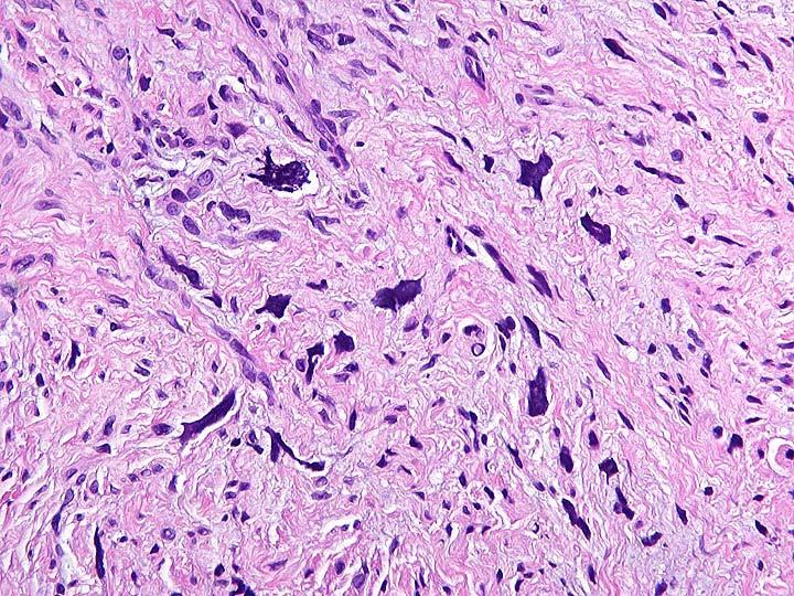
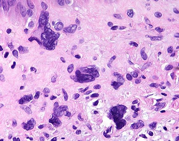
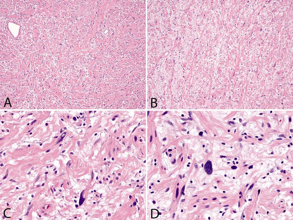
13 Neurofibroma and Cellular Neurofibroma with Atypia Histopathologic features Usual growth pattern of neurofibromas Mild to severe cytologic atypia, nuclear enlargement, hyperchromasia, bizarre giant cells Mitotic activity 1 < 10 HPF (3 cases) or absent Degenerative changes Focal hypercellularity (3 cases) Lack of necrosis Low p53, Ki-67, and S-phase values as compared to MPNST
14 Jokinen CH., Argenyi ZB: Atypical neurofibroma of the skin and subutaneous tissue: clinicopathologic analysis of 11 cases. J Cutan Pathol 2009; 36: in press.
15 Atypical Neurofibroma of the Skin Clinical Data 11 cases, 80% females, 20% males Age range 8-70, mean Predominantly trunk 1 patient with NF No history of PMNST
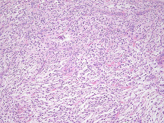
16 Atypical Neurofibromas of the Skin Pathology Dermis/superficial subcutis Growth type: fibrillary and lamellar Cellularity: mild to moderate Cytologic atypia (5-50% of cellularity) Pleomorphic cells Mitotic figures: Absent 9/11 Present 2/11 No necrosis
17

18 S-100P EMA p16 p16
19 Atypical Neurofibromas of the Skin Immunohistochemistry IMMUNOHISTOCHEMISTRY Case number S-100 protein p16 p53 MIB-1 ER/PR EMA (P) ND +(P) ND +(P) ND +(P) ND ++(P) ND +++(D) ND +(D) (P) (D) ND ND ND ND ND
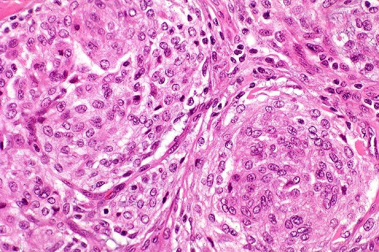
20 Cutaneous Atypical Neurofibroma Clinical Outcome No tumor recurrence No new malignancy Follow-up range 6 63 months (mean: 33 months)
21 Atypical Neurofibroma Conclusions Distinct subset of neurofibromas. The designation should be better accepted. Atypia could be analogous to lesions in: Ancient Schwannomas with degenerative changes Cellular Schwannomas with limited mitotic activity Cytologic atypia alone in NF does not appear to be associated with Neurofibromatosis type-1 There is no apparent short term risk for recurrence or malignancy. Awareness of atypia helps better patient management These lesions can be treated conservatively.
22 Problematic Aspects of Atypical, Cellular Schwannomas 1. The diagnostic criteria are still disputed difficult to apply for cutaneous lesions. 2. Confusing terminology for cutaneous lesions unclear biologic potential. 3. Expanded differential diagnosis in the skin malignant melanoma.
23 Cellular Schwannoma The term was coined by Woodruff in 1981 Synonyms: atypical Schwannoma (Reed) cellular Schwannoma (Woodruff) transformed Schwannoma (Reed) low-grade malignant Schwannoma (Ducatman)
24 Cellular Schwannoma General features 1. Affect mainly females. 2. Tumors of the deep soft tissue (mediastinum, pelvis). 3. Association with NF-1 less than 5%. 4. Evolving histologic criteria. 5. Considered benign. 6. Cutaneous involvement is rare.
25 Cutaneous Cellular Schwannomas 1. Cheek, 59 year old male, Woodruff et al Hand, 90 year old male, Davidson et al Back, 34 year old male, Megahed et al Hand, 66 year old male, De Nuntiis et al Oral mucosa, 34 year old male, Koizumi et al
26 The Spectrum of Cutaneous Schwannomas Common Type Malignant Ancient Cellular Epitheloid
27 Cellular Schwannoma (deep soft tissue type) Key Histopathologic Features Well circumscribed Nodular Hypercellular (Antoni A-Type) Fascicles and whorls Hyperchromasia Spindled atypical nuclei Mitotic rate < 4/10 HPF (depending on authors) Thick-walled vessels Lymphoid aggregates in the wall of the capsule Diffuse, strong S-100 protein expression
28
29
30
31
32 Histopathologic Spectrum of Cellular Schwannomas in the cutaneous literature Atypical Cellular Transformed - Hypercellularity - Interlacing fascicles - Storiform pattern - Cytologic atypia - Hyperchromatism - Mitotic rate - Stellate necrosis - Nuclear palisading - Verocay bodies - Lymphoid follicles - Epithelioid cells - Lipid laden histiocytes /- + +/- <2/10 HPF / /10 HPF +/- +/ >10/10 HPF + +/- - +/- ++ +
33 Cutaneous Cellular Schwannoma Differential Diagnosis Malignant peripheral nerve sheath tumor (MPNST) Malignant melanoma (primary or metastatic) (broad panel of immunohistochemistry, electron microscopy, clinico-pathologic correlation) Leiomyosarcoma (growth pattern + immunohistochemistry) Benign imitators ; ancient, epitheloid schwannomas, angiomyomas, neuromas
34 Cutaneous Cellular Schwannoma Benign Imitators Ancient Schwannoma Epitheloid Schwannoma Palisaded Encapsulated Neuroma Epitheloid Angiomyoma
35
36
37 Differential Diagnosis of Schwannomas with Atypia Epitheliod Cellular MPNST Growth Pattern nodular, oblong nodular, oblong variable Encapsulation usually present well-preserved partial infiltrative Cellularity Architecture trabecular, nodular, syncytial, myxoid Cell Type epitheloid/with or without spindled cells fascicular spindled cells fascicular herring bone spindled or pleomorphic Atypia mild moderate moderate to severe Mitotic Rate < 1/10 HPF < 4/10 HPF > 4/10 HPF Necrosis none none or very focal geographic Vascularity focal mild focal, prominent not characteristic Lymphoid Infiltrate variable patchy aggregates not characteristic
38 Cutaneous Cellular Schwannoma Conclusions 1. No comprehensive studies on cutaneous cellular schwannoma and MPNST 2. Application of diagnostic criteria is often arbitrary 3. Important differential diagnostic challenge (melanoma vs. MPNST)
39 Cellular Neurothekeoma Entity described in 1986 by Rosati et al. Well-established clinico-pathologic features. Controversial histogenesis; neural vs. other Synonyms: - cellular nerve sheath myxoma - immature nerve sheath myxoma - epithelioid nerve sheath myxoma - and a plethora of others. Recently recognized atypical variants
40 Cellular Neurothekeoma Histologic Features 1. Dermal/subcutaneous tumor 2. Multi-lobular or fascicular growth 3. Not encapsulated 4. Scant mucin 5. Epithelioid or spindle cells 6. Characteristic nuclei 7. Rare mitotic figures 8. Heterogeneity of cell components can occur
41
42 Cellular Neurothekeoma Atypical (? malignant variants) Atypical variants (Busam et al. 1998) Clinical - 10 patients - Median age 20.5 years - head and neck Pathological: - Large size - Deep penetration - Diffusely infiltrative borders - Vascular invasion - Marked cytologic atypia - Mitotic rate > 5/10 HPF Follow up: - (1-5 years) - no recurrence Additional Cases: - Bhatia et al Benbenisty et al (advocating Mohs surgery)
43
44
45 Atypical Neurothekeoma Additional observations; Hornick and Fletcher, 2007 Clinical follow-up on 69 cases with the mean F/U of 44 mo. showed recurrence of 10 cases. Atypical morphologic features of 133 cases; size>2cm (10%), mitosis >5/10HPF(21%), pleomorphism (25%), infiltration of fat (25%) were too common to represent increased risk for local recurrence. Conclusions: Only head and neck location and incomplete surgical excision correlated with recurrence.
46 Cellular Neurothekeoma Practical Conclusions CNT has distinct enough clinical and pathological features to accept as a distinct entity, with the notion that the histogenesis is still uncertain. Atypical variants are reported more often than original studies indicated. Currently there are somewhat contradicting data regarding the significance of atypia in Cellular Neurothekeomas. Considering the uncertain biologic potential a cautious clinical approach with adequate excision seems prudent.
47 The Relevance of Atypia in Cutaneous Neural Tumors Conclusions Recognition of several new morphologic subtypes. Biologic potential has not been fully established yet. Most of the atypical variants without other morphologic features of malignancy follows a benign or indolent behavior. Familiarity with these new variants remains important for better patient management.
أملس عضلي غرن = Leiomyosarcoma. Leiomyosarcoma 1 / 5
 Leiomyosarcoma 1 / 5 EPIDEMIOLOGY Exact incidence is unknown, but older studies suggest that leiomyosarcomas comprise approximately 3 percent of soft-tissue sarcomas. Superficial leiomyosarcoma occurs
Selected Pseudomalignant Soft Tissue Tumors of the Skin and Subcutis
 Selected Pseudomalignant Soft Tissue Tumors of the Skin and Subcutis Andrew L. Folpe, M.D. Professor of Laboratory Medicine and Pathology Mayo Clinic, Rochester, MN folpe.andrew@mayo.edu 2016 MFMER slide-1
CASE REPORT Benign epithelioid peripheral nerve sheath tumour resembling schwannoma
 Malaysian J Pathol 2014; 36(3) : 217 221 CASE REPORT Benign epithelioid peripheral nerve sheath tumour resembling schwannoma Thejasvi KRISHNAMURTHY MD and SR NIVEDITHA MD, DNB Department of Pathology,
Malignant Peripheral Nerve Sheath Tumor
 C H A P T E R 120 Malignant Peripheral Nerve Sheath Tumor Currently, malignant peripheral nerve sheath tumor (MPNST) is the most commonly used generic name for the neoplasms known in the past as neurosarcoma,
Cellular Neurothekeoma
 Cellular Neurothekeoma Scott W Binder, MD Pritzker Professor of Pathology & Dermatology Sr. Vice Chair Director, Pathology Clinical Services Chief, Dermatopathology Geffen/UCLA School of Medicine Clinical
Desmoplastic Melanoma R/O BCC. Clinical Information. 74 y.o. man with lesion on left side of neck r/o BCC
 R/O BCC Sabine Kohler, M.D. Professor of Pathology and Dermatology Dermatopathology Service Stanford University School of Medicine Clinical Information 74 y.o. man with lesion on left side of neck r/o
21/07/2017. Hobnail endothelial cells are not the same as epithelioid endothelial cells
 UPDATE IN CUTANEOUS VASCULAR S DERMATOPATHOLOGY SESSION BELFAST PATHOLOGY JUNE 21/2017 Dr E Calonje St John s Institute of Dermatology, London, United Kingdom THE FAMILY OF VASCULAR S WITH EPITHELIOID
1/10/2018. Soft Tissue Tumors Showing Melanocytic Differentiation. Overview. Desmoplastic/ Spindle Cell Melanoma
 2016 MFMER slide-1 2016 MFMER slide-2 2016 MFMER slide-3 Soft Tissue Tumors Showing Melanocytic Differentiation Andrew L. Folpe, M.D. Professor of Laboratory Medicine and Pathology Mayo Clinic, Rochester,
Spindle Cell Lesions Of The Breast. Emad Rakha Professor of Breast Pathology and Consultant Pathologist
 Spindle Cell Lesions Of The Breast Emad Rakha Professor of Breast Pathology and Consultant Pathologist * SCLs comprise a wide spectrum of diseases, ranging from reactive processes to aggressive malignant
Update On Lipomatous Tumors: Old Standbys and New Concepts
 Update On Lipomatous Tumors: Old Standbys and New Concepts John R. Goldblum, M.D. Chairman, Department of Anatomic Pathology Cleveland Clinic Professor of Pathology Cleveland Clinic Lerner College of Medicine
Newer soft tissue entities
 Newer soft tissue entities Examples among fibroblastic tumors Turku, May 6, 2010 Markku Miettinen, M.D. AFIP, Washington, DC Fibroblastic neoplasms Solitary fibrous tumor /Hemangiopericytoma Low-grade
Gastrointestinal stromal tumor
 Gastrointestinal stromal tumor 영남의대병리학교실 최준혁 Classification of gastrointestinal mesenchymal tumor Gastrointestinal stromal tumor(gist) Smooth muscle tumors : leiomyoma, leiomyosarcoma Neurogenic tumors
Financial disclosures
 Mesenchymal Neoplasms with Melanocytic Differentiation By Konstantinos Linos MD, FCAP, FASDP Bone, Soft Tissue and Dermatopathology Assistant Professor of Pathology Dartmouth-Hitchcock Medical Center Geisel
Dermatopathology. Dr. Rafael Botella Estrada. Hospital La Fe de Valencia
 Dermatopathology Dr. Rafael Botella Estrada. Hospital La Fe de Valencia Melanoma and mimics Dr. Martin Mihm Malignant lesions result from the accumulation of mutations Class I lesions (benign) Class II
Diagnostic problems in uterine smooth muscle tumors
 Diagnostic problems in uterine smooth muscle tumors Marina Kos Ljudevit Jurak Clinical Department of Pathology, Clinical Hospital Center Sestre milosrdnice, Zagreb Institute of Pathology, University of
Classification (1) Classification (3) Classification (2) Spindle cell lesions. Spindle cell lesions of bladder (Mills et al.
 Classification (3) Classification (2) Spindle cell lesions. Spindle cell lesions of bladder (Mills et al.") Non-epithelial tumours and nonepithelial tumour-like lesions of the bladder Dr Jonathan H Shanks The Christie NHS Foundation Trust, Manchester, UK Classification (1) Myofibroblastic proliferations and
Non-epithelial tumours and nonepithelial tumour-like lesions of the bladder Dr Jonathan H Shanks The Christie NHS Foundation Trust, Manchester, UK Classification (1) Myofibroblastic proliferations and
SOFT TISSUE TUMOR PATHOLOGY: AN UPDATE
 SOFT TISSUE TUMOR PATHOLOGY: AN UPDATE Jason L. Hornick, MD, PhD July 18, 2013 Department of Pathology Brigham and Women s Hospital Harvard Medical School Boston, MA, USA I have no disclosures. New Soft
G3.02 The malignant potential of the neoplasm should be recorded. CG3.02a
 G3.02 The malignant potential of the neoplasm should be recorded. CG3.02a Conventional adrenocortical neoplasm. Each of the below parameters is scored 0 when absent and 1 when present. 3 or more of these
Pathology of Sarcoma ELEANOR CHEN, MD, PHD, ASSISTANT PROFESSOR DEPARTMENT OF PATHOLOGY UNIVERSITY OF WASHINGTON
 Pathology of Sarcoma ELEANOR CHEN, MD, PHD, ASSISTANT PROFESSOR DEPARTMENT OF PATHOLOGY UNIVERSITY OF WASHINGTON Presentation outline Background and epidemiology of sarcomas Sarcoma classification Sarcoma
5/10. Pathology Soft tissue tumors. Farah Bhani. Mohammed Alorjani
 5/10 Pathology Soft tissue tumors Mohammed Alorjani Farah Bhani Slides are included in this sheet. Objectives: Soft tissue tumors 1. Describe soft tissue tumors. 2. Understand the classification of soft
Update on Cutaneous Mesenchymal Tumors. Thomas Brenn
 Update on Cutaneous Mesenchymal Tumors Thomas Brenn Cutaneous Mesenchymal Tumours Wide morphological and biological spectrum Myofibroblastic, smooth muscle, neural, vascular, apidocytic, undifferentiated;
Fun with Fat. General Rules. Case
 Fun with Fat General Rules Imaging: location (deep vs. superficial) Superficial lesions are seldom liposarcomas Deep lesions may be benign or malignant Myxoid stroma is common in benign and malignant lesions
Slide Seminar Spanish Society of Pathology
 Slide Seminar Spanish Society of Pathology John R. Goldblum, M.D. Chairman, Department of Anatomic Pathology Cleveland Clinic Professor of Pathology Cleveland Clinic Lerner College of Medicine 1921 Original
3/27/2017. Disclosure of Relevant Financial Relationships
 Ophthalmic Pathology Evening Specialty Conference USCAP 2017 5 th March, 2017 Mukul K. Divatia, MD Assistant Professor Department of Pathology & Genomic Medicine Weill Cornell Medical College Houston Methodist
USCAP Pediatrics Evening Subspecialty Conference 2015
 USCAP Pediatrics Evening Subspecialty Conference 2015 Sunday 22 March 2015 Alexander Lazar MD/PhD Department of Pathology S Section of Bone Soft TIssue Pathology Sarcoma Research Center The Case Patient
Tumors of Adipose Tissue Tumors Epidemiology Clinical Features. Morphology. Mature Adipocytes Separated by delicate fibrous septa
 Tumors of Adipose Tissue Lipoma Liposarcoma Most commonly happens in female The most common soft tissue tumor o Originates from matured Adipocytes Most commonly happes at the 4 th and 5 th decade of life
A 25 year old female with a palpable mass in the right lower quadrant of her abdomen
 May 2016 A 25 year old female with a palpable mass in the right lower quadrant of her abdomen Contributed by: Paul Ndekwe, MD, Resident Physician, Indiana University School of Department of Pathology and
Review of the AP Part II Practical Examination. Dr David Clift Co Chief Examiner
 Review of the AP Part II Practical Examination Dr David Clift Co Chief Examiner General Remarks The part II practical examination involved 15 cases which were presented with sufficient clinical data to
Papillary Lesions of the breast
 Papillary Lesions of the breast Emad Rakha Professor of Breast Pathology The University of Nottingham Papillary lesions of the breast are a heterogeneous group of disease, which are characterised by neoplastic
Enterprise Interest Nothing to declare
 Enterprise Interest Nothing to declare Diagnoses one would not like to miss in soft tissue pathology early in your career Marta Sbaraglia, MD Department of Pathology Hospital of Treviso University of Padua
SMOOTH MUSCLE TUMOURS
 SMOOTH MUSCLE TUMOURS NORMAL SMOOTH MUSCLE Cytology Immunohistochemistry Ultrastructure Masson Trichrome Smooth Muscle Ultrastructure Many myofilaments running parallel to the long axis of the smooth
ACCME/Disclosures. Everything is spindle - how far can we go with limited FNA material? Everything is spindle how far can we go? Everything is spindle
 ACCME/Disclosures The USCAP requires that anyone in a position to influence or control the content of CME disclose any relevant financial relationship WITH COMMERCIAL INTERESTS which they or their spouse/partner
Post-test Self-assessment Cases
 Post-test Self-assessment Cases Ibrahim Khalifeh, M.D. Associate Professor Department of Pathology American University of Beirut Medical Center Beirut, Lebanon Case I History A 69 year old gentleman presenting
Disclosures. Parathyroid Pathology. Objectives. The normal parathyroid 11/10/2012
 Disclosures Parathyroid Pathology I have nothing to disclose Annemieke van Zante MD/PhD Assistant Professor of Clinical Pathology Associate Chief of Cytopathology Objectives 1. Review the pathologic features
Molecular biology and impact on modern therapeutical approaches of cutaneous sarcomas
 Molecular biology and impact on modern therapeutical approaches of cutaneous sarcomas Bernhard Zelger Department of Dermatology & Venerology Medical University Innsbruck, Innsbruck 1. Introduction This
Atypical Palisaded Myofibroblastoma of Lymph Node: Report of a rare case.
 ISPUB.COM The Internet Journal of Pathology Volume 10 Number 1 Atypical Palisaded Myofibroblastoma of Lymph Node: Report of a rare case. V Kinnera, R Nandyala, M Yootla, K Mandyam Citation V Kinnera, R
Disclosure of Relevant Financial Relationships
 Neuropathology Evening Specialty Conference Disclosure of Relevant Financial Relationships The USCAP requires that anyone in a position to influence or control the content of all CME activities disclose
Contents Part I Introduction 1 General Description 2 Natural History: Importance of Size, Site, Histopathology
 Contents Part I Introduction 1 General Description... 3 1.1 Introduction... 3 1.2 Incidence and Prevalence... 5 1.3 Predisposing and Genetic Factors... 8 References... 16 2 Natural History: Importance
IN THE NAME OF GOD Dr. Kheirandish Oral and maxillofacial pathology
 IN THE NAME OF GOD Dr. Kheirandish Oral and maxillofacial pathology ORAL FOCAL MUCINOSIS Uncommon Tumorlike Cutaneous myxoid cyst Overproduction of hyaluronic acid by firoblasts Young adults Female Gingiva
Case year old female presented with asymmetric enlargement of the left lobe of the thyroid
 Case 4 22 year old female presented with asymmetric enlargement of the left lobe of the thyroid gland. No information available relative to a prior fine needle aspiration biopsy. A left lobectomy was performed.
Case 4 Diagnosis 2/21/2011 TGB
 Case 4 22 year old female presented with asymmetric enlargement of the left lobe of the thyroid gland. No information available relative to a prior fine needle aspiration biopsy. A left lobectomy was performed.
Special slide seminar
 Special slide seminar Tomáš Rozkoš The Fingerland Department of Pathology Charles University Medical Faculty and Faculty Hospital in Hradec Králové Czech Republic Case history, 33 years old resistance
Diplomate of the American Board of Pathology in Anatomic and Clinical Pathology
 A 33-year-old male with a left lower leg mass. Contributed by Shaoxiong Chen, MD, PhD Assistant Professor Indiana University School of Medicine/ IU Health Partners Department of Pathology and Laboratory
An Overview of Genital Stromal Tumors
 An Overview of Genital Stromal Tumors By Konstantinos Linos MD, FCAP, FASDP Bone, Soft Tissue and Dermatopathology Assistant Professor of Pathology Dartmouth-Hitchcock Medical Center Geisel School of Medicine
Plexiform Tumor of the Orbit
 Plexiform Tumor of the Orbit Anat Stemmer-Rachamimov, MD Department of Pathology Massachusetts General Hospital Harvard Medical School Disclosure of Relevant Financial Relationships USCAP requires that
Essential Dermatopathology: Neoplastic American Academy of Dermatology Annual Meeting NEURAL AND SMOOTH MUSCLE NEOPLASMS
 Essential Dermatopathology: Neoplastic American Academy of Dermatology Annual Meeting NEURAL AND SMOOTH MUSCLE NEOPLASMS Kevin P. White M.D. Oregon Health and Science University Associate Professor of
Soft Tissue Perineurioma
 The Korean Journal of Pathology 2009; 43: 266-70 DOI: 10.4132/KoreanJPathol.2009.43.3.266 Soft Tissue Perineurioma - A Case Report - Jun Mo Kim Joon Hyuk Choi Department of Pathology, Yeungnam University
Female 18. Deeply pigmented lesion on trunk.?warty naevus?seborrhoeic keratosis?malignant melanoma. The best diagnosis is:
 Female 18. Deeply pigmented lesion on trunk.?warty naevus?seborrhoeic keratosis?malignant melanoma. The best diagnosis is: A. deep penetrating naevus B. naevoid malignant melanoma C. pigment synthesising
Nerve Sheath Myxoma Presenting as Finger Nodule in 39 year old Female
 ISPUB.COM The Internet Journal of Pathology Volume 10 Number 1 Nerve Sheath Myxoma Presenting as Finger Nodule in 39 year old Female M Hamodat, A Alhumidi Citation M Hamodat, A Alhumidi.. The Internet
A case of giant cell tumour of soft parts in a horse Francesco Cian 1, Sarah Whiteoak 2, Jennifer Stewart 1
 A case of giant cell tumour of soft parts in a horse Francesco Cian 1, Sarah Whiteoak 2, Jennifer Stewart 1 1 Animal Health Trust, Newmarket, UK 2 608 Equine and Farm Vets, Rowington, UK Signalment: Horse,
Disclosures. Giant Cell Rich Tumors of Bone. Outline. The osteoclast. Giant cell rich tumors 5/21/11
 Disclosures Giant Cell Rich Tumors of Bone Andrew Horvai, MD, PhD Associate Clinical Professor, Pathology This lecture discusses "off label" uses of a number of pharmaceutical agents. The speaker is describing
Slide seminar. Asist. Prof. Jože Pižem, MD, PhD Institute of Pathology Medical Faculty, University of Ljubljana
 Slide seminar Asist. Prof. Jože Pižem, MD, PhD Institute of Pathology Medical Faculty, University of Ljubljana Case 5 A 57-year-old man with a dermal/subcutaneous lesion on the scalp, which was interpreted
Evening Specialty Conference Bone and Soft Tissue Pathology. Diagnostic pitfalls in bone and soft tissue pathology
 Evening Specialty Conference Bone and Soft Tissue Pathology. Case 1 Elizabeth G Demicco, MD, PhD Mount Sinai Hospital, New York Disclosure of Relevant Financial Relationships USCAP requires that all planners
Whitney A. High, MD, JD, MEng
 ADS Dermatopathology Meeting 2014 Selected Adnexal Tumors Whitney A. High, MD, JD, MEng Associate Professor, Dermatology & Pathology Director of Dermatopathology (Dermatology) University of Colorado School
Nervous About Nerve Sheath Tumours?
 Goals Nervous About Nerve Sheath Tumours? Rajiv M. Patel, M.D. RCPA NZ ASM 2017 (3:30-4:15pm, Saturday, 23-09-17) Review spectrum of cutaneous nerve sheath tumors Vast majority are benign & include neurofibromas,
Division of Pathology
 Case 38 Adult woman with a 35mm right breast lump at the 10 o clock position. Excision performed. (Case contributed by Dr Mihir Gudi, KKH) Division of Pathology Merlion, One Fullerton Singapore Diagnosis
Normal endometrium: A, proliferative. B, secretory.
 Normal endometrium: A, proliferative. B, secretory. Nội mạc tử cung Nội mạc tử cung Cyclic changes in endometrium.. Approximate relationship of useful microscopic changes. Arias-Stella reaction in endometrial
From Morphology to Molecular Pathology: A Practical Approach for Cytopathologists Part 1-Cytomorphology. Songlin Zhang, MD, PhD LSUHSC-Shreveport
 From Morphology to Molecular Pathology: A Practical Approach for Cytopathologists Part 1-Cytomorphology Songlin Zhang, MD, PhD LSUHSC-Shreveport I have no Conflict of Interest. FNA on Lymphoproliferative
Endometrial Stromal Tumors
 Endometrial Stromal Tumors WHO Categories: Endometrial Stromal Nodule (ESN) Endometrial Stromal Sarcoma, low grade (LGESS) Endometrial Stromal Sarcoma, high grade (HGESS) Undifferentiated Uterine Sarcoma
Objectives. Atypical Glandular Cells. Atypical Endocervical Cells. Reactive Endocervical Cells
 2013 California Society of Pathologists 66 th Annual Meeting San Francisco, CA Atypical Glandular Cells to Early Invasive Adenocarcinoma: Cervical Cytology and Histology Christina S. Kong, MD Associate
3/24/2017 DENDRITIC CELL NEOPLASMS: HISTOLOGY, IMMUNOHISTOCHEMISTRY, AND MOLECULAR GENETICS. Disclosure of Relevant Financial Relationships
 DENDRITIC CELL NEOPLASMS: HISTOLOGY, IMMUNOHISTOCHEMISTRY, AND MOLECULAR GENETICS Jason L. Hornick, M.D., Ph.D. Director of Surgical Pathology and Immunohistochemistry Brigham and Women s Hospital Professor
Pleomorphic Liposarcoma: A Clinicopathologic Analysis Of 19 Cases
 Pleomorphic Liposarcoma: A Clinicopathologic Analysis Of 19 Cases Katharine A. Downes, M.D., John R. Goldblum, M.D., Elizabeth A. Montgomery, M.D., Cyril Fisher, M.D., F.R.C.Path. Departments of Anatomic
57th Annual HSCP Spring Symposium 4/16/2016
 An Unusual Malignant Spindle Cell Lesion to Involve the Breast Erinn Downs-Kelly, D.O. Associate Professor of Pathology University of Utah & ARUP Laboratories No disclosures Case 39 y/o female with no
Case 1 10/2/17. Myxoid Soft Tissue Tumors & Tumor-like Lesions. Myxofibro- or Fibromyxo-?: Myxoid Soft Tissue Tumours We Are All Mixed Up About
 Myxoid Soft Tissue Tumors & Tumor-like Lesions Myxofibro- or Fibromyxo-?: Myxoid Soft Tissue Tumours We Are All Mixed Up About Rajiv M. Patel, M.D. RCPA NZ ASM 2017 (4:15-5:00pm, Saturday, 23-09-17) Heterogenous
Myxo-inflammatory Fibroblastic sarcoma
 AKA Myxo-inflammatory Fibroblastic sarcoma Acral Myxoinflammatory fibroblastic sarcomaam.j.surg.path1998; 22; 911-924 Inflammatory myxoid tumour of soft parts with bizarre giant cells [Pathol.Res.Pract.
Microcystic/Reticular Schwannoma: Morphological Features Causing Diagnostic Dilemma on Fine-Needle Aspiration Cytology
 ISSN 1941-5923 DOI: 10.12659/AJCR.892196 Received: 2014.08.08 Accepted: 2014.09.06 Published: 2014.12.04 Microcystic/Reticular Schwannoma: Morphological Features Causing Diagnostic Dilemma on Fine-Needle
Ancient schwannoma of the mouth floor A case report and review
 Oral Oncology EXTRA (2006) 42, 281 285 available at www.sciencedirect.com journal homepage: http://intl.elsevierhealth.com/journal/ooex CASE REPORT Ancient schwannoma of the mouth floor A case report and
Conflict of Interest 9/2/2014. Pathogenesis and Comparison of Atypical Spitz Nevi vs Benign Spitz, and Childhood Melanoma
 Pathogenesis and Comparison of Atypical Spitz Nevi vs Benign Spitz, and Childhood Melanoma Martin C. Mihm Jr., M.D., F.A.C.P. Harvard Medical School Brigham and Women s Hospital Dana Farber Cancer Center
A PRACTICAL APPROACH TO ATYPICAL MELANOCYTIC LESIONS BIJAN HAGHIGHI M.D, DIRECTOR OF DERMATOPATHOLOGY, ST. JOSEPH HOSPITAL
 A PRACTICAL APPROACH TO ATYPICAL MELANOCYTIC LESIONS BIJAN HAGHIGHI M.D, DIRECTOR OF DERMATOPATHOLOGY, ST. JOSEPH HOSPITAL OBJECTIVES Discuss current trends and changing concepts in our understanding of
Case Report A Rare Cutaneous Adnexal Tumor: Malignant Proliferating Trichilemmal Tumor
 Case Reports in Medicine Volume 2015, Article ID 742920, 4 pages http://dx.doi.org/10.1155/2015/742920 Case Report A Rare Cutaneous Adnexal Tumor: Malignant Proliferating Trichilemmal Tumor Omer Alici,
Morphologically Benign Lesions of Soft Tissue and Bone Which Metastasize - What Can We Do?
 Andrew L. Folpe, MD Mayo Clinic, Rochester, MN ISBSTP Handout 2010 Morphologically Benign Lesions of Soft Tissue and Bone Which Metastasize - What Can We Do? Introduction Over the past several decades
USCAP 2011: ASDP companion meeting. Steven D. Billings 1
 USCAP 2011: ASDP companion meeting. Steven D. Billings (billins@ccf.org) 1 Spindle cell tumors that make you say, Oh $*&%! This lecture will focus on examples of cutaneous tumors that present particular
Fine Needle Aspiration Biopsy of a Myxoid Leiomyosarcoma with Epithelioid Features and It Metastasized to the Abdominal Wall - A Case Report -
 The Korean Journal of Pathology 2010; 44: 220-4 DOI: 10.4132/KoreanJPathol.2010.44.2.220 Fine Needle Aspiration Biopsy of a Myxoid Leiomyosarcoma with Epithelioid Features and It Metastasized to the Abdominal
Low-grade serous neoplasia. Robert A. Soslow, MD
 Low-grade serous neoplasia Robert A. Soslow, MD soslowr@mskcc.org Outline Orientation Ovarian tumor overview Non serous borderline tumors Serous borderline tumors Clinical summary Morphologic description
Fine Needle Aspiration Cytology of Neurothekeoma DO NOT DUPLICATE
 Fine Needle Aspiration Cytology of Neurothekeoma A Case Report Agustín Vaillo Vinagre, M.D., Antonia Gutiérrez Martín, M.D., Andrés Pérez Barrios, M.D., M.I.A.C., Nuria Alberti Masgrau, M.D., M.I.A.C.,
History A 89 year old gentleman presenting with a scalp/forehead nodule. Patient had squamous cell carcinoma 18 m at same site, excised. Outside diagn
 Case III History A 89 year old gentleman presenting with a scalp/forehead nodule. Patient had squamous cell carcinoma 18 m at same site, excised. Outside diagnoses: Squamous cell carcinoma. R/O: SCC, Melanoma,
Salivary Glands 3/7/2017
 Salivary Glands 3/7/2017 Goals and objectives Focus on the entities unique to H&N Common board type facts Information for your future practice Salivary Glands Salivary Glands Major gland. Paratid. Submandibular.
05/07/2018. Types of challenges. Challenging cases in uterine pathology. Case 1 ` 65 year old female Post menopausal bleeding Uterine Polyp
 Types of challenges Challenging cases in uterine pathology Nafisa Wilkinson Gynaecological Pathologist UCLH London Lack of complete history often, NO clinical history at all! Cases from other centres often
Inflammatory pseudotumor
 Inflammatory pseudotumor Inflammatory pseudotumor (IPT) Heterogeneous group of lesions of obscure etiology On physical and radiographic examination often confused with malignancy Synonyms Plasma cell granuloma
Basal cell carcinoma 5/28/2011
 Goal of this Presentation A practical approach to the diagnosis of cutaneous carcinomas and their mimics Thaddeus Mully, MD University of California San Francisco To review common non-melanoma skin cancers
DEGENERATE NEURILEMMOMA: A DIAGNOSTIC PROBLEM
 J. roy. Army med. Cps. 1977.123,18-24 DEGENERATE NEURILEMMOMA: A DIAGNOSTIC PROBLEM Lieutenant-Colonel W. R STARKE, M.D., M.C.(U.S.A.)* Lieutenant-Colonel J. B. STEW ART M.B., M.R.C.Path., D.RC.Path.,
Case 27 Male 42. Painless, static, well-circumscribed, subcutaneous nodule right lower leg,?lipoma. The best diagnosis is:
 Case 27 Male 42. Painless, static, well-circumscribed, subcutaneous nodule right lower leg,?lipoma. The best diagnosis is: A. Angiosarcoma B. Haemangiopericytoma C.Myopericytoma D.Myofibroma E. Angioleiomyoma
Case Report Malignant Peripheral Nerve Sheath Tumor of the Inguinum and Angiosarcoma of the Scalp in a Child with Neurofibromatosis Type 1
 Hindawi Case Reports in Pathology Volume 2017, Article ID 7542825, 4 pages https://doi.org/10.1155/2017/7542825 Case Report Malignant Peripheral Nerve Sheath Tumor of the Inguinum and Angiosarcoma of the
59 yo male with past medical history of prostate carcinoma, presented with upper abdominal pain
 December 2016 59 yo male with past medical history of prostate carcinoma, presented with upper abdominal pain Contributed by: Divya Sharma, MD. Fellow, Gastrointestinal Pathology, Department of Pathology
BSD 2015 Case 19. Female 21. Nodule on forehead. The best diagnosis is:
 BSD 2015 Case 19 Female 21. Nodule on forehead. The best diagnosis is: A. mixed tumour of skin B. porocarcinoma C. nodular hidradenoma D. metastatic adenocarcinoma BSD 2015 Case 19 Female 21 Nodule on
Case Report Spindle cell lipoma of the wrist, occurring in a distinctly rare location: a case report with review of literature
 Int J Clin Exp Pathol 2015;8(3):3299-3303 www.ijcep.com /ISSN:1936-2625/IJCEP0004992 Case Report Spindle cell lipoma of the wrist, occurring in a distinctly rare location: a case report with review of
"Atypical": Criteria and
 "Atypical": Criteria and Controversies Esther Rossi MD PhD MIAC Division of Anatomic Pathology and Cytology Catholic University of Sacred Heart Rome, Italy CASE HISTORY In 2015, 45 y/o woman underwent
Malignant Phyllodes tumor with necrosis a rare case report
 Quest Journals Journal of Medical and Dental Science Research Volume 1 ~ Issue 1 (2014) pp: 01-06 ISSN(Online) : 2394-076X ISSN (Print):2394-0751 www.questjournals.org Research Paper Malignant Phyllodes
Primary Cutaneous CD30-Positive T-cell Lymphoproliferative Disorders
 Primary Cutaneous CD30-Positive T-cell Lymphoproliferative Disorders Definition A spectrum of related conditions originating from transformed or activated CD30-positive T-lymphocytes May coexist in individual
Pleomorphic Sarcoma of Breast: A Report of Two Cases and Review of Literature
 CASE REPORT Pleomorphic Sarcoma of Breast: A Report of Two Cases and Review of Literature Anju Bansal, Manveen Kaur, and Varsha Dalal Department of Pathology, National Institute of Pathology (ICMR), Safdarjang
Prognostic Significance of Grading and Staging Systems using MIB-1 Score in Adult Patients with Soft Tissue Sarcoma of the Extremities and Trunk
 843 Prognostic Significance of Grading and Staging Systems using MIB-1 Score in Adult Patients with Soft Tissue Sarcoma of the Extremities and Trunk Tadashi Hasegawa, M.D. 1 Seiichiro Yamamoto, Ph.D. 2
Papillary Lesions of the Breast A Practical Approach to Diagnosis. (Arch Pathol Lab Med. 2016;140: ; doi: /arpa.
 Papillary Lesions of the Breast A Practical Approach to Diagnosis (Arch Pathol Lab Med. 2016;140:1052 1059; doi: 10.5858/arpa.2016-0219-RA) Papillary lesions of the breast Span the spectrum of benign,
When Immunostains Can Get You in Trouble: Gynecologic Pathology p16: Panacea or Pandora s Box?
 When Immunostains Can Get You in Trouble: Gynecologic Pathology p16: Panacea or Pandora s Box? Teri A. Longacre, MD Stanford Medicine Stanford California pi6 in Gynecologic Pathology: Panacea or Pandora
Cutaneous Adnexal Tumors
 Cutaneous Adnexal Tumors Lesions with Predominant Follicular Differentiation Special Emphasis on Basal Cell Carcinoma 2014-04-01 Prof. Dr. med. Katharina Glatz Pathologie Cutaneous Adnexal Tumors Hair
Mody. AIS vs. Invasive Adenocarcinoma of the Cervix
 Common Problems in Gynecologic Pathology Michael T. Deavers, M.D. Houston Methodist Hospital, Houston, Texas Common Problems in Gynecologic Pathology Adenocarcinoma in-situ (AIS) of the Cervix vs. Invasive
Thyroid follicular neoplasms in cytology. Ulrika Klopčič Institute of Oncology, Department of Cytopathology, Ljubljana, Slovenia
 Thyroid follicular neoplasms in cytology Ulrika Klopčič Institute of Oncology, Department of Cytopathology, Ljubljana, Slovenia Lecture overview importance of FNAB in assessing thyroid lesions follicular
Case: The patient is a 24 year- old female who was found to have multiple mural nodules within the antrum. Solid and cystic components were noted on
 Case: The patient is a 24 year- old female who was found to have multiple mural nodules within the antrum. Solid and cystic components were noted on imaging. There is no significant past medical history.
Case of Pleomorphic Dermal Sarcoma of the Eyelid Treated with Micrographic Surgery and Secondary Intention Healing
 JI Kim, et al pissn 1013-9087ㆍeISSN 2005-3894 Ann Dermatol Vol. 28, No. 5, 2016 http://dx.doi.org/10.5021/ad.2016.28.5.632 CASE REPORT Case of Pleomorphic Dermal Sarcoma of the Eyelid Treated with Micrographic
My Journey into the World of Salivary Gland Sebaceous Neoplasms
 My Journey into the World of Salivary Gland Sebaceous Neoplasms Douglas R. Gnepp Warren Alpert Medical School at Brown University Rhode Island Hospital Pathology Department Providence RI Asked to present
ARTHUR PURDY STOUT SOCIETY COMPANION MEETING: DIFFICULT NEW DIFFERENTIAL DIAGNOSES IN PROSTATE PATHOLOGY. Jonathan I. Epstein.
 1 ARTHUR PURDY STOUT SOCIETY COMPANION MEETING: DIFFICULT NEW DIFFERENTIAL DIAGNOSES IN PROSTATE PATHOLOGY Jonathan I. Epstein Professor Pathology, Urology, Oncology The Reinhard Professor of Urological