DIFFUSE INTRINSIC PONTINE GLIOMA: A MULTI-FACETTED AND GLOBAL VIEW
|
|
|
- Geoffrey Welch
- 5 years ago
- Views:
Transcription
1 DIFFUSE INTRINSIC PONTINE GLIOMA: A MULTI-FACETTED AND GLOBAL VIEW Sophie E.M. Veldhuijzen van Zanten
2
3 DIFFUSE INTRINSIC PONTINE GLIOMA: A MULTI-FACETTED AND GLOBAL VIEW Sophie Elisabeth Madzy Veldhuijzen van Zanten
4 Diffuse Intrinsic Pontine Glioma: A multi-facetted and global view Sophie Elisabeth Madzy Veldhuijzen van Zanten Thesis, Faculty of Medicine, VU University Medical Center, VU University, The Netherlands ISBN: Author: Sophie E.M. Veldhuijzen van Zanten Cover: Angela Palmer, Brain of the Artist, 2012, hand engraved on multiple sheets of glass, Scottish National Portrait Gallery Lay-out: Nikki Vermeulen - Ridderprint BV Printing: Ridderprint BV - Copyright S.E.M. Veldhuijzen van Zanten, The Netherlands, 2017 All rights reserved. No parts of this publication may be reproduced, stored in a retrieval system, or transmitted in any form or by any means, without written permission from the author of from the Publisher holding the copyright of the published articles. Publication of this thesis was financially supported by: Stichting Semmy, The DIPG Collaborative, Stichting Research Fonds Kindergeneeskunde VUmc, KPMG, Yellow Research BV, 2TCI, 2-BBB Medicines BV, ORTEC Logiqcare BV, Vygon The research in this thesis was performed at the Department of Paediatrics - Division of Oncology-Heamatology, the Department of Neurosurgery, the Department of Radiology & Nuclear Medicine and the Department of Pathology of VU University Medical Center, Amsterdam, The Netherlands
5 VRIJE UNIVERSITEIT DIFFUSE INTRINSIC PONTINE GLIOMA: A MULTI-FACETTED AND GLOBAL VIEW ACADEMISCH PROEFSCHRIFT ter verkrijging van de graad Doctor aan de Vrije Universiteit Amsterdam, op gezag van de rector magnificus prof.dr. V. Subramaniam, in het openbaar te verdedigen ten overstaan van de promotiecommissie van de Faculteit der Geneeskunde op dinsdag 28 november 2017 om uur in de aula van de universiteit, De Boelelaan 1105 door Sophie Elisabeth Madzy Veldhuijzen van Zanten geboren te Amsterdam
6 promotoren: copromotor: prof.dr. G.J.L. Kaspers prof.dr. W.P. Vandertop dr. D.G. van Vuurden
7 PREFACE In the summer of 2006, Semmy, first-born child of John and Nicole, showed increasing tiredness at school. He was four years old at that time. A visit to the pediatrician uncovered abnormalities in the neurological examination and all alarm bells rang. The doctor s disturbing suspicion was confirmed by MR-images, showing a diffuse intrinsic pontine glioma or DIPG, in Semmy s brainstem. With no curative treatment for this malignant tumor available, the neurologist predicted a life expectancy of 3 to 4 months and broached the subject of palliative care on the day of diagnosis. An extensive search on the Internet by the parents revealed that there was no doctor in the world who could cure their child. At that time, there were only few institutions in Europe that performed clinical research into DIPG. In the Netherlands, no prospective clinical trial had yet been conducted. Semmy s parents decided that the chance of success of early-phase clinical trials did not outweigh the fact that the family would need to move abroad for at least three months, which was all the time they were given. To the satisfaction of the family, Semmy received individual-experience based therapy and adequate care in VU University Medical Center. In the beginning of 2007, Semmy rapidly deteriorated. There was just sufficient time and quality of life left for a surprise organized by the Make-A- Wish foundation, and for a visit to the local football club and the zoo. Shortly after his birthday, Semmy died, at the age of five. Diffuse Intrinsic Pontine Glioma (DIPG) is a rare disease. So rare a pediatric oncologist may only see a case like Semmy s once per year. This makes it impossible to recognize patterns, while patterns are important for optimal individual patient care, and for scientific research. For decades, DIPG research has been hampered by the rarity of the disease. Lack of knowledge on disease characteristics led to an ongoing debate about the definition
8 of the disease, and heterogeneous use of in-and exclusion criteria for clinical trials. Moreover, lack of knowledge on possible underlying patient subgroups may have led to selection bias in these trials. At the same time, lack of international, evidence-based treatment guidelines led to predominant application of single-center and individualized therapy. When Semmy died, not only was there a lack of treatment options, but also insufficient knowledge to optimize care for DIPG patients, and most importantly only few research initiatives to change all this. The persistence and perseverance of Semmy s parents to improve prognosis of DIPG patients, even after their own child s death, formed the start of a new era for DIPG research. They established Stichting Semmy (The Semmy Foundation), giving rise to the research of this thesis. In March 2012, at the initiation of this thesis, DIPG research was mostly regionally organized, small-scaled, and geographically scattered. More importantly, in the Netherlands, no prospective DIPG-specific clinical trial had yet been conducted. Thanks to the financial support of Stichting Semmy, the opportunity was provided to initiate the first single- and multi-center trials. The studies that were initiated at VU University Medical Center cover all aspects of DIPG, ranging from clinical symptoms, diagnostics and treatment strategies, to pre-clinical laboratory research on tumor material obtained via autopsy. In Part I of this thesis, the results of these studies are presented in analogy to the disease course of DIPG, from time of diagnosis to death. Since DIPG is so rare, there was not sufficient data to provide all answers to the many research questions raised. Therefore, in Part II of this thesis, the perspective of the research was expanded to a larger scope, both in time and scale. Starting with historical cohort studies and extensive literature reviews to learn from the past, we reached out to our colleagues at national, European and global level. Important subjects such as alternative treatment strategies, survival prediction, palliative care and use of steroids in DIPG patients were investigated. Finally, in Part III, we put into practice what we had learned, which is that regional, small-scaled, and scattered research initiatives are not efficient in a global aim to unravel and cure this rare disease. This part of the thesis describes the establishment of an international research infrastructure, formed by a collaboration of biomedical experts within the SIOPE DIPG Network, and the development and initiation of the SIOPE DIPG Registry and Imaging Repository, in parallel to the International DIPG Registry. These efforts have resulted in the first worldwide initiatives to increase DIPG patient data and
9 improve the integration, speed, quality, and coherence of research into DIPG. For the first time, large datasets have become available for robust analysis of clinical, radiological and biological disease characteristics, as well as treatment strategies. All work described in this thesis was done with the aim to provide a better perspective for children like Semmy.
10 TABLE OF CONTENTS Chapter 1 General introduction and thesis outline 11 PART I Chapter 2 Chapter 3 Chapter 4 Chapter 5 FIRST DIPG-SPECIFIC STUDIES CONDUCTED IN THE NETHERLANDS A phase I/II study of gemcitabine during radiotherapy in children with newly diagnosed diffuse intrinsic pontine glioma J Neurooncol Jul 26 (Epub ahead of print) Molecular drug imaging: 89 Zr-bevacizumab PET in children with diffuse intrinsic pontine glioma J Nucl Med. 2017;58(5): F-FDG PET standard uptake values of the normal pons in children: establishing a reference value for diffuse intrinsic pontine glioma EJNMMI Res. 2014;4(1):8 Deceptive morphologic and epigenetic heterogeneity in diffuse intrinsic pontine glioma Oncotarget. 2017;8: Chapter 6 Multiregional tumor drug-uptake imaging by PET and microvascular morphology in end-stage diffuse intrinsic pontine glioma J Nucl Med Aug 17 (Epub ahead of print) 91 PART II Chapter 7 Chapter 8 Chapter 9 EXPANDING THE SCOPE: HISTORICAL AND INTERNATIONAL RESEARCH INITIATIVES A twenty-year review of diagnosing and treating children with diffuse intrinsic pontine glioma in the Netherlands Expert Rev Anticancer Ther. 2015;15(2): Effective drug delivery in diffuse intrinsic pontine glioma: A theoretical model to identify potential candidates Front Oncol Oct 11 (Epub ahead of print) Palliative and end-of-life care for children with diffuse intrinsic pontine glioma: results from a London cohort study and international survey Neuro Oncol. 2016;18(4):
11 Chapter 10 State of affairs in use of steroids in diffuse intrinsic pontine glioma: an international survey and a review of the literature J Neurooncol. 2016;128(3): Chapter 11 Chapter 12 Chapter 13 PART III Chapter 14 Chapter 15 Survival prediction model of children with diffuse intrinsic pontine glioma based on clinical and radiological criteria Neuro Oncol. 2015;17(1):160 6 External validation of the diffuse intrinsic pontine glioma survival prediction model J Neurooncol. 2017;134(1): Commentary on Histone H3F3A and HIST1H3B K27M mutations define two subgroups of diffuse intrinsic pontine gliomas with different prognosis and phenotypes Acta Neuropathol. 2016;131(5):793 4 A NEW ERA FOR DIPG RESEARCH: LARGE-SCALE, COLLABORATIVE STUDIES Development of the SIOPE DIPG Network, Registry and Imaging Repository: a collaborative effort to optimize research into a rare and lethal disease J Neurooncol. 2017;132(2): Clinical, radiological, histological, and genetic characteristics of long-term survivors of diffuse intrinsic pontine glioma: a collaborative report from the International and SIOPE DIPG Registries Submitted Chapter 16 General discussion and future prospects 269 Chapter 17 English Summary 287 Chapter 18 Nederlandse samenvatting 297 Acknowledgement / Dankwoord 307 PhD portfolio 319 Curriculum Vitae 323 List of dissertations Brain Tumor Center Amsterdam 333
12
13 1 CHAPTER General introduction and thesis outline
14

15 General Introduction and Thesis Outline This chapter provides a general background on diffuse intrinsic pontine glioma (DIPG) and its historical perspective to identify the reigning hypotheses and gaps in knowledge in 2012, at the start of the research projects described in this thesis. This chapter concludes with a detailed outline of the projects and aims. 1 NOMENCLATURE The most commonly used definition of DIPG describes four aspects of the tumor: diffuse, intrinsic, pontine and glioma. Diffuse describes its growth characteristic: tumor cells diffusely infiltrate adjacent and distant brain parenchyma, as opposed to displacing it like focal tumors do. This growth pattern precludes the possibility to surgically resect DIPGs. As opposed to extra-axial or exophytic tumors, intrinsic refers to the intra-axial growth pattern: within the brain parenchyma. The massive infiltration of tumor cells causes elevated pressure, dysfunction and possibly destruction of the normal neuronal structures. Pontine refers to the location in the brainstem (Fig. 1). The pons, first described by anatomist Constantius Varolius in the 16 th century, is also known as the bridge of Varolius FIGURE 1 Localization of the pons; copy from [1]. For the National Cancer Institute 2013 Terese Winslow LLC, U.S. Govt. has certain rights. 13
16 Chapter 1 This delicate and vital area literally bridges higher brain structures with lower nervous centers by longitudinal tracts (separating the ventral and dorsal elements of the pons), and with the cerebellum by transverse tracts that form the cerebellar peduncles [2]. Autonomic functions necessary for life, such as respiratory depth and rate are regulated in the pons. The pons also contains motor and sensory nuclei of several cranial nerves, including the trigeminal nerve (n.v), abducens nerve (n. VI), facial nerve (n.vii), and the vestibulocochlear nerve (n.viii). Glioma finally, refers to the glial origin of the tumor cells. In normal brain development, glial precursor cells have the ability to differentiate into astrocytes, oligodendrocytes, ependymal cells and microglia. They form non-neuronal supportive tissue that provide nerve cell homeostasis, myelin insulation and help maintain the blood-brain barrier (BBB). Upon malignant transformation, different glioma types may arise: astrocytomas, oligodendrogliomas, or ependymomas. DIPGs typically have an astrocytic morphology, although oligodendroglial or rarely a mixed oligodendroglial-astrocytic morphology has also been recognized [3]. It is unknown why and how glial (precursor) cells of the pons undergo malignant transformation to DIPG. Since DIPGs almost exclusively occur in children and have a peak incidence in middle childhood [4], a relationship with early brain development has been suggested [5]. It is also not known if and to what extent malignant transformation changes the function of glial cells, such as maintenance of the BBB, or the local tissue homeostasis of the pontine microenvironment. EPIDEMIOLOGY, CLASSIFICATION AND REGISTRATION Each year approximately 700 children and young adolescents in the Netherlands are diagnosed with cancer among 3.8 million individuals aged 0 20 years [6]. Central nervous system (CNS) tumors are the most common type of solid childhood cancers. Although childhood cancer is rare, it remains the main cause of death in children in our Western society. Childhood brain tumors represent an extremely heterogeneous group of diseases with prognosis depending on age, tumor histology and anatomical localization. Epidemiological data from the Central Brain Tumor Registry of the United States (CBTRUS) show that gliomas account for approximately 47.0% of tumors, and the majority of brain tumors, in children and adolescents age 0-19 years (Fig. 2A). Locations most frequently affected by a childhood brain tumor are the cerebellum (18.5%) and the brainstem (12.4%) (Fig. 2B). Brainstem gliomas, of which 80% grow diffusely (i.e., are DIPGs), cause the largest proportion (±38%) of brain tumor-related death in children. 14
17 General Introduction and Thesis Outline Oligodendrogliomas*,b 1.1% Meningioma 2.6% Glioblastoma* 2.9% Craniopharyngioma 3.4% Germ Cell Tumors, Cysts and Heterotopias 3.9% Nerve Sheath Tumors 5.1% Ependymal Tumors 5.2% Oligoastrocytic Tumors* 0.6% Lymphoma 0.3% Pilocytic Astrocytoma* 15.5% Glioma Malignant, NOS* 11.7% Embryonal Tumors 11.4% Medulloblastoma e 63.7 % ATRT f 14.2 % PNET g 14.0 % 1 Neuronal and Mixed Neuronal Glial Tumors* 6.9% Tumors of the Pituitary 10.8% All Other h 8.1 % Other Astrocytomas*,c 8.2% All Other*,d 10.5% *All or some of this histology are included in the CBTRUS definition of gliomas, including ICD-O-3 histology codes , (Table 2a) FIGURE 2A Distribution in Children and Adolescents (Age 0 19 years) of Primary Brain and CNS Tumors by histology (n = ); copy from [7]. With permission. Olfactory Tumors of the Nasal Cavity Occipital Lobe 0.1% 1% Other Nervous System 1.8% Meninges 2.0% Pineal 2.5% Parietal Lobe 2.7% Spinal Cord and Cauda Equina 4.3% Cerebellum 18.5% Other Brain 15.2% Frontal Lobe 5.3% Cerebrum 5.9% Ventricle 6.3% Brain Stem 12.4% Temporal Lobe 6.5% Cranial Nerves 7.5% Pituitary and Craniopharyngeal duct 8.1% FIGURE 2B Distribution in Children and Adolescents (Age 0 19 years) of Primary Brain and CNS Tumors by location (n = ); copy from [7]. With permission. 15
18 Chapter 1 In the Netherlands, the incidence and survival rates of DIPG are not known. This also applies to most other countries. Over the past decades, various classification systems for brainstem tumors have been proposed, utilizing the best diagnostic modalities available at the time. For years, DIPG patients have been diagnosed based on a typical clinical presentation in combination with specific neuro-imaging findings. The diagnosis did not include pathology and DIPG was no self-contained biological entity: it had no specific code in the International Classification of Diseases for Oncology (ICD-O) [3], potentially having resulted in misinterpretation, misclassification and under-registration. CLINICAL PRESENTATION The clinical presentation of DIPG patients reflects the tumors origin within the delicate brainstem. Symptoms are caused by either direct tumor invasion and destruction of critical pontine structures, or by tumor- and edema-induced compression. At the time of diagnosis, patients usually present with (uni- or bilateral) cranial nerve dysfunction, long tract signs (e.g., increased tone, hyperreflexia, clonus, Babinski sign, motor deficit, etc.) and cerebellar signs (e.g., ataxia, dysmetria, dysarthria). These symptoms may occur solitary, or as a classic triad [4]. Symptoms usually preceed presentation by several weeks, but it is not unusual to have mild symptoms present for several months [4]. Parents often report odd eye movements (with or without the patient reporting double vision), an asymmetric smile or drooping of one side of their child s face, slurred speech, drooling, difficulty swallowing, clumsiness or trouble to maintain balance [8]. Pathological laughter has also been reported [9]. Literature provides only one meta-analysis reviewing symptoms in DIPG patients [10]. This paper, published in 2007, solely addresses symptoms at the time of diagnosis. During the disease course, however, progressive tumor growth, tumor spread, edema formation, bleeding, and/or hydrocephalus cause gradual or sudden neurological deterioration, which severely affects the child s daily functioning and quality of life. No data have been published on the occurrence symptoms towards end-stage disease. IMAGING The earliest description of radiology used for the diagnosis of DIPG dates back to 1946 where Lysholm described air-ventriculography X-rays, showing an upward displacement of the posterior part of the third ventricle, a bow-shaped upward and backward displacement of the aqueduct and fourth ventricle, and a narrowing of the cisterna pontis [11]. In a 1967 Lancet report by Lassman et al., air-venriculography was described as the most helpful radiological investigation, alongside plain X-rays of the skull, to detect signs of increased 16
19 General Introduction and Thesis Outline intracranial pressure [12]. In the 1970s, computerized tomography (CT) revolutionized the field of anatomical imaging, for the first time allowing direct visualization of intracranial structures in a relatively non-invasive fashion [13]. A downside to this technique is that it provides limited differentiation in soft tissue contrast, especially in the posterior fossa (due to beam hardening artifacts of the petrous bone). Patients with any type of tumor in the brainstem were therefore initially uniformly classified and treated as having a brainstem glioma (BSG). The introduction of magnetic resonance imaging (MRI) in the 1980s, allowed for better differentiation in soft tissue contrast and resulted in more specified classification systems [14]. 1 Magnetic Resonance Imaging (MRI) In 1990, Barkovich et al. were the first to publish a classification system for BSG based on MRI. This classification made use of new variables, such as a more detailed topography (midbrain, pons, and/or medulla oblongata), the degree of enlargement of anatomic segment(s), the direction and extent of tumor spread (exophytic, longitudinal and/or axial), and tumor characteristics such as focality (focal versus diffuse), signal intensity as compared to surrounding healthy brain structures (hypo-, iso-, hyperintensity), and the presence of hemorrhage, necrosis, cysts and hydrocephalus [15]. Some of these radiologic variables were found to be significantly associated with survival [15,16]. A primary tumor site in the pons and a diffuse growth pattern were found to correlate with the least favorable survival (Fig. 3A and 3B). FIGURE 3A Survival by primary site; copy from [15]. With permission from S. Karger AG. 17
![Chapter 1 FIGURE 3B Survival by tumor focality; copy from [15]. With permission from S. Karger AG.](/docs-images/81/82860990/images/20-2.jpg "Over the past two decades, MRI has become the gold standard diagnostic tool in case a patient presents with symptoms suggestive for DIPG.")
20 Chapter 1 FIGURE 3B Survival by tumor focality; copy from [15]. With permission from S. Karger AG. Over the past two decades, MRI has become the gold standard diagnostic tool in case a patient presents with symptoms suggestive for DIPG. At the start of this research, the most commonly used definition of a DIPG is based on Barkovich classification system, being a T1-weighted hypointense and T2-weighted hyperintense tumor occupying at least 50% of the pons on T2-images (Fig. 4). DIPGs are herewith separated from focal tumors (often occupying less than 50% of the pons), exophytic tumors, tumors which are sharply demarcated, and other diffuse tumors that occur elsewhere in the midline structures [15]. FIGURE 4 Typical MRI appearance of DIPG: on (A) T1-weighted post-contrast images, (B) T2- weighted images, and (C) FLAIR images; copy from [4]. With permission. 18
21 General Introduction and Thesis Outline Typically, DIPGs are poorly demarcated tumors often accompanied by edema. DIPGs originate from the ventral pons, resulting in encasement of the basilar artery and generally extend continuously along the longitudinal and transverse tracts of the brainstem. Strikingly, only a minority of patients develop hydrocephalus, although DIPGs often compress the aqueduct and fourth ventricle. Distant parenchymal, subependymal, and leptomeningeal metastases in the brain and/or spine are thought to occur in only 13 17% of patients [17,18]. After gadolinium contrast administration, 38% of MR-images obtained at the time of diagnosis show enhancement, which is usually restricted to only a small part of the tumor. The occurrence of hemorrhage and necrosis within the tumor, reflected by ring-like contrast enhancement, seems variable [19]. 1 Contrast enhancement generally reflects extravasation of gadolinium through an altered BBB [20]. In DIPG literature it is hypothesized that limited contrast enhancement reflects an intact BBB [21]. This might also prevent systemically applied chemotherapeutics from reaching larger parts of the tumor properly, which may explain the lack of success of systemic cytotoxic treatment strategies. The relationship between gadolinium contrast enhancement and treatment response or survival, however, has not extensively been studied. In recent years, more advanced MR-techniques have been developed that enable the visualization of (patho)physiological and biochemical processes of the brain, in addition to solely anatomical imaging. Examples are Perfusion Weighted Imaging (PWI), which visualizes blood perfusion, susceptibility-weighted imaging (SWI), showing (micro) hemorrhages [19,22], diffusion-weighted imaging (DWI), which quantifies the number of water molecules [22], diffusion tensor imaging (DTI) which maps the direction of the water molecule diffusion [23 28], and magnetic resonance spectroscopy (MRS), which visualizes the presence and concentration of various metabolites [29 35]. The additional value of these techniques in the classification and prognostication of DIPG patients is yet to be determined. Positron Emission Tomography (PET) Another potentially useful technique in the classification, prognostication and response assessment of DIPG patients is positron emission tomography (PET). Imaging of 18 F-fluorodeoxyglucose ( 18 F-FDG) distribution provides information on normal brain and brain tumor glucose metabolism. A stronger 18 F-FDG PET signal at the site of a tumor has been suggested to correlate with higher grades of malignancy in childhood brainstem tumors [36]. In DIPG patients, the first 18 F-FDG-PET-imaging studies are being conducted [18,36 40]. However, normative values for pontine 18 F-FDG uptake in children are lacking, which hampers the interpretation of the results. 19
22 Chapter 1 PET-technology also enables imaging of radiolabeled drugs, especially monoclonal antibodies and tyrosine kinase inhibitors [41]. By this non-invasive in vivo quantification of drug distribution and tumor uptake, therapeutic potential, as well as toxicity, can be predicted. Especially for DIPG, molecular drug imaging might be of importance, since for most drugs, currently investigated in early phase trials, BBB passage is largely unknown. More in general, children with brain tumors and other solid cancers are particularly likely to benefit from molecular drug imaging, as drugs without therapeutic effect (based on a lack of drug-uptake in the tumor) may only cause (life-long) side effects. Despite a recent boost of molecular drug imaging in adults, showing promising results, to date no molecular drug imaging studies have been performed in children. DIPG TUMOR BIOPSIES AND AUTOPSIES In the pre-imaging eras, up until the early 90 s, biopsies were routinely performed as diagnostic confirmation of DIPG. In addition to determining a glial cell type, grading was generally applied to describe the degree of tumor abnormality. Following the 2007 World Health Organization (WHO) classification of CNS tumors that was commonly used in 2012, about 90% of DIPGs were graded as high-grade glioma. Of these, 65% showed anaplasia, mitotic activity, as well as (foci of) microvascular proliferation and/ or necrosis consistent with WHO Grade IV glioblastoma. The other 25% contained solely anaplasia and mitotic activity, consistent with WHO Grade III anaplastic astrocytoma. The remaining 10% of DIPGs lacked high-grade features and were thus consistent with WHO grade ll low-grade diffuse astrocytoma [3]. Of note in this respect, it is important to emphasize that in 1985, Epstein et al. showed that DIPGs are heterogeneous tumors, with areas varying from high-grade (WHO III and IV) to low-grade (WHO II). These regional differences may result in sampling error if only one area of the tumor is biopsied (Fig. 5). In the early 1990s, routine biopsy was questioned based on (i) the observed heterogeneity of DIPGs, (ii) the fact that histological grading did not alter therapy or outcome, (iii) the possible morbidity associated with the procedure and (iv) the availability of advanced imaging techniques [14,43 45]. In 1993, Albright et al. proclaimed that MR-scans provide images that are virtually diagnostic and yield prognostic information equivalent to that obtainable from biopsies.... As a consequence, for almost a decade, performing a biopsy in case of a suspected DIPG was controversial, resulting in scarcity of tumor material for research purposes. Biopsies were mainly performed in case of a non-typical clinical or radiological presentation. The resulting one-sided selection of only atypical tumor material and pollution by autopsy data (i.e., end-stage disease, and/or post-radiation and/or post-chemotherapy 20
![General Introduction and Thesis Outline material), misrepresented the disease, and limited the understanding of DIPG biology, etiology and pathogenesis [46].](/docs-images/81/82860990/images/23-0.jpg "1 FIGURE 5 Example of possible sampling error from single biopsy in DIPG; copy from [42]. With permission.")
23 General Introduction and Thesis Outline material), misrepresented the disease, and limited the understanding of DIPG biology, etiology and pathogenesis [46]. 1 FIGURE 5 Example of possible sampling error from single biopsy in DIPG; copy from [42]. With permission. In recent years, French colleagues reintroduced biopsies in DIPG and showed that taking biopsies in DIPG patients is relatively safe [47,48]. The procedure is now more frequently considered, especially in the context of clinical trials [49 51]. In addition to taking biopsies, the number of autopsy studies is increasing [52 54]. Autopsy studies have the advantage of providing considerably larger amounts of material than biopsy. Also, it enables the collection of normal brain tissue, which is useful as internal control. Finally, the discrepancy between material obtained from biopsy and autopsy might provide hints about tumor evolution over time caused by the natural course of the disease and/or therapy-induced changes. The recent paradigm shift towards the collection of tumor material opens a whole new area of opportunities to unravel the underlying pathophysiology and find novel effective treatment options. 21
24 Chapter 1 TREATMENT AND TRIALS Novel and effective treatment options for DIPG are urgently needed. To date, there is no curative treatment. The gold standard approach is conventional focal radiotherapy, which is seen as the only effective, albeit palliative, treatment option. Over the past decades, a wide variety of clinical trials have been performed aimed at improving the effect of radiotherapy, but no major differences were observed between hypo- normoand hyperfractionated schedules [55]. The addition of neo-adjuvant, concurrent or adjuvant chemotherapy, showed no significant clinical benefit [56]. In 2006, a review was published assessing the methods and results of 29 clinical trials conducted between 1984 and 2005 [55]. This review brought to light great intra-trial variability in eligibility criteria, response criteria and trial endpoints. Only three studies were randomized controlled trials (RCTs), as opposed to 26 single-arm trials. The number of patients per trial differed greatly (range 6 130), as did the yearly accrual rates. For the latter, significant differences were shown between studies done by institutions (median accrual of 3 patients per year; range 1 10) and those done by cooperative groups (median 17; range 3 51). In the beginning of 2012, an update was published, in essence reiterating these findings [56]. The conduction of non-standardized, singlearm, and largely underpowered clinical trials may have led to biased results. It became clear that DIPG research needed more collaboration and standardization to acquire comprehensive and comparable data on potentially effective treatments. SURVIVAL In DIPG patients, progression-free survival ranges from 5 9 months, and the median overall survival (OS) ranges from 7 14 months, based on studies using the most common definition of DIPG [55,56]. The majority of DIPG studies report a 2-years survival rate of less than 10% and provide Kaplan-Meier curves that have not improved since the first DIPG specific curves published by Barkovich et al. in 1990 (Fig. 4). SUPPORTIVE AND PALLIATIVE CARE Despite the dismal prognosis of DIPG patients, no data have been published on quality of life and the specific needs for palliative and active end-of-life care, including the use of steroids. Steroids are widely prescribed as supportive or palliative treatment, although they are known to cause numerous, sometimes severe, side effects. In addition to the severe symptoms caused by the disease itself, this form of symptom management may also reduce the quality of life of DIPG patients. These issues, so important to patients, urgently need to be addressed in DIPG research. 22
25 General Introduction and Thesis Outline CONCLUSION At the initiation of the research described in this thesis, comprehensive knowledge on DIPG with regard to epidemiology, risk predictors, possible patient subgroups, DIPG biology, potentially effective treatments, and supportive and palliative care, was largely lacking. The conduction of non-standardized, single-arm, and largely underpowered clinical trials may have led to biased results and has certainly led to a lack of comprehensive and comparable data. Despite a wide variety of treatment strategies that have been explored over the past decades, no significant improvement in survival has been established with this strategy. 1 AIMS AND OUTLINE OF THIS THESIS This thesis aims to provide more insight into DIPG epidemiology, risk predictors, patient subgroups, DIPG biology, potentially effective treatments, and supportive and palliative care. Another aim, on an organizational level, is to optimize the efficiency of DIPG research in order to support the search for a cure by collaboration and the establishment of comprehensive and comparable data. This thesis is subdivided into three parts, going from studies on a national level (part I) via international retrospective studies (part II) to the establishment of an international DIPG research infrastructure and registry built for future research (part III). Part I - First DIPG-specific studies conducted in the Netherlands Part I of this thesis (Chapters 2 6) encompasses the first clinical studies for patients with DIPG in the Netherlands. These studies cover multiple aspects of DIPG, ranging from clinical symptoms, diagnostics and treatment strategies, to autopsy. The results of the studies are presented in analogy to the patient journey, starting at the time of diagnosis and ending with death and autopsy. Chapter 2 described the first clinical trial for DIPG in the Netherlands: The DIPG study VUmc 01 - phase A. This study aims to determine the tolerability of radiosensitizer gemcitabine added to standard radiotherapy in patients with newly diagnosed DIPG, and to explore the preliminary efficacy in terms of clinical and radiological response. Chapter 3 describes a molecular imaging study to determine the biodistribution and tumor uptake of systemically applied zirconium-89( 89 Zr)-labeled bevacizumab by means of PET. Chapter 4 describes a functional imaging study to visualize fluor-18 fluorodeoxyglucose ( 18 F-FDG) distribution by means of PET, in order to produce normative values for 18 F-FDG 23
26 Chapter 1 uptake in the pons in children with a non-affected brainstem, and to compare this to 18 F-FDG uptake in DIPG tumors. In Chapter 5 a multi-institutional whole-brain autopsy study is described in which comprehensive morphologic and molecular characterization of multiple affected and non-affected brain samples of nine DIPG patients is performed, with special focus on intratumoral heterogeneity (ITH) and histone 3 K27 trimethylation (H3 K27me3). Chapter 6 describes the case of a 12-year-old patient of whom comprehensive data from biopsy, 89 Zr-bevacizumab PET imaging, and autopsy were obtained in short succession. Part II - Expanding the scope: historical and international research initiatives In Part II of this thesis (Chapters 7 13), the research perspective is expanded to a larger scope, both in time and in scale. Starting with historical cohort studies and extensive literature reviews to learn from the past, we reach out to our colleagues at a national, European and global level. In Chapter 7 the incidence of DIPG in the Netherlands between 1990 and 2010 is determined using a population-based retrospective cohort. Additionally, all treatment strategies that have been applied are reviewed. In Chapter 8 a theoretical model is developed to predict whether chemotherapeutics are suitable for passive diffusion over an intact BBB, or whether local administration via convection-enhanced-delivery (CED) may increase their potential to more efficiently treat these tumors. In Chapter 9 the needs of DIPG patients at end-stage disease are identified through a retrospective cohort study, including all children that received palliative treatment under the care of two London hospitals. In addition, a global questionnaire-study among healthcare professionals is conducted to ascertain information on the (multi) institutional and (multi)national approach to palliative care for DIPG patients, the availability of clinical guidelines, and possible gaps in the current organization of care. Chapter 10 reviews the current use of steroids to reduce peritumoral edema-induced symptoms in DIPG patients. An extensive literature review and global questionnairestudy among health care professionals is performed to ascertain information on the current (multi)institutional and (multi)national use of steroids, the availability of clinical guidelines, and the need for improvements in prescribing steroids to DIPG patients. 24
27 General Introduction and Thesis Outline In Chapter 11 the results of a first European multicenter retrospective cohort study are presented. The aim of this study is to determine the predictive value of multiple clinical and radiological variables for survival, and to develop a DIPG survival prediction model. 1 Chapter 12 builds on chapter 11 and presents the results of an external validation study, in which the validity of the DIPG survival prediction model is determined through external validation in an independent cohort of patient from the United States, Canada, Australia and New Zealand. Chapter 13 presents a critical appraisal on a French DIPG cohort study in which the histone H3 mutation is shown to have stronger predictive value for survival than the DIPG survival prediction model described in chapter 11 and validated in chapter 12. We dispute whether this is a valid conclusion. Part III - A new era for DIPG research: large-scale, collaborative studies Part III of this thesis (Chapters 14 and 15) is focused on the organizational level and looks at the future of DIPG research. In Chapter 14 the methodology and initiation of the SIOPE DIPG Registry and Imaging Repository is described, which is a result of the establishment of an international research infrastructure of biomedical experts: the SIOPE DIPG Network. The aim is to facilitate standardized, large-scale data collection for future collaborative research projects. Chapter 15 builds on Chapter 14. This chapter describes the first worldwide retrospective DIPG patient cohort study, which emerged from the establishment of comprehensive and comparable data within the SIOPE and International DIPG Registries. The aim of this study is to compare the characteristics of long-term survivors (e.g. the few patients that have lived 24 months after diagnosis) and compare these to patients with shorter survival. Chapter 16 provides a general discussion, covering all subjects that are assessed in this thesis. The findings of the individual research studies are placed in the context of recent developments in the field of DIPG research. Current challenges and the implications for future perspectives are discussed. Chapter 17 provides an English summary of the work presented in this thesis. Chapter 18 provides a Dutch summary of the work presented in this thesis. 25
28 Chapter 1 REFERENCES [1] The National Cancer Institute. Available from: [2] Hollis WA. Some points in the histology of the-medulla oblongata, pons Varolii, and cerebellum. J Anat&Phys. 1885;19: [3] Louis DN, Ohgaki H, Wiestler OD, et al. The 2007 WHO classification of tumours of the central nervous system. Acta Neuropathol. 2007;114: [4] Warren KE. Diffuse intrinsic pontine glioma: poised for progress. Front Oncol. 2012;2:2 9. [5] Monje M, Mitra SS, Freret ME, et al. Hedgehogresponsive candidate cell of origin for diffuse intrinsic pontine glioma. Proc Natl Acad Sci USA. 2011;108: [6] Dutch Childhood Oncology Group (DCOG) patient registration. Available from: [7] Ostrom QT, Gittleman H, Jordon Xu, et al. CBTRUS Statistical Report: Primary Brain and Central Nervous System Tumors Diagnosed in the United States in Neuro Oncol. 2016;17:iv1 iv76. [8] Hoffman R. Understanding the journey [9] Hargrave DR, Mabbott DJ, Bouffet E. Pathological laughter and behavioural change in childhood pontine glioma. Journal of neuro-oncology. 2006;77: [10] Wilne S, Collier J, Kennedy C, et al. Presentation of childhood CNS tumours: a systematic review and meta-analysis. The lancet oncol. 2007;8: [11] Lysholm E. Experiences in Ventriculography of Tumours below the Tentorium. Br J Radiol. 1946;19: [12] Lassman LP, Arjona VE. Pontine gliomas of childhood. Lancet. 1967;1: [13] Packer R, Batnitzky S, Cohen M. Magnetic resonance imaging in the evaluation of intracranial tumors of childhood. Cancer. 1985;56: [14] Albright AL, Packer RJ, Zimmerman R, et al. Magnetic resonance scans should replace biopsies for the diagnosis of diffuse brain stem gliomas: A report from the Children s Cancer Group. Neurosurgery. 1993;33: [15] Barkovich AJ, Krischer J, Kun LE, et al. Brain stem gliomas: a classification system based on magnetic resonance imaging. Pediatr Neurosurg. 1990;16: [16] Fischbein NJ, Prados MD, Wara W, et al. Radiologic classification of brain stem tumors: correlation of magnetic resonance imaging appearance with clinical outcome. Pediatr Neurosurg. 1996;24:9 23. [17] Wagner S, Benesch M, Berthold F, et al. Secondary dissemination in children with high-grade malignant gliomas and diffuse intrinsic pontine gliomas. Br J Cancer. 2006;95: [18] Gururangan S, McLaughlin CA, Brashears J, et al. Incidence and patterns of neuraxis metastases in children with diffuse pontine glioma. J Neurooncol. 2006;77: [19] Lobel U, Sedlacik J, Sabin ND, et al. Three-dimensional susceptibility-weighted imaging and two-dimensional T2*-weighted gradient-echo imaging of intratumoral hemorrhages in pediatric diffuse intrinsic pontine glioma. Neuroradiology. 2010;52: [20] Niendorf HP, Laniado M, Semmler W, et al. Dose administration of gadolinium-dtpa in MR imaging of intracranial tumors. AJNR Am J Neuroradiol. 1987;8: [21] Poussaint TY, Kocak M, Vajapeyam S, et al. MRI as a central component of clinical trials analysis in brainstem glioma: a report from the Pediatric Brain Tumor Consortium (PBTC). Neuro Oncol. 2011;13: [22] Lobel U, Sedlacik J, Reddick WE, et al. Quantitative diffusion-weighted and dynamic susceptibilityweighted contrast-enhanced perfusion MR imaging analysis of T2 hypointense lesion components in pediatric diffuse intrinsic pontine glioma. Am J Neuroradiol. 2011;32: [23] Helton KJ, Phillips NS, Khan RB, et al. Diffusion tensor imaging of tract involvement in children with pontine tumors. AJNR Am J Neuroradiol. 2006;27: [24] Chen X, Weigel D, Ganslandt O, et al. Diffusion tensor imaging and white matter tractography in patients with brainstem lesions. Acta Neurochir. 2007;149: [25] de Souza JM, Gasparetto E. Better definition for brainstem tumors: advances in diffusion tensor imaging. World Neurosurg. 2010;74:5 6. [26] Giussani C, Poliakov A, Ferri RT, et al. DTI fiber tracking to differentiate demyelinating diseases from diffuse brain stem glioma. Neuroimage. 2010;52: [27] Chen HJ, Panigrahy A, Dhall G, et al. Apparent diffusion and fractional anisotropy of diffuse intrinsic brain stem gliomas. AJNR Am J Neuroradiol. 2010;31: [28] Prabhu SP, Ng S, Vajapeyam S, et al. DTI assessment of the brainstem white matter tracts in pediatric BSG before and after therapy: A report from the pediatric brain tumor consortium. Child s Nerv Syst. 2011;27:11 8. [29] Curless RG, Bowen BC, Pattany PM, et al. Magnetic resonance spectroscopy in childhood brainstem tumors. Pediatr Neurol. 2002;26: [30] Laprie A, Pirzkall A, Haas-Kogan DA, et al. Longitudinal multivoxel MR spectroscopy study of pediatric diffuse brainstem gliomas treated with radiotherapy. Int J Radiat Oncol Biol Phys. 2005;62:
29 General Introduction and Thesis Outline [31] Thakur SB, Karimi S, Dunkel IJ, et al. Longitudinal MR spectroscopic imaging of pediatric diffuse pontine tumors to assess tumor aggression and progression. Am J Neuroradiol. 2006;27: [32] Panigrahy A, Nelson MD, Finlay JL, et al. Metabolism of diffuse intrinsic brainstem gliomas in children. Neuro Oncol. 2008;10: [33] Hipp SJ, Steffen-Smith E, Hammoud D, et al. Predicting outcome of children with diffuse intrinsic pontine gliomas using multiparametric imaging. Neuro Oncol. 2011;13: [34] Steffen-Smith E, Shih JH, Hipp SJ, et al. Proton magnetic resonance spectroscopy predicts survival in children with diffuse intrinsic pontine glioma. J Neurooncol. 2011;105: [35] Yamasaki F, Kurisu K, Kajiwara Y, et al. Magnetic resonance spectroscopic detection of lactate is predictive of a poor prognosis in patients with diffuse intrinsic pontine glioma. Neuro Oncol. 2011;13: [36] Kwon JW, Kim IO, Cheon J-E, et al. Paediatric brainstem gliomas: MRI, FDG-PET and histological grading correlation. Pediatr Radiol. 2006;36: [37] Bruggers CS, Friedman HS, Fuller GN, et al. Comparison of serial PET and MRI scans in a pediatric patient with a brainstem glioma. Med Pediatr Oncol. 1993;21: [38] Pirotte BJM, Lubansu A, Massager N, et al. Results of positron emission tomography guidance and reassessment of the utility of and indications for stereotactic biopsy in children with infiltrative brainstem tumors. J Neurosurg. 2007;107: [39] Rosenfeld A, Etzl M, Bandy D, et al. Use of positron emission tomography in the evaluation of diffuse intrinsic brainstem gliomas in children. J Pediatr Hematol Oncol. 2011;33: [40] Zukotynski KA, Fahey FH, Kocak M, et al. Evaluation of 18F-FDG PET and MRI associations in pediatric diffuse intrinsic brain stem glioma: a report from the Pediatric Brain Tumor Consortium. J Nucl Med. 2011;52: [41] Van Dongen GAMS, Poot AJ, Vugts DJ. PET imaging with radiolabeled antibodies and tyrosine kinase inhibitors: Immuno-PET and TKI-PET. Tumor Biol. 2012;33: [42] Buczkowicz P, Hawkins C. Pathology, molecular genetics, and epigenetics of diffuse intrinsic pontine glioma. Front Oncol. 2015;5:1 9. [43] Epstein F. A staging system for brain stem gliomas. Cancer. 1985;56: [44] Epstein F, McCleary EL. Intrinsic brain-stem tumors of childhood: surgical indications. J Neurosurg. 1986;64:11 5. [45] Stroink AR, Hoffman HJ, Hendrick EB, et al. Diagnosis and management of pediatric brain-stem gliomas. J Neurosurg. 1986;65: [46] Leach PA, Estlin EJ, Coope DJ, et al. Diffuse brainstem gliomas in children: should we or shouldn t we biopsy? Br J Neurosurg. 2008;22: [47] Roujeau T, Machado G, Garnett MR, et al. Stereotactic biopsy of diffuse pontine lesions in children. J Neurosurg. 2007;107:1 4. [48] Puget S, Blauwblomme T, Grill J, et al. Is Biopsy Safe in Children with Newly Diagnosed Diffuse Intrinsic Pontine Glioma? Am Soc Clin Oncol Educ Book. 2012;32: [49] Walker DA, Liu J, Kieran M, et al. A multi-disciplinary consensus statement concerning surgical approaches to low-grade, high-grade astrocytomas and diffuse intrinsic pontine gliomas in childhood (CPN Paris 2011) using the Delphi method. Neuro Oncol. 2010;12: [50] MacDonald T. Diffuse intrinsic pontine glioma: time to biopsy again? Pediatr Blood Cancer. 2012;58: [51] Rutka JT. Biopsy of diffuse intrinsic pontine gliomas? J Neurosurg Pediatr. 2012;10: [52] Yoshimura J, Onda K, Tanaka R, et al. Clinicopathological Study of Diffuse Type Brainstem Gliomas : Analysis of 40 Autopsy Cases. Neurol Med Chir. 2003;43: [53] Broniscer A, Baker JN, Baker SJ, et al. Prospective collection of tissue samples at autopsy in children with diffuse intrinsic pontine glioma. Cancer. 2010;116: [54] Angelini P, Hawkins C, Laperriere N, et al. Post mortem examinations in diffuse intrinsic pontine glioma: challenges and chances. J Neurooncol. 2011;101: [55] Hargrave D, Bartels U, Bouffet E. Diffuse brainstem glioma in children: critical review of clinical trials. Lancet Oncol. 2006;7: [56] Jansen MHA, van Vuurden DG, Vandertop WP, et al. Diffuse intrinsic pontine gliomas: A systematic update on clinical trials and biology. Cancer Treat Rev. 2012;38:
30
31 PART I First DIPG-specific studies conducted in the Netherlands
32
33 CHAPTER 2 A phase I/II study of gemcitabine during radiotherapy in children with newly diagnosed diffuse intrinsic pontine glioma Veldhuijzen van Zanten SEM*, El-Khouly FE*, Jansen MHA, Bakker DP, Sanchez Aliaga E, Haasbeek CJA, Wolf NI, Zwaan CM, Vandertop WP, van Vuurden DG*, Kaspers GJL* * shared authorship J Neurooncol Jul 26
34 Chapter 2 ABSTRACT INTRODUCTION The purpose of this phase I/II, open-label, single-arm trial is to investigate the safety, tolerability, maximum tolerated dose and preliminary efficacy of the potential radiosensitizer gemcitabine, administered concomitantly to radiotherapy, in children with newly diagnosed diffuse intrinsic pontine glioma (DIPG). METHODS Six doses of weekly gemcitabine were administered intravenously, concomitantly to 6 weeks of hyperfractionated radiotherapy. Successive cohorts received increasing doses of 140, 175 and 200 mg/m 2 gemcitabine, respectively, following a dose-escalation schedule without expansion cohort. Dose-limiting toxicities (DLT) were monitored during treatment period. Clinical response was assessed using predefined case report forms and radiological response was assessed using the modified RANO criteria. Quality of life (QoL) was assessed using PedsQL questionnaires. RESULTS Between June 2012 and December 2016, nine patients were enrolled. Treatment was well tolerated, and no DLTs were observed up to the maximum dose of 200 mg/m 2. All patients experienced reduction of tumor-related symptoms. QoL tended to improve during treatment. PFS and MOS were 4.8 months (95% CI ) and 8.7 months (95% CI ). Classifying patients according to the recently developed DIPG survival prediction model, intermediate risk patients (n = 4), showed a PFS and MOS of 6.4 and 12.4 months, respectively, versus a PFS and MOS of 4.5 and 8.1 months, respectively, in high risk patient (n = 5). DISCUSSION Gemcitabine up to 200 mg/m 2 /once weekly, added to radiotherapy, is safe and well tolerated in children with newly diagnosed DIPG. PFS and MOS were not significantly different from literature. 32
35 Gemcitabine and radiotherapy in DIPG INTRODUCTION Patients with diffuse intrinsic pontine glioma (DIPG) face a dismal prognosis, with less than 10% of the patients being alive at 2 years after initial diagnosis, and a median overall survival (MOS) of 9 months [1, 2]. To date, radiotherapy is the only effective, albeit palliative, treatment option, with temporary improvement of symptoms and a survival benefit of approximately 3 months [3]. 2 Gemcitabine, a pyrimidine antimetabolite of cytosine, has proven to penetrate the blood-brain-barrier (BBB) reaching radiosensitizing levels in adults with glioblastoma multiforme (GBM), when administered in single doses of 500 or 1000 mg/m 2 [4]. Furthermore, gemcitabine displays radiosensitizing effects at concentrations 1000 times lower than cytotoxic plasma levels [5]. The radiosensitizing doses in the current trial were based on studies by Fabi et al. [6]and Metro et al. [7], showing a maximum tolerated dose (MTD) of 175 mg/m 2 gemcitabine once weekly, concomitant to radiotherapy in adult GBM [6, 7]. In children, gemcitabine monotherapy has proven to be safe and tolerable up to cytotoxic dosages of 3600 mg/m 2 /week in leukemia, Hodgkin s lymphoma and solid tumor patients [8-11]. Toxicities observed in these pediatric studies are mainly myelotoxicity, elevation of liver enzymes and mucositis. Since the present study aims for the radiosensitizing effect of gemcitabine, much lower doses were used, and only limited additional toxicity was expected. The purpose of this study is to (i) determine the safety and tolerability of adding the radiosensitizer gemcitabine, to standard radiotherapy in patients with newly diagnosed DIPG, using three pre-specified dose levels, (ii) explore the preliminary efficacy in terms of clinical and radiological response and to compare progression free survival (PFS) and MOS at these dose levels, and to (iii) evaluate the quality of life (QoL) during treatment. METHODS Approval This study (EudraCT , Dutch Trial Register NTR2391) was approved by the institutional review board of VU University Medical Center (METc VUmc, study number: VUMC2010/164), and the Scientific Committee of the Dutch Childhood Oncology Group (DCOG). The use of gemcitabine has been approved by U.S. Food and Drug Administration (FDA) and European Medicines Agency (EMA) for adults, and has been tested and proven to be safe in children up to 1000 mg/m 2 /dose (off-label) when combined with other chemotherapeutic drugs [12]. All parents signed informed consent, and patients between 12 and 18 years of age also signed informed assent. 33
36 Chapter 2 In- and exclusion criteria Children aged 3 18 years with newly diagnosed DIPG were eligible for this study. Inclusion criteria were: (i) diagnosis of a typical DIPG: symptoms <6 months and MRIconfirmed ( 50% involvement of the transverse area of the pons, T1 hypointensity, T2 hyperintensity, with a clear origin in the pons), (ii) written informed consent, (iii) transfusion-independent platelet count /L, peripheral absolute neutrophil count (ANC) /L, (iv) adequate liver function, defined as direct bilirubin 1.5 upper limit of normal (ULN) for age and alanine aminotransferase (ALAT) <5 upper limit of normal (ULN) for age, (v) adequate renal function, defined as serum creatinine 1.5 upper limit of normal (ULN) for age, (vi) willingness to perform a pregnancy test and apply contraceptives in females of childbearing age. Biopsy was offered as an option, but was not mandatory for the diagnosis of a DIPG. Exclusion criteria were: (i) clinically-diagnosed neurofibromatosis (NF) type I (DNA-diagnostics not mandatory), (ii) patients who received prior therapeutic treatment for DIPG (except corticosteroids), (iii) presence of diffuse leptomeningeal disease, (iv) performance status (Lansky or Karnofsky score) of 40 or less (v) contra-indications for chemotherapy. Study objectives The primary objective was to determine the safety and tolerability of gemcitabine at three pre-specified different dose levels; 140, 175 and 200 mg/m 2. The secondary objective was to evaluate the preliminary efficacy in terms of PFS and MOS at these dose levels. Progressive disease was defined as clinical signs of disease progression (i.e., increase of symptoms or new symptoms) and/or radiological progression based on the modified RANO criteria. The tertiary objective was to evaluate the quality of life (QoL) using QoL questionnaires. Study procedures Patients received weekly gemcitabine, concomitant with 6 weeks of radiotherapy: 54 Gy in 30 fractions of 1.8 Gy directed at the tumor using volumetric modulated arc therapy (VMAT). The VMAT technique was used to reduce toxicity as much as possible. Gemcitabine was administered intravenously once weekly for 6 weeks, starting 24 h before the first day of radiotherapy. This schedule was based on a previous gemcitabine and radiotherapy study [6, 7]. Doses were escalated in successive patients following a traditional dose-escalation, without a dose expansion cohort [13]. If a dose limiting toxicity (DLT) was observed in one out of three patients in a specific cohort, three additional patients were enrolled in that cohort. The MTD was reached if more than one out of six patients (in 1 cohort) developed a DLT. In that case further dose 34
37 Gemcitabine and radiotherapy in DIPG escalation was not pursued. If no DLT was observed in any of the patients in a specific cohort at 2 weeks after gemcitabine administration, additional trial patients were treated following the next higher dose cohort until the predefined highest dose of 200 mg/m 2 was reached [13]. Following this method, a minimum of three and a maximum of 18 patients were expected to be included. The starting dose was 140 mg/ m 2, which is 80% of the recommended dose of 175 mg/m 2 by Fabi et al. [6, 7]. Successive cohorts received doses of 175, and 200 mg/m 2 as the predefined highest dose. Gemcitabine was reconstituted in 50 ml 0.9% sodium chloride solution and administered intravenously at a rate of 10 mg/m 2 /min. Prior to each cycle of gemcitabine, patients were required to qualify based on hematological examination; the ANC had to be equal to or > /L, and the platelet count equal to or > /L. If required, ondansetron (Zofran ) was administered intravenously before gemcitabine administration or orally before radiotherapy. All patients were treated in one center, VU University Medical Center (VUMC) Amsterdam. 2 Safety assessments and response evaluation A DLT was defined as any clinically relevant, and likely drug-related, grade 3 adverse event (AE), according to criteria outlined in the NCI Common Terminology Criteria for Adverse Events (CTCAE), version 4.0 [14]. Asymptomatic laboratory abnormalities were not considered a DLT. DLTs were evaluated during the 6 weeks treatment period. Safety assessments (i.e., evaluation of DLTs) included weekly examination of urine and blood (hemoglobin), platelets, white blood cell count and differentiation, creatinine, blood urea nitrogen, uric acid, albumin, sodium, potassium, calcium, magnesium, phosphate, ASAT, ALAT, G-GT, bilirubin, LDH, bicarbonate, glucose, and extra collections according to the treating physician. Urine and blood were examined weekly during the 6-week treatment period. In case of abnormalities, safety assessments proceeded until aberrant values normalized. Additionally, patients underwent regular full physical and neurological examination by either a pediatric oncologist or a child neurologist in order to assess possible DLTs and possible disease progression. This was performed once every 2 weeks during treatment to assess DLTs, and for disease progression once monthly until 3 months post-treatment (i.e. week 19) or until disease progression. In case of stable disease after week 19, clinical follow-up was performed every 3 months to determine the PFS and MOS. Disease progression was defined as clinical signs of disease progression (i.e., increase of symptoms or new symptoms) and/or radiological tumor progression, whichever came first. Radiologic response was evaluated by a neuro-radiologist using the modified RANO criteria, which takes into account the change in T1 gadolinium enhancement and T2 35
38 Chapter 2 tumor size, development of metastasis, use of corticosteroids and clinical status [15]. MR-scans of the brain and spinal cord were performed at baseline, 3 months posttreatment and/or at disease progression. QoL was assessed at baseline, and 3 months post-treatment by three categories of the PedsQL questionnaires (Pediatric Quality of Life Inventory TM [16, 17]): (i) the PedsQL TM 4.0 Generic Core Scales, addressing physical performance and psychosocial health, (ii) the PedsQL TM Multidimensional Fatigue Scale, addressing general fatigue, sleep rhythm and cognitive fatigue and (iii) the PedsQL TM 3.0 Cancer Module, addressing pain during treatment, nausea, fear of treatment and procedures, worrying about disease course and appearances and communication with other people. PedsQL provides ageappropriate questionnaires that take approximately 10 min per category. Supportive care In the first week of radiotherapy, the use of dexamethasone was allowed in order to reduce symptoms related to edema formation. After the first week, physicians were encouraged to stop or taper dexamethasone as soon as possible because of associated side-effects [18] and possible negative effects on blood-brain-barrier penetrance and activity of gemcitabine [19, 20]. Statistical methods Data were analyzed using IBM SPSS Statistics for Windows, Version 22.0 (Armonk, NY: IBM Corp. Released 2013). Patient data regarding demographics were analyzed by descriptive statistics. PFS and MOS were determined by means of Kaplan-Meier method. PFS and MOS of the total study cohort were compared to historical survival data of DIPG patients receiving radiotherapy only found in literature. Subsequently, each patient was classified in a risk-category according to the recently developed DIPG survival prediction model, to evaluate whether the risk-classification of the patients might have influenced the observed survival [21, 22]. For each patient, an individual riskscore was calculated based on the following formula: symptom duration (in months) at time of diagnosis (times minus one), added with seven points if age 3 years, added with four points if ring enhancement is present on diagnostic MRI. Based on the riskscore, patients were categorized as being standard- (score 1), intermediate- (1 6) or high-risk ( 7). For each risk-group subgroup specific PFS and MOS were calculated, and compared to the survival data reported by Jansen et al. [21] QoL data before and after treatment were compared using the Wilcoxon signed ranks test. Statistical significance was defined as a 2-sided P <
39 Gemcitabine and radiotherapy in DIPG RESULTS Patients Between June 2012 and December 2015, nine patients with newly diagnosed DIPG were included in this study. Patient data regarding demographics, clinical characteristics and diagnosis are summarized in Table 1. The median age was 10.8 years (range ). Signs of disease preceded the hospital presentation by a median time of 2 weeks. Symptoms most commonly observed were abducens nerve palsy, diplopia, impaired coordination and ataxia. None of the patients received any treatment prior to participating in this study. Based on the DIPG survival prediction model, four patients were intermediate- and five patients were high-risk patients, with scores varying from 6.0 to [21]. 2 Toxicity All patients received radiotherapy up to a total dose of 54 Gy following the predefined schedule of 30 fractions of 1.8 Gy. Cohort 1, 2 and 3, received gemcitabine once weekly for 6 weeks, in doses of 140, 175 and 200 mg/m 2, respectively. No DLTs, SUSARs or SAEs occurred. In each cohort, the minimum of three patients was included since radiotherapy and concomitant gemcitabine were well tolerated. No grade 4 or 5 laboratory toxicities were reported. Two patients showed laboratory grade 3 hepatotoxicity: an increase in ALAT during their 6 weeks of treatment, up to 241 U/L in week 5, and 227 U/L in week 6, respectively (normative value 0 35 U/L). Laboratory grade 2 and 3 neutropenia was observed in two patients. None of the aberrant values had consequences for the clinical well-being of the patient, and all values normalized directly after treatment without any intervention. Therefore, no clinically relevant grade 3 toxicities were reported. All patients experienced grade 2 nausea and vomiting during the 6-week treatment period. Therefore, ondansetron (Zofran ) was administered intravenously before gemcitabine administration, and additional oral ondansetron was administered if needed before radiotherapy. Nausea and vomiting disappeared directly after treatment was completed. Local alopecia, restricted to the target area of the radiation beam, was observed in all patients. Five out of nine patients experienced enhanced smell and altered taste during treatment, rated as grade 1 toxicity. Efficacy At diagnosis, clinical symptoms associated with DIPG such as ataxia, walking disturbances, and abducens nerve palsy resulting in diplopia were observed in most patients. 37
40 Chapter 2 TABLE 1 Baseline characteristics. Patient ID Age at diagnosis (years) Gender Histology Encasement a. Basilaris F Anaplastic Astrocytoma (WHO III) 181 < encasement < 360 Ring enhancement Metastases Symptom duration (weeks) Dexa use Risk group Study cohort Yes No 4 No High M n.a. 181 < encasement Yes No 2 Yes High 1 < M Glioblastoma 181 < encasement Yes No 1.5 Yes High 1 (WHO IV) < M n.a.* 181 < encasement No No 4 Single dose Intermediate 2 < F Glioblastoma Full (360 ) No No 0.5 Yes Intermediate 2 (WHO IV) M Astrocytoma 181 < encasement No No 2 No Intermediate 2 (WHO II) < F n.a. 181 < encasement Yes No 3 No High 3 < M n.a. Full (360 ) No No 2 No Intermediate F n.a. Full (360 ) Yes No 1 Yes High 3 Median n.a.: not applicable, meaning no biopsy performed; *: biopsy performed, inconclusive results. 38
41 Gemcitabine and radiotherapy in DIPG Four out of nine patients received dexamethasone directly at time of diagnosis. Upon treatment, dexamethasone was tapered in three, and continued in one patient at a low dose (0.25 mg/day coming from 2 2 mg/day). One patient received a single dose of dexamethasone during treatment. In all patients, the clinical symptoms improved during treatment. No difference in clinical response was observed between the doselevel cohorts. 2 In the radiological response evaluation by the neuro-radiologist at 3 months posttreatment, patients four and six had stable disease (SD), based on the modified RANO response criteria [15]. Two patients (patient three and nine) had progressive disease (PD) based on the occurrence of metastases. In the first, metastases spread diffusely via the leptomeninges, infra- and supratentorial, along the spinal cord and intraventricular. In the second, leptomeningeal spread was observed. Four patients showed a pattern fitting either progressive disease or pseudo-progression (PD/psPD). Upon clinical/ radiological follow-up, these patients (1, 2, 7, and 8) were retrospectively classified as having PD based on clinical and/or further radiological progression. Finally, one patient showed PD within a month after finishing treatment with the occurrence of metastases (the MRI showed a metastasis in the septum pellucidum and diffuse leptomeningeal spread), and died 1.5 months after treatment (no week 19 scan was made). Supplementary Table 1 provides radiologic response assessments at baseline and 3 months post-treatment. No difference in radiological response was observed between the dose-level cohorts. The median overall PFS and MOS of all nine patients were 4.8 months (95% CI ) and 8.7 months (95% CI ), respectively (Supplementary Fig. 1). According to the DIPG survival prediction model, our cohort included four intermediate- and five high-risk patients. PFS and MOS for intermediate-risk patients were 6.4 and 12.4 months, respectively, and for the high-risk patients 4.5 and 8.1 months, respectively (Supplementary Fig. 1). Figure 1 shows a detailed course of the disease for each patient. The median time from diagnosis to start of treatment was 18 days (range 8 28). One patient has not experienced disease progression and is alive at 34.2 months postdiagnosis (March 2017). No difference in survival was observed between the dose-level cohorts. Quality of Life Table 2 contains total scores per questionnaire-category, and separate scores per subcategory within the questionnaire-categories. The higher the score, the better the QoL. All patients scored relatively high on all categories of the PedsQL TM 4.0 Generic Core Scales and the PedsQL TM Multidimensional Fatigue Scale questionnaires, except 39
42 Chapter 2 on the psychosocial health, which includes school absence. Although not statistically significant, all median scores of the PedsQL TM 4.0 Generic Core Scales questionnaire increased, suggesting a better QoL after treatment. Furthermore, the PedsQL TM Multidimensional Fatigue Scale scores decreased after treatment. In the subcategories of the PedsQL TM 3.0 Cancer Module questionnaire, nausea and fear of procedure scored lower after treatment compared to baseline. No difference in QoL was observed between the dose-level cohorts. 1 2 PFS OS 3 Patient-ID Months FIGURE 1 Detailed disease course for each patient included in this study. The frame marks the treatment period of 6 weeks. TABLE 2 Quality of Life assessment. Median [IQR] P value (<0.05) Week 0 Week 19 PedsQL TM 4.0 Generic Core Scales Self-report Physical performance [37.50;96.88] [64.06;93.75] Psychosocial health [73.33;86.67] [76.67;93.33] Total score [60.87;86.96] [74.46;93.48] Parent report Physical performance [28.13;90.63] [62.50;86.72] Psychosocial health [42.39;54.35] [46.47;49.73] Total score [42.39;54.35] [46.47;49.73]
43 Gemcitabine and radiotherapy in DIPG TABLE 2 Quality of Life assessment. (Continued) Median [IQR] P value (<0.05) Week 0 Week 19 PedsQL TM Multidimensional Fatigue Scale Self-report General score [58.33;89.58] [75.00;91.67] Sleep score [56.25;97.92] [70.83;93.75] Cognitive fatigue score [66.67;100.00] [62.50;97.92] Total score [61.81;88.19] [73.61;90.97] Parent report General score [50.00;77.08] [68.75;93.75] Sleep score [54.17;93.75] [83.33;95.83] Cognitive fatigue score [56.25;89.58] [77.08;97.92] Total score [54.86;84.72] [81.11;92.36] PedsQL TM 3.0 Cancer Module Self-report Pain [68.75;100.00] [75.00;100.00] Nausea [60.00;97.50] [60.00;90.00] Fear of procedures [25.00;100.00] [4.17;50.00] Fear of treatment [62.50;100.00] [54.17;100.00] Worry [66.67;100.00] [75.00;100.00] Cognitive functioning [62.50;100.00] [50.00;100.00] Appearance [75.00;100.00] [87.50;100.00] Communication [70.83;100.00] [70.83;100.00] Total score [75.93;90.28] [69.44;80.56] Parent report Pain [68.75;100.00] [87.50;100.00] Nausea [62.50;97.50] [40.00;85.00] Fear of procedures [25.00;100.00] [00.00;58.33] Fear of treatment [45.83;100.00] [54.17;100.00] Worry [62.50;100.00] [75.00;100.00] PedsQL TM 3.0 Cancer Module Parent report Cognitive functioning [44.38;75.00] [55.63;75.00] Appearance [66.67;100.00] [75.00;95.83] Communication [54.17;100.00] [87.50;100.00] Total score [64.35;86.22] [68.32;81.02]
44 Chapter 2 DISCUSSION This phase I/II open-label single arm trial demonstrates that conventionally-fractionated radiotherapy (54 Gy), combined with weekly gemcitabine in a dose up to 200 mg/m 2 / week, is safe and well tolerated in children aged 7 17 years with newly-diagnosed DIPG. PFS and MOS were not different from historical control patients from equal risk-score cohorts. QoL tended to improve, but not to a statistically significant extent. In previous studies in children, in which up to 18-fold higher doses of gemcitabine were given without radiotherapy, myelotoxicity was reported, as well as elevation of liver enzymes and mucositis [8-11]. In our study, no DLTs, defined as clinically relevant grade 3 toxicities, were observed. During the 6-week treatment period, only decreased neutrophil counts and elevated ALAT were observed. These aberrant laboratory values all normalized directly after treatment and did not require treatment interruption or other medical management. By using the VMAT technique, toxicity of the irradiated mucosa was prevented and no mucositis was observed. All patients reported nausea and vomiting, occurring directly after gemcitabine administration and during radiotherapy, which was considered the main disadvantage of participation in this study. Efficacy, in terms of clinical and radiological response, PFS and MOS, was not different when compared to historical control patients from equal risk-score cohorts. In patients with pspd, the clinical course of the patients provided the distinction between PD and pspd. Possibly, radiologic responses could have been more conclusive if successive MRscans had been made with shorter time-intervals. The overall PFS and MOS found in this study, were relatively short (4.8 and 8.7 month, respectively) compared to survival data of DIPG patients receiving radiotherapy only found in literature (i.e. PFS of 6.0 months, MOS of 10.0 months) [23]. This could be explained by the fact that all patients included in this study were either intermediate- (four) or high-risk (five) patients, when classified according to the recently developed and validated DIPG survival prediction model [21, 22]. In the first study, intermediate- and high-risk DIPG patients showed a PFS of 7.0 and 5.1 months, respectively (unpublished data), and a MOS of 9.7 and 7.0 months, respectively. It should be taken into account, however, that this cohort included patients that where treated with radiotherapy only, but also patients that received successive treatments after disease progression. Direct comparison of survival times might therefore not be reliable, but the observed difference between the risk-groups is comparable in both studies and could be relevant in the interpretation of our results. Finally, in contrast to the relative short MOS of the total cohort, our study cohort includes two so-called long-term survivors (arbitrarily defined as patients who survive 24 months post-diagnosis). Both patients were typical DIPG patients based on their clinical symptoms and MR-imaging characteristics at 42
45 Gemcitabine and radiotherapy in DIPG time of diagnosis. Histology from biopsy of one of these patients showed WHO grade III anaplastic astrocytoma. Results of the other long-term survivor were, unfortunately, inconclusive and thus far repeat biopsy has not been performed. The patient showing WHO grade II astrocytoma histology had a PFS of 6.4 months, in accordance with literature, and a relatively long OS of 19.1 months. Since it is well known that DIPGs are heterogeneous tumors [24], with areas varying from low (WHO grade II) to highgrade (WHO grade III and IV) tumors, we did not take histology into account in the interpretation of our results. Moreover, WHO grade II IV was previously found not to correlate with survival in DIPG [25]. Determination of histone mutation status would be of interest, but is not known for the patients included in this study, as the analysis was not readily available when the study was designed in However, the prolonged survival (i.e. >18 months) of three (out of nine) patients might be caused by a different underlying tumor biology. To conclude, with the current study design with limited patient numbers, it cannot be determined whether the prolonged survival of these patients is a result of the treatment itself, or influenced by the underlying biological background of their tumors. 2 Current knowledge about the BBB in DIPG suggests that gemcitabine has limited BBB passage. However, as radiotherapy has been shown to temporarily disrupt the BBB, administration of gemcitabine is expected to be more efficient and effective during radiotherapy [26]. Based on recent studies, dosages up to 750 mg/m 2 can safely be explored in future gemcitabine-radiotherapy studies [27]. Potentially gemcitabine dosing in combination with radiotherapy can be escalated even further, as in children DLTs of gemcitabine monotherapy were only observed at doses as high as 3600 mg/m 2. Since these DLTs were mainly hepatotoxicity and hematological toxicity, it is unlikely that local radiotherapy to the brainstem will aggravate or increase the incidence of these DLTs. In vitro studies have described a decreased activity of gemcitabine when combined with dexamethasone [19, 20]. Unfortunately, due to the limited number of patients enrolled in this study and the different treatment schedules used, it is not possible to determine whether dexamethasone use in the current study affected the outcome. Of specific note, in four out of nine patients, including one of the long-term survivors, no dexamethasone was given at diagnosis or during chemo-radiotherapy, and the other long-term survivor used only a single dose of dexamethasone during treatment. This indicates that corticosteroids may not be a stringent necessity in newly diagnosed DIPG patients. In the assessment of QoL in DIPG patients under treatment, our study showed remarkably low scores for psychosocial health compared to the other scores assessed in this study. 43
46 Chapter 2 This could be attributed to the fact that one question assesses school absence, and due to the intensive treatment schedule most children did not attend school during the 6-week treatment period. The observed decrease in the PedsQL TM 3.0 Cancer Module questionnaire scores might partly be explained by nausea, being the main side effect of this treatment. Off note, to reduce edema and to control nausea during radiotherapy, dexamethasone is commonly used in DIPG patients. In this study, however, the use of dexamethasone was limited because of the possible drug-interaction with gemcitabine, its negative effects on BBB passage of drugs [19, 20] and its well-known side effects [18]. Instead, nausea was treated with ondansetron (Zofran ). The observed mild decrease in the PedsQL TM 3.0 Cancer Module questionnaire scores might furthermore be influenced by anxiety around the weekly placement of the IV cannula for the infusion of gemcitabine. In future trials this could be prevented by placement of a central line. Unfortunately, no historical data on QoL are available for DIPG patients. Nevertheless, all patients and parents evaluated participation in this study as a positive experience. Even though this treatment had a great impact on a family s daily routine for 6 weeks, none of the parents or patients experienced a significant burden from the procedures. Instead, families reported to have experienced support from the intensive level of support and care from the treating physician and the research team. A limitation of the QoL evaluation in this study is that QoL was only measured at two time points (baseline and 3 months after treatment), while longer follow-up could have provided more information. In the design of the study we took into account the burden these questionnaires might have, the duration of administration of the study drug (i.e. 6 weeks), and the short survival of DIPG patients. Recently, Mandrell et al. were the first to demonstrate that frequent evaluation and follow-up of QoL is feasible in DIPG patients [28]. Unfortunately, in that study other types of PedsQL questionnaires were used. Therefore, no one-on-one comparison could be made with the results found in our current study. In conclusion, this study demonstrates that weekly gemcitabine concomitant to radiotherapy is safe and well tolerated in doses up to 200 mg/m 2 in children suffering from newly diagnosed DIPG. Because of the limited number of patients included in this study, and the limited knowledge on the biology of each patient s tumor, no definite conclusions on the preliminary efficacy can be drawn. Based on promising results from recent studies in adult glioblastoma [27], a study to assess the safety, tolerability and efficacy of higher doses of gemcitabine, in combination with radiotherapy, is being prepared. Furthermore, investigating the safety, tolerability and efficacy of the use of gemcitabine concurrently as a radiosensitizer and adjuvant as a cytotoxic therapy could be of interest. 44
47 Gemcitabine and radiotherapy in DIPG Response (RANO): Follow-up 2 SUPPLEMENTARY DATA SUPPLEMENTARY TABLE 1 Radiologic response assessment. Pat_ID Week Extension Pontine involvement (%) T2 hypointensity (%) Encasement ( ) Metastasis Diffusion restriction (%) Heterogeneity Hemorrhage Enhancement extent (%) Ring-like enhancement Necrosis (%) Perfusion Perfusion extent (%) Hydrocephalus Response (RANO) PD/psPD PD 3 0 post/sup/ inf >67 None No None marked None 1-33 Yes 1-33 Hyper no PD/psPD PD post/sup/ inf post/sup/ inf > No None marked None Yes 1-33 n.a. n.a. yes > No 1-33 mild Yes 1-33 Yes 1-33 Normal n.a. no PD post/sup/ inf post/sup/ inf None No None marked None Yes Hypo no > No 1-33 marked None Yes Normal 1-33 no 19 post/sup >67 None Yes None marked Yes Yes n.a. n.a. no 4 0 post/inf >67 None No None Homogeneous None None n.a. None Normal n.a. no SD PD 19 inf None No None Homogeneous None None n.a. None n.a. n.a. no post/sup/ inf > Full No None marked Yes No None Hyper no 19 n.a. n.a. n.a. n.a. n.a. n.a. n.a. n.a. n.a. n.a. n.a. n.a. n.a. n.a. SD 19 post/sup/ inf post/sup/ inf >67 None No None mild None 1-33 No None Hyper 1-33 no PD/psPD PD None No None mild None 1-33 No None Normal n.a. no 7 0 post/sup > No 1-33 marked None 1-33 Yes 1-33 n.a. n.a. no post/sup No None marked Yes Yes Hyper 1-33 no PD/psPD PD post/sup/ inf Full No None mild no 1-33 No no Hyper 10 no 19 post/sup No None mild no No no No no no 9 0 inf Full No 1-33 marked Yes 1-33 Yes 1-33 Hyper 20 no PD 19 post/sup > Full Yes None marked Yes Yes Hyper yes 45
48 Chapter 2 A PFS OS PFS - censored OS - censored Cumulative survival Survival time (months) 40 B Intermediate risk High risk Intermediate risk - censored High risk - censored Cumulative survival Progression free survival time (months) 40 C Intermediate risk High risk Intermediate risk - censored High risk - censored Cumulative survival Overall survival time (months) SUPPLEMENTARY FIGURE 1 Kaplan-Meier curves showing (A) overall PFS and MOS of nine patients (B) PFS per risk-group, and (C) MOS per risk-group
49 Gemcitabine and radiotherapy in DIPG REFERENCES [1] Hargrave D, Bartels U, Bouffet E. Diffuse brainstem glioma in children: critical review of clinical trials. Lancet Oncol. 2006;7: [2] Jansen MHA, van Vuurden DG, Vandertop WP, et al. Diffuse intrinsic pontine gliomas: A systematic update on clinical trials and biology. Cancer Treat Rev. 2012;38: [3] Langmoen IA, Lundar T, Storm-Mathisen I, et al. Management of pediatric pontine gliomas. Childs Nerv Syst. 1991;7:13 5. [4] Sigmond J, Honeywell RJ, Postma TJ, et al. Gemcitabine uptake in glioblastoma multiforme: potential as a radiosensitizer. Annals of Oncol. 2008;20: [5] Pauwels B, Korst AEC, Lardon F, et al. Combined modality therapy of gemcitabine and radiation. Oncologist. 2005;10: [6] Fabi A, Mirri A, Felici A, et al. Fixed dose-rate gemcitabine as radiosensitizer for newly diagnosed glioblastoma: a dose-finding study. J Neurooncol. 2008;87: [7] Metro G, Fabi A, Mirri MA, et al. Phase II study of fixed dose rate gemcitabine as radiosensitizer for newly diagnosed glioblastoma multiforme. Cancer Chemother Pharmacol. 2010;65: [8] Steinherz PGG, Seibel NLL, Ames MMM, et al. Phase I Study of gemcitabine (difluorodeoxycytidine) in children with relapsed or refractory leukemia (CCG- 0955): a report from the Children s Cancer Group. 2002;43: [9] Wagner-Bohn A, Paulussen M, Vieira Pinheiro JP, et al. Phase II study of gemcitabine in children with solid tumors of mesenchymal and embryonic origin. Anticancer Drugs. 2006;17: [10] Angiolillo AL, Whitlock J, Chen Z, et al. Phase II study of gemcitabine in children with relapsed acute lymphoblastic leukemia or acute myelogenous leukemia (ADVL0022): A Children s Oncology Group Report. Pediatr Blood Cancer. 2006;46: [11] Cole PD, Schwartz CL, Drachtman RA, et al. Phase II study of weekly gemcitabine and vinorelbine for children with recurrent or refractory Hodgkin s disease: a children s oncology group report. J Clin Oncol. 2009;27: [12] Kinderformularium - Geneesmiddelen - Gemcitabine. Available from: geneesmiddel/2882/gemcitabine. [13] Le Tourneau C, Lee JJ, Siu LL. Dose escalation methods in phase I cancer clinical trials. J Natl Cancer Inst. 2009;101: [14] Common Terminology Criteria for Adverse Events (CTCAE) version 4.0. U.S. Department of Health and Human Services. Published May 28, 2009 (v4.03: June 14, 2010). [15] Jaspan T, Morgan PS, Warmuth-Metz, et al. Response Assessment in Pediatric Neuro-Oncology: Implementation and Expansion of the RANO Criteria in a Randomized Phase II Trial of Pediatric Patients with Newly Diagnosed High-Grade Gliomas. AJNR Am J Neuroradiol. 2016;37: [16] Varni JW. The PedsQL TM Measurement Model for the Pediatric Quality of Life Intentory TM. Available from: [17] Varni JW. Pediatric Quality of Life Inventory TM (PedsQL TM ). Scaling and scoring. Available from: org/pedsql-scoring.pdf. [18] Veldhuijzen van Zanten SEM, Cruz O, Kaspers GJL, et al. State of affairs in use of steroids in diffuse intrinsic pontine glioma: an international survey and a review of the literature. J Neurooncol 2016;128: [19] Bergman AM, Pinedo HM, Peters GJ. Steroids affect collateral sensitivity to gemcitabine of multidrugresistant human lung cancer cells. Eur J Pharmacol. 2001;416: [20] Gassler N, Zhang C, Wenger T, et al. Dexamethasoneinduced cisplatin and gemcitabine resistance in lung carcinoma samples treated ex vivo. Br J Cancer. 2005;92: [21] Jansen MH, Veldhuijzen van Zanten SE, Sanchez Aliaga E, et al. Survival prediction model of children with diffuse intrinsic pontine glioma based on clinical and radiological criteria. Neuro Oncol. 2015;17: [22] Veldhuijzen van Zanten SEM, Lane A, Heymans MW, et al. External validation of the diffuse intrinsic pontine glioma survival prediction model: a collaborative report from the International DIPG Registry and the SIOPE DIPG Registry. J Neurooncol. 2017;134: [23] Veldhuijzen van Zanten SEM, Jansen MHA, Sanchez Aliaga E, et al. A twenty-year review of diagnosing and treating children with diffuse intrinsic pontine glioma in The Netherlands. Expert Rev Anticancer Ther. 2015;15: [24] Epstein F. A staging system for brain stem gliomas. Cancer. 1985;56: [25] Wagner S, Warmuth-Metz M, Emser A, et al. Treatment options in childhood pontine gliomas. J Neurooncol. 2006;79: [26] Van Vulpen M, Kal HB, Taphoorn MJB, et al. Changes in blood-brain barrier permeability induced by radiotherapy: Implications for timing of chemotherapy? (Review). Oncol Rep. 2002;9: [27] Kim MM, Camelo-Piragua S, Schipper M, et al. Gemcitabine Plus Radiation Therapy for High-Grade Glioma: Long-Term Results of a Phase 1 Dose-Escalation Study. Int J Radiat Oncol. 2016;94: [28] Mandrell BN, Baker J, Levine D, et al. Children with minimal chance for cure: parent proxy of the child s health-related quality of life and the effect on parental physical and mental health during treatment. J Neurooncol. 2016;129:
50
51 CHAPTER 3 Molecular drug imaging: 89 Zr-bevacizumab PET in children with diffuse intrinsic pontine glioma Veldhuijzen van Zanten SEM*, Jansen MHA*, van Vuurden DG, Huisman MC, Vugts DJ, Hoekstra OS, van Dongen GAMS, Kaspers GJL * shared authorship J Nucl Med. 2017;58(5):711 6
52 Chapter 3 ABSTRACT INTRODUCTION Predictive tools for guiding therapy in children with brain tumors are urgently needed. In this first molecular drug imaging study in children, we investigated whether bevacizumab can reach tumors in children with diffuse intrinsic pontine glioma (DIPG) by measuring the tumor uptake of 89 Zr-labeled bevacizumab by PET. In addition, we evaluated the safety of the procedure in children and determined the optimal time for imaging. METHODS Patients received 89 Zr-bevacizumab (0.1 mg/kg; 0.9 MBq/kg) at least 2 wk after completing radiotherapy. Whole-body PET/CT scans were obtained 1, 72, and 144 h after injection. All patients underwent contrast (gadolinium)-enhanced MRI. The biodistribution of 89 Zr-bevacizumab was quantified as SUVs. RESULTS Seven DIPG patients (4 boys; 6 17 y old) were scanned without anesthesia. No adverse events occurred. Five of 7 primary tumors showed focal 89 Zr-bevacizumab uptake (SUVs at 144 h after injection were ), whereas no significant uptake was seen in the healthy brain. In 1 patient, multiple metastases all showed positive PET results. We observed inter- and intratumoral heterogeneity of uptake, and 89 Zr-bevacizumab uptake was present predominantly (in 4/5 patients) within MRI contrast-enhanced areas, although 89 Zr-bevacizumab uptake in these areas was variable. Tumor targeting results were quantitatively similar at 72 and 144 h after injection, but tumor to blood-pool SUV ratios increased with time after injection (P = 0.045). The mean effective dose per patient was 0.9 msv/mbq (SD, 0.3 msv/mbq). DISCUSSION 89 Zr-bevacizumab PET studies are feasible in children with DIPG. The data suggest considerable heterogeneity in drug delivery among patients and within DIPG tumors and a positive, but not 1:1, correlation between MRI contrast enhancement and 89 Zr-bevacizumab uptake. The optimal time for scanning is 144 h after injection. Tumor 89 Zr-bevacizumab accumulation assessed by PET scanning may help in the selection of patients with the greatest chance of benefit from bevacizumab treatment. 50
53 89 Zr-bevacizumab PET in DIPG INTRODUCTION The need for predictors to guide therapy in individual patients applies to the entire field of pediatric oncology but, in particular, to malignant brain tumors. One of the most challenging brain tumors in children is diffuse intrinsic pontine glioma (DIPG), a lethal childhood malignancy of the brain stem comprising 10% of all pediatric central nervous system tumors [1]. DIPG tumors are resistant to all kinds of systemic therapies, including targeted agents, and hardly any patient survives beyond 2 y from diagnosis [2,3]. One hypothesis for therapy failure is that drugs actually do not reach the tumors, as in most DIPG tumors at diagnosis MRI shows little or no gadolinium contrast enhancement, suggesting an intact blood brain barrier for large molecules [4]. Molecular drug imaging may help in the investigation of this hypothesis; however, despite a recent boost in molecular drug imaging in adults, no immuno-pet imaging studies have yet been performed in children [5]. 3 Recent messenger RNA profiling studies revealed overexpression of the proangiogenic vascular endothelial growth factor (VEGF) in DIPG compared with the normal brain and compared with adult high-grade glioma, a result that makes proangiogenic VEGF a potential drug target [6,7]. The biologic activity of VEGF can be neutralized by bevacizumab, a recombinant humanized monoclonal antibody. The potential of bevacizumab in DIPG is being studied in several trials (clinicaltrials.gov: NCT ; Trialregister.nl: NTR2391), but few trials have been published [8 10]. The overall survival outcome in these DIPG trials was as poor as in patients receiving standard treatment [8,9], but individual patients with significant responses and prolonged survival after bevacizumab treatment have been reported [10,11]. Hence, the challenge is to identify patients who will benefit from bevacizumab treatment 89 Zr-labeled bevacizumab PET imaging may help in assessment of the inter- and intrapatient heterogeneity of drug biodistribution in vivo, thereby predicting the presence or absence of a response in patients subsequently treated with bevacizumab. Studies in both mice and adult patients confirmed that 89 Zr-bevacizumab PET imaging was feasible and able to reveal bevacizumab accumulation in VEGF-positive tumors [7,12 14]. In addition, in adult renal cancer tumors, 89 Zr-bevacizumab uptake was correlated with a response to bevacizumab treatment [13]. The results of our preclinical research with 89 Zr-bevacizumab in mice suggested poor bevacizumab uptake in intracranial tumors; using pontine, striatal, and subcutaneous glioma mouse models, we found no significant uptake of 89 Zr-bevacizumab in intracranial tumors at any stage of the disease or in the normal nonneoplastic surrounding brain in any of the tumor models used [7]. In contrast, a high level of accumulation of 89 Zr-bevacizumab was observed in the subcutaneous glioma xenograft. Therefore, poor bevacizumab distribution in the brain seemed to be 51
54 Chapter 3 a major issue, but we also observed a significantly lower level of VEGF expression in intracranial tumors than in the subcutaneous glioma xenograft. As a first step in investigating the clinical utility of immuno-pet in this setting, we performed a pilot study to investigate whether bevacizumab can reach tumors in children with DIPGs by measuring the tumor uptake of 89 Zr-bevacizumab. In addition, we assessed the optimal time for PET imaging, evaluated the biodistribution and radiation dosimetry of 89 Zr-bevacizumab, and ascertained the safety and tolerability of scanning procedures without anesthesia. METHODS Study population The study was conducted at VU University Medical Center, Amsterdam, The Netherlands. DIPG patients who were 4 18 y old and had completed radiotherapy (the standard DIPG therapy) at least 2 wk earlier were eligible. Patients with a known hypersensitivity to humanized monoclonal antibodies were excluded, as were those previously treated with bevacizumab or any other anti-vegf agent. To reduce patient burden, PET-related blood sampling and the use of anesthetics were not allowed. The latter was the reason for the minimum age of 4 y, as children younger than 4 y were not expected to complete a PET scan with no movement artifacts without the use of anesthesia. For all subjects, both parents signed a written informed consent form. In addition, all subjects who were at least 12 y old signed a written informed consent form. The study (registered as NTR3518 in The Netherlands National Trial Register) was approved by the Dutch Central Committee on Research Involving Human Subjects and the Institutional Review Board of VU University Medical Center and was performed in accordance with the Declaration of Helsinki. To determine the optimal time for scanning (72 or 144 h after injection), we aimed to include at least 5 patients with visible tumor uptake of 89 Zr-bevacizumab. These postinjection times were based on previous research in mice and clinical studies in adults with 89 Zr-bevacizumab [7,12,14]. 89 Zr-Bevacizumab labeling, infusion, and PET procedures The production and purification of 89 Zr and its coupling to bevacizumab were performed in accordance with good manufacturing practice at VU University Medical Center. The labeling process as well as quality controls were described previously [15 17], and the results were similar to those described by Bahce et al. [14]. The administered dose of 52
55 89 Zr-bevacizumab PET in DIPG 89 Zr-bevacizumab (0.1 mg/kg; 0.9 MBq/kg) was 1% of the therapeutic bevacizumab dose in humans. Patients were imaged with a whole-body Philips Gemini TF64 PET/ CT scanner [18]. Each PET scan was preceded by a low-dose CT scan with routinely applied settings. The CT scan was followed by a 10-min static PET/CT scan covering the brain and a whole-body PET scan (4 min per bed position covering the neck to the upper legs and 1 min covering the legs). PET scans were performed at 1, 72, and 144 h after injection. Patients were clinically observed for 3 h after injection to check for allergic reactions, and they were instructed to contact the hospital in case of later adverse events. All patients underwent T1-weighted pre- and post-gadolinium MRI and T2-weighted MRI without anesthesia (Siemens Sonata 1.5-T MRI scanner) within a 2-wk period before PET. 3 Image analysis Visual analysis of the PET scans was first performed without knowledge of the MRI results. Focal uptake exceeding the local background was considered to be a positive result. Volumes of interest were generated manually for tumors and metastases with enhanced tracer uptake and for the blood pool (1.6 ml in the aortic arch), liver, kidneys, spleen, lungs, and bone (vertebrae). SUVs were calculated as the decay-corrected activity concentration (kbq/ml) per injected dose (MBq) per body weight (kg). PET images of the brain were coregistered with gadolinium-enhanced T1-weighted MR images to enable comparison of post contrast MR images with PET images. T2-weighted MRI was used to determine whether 89 Zr-bevacizumab uptake was present in the whole tumor or focally. Whole-body dosimetry was performed with OLINDA software [19]. This dosimetry model takes into account an age-dependent weight factor. Statistics Statistical analyses were performed with SPSS version 19 (IBM SPSS). For correlations between SUV mean ratios and time after injection, a nonparametric correlation test (Kendall τ b ) was used. RESULTS Population: safety and feasibility We included 7 patients with primary DIPG tumors; 1 patient had metastatic disease in the spinal cord and subependymal space, and another had tumor extension in the facial nerve. The baseline characteristics of the patients are shown in Table 1. The median age was 8 y (range, 6 17 y), and all patients had been pretreated with radiotherapy (in 53
56 Chapter 3 TABLE 1 Baseline characteristics and scanning results Patient Sex Age (y) Weight (kg) Metastases or tumor extension Treatment before PET study: 89 Zr dose (Mbq) Adverse events Tumor size transversal diameter (mm) Gd-Contrast enhancement on MRI 89 Zr-Bmab tumor uptake 1 F 6 33 None RT 30.7 None 25 Small nodular Yes F None RT and 37.1 None 37 Ring Yes gemcitabine 3 M 7 28 None RT 24.2 None 36 Ring No No No 4 M 8 30 None RT 25.6 None 33 Patchy Yes* M None RT and 31.2 None 39 No No No No gemcitabine 6 F Leptomeningeal RT and 34.7 None 32 Patchy Yes and arachnoid gemcitabine metastases 7 M Tumor extension RT and 36.7 None 37 Ring Yes to nvii temozolomide Total 57% M *PET uptake was in area without gadolinium contrast enhancement. Median. Mean. RT = radiotherapy; nvii = facial nerve; p.i. = post-injection. Tumor SUV 72 hrs p.i. Tumor SUV 144 hrs p.i. 54

57 89 Zr-bevacizumab PET in DIPG combination with gemcitabine for 3 patients and in combination with temozolomide for 1 patient). No adverse events were observed after injection of 89 Zr-bevacizumab, and all patients tolerated the scans and related procedures well. The duration of the wholebody PET scans (including the brain) ranged from 40 to 50 min. All PET and MR images were of good quality and had no movement artifacts. 89 Zr-Bevacizumab uptake in DIPG 3 The PET scans of 5 patients showed focally enhanced 89 Zr-bevacizumab accumulation in their primary DIPG tumors (tumor transverse size range on MRI, mm), whereas the PET scans for 2 patients did not show 89 Zr-bevacizumab accumulation (tumor transverse size range on MRI, mm) (Figs. 1C and 1E). FIGURE 1 MR and PET/MR fusion images of patients with DIPG. (Top) 89 Zr-bevacizumab PET image (144 h after injection) fused with gadolinium-enhanced T1-weighted MRI image for each patient. (Middle) Gadolinium-enhanced T1-weighted MR images. (Bottom) T2-weighted/ fluid-attenuated inversion recovery (FLAIR) MR images. Five tumors showed variable uptake of 89 Zrbevacizumab (blue arrows), with areas within each tumor showing both negative and positive PET results. Two primary tumors (C and E) showed completely negative PET results, whereas T2-weighted images showed tumor infiltration in whole pons of both patients. Red arrows represent areas of contrast enhancement within tumor. In 4 of 5 primary tumors, areas showing positive PET results corresponded to contrast-enhanced areas on MRI (A, B, F, and G). Tumor in C showed MRI contrast-enhanced area but no 89 Zr-bevacizumab uptake. Tumor in D showed positive PET results but no gadolinium contrast enhancement on MRI. 55
58 Chapter 3 The results of all tumor PET scans were negative at 1 h after injection and became positive as of 72 h after injection (Fig. 2A). In none of the patients was 89 Zr-bevacizumab uptake present throughout the entire tumor, as depicted by T2-weighted MRI. For example, in the T2-weighted image (Fig. 1B), 89 Zr-bevacizumab uptake was concentrated on the lower right side of the tumor, but the tumor extended into the whole pons. There was no visually detectable 89 Zr-bevacizumab uptake in healthy brain tissue in any patient. Tumor uptake of the tracer among patients was heterogeneous, with tumor SUVs ranging from 1.0 to 5.3 and from 1.0 to 6.7 at 72 and 144 h after injection, respectively (Fig. 2B). Five DIPG tumors showed focal areas of modest to strong contrast enhancement on gadolinium-enhanced T1-weighted MRI (Fig. 1). Coregistration of PET/MR images revealed focal 89 Zr-bevacizumab uptake in these contrast-enhanced areas in 4 of 5 DIPG tumors. For 1 tumor with an area of gadolinium contrast enhancement (transverse enhancement size, 13 mm), the PET results were negative (Fig. 1C). In 1 DIPG tumor, the area of 89 Zr-bevacizumab uptake on PET was not in an area of gadolinium contrast enhancement (Fig. 1D). SUVs in gadolinium-positive tumors varied widely (Table 1). In 1 DIPG patient, 11 metastases were observed, and the PET results for all of them were positive (Fig. 3). The 2 largest metastases were in the spinal cord at the C1 level and in the subependymal space (volumes of 2.0 and 1.2 ml, respectively); the SUV mean s for these metastases were 2.4 and 6.5 at 72 h after injection and 2.5 and 7.2 at 144 h after injection, respectively. In 1 patient, extension of the tumor in the facial nerve was seen on T2-weighted MRI, but there was no visually detectable PET signal. Optimal moment of scanning and dosimetry There was no significant difference in the SUV mean s for the 5 tumors with positive PET results at 72 and 144 h after injection (Fig. 2B). Because blood-pool activity declined over time, tumor to blood-pool SUV ratios were positively correlated with time after injection (P = 0.045) (Fig. 2C). Whole-body PET evaluation revealed that organ uptake of 89 Zr-bevacizumab among patients was, in contrast to that in tumors, quite homogeneous; uptake was highest in the liver and then in the kidneys, spleen, lungs, and vertebrae (Fig. 4). The mean effective dose per patient was 0.9 msv/mbq (SD, 0.3 msv/mbq) (Table 2). 56
 0 72 144 Time after injection (h) FIGURE 2 Correlation of tumor uptake of 89 Zr-bevacizumab with blood pool.")
59 89 Zr-bevacizumab PET in DIPG A 3 1h p.i. 72h p.i. 144h p.i. B SUV mean (tumor) C Mean tumor/blood SUV ratio PAT number Time after injection (h) Time after injection (h) FIGURE 2 Correlation of tumor uptake of 89 Zr-bevacizumab with blood pool. (A) Whole-body PET images for 1 DIPG patient (patient 2) at 1, 72, and 144 h after injection. At 1 h after injection, maximum uptake was observed within blood pool. With increasing times after injection, blood-pool activity decreased significantly. There was no uptake of 89 Zr-bevacizumab in tumor at 1 h after injection, but there was clear uptake at both 72 and 144 h after injection (arrows). Uptake in liver was stable and represented vascularization (1 h after injection) and metabolism (72 and 144 h after injection) of 89 Zr-bevacizumab. p.i. = post-injection. (B) Box plots of tumor SUVs at 72 and 144 h after injection. SUVs were not significantly different (P = 0.6). (C) Tumor to blood-pool SUV ratios (measured in aortic arch) for each patient (PAT) as function of time after injection. SUV ratios were positively correlated with increasing times after injection (Kendall τ b correlation coefficient, 0.524; P = 0.045; 2-tailed). 57
, which were all confirmed by gadolinium-enhanced T1-weighted MRI to be")
60 Chapter 3 FIGURE 3 MR and PET/MR fusion images for 1 patient with primary DIPG and metastases in spinal cord. 89 Zr-bevacizumab PET/MR image in sagittal plane of spinal cord showed 6 89 Zr-bevacizumab PET hot spots (left, arrows), which were all confirmed by gadolinium-enhanced T1-weighted MRI to be metastases (right, arrows). Some metastases were even clearer on PET than on MRI. All (11 in total) metastases showed positive PET results in this patient, whose primary tumor also showed high but focal 89 Zr-bevacizumab uptake (Fig. 1F). TABLE 2 Absorbed doses and effective dose for individual subjects. Patient Absorbed doses (mgy/mbq) MIRDage WB Kidneys Liver Lungs Spleen Bone RoB (y) ROI Marrow Sex Effective dose (msv/mbq) F F M M M F M 0.73 Total (median) WB = whole body; ROI = region of interest; RoB = remainder of body; MIRD = Medical Internal Radiation Dose. 58
61 89 Zr-bevacizumab PET in DIPG SUVmean Organs Blood Kidney Liver Lung Spleen Vertebra Time after injection (h) FIGURE 4 SUVs of normal organs. Data represent correlation of SUVmeans with time after injection (error bars = 1 SD). 89 Zr-bevacizumab uptake in organs was constant among patients, in contrast to that in tumors. DISCUSSION There is an urgent need to predict the potential of a drug before therapy in children with brain tumors. In this first (to our knowledge) immuno-pet imaging study of pediatric oncology patients, we performed 89 Zr-bevacizumab PET imaging of children with DIPG and showed that the procedure is feasible without the use of anesthetics from the age of 6 y. We observed inter- and intratumoral heterogeneity of 89 Zr-bevacizumab uptake in 5 of 7 patients with DIPG, whereas the normal brain showed no uptake at all. Two of 7 primary tumors showed high-level but focal uptake, whereas 89 Zr- bevacizumab was not detectable at all in 2 DIPG tumors. The optimal time for scanning was 144 h after injection, as the tumor to blood-pool SUV ratios increased significantly over time. Therefore, we suggest the use of a single PET/CT scan at 144 h after injection in future studies. The 89 Zr-bevacizumab uptake pattern in all tumors was focal; interestingly, 4 of 5 tumors showed significant 89 Zr-bevacizumab uptake only within MRI contrast-enhanced areas. Gadolinium uptake in the brain is associated with degradation of the blood brain 59
62 Chapter 3 barrier; thus, when gadolinium DPTA, with an average molecular weight of 545 kda, is able to pass the blood brain barrier, other large molecules, such as bevacizumab (149 kda), may be able to pass as well; although blood brain barrier permeability is, of course, dependent on more than molecular weight alone. In addition, contrast-enhanced leaky tumors have been associated with a higher level of local VEGF expression [20]. However, we observed high variability in the level of 89 Zr-bevacizumab uptake in gadolinium-enhanced areas (from intense to absent 89 Zr-bevacizumab uptake), suggesting large differences in local VEGF expression among DIPG tumors. Moreover, 1 tumor showed 89 Zr-bevacizumab uptake in an area without gadolinium contrast enhancement. Unfortunately, we were not able to validate VEGF expression in tissue, as biopsies are not done routinely in DIPG patients. At least the clear differences in SUVs among gadolinium-enhanced tumors (reflecting differences in local drug uptake) and the presence of drug uptake in tumor areas without gadolinium contrast enhancement suggested that MRI alone is insufficient to predict the tumor accumulation of large molecules such as bevacizumab and that immuno-pet is of additional value. We realize that the influence of radiotherapy is important. In our preclinical study with nonirradiated DIPG mouse models, we observed poor uptake of 89 Zr-bevacizumab in intracranial tumors [7]. In the present study, the fact that the patients had completed radiotherapy at least 2 wk earlier may have induced a temporary disruption of the blood brain barrier as well as increased VEGF expression via the mitogen-activated protein kinase pathway [21]. We expected that DIPG patients would be more eligible for bevacizumab treatment after radiotherapy and that the effect on blood brain barrier integrity would dissipate with increasing time after radiotherapy. Therefore, ideally, scans should be performed before, during, and after radiotherapy in each patient to determine the susceptibility of tumors to bevacizumab over time. More studies are needed to show the correlation of tumor uptake of 89 Zr-bevacizumab and the response to treatment in DIPG patients. In adults with renal cell carcinoma, it has been shown that a high level of baseline 89 Zr-bevacizumab uptake in tumors before treatment is positively associated with time to progression in bevacizumab-treated patients [13]. A difficulty in performing such a study with DIPG patients is that current DIPG trials involve multi-agent therapy regimens. The finding that bevacizumab can reduce the penetration of other drugs into tumors [22,23] underlines the need for labeled-drug imaging in this multi-agent setting. On the other hand, in contrast to single-agent settings, multi-agent therapy may confound the association of the imaging biomarker and outcome so that clinical validation of this immuno-pet method as a predictive biomarker of the response to therapy requires larger datasets. However, because immuno-pet allows for international application (long physical half-life of 89 Zr; 60
63 89 Zr-bevacizumab PET in DIPG relatively simple, centrally or locally performed radiochemistry), we suggest (on the basis of the data from the present pilot study) the inclusion of this method in future international DIPG clinical trials. Furthermore, molecular imaging data from different trials could be included in the recently established SIOPE DIPG Registry to enable comparison of results. Whole-body molecular drug imaging can help to predict organ-related toxicity. These data are particularly important in children because drugs developed to target overexpressed cancer-specific signal proteins (such as VEGF) also target tissues in which these proteins are expressed during children s development. In the present study, the whole-body 89 Zr-bevacizumab biodistribution revealed a relatively high level of organ uptake in the liver and then in the blood, kidneys, lungs, and bone. These results are comparable to the results of the 2 89 Zr-bevacizumab clinical trials in adults and correspond to the results of toxicity trials with bevacizumab in children; in these trials, hypertension, bleeding, elevation of the aspartate aminotransferase level, and proteinuria were reported as the main side effects [13,14,24]. We observed moderate bone uptake, but osteonecrosis is a rarely reported symptom in bevacizumab-treated children (1%). Whether 89 Zr-bevacizumab uptake also correlates with long-term organ or bone toxicity needs to be addressed in long-term follow-up studies of children treated with bevacizumab. 3 The mean effective dose in the present study was slightly higher than doses of 89 Zrlabeled compounds in studies with adults [25]; this finding likely was due to the agedependent weight factors in the dosimetry models. In the present study, the 29-mSv radiation burden of immuno-pet (including 3 low-dose total-body CT scans) for an average DIPG patient (25 kg) was considerable, although in future studies the radiation dose will be reduced to 22 msv because only 1 PET/CT (low-dose) scan of the brain will be performed. However, the possible benefits may outweigh the risks, especially in light of the poor prognosis (2-y survival of <10%) for patients with DIPG. CONCLUSION In the present study, we used immuno-pet imaging for pediatric cancer patients. The procedure was found to be safe and feasible in children without the use of anesthetics, and the optimal time for scanning was 144 h after injection. Clear differences in 89 Zrbevacizumab uptake among DIPG tumors were observed, with 2 tumors showing no uptake at all. Interestingly, 4 of 5 tumors showed significant 89 Zr-bevacizumab uptake on PET within MRI contrast-enhanced areas. However, we observed high variability in SUVs in these contrast-enhanced areas, suggesting differences in local VEGF expression, 61
64 Chapter 3 and we observed positive PET results in a non gadolinium-enhanced area in 1 patient. Therefore, MRI alone seems to be insufficient for predicting drug accumulation in tumors. The results suggest that the addition of 89 Zr-bevacizumab PET imaging may be helpful in the selection of potential candidates for bevacizumab treatment of DIPG because this procedure assesses both target availability and drug accessibility of the tumor. 62
65 89 Zr-bevacizumab PET in DIPG REFERENCES [1] Kaatsch P, Rickert CH, Kuhl J, et al. Population-based epidemiologic data on brain tumors in German children. Cancer. 2001;92: [2] Hargrave D, Bartels U, Bouffet E. Diffuse brainstem glioma in children: critical review of clinical trials. Lancet Oncol. 2006;7: [3] Jansen MH, Van Vuurden DG, Vandertop WP, et al. Diffuse intrinsic pontine gliomas: a systematic update on clinical trials and biology. Cancer Treat Rev. 2012;38: [4] Jansen MH, Veldhuijzen van Zanten SE, Sanchez Aliaga, et al. Survival prediction model of children with diffuse intrinsic pontine glioma based on clinical and radiological criteria. Neuro Oncol. 2015;17: [5] Van Dongen GAMS, Huisman MC, Boellaard R, et al. 89 Zr-immuno-PET for imaging of long circulating drugs and disease targets: why, how and when to be applied? Q J Nucl Med Mol Imaging. 2015;59: [6] Puget S, Philippe C, Bax DA, et al. Mesenchymal transition and PDGFRA amplification/mutation are key distinct oncogenic events in pediatric diffuse intrinsic pontine gliomas. PLoS One. 2012;7:e [7] Jansen MJ, Lagerweij T, Sewing AC, et al. Bevacizumab targeting diffuse intrinsic pontine glioma: results of 89Zr-bevacizumab PET imaging in brain tumor models. Mol Cancer Ther. 2016;15: [8] Gururangan S, Chi SN, Young PT, et al. Lack of efficacy of bevacizumab plus irinotecan in children with recurrent malignant glioma and diffuse brainstem glioma: a Pediatric Brain Tumor Consortium study. J Clin Oncol. 2010;28: [9] Hummel TR, Salloum R, Drissi R, et al. A pilot study of bevacizumab-based therapy in patients with newly diagnosed high-grade gliomas and diffuse intrinsic pontine gliomas. J Neurooncol. 2016;127: [10] Zaky W, Wellner M, Brown RJ, et al. Treatment of children with diffuse intrinsic pontine gliomas with chemoradiotherapy followed by a combination of temozolomide, irinotecan, and bevacizumab. Pediatr Hematol Oncol. 2013;30: [11] Aguilera DG, Mazewski C, Hayes L, et al. Prolonged survival after treatment of diffuse intrinsic pontine glioma with radiation, temozolamide, and bevacizumab: report of 2 cases. J Pediatr Hematol Oncol. 2013;35:42 6. [12] Van der Bilt AR, Terwisscha van Scheltinga AG, Timmer- Bosscha H, et al. Measurement of tumor VEGF-A levels with 89Zr-bevacizumab PET as an early biomarker for the antiangiogenic effect of everolimus treatment in an ovarian cancer xenograft model. Clin Cancer Res. 2012;18: [13] Oosting SF, Brouwers AH, Van Es SC, et al. 89Zrbevacizumab PET visualizes heterogeneous tracer accumulation in tumor lesions of renal cell carcinoma patients and differential effects of antiangiogenic treatment. J Nucl Med. 2015;56: [14] Bahce I, Huisman MC, Verwer EE, et al. Pilot study of (89)Zr-bevacizumab positron emission tomography in patients with advanced non-small cell lung cancer. EJNMMI Res. 2014;4:35. [15] Verel I, Visser GW, Boellaard R, et al. 89Zr immuno- PET: comprehensive procedures for the production of 89 Zr-labeled monoclonal antibodies. J Nucl Med. 2003;44: [16] Cohen R, Stammes MA, de Roos IH, et al. Inert coupling of IRDye800CW to monoclonal antibodies for clinical optical imaging of tumor targets. EJNMMI Res. 2011;1:31. [17] Cohen R, Vugts DJ, Stigter-van Walsum M, et al. Inert coupling of IRDye800CW and zirconium-89 to monoclonal antibodies for single- or dualmode fluorescence and PET imaging. Nat Protoc. 2013;8: [18] Surti S, Kuhn A, Werner ME, et al. Performance of Philips Gemini TF PET/CT scanner with special consideration for its time-of-flight imaging capabilities. J Nucl Med. 2007;48: [19] Stabin MG, Sparks RB, Crowe E. OLINDA/EXM: the second-generation personal computer software for internal dose assessment in nuclear medicine. J Nucl Med. 2005;46: [20] Johansson M, Brannstrom T, Bergenheim AT, et al. Spatial expression of VEGF-A in human glioma. J Neurooncol. 2002;59:1 6. [21] Park JS, Qiao L, Su ZZ, et al. Ionizing radiation modulates vascular endothelial growth factor (VEGF) expression through multiple mitogen activated protein kinase dependent pathways. Oncogene. 2001;20: [22] Arjaans M, Oude Munnink TH, Oosting SF, et al. Bevacizumab-induced normalization of blood vessels in tumors hampers antibody uptake. Cancer Res. 2013;73: [23] Van der Veldt AA, Lubberink M, Mathijssen RH, et al. Toward prediction of efficacy of chemotherapy: a proof of concept study in lung cancer patients using ((1)(1) C)docetaxel and positron emission tomography. Clin Cancer Res. 2013;19: [24] Fangusaro J, Gururangan S, Poussaint TY, et al. Bevacizumab (BVZ)-associated toxicities in children with recurrent central nervous sys tem tumors treated with BVZ and irinotecan (CPT-11): a Pediatric Brain Tumor Consortium Study (PBTC-022). Cancer. 2013;1;119: [25] Börjesson PK, Jauw YW, Boellaard R, et al. Performance of immuno-positron emission tomography with zirconium-89-labeled chimeric monoclonal antibody U36 in the detection of lymph node metastases in head and neck cancer patients. Clin Cancer Res. 2006;12:
66
67 CHAPTER 4 18 F-FDG PET standard uptake values of the normal pons in children: establishing a reference value for diffuse intrinsic pontine glioma Jansen MHA, Kloet RW, van Vuurden DG, Veldhuijzen van Zanten SEM, Witte BI, Goldman S, Vandertop WP, Comans EFI, Hoekstra OS, Boellaard R, Kaspers GJL EJNMMI Res. 2014;4(1):8
68 Chapter 4 ABSTRACT INTRODUCTION Positron emission tomography (PET) scanning with [ 18 F]- fluorodeoxyglucose ( 18 F-FDG) is a useful diagnostic and prediction tool in brain tumors, but its value in childhood diffuse intrinsic pontine glioma (DIPG) is still unclear. For interpretation of 18 F-FDG PET results in DIPG, uptake values of the normal pons of children of increasing ages are mandatory. The aim of this study was to determine 18 F-FDG standard uptake value ratios (SUVr) of the normal pons and to compare these to those of DIPG. METHODS We studied 36 subjects with a normal, non-affected pons (aged 5 to 23 years) and 6 patients with DIPG (aged 4 to 17 years) who underwent 18 F-FDG PET scanning. Magnetic resonance imaging (MRI) was co-registered to define the regions of interest. SUVr and SUVrmax for the pons/cerebellum (SUVr p/c ) and the pons/occipital lobe (SUVr p/o ) were calculated. Independent-samples t tests and Mann Whitney U tests were used to compare the mean SUVr and Pearson s test for correlations. RESULTS For the normal pons, mean SUVr p/c and SUVr p/o were 0.65 (±0.054) and 0.51 (±0.056), respectively. No significant correlations were found between the SUVr of the normal pons and sex, age, nor pontine volume. A modest but statistically significant correlation was found between SUVr and post-injection time acquisition timing. For DIPG, mean SUVr p/c and SUVr p/o were 0.74 (±0.20) and 0.65 (±0.30), respectively, while mean SUVr p(max)/c and SUVr p(max)/o were 1.95 (±0.48) and 1.81 (±0.20), respectively. DISCUSSION The SUVr of the unaffected pons are strikingly constant between children, irrespective of sex and age, and can therefore be well used as a reference value for 18 F-FDG PET studies in DIPG. 66
69 Normal values of 18 F-FDG PET in children as reference for DIPG INTRODUCTION Positron emission tomography (PET) scanning with [ 18 F]-fluorodeoxyglucose ( 18 F-FDG) provides information on glucose metabolism. 18 F-FDG PET positively correlates with an increasing WHO grade in astrocytomas [1]. In high-grade glioma (HGG), 18 F-FDG PET is an indicator of response to therapy and is used for PET-guided planning of stereotactic brain biopsy [2 5]. In the past few years, 18 F-FDG PET studies have been introduced in diffuse intrinsic pontine glioma (DIPG) [6 10], a fatal disease that almost exclusively occurs in children [11]. Interestingly, 18 F-FDG metabolism in the majority of the DIPG was lower than that in the non-affected occipital lobe, but increased 18 F-FDG uptake correlated with decreased overall survival [10]. However, reference values of 18 F-FDG uptake in the normal pons of children of increasing age are mandatory to know what increased uptake is in the pons, and these data are lacking. Therefore, the aim of this study was to calculate the standard uptake value ratios (SUVr) for the pons/cerebellum (SUVr p/c ) and for the pons/occipital lobe (SUVr p/o ) in subjects with a normal pons and to investigate the influence of age, pontine size, and post-injection interval on the SUVr. The SUVr of the normal pons were then compared to the SUVr and SUVrmax of DIPG. 4 METHODS Subjects To study the 18 F-FDG uptake of the normal pons, a retrospective cohort was used. Thirty-six children and adolescents aged 6 to 23 years who underwent 18 F-FDG PET scans for epilepsy surgery planning in the period of 2002 until 2012 were included. All controls had focal epilepsy and were in a non-ictal state at the moment of scanning. We inventoried the anti-epileptic agents used at the day of scanning. We excluded scans that revealed space-occupying lesions anywhere in the brain or epilepsy-induced changes in the pons, occipital lobe, and cerebellum and scans that did not meet the criteria as described under Scanning procedure. The affected population consisted of six children with a newly diagnosed DIPG, based on criteria as described elsewhere from VU University Medical Center (VUmc), Amsterdam, the Netherlands, who underwent an 18 F-FDG PET scan at diagnosis [11]. The study was approved by the institutional review board of VUmc. Scanning procedure Scans of controls and DIPG patients were performed using an ECAT EXACT HR + PET scanner (Siemens/CTI, Knoxville, TN, USA), as previously described [12]. Patients and controls fasted for at least 4 h before the PET scan. Fifteen minutes before injection, 67
70 Chapter 4 they were positioned in a quiet, darkened room, with their eyes closed and no noise. After injection of 185 MBq 18 F-FDG (mean MBq ± 5.6), subjects remained in the quiet, darkened room for 35 min followed by a 10-min 2D transmission scan, acquired using retractable rotating 68 Ge sources, used for attenuation correction purposes. Approximately 45 min post-injection, a static 3D emission scan of 15 min was acquired. All emission scans were reconstructed using ordered subset expectation maximization (OSEM, 4 iterations, 16 subsets) with a Hanning filter with a cutoff at 0.5 times the Nyquist frequency and included the usual corrections for normalization, decay, dead time, attenuation, scatter, and randoms [13]. During reconstruction, a zoom factor of and a matrix of were used, resulting in voxel sizes of mm 3. All subjects underwent structural magnetic resonance imaging (MRI) T1-T2 for diagnostic purposes. PET characteristics are summarized in Table 1. Image analysis Each patient s T1-weighted MR image was co-registered to their 18 F-FDG PET using VINCI software (Max Planck Institute, Cologne, Germany) and subsequently used to manually define the regions of interest (ROIs) of the pons, occipital lobe, and cerebellum in normal subjects (Fig. 1). For DIPG, the ROI was defined as the hypointense pontine lesion on T1 MRI, independent of contrast enhancement. The ROIs were projected on the PET, and the mean uptake (becquerel per cubic centimeter) was calculated for the entire defined ROI. Next, the SUV ratios were calculated by dividing the activity (becquerel per cubic centimeter) of the pons by the reference regions. Control group reference regions were the occipital lobe (SUVr pons/occipital = SUVr p/o ) and cerebellum (SUVr pons/cerebellum = SUVr p/c ). Temporo-parietal lobe was excluded as a reference region in this control group as FDG uptake may have been affected by epilepsy-induced changes in this region. For DIPG, the maximal SUV ratios (SUVr p(max)/c and SUVr p(max)/o ) were calculated by dividing the hottest pixel of the pons (becquerel per cubic centimeter) by the mean uptake of the reference region (becquerel per cubic centimeter). Finally, SUV ratios were correlated to post-injection time, age, sex, and pontine volume (calculated on MRI) in the control cohort. Statistics SPSS 18.0 for Windows was used for statistical analyses. The range and distribution of the SUVr p/c and SUVr p/o are illustrated in histograms and boxplots. To determine whether the observations followed a normal (Gaussian) distribution, histograms and QQ plots were established. The mean, standard deviation, and corresponding confidence intervals were calculated accordingly. Based on a Gaussian distribution in both groups, 68
 were used to compare the SUVr of DIPG versus the SUVr of controls. Pearson s correlation test was used to correlate parameters with SUV ratios. 4 20000.")
71 Normal values of 18 F-FDG PET in children as reference for DIPG independent- samples t tests were used to compare the mean SUV ratios of male versus female subjects. Non-parametric tests (Mann-Whitney U tests) were used to compare the SUVr of DIPG versus the SUVr of controls. Pearson s correlation test was used to correlate parameters with SUV ratios [Bq/cc] FIGURE 1 Co-registered T1-MR and FDG PET of a control. The ROI was defined on the co-registered T1-MR on sagittal, coronal, and axial slices. The upper row shows the ROI of the pons, the second row of the occipital lobe, and the third row of the cerebellum. For the occipital lobe, five slices were taken as the ROI from the coronal angle. The lower row shows the PET scan after T1-MRI fusion. RESULTS Baseline characteristics Baseline characteristics are summarized in Table 1. 69
72 Chapter 4 TABLE 1 Baseline and PET characteristics of controls and patients with DIPG. Controls Number of subjects 36 a 6 Male 21 2 Female 15 4 DIPG Median age (years) 12 (±4) 6 (±5) 0 to to to to to Anti-epileptic drugs Valproic acid 3 0 Clobazam 4 0 Carbamazepine 12 0 Levetiracetam 6 0 Lamotrigine 9 0 Other 3 0 Histology Anaplastic astrocytoma 2 (biopsy) Glioblastoma multiforme 1 (autopsy) DIPG histology unknown 3 PET characteristics Mean 18 F-FDG dose (MBq) 187 (±11) 170 (±29) Mean scan duration (min) 15 (±0) 16 (±2) 15 min min F-FDG uptake interval time Mean (min) 48 (±16) 50 (±27) PET reconstruction parameters Method OSEM OSEM Matrix Matrix OSEM, ordered subset expectation maximization. a The controls consisted of 6 subjects with temporal lobe, 1 with parietal lobe, and 2 with frontal lobe epileptogenic foci; 5 with hypomethabolism of the hippocampus; 5 with focal cortical dysplasia; 3 with mesial temporal sclerosis; and 14 without structural or 18 F-FDG PET epileptogenic foci. 70
73 Normal values of 18 F-FDG PET in children as reference for DIPG SUV ratios of the normal pons Controls showed consistent SUV ratios of the normal pons: a mean SUVr p/c of 0.65 (±0.054) and a mean SUVr p/o of 0.51 (±0.056). SUVr p/c and SUVr p/o showed normal Gaussian distributions as confirmed by histograms and QQ plots (Supplementary Figure). Figure 2 shows the SUV ratios of the normal pons. A SUVr pons/cerebellum B SUVr pons/occipital Normal pons DIPG Normal pons DIPG FIGURE 2 Boxplots of SUVr p/c (A) and SUVr p/o (B) for the normal pons versus DIPG. The SUVr deviation between controls is limited compared to that between patients with DIPG. The mean SUVr p/c and SUVr p/o are both not significantly higher in DIPG compared to controls. In the majority of the DIPG patients, the SUVr p/c and SUVr p/o are less than 1.0. Some patients with DIPG even show SUVr at the lower end of the SUVr of controls. Pontine SUV ratios in relation to pontine volume, sex, and age The average volume of the normal pons was 10 cm 3 (±1.4). The pontine volume linearly increased with age (regression coefficient 0.17, r = 0.51, p = 0.001; Fig. 3A). There was no significant correlation between SUVr p/o (r = 0.18, p = 0.28; Fig. 3B) and pontine volume nor SUVr p/c (r = 0.13, p = 0.45) and pontine volume. Furthermore, SUV ratios were found to be age independent, with r values of 0.17 (p = 0.324) and 0.18 (p = 0.305) for SUVr p/c and SUVr p/o (Fig. 3C), respectively. We also found no significant difference between male and female subjects for SUVr p/c (p = 0.86) nor SUVr p/o (p = 0.98). Pontine FDG SUV ratios as a function of post-injection uptake time To determine whether uptake time influenced the 18 F-FDG uptake, we investigated the correlation between the SUV ratios and the post-injection uptake time in the control group (Fig. 4). A modest positive correlation was found with both SUVr p/c (r = 0.37, p = 0.034) and SUVr p/o (r = 0.43, p = 0.012) and increasing post-injection time. The regression coefficients were small (0.0011/min and /min, respectively). 71
74 Chapter 4 A 15 SUVr pons/occipital SUVr pons/occipital Pontine volume (ml) controls B C Age (years) Volume (ml) Age (years) FIGURE 3 Correlation between SUVr and pontine volume, sex and age. Age is significantly correlated with the pontine volume of controls as measured on MRI (A). The line shown is the regression curve. The SUVr p/o of controls and DIPG is plotted against pontine volume (B) and age (C). No correlation was found between SUVr p/o and these parameters. This also applies to SUVr p/c (figures not shown). 72
75 Normal values of 18 F-FDG PET in children as reference for DIPG A 0.7 SUVr pons/occipital B Post-injection time to start scan (min) SUVr pons/cerebellum Post-injection time to start scan (min) FIGURE 4 Correlation between SUVr and post-injection (PI) time. The SUVr p/o (A) and SUVr p/c (B) are plotted against the PI time. Both SUV ratios slightly increase over time; in other words, the pons shows a delayed uptake of 18 F-FDG compared to the cerebellum and occipital lobe. The line shown is the regression curve. 18 F-FDG uptake in the normal pons versus DIPG The average DIPG volume on MRI was 27 cm 3 (±4.1). The mean SUVr p/c in DIPG patients was 0.74 (±0.20), whereas in controls a SUVr p/c of 0.65 (±0.054) was found (p = 0.64) (Fig. 2). The mean SUVr p/o in DIPG patients was 0.65 (±0.30), which was 0.51 (±0.056) in controls (p = 0.37). In only one out of six DIPGs, a SUVr p/o and SUVr p/c 1.0 was found. In three patients with increased local 18 F-FDG tumor uptake, the SUVrmax was calculated. The mean SUVr p(max)/o was 1.81 (±0.20) and SUVr p(max)/c was 1.95 (±0.48) which was significantly higher than the mean SUVr of the normal pons (p = and p = 0.005). DISCUSSION In an era where numerous drug trials in DIPG are ongoing or will be initiated shortly, it is essential to develop tools to predict disease evolution and to monitor response to 73
76 Chapter 4 therapy [14]. 18 F-FDG PET has the potential to be such a tool. However, the interpretation of 18 F-FDG PET results in DIPG is hampered by a lack of data on normal pontine glucose metabolism in children. We show in this study that 18 F-FDG SUV ratios of the normal pons versus those of the cerebellum and occipital lobe are very consistent in between controls, independent of sex, age, and pontine volume, and are therefore suitable as a reference value for 18 F-FDG PET studies in DIPG. Not only the pons of controls but also the pons infiltrated by tumor often showed lower 18 F-FDG uptake than the cerebellum and occipital lobe, a phenomenon that has been reported before [10]. Moreover, the mean SUVr of DIPG were not significantly higher than those of the normal pons, but this is probably due to the small DIPG sample size as the standard deviations were high. One may therefore question the role of 18 F-FDG PET in DIPG; however, the mean SUVrmax clearly increased in DIPG compared to the normal pons. Indeed, a recent study showed a significant correlation between increased 18 F-FDG tumor uptake and decreased survival in patients with this disease [10]. This correlation might be even stronger when considering that a SUVr p/o in DIPG between 0.5 and 1.0 already reflects increased 18 F-FDG uptake in comparison with the normal pons. This consideration is not taken into account in studies using semi-quantitative measurements that lead to classification as hypo/iso/ hypermetabolic compared to other brain areas [6 10]. An explanation for the limited 18 F-FDG uptake in DIPG compared to supratentorial HGG is that DIPGs are heterogeneous tumors with a mixed histologic tumor grade, as local uptake of the tracer is related to the presence of anaplastic features [11,15,16]. Calculating the SUVrmax, reflecting the highest local uptake in the tumor, is helpful in those tumors with heterogeneous 18 F-FDG uptake. Other explanations of the limited uptake are the frequently observed integrity of the blood-brain barrier in DIPG and the presence of white matter in the pontine region, which has low glucose metabolism [17]. We further investigated whether the time between injection and PET scanning had an influence on the 18 F-FDG uptake in the pons of controls compared to other brain areas. Indeed, SUVr p/c and SUVr p/o were positively correlated with increasing post-injection time. This suggests a delayed uptake of this tracer in the pons compared to the cerebellum and occipital lobe. However, the SUVr regression coefficients were small, and therefore, the influence of the uptake interval in clinical practice is negligible. The main advantage of SUV ratios is that the possible errors in the measurement of weight or transcription and dose administered are minimized by the ratio between the two SUV measurements [18]. This applies especially for pediatric cancer, with low patient numbers and therefore often multi-national multi-center trials. In this study, we showed that SUV ratios of the normal pons are independent of sex, pontine volume, and age, although we had an under-representation of the youngest children (<5 years) in the 74
77 Normal values of 18 F-FDG PET in children as reference for DIPG control group. Although SUV ratios may give useful information in serial measurements, they have their limitations. In situations in which the 18 F-FDG uptake of the reference tissue varies, changes in SUV ratios can be misleading. For example, this may be the case when patients use steroids, which influence the glucose metabolism of the brain [19]. A methodological issue in this study was the use of epilepsy patients as controls, as 18 F-FDG PET data of healthy children could not be obtained due to ethical reasons regarding radiation exposure. We, however, do not expect significant changes in glucose metabolism of the pons due to epilepsy as all our subjects were in an interictal state, which is not associated with changed glucose metabolism [20]. Furthermore, several anti-epileptic drugs including phenobarbital, phenytoin, benzodiazepines, and valproic acid have been associated with hypometabolism of the brain and especially the cerebellum and may therefore overestimate the SUVr p/c. Of these drugs, only valproic acid and clobazam were used in this study by, respectively, 3 and 4 out of 37 controls [21,22]. The lack of variance in between controls of both SUVr p/c and SUVr p/o presumes that the use of anti-epileptic drugs has not influenced our results significantly. In addition, the use of the cerebellum as a reference in epileptic patients in 18 F-FDG PET studies is not uncommon [23,24]. 4 Future 18 F-FDG PET studies in DIPG may now compare SUVr and SUVrmax in DIPG to the here reported mean SUV ratios of the normal pons. By comparing SUV ratios to the normal pons, smaller increases in glucose metabolism can be detected in comparison with semi-quantitative measurements, as DIPGs often show lower glucose metabolism than the reference brain tissue (occipital lobe). In this way, the sensitivity and applicability of 18 F-FDG PET as a predictive and response monitoring tool for patients with DIPG can be increased. CONCLUSION We established a reference SUVr for 18 F-FDG uptake in the normal pons. SUV ratios are very consistent in between controls and independent of pontine volume, sex, or age. Not only was the 18 F-FDG uptake in the normal pons low compared to that in the reference brain areas, but also the uptake in DIPG was often lower than that in the occipital and cerebellar tissues. We encourage a study in controls to validate our results and propose that future 18 F-FDG PET trials in DIPG calculate SUV and SUV(max) ratios in order to relate these to the here reported mean SUV ratios of the normal pons. Smaller changes in the tumor s glucose metabolism can be detected in this way, which may have prognostic relevance for the patient. 75


 and boxplots")
78 Chapter 4 SUPPLEMENTARY DATA A D Frequency 4 2 Frequency SUVr pons/occipital lobe SUVr pons/cerebellum B E SUVr pons/occipital lobe SUVr pons/cerebellum C 2 F 2 Expected normal Expected normal SUVr pons/occipital lobe SUVr pons/cerebellum SUPPLEMENTARY FIGURE SUVr p/o and SUVr p/c of normal controls show a normal Gaussian distribution as presented in histograms (A,D) and boxplots (B,E). The Gaussian distribution was confirmed by QQ plots (C,F). 76
79 Normal values of 18 F-FDG PET in children as reference for DIPG REFERENCES [1] Di CG, Oldfield E, Bairamian D, et al. Metabolic imaging of the brain stem and spinal cord: studies with positron 18 emission tomography using F-2-deoxyglucose in normal and pathological cases. J Comput Assist Tomogr. 1983;7: [2] Colavolpe C, Chinot O, Metellus P, et al. FDG-PET predicts survival in recurrent high-grade gliomas treated with bevacizumab and irinotecan. Neuro Oncol. 2012;14: [3] Colavolpe C, Metellus P, Mancini J, et al. Independent prognostic value of pre-treatment 18 -FDG-PET in highgrade gliomas. J Neurooncol. 2012;107: [4] Goldman S, Levivier M, Pirotte B, et al. Regional methionine and glucose uptake in high-grade gliomas: a comparative study on PET-guided stereotactic biopsy. J Nucl Med. 1997;38: [5] Pirotte B, Goldman S, Massager N, et al. Combined use of 18 F-fluorodeoxyglucose and 11C-methionine in 45 positron emission tomography-guided stereotactic brain biopsies. J Neurosurg. 2004;101: [6] Bruggers CS, Friedman HS, Fuller GN, et al. Comparison of serial PET and MRI scans in a pediatric patient with a brainstem glioma. Med Pediatr Oncol. 1993;21: [7] Kwon JW, Kim IO, Cheon JE, et al. Paediatric brainstem gliomas: MRI, FDG-PET and histological grading correlation. Pediatr Radiol. 2006;36: [8] Pirotte BJ, Lubansu A, Massager N, et al. Results of positron emission tomography guidance and reassessment of the utility of and indications for stereotactic biopsy in children with infiltrative brainstem tumors. J Neurosurg. 2007;107: [9] Rosenfeld A, Etzl M, Bandy D, et al. Use of positron emission tomography in the evaluation of diffuse intrinsic brainstem gliomas in children. J Pediatr Hematol Oncol. 2011;33: [10] Zukotynski KA, Fahey FH, Kocak M, et al. Evaluation of 18 F-FDG PET and MRI associations in pediatric diffuse intrinsic brain stem glioma: a report from the pediatric brain tumor consortium. J Nucl Med. 2011;52: [11] Jansen MH, van Vuurden DG, Vandertop WP, et al. Diffuse intrinsic pontine gliomas: a systematic update on clinical trials and biology. Cancer Treat Rev. 2012;38: [12. Brix G, Zaers J, Adam LE, et al. Performance evaluation of a whole-body PET scanner using the NEMA protocol. National electrical manufacturers association. J Nucl Med. 1997;38: [13] Boellaard R, Lubberink M, De Jong HW, et al. Application of various iterative reconstruction methods for quantitative 3D dynamic brain PET studies. IEEE Nucl Sci Symp Conf Rec. 2004;4: [14] Jansen MH, Kaspers GJ. A new era for children with diffuse intrinsic pontine glioma: hope for cure? Expert Rev Anticancer Ther. 2012;12: [15] Goldman S, Levivier M, Pirotte B, et al. Regional glucose metabolism and histopathology of gliomas. A study based on positron emission tomography-guided stereotactic biopsy. Cancer. 1996;78: [16] Caretti V, Jansen MH, van Vuurden DG, et al. Implementation of a multi-institutional diffuse intrinsic pontine glioma autopsy protocol and characterization of a primary cell culture. Neuropathol Appl Neurobiol. 2012;39: [17] Hargrave D, Chuang N, Bouffet E. Conventional MRI cannot predict survival in childhood diffuse intrinsic pontine glioma. J Neurooncol. 2008;86: [18] Boellaard R. Need for standardization of 18 F-FDG PET/ CT for treatment response assessments. J Nucl Med. 2011;52:93S 100S. [19] Roelcke U, Blasberg RG, von Ammon K, et al. Dexamethasone treatment and plasma glucose levels: relevance for fluorine-18-fluorodeoxyglucose uptake measurements in gliomas. J Nucl Med. 1998;39: [20] Theodore WH, Fishbein D, Dietz M, et al. Complex partial seizures: cerebellar metabolism. Epilepsia. 1987;28: [21] Theodore WH. Antiepileptic drugs and cerebral glucose metabolism. Epilepsia. 1988;29:S48 S55. [22] Leiderman DB, Balish M, Bromfield EB, et al. Effect of valproate on human cerebral glucose metabolism. Epilepsia. 1991;32: [23] Ferrie CD, Marsden PK, Maisey MN, et al. Visual and semiquantitative analysis of cortical FDG-PET scans in childhood epileptic encephalopathies. J Nucl Med. 1997;38: [24] Ferrie CD, Marsden PK, Maisey MN, et al. Cortical and subcortical glucose metabolism in childhood epileptic encephalopathies. J Neurol Neurosurg Psychiatry. 1997;63:
80
81 CHAPTER 5 Deceptive morphologic and epigenetic heterogeneity in diffuse intrinsic pontine glioma Veldhuijzen van Zanten SEM*, Bugiani M*, Caretti V, Schellen P, Aronica E, Noske DP, Vandertop WP, Kaspers GJL, van Vuurden DG, Wesseling P, Hulleman E * shared authorship Oncotarget. 2017;8:
82 Chapter 5 ABSTRACT Historically, the diagnosis of diffuse intrinsic pontine glioma (DIPG) was based on typical imaging findings and clinical characteristics instead of pathology. However, the discovery of mutations in histone H3 variants, and the availability of tumor material for molecular analysis, has led to a paradigm shift in DIPG research and clinical practice. Using data from whole-brain autopsies in a series of nine DIPG patients with known histone mutational status, we here aim to review histopathological characteristics with special focus on intratumoral heterogeneity (ITH) and histone 3 K27 trimethylation (H3 K27me3). All DIPGs showed marked histologic ITH, with 56% even showing focal areas resembling a WHO grade I phenotype. As expected, H3 K27me3 immunoreactivity was lost in the tumors that were H3 K27M-mutated (seven patients; 67% H3.3, 11% H3.1). Strikingly, the H3 K27 wildtype tumors (two patients; 22%) also contained H3 K27me3-immunonegative areas. Our study underscores the importance of the choice of the biopsy site, as ITH in DIPGs could theoretically lead to erroneous histological diagnoses with small biopsies. New in this respect is our finding that a substantial number of otherwise typical DIPGs has areas resembling WHO grade I tumors (esp. pilocytic astrocytoma, subependymoma). Furthermore, our study shows that negative H3 K27me3 immunohistochemistry in a DIPG does not imply a H3 K27-mutant tumor. 80
83 Morphologic and epigenetic heterogeneity in DIPG INTRODUCTION Diffuse intrinsic pontine glioma (DIPG), one of the deadliest childhood cancers [1], shows marked intratumoral heterogeneity (ITH) in terms of histological phenotype and malignancy grade [2]. Because of this heterogeneity and the possible risks associated with biopsy procedures, DIPG diagnoses have been made solely on clinical and radiological grounds for decades. Owing to the reintroduction of stereotactic and open surgical biopsies and subsequent molecular characterization of these tumors, it was recently demonstrated that over 90% of DIPGs carry a histone H3 K27M mutation in the genes encoding H3.3 (H3F3A), H3.2 (HIST2H3C), or H3.1 (HIST1H3B and HIST1H3C). Moreover, it was shown that these mutations are spatially conserved and result in both loss of H3 K27 trimethylation (H3 K27me3) and alteration of gene expression profiles, putatively driving gliomagenesis [3]. Since H3 K27 mutations are significantly associated with survival, the 2016 World Health Organization (WHO) Classification of Tumours of the Central Nervous System introduced Diffuse midline glioma, H3 K27Mmutant as a separate WHO grade IV entity, wherein DIPG is a relatively frequent subgroup [4]. Consequently, immunohistochemistry for H3 K27M- mutant protein and for H3 K27me3 is now increasingly used to assess the H3 K27 status of DIPGs. Based on these recent discoveries, this study aims to perform integrated morphologic and molecular characterization in a series of DIPG patients in whom whole-brain autopsies were performed. Autopsies were performed according to an established protocol [5] that was approved by the medical ethical committee of the VU University Medical Centre, Amsterdam, The Netherlands. 5 RESULTS Patient characteristics and autopsy procedure All patients fulfilled the diagnostic MRI criteria for DIPG according to Barkovich et al [6], i.e. a T1-weighted hypointense and T2-weighted hyperintense tumor with at least 50% involvement of the pons. Table 1 shows the patient and treatment characteristics. Median age at diagnosis was 9.0 years (range years). In most patients, symptoms preceded presentation by less than 6 weeks. In two patients, symptom duration was longer than three but less than six months. The complaints in the latter cases included gait disturbances and strabismus. All patients except the youngest one received radiotherapy at time of diagnosis (range Gy; Table 1). The median progression-free survival was 5 months (range 1-28 months). At time of progression, three patients received chemotherapy and two underwent re-irradiation. Median overall survival was 11 months (range 4-41 months). 81
84 Chapter 5 TABLE 1 Clinical characteristics of DIPG patients. Patient ID Age (y) Gender Symptom duration (w) Treatment at diagnosis Treatment at disease progression M <6 XRT (15x3=45 Gy) F XRT (16x2.8 = 44.8 Gy) Temozolomide F Vincristine, carboplatin M <6 XRT (13x3.0=39 Gy) temozolomide M <6 XRT (30x1.8=54Gy) + Temozolomide, gemcitabine / HDC / combined targeted therapy imatinib, dichloroacetate M <6 XRT (6x3.0=18 Gy) M <6 XRT (13x3.0=39 Gy) XRT (8x3.0=24 Gy) M <6 XRT (13x3.0=39 Gy) XRT (10x3.0=30 Gy) F <6 XRT (30x1.8=54Gy) + gemcitabine M: male, F: female, y: years, m: months, w: weeks, XRT: radiotherapy, HDC: high-dose chemotherapy, PFS: progression-free survival, OS: overall survival PFS (m) OS (m) Autopsies were performed within 7 hours after death (mean post-mortem time 3 hours). Per autopsy, an average total of 7 tumor samples from the pons, cerebellar peduncles, and cerebellar hemispheres were obtained. Additionally, an average of 27 samples of histopathologically normal tissue were obtained from adjacent areas cranially and caudally within the brainstem and from more distant cerebral regions, resulting in a total of 306 study samples across all patients. Immunohistochemistry and molecular analysis All DIPGs showed marked histologic ITH including areas with WHO grade II IV histology. Additionally, focal areas were present with a pilocytic astrocytoma- and/or subependymoma-like phenotype (3 and 5 cases, respectively), thus resembling WHO grade I lesions (Fig. 1). Pilocytic astrocytoma-like regions were characterized by piloid tumor cell processes and presence of Rosenthal fibers, while subependymoma-like areas were paucicellular with marked clustering of tumor cells, some of which showing dot-like EMA-positivity, and some expressing the glial-restricted progenitor cell marker Olig2. Immunohistochemistry confirmed the substantial ITH with respect to astrocytic differentiation and expression of stem cell markers (GFAP, GFAPδ, nestin and CD44), only partly overlapping with the degree of cellular atypia, and revealed at least focal expression of neuronal markers (including synaptophysin and neurofilament kDa). 82
 Gemistocitic morphology of tumor cells and calcifications. (C) High-grade component with necrosis surrounded by pseudopalissading of tumor cells.")
85 Morphologic and epigenetic heterogeneity in DIPG 5 FIGURE 1 Intratumoral heterogeneity of DIPG (patient VUMC-DIPG-6). (A) axial T2-weighted MR-image; letters correspond to tumor areas illustrated. (B) Gemistocitic morphology of tumor cells and calcifications. (C) High-grade component with necrosis surrounded by pseudopalissading of tumor cells. (C ) In the same area, tumor cells expressing the stem cell markers GFAPδ and CD44. (D) Subependymoma-like component showing negligible mitotic activity (D ), dot-like cytoplasmic immunopositivity for the epithelial membrane antigen (EMA, D ), indicating ependymal differentiation, and immunopositivity for olig2 (D ). (E) High-grade component with microvascular proliferation. (F) Tumor cells infiltrating the gray matter of the pontine nuclei. (G) High-grade component with small cell morphology and brisk mitotic activity (G ). (H) Low-grade component with isolated tumor cells infiltrating the distant white matter and expressing olig2 (H ). In the same area, perivascular clustering of tumor cells immunopositive for neurofilament (NF) kDA (h ). (I) Isolated tumor cells in the distant cerebellar cortex. Bars: 100μm, C 200μm. Table 2 shows the results of the whole-genome paired-end sequencing and Sanger sequencing. Six tumors (67%) carried a H3.3 K27M mutation, one (11%) carried a H3.1 K27M mutation, and two tumors were H3 K27M wildtype (22%). All H3.3 K27M and H3.1 K27M tumors were immunopositive for the mutant protein. Strikingly, the H3 K27 wildtype tumors (two patients; 22%) also contained H3 K27me3-immunonegative areas (Fig. 2). 83

86 Chapter 5 TABLE 2 Whole-genome paired-end and Sanger sequencing results. Patient ID H3.3 K27M H3.3 G34R/V H3.1 K27M 1 WT WT WT 2 WT WT mut 3 mut WT WT 4 mut WT WT 5 mut WT WT 6 mut WT WT 7 mut WT WT 8 mut WT WT 9 WT WT WT WT: wildtype, mut: mutant FIGURE 2 Immunohistochemistry for the H3 K27M-mutant protein and H3 K27me3. Patient VUMC-DIPG-6 (H3.3 K27M- mutant, left panels) is immunopositive for the mutant protein in tumor nuclei and shows H3 K27me3-loss in both high-grade (left) and low-grade (middle-left) components. Patient VUMC-DIPG-1 (H3 K27 wildtype, right panels) is immunonegative for the mutant protein in tumor cells, with aspecific microglia staining. Stain against H3 K27me3 shows H3 K27me3-conservation in some areas (middleright), whereas other areas display H3 K27me3-loss (right). Bars: 100μm. DISCUSSION Based on our findings in an autopsy series of nine DIPG patients evaluated in the last eight years at the VU University Medical Center Amsterdam, we underscore that DIPG ITH may lead to misinterpretation in biopsy specimens at two distinct levels when using (immuno) histochemistry only: 84
87 Morphologic and epigenetic heterogeneity in DIPG 1. Histologic phenotype. Without exception, all DIPGs showed marked histologic ITH. Remarkably, 56% also showed focal areas with a pilocytic astrocytoma- and/or subependymoma-like phenotype resembling WHO grade I lesions. This finding is of interest, since many consider WHO grade I lesions not part of the histologic spectrum of DIPG, even in case of typical imaging features. Patients with a WHO grade I lesion upon biopsy were therefore often excluded from clinical DIPG trials. With our results we show that this may have potentially been a false negative decision. Of note, the survival of the patients with WHO grade I-like tumor regions was as poor as that of the others. 2. Immunohistochemical assessment of H3 K27me3/H3 K27 status. As expected, all H3 K27M tumors (78%) lost H3 K27me3 immunoreactivity, irrespective of histological phenotype and grade. However, the two H3 K27 wildtype neoplasms also contained H3 K27me3-immunonegative areas, which were not clearly related to tumor morphology or grade. Using only H3 K27me3 immunohistochemistry may thus lead to a false positive diagnosis of H3 K27-mutant glioma. Focal loss of H3 K27me3 has been reported only once in 2 out of 76 high-grade gliomas [7] and may indicate additional mechanisms underlying the previously described dominant-negative effect of the H3 K27-mutant protein on H3 K27me3. Further research investigating these mechanisms may help to better understand the H3 K27 wildtype subgroup, with possible diagnostic and therapeutic consequences. If loss of H3 K27me3 actively contributes to a more malignant course of the disease, H3 K27me3 immunodetection could add important information. Multiple biopsies for multiregional H3 K27me3 immunodetection should then be considered, especially when therapies targeting loss of H3 K27me3 could also be partly effective in patients with H3 K27 wildtype tumors. Notably, interpretation of immunohistochemical staining for the H3 K27- mutant protein may be challenging, especially as discriminating positive microglial cells from positive tumor cell nuclei can be difficult. Ideally, molecular analysis should be performed for a definitive assessment the H3 K27 status of the tumor. 5 In conclusion, here we demonstrate that histologic phenotype and immunohistochemical staining for H3 K27 status in small DIPG biopsies can be deceptive. From a diagnostic point of view this means that to make the diagnosis DIPG, typical imaging- and clinical signs remain crucial. And, to make the diagnosis of the (overarching) entity diffuse midline glioma, H3 K27M-mutant, as defined in the WHO 2016 classification, next generation sequencing/sanger sequencing for now seems more reliable than H3 K27M/ H3 K27me3 immunohistochemistry. Future studies are needed to assess the correlation between (heterogeneous) focal loss of H3 K27me3 in H3 K27-wildtype DIPGs and the clinical behavior of these tumors. Last but not least, from a therapeutic point of 85
88 Chapter 5 view, the observed spatial heterogeneity in DIPGs suggests that future chemotherapy should be directed against intratumoral polyclonality using multiple drugs, instead of therapies targeting only a single (putative) target. Moreover, combining this with various administration modalities (i.e. systemic and local drug delivery, such as convection enhanced delivery or ultrasound mediated blood-brain barrier disruption) and (re) irradiation schedules may help to better target the full range of this heterogeneous disease. METHODS Whole-brain autopsy was performed in nine DIPG patients diagnosed based on clinical and MRI features [6] according to the ethical approved protocol [5]. The brainstem and cerebellum were separated from the cerebral hemispheres and cut axially. The cerebral hemispheres were cut coronally. Tumor location and extension via direct parenchymal infiltration and leptomeningeal or intraventricular invasion was evaluated macroscopically. Multiple tissue samples were collected from the pons, cerebellar peduncles, cerebellar hemispheres, medulla oblongata, cervical spinal cord, midbrain, thalami, wall of the third ventricle, subventricular zone, basal nuclei, hippocampi, and all cerebral lobes. Both tumorous and histopathologically normal brain tissue were collected. Four-μm-thick sections of formalin-fixed paraffin-embedded material were histochemically stained for Hematoxylin & Eosin (H&E). After heat-induced antigen retrieval in 0.01M citrate buffer (ph6), immunohistochemical staining was performed with antibodies against glial fibrillary acidic protein (GFAP; Sigma, G3893, 1:1000), neurofilaments kda (Monosan, 1:10), oligodendrocyte transcription factor 2 (olig2; Abcam, ab33427, 1:400), Ki67 (Dako, M7240, 1:160), epithelial membrane antigen (EMA; Dako, M0613, 1:100), H3.3 K27M (Merck Millipore, ABE419, 1:500) and H3 K27me3 (Abcam, ab24684, 1:100). Immunopositivity was detected with 3,3 -Diaminobenzidine (DAB) as chromogen. Six-μm-thick frozen tissue sections were used for fluorescence immunohistochemistry. Tissue sections were fixed in 4% paraformaldehyde, blocked for 30 minutes in phosphate buffered saline supplemented with 0.1% saponin and 5% normal goat serum, and incubated with antibodies isoform GFAP delta (GFAPδ; kind gift of E. Hol, University Medical Centre Utrecht, 1:250 [8]) and CD44 (Hermes3, 1:100 [8]). After incubating with secondary antibodies (Alexa Fluor 488- or 594-tagged; Molecular Probes, 1:400), sections were counterstained with 4,6-diamidino- 2-phenylindole (DAPI; Molecular Probes, 10 ng/ml) and embedded in Fluoromount G (Southern Biotech). All tissue sections were photographed using a Leica DM6000B microscope (Leica Microsystems). Omitting primary antibodies yielded no significant staining. 86
89 Morphologic and epigenetic heterogeneity in DIPG All nine tumors were molecularly characterized by next generation sequencing, and histone variants were validated with Sanger sequencing. Genomic DNA was isolated from DIPG tumors and from non-affected counterpart brain regionsusing the QIAamp DNA mini isolation kit (Qiagen, USA). Next generation sequencing was executed by wholegenome paired-end sequencing as performed by Complete Genomics [9]. Primary data analysis including variant calling was performed and mapped to reference build hg19. Genomic data was further analyzed using the open source CGAtools ( cgatools.sourceforge.net), generating somatic calls for splice sites or coding exons. For Sanger sequencing, the histone 3 sequences of interest were amplified by PCR andsubsequently sequenced by the dideoxy chain- termination method using the ABI PrismTM BigDye Terminator kit (Perkin Elmer, USA), which was run on the ABI Prism Genetic Analyser 3100 automatic DNA autosequencer (Perkin Elmer) and analysed with ABI sequence Alignment Editor software. Primers were designed using Oligo explorer 1.5 software (Genelink primer sequences available upon request) and blasted against the human genome for specificity (NCBI). 5 ABBREVIATIONS DAB 3,3 -Diaminobenzidine DIPG diffuse intrinsic pontine glioma EMA epithelial membrane antigen GFAP glial fibrillary acidic protein GFAPδ isoform delta of GFAP GFAP HIST2H3C gene encoding H3.2 HIST1H3B gene encoding H3.1 HIST1H3C gene encoding H3.1 H3F3A gene encoding H3.3 H3 K27me3 H3 K27 trimethylation H&E hematoxylin & eosin ITH intratumoral heterogeneity olig2 oligodendrocyte transcription factor 2 WHO World Health Organization 87
90 Chapter 5 REFERENCES [1] Kambhampati M, Perez JP, Yadavilli S, et al. A standardized autopsy procurement allows for the comprehensive study of DIPG biology. Oncotarget. 2015;6:1 8. [2] Epstein F. A staging system for brain stem gliomas. Cancer. 1985;56: [3] Jones C, Karajannis MA, Jones DT, et al. Pediatric highgrade glioma: biologically and clinically in need of new thinking. Neuro Oncol. 2017;19(2): [4] Louis DN, Ohgaki H, Wiestler OD, et al. WHO Classification of Tumours of the Central Nervous System. IARC Press, Lyon 2016 [5] Caretti V, Jansen MH, van Vuurden DG, et al. Implementation of a multi-institutional diffuse intrinsic pontine glioma autopsy protocol and characterization of a primary cell culture. Neuropathol Appl Neurobiol. 2013;39: [6] Barkovich AJ, Krischer J, Kun LE, et al. Brain stem gliomas: a classification system based on magnetic resonance imaging. Pediatr Neurosurg. 1990;16: [7] Bender S, Tang Y, Lindroth AM, et al. Reduced H3K27me3 and DNA hypomethylation are major drivers of gene expression in K27M mutant pediatric high-grade gliomas. Cancer Cell. 2013;24: [8] Bugiani M, Boor I, van Kollenburg B, et al. Defective glial maturation in vanishing white matter disease. J Neuropathol Exp Neurol. 2011;70: [9] Drmanac R, Sparks AB, Callow MJ, et al. Human genome sequencing using unchained base reads on selfassembling DNA nanoarrays. Science. 2010;327:
91
92
93 CHAPTER 6 Multiregional tumor drug-uptake imaging by PET and microvascular morphology in end-stage diffuse intrinsic pontine glioma Veldhuijzen van Zanten SEM*, Sewing ACP*, van Lingen A, Hoekstra OS, Wesseling P, Meel MH, van Vuurden DG, Kaspers GJL, Hulleman E, Bugiani M * shared authorship J Nucl Med Jul 26
94 Chapter 6 ABSTRACT INTRODUCTION Inadequate tumor uptake of the vascular endothelial growth factor antibody bevacizumab could explain lack of effect in diffuse intrinsic pontine glioma. METHODS By combining data from a PET imaging study using 89 Zr-labeled bevacizumab and an autopsy study, a 1-on-1 analysis of multiregional in vivo and ex vivo 89 Zrbevacizumab uptake, tumor histology, and vascular morphology in a DIPG patient was performed. RESULTS In vivo 89 Zr-bevacizumab measurements showed heterogeneity between lesions. Additional ex vivo measurements and immunohistochemistry of cervicomedullary metastasis samples showed uptake to be highest in the area with marked microvascular proliferation. In the primary pontine tumor, all samples showed similar vascular morphology. Other histologic features were similar between the samples studied. DISCUSSION In vivo 89 Zr- bevacizumab PET serves to identify heterogeneous uptake between tumor lesions, whereas subcentimeter intralesional heterogeneity could be identified only by ex vivo measurements. 89 Zr-bevacizumab uptake is enhanced by vascular proliferation, although our results suggest it is not the only determinant of intralesional uptake heterogeneity. 92
95 89 Zr-bevacizumab-uptake and tumor morphology in end-stage DIPG INTRODUCTION End-stage diffuse intrinsic pontine glioma (DIPG) shows glioblastoma histology [1] and overexpression of proangiogenic factors, including vascular endothelial growth factor (VEGF) [1,2]. However, whether DIPG patients benefit from anti-vegf treatment, such as the monoclonal antibody bevacizumab, is still unclear [3 5]. So far, the results of trials with bevacizumab have been disappointing [6,7]. A recent drug imaging study assessing tumor uptake of 89 Zr-labeled bevacizumab in DIPG showed inter- and intrapatient heterogeneity, but factors determining uptake are currently unknown [8]. Therefore, in this study we performed a 1-on-1 analysis of multiregional in vivo and ex vivo 89 Zrbevacizumab uptake, tumor histology, and vascular morphology in a DIPG patient. METHODS A 12-y-old girl presented with paralysis of the left abducens nerve. Brain MR imaging revealed a T1-weighted hypointense, T2-weighted hyperintense lesion infiltrating 50% of the pons [9], suggestive of DIPG. Biopsy demonstrated high-grade diffuse glioma features, and Sanger sequencing of H3F3A and HIST1H3B revealed a wild-type status [10]. Whole-exome sequencing showed no mutations in HIST1H3C or HIST2H3C either. A diagnosis of histone H3 wild-type DIPG was made [11]. The patient was enrolled in phase A of the VUmc-01 DIPG study (Dutch Trial Register: NTR2391) and treated with radiotherapy (54 Gy [30 treatments of 1.8-Gy each]) and weekly gemcitabine (175 mg/ m 2 /wk) for 6 wk. Progression-free survival was 3.8 mo. After disease progression, the patient participated in a molecular imaging study (NTR3518), including an immuno-pet scan at 145 h after injection of a 0.1 mg/kg dose of bevacizumab labeled with 89 Zr (0.9 MBq/kg), as previously described [8]. Four days afterward, the patient died (death was unrelated to study participation) and participated in an autopsy study. All 3 studies were approved by the Institutional Review Board of VUmc and performed in accordance with the Declaration of Helsinki. For each study, both parents gave written informed consent [8,12,13]. 6 In vivo 89 Zr-bevacizumab uptake measurements In vivo 89 Zr-bevacizumab uptake was quantified as decay-corrected maximum activity concentration (in Bq/mL) in manually delineated volumes of interest, that is, tumor areas with visually enhanced uptake. Activity concentrations were also converted into SUVs, that is, decay-corrected maximum activity concentration normalized to injected dose per body weight. 93
96 Chapter 6 Ex vivo 89 Zr-bevacizumab uptake measurements Brain autopsy was performed 2 h after death. Multiple 0.5-cm 3 tumor and control samples (macroscopically nonaffected brain, cerebrospinal fluid, and blood) were obtained for ex vivo 89 Zr-radioactivity measurements and histologic analysis. Ex vivo radioactivity concentrations were measured with a g-well counter (1480 Wizard; Wallac). The in vivo PET scanner (Ingenuity TF-128 PET/CT scanner; Philips Healthcare) was crosscalibrated with the well counter [14,15]. Ex vivo 89 Zr-radioactivity concentrations were also quantified as corrected maximum activity concentration and converted into SUV. Immunohistochemistry Four-micrometer-thick formalin-fixed, paraffin-embedded tissue sections were stained with hematoxylin and eosin and immunostained as previously validated and described [16,17] against glial fibrillary acidic protein (1:400; Dako); MIB-1 (1:40; Dako); glucose transporter 1 (1:200; Thermo Scientific), smooth muscle actin (1:200; Dako), collagen-iv (1:50; Dako), CD34 (1:50; Dako), and VEGF (1:50; Pharmagen). Specific stainings were chosen to determine differences in proliferation grade in the vascular endothelial cells (MIB-1) of the tumor; to highlight vascular morphology by identifying endothelial cells (CD34), vascular smooth muscle cells, activated pericytes (smooth muscle actin), and the vascular basal membrane (collagen-iv); to visualize tumor morphology and resident astrocytes (glial fibrillary acidic protein); to identify blood brain barrier integrity and areas of increased hypoxia (glucose transporter 1); and to determine VEGF expression. Immunopositivity was detected with 3,39-diaminobenzidine. Omitting primary antibodies yielded no significant staining. Sections were photographed using a Leica DM6000B microscope. RESULTS In vivo 89 Zr-bevacizumab uptake The T1-weighted postgadolinium MR images obtained 8 d before death showed a 3.6 x 3.3 cm primary pontine tumor, with strong contrast enhancement but no apparent necrosis. Furthermore, extensive meningeal and subependymal metastases in the right ventricular trigone (VTM) and cervicomedullary junction (CMM) (Fig. 1), as well as in the right frontal lobe, left Sylvian fissure, left lateral ventricle posterior horn, and along the spinal cord, were observed (Supplementary Figs. 1A 1F). At 145 h, the most intense 89 Zr-bevacizumab uptake positivity was seen in the CMM (corrected maximum activity concentration, 7,800 Bq/mL; SUV, 9.9; volume, 2.0 ml), followed by the primary pontine tumor (5,607 Bq/mL; SUV, 7.1; volume, 13.1 ml) and the VTM (2,537 Bq/mL; SUV, 3.2; volume, 1.2 ml). 94
 FIGURE 1 Gadolinium-enhanced T1-weighted MR images obtained 8 d before death, and corresponding PET slices obtained 4 d before death (145 h after 89 Zr-bevacizumab injection),")

, and macroscopically normal areas of the pons, cerebellum, and dura. Tracer uptake was highest in the dural metastasis (22,652 Bq/mL; SUV, 28.")
 and caudal (11,343 Bq/mL; SUV, 14.4) parts of the primary tumor.")
97 89 Zr-bevacizumab-uptake and tumor morphology in end-stage DIPG v 6 VTM Pons CMM 2,537 5,607 7,800 Corr. Acmax (Bq/mL) FIGURE 1 Gadolinium-enhanced T1-weighted MR images obtained 8 d before death, and corresponding PET slices obtained 4 d before death (145 h after 89 Zr-bevacizumab injection), showing primary pontine tumor and metastases in VTM and CMM. Corr. Acmax = corrected maximum activity concentration; VTM = ventricular trigone metastasis; CMM = cervicomedullary metastasis. Arrows represent areas of disease. Ex vivo 89 Zr-bevacizumab uptake Ex vivo radioactivity measurements at 242 h were obtained for multiple tumor samples (Fig. 2A), including the VTM, primary tumor, CMM, and a small 0.02-g fragment of dural metastasis that was previously undetected by MRI and PET (Supplementary Fig. 2), and macroscopically normal areas of the pons, cerebellum, and dura. Tracer uptake was highest in the dural metastasis (22,652 Bq/mL; SUV, 28.7) and CMM samples, with a considerable difference between the cranial (27,152 Bq/mL; SUV, 34.4) and caudal (16,184 Bq/mL; SUV, 20.5) parts. Differences in uptake were also observed between the cranial (2,949 Bq/mL; SUV, 3.7) and caudal (11,343 Bq/mL; SUV, 14.4) parts of the primary tumor. Radioactivity was low in normal tissue samples (<1,844 Bq/mL; SUV, <2.3), cerebrospinal fluid (62 Bq/mL; SUV, 0.1), and blood (716 Bq/mL; SUV, 0.9). Correlation between in vivo and ex vivo 89 Zr-bevacizumab uptake Figure 2B shows the ratio between the in vivo and ex vivo 89 Zr-bevacizumab uptake of the primary tumor, VTM, and CMM. 95
 Ex vivo 89 Zr-bevacizumab uptake (242 h after injection) in tumor samples and macroscopically normal tissue samples, cerebrospinal fluid, and blood.")
98 Chapter 6 A Tumor Non tumor Bq/mL VTM Cranial pons Cranial CMM Caudal CMM DM Caudal pons Pons Cerebellum Dura CSF Blood B Ex vivo corr. ACmax Location In vivo corr. ACmax Ratio * 11,447 2,949 11,343 27,152 16,184 VTM Cranial pons Caudal pons Cranial CMM Caudal CMM } } 2,537 5,607 7, FIGURE 2 (A) Ex vivo 89 Zr-bevacizumab uptake (242 h after injection) in tumor samples and macroscopically normal tissue samples, cerebrospinal fluid, and blood. (B) Correlation with in vivo 89 Zr-bevacizumab uptake (arrows, 145 h after injection). VTM = ventricular trigone metastasis; CMM = cervicomedullary metastasis; DM = dural metastasis; CSF = cerebrospinal fluid; Corr. Acmax = corrected maximum activity concentration. *Numerator for ex vivo uptake of primary tumor and CMM is average of cranial and caudal corr. ACmax. Correlation between ex vivo 89 Zr-bevacizumab uptake and histology After ex vivo radioactivity measurements, multiple samples from the primary tumor and CMM were investigated with histochemistry and immunohistochemistry to explore differences in histology as a possible explanation for tracer uptake heterogeneity. Hematoxylin and eosin staining showed a diffuse growth pattern, high cellularity, and variable cytonuclear atypia (Figs. 3A and 3B) in all tumor samples and confirmed the absence of tumor in macroscopically normal tissue. Necrosis and florid glomeruloid microvascular proliferation were present only in the cranial CMM (with the highest ex vivo radioactivity) (Fig. 3A). In this sample, MIB-1 staining was positive in up to 50% of tumor cells and in numerous endothelial cells in the microvascular walls (Fig. 3B). 96


; MIB-1 (for")
")


 (D);")

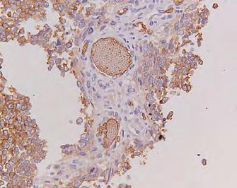
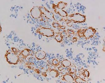
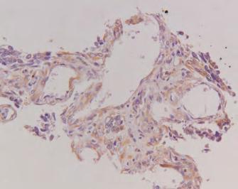
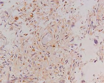
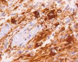
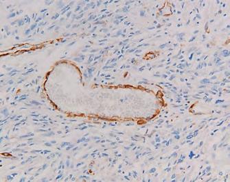
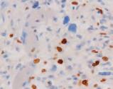
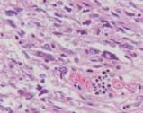
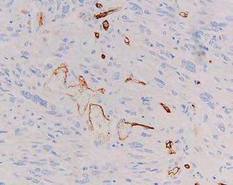
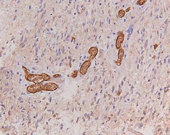
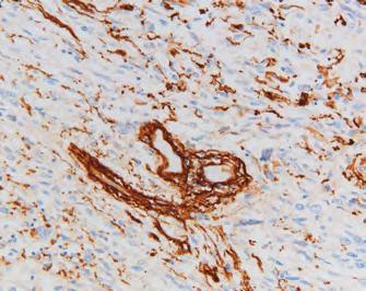
99 89 Zr-bevacizumab-uptake and tumor morphology in end-stage DIPG Cranial pons Caudal pons Cranial CMM Caudal CMM A H&E H&E H&E H&E B MIB-1 MIB-1 MIB-1 MIB-1 C 6 GFAP GFAP GFAP GFAP D CD34 Glut-1 CD34 Glut-1 CD34 Glut-1 CD34 Glut-1 E SMA Coll-IV SMA Coll-IV SMA Coll-IV SMA Coll-IV F VEGF VEGF VEGF VEGF FIGURE 3 Histology and immunohistochemistry of samples from cranial and caudal parts of primary pontine tumor and CMM, respectively: hematoxylin and eosin (A); MIB-1 (for detection of proliferating cells) (B); astrocyte marker glial fibrillary acidic protein (C); CD34 (endothelial cell marker) and Glut-1 (glucose transporter expressed by brain endothelial cells as part of blood brain barrier, and often deregulated in areas with hypoxia) (D); smooth muscle actin (marker of vascular smooth muscle cells and activated pericytes) and collagen-iv (component of vascular basal membrane) (E); and VEGF staining (F). All images are 200x original magnification. Diffuse growth pattern is seen in all samples. Extensive necrosis and glomerular microvascular proliferation is seen with multiple MIB-1 positive (endothelial) cells in microvascular walls in caudal CMM. Partial loss of CD-34 is seen in caudal pons. Similar staining against glial fibrillary acidic protein, glucose transporter 1, and VEGF is seen in all samples. All vessels and microvessels are highlighted in stain against collagen-iv and smooth muscle actin. 97
100 Chapter 6 In the remaining samples, microvascular proliferation was less pronounced: here, 30% (in both primary tumor samples) to 50% (in the caudal CMM sample) of tumor cell nuclei were MIB-1 positive, whereas endothelial cells were MIB-1 negative (Fig. 3B). Glial fibrillary acidic protein was expressed in tumor cells in the primary tumor and CMM, as well as in residual nonneoplastic astrocytes (Fig. 3C). Staining of CD34 showed partial loss of microvascular immunoreactivity in the caudal part of the primary tumor. In the other samples, CD34 immunopositivity was maintained (Fig. 3D). Immunoreactivity for glucose transporter 1 was similar in all samples (Fig. 3E). Staining of collagen-iv and smooth muscle actin highlighted the vascular basal lamina and tunica media, respectively, in all samples without obvious differences (Fig. 3E). Staining of VEGF was similar among all samples (Fig. 3F). DISCUSSION This study directly correlated multiregional 89 Zr-bevacizumab uptake with tumor histology and vascular morphology in a patient with histone H3 wild-type DIPG [12]. Observed differences between in vivo and ex vivo radioactivity measurements reflected tracer pharmacokinetics (e.g., ongoing tracer clearance from blood over time) in combination with spatially or temporally heterogeneously increasing 89 Zr-bevacizumab tissue deposition. Moreover, in vivo PET underestimates the true activity in small lesions (in this case, the CMM and VTM) because of partial-volume effects. Because of this effect [14], the actual radioactivity concentration as assessed by imaging at 145 h after 89 Zr-bevacizumab injection was likely underestimated by approximately 70%. Finally, as opposed to ex vivo measurements, at 145 h intravascular 89 Zr-bevacizumab still contributes to the in vivo PET tumor signal (blood pool SUV, ~4) [8]. Thus, whereas in vivo PET serves to identify whole-body tracer distribution and heterogeneity between lesions, the subcentimeter intralesional heterogeneity of tracer distribution can be defined only with ex vivo measurements. Interestingly, hematoxylin and eosin and MIB-1 staining showed prominent microvascular proliferation, with higher 89 Zr-bevacizumab uptake, in the cranial part of the CMM versus the caudal part, with lower uptake. No other histologic differences were found between the two samples, such as pattern of tumor growth, extent of necrosis, or difference in immunoreactivity for endothelial markers. In the two samples from the pons, intralesional uptake heterogeneity was also observed, although here both areas showed similar vascular morphology, tumor growth, and extent of necrosis. These findings suggest that vascular proliferation is an important, yet not the only, determinant of intralesional heterogeneity in 89 Zr-bevacizumab uptake. 98
101 89 Zr-bevacizumab-uptake and tumor morphology in end-stage DIPG The presence of microvascular proliferation and disorganized vessels [18] in World Health Organization grade IV glioma is considered a consequence of hypoxia-induced overexpression of proangiogenic factors, including VEGF [1,2]. In the cranial CMM sample, glomeruloid microvascular structures with increased endothelial cell turnover (as determined by MIB-1-immunoreactivity) were prominent. On this basis, VEGF was expected to be overexpressed. VEGF immunoreactivity, how- ever, showed mostly aspecific staining and did not differ between samples. This may be explained by the fact that the very short half-life of the VEGF protein (15 20 min [19]) was incompatible with its immunohistochemical detection 2 h after death. Glucose transporter 1 staining was similar in all samples analyzed, suggesting a partially intact blood brain barrier. However, our histologic analysis did not provide definitive clues to determine the role of blood brain barrier integrity in 89 Zr-bevacizumab uptake in this case. 6 CONCLUSION Although concerning a single case, we conclude that in vivo PET is capable of detecting heterogeneity in 89 Zr-bevacizumab uptake between lesions, which correlates well with ex vivo measurements. However, PET cannot detect subcentimeter intralesional uptake heterogeneity. Furthermore, our results suggest that targeting of 89 Zr-bevacizumab is enhanced in areas with vascular proliferation. However, because significant inter- and intralesional heterogeneity was also observed in areas that did not show differences in vascular proliferation, other factors present in the DIPG microenvironment likely also play a role. Because the DIPG microenvironment is heterogeneous and dynamic [3], patients are likely to fail treatment if timing and patient selection are not optimized. The results of this study underline the potential of immuno-pet studies, especially when combined with biopsy or autopsy studies, in the quest for optimal selection and timing of treatment schedules. 99
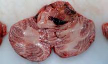
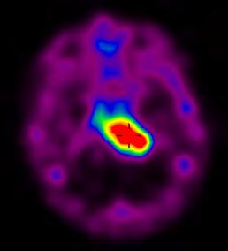
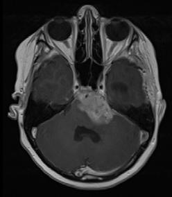
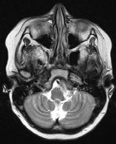
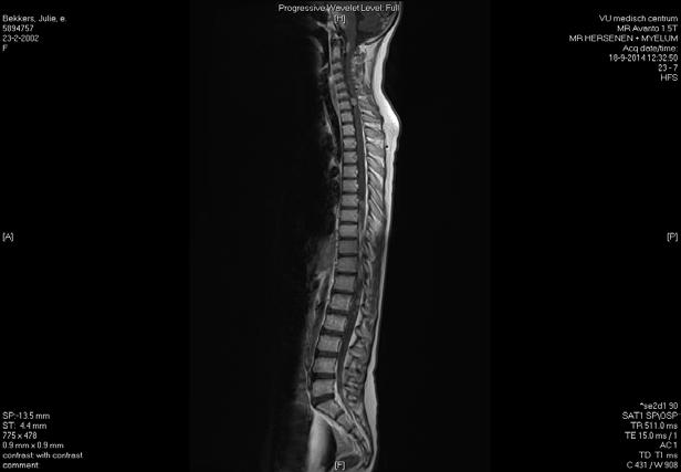
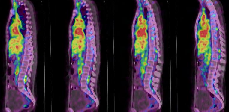
102 Chapter 6 SUPPLEMENTARY DATA A Primary pontine tumor MRI + / PET + B Cervicomedullary junction MRI + / PET + C Spinal cord - MRI + - PET + 100


")


103 89 Zr-bevacizumab-uptake and tumor morphology in end-stage DIPG D 6 Posterior horn of left lateral ventricle MRI + / PET - E Right frontal lobe - MRI + - PET +/- F Left Sylvian fissure - MRI + - PET - SUPPLEMENTARY FIGURE 1 Gadolinium-enhanced T1-MRI, PET and macroscopy (if available) images of (A) the primary pontine tumor and metastases at the (B) cervicomedullary junction, (C) spinal cord, (D) posterior horn of the left lateral ventricle, (E) right frontal lobe and (F) left Sylvian fissure. White arrows represent areas of 89 Zr-bevacizumab uptake and/or disease. 101

104 Chapter 6 SUPPLEMENTARY FIGURE 2 Macroscopic picture of the dural metastases (white arrows), which were detected during autopsy but were undetected by MRI and PET. The dotted arrow indicates the primary pontine tumor. 102
105 89 Zr-bevacizumab-uptake and tumor morphology in end-stage DIPG REFERENCES [1] Buczkowicz P, Bartels U, Bouffet E, et al. Histopathological spectrum of paediatric diffuse intrinsic pontine glioma: diagnostic and therapeutic implications. Acta Neuropathol. 2014;128: [2] Puget S, Philippe C, Bax DA, et al. Mesenchymal transition and PDGFRA amplification/mutation are key distinct oncogenic events in pediatric diffuse intrinsic pontine gliomas. PLoS One. 2012;7:e [3] Ballester LY, Wang Z, Shandilya S, et al. Morphologic characteristics and immunohistochemical profile of diffuse intrinsic pontine gliomas. Am J Surg Pathol. 2013;37: [4] Gururangan S, Chi SN, Young Poussaint T, et al. Lack of efficacy of bevacizumab plus irinotecan in children with recurrent malignant glioma and diffuse brainstem glioma: a Pediatric Brain Tumor Consortium study. J Clin Oncol. 2010;28: [5] Jansen MHA, Lagerweij T, Sewing ACP, et al. Bevacizumab targeting diffuse intrinsic pontine glioma: results of 89Zr-bevacizumab PET imaging in brain tumor models. Mol Cancer Ther. 2016;15: [6] Parekh C, Jubran R, Erdreich-Epstein A, et al. Treatment of children with recurrent high grade gliomas with a bevacizumab containing regimen. J Neuro-oncol. 2011;103: [7] Zaky W, Wellner M, Brown RJ, et al. Treatment of children with diffuse intrinsic pontine gliomas with chemoradiotherapy followed by a combination of temozolomide, irinotecan, and bevacizumab. Pediatr Hematol Oncol. 2013;30: [8] Jansen MH, Veldhuijzen van Zanten SE, Van Vuurden DG, et al. Molecular drug imaging: 89Zr-bevacizumab PET in children with diffuse intrinsic pontine gli- oma. J Nucl Med. 2017;58: [9] Barkovich AJ, Krischer J, Kun LE, et al. Brain stem gliomas: a classification system based on magnetic resonance imaging. Pediatr Neurosurg ;16: [10] Wu G, Broniscer A, McEachron TA, et al. Somatic histone H3 alterations in pediatric diffuse intrinsic pontine gliomas and non-brainstem glioblastomas. Nat Genet. 2012;44: [11] Louis DN, Perry A, Reifenberger G, et al. The 2016 World Health Organization classification of tumors of the central nervous system: a summary. Acta Neuropathol. 2016;131: [12] Caretti V, Jansen MHA, van Vuurden DG, et al. Implementation of a multi- institutional diffuse intrinsic pontine glioma autopsy protocol and characterization of a primary cell culture. Neuropathol Appl Neurobiol. 2013;39: [13] Bugiani M, Veldhuijzen van Zanten SEM, Caretti V, et al. Deceptive morphologic and epigenetic heterogeneity in diffuse intrinsic pontine glioma. Oncotarget. 2017;8: [14] Makris NE, Boellaard R, Visser EP, et al. Multicenter harmonization of 89Zr PET/CT performance. J Nucl Med. 2014;55: [15] Greuter HNJM, Boellaard R, van Lingen A, et al. Measurement of 18F-FDG concentrations in blood samples: comparison of direct calibration and standard solution methods. J Nucl Med Technol. 2003;31: [16] Kevelam SH, Bugiani M, Salomons GS, et al. Exome sequencing reveals mutated SLC19A3 in patients with an early-infantile, lethal encephalopathy. Brain. 2013;136: [17] Navis AC, Hamans BC, Claes A, et al. Effects of targeting the VEGF and PDGF pathways in diffuse orthotopic glioma models. J Pathol. 2011;223: [18] Wagemakers M. Angiogenesis and Angiopoietins in Human Gliomas [thesis]. Groningen, The Netherlands: University of Groningen; [19] Levy NS, Chung S, Furneaux H, et al. Hypoxic stabilization of vascular endothelial growth factor mrna by the RNA-binding protein HuR. J Biol Chem. 1998;273:
106
107 PART II Expanding the scope: Historical and International research initiatives
108
109 CHAPTER 7 A twenty-year review of diagnosing and treating children with diffuse intrinsic pontine glioma in the Netherlands Veldhuijzen van Zanten SEM*, Jansen MHA*, Sanchez Aliaga E, van Vuurden DG, Vandertop WP, Kaspers GJL * shared authorship Expert Rev Anticancer Ther. 2015;15(2):157 64
110 Chapter 7 ABSTRACT INTRODUCTION Children with diffuse intrinsic pontine glioma (DIPG) face a dismal prognosis, with a median overall survival of 9 months. Our aims are to determine the incidence of DIPG in the Netherlands and to identify points for improvement in clinical research, a prerequisite for increasing the chance to find a cure. METHODS We performed a population-based retrospective cohort study by evaluating all children diagnosed with DIPG in the Netherlands between 1990 and RESULTS The incidence of DIPG in the Netherlands corresponds with international literature. Between 1990 and 2010, a large heterogeneity of treatment schedules was applied and only a minority of patients was included in clinical trials. DISCUSSION Given the rarity of DIPG, we emphasize the need for (inter-)national trials to facilitate the identification of potentially effective therapeutics in the future. This can be supported by the recent development of a European DIPG registry enabling international study collaborations. 108
111 A twenty-year review of DIPG in the Netherlands INTRODUCTION Diffuse intrinsic pontine glioma (DIPG) is a rare and childhood-specific malignancy of the brainstem. Children with DIPG face a dismal prognosis, with a median overall survival (MOS) of 9 months [1]. Over the years, DIPG has been distinguished from other brainstem entities by numerous classification systems, using criteria based on location, growth pattern, size, and histopathological grade [2 4]. It was, however, not until the 1990s that DIPG was classified separately from more focally growing tumors, and showed to have its specific, dismal prognosis [5]. Most epidemiological data about DIPG come from America and Canada. It is estimated that gliomas arising in the brainstem account for 10 20% of pediatric CNS tumors and that 80% of these can be classified as a typical DIPG [6]. There is, however, a lack of epidemiological data from European countries. Although better and more uniform classification criteria were developed, none of the applied treatment options so far has yielded a substantial survival benefit in patients suffering from a typical DIPG [7]. Radiotherapy remains the only effective, albeit palliative, treatment option, with a few months survival increase compared to best supportive care and a transient improvement of clinical performance and quality of life [8,9]. 7 We evaluated the 20-year history of diagnosing and treating patients with DIPG in the Netherlands, to gain more insight in the epidemiology and current treatment approach. The main purpose of this study is to determine the incidence of DIPG, in a country which is representative of Western Europe. In addition, we will evaluate the applied treatments, including the application of clinical trials. With this, we aim to find possible points for improvement in clinical research into this rare disease. METHODS This study was approved by the institutional review board of VU University Medical Center (VUmc, Amsterdam, The Netherlands) and the scientific committee of the Dutch Childhood Oncology Group (DCOG). Identification of study cohort A search was performed in the Pathological Anatomy National Automated Archive, the databases of the centers of the DCOG and pediatric radiotherapy centers, a total of eight academic university medical centers and one regional hospital. To ensure the inclusion of typical DIPG patients, we solely evaluated patients of whom MR images from time of 109
112 Chapter 7 diagnosis could be obtained for central review. In this central review, DIPG was defined as a T1-weighted hypointense and T2-weighted hyperintense tumor with at least 50% involvement of the pons on T2 [5]. Histological determination was not required for this study. If, however, a biopsy was performed and pathology data could be retrieved, than the data were analyzed. We did not exclude patients on the basis of their duration of symptoms before diagnosis, since this criterion was not well documented and not systematically used to define DIPG during our study period. However, since nowadays a longer symptom duration is assumed to be atypical for DIPG patients [1], we did perform additional analyses on patients with a symptom duration of more than 6 months. Data collection Demographics and clinical data were obtained from the patient records. The presence and duration of cranial nerve deficits, long tract symptoms and ataxia were scored when available. MR images were centrally reviewed. Information from pathology reports was retrieved (i.e., biopsy or autopsy). Data on treatment strategies (surgery, chemo- and radiotherapy) and participation in clinical trials, both at time of diagnosis and at time of disease progression, were collected. Unfortunately, we could not in all cases retrieve the exact nature of (clinical and/or radiological) responses to (first or subsequent) treatments and could therefore not describe the exact pattern of progression (i.e., local and/or metastatic). Progressive disease (yes or no) was defined as clinical signs of disease progression (i.e., increase of symptoms or new symptoms) and/or radiological tumor progression, obtained from the patient records and radiology reports. Statistics Statistical analyses were performed using IBM SPSS Statistics for Windows, Version 20.0 (IBM Corp., Armonk, NY, USA: released 2011). Patient data regarding demographics, diagnosis and treatment schedules were analyzed by descriptive statistical methods. The incidence of DIPG was calculated for the period between 2004 and 2009, these being the most reliable years in terms of complete registration. Survival analysis was performed by Kaplan-Meier estimates. RESULTS Patient population The search resulted in a list of 207 patients diagnosed with a brainstem tumor between 1990 and 2010 in the Netherlands, illustrated in Figure 1. We excluded patients with non-glioma lesions (n = 11) and patients diagnosed with neurofibromatosis (n = 7). 110
113 A twenty-year review of DIPG in the Netherlands Based on the current classification of a typical DIPG, we also excluded patients with pilocytic astrocytoma (WHO grade I; n = 3), or with dorsally exophytic tumors and tumors occurring elsewhere in the brainstem, that is, mesencephalic or medullary tumors (n = 29). Out of 207 patients diagnosed with a brainstem tumor, 157 patients were therewith identified as having a PG. In 117 cases (75%), MR images were available for central review, of which 88% of patients were classified as having a typical DIPG (n = 103). Further analyses concerning the applied treatment approach were performed in this cohort of 103 MRI-confirmed typical DIPG patients. Excluded: * Neurofibromatosis * Other location than pons or dorsally exophytic * Other histopathology Brainstem tumours (Netherlands ) n = 50 n = 207 Pontine glioma (PG) n = MR-image available for review n = 117 Local radiology report: PG n = 40 Typical DIPG Focal PG n = 103 n = 14 No biopsy n = 80 No biopsy n = 10 No biopsy n = 30 WHO grade II n = 8 WHO grade II n = 1 WHO grade II n = 4 WHO grade III n = 7 WHO grade III n = 2 WHO grade III n = 4 WHO grade IV n = 8 WHO grade IV n = 1 WHO grade IV n = 2 FIGURE 1 Overview of patients diagnosed with diffuse intrinsic pontine glioma in the Netherlands ( ). The dotted square indicates the evaluated population in this retrospective study. Incidence We could not retrieve all MRIs from patients diagnosed before 2004, whereas for the time period from 2004 to 2009, MRI scans from all patients could be collected and centrally reviewed. Therefore, this time period was used to ascertain the incidence of 111
114 Chapter 7 DIPG. In 6 years time, 55 patients were diagnosed as having a typical DIPG and 9 as having a focal PG. The incidence of DIPG in the Netherlands was therefore estimated to be nine patients per year (0.54 per 1,000,000; 2.32 per 1,000,000 aged 0 20 years). However, annual variations were observed (range 5 13 per year). No seasonal variations were observed. Distribution of patients in the Netherlands The 103 DIPG patients were diagnosed and/or treated in nine hospitals distributed throughout the Netherlands. These hospitals are all the possible sites where DIPG patients can be referred to within our boarders. The number of patients per center over the course of 20 years ranged from 1 to 26 (median 10). Demographics The mean age of patients was 7.2 years (standard deviation 3.4). There was a balanced gender distribution (boys n = 49, girls n = 54). History & physical examination From 96% of patients (99/103), information on the duration of symptoms before diagnosis could be retrieved from the patient records. The median duration of symptoms before diagnosis was 1.0 month (range 0 16 months). The exclusion of six patients with a symptom onset duration of more than 6 months did not influence the median duration of 1 month (range 0 5 months). Neurological examination at the time of initial diagnosis showed cranial nerve deficits to be present in 98 patients (95%), long-tract symptoms in 78 (76%) and ataxia in 78 patients (76%). Diagnosis In each hospital, the original diagnosis was made based on MR imaging. In 23 patients (22%), MRI of the brain was supplemented with an MRI of the spine. None of the local radiology reports at time of diagnosis described neuraxis metastases. In 23 children (22%), imaging was supplemented with pathology (Fig. 1), showing WHO grade II, III and IV astrocytomas. Additional analyses of the six patients with a symptom duration of more than 6 months, showed typical DIPG images on MR in all patients. In one of these six cases a biopsy was performed, showing astrocytoma grade II. In further analyses concerning the applied treatment approach, these six patients were therefore not separated from the other patients. 112
115 A twenty-year review of DIPG in the Netherlands Therapy at initial diagnosis At the time of initial diagnosis, 89 out of 103 DIPG patients (86%) received anti-tumor treatment (Fig. 2). When only supportive care was applied (n = 14; 14%), this was either due to very young age of the child (n = 2), poor neurological state (n = 4), parental refusal (n = 3) or parental preference for alternative medicine or supportive care only (dexamethasone) (n = 5). Reports on dexamethasone schedules were incomplete and could therefore not be analyzed. MRI reviewed patients Therapy at diagnosis (n = 89 out of 103) Therapy at 1 st progression (n = 42 out of 92) Therapy at 2 nd progression (n = 2 out of 46) No therapy n = 14 No further No therapy therapy n = 50 Radiotherapy n = 1 Chemotherapy n = 2 No further therapy n = 44 Radiotherapy n = 1 7 Radiotherapy n = 1 ^ DIPG n = 103 Radiotherapy n = 68 * Chemotherapy n = 30 Surgery n = 2 Hyperthermia n = 1 Chemotherapy n = 5 Chemotherapy n = 1 Hyperthermia n = 1 Chemoradiotherapy n = 14 # Chemotherapy n = 2 Surgery n = 1 Surgery n = 2 Chemotherapy n = 1 FIGURE 2 Treatment schedules applied to diffuse intrinsic pontine glioma patients in the Netherlands ( ). *Includes one patient who received radiotherapy together with homeopathic Ruta-6 and calcium phosphate. # Includes one patient who stopped radiotherapy after the second day of treatment because of neurologic deterioration. The patient continued with temozolomide instead. ^Includes one patient who received spinal irradiation after initial radiotherapy treatment because of leptomeningeal metastasis. Of the 89 treated patients at time of diagnosis, a total of 82 patients (92%) received radiotherapy, of which the majority (n = 68; 83%) received radiotherapy only. One of these patients received concomitant homeopathic Ruta-6 and calcium phosphate. A total of 19 treated patients (21%) received some form of chemotherapy, either combined with radiotherapy (n = 14) or as single therapy (n = 5). The most widely prescribed 113
116 Chapter 7 chemotherapeutic agents at the time of diagnosis were temozolomide and vincristine/ carboplatin (Table 1). Four patients (7%) underwent a partial resection of their tumor together with initial therapy (not shown), whereas two patients (2%) underwent surgery only, which consisted of a partial resection combined with the placement of an extra ventricular drain (Fig. 2). Therapy at disease progression In 11 cases, data on progressive disease and therapy were not available for evaluation. In all 92 evaluable DIPG patients, progressive disease was reported after an initial response to therapy. Forty-two of these evaluable patients (46%) received second-line treatment (Fig. 2). The majority, 36 patients (39%) were treated with chemotherapy, which mainly consisted of temozolomide (Table 1). Three of these patients received a second chemotherapy regimen, which in all cases differed (i.e., in drugs or doses) from the regimen at time of diagnosis. Irradiation at time of disease progression was performed in two patients (2%), but these patients were not radiated at time of diagnosis. One patient receiving second-line therapy underwent a partial resection of the tumor prior to chemotherapy (not shown) and three patients (3%) underwent a partial resection of tumor tissue as single therapy at time of disease progression (Fig. 2). In all cases, this was the first surgical intervention. In one patient, an alternative treatment strategy, hyperthermia, was applied at the time of progressive disease. In 46 patients (50%), a second episode of disease progression was reported (Fig. 2) after a period of stable disease. At this point, the majority of these patients (n = 44; 96%) received no further therapy and palliative care was initiated. Two patients (4%) with secondary progressive disease received third-line therapy: one patient, who did not receive radiotherapy before, was irradiated and the other patient received alternative therapy (hyperthermia) (Fig. 2). Uniformity of applied treatments Radiotherapy was applied to a total of 85 out of 103 patients (83%) at some point during their disease course. Table 2 shows the radiotherapy schedules that were applied at the time of diagnosis and at the time of progression. Radiotherapy was applied in each of the treatment centers. Table 1 shows the various chemotherapeutic regimens that were applied at initial diagnosis and at time of progressive disease. Chemotherapy at the time of diagnosis was applied in six hospitals (with n = 9, n = 5 and n = 2 in three hospitals, and n = 1 in the remaining hospitals). Chemotherapy at the time of progressive disease was applied 114
117 A twenty-year review of DIPG in the Netherlands TABLE 1 Chemotherapeutic agents and schedules. Chemotherapeutic agents At diagnosis (n) At progressive disease (n) Temozolomide 5 30 (inter alia TMZ study NTR227) [12] Temozolomide & thalidomide & erlotinib - 1 Temozolomide & imatinib & dichloroacetate - 1 Vincristine & procarbazine& methotrexate & dexamethasone 1 - followed by vincristine & lomustine Vincristine & lomustine 1 - Vincristine & cyclophosphamide 1 1 followed by carboplatin & procarbazine and etoposide & cisplatin Vincristine & carboplatin 3 - followed by temozolomide Vincristine & carboplatin 1 1 Vincristine & carboplatin 1 - followed by temozolomide & lomustine Gemcitabine followed by 2 - high dose chemo with stem cell reinfusion and irinotecan & bevacizumab & erlotinib (& everolimus) (VUmc DIPG 01 study NTR2391) [13] Etoposide (& carboplatin) 2 2 Cisplatin Patients receiving chemotherapy treatment n: Number of patients TABLE 2 Radiotherapy schedules. Schedule Total Gy n 13 x x x Unknown x x x x x x x Hyperfractionated Other Patients receiving RT treatment 85 Gy: Gray; n: Number of patients; RT: Radiotherapy 115
118 Chapter 7 in eight hospitals (with n = 5 in two hospitals, n = 2 in two, n = 1 in two and n = 3 and n = 17 in the remaining hospitals). As with radiotherapy, no general guidelines were available or used. The majority of patients was treated off-trial with temozolomide, mainly at progression of disease. Reports on dosage and schedules were incomplete and could therefore not be further analyzed. Clinical trials Over the course of our study period, 19 patients (18%) of all children diagnosed with DIPG in the Netherlands were enrolled into a prospective clinical trial. Only trials that were approved by an institutional review board, the scientific committee of the DCOG, and trials that were included in a trial registry, were evaluated. The Childhood Oncology Group ACNS-0126 study, a multicenter, Phase II prospective cohort study was initiated in 2002, exploring the toxicity and efficacy of temozolomide as adjuvant therapy [10,11] in pediatric high-grade glioma and DIPG. One center in the Netherlands participated in this study. From January 2004, patients with recurrent or progressive PNET or highgrade glioma (including DIPG) could be included in a similar Phase II study exploring the use of temozolomide [12]. Both studies have been closed for accrual. In 2010, a single-center study was opened, which offers a broad and at the same time targeted approach of DIPG. Therapy consists of standard radiotherapy (54 Gy) combined with gemcitabine as radiosensitizer at diagnosis. At disease progression, therapy consists of a backbone of irinotecan and bevacizumab combined with escalating doses of erlotinib and everolimus. This is the first study in the Netherlands that offers the option of undergoing a biopsy. A biopsy, however, is not mandatory for enrolment [13]. This study is still accruing patients. Survival In the total cohort of MRI-confirmed typical DIPG patients, the progression-free survival (PFS) was 6 months and the MOS was 9.5 months (Fig. 3 & Table 3). Children <3 years of age showed a relatively short PFS of 4.0 months and a relatively long MOS of 11.0 months (log-rank p = and 0.015, respectively). For the children aged 9 18 years, no longer PFS or MOS was observed (log-rank p = and 0.868, respectively). Table 3 shows the result of additional analysis of the separate treatment groups at time of diagnosis. We could not perform specific analyses per treatment schedule, drug or dosage due to low patient numbers. In this large, unselected, nationwide population-based cohort, six patients (6%) with a typical DIPG on MRI were classified as long-term survivors. Long-term survival was 116
119 A twenty-year review of DIPG in the Netherlands defined as having a survival from initial diagnosis beyond 24 months (Fig. 3). The PFS of these patients was 42 months and the MOS 46 months, with three patients being alive at 27, 79 and 161 months, respectively (Table 3; log-rank p = and 0.000, respectively) Survival (%) Follow up (months) FIGURE 3 Survival of diffuse intrinsic pontine glioma patients in the Netherlands ( ). The dashed line indicates a median overall survival of 9.5 months. The dotted line indicates a 2-year survival of 6%. TABLE 3 Survival analysis of treatment groups at time of diagnosis. This group is too small to analyse (n = 2). Group PFS 95% CI MOS 95% CI Overall (n = 103) Age <3 years (n = 8) Age 9-18 years (n = 24) LTS (n = 6) Therapy group (at diagnosis) PFS 95% CI MOS 95% CI No therapy Radiotherapy Chemotherapy Chemo-radiotherapy Surgery LTS: Long-term survival (>24 months); MOS: Median overall survival; PFS: Progression-free survival 117
120 Chapter 7 DISCUSSION We determined the population-based, and MRI-confirmed, incidence of patients diagnosed with DIPG in the Netherlands, and showed how these patients were treated over the past 20 years. Each year, on average, nine patients with typical DIPG are diagnosed, equally distributed geographically throughout the Netherlands, without an indication for seasonal variations. The incidence of 2.32 per 1,000,000 individuals aged 0 20 years corresponds to international data from the USA [14]. It is noteworthy that the limited number of patients was diagnosed and/or treated in nine different hospitals, resulting in very low patient numbers per hospital. More importantly, only a minority of patients (18%) was enrolled into a prospective clinical trial, although during our study period three clinical trials were available for patient inclusion. Only the first study, which was an international collaboration initiated by the Children s Oncology Group comprising 230 study locations, was successfully completed [11]. The second study was a Dutch multicenter study. This study, however, was discontinued and the results are yet to be published. The third study is a single-center study, which is still open of accrual, but with an inclusion rate of only one patient (11%) per year on average. This retrospective study shows that DIPG patients are mainly treated according to single-center guidelines and even individualized therapy. There are still no strict national guidelines for both radio- and chemotherapy, resulting in a heterogeneous application of treatment schedules. More importantly, DIPG patients are often not included in clinical trials. This could lead to selection bias when a limited number, or a selected group of patients, participate in clinical trials. Further research is needed to investigate why patients are mainly treated off-study. In view of the rarity of the disease, this heterogeneity might be caused by the limited number of patients treated per center, with a median of 10 patients over the course of 20 years. A rare disease such as DIPG might benefit from centralization in a limited number of specialized hospitals, in this case pediatric cancer centers [15], together with (inter)national consensus on the approach of these patients and optional inclusion in collaborative clinical trials [1,7]. In addition to co-operation in existing clinical trials, we emphasize the need for collaboration in other fields of DIPG research, like biological studies and molecular drug imaging using PET. Between 1990 and 2010, 78% of patients did not undergo a biopsy in addition to radiological diagnoses, and 98% of patients did not get the chance to participate in an autopsy study. From the introduction of offering biopsies in the VUmc DIPG 01 study and the opening of the autopsy study [16], this is increasingly performed. However, in view of the low incidence of DIPG in the Netherlands, international collaboration is urgently needed. This is underlined by the fact that DIPG biopsy and 118
121 A twenty-year review of DIPG in the Netherlands autopsy studies have advanced the understanding of the disease, with the discovery of a K27M mutation in H3.3 or H3.1, perturbations in genes of the receptor tyrosine kinase/ Ras/PI3K signaling pathway and the overexpression of VEGF, which provides useful information for drug development, design of novel therapies and possibly treatment stratification [17 19]. Future international studies comparing biopsy and autopsy material will also reveal more about the biological changes, either due to the natural course of the disease, or influenced by therapy. In addition, emerging PET studies will reveal more about the in vivo behavior of drugs, such as blood-brain barrier passage and tumor pharmacokinetics [20]. Collaborative international biological and diagnostic studies will thus contribute to the discovery of novel therapies for future clinical trials. Our complete cohort of 103 MRI-confirmed DIPG patients showed a PFS of 6 months and a MOS of 9.5 months, with a significant longer survival time in children less than 3 years of age. The relatively short PFS in this age group is possibly due to the fact that these children did not receive radiotherapy. These data, as well as the data from the subgroups of patients receiving radiotherapy alone, or radiotherapy combined with chemotherapy, are in accordance with previously published data [1,7,21,22]. The relatively long MOS of the patients receiving chemotherapy only is probably due to confounding bias based on the relative young age of patients in this group. Other studies have reported longer survival times for children aged 9 18 years, however, this could not be confirmed in our cohort of patients [23]. Our cohort harbored a total of six, relatively young, long-term survivors, which is in agreement with international literature [24]. Further research is needed to better understand this intriguing group of DIPG patients, which might have a distinct survival based on specific biological features. 7 With this nationwide, population-based retrospective cohort study, we confirm the assumed low incidence of DIPG and show that a poor accrual of DIPG patients in clinical trials results in a lack of comprehensive data on demographics, history, physical examination, diagnosis (both imaging and biology), outcome, but most importantly effectiveness per treatment schedule, drug or dosage. Given the rarity of DIPG, we emphasize the need for (inter-)national data and trials to facilitate the identification of potentially effective therapeutics in the future. This can be supported by the recently developed European DIPG registry [25] by the DIPG network of the International Society of Pediatric Oncology Europe. The International Society of Pediatric Oncology Europe DIPG registry was developed in parallel with that in the USA [26]. The first large-scale international study that will be performed in the setting of the European DIPG registry will be an evaluation of all European long-term survivors. 119
122 Chapter 7 EXPERT COMMENTARY In view of the very poor prognosis for children with DIPG, patients should be able to participate in clinical trials. Given the rarity of DIPG, we emphasize the need for (inter-) national collaboration and trials to facilitate the identification of potentially effective therapeutics in the future. This can be supported by the recent development of a European DIPG registry. FIVE-YEAR VIEW Given the rarity of DIPG and lack of comprehensive data from large unselected cohorts of DIPG patients, we envision that the European DIPG registry will contribute to future clinical research. A comprehensive database will for instance facilitate studies on subgroups of DIPG, the analysis of long-term survival and the evaluation of decisionmaking. The database will also function as a control cohort for (inter)national clinical trials without randomization, which is most useful in a rare disease such as DIPG. KEY ISSUES Patients with diffuse intrinsic pontine glioma (DIPG) face a dismal prognosis, with a median overall survival of 9 months. In recent decades, the survival has not improved despite several treatment strategies that have been explored. The incidence of DIPG in the Netherlands is 9 (5 13) patients per year (0.54 per 1,000,000; 2.32 per 1,000,000 aged 0 20 years). Between 1990 and 2010, for both radio- and chemotherapy, more than 10 different schedules or agents were used. In this period, only 18% of patients were formally included in clinical trials. Poor accrual of DIPG patients in clinical trials results in a lack of comprehensive data on demographics, history, physical examination, diagnosis (both imaging and biology), outcome, but most importantly effectiveness per treatment schedule, drug or dosage. Given the rarity of DIPG, we emphasize the need for (inter-)national trials to facilitate the identification of potentially effective therapeutics in the future. Recently, a European DIPG registry [25] has been developed by the DIPG network of the International Society of Pediatric Oncology Europe. This registry will enable the evaluation and follow-up of clinical and centrally reviewed radiology data of all European patients with DIPG, both in- and outside clinical trials, which will give a realistic picture of the actual spectrum of DIPG patients. Subsequently, a comprehensive European DIPG registry will pave the way for further international collaborations and, hopefully, European clinical DIPG trials. 120
123 A twenty-year review of DIPG in the Netherlands REFERENCES Papers of special note have been highlighted as: * of interest, or ** of considerable interest [1]* Hargrave D, Bartels U, Bouffet E. Diffuse brainstem glioma in children: critical review of clinical trials. Lancet Oncol. 2006;7: Hargrave et al. evaluated the consistency between diffuse intrinsic pontine glioma (DIPG) trial protocols and showed that most studies varied substantially, which could explain the differences in outcome. In addition, they showed that no treatment has benefit over conventional radiotherapy. [2] Berger MS, Edwards MS, LaMasters D, et al. Pediatric brain stem tumors: radiographic, pathological, and clinical correlations. Neurosurgery. 1983;12: [3] Epstein F. A staging system for brain stem gliomas. Cancer 1985;56: [4] Stroink AR, Hoffman HJ, Hendrick EB, et al. Diagnosis and management of pediatric brain-stem gliomas. JNS. 1986;65: [5]* Barkovich AJ, Krischer J, Kun LE, et al. Brain stem gliomas: a classification system based on magnetic resonance imaging. Pediatr Neurosurg. 1990;16: Barkovich et al. developed a classification system for DIPG, based on MR imaging, which is nowadays mostly used. [6]* Bartels U, Hawkins C, Vezina G, et al. Proceedings of the diffuse intrinsic pontine glioma (DIPG) Toronto Think Tank: advancing basic and translational research and cooperation. J Neurooncol. 2011;105: Bartels et al. appoint that the exact incidence of DIPG is still undefined, but suggest that each year children are diagnosed in the USA. They inform the public about recent breakthroughs in the understanding of the molecular biology of DIPG, which have raised new hope and opened new avenues for therapeutic options. [7]** Jansen MH, van Vuurden DG, Vandertop WP, et al. Diffuse intrinsic pontine gliomas: a systematic update on clinical trials and biology. Cancer Treat Rev. 2012;38: Jansen et al. state that the knowledge on DIPG biology is increasing, mainly due to the (re)introduction of biopsies and autopsies, the possibility of gene expression profiling, and the development of in vivo models. Translation of this knowledge into clinical trials may eventually lead to more effective treatment options. [8] Grimm SA, Chamberlain MC. Brainstem glioma: A Review. Curr Neurol Neurosci Rep. 2013;13:346. [9] Schild SE, Stafford SL, Brown PD, et al. The results of radiotherapy for brainstem tumors. J Neurooncol. 1998;40: [10] Chemotherapy and Radiation Therapy After Surgery in Treating Children With Newly Diagnosed Astrocytoma, Glioblastoma Multiforme, Gliosarcoma, or Diffuse Intrinsic Pontine Glioma. Available from: clinicaltrials.gov/show/ NCT [11] Cohen KJ, Heideman RL, Zhou T, et al. Temozolomide in the treatment of children with newly diagnosed diffuse intrinsic pontinegliomas: a report from the Children s Oncology Group. Neuro Oncol. 2011;13: [12] Temozolomide in children with recurrent or refractory CNS tumors. Available from: trialreg/admin/rctview.asp?tc=227 [13] Combination therapy for children with malignant pontine gliomas. Available from: trialregister.nl/trialreg/admin/rctview.asp?tc=2391 [14]* Tonn JC. Neuro-Oncology of CNS Tumors. Heidelberg: Springer, Ronghe et al. wrote a chapter about DIPG. This chapter reports an expected incidence of DIPG patients each year in the USA. [15]* Knops RR, van Dalen EC, Mulder RL, et al. The volume effect in pediatric oncology: a systematic review. Ann Oncol. 2013;24: Knops et al. performed a systematic review and provided an overview of the existing evidence regarding the volume effect in pediatric oncology. They conclude to support the statement that higher volume hospitals, higher case volume providers and specialized hospitals are related to the better outcome in pediatric oncology. [16] Caretti V, Jansen MH, van Vuurden DG, et al. Implementation of a Multi-Institutional Diffuse Intrinsic Pontine Glioma Autopsy Protocol and Characterization of a Primary Cell Culture. Neuropathol Appl Neurobiol. 2013;39: [17] Wu G, Broniscer A, McEachron TA, et al. Somatic histone H3 alterations in pediatric diffuse intrinsic pontine gliomas and non-brainstem glioblastomas. Nature Genetics. 2012;44: [18] Khuong-Quang DA, Buczkowicz P, Rakopoulos P, et al. K27M mutation in histone H3.3 defines clinically and biologically distinct subgroups of pediatric diffuse intrinsic pontine gliomas. Acta Neuropathol. 2012;124: [19] Puget S, Philippe C, Bax DA, et al. Mesenchymal transition and PDGFRA amplification/mutation are key distinct oncogenic events in pediatric diffuse intrinsic pontine gliomas. PLoS One. 2012;7:e [20] Jansen MH, Veldhuijzen van Zanten SE, Van Vuurden DG, et al. Molecular drug imaging: 89 Zr-bevacizumab PET in children with diffuse intrinsic pontine glioma. JNucl Med. 2017;58: [21] Broniscer A, Laningham F, Sanders R, et al. Young Age May Predict a Better Outcome for Children With Diffuse Pontine Glioma. Cancer. 2008;113: [22] Jansen MH, Veldhuijzen van Zanten SE, Sanchez Aliaga E, et al. Survival prediction model of children with diffuse intrinsic pontine glioma based on clinical and radiological criteria. Neuro Oncol. 2015;17: [23] Bailey S, Howman A, Wheatley K, et al. Diffuse intrinsic pontine glioma treated with prolonged temozolomide and radiotherapy Results of a United Kingdom phase II trial (CNS ). Eur J Cancer. 2013;49: [24] Jackson S, Patay Z, Howarth R, et al. Clinico-radiologic characteristcs of long-term survivors of diffuse intrinsic pontineglioma. J Neurooncol. 2013;114: [25] SIOPE DIPG Registry. Available from: dipgregistry.eu [26] International DIPG Registry. Available from:
124
125 CHAPTER 8 Effective drug delivery in diffuse intrinsic pontine glioma: A theoretical model to identify potential candidates El-Khouly FE, van Vuurden DG, Stroink T, Hulleman E, Kaspers GJL, Hendrikse NH, Veldhuijzen van Zanten SEM Front Oncol Oct 11
126 Chapter 8 ABSTRACT INTRODUCTION Despite decades of clinical trials for diffuse intrinsic pontine glioma (DIPG), patient survival does not exceed 10% at two years post-diagnosis. Lack of benefit from systemic chemotherapy may be attributed to an intact blood-brain-barrier (BBB). We aim to develop a theoretical model including relevant physicochemical properties in order to review whether applied chemotherapeutics are suitable for passive diffusion through an intact BBB or whether local administration via convection-enhanced delivery (CED) may increase their therapeutic potential. METHODS Physicochemical properties (lipophilicity, molecular weight, and charge in physiological environment) of anti-cancer drugs historically and currently administered to DIPG patients, that affect passive diffusion over the BBB, were included in the model. Subsequently, the likelihood of BBB passage of these drugs was ascertained, as well as their potential for intratumoral administration via CED. RESULTS As only non-molecularly charged, lipophilic, and relatively small sized drugs are likely to passively diffuse through the BBB, out of 51 drugs modeled, only 8 (15%) carmustine, lomustine, erlotinib, vismodegib, lenalomide, thalidomide, vorinostat, and mebendazole are theoretically qualified for systemic administration in DIPG. Local administration via CED might create more therapeutic options, excluding only positively charged drugs, and drugs that are either prodrugs and/or only available as oral formulation. DISCUSSION A wide variety of drugs have been administered systemically to DIPG patients. Our model shows that only few are likely to penetrate the BBB via passive diffusion, which may partly explain the lack of efficacy. Drug distribution via CED is less dependent on physicochemical properties, and may increase the therapeutic options for DIPG. 124
127 Drug delivery in DIPG INTRODUCTION Diffuse intrinsic pontine glioma (DIPG) is a rare, aggressive childhood malignancy of the brainstem with a 2-year survival rate of 10% [1,2]. Unlike the spectacular increase in survival of childhood leukemia patients from <10% to over 80% in the last 50 years, the prospect for patients suffering from DIPG has not changed [3 5], likely due to a lack of success from (chemo)therapeutic strategies. Historically, this was blamed on the supposed resistance of DIPG tumor cells to cytotoxic agents. Pre-clinical studies have, however, recently shown that primary cultures derived from DIPG patients, are actually not resistant to a number of traditionally used cytotoxic drugs and novel targeted chemotherapeutics [5,6]. These results from pre-clinical studies contrasting with disappointing outcomes of clinical studies led to a shifting paradigm towards the hypothesis of a possible delivery problem of chemotherapeutics over the blood-brain barrier (BBB). This supposedly prevents drugs from reaching the tumor properly [8]. In many brain tumors, the integrity of the BBB is affected by the formation of disordered and highly permeable tumor neovasculature. Infiltrating tumors, such as DIPG, however, make use of the existing brain vasculature with normal BBB integrity [9]. Noteworthy in this respect, especially early-disease-dipgs show limited contrast enhancement after intravenous administration of gadolinium compared to glioblastoma multiforme tumors that harbor highly-neovascular regions. As gadolinium has an average molecular weight of 545 kda, which largely exceeds the penetration cut-off of the BBB (e.g Da), limited contrast enhancement in these tumors is suggestive of a largely intact BBB [10 12]. Furthermore, to illustrate, studies investigating biopsy-derived intratumoral drug concentrations of systemically delivered drugs in adults with high-grade glioma show low, potentially sub-therapeutic local drug concentrations, especially in non-enhancing tumor regions [13 15]. 8 To overcome the BBB, novel drug delivery techniques, such as convection-enhanced delivery (CED), have been developed. With CED, chemotherapeutic agents are administered directly into the tumor microenvironment via a highly controlled positive pressure gradient and a constant flow induced by a pump. This enables homogeneous distribution of high drug concentrations over an easy-to-define distance, presumably increasing the therapeutic potential and avoiding systemic toxicities [15,16]. In this study, we aim to review all chemotherapeutic drugs (previously) administered systemically to DIPG patients by means of a theoretical model including all physicochemical properties that influence passive diffusion, to indicate their likeliness of passage over an intact BBB in DIPG. Furthermore, we aim to indicate whether local administration of these drugs via convection-enhanced delivery (CED) may increase their therapeutic potential. 125
128 Chapter 8 METHODS - MODEL DESIGN An extensive search of the literature and trial databases was performed to identify all chemotherapeutics historically employed in DIPG patients. Databases of Medline/ PubMed and The Cochrane Library were searched for potentially relevant articles. The search strategy combined controlled and free text words for the target population (e.g. children), the tumor type (e.g. DIPG or pontine glioma) and the application of chemotherapeutic drugs. The reference lists of all included articles were searched for additional studies. In addition, trial registries ( www. clinicaltrialsregister.eu) as well as websites from consortia treating children with brain tumors ( were searched for clinical trials in DIPG. The complete search strategy can be found in the supplementary data (available upon request). Subsequently, drug simulations were performed for both systemic administration, to predict passive diffusion over an intact BBB and for local intratumoral drug delivery via CED, to predict convection-distribution efficacy, using relevant physicochemical properties (molecular weight, lipophilicity and molecular charge). To this aim, physicochemical property data were extracted from various chemical databases: PubChem chemistry database, Drugbank.ca and Clarke's Analysis of Drugs and Poisons. The molecular charge in physiologic environment was simulated by MarvinSketch (Chemaxon), an advanced chemical editor for drawing chemical structures and calculating basic physicochemical properties (e.g. molecular charge, logp), using specific algorithms. Drug simulation for systemic drug administration The BBB, formed by tightly interconnected endothelial cells in the capillary walls of the brain vasculature, protects the brain by limiting the inter- and paracellular transport of foreign substances from the systemic blood flow [18]. Systemically applied chemotherapeutics enter the brain via passive diffusion or active transport mechanisms. The balance between in- and outflow depends both on the physicochemical properties of the drug itself and on its affinity for drug in- and efflux transporters and receptors expressed in the BBB. The Lipinski Rule of 5 is used to determine a drug s permeability. According to this rule good permeability is likely if: (i) the molecular weight is 500 Da, (ii) the lipophilicity, measured by the partition coefficient (log P), is 5 (optimal value of ), (iii) the structure has no more than 5 hydrogen bond donors, and (iv) no more than 10 hydrogen bond donor acceptors [18 20]. Taking these rules into consideration, the physicochemical properties that determine passive diffusion through the BBB, are molecular weight, lipophilicity, and molecular charge. For every drug, molecular weight and Log P were 126
129 Drug delivery in DIPG included in the model and subsequently, the chemical structure was used to simulate the molecular charge of a drug in physiological environment (ph 7.4). Drugs with a molecular (positive or negative) charge of 10% were considered to be able to passively diffuse through the BBB. Drugs with a (positive or negative) molecular charge of 90% are considered to have a higher affinity for the hydrophilic environment of the blood and are therefore not likely to passively diffuse through the BBB. Drugs with a molecular charge between 10-90% are partly able to diffuse through the BBB, but are likely not to reach their therapeutic concentration after systemic administration. In case of prodrugs (i.e. inactive compounds that require metabolization into a pharmacologically active form, the physicochemical properties of the active metabolites were evaluated in the model. Drug simulation for local administration via CED For CED, drug distribution over the tumor volume mainly depends on two determinants: positive pressure gradient created by the drug infusion system and the molecular charge of a drug: positively charged molecules tend to form complexes with negatively charged cell membrane components, leading to lower distribution volumes [17, 21, 22]. Mackay et al. demonstrated that drugs with a positive charge of 10% show a significantly lower distribution than neutral drugs [21]. Neutral or negatively charged drugs seem to optimally convect and distribute via CED. However, as negatively charged molecules have previously only been studied up to a charge of 10%, evidence of better convection and distribution of more negatively charged molecules is lacking. In addition, since CED circumvents the systemic circulation and thus the first pass effect, prodrugs are not suitable for local administration. 8 RESULTS - EFFICACY SIMULATION FOR DRUG DELIVERY IN DIPG Table 1 shows the variables (i.e. the relevant physicochemical properties discussed above) included in the theoretical model and all drugs found in the literature search. The drugs were grouped based on their mechanisms of action (e.g. alkylating agents, topoisomerase inhibitors, signal transduction inhibitors, cytostatic antibiotics, antimitotic agents, antimetabolites, platinum-containing cytotoxics, anti-hormones and others). An efficacy simulation was performed including the physicochemical properties of each chemotherapeutic agent and the environmental properties (i.e. ph 7.4). In this efficacy simulation every property was scored to be either optimal (1) or poor (0) based on the Lipinski rule of five. The sum of the scores for each variable was used to determine the likelihood of the drug to penetrate the BBB after systemic administration, or the likeliness of drug distribution over the tumor volume using CED. The colors in Table 1 indicate which chemotherapeutic agents are theoretically well suited for systemic and/ 127
130 Chapter 8 or local administration via CED (score 3: marked green), or which chemotherapeutic agents are not (score 0/1: marked red). Chemotherapeutic agents with intermediate BBB penetration (score 2) have the potential to passively diffuse through the BBB, but likely in such low concentrations that they presumably do not reach therapeutic concentrations. These drugs are therefore marked yellow. Drug affinity for efflux transporters (ATP-binding cassette transporters - ABC-transporters) is an important factor in the prediction of ultimate brain uptake. These transporters include P-glycoprotein (P-gp/MDR1/ABCB1), Breast Cancer-Resistant Protein (BCRP/ ABCG2) and Multidrug Resistance Protein 1 (MRP1/ABCC1) [18,19,23]. Drug affinity for these transporters has not been investigated for the majority of chemotherapeutics. Besides, as efflux transporter affinity strongly depends on the concentration of a drug, it is difficult to give uniform values on these transporters affinity. Therefore, the influence of ABC-transporters could not be included in the model. For completeness, Table 2 was designed to summarize all known drug affinities for the efflux transporters located in the BBB. Based on our model, including 51 drugs, only 8 (15%) - carmustine, lomustine, erlotinib, vismodegib, lenalidomide, thalidomide, vorinostat and mebendazole - appear to be of use for systemic administration (green cells in Table 1 column 5), presumably resulting in adequate brain uptake in case of an intact BBB. These drugs will very likely have good BBB passage due to their relatively small molecular weight, lipophilicity and limited molecular charge. Drugs expected to have limited BBB passage (marked in yellow) are either (partly) charged (e.g. dasatinib) or too hydrophilic (e.g. temozolomide), limiting their passive diffusion. Drugs that are marked in red are unlikely to penetrate an intact BBB, mostly due to their molecular charge in physiological environment (up to 100%). Potential candidates for CED, listed green in Table 1 column 6, are carmustine, etoposide, tacrolimus, temsirolimus, cabazitaxel, cytarabine, gemcitabine, carboplatin and cisplatin. These drugs have a neutral charge in physiological environment. Methotrexate and valproic acid (in yellow), being negatively charged, might also be suitable for CED, but this is speculative, as only molecules with a negative charge up to 10% have been investigated [21]. The other drugs, marked in red, are theoretically not suitable for CED, mainly due to their positive charge (e.g. topotecan). Furthermore, prodrugs such as cyclophosphamide (indicated with an asterix *) are not suitable for CED since these drugs require to be metabolized into their active (effective) metabolite. Additionally, drugs indicated with are currently only available for oral administration and not in liquid form. These drugs, however, are theoretically suitable for CED based on their favorable neutral charge. It might be worth to investigate reformulation of these drugs into liquid formula for local brain delivery applications. 128
131 Drug delivery in DIPG TABLE 1 Overview of the physicochemical properties of all chemotherapeutic drugs historically applied to DIPG patients. Drug Log P Molecular weight (g/mol) Charge* (%) Systemic delivery Convection enhanced delivery Alkylating agents Carmustine 1,53 214, Cyclophosphamide* 0,20 277,09 0 +/ Dacarbazine* - 0,5 126,12 0 +/ Ifosfamide* 0,20 277,09 0 +/ Lomustine 2,83 233, Melphalan - 0,5 305,20 99 (+/ ) Temozolomide - 1,1 194,15 0 +/ Topo-isomerase inhibitors Etoposide 0,60 588,56 1 ( ) +/ + Irinotecan 3,50 586, (+) +/ Topotecan - 0,88 412, (+) Signal transduction inhibitors Monoclonal antibodies Bevacizumab unknown ,00 unknown Cetuximab unknown ,60 unknown Nimotuzumab unknown ,00 unknown Pembrolizumab unknown ,29 unknown Tyrosine Kinase Inhibitors Afatinib 3,60 485, (+) +/ Cobimetinib 3,90 531, (+) +/ Crenolanib 3,70 443, (+) +/ Crizotinib 3,70 450,34 98 (+) +/ Dasatinib 3,60 488,01 40 (+) +/ Erlotinib 2,95 393, Imatinib 3,25 493,60 88 (+) +/ Gefitinib 3,65 446,90 21 (+) +/ Vandetanib 4,82 475,35 98 (+) +/ Proliferation signal inhibitors (mtor) Everolimus 5,90 958,22 0 Sirolimus 4,81 914,17 0 Tacrolimus 3,30 804,02 0 +/ + Temsirolimus 4, , Other signal transduction inhibitors Vismodegib 2,70 421, Cytotoxic antibiotics Dactinomycin 3, ,42 99 (+) Daunorubicin 1,83 527,52 98 (+) Doxorubicin 1,28 543,51 98 (+) Mitoxantrone 1,19 444,48 99 (+) 8 129
132 Chapter 8 TABLE 1 Overview of the physicochemical properties of all chemotherapeutic drugs historically applied to DIPG patients. (Continued) Drug Log P Molecular weight (g/mol) Charge* (%) Systemic delivery Convection enhanced delivery Antimitotic agents Cabazitaxel 2,70 835,93 0 +/ + Vincristine 2,82 824,96 98 (+) Vinorelbine 4,84 778, (+) Antimetabolites Capecitabine 0,56 359,35 12 ( ) +/ Cytarabine - 2,46 243,22 0 +/ + Gemcitabine - 2,01 263,19 0 +/ + Methotrexate - 1,85 454, ( ) +/ Platinum containing cytotoxics Carboplatin - 0,19 371,25 0 +/ + Cisplatin - 2,19 300,05 0 +/ + Antihormones Tamoxifen 6,70 371,51 95 (+) Other Chemotherapeutics Abemaciclib 3,8 506,59 77 (+) Cilengitide , (+) Imetelstat sodium na 4895, (+) lenalidomide - 0,4 259, Panobinostat 3,0 349,43 98 (+) +/ Ribociclib 2,2 434,54 96 (+) +/ Thalidomide 0,33 258, Veliparib 0,5 244,29 99 (+) +/ Vorinostat 1,44 264,32 3 ( ) + Other Drugs Mebendazole 2,83 295,29 8 ( ) + Valproic acid 2,75 144,21 99 ( ) +/ +/ Green = drugs with good BBB penetration (systemic) or high distribution volume (CED); yellow = drugs with moderate BBB penetration (systemic) or moderate distribution volume (CED); red = drugs with limited BBB penetration (systemic) or limited distribution volume (CED). * = Prodrug; = Both drug and metabolite are active, drug does not penetrate the BBB and metabolite does; = based on physicochemical properties, suitable for CED, however only available for oral administration. 130
133 Drug delivery in DIPG TABLE 2 Overview of efflux transporter affinity of all chemotherapeutic drugs historically applied to DIPG patients. P-gp substrate BCRP substrate MRP1 substrate References Alkylating agents Carmustine [6] Cyclophosphamide [28, 29] Dacarbazine + na na [30] Ifosfamide + na + [31, 32] Lomustine [33] Melphalan [6] Temozolomide [6, 34] Topo-isomerase inhibitors Etoposide [6] Irinotecan [35 37] Topotecan [38, 39] Signaltransduction inhibitors Monoclonal antibodies Bevacizumab na na na Cetuximab na na na Nimotuzumab na na na Pembrolizumab na na na Tyrosine Kinase Inhibitors Afatinib + + na [30] Cobimetinib + - na [30] Crenolanib na na na Crizotinib [40] Dasatinib [6, 41, 42] Erlotinib [6, 43, 44] Imatinib [42, 45, 46] Gefitinib [36, 44, 47, 48] Vandetanib [49 51] Immosuppressives Everolimus + - na [52] Sirolimus + + na [53, 54] Tacrolimus + + na [54] Temsirolimus [6] Other signal transduction inhibitors Vismodegib + na na [55, 56] Cytotoxic antibiotics Dactinomycin [57, 58] Daunorubicin [35] Doxorubicin [6] 8 131
134 Chapter 8 TABLE 2 Overview of efflux transporter affinity of all chemotherapeutic drugs historically applied to DIPG patients. (Continued) P-gp substrate BCRP substrate MRP1 substrate References Mitoxantrone [6] Antimitotic agents Cabazitaxel +/- - na [30] Vincristine [35 37] Vinorelbine + na - [58 60] Antimetabolites Capecitabine na na na Cytarabine +/- - na [29, 58, 61] Gemcitabine +/- na + [29, 62] Methotrexate [35, 36, 63] Platinum containing cytotoxics Carboplatin [6, 64] Cisplatin +/- + + [28] Antihormones Tamoxifen [65 68] Other Chemotherapeutics Abemaciclib + + na Cilengitide + na na [69] Imetelstat sodium na na na lenalidomide [30] Panobinostat - na - [70] Ribociclib na na na Thalidomide +/- na na [71] Veliparib + + na [72 74] Vorinostat na na na [70] Other Drugs Mebendazole - na na [30] Valproic acid [75 77] DISCUSSION In this study we developed a theoretical model to review whether chemotherapeutics are suitable for passive diffusion through an intact BBB (after systemic administration) or whether local administration via convection-enhanced delivery (CED) may increase their therapeutic potential. We demonstrate that most systemically (intravenously or orally) administered chemotherapeutic drugs thus far investigated in clinical trials in DIPG are not likely to reach adequate intratumoral concentrations in case of intact BBB, rendering most of these therapies likely ineffective for use in DIPG. 132
135 Drug delivery in DIPG To date, only few studies investigated the actual brain concentration of chemotherapeutic drugs after systemic administration. In 2007 Muldoon et al. reviewed these studies and showed that (from our list of chemotherapeutic drugs historically administered to DIPG patients) there was no brain uptake of doxorubicin, vincristine, and methotrexate after systemic administration in healthy and glioma-bearing dogs and rats [9]. Cytarabine and etoposide showed low brain concentrations in these pre-clinical models. In adults, ontherapy biopsies showed low brain concentrations of cisplatin, imatinib gemcitabine, and methotrexate after systemic administration [9,13 15]. These results are in line with the results of our theoretical review. Muldoon et al. also showed that the brain:plasma ratio found in preclinical and clinical pharmacology studies was variable, mainly due to the inconsistent interstitial fluid pressure and location of the samples taken, indicating heterogeneous drug distribution [9]. Recently, we introduced PET imaging of zirconium-89 ( 89 Zr)-labeled bevacizumab in children with DIPG, and demonstrated considerable heterogeneity in drug delivery between patients and within DIPG tumors [24]. PET-technology enables imaging of radiolabeled drugs, especially monoclonal antibodies and tyrosine kinase inhibitors [25]. By this non-invasive in patient quantification of tumor uptake and drug distribution not only therapeutic potential but also toxicity can be predicted. We advocate the development of molecular drug imaging studies additional or parallel to clinical trials because especially in children with cancer, drugs without therapeutic effect (based on a lack of drug-uptake in the tumor), may only cause (life-long) side effects. 8 In addition to the potential to reach therapeutic concentrations, a drug s maximum tolerated dose and toxicity profile could be a limiting factor, even when a drug has proven to penetrate the BBB. Recent clinical studies in DIPG have, therefore, started focusing on alternative routes of drug delivery. Alternatives include CED, for which we here show that it theoretically increases the therapeutic potential of suited drugs previously administered systemically in DIPG patients without effect. It should be noted, however, that CED targets only the primary tumor site and will not target disseminated disease. As 13 17% of DIGP patients show distant parenchymal, subependymal, and leptomeningeal metastases in the brain and/or spine, CED should very likely be complemented with whole-brain therapy [26]. Other alternative drug delivery techniques that are currently being investigated include (i) encapsulation of cationic substances into liposomes (micro- and nanoparticles) to decrease their tissue affinity and thus increase their volume of distribution, and (ii) temporary BBB disruption techniques, such as focused or unfocused ultrasound-mediated drug delivery, to enhance uptake of systemically delivered drugs [17, 21, 22, 27]. Since the variables that determine local drug concentrations in these techniques are heterogeneous and not widely investigated yet, these alternative techniques could not be taken into account in 133
136 Chapter 8 our model design and review. The exact extent and timing of radiotherapy-induced BBB disruption in DIPG is unknown and warrants further investigation. In conclusion, this review raises awareness for the impact of physicochemical properties of anti-cancer drugs that influence their passive diffusion through an intact BBB after systemic administration. In diffuse gliomas such as DIPG, in which the BBB is largely intact in large parts of the tumor, during most time of the disease course, one must critically weigh drug candidates that a priori are unlikely to pass the BBB and thus are unlikely to have therapeutic effect in patients suffering from these tumors. This might also be valid for other diffuse growing brain tumors in child- and adulthood that show areas of tumor infiltration behind an intact BBB. We furthermore postulate that a novel drug delivery technique, such as CED theoretically increases the therapeutic potential of some of the drugs previously administered systemically. This might require repositioning of these drugs, and reformulation to render them suitable for local delivery strategies. FUTURE PROSPECTS Our review calls for further preclinical BBB drug delivery models, as well as clinical research on actual intratumoral drug uptake and local drug delivery techniques such as CED. The model itself may be helpful in the design of future treatment regimens in which the combination of systemic administration and local or alternative delivery of different chemotherapeutics is explored. It aims to provide a first selection of drugs that have the highest potential to penetrate the BBB. Ultimately, best (combinations) of potentially effective drugs against DIPG can be sought combining these data with IC50 data from preclinical studies, and information on drug efflux mechanisms. Ideally, this theoretical BBB-passage model needs (pre)clinical validation. Although research into intratumoral drug concentrations remains challenging, especially in DIPG, a preclinical validation study of our model is currently being developed. Clinical validation using on-therapy tumor biopsy studies (i.e. to directly measure intratumoral drug uptake after systemic administration of chemotherapeutic agents), or less invasively with PET imaging [24] are currently emerging and will provide further information on drug uptake in these detrimental diseases. 134
137 Drug delivery in DIPG REFERENCES [1] Veldhuijzen van Zanten SEM, Jansen MHA, Sanchez Aliaga E, et al. A twenty-year review of diagnosing and treating children with diffuse intrinsic pontine glioma in The Netherlands. Expert Rev Anticancer Ther. 2015;15: [2] Veldhuijzen van Zanten SEM, Baugh J, Chaney B, et al. Development of the SIOPE DIPG network, registry and imaging repository: a collaborative effort to optimize research into a rare and lethal disease. J Neurooncol. 2017;132: [3] Thomas A. How can we improve on the already impressive results in pediatric ALL? ASH Educ Progr B. 2015; [4] Hargrave D, Bartels U, Bouffet E. Diffuse brainstem glioma in children: critical review of clinical trials. Lancet Oncol. 2006;7: [5] Jansen MHA, van Vuurden DG, Vandertop WP, et al. Diffuse intrinsic pontine gliomas: A systematic update on clinical trials and biology. Cancer Treat Rev. 2012;38: [6] Veringa SJE, Biesmans D, van Vuurden DG, et al. In vitro drug response and efflux transporters associated with drug resistance in pediatric high grade glioma and diffuse intrinsic pontine glioma. PLoS One. 2013;8:e [7] Grasso CS, Tang Y, Truffaux N, et al. Functionally defined therapeutic targets in diffuse intrinsic pontine glioma. Nat Med. 2015;21: [8] Warren KE. Diffuse intrinsic pontine glioma: poised for progress. Front Oncol. 2012;2:2 9. [9] Muldoon LL, Soussain C, Jahnke K, et al. Chemotherapy Delivery Issues in Central Nervous System Malignancy: A Reality Check. J Clin Oncol. 2007;25: [10] Bartels U, Hawkins C, Vézina G, et al. Proceedings of the diffuse intrinsic pontine glioma (DIPG) Toronto Think Tank: advancing basic and translational research and cooperation in DIPG. J Neurooncol. 2011;105: [11] Grimm SA, Chamberlain MC. Brainstem glioma: a review. Curr Neurol Neurosci Rep. 2013;13:346. [12] Warren KE. Novel therapeutic delivery approaches in development for pediatric gliomas. CNS Oncol. 2013;2: [13] Holdhoff M, Supko JG, Gallia GL, et al. Intratumoral concentrations of imatinib after oral administration in patients with glioblastoma multiforme. J Neurooncol. 2010;97: [14] Sigmond J, Honeywell RJ, Postma TJ, et al. Gemcitabine uptake in glioblastoma multiforme: potential as a radiosensitizer. Ann Oncol. 2009;20: [15] Blakeley JO, Olson J, Grossman SA, et al. Effect of blood brain barrier permeability in recurrent high grade gliomas on the intratumoral pharmacokinetics of methotrexate: A microdialysis study. J Neurooncol. 2009;91:51 8. [16] Allard E, Passirani C, Benoit J-P. Convection-enhanced delivery of nanocarriers for the treatment of brain tumors. Biomaterials. 2009;30: [17] Barua NU, Gill SS, Love S. Convection-Enhanced Drug Delivery to the Brain: Therapeutic Potential and Neuropathological Considerations. Brain Pathol. 2014;24: [18] Abbott NJ, Patabendige AAK, Dolman DEM, et al. Structure and function of the blood-brain barrier. Neurobiol Dis. 2010;37: [19] Löscher W, Potschka H. Role of drug efflux transporters in the brain for drug disposition and treatment of brain diseases. Prog Neurobiol. 2005;76: [20] Pike VW. PET radiotracers: crossing the blood-brain barrier and surviving metabolism. Trends Pharmacol Sci. 2009;30: [21] MacKay JA, Deen DF, Szoka FC. Distribution in brain of liposomes after convection enhanced delivery; modulation by particle charge, particle diameter, and presence of steric coating. Brain Res. 2005;1035: [22] Saito R, Krauze MT, Noble CO, et al. Tissue affinity of the infusate affects the distribution volume during convection-enhanced delivery into rodent brains: implications for local drug delivery. J Neurosci Methods. 2006;154: [23] Deeken JF, Löscher W. The blood-brain barrier and cancer: transporters, treatment, and Trojan horses. Clin Cancer Res. 2007;13: [24] Jansen M, Veldhuijzen van Zanten SE, Van Vuurden DG, et al. Molecular Drug Imaging: 89Zr-bevacizumab PET in Children with Diffuse Intrinsic Pontine Glioma. J Nucl Med. 2017;58: [25] Van Dongen GAMS, Poot AJ, Vugts DJ. PET imaging with radiolabeled antibodies and tyrosine kinase inhibitors: Immuno-PET and TKI-PET. Tumor Biol. 2012;33: [26] Gururangan S, McLaughlin CA, Brashears J, et al. Incidence and patterns of neuraxis metastases in children with diffuse pontine glioma. J Neurooncol. 2006;77: [27] Saito R, Tominaga T. Convection-enhanced delivery: from mechanisms to clinical drug delivery for diseases of the central nervous system. Neurol Med Chir. 2012;52: [28] Pawłowski KM, Mucha J, Majchrzak K, et al. Expression and role of PGP, BCRP, MRP1 and MRP3 in multidrug resistance of canine mammary cancer cells. BMC Vet Res. 2013;9:119. [29] Takara K, Sakaeda T, Yagami T, et al. Cytotoxic effects of 27 anticancer drugs in HeLa and MDR1-overexpressing derivative cell lines. Biol Pharm Bull. 2002;25: [30] Drugbank Canada. Drug & Drug Target Database. Version 5.0. Available from:
138 Chapter 8 [31] Haimeur A, Conseil G, Deeley RG, et al. The MRP-related and BCRP/ABCG2 multidrug resistance proteins: biology, substrate specificity and regulation. Curr Drug Metab. 2004;5: [32] Munoz M, Henderson M, Haber M, et al. Role of the MRP1/ABCC1 multidrug transporter protein in cancer. IUBMB Life. 2007;59: [33] Flory AB, Rassnick KM, Al-Sarraf R, et al. Combination of CCNU and DTIC chemotherapy for treatment of resistant lymphoma in dogs. J Vet Intern Med. 2008;22: [34] Schaich M, Kestel L, Pfirrmann M, et al. A MDR1 (ABCB1) gene single nucleotide polymorphism predicts outcome of temozolomide treatment in glioblastoma patients. Ann Oncol Off J Eur Soc Med Oncol. 2009;20: [35] Nakanishi T. Drug transporters as targets for cancer chemotherapy. Cancer Genomics Proteomics. 2007;4: [36] Sharom FJ. ABC multidrug transporters: structure, function and role in chemoresistance. Pharmacogenomics. 2008;9: [37] Steeg PS, Camphausen KA, Smith QR. Brain metastases as preventive and therapeutic targets. Nat Rev Cancer. 2011;11: [38] Litman T, Brangi M, Hudson E, et al. The multidrugresistant phenotype associated with overexpression of the new ABC half-transporter, MXR (ABCG2). J Cell Sci. 2000;113: [39] Cihalova D, Staud F, Ceckova M. Interactions of cyclindependent kinase inhibitors AT-7519, flavopiridol and SNS-032 with ABCB1, ABCG2 and ABCC1 transporters and their potential to overcome multidrug resistance in vitro. Cancer Chemother Pharmacol. 2015;76: [40] Zhou WW, Zhang X, Cheng C, et al. Crizotinib (PF ) reverses multidrug resistance in cancer cells by inhibiting the function of P-glycoprotein. Br J Pharmacol. 2012;166: [41] Tang SC, de Vries N, Sparidans RW, et al. Impact of P-glycoprotein (ABCB1) and breast cancer resistance protein (ABCG2) gene dosage on plasma pharmacokinetics and brain accumulation of dasatinib, sorafenib, and sunitinib. J Pharmacol Exp Ther. 2013;346: [42] Robey RW, Ierano C, Zhan Z, et al. The challenge of exploiting ABCG2 in the clinic. Curr Pharm Biotechnol. 2011;12: [43] de Vries NA, Buckle T, Zhao J, et al. Restricted brain penetration of the tyrosine kinase inhibitor erlotinib due to the drug transporters P-gp and BCRP. Invest New Drugs. 2012;30: [44] Lainey E, Sébert M, Thépot S, et al. Erlotinib antagonizes ABC transporters in acute myeloid leukemia. Cell Cycle. 2012;11: [45] Zhou L, Schmidt K, Nelson FR, et al. The effect of breast cancer resistance protein and P-glycoprotein on the brain penetration of flavopiridol, imatinib mesylate (Gleevec), prazosin, and 2-methoxy-3-(4-(2-(5-methyl- 2-phenyloxazol-4-yl)ethoxy)phenyl)propanoic acid (PF ) in mice. Drug Metab Dispos. 2009;37: [46] Dohse M, Scharenberg C, Shukla S, et al. Comparison of ATP-binding cassette transporter interactions with the tyrosine kinase inhibitors imatinib, nilotinib, and dasatinib. Drug Metab Dispos. 2010;38: [47] Agarwal S, Sane R, Oberoi R, et al. Delivery of molecularly targeted therapy to malignant glioma, a disease of the whole brain. Expert Rev Mol Med. 2011;13:1 27. [48] Ozvegy-Laczka C, Hegedus T, Várady G, et al. Highaffinity interaction of tyrosine kinase inhibitors with the ABCG2 multidrug transporter. Mol Pharmacol. 2004;65: [49] Jovelet C, Bénard J, Forestier F, et al. Inhibition of P-glycoprotein functionality by vandetanib may reverse cancer cell resistance to doxorubicin. Eur J Pharm Sci. 2012;46: [50] Minocha M, Khurana V, Qin B, et al. Co-administration strategy to enhance brain accumulation of vandetanib by modulating P-glycoprotein (P-gp/Abcb1) and breast cancer resistance protein (Bcrp1/Abcg2) mediated efflux with m-tor inhibitors. Int J Pharm. 2012;434: [51] Zheng L, Wang F, Li Y, et al. Vandetanib (Zactima, ZD6474) antagonizes ABCC1- and ABCG2-mediated multidrug resistance by inhibition of their transport function. PLoS One. 2009;4:e5172. [52] Food and Drug Administration (FDA). Pharmacologic Class Indexing. Everolimus. Available from: [53] Pop I V, Pop LM, Ghetie M-A, et al. Targeting mammalian target of rapamycin to both downregulate and disable the P-glycoprotein pump in multidrug-resistant B-cell lymphoma cell lines. Leuk Lymphoma. 2009;50: [54] Pawarode A, Shukla S, Minderman H, et al. Differential effects of the immunosuppressive agents cyclosporin A, tacrolimus and sirolimus on drug transport by multidrug resistance proteins. Cancer Chemother Pharmacol. 2007;60: [55] Sandhiya S, Melvin G, Kumar SS, et al. The dawn of hedgehog inhibitors: Vismodegib. J Pharmacol Pharmacother. 2013;4:4 7. [56] Zhang Y, Laterra J, Pomper MG. Hedgehog pathway inhibitor HhAntag691 is a potent inhibitor of ABCG2/ BCRP and ABCB1/Pgp. Neoplasia. 2009;11: [57] Hill CR, Jamieson D, Thomas HD, et al. Characterisation of the roles of ABCB1, ABCC1, ABCC2 and ABCG2 in the 136
139 Drug delivery in DIPG transport and pharmacokinetics of actinomycin D in vitro and in vivo. Biochem Pharmacol. 2013;85: ] Prenkert M, Uggla B, Tina E, et al. Rapid induction of P-glycoprotein mrna and protein expression by cytarabine in HL-60 cells. Anticancer Res. 2009;29: [59] Bessho Y, Oguri T, Ozasa H, et al. ABCC10/MRP7 is associated with vinorelbine resistance in non-small cell lung cancer. Oncol Rep. 2009;21: [60] Attaoua C, Vincent L-A, Abdel Jaoued A, et al. Differential involvement of glutathione S-transferase mu 1 and multidrug resistance protein 1 in melanoma acquired resistance to vinca alkaloids. Fundam Clin Pharmacol. 2015;29: [61] Stam RW, van den Heuvel-Eibrink MM, den Boer ML, et al. Multidrug resistance genes in infant acute lymphoblastic leukemia: Ara-C is not a substrate for the breast cancer resistance protein. Leukemia. 2004;18: [62] Kohan HG, Boroujerdi M. Time and concentration dependency of P-gp, MRP1 and MRP5 induction in response to gemcitabine uptake in Capan-2 pancreatic cancer cells. Xenobiotica. 2015;45: [63] Peng XX, Tiwari AK, Wu HC, et al. Overexpression of P-glycoprotein induces acquired resistance to imatinib in chronic myelogenous leukemia cells. Chin J Cancer. 2012;31: [64] Ceckova M, Vackova Z, Radilova H, et al. Effect of ABCG2 on cytotoxicity of platinum drugs: interference of EGFP. Toxicol In Vitro. 2008;22: [65] Iusuf D, Teunissen SF, Wagenaar E, et al. P-glycoprotein (ABCB1) transports the primary active tamoxifen metabolites endoxifen and 4-hydroxytamoxifen and restricts their brain penetration. J Pharmacol Exp Ther. 2011;337: [66] Callaghan R, Higgins CF. Interaction of tamoxifen with the multidrug resistance P-glycoprotein. Br J Cancer. 1995;71: [67] Sugimoto Y, Tsukahara S, Ishikawa E, et al. Breast cancer resistance protein: molecular target for anticancer drug resistance and pharmacokinetics/ pharmacodynamics. Cancer Sci. 2005;96: [68] Filipits M, Pohl G, Rudas M, et al. Clinical role of multidrug resistance protein 1 expression in chemotherapy resistance in early-stage breast cancer: the Austrian Breast and Colorectal Cancer Study Group. J Clin Oncol. 2005;23: [69] MacDonald TJ, Stewart CF, Kocak M, et al. Phase I clinical trial of cilengitide in children with refractory brain tumors: Pediatric Brain Tumor Consortium Study PBTC-012. J Clin Oncol. 2008;26: [70] Basseville A, Tamaki A, Ierano C, et al. Histone deacetylase inhibitors influence chemotherapy transport by modulating expression and trafficking of a common polymorphic variant of the ABCG2 efflux transporter. Cancer Res. 2012;72: [71] Zimmermann C, Gutmann H, Drewe J. Thalidomide does not interact with P-glycoprotein. Cancer Chemother Pharmacol. 2006;57: [72] Li X, Delzer J, Voorman R, et al. Disposition and drugdrug interaction potential of veliparib (ABT-888), a novel and potent inhibitor of poly(adp-ribose) polymerase. Drug Metab Dispos. 2011;39: [73] Hauswald S, Duque-Afonso J, Wagner MM, et al. Histone deacetylase inhibitors induce a very broad, pleiotropic anticancer drug resistance phenotype in acute myeloid leukemia cells by modulation of multiple ABC transporter genes. Clin Cancer Res. 2009;15: [74] Chao KC, Chang CC, Yen MS, et al. Anti-tumor activity of histone deacetylase inhibitors and the effect on ATP-binding cassette in ovarian carcinoma cells. Eur J Gynaecol Oncol. 2010;31: [75] Baltes S, Fedrowitz M, Tortós CL, et al. Valproic acid is not a substrate for P-glycoprotein or multidrug resistance proteins 1 and 2 in a number of in vitro and in vivo transport assays. J Pharmacol Exp Ther. 2007;320: [76] Cerveny L, Pavek P, Malakova J, et al. Lack of interactions between breast cancer resistance protein (bcrp/abcg2) and selected antiepileptic agents. Epilepsia. 2006;47: [77] Eyal S, Lamb JG, Smith-Yockman M, et al. The antiepileptic and anticancer agent, valproic acid, induces P-glycoprotein in human tumour cell lines and in rat liver. Br J Pharmacol. 2006;149:
140
141 CHAPTER 9 Palliative and end-of-life care for children with diffuse intrinsic pontine glioma: Results from a London cohort study and international survey Veldhuijzen van Zanten SEM, van Meerwijk CLLI, Jansen MHA, Twisk JWR, Anderson AK, Coombes L, Breen M, Hargrave OJ, Hemsley J, Craig F, Cruz O, Kaspers GJL, van Vuurden DG, Hargrave DR, on behalf of the SIOPE DIPG Network Neuro Oncol. 2016;18(4):582 8
142 Chapter 9 ABSTRACT INTRODUCTION More than 90% of patients with diffuse intrinsic pontine glioma (DIPG) will die within 2 years of diagnosis. Patients deteriorate rapidly during the disease course, which severely impairs their quality of life. To date, no specific research on this clinically important subject has been conducted. This study aimed to compile an inventory of symptoms experienced, interventions applied, and current service provision in end-oflife care for DIPG. METHODS We performed a retrospective cohort study of children with DIPG, aged 0 18 years, who received treatment under the care of 2 London hospitals. Symptoms, interventions, and services applied during the 12 weeks before death were analyzed. In addition, we conducted a global questionnaire-study among health care professionals. RESULTS In more than 78% of DIPG patients, problems concerning mobility, swallowing, communication, consciousness, and breathing arose during endstage disease. Supportive drugs were widely prescribed. The use of medical aids was only documented in 15% of patients. Palliative and end-of-life care was mostly based on the health care professional s experience; only 21% of the questionnaire respondents reported to have a disease-specific palliative care guideline available. DISCUSSION This research assessed the current state of palliative and end-of-life care for children with DIPG. Our results show the variability and complexity of symptoms at end-stage disease and the current lack of disease-specific guidelines for this vulnerable group of patients. This first descriptive paper is intended to act as a solid basis for developing an international clinical trial and subsequent guideline to support high-quality palliative and end-of-life care. 140
143 Palliative and end-of-life care in DIPG INTRODUCTION Despite decades of clinical research, the dismal prognosis and inevitable neurological decline has not changed for patients suffering from diffuse intrinsic pontine glioma (DIPG) [1,2]. Tumor growth and associated peritumoral edema lead to serious dysfunction of internal pontine and brainstem structures. The pons regulates vital autonomic functions, contains nuclei of the cranial nerves, and serves as a bridge for neuronal tracts from the brain to the spinal cord. Local disturbance results in symptoms that severely affect the child s daily functioning and quality of life, especially at end-stage disease. To the best of our knowledge, no data have been published that describe the symptoms in DIPG patients at end-stage disease and, most importantly, the associated specific needs for palliative and active end-of-life care. Eighty-nine percent of parents whose child died of cancer report at least one distressing symptom in the last month of life [3]. Since DIPG is a rapidly progressive and severely disabling disease, it is important to examine the disease-specific distressing symptoms and their evolution over time to optimally anticipate the interventions and services needed for holistic palliative and active end-of-life care as defined by the World Health Organization (WHO) [4]. Currently, radiotherapy may temporarily reduce symptoms for DIPG patients, but premature death remains inevitable. This raises the important questions of when to introduce the concept of palliative care and when a more active end-of-life phase is indicated. The aim of this first study was therefore (i) to investigate DIPG-specific symptoms and their evolution during the 12 weeks before death, (ii) describe the current palliative and end-of-life care approach, including the timing of initiation and the use of clinical guidelines, and (iii) evaluate the potential need for uniform international disease-specific palliative and end-of-life care guidelines for DIPG. 9 METHODS To obtain disease-specific data, a retrospective cohort study was performed. This study was supplemented with an online international questionnaire among health care professionals to ascertain information on the (multi-)institutional and (multi-)national approach to palliative care for DIPG patients, availability of clinical guidelines, and possible gaps in the current organization of care. Retrospective cohort study This study was approved by the institutional review boards of the Royal Marsden Hospital and Great Ormond Street Hospital. A retrospective chart review was performed for children with DIPG who were diagnosed between 1996 and Eligible patients 141
144 Chapter 9 were those aged 0 18 years receiving palliative care following the diagnosis of a typical DIPG on MR-imaging, defined as a T1-weighted hypointense and T2-weighted hyperintense tumor with at least 50% involvement of the pons on T2 as confirmed by the local radiologist [5]. For each patient, demographics and clinical data, including all symptoms and applied interventions, covering the period of the last 12 weeks (i.e. 3 months) until death, were extracted from case notes for further analysis. In addition, data on the disease course (i.e. progression-free survival [PFS] and overall survival [OS]) were obtained. The date of diagnosis was defined as the date of the first MRI. Progressive disease was defined as clinical disease progression (i.e. increase of symptoms or new symptoms) and/or radiological tumor progression as obtained from the patient records and radiology reports. When available, the dates of treatment and date of initiation of active end-oflife care were obtained. In both institutions, poor-prognosis patients and their families were introduced to the pediatric oncology outreach and palliative care team at the time of diagnosis. This team provides oncology outreach and symptomatic supportive care from diagnosis; as disease progresses, the focus of care shifts to active palliation and end-of-life care. Following identification of disease progression, the date at which the palliative care team renewed patient contact was defined as the point of initiation of active end-of-life care. Online international questionnaire In addition to the analysis of patient data, an online questionnaire was conducted among health care professionals specializing in DIPG (Supplementary Material; available from: The questionnaire provided information about the local approach to palliative care, including interventions and clinical guidelines, and the expert s experience of acknowledged anticipated signs and symptoms in DIPG patients. The questionnaire was primarily distributed via the International Society of Paediatric Oncology (SIOPE), Europe s DIPG Network, which currently includes 20 of the 28 European Union member states, and three non-eumember states (Iceland, Russia, and Turkey). Via this Network, the questionnaire was distributed worldwide using electronic mailing lists from SIOPE, the International Society of Pediatric Neuro-oncology (ISPNO), and the International Brain Tumour Alliance (IBTA). Statistical analysis Statistical analyses were performed using IBM SPSS Statistics for Windows, version 20.0 (IBM Corp). Patient data regarding demographics, symptoms, and interventions were first analyzed by descriptive statistical methods. Subsequently, for each of the 6 most 142
145 Palliative and end-of-life care in DIPG prevalent symptoms, their weekly presence from week 12, and daily presence in the last week prior to death were scored. With these data, logistic generalized estimating equation (GEE) analyses were run to display the evolution over time at end-stage disease. As a result of the logistic GEE analyses, the probabilities of having a symptom at different time points were calculated. PFS and OS were estimated using the Kaplan- Meier method. For the questionnaire study, descriptive statistics were conducted. RESULTS - RETROSPECTIVE COHORT STUDY Patient population Sixty-three patients met the criteria for inclusion in the study from the Great Ormond Street and Royal Marsden Hospital cohorts (Table 1). TABLE 1 Demographics. Characteristic Number of DIPG patients 63 Age (SD in years) 7.2 (3.6) Gender (male/female) 35 / 28 PFS (Q1 - Q3 a in months) 5.7 ( ) Median OS (Q1 - Q3 in months) 7.9 ( ) DIPG: diffuse intrinsic pontine glioma; OS: overall survival; PFS: progression-free survival; SD: standard deviation. a Quartile 1 Quartile 3. 9 DIPG-specific symptoms at end-stage-disease Patients experienced an average of 13 symptoms (range: 5 19) during the 12 weeks prior to death. The most common symptoms were impaired mobility, dysphagia, dysarthria, communication difficulties, loss of consciousness, and breathing difficulties (Table 2). The results from the GEE analyses on these symptoms display their evolution over time during the 12 weeks before death (Fig. 1A and B). The trajectory and duration of DIPGrelated problems varied according to the individual deficits, with mobility problems occurring 3 months before death in many patients. Problems with speech, swallowing, and (nonverbal) communication started to arise around 8 weeks prior to death, and in the days immediately preceding death there was a steep increase in the incidence of breathing difficulties and loss of consciousness. 143
146 Chapter 9 TABLE 2 Occurrence of symptoms in diffuse intrinsic pontine glioma patients during last 12 weeks of life. Symptoms Impaired mobility (e.g. paresis) 90 Dysphagia 83 Dysarthria 79 Communication difficulties (i.e. verbal and non-verbal) 79 Unconsciousness 79 Breathing difficulties 78 Nutrition problems 63 Headache 45 Visual impairment 45 Vomiting 39 Spasticity 39 Nausea 29 Constipation 26 Pyrexia 26 Behavior abnormalities 25 Urinary retention 23 Posturing (e.g. decorticate/decerebrate) 22 Neuropathic pain 21 Dehydration 21 Urinary incontinence 20 Seizures 17 Coughing 15 Pneumonia 10 Diarrhea 10 Decubitus (confined to bed) 9 Hearing loss 4 Depression 4 % of patients Interventions at end-stage-disease Analysis of the patient charts showed a wide variety of prescribed drugs and applied interventions (Table 3). Analgesics were prescribed in all DIPG patients, antiemetics in 94%, and antisecretory drugs in 81%. Other commonly prescribed medications included steroids (57%) and anticonvulsants (47%). Medical aids to support communication, vision, or hearing were not commonly recorded as being used. The use of aids to support mobility problems (e.g. wheelchairs, commodes, etc.) or feeding (e.g. nasogastric tubes or gastrostomy) were not specifically documented in the patients charts but were known to have been used. 144
147 Palliative and end-of-life care in DIPG A B 1 1 0,9 0,9 0,8 0,8 0,7 0,7 0,6 0,6 0,5 0,5 0,4 0,4 0,3 0,3 0,2 0,2 0,1 0, FIGURE 1 Estimated probabilities of the 6 most frequent symptoms as a result of logistic generalized estimated equation analysis. Evaluation of symptoms in diffuse intrinsic pontine glioma patients during the last 12 weeks (A) and last days (B) prior to death
148 Chapter 9 TABLE 3 Interventions applied during last 12 weeks of life. Interventions % of patients Analgesics 100 Antiemetics 94 Antisecretory drugs 81 Steroids 57 Anticonvulsants 47 Laxatives 45 Nasogastric feeding 29 Sedatives 26 Communication aids 15 Visual aids 11 Antidepressants 3 Hearing aids 0 Timing of palliative care Figure 2 shows the relationships between the disease course, the timing of anticancer treatment, and the commencement of palliative care and the active end-of-life phase. In 52 patients (83%), active anticancer treatment was given up to a median of 5.4 months (i.e. 22 weeks) before death. Disease progression started at a median of 2.5 months (i.e. 10 weeks) before death in 60 patients (95%). The time at which active end-of-life care was initiated was only recorded in 29 charts. There was a median duration of 0.8 months (i.e. 24 days) between the formal initiation of active end-of-life care and the date of death. STUD Y PERIOD 1 2 w eeks Diagnosis MOS = 7.9 months ( ) n=63 Palliative care Anticancer treatment 2.5 months ( ) n=52 Progression 2.5 months ( ) n=60 End-of-life care 0.8 months ( ) n=29 D E A T H FIGURE 2 Disease course and treatment. MOS: median overall survival in months (Quartile 1 Quartile 3); n: number of patients with data available. 146
. In total, 26 countries were represented: 18 European and 8 non-european.")
; while the median is 4 patients per year (range: 1 25) outside of")
149 Palliative and end-of-life care in DIPG ONLINE INTERNATIONAL QUESTIONNAIRE One hundred health care professionals specializing in DIPG completed the online questionnaire. Sixty-eight respondents had a clinical practice based in Europe, and 32 were outside Europe (Fig. 3). In total, 26 countries were represented: 18 European and 8 non-european. Most respondents were pediatric oncologists (82%), while others included pediatric neurologists (6%), radiotherapists (5%), and nurses (3%) (Supplementary Data). Responses indicated that European health care professionals each treat a median of 2 DIPG patients per year (range: 1 10 patients); while the median is 4 patients per year (range: 1 25) outside of Europe. There were 6 centers in which physicians treat 10 or more DIPG patients each year. These centers are located in the United States (n = 3), Argentina (n = 1), and Italy (n = 2) (data not shown). Total n = 100 Europe n = 68 9 FIGURE 3 Geographical distribution of respondents to the international online survey. 147
150 Chapter 9 DIPG-specific symptoms at end-stage-disease Respondents reported ataxia, motor deficits, immobility, speech and communication problems (i.e. verbal and nonverbal), swallowing difficulties, and feeding problems as the most prevalent symptoms (>90%), followed by nausea and vomiting, headache, and constipation (+/-70%), urinary difficulties and behavioral problems (+/-50%), backache, neuropathic pain, and seizures (20% 35%). Interventions at end-stage disease The most prevalent interventions reported were the use of analgesics (100%), laxatives and steroids (85% 95%), followed by sedative medication (75%) and the prescription of antisecretory medication and the commencement of nasogastric feeding (+/-60%). Only a minority of respondents (32% and 20%, respectively) stated that they use chemotherapy and re-irradiation with the aim of relieving symptoms at end-stage disease. Timing of palliative care and current approach Seventy-two percent of the respondents stated that they often mention palliative care at the time of diagnosis (45% always, 27% frequently). Seventy-nine percent of health care professionals reported they did not have access to institutional guidelines for palliative care in DIPG patients. No difference was seen in the use of guidelines between centers with higher or lower numbers of DIPG patients. According to the respondents, palliative care is predominantly organized by pediatric oncology teams (76%) and less frequently by multidisciplinary pediatric palliative care teams (37%). These specialized palliative care teams, however, are mainly seen in Germany, United Kingdom, Canada, and United States. Specialists involved in palliative care are the pediatric oncology or palliative care physician (96% and 63%, respectively), the child s family doctor (64%), a social worker (80%), a psychologist (78%), physiotherapist (63%), pediatric oncology outreach nurse (62%), and spiritual input (65%). In 62% of cases, these health care specialists come together in a specific palliative care meeting. The most common place for children with DIPG to die is at home (77%). Children die less frequently in the hospital (19%) or a hospice (5%). Brain or whole-body autopsy is rarely routinely discussed (21% and 10%, respectively). Almost all respondents (90%) stated that parents receive bereavement support and/or follow-up. This is mostly carried out by the pediatric oncology physician (55%), social worker (42%), psychologist (37%), outreach nurse (29%) or pediatric palliative care physician (22%). 148
151 Palliative and end-of-life care in DIPG DISCUSSION In this study, we surveyed the reported symptoms during the 12 weeks before death in a cohort of typical DIPG patients with the aim of (i) investigating DIPG-specific symptoms and their evolution at end-stage disease. A number of studies have been published specifically describing the most common symptoms of children with a progressive (non-dipg) brain tumor [6 11]. There is one literature review that describes symptoms in DIPG patients, but this review only addresses symptoms at diagnosis and not during disease progression [12]. At this time during the disease course, DIPG patients often present with a classic triad of cranial neuropathies, long tract signs, and ataxia. These symptoms are described in almost all studies published on DIPG. Our current study is the first to describe the many additional symptoms experienced by patients with DIPG as the disease progresses and during end-stage disease. Our results, from both the cohort study and questionnaire, show that DIPG patients suffer a high number of symptoms that could severely affect their quality of life in the last 12 weeks (e.g. impaired mobility and problems with swallowing, communication, consciousness, and breathing.) Seizures and neurocognitive decline, however, were less commonly reported in DIPG than in studies of other types of brain tumors [13]. The high prevalence of symptoms experienced in children with DIPG, who often remain cognitively intact while their disease evolves to a locked-in-syndrome with total motor impairment, including the inability to swallow or speak [14,15] confirms the importance of high-quality palliative care for these patients. 9 In addition to the high number of symptoms that occur at end-stage disease, our study demonstrates the pattern in which the 6 most prevalent symptoms arise, illustrating the trajectory by which patients deteriorate as a consequence of increasing brainstem dysfunction. Taking into account the rapid decline, our aim was to (ii) describe the current palliative and end-of-life care approach, including the timing of initiation and use of clinical guidelines. By presenting the relationship between the disease course (i.e. the date of diagnosis, disease progression, and death), the treatment approach (i.e., the date of last anticancer treatment), and the supportive care approach (i.e. commencement of palliative and active end-of-life care), together with the trajectory by which patients deteriorate during the last 12 weeks (i.e. 3 months) of life, we have demonstrated a role for palliative care from the time of diagnosis rather then the phase of rapid symptom escalation in the last few days of life (Fig. 1 and 2). Based on the results from our study, we recommend a model in which palliative care begins at diagnosis and continues throughout the child s life and into bereavement. In the 2 London hospitals included in this study, all DIPG patients are introduced to the pediatric oncology outreach and palliative care team at the time of diagnosis. This team is 149
152 Chapter 9 composed of pediatric palliative care nurse specialists and medical or nurse consultants who work in partnership with the pediatric oncology teams. They provide supportive palliative care, including symptom management, from diagnosis throughout treatment, in addition to care through the end-of-life phase and bereavement. The advantage of this model is that specialist symptom management and supportive care can be provided from the time of diagnosis, along with anticancer treatments, rather than just through disease progression or the end of life. It avoids the need to transfer care or introduce, a new team during the child s last days because it promotes seamless continuation of the already-existing partnerships between the family and the oncology and palliative care teams. This model also allows for anticipatory prescribing and preparing families for what may occur (e.g. with written information or early referral to occupational therapy services). When introducing palliative care, it is important that families and clinicians understand that it does not refer to end-of-life care only but rather includes the whole spectrum of symptoms and family support needs from diagnosis throughout the disease trajectory. The survey among health care professionals investigated the worldwide approach of palliative and end-of-life care in our aim to (iii) evaluate the potential need for uniform international disease-specific palliative and end-of-life care guidelines for DIPG. The results of the survey show a wide variety of applied interventions as well as the diversity in the ways palliative care is organized, which could potentially lead to less efficient or less effective care. An example includes mobility and communication difficulties, both of which cause major problems for patients progressing with DIPG and for which specific interventions should be employed in time to support the physical dysfunction. Standardized protocols for these interventions and the best supportive care plans (e.g. to teach children how to use communication aids before they decline) should be developed to minimize possible delays. This not only applies to the frequently observed symptoms but also to symptoms that occur less frequently, such as nausea (present in 29% of patients) and/or vomiting (present in 39%). Interventions for vomiting could, for example, range from prescribing antiemetics to placement of a drain to reduce intracranial pressure - although vomiting may also occur from less common causes (for which interventions are still limited), such as disturbance of vagus nerve projections to the esophagus [16 18]. A final example is pyrexia (present in 26% of patients), which could be caused by tumor disturbance of autonomic brainstem structures and thus does not always indicate an infection requiring diagnostics and antibiotics. More knowledge and standardized protocols may avoid under- or overtreatment. With these examples, and the described diversity in the way palliative care is currently organized, we emphasize the need to develop evidence-based, standardized disease-specific (multi-) institutional and (multi-)national palliative care guidelines for DIPG patients. 150
153 Palliative and end-of-life care in DIPG This is underlined by our observation that currently only a few countries have regionally or nationally organized palliative care for these patients and that many centers treat only small numbers of patients. A limitation of our study is the fact that the data from the patient cohort were retrieved retrospectively and from one city in the United Kingdom. The results rely on the completeness of historical records for symptoms and interventions. Although the patient charts were generally well documented, our study could represent an underestimation of the true number of symptoms at end-stage disease since the records did not have any validated prospective scoring of symptoms or quality of life. Finally, we were unable to correlate interventions to targeted symptoms, in order to determine their effect, because of the limited patient numbers and the variation of observed effects over time. The international survey among health care professionals could have been influenced by recall bias as clinicians were not required to look back at notes. Future studies should therefore include prospective registration of DIPGspecific symptoms, from diagnosis and throughout the disease trajectory, and accurate recording of applied interventions and their effect on symptoms and quality of life. Such studies should also include clear definitions for the curative, palliative, and active endof-life phases as the timing of initiation of each phase will be an important focus when developing a clinical guideline. 9 With this first inventory of symptom prevalence during DIPG patients disease trajectory and the palliative and end-of-life care approach, we are able to describe the current state of affairs and its gaps. We have proposed a list of focus points that could be used to develop an international prospective clinical study. Such a trial can be easily conducted within the recently developed European DIPG registry ( eu) by the SIOPE DIPG Network and International DIPG Registry (www. dipgregistry.org) [19]. Our current study, exploratory and descriptive in nature, is a first step towards the development of a collaborative clinical study and subsequent evidence-based diseasespecific (multi-) institutional and (multi-) national palliative care guidelines for patients suffering from DIPG. 151
154 Chapter 9 SUPPLEMENTARY DATA What is your profession? (%) * Pediatric Oncologist 82 * Radiotherapist 6 * Pediatric Neurologist 5 * Nurse 3 * Pediatric Surgeon 1 * Neuro-Oncologist 1 * Consultant pediatric palliative medicine 1 * Psychiatrist 1 Number of DIPG patients each year Overall * Median (Q1-Q3) 2 (1-4) * Min - Max 1-25 Which of the following symptoms do you encounter during end of life care for patients with DIPG? (% Yes) * Motor deficits 99 * Feeding problems 96 * Ataxia 96 * Immobility 96 * Swallow difficulties 96 * Speech/communication difficulties 94 * Headache 71 * Constipation 71 * Nausea & Vomiting 70 * Behavioral problems 56 * Urinary difficulties 49 * Backache 33 * Neuropathic pain 30 * Seizures 18 Which of the following interventions do you offer during end of life care for patients with DIPG? (% Yes) * Opiate analgesics (e.g. morphine, fentanyl etc.) 94 * Non-opiate analgesics (e.g. non-steroidals, paracetamol) 91 * Oral analgesics 89 * Laxatives
155 Palliative and end-of-life care in DIPG * Steroids 87 * Sedative medication (e.g. midazolam) 74 * Anti-secretory drugs (e.g. hyoscine, glycopyrrolate) 60 * Nasogastric feeding 59 * IV analgesics 53 * Anti-consultants 39 * IM analgesics 36 * Chemotherapy 32 * Irradiation 20 At what time point is palliative care mentioned in your DIPG patients? (% Yes) * Diagnosis 94 - Always 45 - Often 27 - Sometimes 22 * During Therapy 98 - Always 39 - Often 37 - Sometimes 22 * Relapse Always 90 - Often 9 - Sometimes 1 9 Questions regarding the use of palliative care guidelines for DIPG: (% Yes) * Do all children with DIPG have an individualized palliative care guideline 63 * Do you use a specific palliative care guideline in your institution? 21 * If so, would you be willing to share your guideline with the SIOPE DIPG Network? 23 Who leads on palliative care for DIPG patients in your center? (% Yes) * Pediatric oncology team 76 * Pediatric palliative care team 37 * Adult palliative care team 3 * Childs pediatric team 1 * Childs family doctor 1 * Neurology 1 153
156 Chapter 9 Please tick all of those involved in palliative care for DIPG patients in your center? (% Yes) * Pediatric oncology physician 96 * Social worker 80 * Psychologist 78 * Spiritual (Religious or Faith) input 65 * Childs family doctor 64 * Pediatric palliative care physician 63 * Physiotherapist/Occupational therapist 63 * Pediatric oncology outreach nurse 62 * Play therapy 51 * Dietician 49 * Speech and language therapist 39 * Children s Hospice team 39 * Community children s nurse 37 * Childs pediatric team 27 * Counselor 19 * Adult palliative care physician 17 Are all children with DIPG discussed in a specific palliative care meeting? (% Yes) If yes who attends this palliative care meeting? * Yes 61 * Pediatric oncology physician 58 * Pediatric oncology nurse 51 * Social worker 48 * Pediatric palliative care physician 43 * Psychologist 43 * Physiotherapist/Occupational therapist 25 * Play therapy 20 * Children s Hospice team 18 * Dietician 16 * Spiritual (Religious or Faith) input 15 * Community children s nurse 12 * Speech and language therapist 11 * Childs pediatric team 8 * Childs family doctor 8 * Adult palliative care physician 7 * Counselor 5 154
157 Palliative and end-of-life care in DIPG Please list in your experience/opinion the most common place of death for children with DIPG in your area? (% Yes) * Home 77 * Hospital 19 * Hospice 5 Is (brain/whole-body) autopsy routinely discussed as an option for patients/ families with DIPG at your center? (% Yes) * Brain 21 * Whole body 10 Do all families have bereavement support/ follow up? If yes who conducts this? (% Yes) * Yes 90 * Pediatric oncology physician 55 * Social worker 42 * Psychologist 37 * Pediatric oncology outreach nurse 29 * Pediatric palliative care physician 22 * Bereavement specialist 13 *Childs family doctor 9 * Childs pediatric team 5 * Community children s nurse 3 * Adult palliative care physician
158 Chapter 9 REFERENCES [1] Jansen MHA, van Vuurden DG, Vandertop WP, et al. Diffuse intrinsic pontine gliomas: A systematic update on clinical trials and biology. Cancer Treat Rev. 2012;38: [2] Hargrave D, Bartels U, Bouffet E. Diffuse brainstem glioma in children: critical review of clinical trials. Lancet Oncol. 2006;7: [3] Michelson KN, Steinhorn DM. Pediatric end-of-life issues and palliative care. Clin Pediatr Emerg Med. 2007;8: [4] World Health Organization. (1998) WHO Definition of Palliative Care. Available from: cancer/palliative/definition/en/. [5] Barkovich AJ, Krischer J, Kun LE, et al. Brain stem gliomas: a classifiation system based on magnetic resonance imaging. Pediatr Neurosurg. 1990;16: [6] Goldman A, Hewitt M, Collins GS, et al. Symptoms in children/young people with progressive malignant disease: United Kingdom Children s Cancer Study Group/Paediatric Oncology Nurses Forum survey. Pediatrics. 2006;117: [7] Hendricks-Ferguson V. Physical symptoms of children receiving pediatric hospice care at home during the last week of life. Oncol Nurs Forum. 2008;35: [8] Kang T, Hoehn KS, Licht DJ, et al. Pediatric palliative, end-of-life, and bereavement care. Pediatr Clin North Am. 2005;52: [9] Van Cleve L, Munoz CE, Riggs ML, et al. Pain experience in children with advanced cancer. J Pediatr Oncol Nurs. 2012;29: [10] Wolfe J, Friebert S, Hilden J. Caring for children with advanced cancer integrating palliative care. Pediatr Clin North Am. 2002;49: [11] Wolfe J, Hammel JF, Edwards KE, et al. Easing of suffering in children with cancer at the end of life: is care changing? J Clin Oncol. 2008;26: [12] Wilne S, Collier J, Kennedy C, et al. Presentation of childhood CNS tumours: a systematic review and meta-analysis. Lancet Oncol. 2007;8: [13] Flechl B, Ackerl M, Sax C, et al. The caregivers perspective on the end-of-life phase of glioblastoma patients. J Neurooncol. 2013;112: [14] Masuzawa H, Sato J, Kamitani H, et al. Pontine gliomas causing locked-in syndrome. Childs Nerv Syst. 1993;9: [15] Rosenblum RK. Brain stem glioma: two case studies. J Pediatr Oncol Nurs. 2005;22: [16] Mann SD, Danesh BJ, Kamm MA. Intractable vomiting due to a brainstem lesion in the absence of neurological signs or raised intracranial pressure. Gut. 1998;42: [17] Mahony MJ, Kennedy JD, Leaf A, et al. Brain stem glioma presenting as gastro-oesophageal reflux. Arch Dis Child. 1987;62: [18] Wood JR, Camilleri M, Low PA, et al. Brainstem tumor presenting as an upper gut motility disorder. Gastroenterology. 1985;89: [19] Veldhuijzen van Zanten SEM, Jansen MHA, Sanchez Aliaga E, et al. A twenty-year review of diagnosing and treating children with diffuse intrinsic pontine glioma in The Netherlands. Expert Rev Anticancer Ther. 2015;15:
159
160
161 CHAPTER 10 State of affairs in use of steroids in diffuse intrinsic pontine glioma: An international survey and a review of the literature Veldhuijzen van Zanten SEM, Cruz O, Kaspers GJL, Hargrave DR, van Vuurden DG, on behalf of the SIOPE DIPG Network. J Neurooncol. 2016;128(3):387 94
162 Chapter 10 ABSTRACT INTRODUCTION Children diagnosed with diffuse intrinsic pontine glioma (DIPG) face a dismal prognosis, with severe neurologic deterioration and inevitable death at a median of nine months from diagnosis. Steroids are widely prescribed as supportive or palliative treatment although they are known to cause severe side effects that may reduce the quality of life. This study aims to review the current knowledge on, and use of, steroids in DIPG patients. METHODS A global questionnaire-study among health care professionals was performed to ascertain information on the current (multi-)institutional and (multi-)national use of steroids, the availability of clinical guidelines, and the need for improvements in prescribing steroids to DIPG patients. In addition, an extensive literature search was performed to review studies investigating steroids in pediatric brain tumor patients. RESULTS From 150 responding health care professionals, only 7% had clinical guidelines. The use of steroids was heterogeneous and over 85% of respondents reported serious side effects. Fourteen articles, with low level of evidence, described the use of steroids in pediatric brain tumor patients. Clinical trials investigating optimal dose or regimen were lacking. DISCUSSION This study is a first inventory of the availability of evidence-based information and clinical guidelines, and the current attitude towards the use of steroids in DIPG patients. To date, the riskbenefit ratio of steroids in this disease is yet to be determined. We emphasize the need for clinical trials resulting in guidelines on steroids, and possibly alternative drugs, to optimize the quality of care and quality of life of DIPG patients. 160
163 Steroids in DIPG INTRODUCTION Diffuse intrinsic pontine glioma (DIPG) is a childhood brain tumor that grows diffusely in between the critical structures of the brainstem. Patients have a two-year survival rate of less than 10% [1]. Given the poor prognosis and lack of effective treatment options, maintenance of a good quality of life for as long as possible should be a major goal in the management approach of DIPG patients. Steroids are widely prescribed as supportive or palliative treatment. They are, however, well known to cause numerous side effects, which in turn may comprise the patient s quality of life. To date, little research into the risk-benefit ratio and use of steroids in DIPG patients has been performed. Symptoms in DIPG patients are a result of either direct tumor invasion and destruction of the critical brainstem structures, or tumor- or edema induced compression. Elevated tissue pressure by tumor and edema results in the classical neurological triad of cranial nerve deficits, extremity weakness and ataxia at time of diagnosis [2]. As the tumor grows, the increase in clinical symptoms inexorably leads to a further decrease in the quality of life, and tumor-induced compression and destruction of critical structures for autonomic functioning inside the brainstem eventually heralds death. Edema formation or bleeding within the tumor may accelerate this deterioration. Steroids are used to temporarily relieve symptoms caused by peritumoral edema and are thought to prolong life at end-stage disease. Although steroids have proven to be very effective in reducing peritumoral edema, they are known to cause substantial side effects, especially with continuous use. Side effects include sleep disorders, mood and behavioral changes, insatiable appetite, weight gain and Cushing s syndrome, often accompanied by disfiguring striae and a moon face, completely changing the appearance of the child [3]. These side effects substantially compromise quality of life. It is therefore of the utmost importance to weigh the risks and benefits of steroid treatment. 10 In this study we aim to survey how health care professionals who manage DIPG patients use steroids in daily practice (e.g. which drugs, dosages, duration and schedules). In addition, we provide an evidence-based overview of the current literature on the use of steroids in DIPG and other pediatric brain tumor patients. The ultimate purpose of this study is to provide a needs-assessment, to facilitate the development of a steroid treatment guideline to optimize care and quality of life of DIPG patients. 161
164 Chapter 10 MATERIALS AND METHODS International online survey To ascertain information on the current multi-institutional and multi-national use of steroids in DIPG patients, the availability of clinical guidelines, and to learn possible points for improvement in prescribing steroids to DIPG patients, an international survey, using an online questionnaire, was developed and distributed among health care professionals specialized in DIPG. The survey assessed the institutional use of steroids; e.g. prescribed drugs (type of steroid), dose and dosing schedule, route of administration, duration of steroid therapy, time of initiation during disease course, and tapering regimens. Different types of steroid doses were converted to dexamethasone equivalents in mg per m 2 per day by multiplying with for methylprednisolone, 0.15 for prednisone/prednisolone and 1.25 for betamethasone. Doses given in mg/kg/day were converted by multiplication with 30 [4]. An average prescribed dose was thus calculated for each respondent. For tapering regimens, the duration over which the dose was reduced, and to what extent, was analyzed. No differentiation was made between the use of steroids at the time of diagnosis and/or at the time of palliation. Instead, the respondents were asked a general question on when steroids are usually prescribed during the disease course (e.g. at time of diagnosis, during radiotherapy, at relapse and/or at the terminal phase of the disease). The observed effects and side effects of steroid treatment were ascertained. Availability of local clinical guidelines was asked. Items of this questionnaire may be found in the Supplementary Material; available from: pubmed/ The survey was distributed world-wide to experts treating children with brain tumors, via the electronic mailing lists of the International Society of Paediatric Oncology Europe (SIOPE) Brain Tumour Group, the International Society of Paediatric Neurooncology (ISPNO) and the International Brain Tumor Alliance (IBTA). Two weeks after the initial distribution, a reminder was sent. The survey was also promoted in the online newsletter from the IBTA. Statistical analysis The data from the survey were analyzed by descriptive statistical methods using IBM SPSS Statistics for Windows, Version 20.0 (Armonk, NY: IBM Corp. Released 2011). Nominal categorical variables were descriptively analyzed regarding to the observed frequencies. Numerical variables were evaluated using central tendency and dispersion measures. 162
165 Steroids in DIPG Review of the literature To identify all possible information on the use of steroids in children with brain tumors, a review of the current literature was performed to obtain evidence-based information, especially on clinically studied drugs and dosages, and the duration and/or schedules that have been tested thus far. In advance of the search, it was known that literature on the use of steroids in DIPG patients is scarce. It was therefore our explicit purpose to broaden the search to all pediatric brain tumors, in an aim to find information that is translatable to DIPG patients. The complete databases of Medline/PubMed, Embase and The Cochrane Library were searched for relevant articles. The search strategy combined controlled and free text words for the target population (e.g. children), the tumor type (e.g. brain tumors), the underlying pathophysiology (e.g. edema) and all types of steroidal drugs. Inclusion criteria were: case reports, clinical studies and literature reviews investigating the use of steroids for pediatric brain tumor patients. The complete search strategy may be found in the Supplementary Material; available from: RESULTS Online international questionnaire-study One hundred and fifty health care professionals who manage DIPG patients from 31 countries responded to the online survey (Fig. 1). It was not possible to determine the response rate from the electronic mailing lists of the SIOPE, ISPNO and the IBTA, as (1) it was unclear how many professionals were included, (2) these lists also include professionals not directly involved in the treatment of DIPG patients, and (3) professionals were also asked to forward this invitation to the survey to colleagues within their institution and/or national groups. In total, professionals from 20 European and 11 non-european countries completed the survey. Most respondents were pediatric oncologists (121, 81%). Others included radiotherapists, pediatric neurologists or pediatric neurosurgeons (Supplementary Data). The health care professionals answering the questionnaire treated a median of two DIPG patients per year (range 1 25 patients). There were 11 professionals, working in larger volume centers located in Italy (n = 3), the United Kingdom (n = 1), the United States of America (n = 6) and Argentina (n = 1) who indicated treating eight or more DIPG patients each year. 10 The vast majority, 93% of respondents, responded that no specific guideline for the prescription of steroids was used in their institution. Guidelines were available in all but one of the larger volume centers. Sixty-seven percent of those that used a guideline were willing to share this with the community. 163
.")
, without a clear pattern, but prescription is mainly driven by clinical symptoms.")

166 Chapter 10 Steroid therapy was in most cases initiated by a pediatric oncologist (82%) or radiotherapist (18%). Neurosurgeons, general pediatricians, family doctors or parents are less likely to lead on the initiation (Supplementary Data). Steroids are prescribed during the entire disease course (e.g. at time of diagnosis, during radiotherapy, at relapse and at the terminal phase of the disease; Fig. 2), without a clear pattern, but prescription is mainly driven by clinical symptoms. Total n = 150 FIGURE 1 Geographical distribution of respondents to the international online survey. always often sometimes never 100% 80% 60% 40% 20% 0% At diagnosis Preemptive at start RT With progression of symptoms during RT At relapse FIGURE 2 The pattern of steroid prescription during the disease course. At terminal phase 164
167 Steroids in DIPG The types of steroids used are dexamethasone, methylprednisolone, betamethasone, prednisolone and prednisone. The vast majority of respondents (91%) prescribe dexamethasone (Supplementary Data). The most common route of administration is oral, which is rarely combined or interspersed with intravenous administration (Supplementary Data). Heterogeneous dosing was observed, both in dose as in duration. Converted doses varied from 1.5 to 52.5 mg/m 2 /day with a median of 8.5 mg/m 2 /day (Fig. 3). Steroid prescription was slightly less heterogeneous in European countries and in larger volume centers, ranging from 2.25 to 22.5 mg/m 2 /day (median 8 mg/m 2 /day) and 2.25 to 30 mg/m 2 /day (median 8.5 mg/m 2 /day), respectively. The duration varied from 3 to 75 days with a median of 15 days (Fig. 4). Stopping or tapering regimens also varied; most respondents (75%) always taper, whereas 9% of respondents do not taper steroid therapy but stop instantly and 16% taper if the steroids were prescribed over a longer period of time (i.e. prescription ranging from >5 to >14 days). The tapering regimen duration varied from 1 to 35 days and the dose reduction per step varied from 10 to 50% per day, or mg/m 2 /day. For tapering and duration there was no difference observed between larger or smaller volume centers. Number of respondents Dexamethasone dose (mg/m2/day) FIGURE 3 Distribution of steroid dosage. Starting dose converted to dexamethasone equivalents. Commonly observed steroid side effects, as described by more than 85% of respondents were: mood changes, obesity, food craving, personality changes, depression, Cushing s syndrome, insomnia, muscle atrophy, skin thinning, hypertension and edema (Fig. 5). Long-term steroid-induced side effects of steroid administration were less often 165
, bone demineralization, diabetes mellitus, elevated liver enzymes, adrenal insufficiency and fatty liver")
 FIGURE 4 Distribution of steroid treatment")
168 Chapter 10 described; immunosuppression (e.g. increased infections), bone demineralization, diabetes mellitus, elevated liver enzymes, adrenal insufficiency and fatty liver degeneration. Occurrence of allergic reactions along with steroid administration was described by 23% of the respondents. 15 Number of respondents Average duration (days) FIGURE 4 Distribution of steroid treatment duration FIGURE 5 Reported steroid side effects. Dotted line reported by >85% of respondents. 166
169 Steroids in DIPG Sixty-eight percent of respondents believe that steroids are of great help in management of symptoms in DIPG patients, although it is acknowledged that there is a tight balance between benefits and side effects of steroids in these patients. Forty-six percent of respondents hold the opinion that the observed side effects do not outweigh the established efficacy and 77% of respondents state that steroid alternatives are urgently needed. To overcome or reduce side effects, 16% of respondents use alternatives to steroids. Reported alternatives included bevacizumab, boswelic acids, osmotic diuretics, mannitol, acetazolamide, celecoxib, and high dose bicarbonate (Supplementary Data). More than 70% of the respondents agree with the statement that steroid regimens are to be investigated in DIPG patients and that an international DIPG steroid guideline should be developed for these patients. TABLE 1 Results of the literature search. Ref.nr. Year Author Study type Patient type Disease What was reported [5] 1986 Schmid Retrospective analysis [6] 1988 Freeman Single arm Phase 1 [7] 1991 Freeman Single arm Phase 1 38 adults/23 children 34 children (aged 3-21) 57 children (aged 3-21) [8] 1995 Toftegaard Case report 15 year-old girl Fossa posterior tumors Brain stem tumors Brain stem tumors Brain tumor Effect of steroids Side effects of steroids Side effects of steroids Side effect of steroids [3] 1997 Glaser Descriptive 62 children CNS tumors Side effects of steroids [9] 1998 Wolff Retrospective analysis [10] 2000 Mursch Retrospective analysis [11] 2002 Edelbauer Double arm study 20 children Brain tumors Side effects of steroids 55 children Brain stem tumors Effect of steroids 60 children (aged 1-18) [12] 2008 Mallur Case report 5 year-old boy [13] 2010 Broniscer Single arm Phase 1 21 children (aged 2-20) Brain tumors JPA DIPG [14] 2010 Meyzer Case report 10 year-old boy Oligodendroglioma [15] 2011 Beltran Single arm 15 children DIPG (aged 2-13) [16] 2012 Wheeler Case report 12 year-old Supratentorial GBM boy [17] 2012 Yamasaki Survey children Cancer and brain tumours Side effects of steroids Effect of steroids Side effects of steroids Effect of steroids Side effects of steroids Side effects of steroids Side effects of steroids Level of Evidence [24] 2B C 2B 2B 1B B Complete list of studies investigating or describing the use of steroids in pediatric brain tumor patients, and their level of evidence Ref. nr.: reference number; JPA: Juvenile pilocytic astrocytoma; GBM: Glioblastoma multiforme 167
170 Chapter 10 TABLE 2 Reported side effects of steroid use in pediatric brain tumor patients. Reference number [6] [7] [8] [3] [9] [11] [14] [16] [17] [18] Personality-/mental-/behavioral-/mood changes * * * Gastrointestinal (peptic) ulcer, hemorrhage and perforation / gastritis * * Increased/insatiable appetite * Moon face/obesity/weight gain * * * * Altered body habitus * Iatrogenic Cushing syndrome * Abnormal glucose tolerance/elevated blood glucose * * * Diabetic ketoacidosis * Osteoporosis * * * Aseptic bone necrosis * Hypertension * * * * Posterior Reversible Encephalopathy Syndrome (PRES) * Glaucoma * Addisonian crisis risk up to 1 year after cessation * (Proximal) myopathy * * Cataract * Reduced permeability of BBB to chemotherapeutic agents * Skin atrophy/abdominal striae (striae sistensae)/ interference with wound healing * Glucocorticoid-induced and vasopressin-resistant polyuria * Hepatotoxicity * * Glutamate oxalacetate transaminase (GOT) * * Glutamate pyrovate transaminase (GPT) * Altered immune response/immunosuppression/ opportunistic infections * * * * * * Inhibition of the transcription of IL-4/IFN-c/TNF-a/ IL-3/IL-5 * * Enhancement of in vitro IL-4 production in PBMCs * * Shift towards the Th2 humoral immune response * * IgE antibody production with danger of anaphylactic reactions * * Pneumocystis jiroveci pneumonia (PJP), formerly known as Pneumocystis carinii pneumonia (PCP) * * Disseminated varicella * * Mucocutaneous Candidiasis * Significant decrease in Quality of Life * 168
171 Steroids in DIPG Literature review The literature search resulted in 844 records. Fourteen papers were selected for full text analysis [3,5 17]. Excluded papers investigated the use of steroids in animals, adults, and children with steroid use for other indications (i.e. asthma, nephrotic syndrome, leukemia, trauma, meningitis, etc.). Table 1 shows the complete list of studies investigating or describing the use of steroids in pediatric brain tumor patients, and their level of evidence. Full text analysis, revealed that most papers describe the use of dexamethasone, with doses ranging from 0.15 mg/ kg/day (4.5 mg/m 2 /day) [11] to 2.0 mg/kg/day (60 mg/m 2 /day) [15]. Only four studies describe the effects of steroid use [5,10,12,14]. Table 2 shows the side effects of steroid use that were reported in the papers. DISCUSSION Our extensive worldwide survey with 150 respondents from all over the world provides a good overview of the current use of steroids in DIPG patients. This international survey uncovers an absence of clinical guidelines, a strikingly heterogeneous use of steroids in DIPG patients, and a significant amount of reported side effects. A meager 11 (7%) of the surveyed health care professionals indicated to have a clinical guideline available. These guidelines were mainly used in larger volume centers. Steroids are prescribed throughout the entire DIPG disease course and almost all known side effects from steroid use occur in over 50% of patients. Oral dexamethasone is by far the most common prescription, but the dosages, duration and tapering regiments showed such a wide variety that the current data are insufficient to develop clinical recommendations. In some cases it was even questionable whether the responses reflect the reality, with respondents indicating to dose as low as 1.5 mg/m 2 /day up to as high as 52.5 mg/m 2 /day. A limitation of the survey, however, is the fact that it did not separately address steroid prescription at the time of diagnosis from prescription at time of disease progression. Furthermore, respondents were not asked whether steroids were prescribed in the context of a clinical trial. This may have influenced the results of the obtained dose-range and should be taken into account in future studies. Finally, health care professionals indicate that both optimization of steroid therapy and exploration of alternative options to steroid therapy are urgently needed in an aim to provide better supportive and palliative care for patients suffering from a DIPG. 10 For children with DIPG, treatment is essentially palliative and quality of life is of paramount importance [18]. Although steroids are widely prescribed, we show that 169
172 Chapter 10 there is a striking lack of literature and evidence available on this important topic. Only 14 studies report on the effects and/or side effects of steroid use in pediatric brain tumor patients. Only four studies focus on DIPG patients specifically [6,7,13,15]. Most articles have low level of evidence, do not specifically study the use of steroids and more importantly, clinical trials determining the optimal drugs, dosage and schedules are lacking. The dosages that were found in the literature show a great diversity when converted to dexamethasone equivalents: from a 4 mg single dose up to a cumulative 24- hour dose of 66 mg/m 2 in young patients undergoing brainstem surgery/biopsy [8,10]. The observed diversity was partly related to the indication for steroid therapy. Merely four articles report on the positive effects of steroids, which in most cases is a reduction of clinical symptoms [5,10,12,14] and in one case changes observed by MR-imaging [14]. One article reports a possible negative effect of steroids: a decrease of the blood-brain barrier (BBB) permeability to (water soluble) cytotoxic agents aimed at treating the tumor [3]. The possibly more important negative effect of immunosuppression, namely a reduction of anti-tumor immunogenicity, is not mentioned in any of the articles from the literature search [19]. Twelve articles report on steroid side effects, but only two studies performed active prospective registration [11,15]. To conclude, in literature there currently is no high-level evidence on the (side) effects and optimum steroidal drugs, dosages, duration and schedules for children suffering DIPG. The results of our study show that clinical guidelines on the use of steroids are urgently needed. To substantiate these guidelines, further research is warranted, investigating both steroid schedules, for instance by means of randomizing between short courses of high dosages and prolonged use of lower dosages, as well as studies into more patient friendly alternatives to steroids, such as bevacizumab, boswelic acids, and possibly corticotropin-releasing factor (hcrf) analogue corticorelin acetate [18,20 22], or palliative re-irradiation [23]. Prospective (randomized) clinical trials specifically developed for DIPG patients are a prerequisite, since edema-related symptoms in these patients occur more frequently and rapidly than in patients suffering from other types of brain tumors. Also, the balance of the risk-benefit ratio may be different in DIPG patients: as there is no curative treatment yet, it should be judiciously considered whether one should extend life by the use of steroids, whilst incurring side effects that decrease the quality of life. Outcome measures such as performance score and quality of life should therefore be carefully recorded, in parallel to recording of BMI and observed side effects. In the design of a prospective clinical trial, there will be a number of challenges, such as the rarity of the disease resulting in problems to power the trial or to address the many variations of use obtained from our worldwide survey, but possibly also breaking wellestablished individual-experience based practices that oncologists may not be willing to stop, and finally human subjects protection concerns related to this vulnerable group 170
173 Steroids in DIPG of patients. We therefore would recommend commencing the prospective registration of current practices in the prescription of steroids in DIPG patients. Such aims can be facilitated by the recent establishment of the SIOPE DIPG registry ( eu) by the SIOPE DIPG network, and the International DIPG registry ( org). Prospective registration should include all items from the survey, and should be designed for longitudinal registration (e.g. during all phases of the disease course). Prophylaxis during prolonged steroid prescription, such as for Pneumocystis jiroveci pneumonia (PJP), should also be assessed. In order to allow the collection of uniform data, standardized case report forms will be developed and included in the registries shortly. To conclude, this study has provided insight in the current wide variety of steroid use in DIPG. We believe that prospective registration of current practices is a prerequisite step in advance of the development of a multinational clinical trial and a future guideline. Determining the risk-benefit ratio of steroid use will be challenging, but is needed to optimize supportive care and quality of life for patients suffering from DIPG
174 Chapter 10 SUPPLEMENTARY DATA Number of respondents Overall See Figure What is your profession? (%) * Pediatric Oncologist 81 * Radiotherapist 6 * Pediatric Neurologist 5 * Pediatric Neuro-surgeon 3 * Pediatrician 1 * Oncologist 1 * Pediatric palliative medicine consultant 1 * Nurse * 1 * Pathologist * 1 * did not participate as prescriber of steroids, but as members of the multidisciplinary team; able to identify adverse effects and tissue changes. How many DIPG patients do you treat on average each year? Overall * Median (Q1-Q3) 2 (1-4) * Min Max 1 25 Questions regarding the use of steroid guidelines for DIPG (% yes) * Do you use a specific steroid guideline in your institution? 7 * If so, would you be willing to share your guideline with the SIOPE DIPG Network? 67 Who leads on the initiation of steroid therapy in your practice? (answer yes/no) (% yes) * Pediatric oncology team 82 * Radiotherapy team 18 * Neurosurgeon 7 * Child s pediatric team 3 * Parents 2 * Child s family doctor 0 At what time in the disease course do you prescribe steroids to your DIPG patients? See Figure
175 Steroids in DIPG Which steroids (generic name) do you usually prescribe in DIPG patients? (%) * dexamethasone 91 * methylprednisolone 4 * betamethasone 2 * prednisolone 2 * prednisone 1 How do you administer steroids? (%) * Oral (p.o.) 80 * Oral (p.o.) and Intravenous (i.v.) 14 * Intravenous (i.v.) 2 * Subcutaneous infusion (csci) 1 * Oral nasogastric (ng) 1 * Percutaneous endoscopic gastrostomy (PEG) 1 * Subcutaneous (SubQ) 1 What dose, frequency and tapering regime to you use in DIPG patients? See Figure 3 & 4 Which of the following side effects do you encounter in patients with DIPG? See Figure 5 (% yes) 10 Do you use alternatives to steroids? If yes, please specify (%) * Yes 16 * Please specify: - boswelic acids (or frankincense) n = 8 - bevacizumab n = 5 - mannitol n = 3 - shunt placement n = 2 - osmotic diuretics n = 1 - acetazolamide n = 1 - colecoxib n = 1 - pain therapy n = 1 - ondansetron n = 1 - high dose bicarbonate n = 1 173
176 Chapter 10 Do you agree or disagree with each of the following statements? Or are you neutral? (%) * Steroids are of great help in management of symptoms - Agree 68 - Disagree 5 * There is a close balance between effect and side effects of steroids in DIPG - Agree 67 - Disagree 15 * The observed side effects outweigh the established efficacy - Agree 24 - Disagree 46 * Steroid alternatives are urgently needed - Agree 77 - Disagree 8 * Steroid regimens should be investigated in DIPG patients - Agree 73 - Disagree 11 * A (European) DIPG steroid guideline should be developed - Agree 76 - Disagree 7 174
177 Steroids in DIPG REFERENCES [1] Hargrave D, Bartels U, Bouffet E. Diffuse brainstem glioma in children: critical review of clinical trials. Lancet Oncol. 2006;7: [2] Warren KE. Diffuse intrinsic pontine glioma: poised for progress. Front Oncol. 2012;2:205. [3] Glaser A, Buxton N, Walker D. Corticosteroids in the management of central nervous system tumours. Arch Dis Child. 1997;76:76 8. [4] Chemotherapy Standardisation Group. Estimation of Body-Surface Area in Infants and Children. Children s Cancer and Leukaemia Group. Available from: Body-surfaceareaCCLGChart1.pdf. [5] Schmid UD, Seiler RW. Management of obstructive hydrocephalus secondary to posterior fossa tumors by steroids and subcutaneous ventricular catheter reservoir. J Neurosurg. 1986;65: [6] Freeman CR, Krischer J, Sanford RA, et al. Hyperfractionated radiotherapy in brain stem tumors: results of a Pediatric Oncology Group study. Int J Radiat Oncol Biol Phys. 1988;15: [7] Freeman CR, Krischer J, Sanford RA, et al. Hyperfractionated radiation therapy in brain stem tumors. Results of treatment at the 7020 cgy dose level of Pediatric Oncology Group study #8495. Cancer. 1991;68: [8] Toftegaard M, Knudsen F. Massive vasopressinresistant polyuria induced by dexamethasone. Intensive Care Med. 1995;21: [9] Wolff JE, Hauch H, Kühl J, et al. Dexamethasone increases hepatotoxicity of MTX in children with brain tumors. Anticancer Res. 1998;18: [10] Mursch K, Buhre W, Behnke-Mursch J, et al. Peroperative cardiovascular stability during brainstem surgery The use of high-dose methylprednisolone compared to dexamethasone. A retrospective analysis. Acta Anaesthesiol Scand. 2000;44: [11] Edelbauer M, Gerstmayr M, Loibichler C, et al. Glucocorticoids enhance interleukin-4 production to neo-antigen (hyaluronidase) in children immunocompromised with cytostatic drugs. Pediatr Allergy Immunol. 2002;13: [12] Mallur PS, Wisoff JH, Lalwani AK. Steroid responsive fluctuating sensorineural hearing loss due to juvenile pilocytic astrocytoma involving the cerebellopontine angle. Int J Pediatr Otorhinolaryngol. 2008;72: [13] Broniscer A, Baker JN, Tagen M, et al. Phase I study of vandetanib during and after radiotherapy in children with diffuse intrinsic pontine glioma. J Clin Oncol. 2010;28: [14] Meyzer C, Dhermain F, Ducreux D, et al. A case report of pseudoprogression followed by complete remission after proton-beam irradiation for a low-grade glioma in a teenager: the value of dynamic contrast-enhanced MRI. Radiat Oncol. 2010;5:9. [15] Beltran C, Sharma S, Merchant TE. Role of adaptive radiation therapy for pediatric patients with diffuse pontine glioma. J Appl Clin Med Phys. 2011;12:3421. [16] Wheeler H, Black J, Webb S, et al. Dehiscence of corticosteroid-induced abdominal striae in a 14-yearold boy treated with bevacizumab for recurrent glioblastoma. J Child Neurol. 2011;27: [17] Yamasaki F, Nakamura K, Sugiyama K, et al. A survey of pediatricians and neurosurgeons policies regarding the use of corticosteroids in children with cancer and brain tumors. No Shinkei Geka. 2012;40: [18] Liu AK, Macy ME, Foreman NK. Bevacizumab as therapy for radiation necrosis in four children with pontine gliomas. Int J Radiat Oncol Biol Phys. 2009;75: [19] Grossman SA, Ye X, Lesser G, et al. Immunosuppression in Patients with High Grade Gliomas Treated with Radiation and Temozolomide. Clin Cancer Res. 2011;17: [20] Janssen G, Bode U, Breu H, et al. Boswellic acids in the palliative therapy of children with progressive or relapsed brain tumors. Klin Pädiatrie. 2000;212: [21] Villalona-Calero MA, Eckardt J, Burris H, et al. A phase I trial of human corticotropin-releasing factor (hcrf) in patients with peritumoral brain edema. Ann Oncol. 1998;9:71 7. [22] Moliterno JA, Henry E, Pannullo SC. Corticorelin acetate injections for the treatment of peritumoral brain edema. Expert Opin Investig Drugs. 2009;18: [23] Fontanilla HP, Pinnix CC, Ketonen LM, et al. Palliative reirradiation for progressive diffuse intrinsic pontine glioma. Am J Clin Oncol. 2012;35:51 7. [24] Oxford Centre for Evidence-based Medicine. Levels of Evidence. Centre for Evidence-based Medicine. Available from:
178
179 CHAPTER 11 Survival prediction model of children with diffuse intrinsic pontine glioma based on clinical and radiological criteria Jansen MHA, Veldhuijzen van Zanten SEM, Sanchez Aliaga E, Heymans MW, Warmuth-Metz M, Hargrave D, van der Hoeven EJ, Gidding CE, de Bont ES, Eshghi OS, Reddingius R, Peeters CM, Schouten-van Meeteren AYN, Gooskens RHJ, Granzen B, Paardekooper GM, Janssens GO, Noske DP, Barkhof F, Kramm CM, Vandertop WP, Kaspers GJL, van Vuurden DG Neuro Oncol. 2015;17(1):160 6
180 Chapter 11 ABSTRACT INTRODUCTION Although diffuse intrinsic pontine glioma (DIPG) carries the worst prognosis of all pediatric brain tumors, studies on prognostic factors in DIPG are sparse. To control for confounding variables in DIPG studies, which generally include relatively small patient numbers, a survival prediction tool is needed. METHODS A multicenter retrospective cohort study was performed in the Netherlands, the UK, and Germany with central review of clinical data and MRI scans of children with DIPG. Cox proportional hazards with backward regression was used to select prognostic variables (P <.05) to predict the accumulated 12-month risk of death. These predictors were transformed into a practical risk score. The model s performance was validated by bootstrapping techniques. RESULTS A total of 316 patients were included. The median overall survival was 10 months. Multivariate Cox analysis yielded 5 prognostic variables of which the coefficients were included in the risk score. Age 3 years, longer symptom duration at diagnosis, and use of oral and intravenous chemotherapy were favorable predictors, while ring enhancement on MRI at diagnosis was an unfavorable predictor. With increasing risk score categories, overall survival decreased significantly. The model can distinguish between patients with very short, average, and increased overall survival (medians of 7.0, 9.7, and 13.7 mo, respectively). The area under the receiver operating characteristic curve was DISCUSSION We developed a DIPG survival prediction tool that can be used to predict the outcome of patients and for stratification in trials. Validation of the model is needed in a prospective cohort. 178
181 Survival prediction in DIPG INTRODUCTION Pediatric brain tumors comprise 20% 25% of childhood cancer. Among these, diffuse intrinsic pontine glioma (DIPG) carries the worst prognosis [1]. The median overall survival (OS) is 9 months, and 10% of patients are alive at 2 years after diagnosis [2,3]. With the introduction of MRI in the 1990s, specific radiological characteristics of brainstem tumors have been associated with prognosis. This led to the important distinction of diffuse gliomas arising in the pons from more focal tumors of the midbrain, cervico-medullary junction, and medulla oblongata that have a better prognosis [4]. Since then, study populations in DIPG trials have been more homogeneous, as the general consensus is to include patients with a T1-hypointense and T2-hyperintense tumor involving 50% of the pons, sometimes complemented by the presence of one of the classical triad of symptoms [5]. However, among DIPG study populations, although the long-term outcome is invariably dismal, the median OS varies among studies from 7 to 16 months [2,3]. It is important to know whether these variations are caused by treatment effects or by confounders, as virtually all studies are nonrandomized. To make this distinction in future trials, prognostic factors at diagnosis of DIPG should be identified. Significant prognostic factors can be useful for risk-group adapted therapy and subgroup analysis. Until now, studies have been inconclusive as to whether MRI can predict the prognosis of children with DIPG [6 9]. At diagnosis, clinical factors (like age and symptom duration) have been associated with prognosis [10,11]. These prediction studies, however, included relatively few patients. 11 This study therefore aims to develop the first multivariable prediction model for DIPG survival based on radiological and clinical variables in a large retrospective, multiinstitutional, multinational cohort. METHODS Study population The study cohort consisted of children aged 0 18 years with a DIPG. The availability of a diagnostic MRI for review was mandatory to be included in the study. DIPG was defined as a T1-hypo (or iso) intense and T2-hyperintense tumor involving at least 50% of the pons. The diagnosis was established by an experienced neuroradiologist. A search covering the time period from January 1990 to January 2010 was performed in the database of the Dutch Childhood Oncology Group, as well as in local registries of the Dutch childhood cancer and pediatric radiotherapy centers, the patient registry of Great Ormond Street Hospital (GOSH; London, UK), and the HirnTumor Glioblastoma 179


182 Chapter 11 Multiforme/High-grade Glioma (HIT-GBM/HGG) database of the GPOH (Gesellschaft für Pädiatrische Hämatologie und Onkologie; Germany, Austria, Switzerland). From the HIT- GBM/HGG only MRI's from the time period were available for central review. No histological confirmation was required. The local authorities of the Dutch, German, and UK institutions gave permission to use the anonymized patient data. The study was reviewed by the scientific committee of the Dutch Childhood Oncology Group. Variables MRI scans at diagnosis were scored by 3 independent reviewers (M.J., S.V., E.S.) on tumor-specific radiological characteristics. Clinical data, histology (if available), and information on the applied treatment were obtained from the patient charts and from the GOSH and HIT-GBM/HGG databases. Table 1 presents the clinical and radiological variables included in the present analysis. Percentage of the pons involved (50% 67% or 67% 100%) and tumor growth in the medulla and mesencephalon were determined 2-dimensionally on axial and sagittal T2-weighted MRI, while the degree of encasement of the basilar artery was determined on T1-weighted images or fluid attenuated inversion recovery (if available). Ring enhancement was defined as one or more areas of a ring-shaped enhancement with a hypointense center on T1-weighted images after gadolinium administration (Fig. 1). Leptomeningeal dissemination was not included, as most patients did not undergo MRI scanning of the whole neuraxis. As patients received different treatments, we categorized these into either oral or intravenous chemotherapy in addition to radiotherapy (RT). If patients received both oral and intravenous chemotherapy, the therapy was categorized as intravenous chemotherapy. FIGURE 1 Two patients with DIPG who underwent T1-weighted MRI with contrst: (A) the tumor shows a small nodular enhancement (arrow) which was therefore not scored as ring enhancement; (B) the tumor shows a large area of ring enhancement (arrow). 180
183 Survival prediction in DIPG TABLE 1 Baseline characteristics of children with diffuse intrinsic pontine glioma. Category Variable Number (%) Total 316 Sex Female 156 (51%) Male 160 (49%) Age Mean age, y (range) 7.2 (0-18) Age <3 y 20 (6%) Symptom Mean symptom duration prediagnosis 2.0 (0-30) months Symptom duration 6 months 21 (7%) Symptom duration <6 months 264 (93%) Missing 31 (10%) Cranial nerve palsy 226 (72%) Ataxia 192 (61%) Pyramidal tract symptoms 133 (42%) Histology WHO II 14 (21%) WHO III 21 (31%) WHO IV 26 (38%) High-grade glioma not defined 7 (10%) Unknown (not biopsied) 248 (79%) MRI Pontine involvement 50-67% 33 (10%) >67% 283 (90%) Ring enhancement 114 (36%) No contrast given 14 (4%) Encasement basilar artery: 180 < encasement < (67%) Full encasement (360 ) No encasement 71 (23%) 33 (10%) Hydrocephalus 65 (21%) Growth in mesencephalon 183 (58%) Growth in medulla oblongata 124 (39%) Treatment Radiotherapy 272 (91%) Oral chemotherapy a 159 (50%) Intravenous chemotherapy b 33 (10%) Outcome Median OS 10 (±0.38) months 12-mo OS 35% 24-mo OS 9% 5-y OS 2% Median PFS 6 (±0.25) months 11 WHO: World Health Organization; PFS: Progression-free survival. a Patients were mainly treated with temozolomide concurrent with and/or adjuvant to RT or with vincristine and carboplatin according to the International Society of Paediatric Oncology low-grade glioma protocol. b HIT-GBM-D: preirradiation methotrexate, radiation, and cisplatin, etoposide, vincristine, and ifosfamide. HITSKK: cyclofosfamide, methotrexate and vincristine or DIPG-VU University Medical Center-1 containting high dose chemotherapy with stem cell reinfusion. 181
184 Chapter 11 Statistical analysis Statistical analyses were performed using the SPSS statistical package version 18.0 and R. The cumulative probability of dying before or at 12 months after diagnosis (12-mo risk of death) was chosen as the cutoff, which is commonly used for clinical trials. For development of the prediction model we added all variables with 10% missing values to the Cox proportional hazards model; 10 (non)events should occur in each variable to be included in the model [12]. Predictors were removed from the model when P.05 [13,14]. The regression coefficients from this model were used to obtain the 12-month probability of dying. This probability was calculated using the baseline probability of dying for an individual patient with a follow-up period of 12 months. Next, the patients were categorized into 5 equally sized groups based on these regression coefficients, ranging from low to high. We compared the mean risk of death of each group to the actual survival time of the group using the Kaplan-Meier method. To test the generalizability of the model, bootstrapping techniques were applied [15], from which 250 new databases were created, each consisting of at least 100 patients randomly selected from the original database. Bootstrapping yielded a shrinkage factor, correcting for overfitting of the model. This shrinkage factor was applied to the regression coefficients before a calibration plot was generated to consider the agreement between predicted and observed probabilities of dying. Subsequently, the area under the receiver operating characteristic (ROC) curve was calculated to test the discriminative ability. To make our prediction tool suitable for clinical research, each coefficient from the model was transformed to a round number of risk scores. The total risk score for each individual patient could be determined by adding the risk score of each present predictor. The sensitivity, specificity, positive predictive value (PPV), and negative predictive value (NPV) of increasing risk score categories were calculated for the 12-month cumulative risk of death. Finally, we defined 3 risk groups based on the risk score categories and compared them using the Kaplan-Meier method, to obtain information on the predictive capability of the prognostic model beyond 12 months follow-up. Subgroup analysis To investigate whether the model had predictive value in typical DIPG trial patients, defined as patients aged 3 18 years and treated with RT, we repeated the Cox proportional hazards analysis in this subgroup and performed Kaplan-Meier estimates using the established risk scores. 182
185 Survival prediction in DIPG RESULTS A total of 316 patients met the inclusion criteria (Table 1); of these, 106 were included from Dutch centers, 65 from GOSH, and 145 from Germany. The median OS of the whole cohort was 10 months, and the 12-month OS was 35%. Males and females were equally represented, and the median age was 7 years. Of all patients, 91% received RT. Patients who did not receive RT were very young, had progressed too fast to receive RT, or had parents who decided not to provide any therapy for their child. Sixty percent of patients received additional chemotherapy: oral chemotherapy in 50% of cases (mostly temozolomide) and intravenous chemotherapy in 10% (Table 1). Univariate cox proportional hazards analysis Results of the univariate Cox proportional hazards analysis are shown in Table 2. Age 3 years, longer duration of symptoms at diagnosis, and use of oral and/or intravenous chemotherapy all showed a significant correlation with prolonged OS. The presence of ataxia at diagnosis and ring enhancement were negative predictors of OS. None of the variables was excluded from the model based on the outcome of the univariate analysis. Multivariate cox proportional hazards analysis and development of the prediction model All variables from the univariate analysis met the inclusion criteria ( 10% missing values and 10 events) to be included in the multivariate Cox analysis, except for histology, which was available in only 21% of the patients. Backward selection yielded 5 significant prognostic variables. Based on the regression coefficients of these variables, duration of survival was predicted for all participants. The resulting model consists of positive predictors of prognosis (longer symptom duration, age 3 y, and use of oral and intravenous chemotherapy as additive to RT) and one negative predictor (presence of ring enhancement; Table 3). Ataxia was not a significant predictor in the multivariate analysis. 11 The risk score of a patient is calculated from the coefficients (transformed to a round number) of each predictor (Table 3). For example, a newly diagnosed patient of 8 years of age (+7) with 5 months existing symptoms pre-diagnosis (-5) with a ring-enhancing DIPG (+4), who is not planned to receive chemotherapy in addition to the standard RT, has a total risk score of 6. The predicted risk of death can then be extracted from Table 4: the predicted risk of death for this patient is 74% at 12 months. 183
186 Chapter 11 TABLE 2 Results fof the univariate Cox proportional hazards regression analysis. Baseline variables Hazard ratio (95% CI) P Increasing age, y 1.01 ( ).68 Age 3 y 2.19 ( ).006 Sex, male vs female 0.92 ( ).49 Signs and symptoms Increasing symptom duration, mo 0.90 ( ).0001 Cranial nerve palsy 1.29 ( ).08 Pyramidal tract symptoms 1.18 ( ).17 Ataxia 1.38 ( ).02 MRI characteristics Pontine involvement: 50-67% vs >67% 1.29 ( ).21 Ring enhancement 1.53 ( ).001 Encasement basilar artery:.49 1) >180 ; < 360 vs no encasement 1.15 ( ) 2) 360 vs no encasement 1.30 ( ) Hydrocephalus 0.95 ( ).75 Growth in mesencephalon 0.93 ( ).54 Growth in medulla oblongata 1.17 ( ).22 Histology WHO grade III-IV vs grade II 1.55 ( ).20 Treatment RT + chemotherapy vs RT:.004 1) Oral chemotherapy 0.64 ( ) 2) Intensive chemotherapy 0.68 ( ) TABLE 3 Results of the multivariate Cox proportional hazards analysis and translation into risk score. Predictor Hazard ratio (95% CI) P Coefficient after shrinkage Age 3 y 1.95 ( ) Symptom duration, mo 0.92 ( ) Ring enhancement 1.41 ( ) Chemotherapy:.013 Oral chemotherapy 0.66 ( ) Intensive chemotherapy 0.63 ( ) Contribution to risk score The formula to calculate the DIPG risk score for an individual patient = months of symptom duration (x 1) + age 3y (+7) + ring enhancement (+4) the use of oral/intensive chemotherapy (=4). 184
187 Survival prediction in DIPG TABLE 4 Study cohort 12-mo predicted risk of death vs. observed death. Risk score Died at 12 Mo * Censored* Predicted death Observed death (KM) < * Number of patients; KM: Kaplan-Meier estimate The specificity, sensitivity, PPV, and NPV of the risk score on 12-month risk of death are presented in Table 5. Patients with a risk score of <1 had a NPV of 73%; this implies that they had a 27% chance to die within the first 12 months after diagnosis. On the other hand, a patient with a risk score of 7 had an 80% chance (PPV at 12 months) to die within 12 months after diagnosis. Internal validation by bootstrapping revealed a 15% overfitting of the model. The predicted and observed probabilities differed by 7% (calibration plot after shrinkage is given in Supplementary Fig. 1). The discriminatory capacity of the model was estimated by the area under the ROC curve, which was 0.68 (95%CI: ) (Supplementary Fig. 2). TABLE 5 Study cohort prognostic test characteristics for 12-mo cumulative risk of death. Risk score True positive* True negative* False positive* False negative* Sensitivity Specificity PPV NPV * Number of patients 11 Identification of DIPG risk groups Figure 2 shows the predictive capability of the risk score over the entire follow-up period. The median OS rates for patients with risk scores of <1, 1 6, and 7 were 13.7 (+1.7), 9.7 (+0.4), and 7.0 (+0.9) months, respectively. Therefore, the risk score enables definition of a standard, an intermediate, and a high-risk group within DIPG. Subgroup analysis (Supplementary data) The results of the Cox proportional hazards analysis for the sub-group of patients aged 3 18 years who were treated with RT are shown in Supplementary Table 1. The analysis revealed the same predictors as in the original cohort except age 3 years. 185
188 Chapter 11 Additionally, extension of the tumor in the medulla was a negative predictor in this cohort. Supplementary Figure 3 shows the Kaplan-Meier survival curves when applying the established risk scores to this subgroup. Increasing risk score intervals correlate with decreasing OS; in other words, also within this subgroup a standard, an intermediate, and a high-risk group was identified p < RS < 1: standard risk _ RS = 1-6: intermediate risk Cumulative survival à RS >= 7: high risk Time to last follow up or death (months) à FIGURE 2 Kaplan-Meier estimates of the DIPG risk score (RS). Based on the risk score, 3 categories were identified: a standard risk arm (RS <1), an intermediate risk arm (RS 1 6), and a high-risk arm (RS 7). The increasing risk arms correlated with decreasing OS time (log-rank P <.0001 and generalized Wilcoxon P <.0001). DISCUSSION In this large retrospective cohort study we show that the OS of patients with a DIPG can be predicted at diagnosis by clinical and radiological characteristics, including duration of symptoms, age, and ring enhancement on MRI. We also found chemotherapy to contribute positively to OS, but we cannot exclude that survivorship bias is responsible for this result, as we will discuss later. The combination of these variables results in a DIPG risk score that can be used in future clinical studies. The DIPG risk score predicts the outcome of a study cohort based on standard therapy; thus, the score helps to 186
189 Survival prediction in DIPG conclude whether an apparent change in OS can be attributed to the novel therapeutic intervention or, alternatively, to selection bias. In trials, the DIPG risk score enables stratification of patients into standard, intermediate, and high-risk groups. Our subgroup analysis presented in the Supplementary Data show that the predictors and the DIPG risk score both keep their predictive capacity in the cohort of DIPG patients typically included in trials: those aged 3 18 years and treated with RT. Interestingly, in the whole cohort, the 3-year OS of the high-risk group (risk score 7) was 0% versus 20% in the standard risk group (risk score <1). Although this might eventually allow risk-group adapted therapy, because the long-term outcome is currently poor in all 3 groups, it seems that such an approach is not yet indicated in DIPG. Longer duration of symptoms before diagnosis correlated with improved OS, as previously suggested in a nonmultivariate analysis [11]. Apparently, a less acute presentation reflects a more indolent disease course. In contrast to previous studies, we show the presence of ring enhancement to be a negative predictor of OS. Previous studies did not perform subgroup analyses for specific ring enhancement, and smaller patient numbers were included [6,8]. Our results are in agreement with Poussaint et al. [9] and suggest that ring enhancement matches glioblastoma multiforme histology [9,16]. On the multivariate Cox proportional hazards analysis, we confirmed a survival benefit for patients aged 3 years. The cutoff we used was based on 2 reports in which this age group was suggested to have a more favorable prognosis [10,17]. In our and other cohorts, oral and intravenous chemotherapy in addition to RT slightly improved OS in DIPG compared with RT alone [18,19]. However, there are no prospective randomized controlled trials that really prove or disprove a survival benefit for chemotherapy in addition to RT in DIPG. The broad range in median OS (7 16 mo) of all observational, single-arm trials in the past 7 years suggests that there might be an effect of at least some of the drugs, although selection bias cannot be excluded [3,18,19]. In contrast, Cohen et al. [20] reported no survival benefit in a large trial cohort treated with temozolomide, the most commonly used oral drug in DIPG, when compared with a historical DIPG cohort treated with RT only [20]. We are well aware that our results may be biased by survivorship, as patients presented in the RT-only arm may have died too early to receive any further chemotherapy. No further subdivision of specific chemotherapy schedules was possible, since multiple treatments were applied within this cohort, many of them off trial. Obviously, randomized controlled trials are needed to show whether there is a benefit of the addition of chemotherapy to radiotherapy, but it can be questioned whether it is ethical to execute such a randomized study in a population with a dismal prognosis such as DIPG. 11 With a total of 316 patients, ours is the largest prognostic study in DIPG [4,6 9,11,17,18,21 28]. Another strength of our study is the internal validation of the prognostic model by 187
190 Chapter 11 bootstrapping. The area under the ROC curve of our model (68%) is modest compared with other diagnostic prediction tests [13,14]. However, prognostic tools are known to achieve lower values [14]. Notably, the curve of the predicted and observed risk of death was well calibrated. The main limitation of the present study is the heterogeneity of treatment regimens applied and the possibility of the previously explained survivorship bias. We included all patients of the participating institutions , on and off trial, and therefore limited the chance of selection bias. However, from the German cohort, only patients diagnosed from 2004 were included, as MRI scans from this time period only could be reviewed. The presented model is to be validated in a large, prospective, and (preferably) homogeneously treated group of DIPG patients. This will be feasible within the recently initiated European Society of Paediatric Oncology DIPG Network, which created a European DIPG registry of clinical and imaging data ( and by use of the International DIPG Registry created in the US ( In addition, new prognostic variables resulting from other imaging modalities (e.g., MR spectrometry, PET) may be integrated into this model to increase its accuracy [29,30]. Furthermore, with the reintroduction of biopsies into treatment of DIPG, biological predictors may be defined and integrated into the model [31 33]. If, in the near future, studies show that biological features of DIPG are of predictive value and therefore should be used in treatment stratification, this might reinstate biopsy as a common procedure in DIPG therapy. In this respect, it might be worthwhile to investigate in a new study whether long-term survivors cluster in a certain favorable metabolic or molecular profile, such as the recently discovered mutation in the H3.3 histone. Recent published data have suggested that histone mutation status may be prognostic; that is, DIPG tumors expressing wild-type H3.3 showed a more favorable prognosis than those that harbored the H3.3 mutation [32,33]. In conclusion, the present study shows that a risk score based on clinical and radiological variables obtained at diagnosis is able to predict the prognosis of patients with DIPG. Negative predictors were age 3 years and the presence of ring enhancement on MRI, whereas longer duration of symptoms at diagnosis was a positive predictor. Furthermore, the use of oral and intravenous chemotherapy contributed positively to survival, although this could be subject to survivorship bias. Our model predicts the outcome of a study cohort treated with standard therapy, thus allowing the possible benefit of a new intervention. In addition, the definition of standard, intermediate, and high-risk groups based on risk score enables stratification of patients in trials, controlling for confounding variables in DIPG. In the future, the model should be validated in a prospective cohort. 188
191 Survival prediction in DIPG SUPPLEMENTARY DATA 1.0 Observed 12 month risk Risk score <1 Risk score 7-11 Risk score 6 Risk score 4-5 Risk score Predicted 12 month risk SUPPLEMENTARY FIGURE 1 Calibration plot of the Cox proportional hazards model for the observed versus predicted risk of death at 12-months follow-up. The dotted line represents the line of identity that presents the perfect model. Each data point corresponds with a risk score interval Sensitivity Specificity SUPPLEMENTARY FIGURE 2 The receiver operating characteristic curve (ROC) of the multivariate Cox proportional-hazards analysis for the prediction of 12-months risk of death in DIPG is presented after applying the shrinkage factor resulting from bootstrapping. The area under the ROC is 0.68 (95% CI ). 189
192 Chapter 11 SUPPLEMENTARY TABLE 1 Results from multivariate Cox proportional hazards analysis in the subgroup of patients aged 3-18 years receiving radiotherapy. Predictor Hazard ratio (95% CI) P Coefficient Symptom duration, mo Ring enhancement Extension into the medulla.005 0,415 Chemotherapy: Oral chemotherapy Intensive chemotherapy Cumulative survival à p < RS < 1: standard risk RS = 1-6: intermediate risk RS >= 7: high risk Time to last follow-up or death (months) à SUPPLEMENTARY FIGURE 3 Kaplan-Meier estimates of the DIPG risk score in the DIPG subgroup of patients aged 3 and treated with radiotherapy. The increasing risk score categories within this subgroup correlate with decreasing overall survival time (log rank P < and generalized Wilcoxon P < ). 190
193 Survival prediction in DIPG REFERENCES [1] Grill J, Bhangoo R. Recent development in chemotherapy of paediatric brain tumours. Curr Opin Oncol. 2007;19: [2] Hargrave D, Bartels U, Bouffet E. Diffuse brainstem glioma in children: critical review of clinical trials. Lancet Oncol. 2006;7: [3] Jansen MH, van Vuurden DG, Vandertop WP, et al. Diffuse intrinsic pontine gliomas: a systematic update on clinical trials and biology. Cancer Treat Rev. 2012;38: [4] Barkovich AJ, Krischer J, Kun LE, et al. Brain stem gliomas: a classifi tion system based on magnetic resonance imaging. Pediatr Neurosurg. 1990;16: [5] Jansen MH, Kaspers GJ. A new era for children with diffuse intrinsic pontine glioma: hope for cure? Expert Rev Anticancer Ther. 2012;12: [6] Hargrave D, Chuang N, Bouffet E. Conventional MRI cannot predict survival in childhood diffuse intrinsic pontine glioma. J Neurooncol. 2008;86: [7] Hipp SJ, Steffen-Smith E, Hammoud D, et al. Predicting outcome of children with diffuse intrinsic pontine gliomas using multiparametric imaging. Neuro Oncol. 2011;13: [8] Liu AK, Brandon J, Foreman NK, et al. Conventional MRI at presentation does not predict clinical response to radiation therapy in children with diffuse pontine glioma. Pediatr Radiol. 2009;39: [9] Poussaint TY, Kocak M, Vajapeyam S, et al. MRI as a central component of clinical trials analysis in brainstem glioma: a report from the Pediatric Brain Tumor Consortium (PBTC). Neuro Oncol. 2011;13: [10] Broniscer A, Laningham FH, Sanders RP, et al. Young age may predict a better outcome for children with diffuse pontine glioma. Cancer. 2008;113: [11] Ueoka DI, Nogueira J, Campos JC, et al. Brainstem gliomas - retrospective analysis of 86 patients. J Neurol Sci. 2009;281: [12] Harrell FE Jr., Lee KL, Mark DB. Multivariable prognostic models: issues in developing models, evaluating assumptions and adequacy, and measuring and reducing errors. Stat Med. 1996;15: [13] Heymans MW, Anema JR, Van Buuren S, et al. Return to work in a cohort of low back pain patients: development and validation of a clinical prediction rule. J Occup Rehabil. 2009;19: [14] Spijker J, De Graaf R, Ormel J, et al. The persistence of depression score. Acta Psychiatr Scand. 2006;114: [15] Efron B. Bootstrap methods: another look at the jackknife. Ann Statist. 1979;7:1 26. [16] Butler AR, Horii SC, Kricheff II, Shannon MB, Budzilovich GN. Computed tomography in astrocytomas. A statistical analysis of the parameters of malignancy and the positive contrast-enhanced CT scan. Radiology. 1978;129: [17] Jackson S, Patay Z, Howarth R, et al. Clinicoradiologic characteristics of long-term survivors of diffuse intrinsic pontine glioma. J Neurooncol. 2013;114: [18] Wagner S, Warmuth-Metz M, Emser A, et al. Treatment options in childhood pontine gliomas. J Neurooncol. 2006;79: [19] Chitnavis B, Phipps K, Harkness W, Hayward R. Intrinsic brainstem tumours in childhood: a report of 35 children followed for a minimum of 5 years. Br J Neurosurg. 1997;11: [20] Farmer JP, Montes JL, Freeman CR, et al. Brainstem Gliomas. A 10-year institutional review. Pediatr Neurosurg. 2001;34: [21] Fisher PG, Breiter SN, Carson BS, et al. A clinicopathologic reappraisal of brain stem tumor classification. Identification of pilocystic astrocytoma and fibrillary astrocytoma as distinct entities. Cancer. 2000;89: [22] Kansal S, Jindal A, Mahapatra AK. Brain stem glioma - a study of 111 patients. Indian J Cancer. 1999;36: [23] Rosenthal MA, Ashley DM, Drummond KJ, et al. Brain stem gliomas: patterns of care in Victoria from J Clin Neurosci. 2008;15: [24] Selvapandian S, Rajshekhar V, Chandy MJ. Brainstem glioma: comparative study of clinicoradiological presentation, pathology and outcome in children and adults. Acta Neurochir. 1999;141: [25] Villani R, Gaini SM, Tomei G. Follow-up study of brain stem tumors in children. Childs Brain. 1975;1:
194 Chapter 11 [26] Dellaretti M, Reyns N, Touzet G, et al. Diffuse brainstem glioma: prognostic factors. J Neurosurg. 2012;117: [27] Wolff JE, Driever PH, Erdlenbruch B, et al. Intensive chemotherapy improves survival in pediatric high-grade glioma after gross total resection: results of the HIT-GBM-C protocol. Cancer. 2010;116: [28] Cohen KJ, Heideman RL, Zhou T, et al. Temozolomide in the treatment of children with newly diagnosed diffuse intrinsic pontine gliomas: a report from the Children's Oncology Group. Neuro Oncol. 2011;13: [29] Yamasaki F, Kurisu K, Kajiwara Y, et al. Magnetic resonance spectroscopic detection of lactate is predictive of a poor prognosis in patients with diffuse intrinsic pontine glioma. Neuro Oncol. 2011;13: [30] Zukotynski KA, Fahey FH, Kocak M, et al. Evaluation of 18F-FDG PET and MRI associations in pediatric diffuse intrinsic brain stem glioma: a report from the Pediatric Brain Tumor Consortium. J Nucl Med. 2011;52: [31] Paugh BS, Broniscer A, Qu C, et al. Genome- Wide Analyses Identify Recurrent Amplifications of Receptor Tyrosine Kinases and Cell-Cycle Regulatory Genes in Diffuse Intrinsic Pontine Glioma. J Clin Oncol. 2011;29: [32] Wu G, Broniscer A, McEachron TA, et al. Somatic histone H3 alterations in pediatric diffuse intrinsic pontine gliomas and non-brainstem glioblastomas. Nat Genet. 2012;44: [33] Khuong-Quang DA, Buczkowicz P, Rakopoulos P, et al. K27M mutation in histone H3.3 defines clinically and biologically distinct subgroups of pediatric diffuse intrinsic pontine gliomas. Acta Neuropathol. 2012;124:
195
196
197 CHAPTER 12 External validation of the diffuse intrinsic pontine glioma survival prediction model: A collaborative report from the International DIPG Registry and the SIOPE DIPG Registry Veldhuijzen van Zanten SEM, Lane A*, Heymans MW*, Baugh J*, Chaney B*, Hoffman LM, Doughman R, Jansen MHA, Sanchez E, Vandertop WP, Kaspers GJL, van Vuurden DG, Fouladi M, Jones BV*, Leach J* * shared authorship For this project, the first author received the Ter Meulen Grant of the Royal Netherlands Academy of Arts and Sciences J Neurooncol. 2017;134(1):231 40
198 Chapter 12 ABSTRACT INTRODUCTION We aimed to perform external validation of the recently developed survival prediction model for diffuse intrinsic pontine glioma (DIPG), and discuss its utility. The DIPG survival prediction model was developed in a cohort of patients from the Netherlands, United Kingdom and Germany, registered in the SIOPE DIPG Registry, and includes age <3 years, longer symptom duration and receipt of chemotherapy as favorable predictors, and presence of ring-enhancement on MRI as unfavorable predictor. Model performance was evaluated by analyzing the discrimination and calibration abilities. METHODS External validation was performed using an unselected cohort from the International DIPG Registry, including patients from United States, Canada, Australia and New Zealand. Basic comparison with the results of the original study was performed using descriptive statistics, and univariate- and multivariable regression analyses in the validation cohort. External validation was assessed following a variety of analyses described previously. RESULTS Baseline patient characteristics and results from the regression analyses were largely comparable. Kaplan-Meier curves of the validation cohort reproduced separated groups of standard (n = 39), intermediate (n = 125), and high-risk (n = 78) patients. This discriminative ability was confirmed by similar values for the hazard ratios across these risk groups. The calibration curve in the validation cohort showed a symmetric underestimation of the predicted survival probabilities. DISCUSSION In this external validation study, we demonstrate that the DIPG survival prediction model has acceptable cross-cohort calibration and is able to discriminate patients with short, average, and increased survival. We discuss how this clinico-radiological model may serve a useful role in current clinical practice. 196
199 External validation of the DIPG survival prediction model INTRODUCTION Diffuse intrinsic pontine glioma (DIPG) is a highly aggressive tumor in the pons that nearly exclusively affects children. Prognosis is dismal, with a median overall survival (OS) of nine months [1]. Despite decades of research, survival has not improved, although variations in outcome have been reported [2]. Given the rarity of DIPG, clinical trials are mostly non-randomized and include low patient numbers. Results are, therefore, possibly influenced by selection bias since prognostic variables for patient stratification are rarely taken into account. This makes it difficult to determine whether the observed variations in survival are caused by true treatment effects or (patient- or disease-related) confounders [3]. At the same time, by not taking into account significant prognostic variables in small-scaled clinical trial cohorts, the detection of potential subgroupspecific efficacy may be hampered [4]. To better understand the variables influencing the outcome of DIPG patients, a multivariable prediction model was developed to assess survival, based on radiographic and clinical variables [5]. For this, patient data from the Netherlands, United Kingdom and Germany, now included in the SIOPE DIPG Registry [6], were used. The DIPG survival prediction model that was developed contained four prognostic variables, including patient age and symptom duration at time of diagnosis, presence of ring enhancement on diagnostic MR-imaging, and receipt of any chemotherapy at any time during the disease course, and could distinguish patients with short, average, and increased survival. Internal validation of the model by bootstrapping of the original dataset showed acceptable calibration. External validation, however, could not be performed because a large-scale independent dataset was lacking. With the recently established close international collaboration between the SIOPE DIPG Registry and International DIPG Registry [6,7], such dataset became available. 12 The primary aim of this study was therefore to perform external validation of the DIPG survival prediction model, using an independent and unselected cohort of patients from the International DIPG Registry [7]. External validation is essential to determine a model s accuracy and examine its generalizability [8-10]. An accurate and generalizable model not only discriminates well between patient outcomes, thereby dividing the cohort into distinguishable risk groups, but also calibrates well to prevent under- or over prediction of the survival probabilities. A secondary aim of this study was to discuss the utility of the current clinico-radiological DIPG survival prediction model, considering the rapid developments in the field of DIPG research, especially the discovery of biological variables that correlate with survival. 197
200 Chapter 12 METHODS Study population For external validation of the DIPG survival prediction model, an independent and unselected cohort from the International DIPG Registry was utilized. This cohort contained comparable data to the original cohort. However, data collected differed based on participating countries and sites, time frame of data collection, and participating investigators and coordinators responsible for data collection. The same inclusion criteria were used for patients registered on both the International DIPG Registry and the SIOPE DIPG Registry; the common definition of DIPG included a T1-weighted hypointense and T2-weighted hyperintense tumor with at least 50% involvement of the pons [6,7,11]. As described previously, all patients had central review of diagnostic MRimages by two board certified neuroradiologists (JL and BVJ) to confirm the diagnosis DIPG [7]. As in the original study, only patients aged 0-18 years were included. The derivation cohort used in the original study included patients diagnosed between 1990 and In that study, data were abstracted from two nationwide cohorts (the Netherlands and Germany) and one single center cohort (United Kingdom) [5]. The validation cohort consisted of patients diagnosed between 1999 and Patients from the United States, Canada, Australia and New Zealand were enrolled in the International DIPG Registry through the website, dipgregistry.org, or via collaborating medical centers. Data for this study were abstracted by registry staff. DIPG survival prediction model The prognostic variables in the DIPG survival prediction model were age (age 3 years = 1 / age <3 years = 0) and symptom duration at time of diagnosis (as continuous variable), presence of ring enhancement on diagnostic MR-imaging (yes = 1 / no = 0), and the use of oral or intensive (i.v.) chemotherapy at any time during the disease course (yes = 1 / no = 0). This model was converted into the following clinical prediction rule: (age 3 years 7) + (symptom duration in months -1) + (ring enhancement 4) + (oral chemotherapy and/or intensive chemotherapy -4). With the resulting risk score, the predicted risk of death at 12 months can be calculated for each individual DIPG patient to whom the rule is applied. In the original study, patients with a risk-score <1 were considered to represent a standard risk group and showed a median survival of 13.7 (+1.7) months, patients with a risk-score of 1 6 were considered to represent an intermediate risk group with a median survival 9.7 (+0.4) months, and patients with a risk-score >6 were considered to represent a high-risk group with a median survival of 7.0 (+0.9) months. 198
201 External validation of the DIPG survival prediction model Variables The variables as used in the original study were retrieved from the International Registry. For most variables, the exact same definition and scoring system were used. One radiographic variable, encasement of the basilar artery, was not collected in the validation cohort, but this was not a significant predictor of prognosis in the original study. Also, due to the similarity of the hazard ratios for oral and intensive chemotherapy, these variables were combined to form the variable any chemotherapy for the prediction rule in the original study. For this reason, we have considered only any chemotherapy in this validation study, defined as the receipt of chemotherapeutics at any time during the disease course. For histology, the 2007 WHO grading system [12] was used in both cohorts, however, tissue was collected at different time points during the disease course: in the derivation cohort from biopsy alone, and in the validation cohort from biopsy and autopsy. The outcome variables collected were the event (e.g. death) and time until the event (e.g. OS). OS was defined as the time from diagnosis to death. Missing data Multivariable analyses and external validation steps were performed using the complete cases, single and multiple imputation from the validation cohort [13,14]. The complete cases were patients with complete data on the four prognostic and two outcome variables. Data analyses All analyses were performed using data from the validation cohort only. The results from the analysis in the validation cohort were compared to the original results previously published [5]. Continuous and categorical patient characteristics were summarized by median (range) and frequency (percent), respectively, to enable basic comparison with the results from the original study [5]. Univariate and multivariable hazard ratios were found using Cox proportional hazards regression analysis for all variables of interest, and compared to the Hazard ratios found in the original study [5]. To externally validate the DIPG survival prediction model, the methods as described by Royston et al. were performed [15]. Statistical significance was assessed at the 0.05 level. 12 Method 1: Regression on the Prognostic Index The Prognostic Index (PI) is the weighted sum of the prognostic variables, where the weights are the regression coefficients from the derivation cohort A Cox proportional hazards model was fit with the PI as the only prognostic variable. A calibration slope 199
202 Chapter 12 smaller than 1 indicates suboptimal discrimination. A score test was performed to test for if the slope was significantly different from 1 Averaged values were reported as a result of multiple imputations. Method 2: Model misspecification/fit Model fit was defined as the agreement of the regression coefficients between the derivation and validation cohorts. It was assessed by fitting a Cox model that included the prognostic variables and the PI (using the original coefficients from the derivation cohort) as an offset variable. The model is considered to fit well if the regression coefficients for the prognostic variables were not statistically significantly different from 0. This was tested jointly for significance using a pooled Likelihood ratio (LR) test from each multiple imputation. Method 3: Measures of discrimination To determine the discriminative ability of the DIPG survival prediction model, the Harrell s c-index of concordance was calculated in the validation cohort. Harrell s c-index reflects the proportion of all patient pairs in which the predicted and observed outcomes are accordant [16]. An index value close to 1 is considered to reflect good performance of the model. Results were pooled over multiple imputed datasets by taking the average. Method 4: Kaplan-Meier curve for risk groups Kaplan-Meier curves for OS were created based on the three risk groups from the original study, including standard risk (score <1), intermediate risk (score 1-6), and high-risk (score >6) groups. The Kaplan-Meier curves allowed a visual evaluation of the discriminative ability of the DIPG survival prediction model when applied to the data from the validation cohort. The Kaplan-Meier curves also indicated how well the model is calibrated by means of comparing agreement of the curves from the derivation and validation cohorts. Method 5: Hazard ratios across risk groups To check the discriminative ability represented by the Kaplan-Meier curves, hazard ratios across the risk groups were calculated. Ideally, each value would correspond well with what was observed in the results from the original study. Method 6: Probability of death The calibration of the DIPG survival prediction model in the validation cohort was also checked by using a calibration curve. On this curve the predicted and observed 200
203 External validation of the DIPG survival prediction model probabilities to die at 12 months were plotted. The baseline survival probability for 12 months survival in the validation cohort was determined using (S 0 (12)). The survival probabilities at 12 months were calculated using S(12) = S 0 (12) exp(pi), where S 0 (12)= and the probability of dying at 12 months was 1- S(12).The results were compared with the results in the original study. Statistical software Data cleaning and statistical evaluation was carried out using R (Vienna, Austria, R foundation for Statistical Computing, Version 3.1.3). Multiple Imputation (MI) and single imputation (SI) were performed by use of the mi package [17]. For MI, total of 100 imputations was used. RESULTS Derivation and validation cohorts The derivation cohort comprised 316 typical DIPG patients [5]. The validation dataset includes 249 patients (Table 1). Following the inclusion criteria from the original study, patients >18 years of age and patients with non-typical pontine tumors, based on the classification criteria of Barkovich et al. [11] were excluded. Out of 249 patients, 205 are considered complete cases based on the prognostic and outcome variables from the DIPG survival prediction model. Out of 249 patients, seven patients had missing values in at least one of the outcome variables. In the remaining 242 patients, missing values for the prognostic variables were substituted by single and multiple imputation methods. These datasets were used for the multivariable analyses and external validation steps. Results from the multiple imputation methods are discussed below, results from complete case and single imputation methods can be found in the Supplementary Data. 12 Comparison of the study populations Table 1 presents the patient characteristics of both the validation cohort and the derivation cohort (copied from the original paper). The distribution of most variables within the cohorts is remarkably similar, however, small differences are seen in the prevalence of cranial nerve palsies between the derivation and the validation cohort (72 vs. 63%, respectively). Also, the validation cohort shows a shorter duration of symptoms pre-diagnosis (max 12 vs. 30 months), an 11% higher prevalence of WHO grade IV histology, a higher prevalence of tumors that affect >67% of the pons (96 vs. 90%) and a higher prevalence of tumors that extend towards the mesencephalon and medulla oblongata (12% and 36% higher, respectively). 201
204 Chapter 12 TABLE 1 Baseline characteristics of children with a diffuse intrinsic pontine glioma. Category Baseline variable Derivation a Validation n (%) n (%) Total Sex Female 156 (51) 137 (55) Male 160 (49) 110 (45) Age Mean age [years (range)] 7.2 (0 18) 7.1 ( ) Age <3 years 20 (6) 16 (7) Missing - 3 Signs & Mean symptom duration pre-diagnosis, mo (range) 2.0 (0 30) 1.4 (0 12) symptoms Symptom duration 6 months 21/285 (7) 7/230 (3) Symptom duration <6 months 264/285 (93) 223/230 (97) Cranial nerve palsy 226/310 (72) 130/206 (63) Ataxia 192/315 (61) 127/208 (61) Pyramidal tract symptoms 133/317 (42) 84/210 (40) Histology WHO II 14/68 (21) 10/57 (18) WHO III 21/68 (31) 15/57 (26) WHO IV 26/68 (38) 28/57 (49) High-grade glioma not defined 7/68 (10) 4/57 (7) Unknown (no biopsy or biopsy/autopsy) b 248/316 (79) 192/249 (77) MRI Pontine involvement 50 66% 33/316 (10) 9/249 (4) characteristics >67% 283/316 (90) 240/249 (96) Ring enhancement 114/316 (36) 73/235 (31) No contrast given 14/316 (4) Not collected Encasement basilar artery: Not collected 180 < encasement < /316 (67) - Full encasement (360 ) 71/316 (23) - No encasement 33/316 (10) - Hydrocephalus 65/316 (21) 57/228 (25) Growth in mesencephalon 183/316 (58) 174/249 (70) Growth in medulla oblongata 124/316 (39) 186/249 (75) Treatment Radiotherapy 272/299 (91) 234/241 (97) Oral chemotherapy c 159/316 (50) - Intravenous chemotherapy d 33/316 (10) - Any chemo - 182/236 (77) Outcome Median overall survival (OS), mo 10 (±0.38) 10.7 (±0.35) 12-month OS 35% 40% 24-month OS 9% 8% 5-year OS 2% 0% Median PFS, mo 6 (±0.25) 6 (±0.5) 202
205 External validation of the DIPG survival prediction model a Data directly copied from the original study [5] b In the derivation cohort, tissue was collected from biopsy (n = 68). In the validation cohort, tissue was collected from biopsy and autopsy (n = 57) c Patients were mainly treated with temozolomide concurrent with and/or adjuvant to radiotherapy or with vincristine and carboplatin according to the SIOP LGG protocol d HITGBM-D: pre-irradiation methotrexate, radiation & cisplatin, etoposide, vincristine and ifosfamide, HITSKK: cyclofosfamide, methotrexate and vincristine or DIPG-VUMC-1 containing high dose chemotherapy with stem cell reinfusion The validation cohort also contains a higher percentage of patients who have been treated with either radiotherapy (97 versus 91%) and/or chemotherapy at any time during the disease course (77 versus 60%). The 5-years OS of the validation cohort was 0% (versus 2% in the derivation cohort), however, with a median OS of 10.7 (±0.35) versus 10 (±0.38). Table 2 shows the comparison of hazard ratios for each variable investigated in the original study, resulting from univariate analysis. The variables included in the DIPG survival prediction model are indicated with an arrow. The hazard ratios for these variables, i.e. age 3 years, symptom duration, presence of ring enhancement, and chemotherapy, point in the same direction in both cohorts. In the validation cohort, significance is only found for the use of chemotherapy. Table 3 shows the comparison of hazard ratios resulting from multivariable analyses. Again, all predictor variables point in the same direction, but in the validation cohort significance is only found for the use of chemotherapy. External validation steps Method 1: Regression on the Prognostic Index The slope in the Cox proportional hazards model on the PI in the validation cohort was 0.72 and different from 1 (p = 0.01). This suggests a suboptimal discrimination and some mis-calibration of the model. 12 Method 2: Model misspecification/fit The agreement, or rather the above suggested difference (i.e. a slope of 0.72), in one or more regression coefficients between the derivation and validation cohort was tested by creating and offset Cox proportional hazards model. The joint test of all the prognostic variables resulted in a chi 2 of 9.77, which was different from 0 (p = 0.002), suggesting not a good fit of the PI in the validation cohort. 203
206 Chapter 12 TABLE 2 Results of the univariate Cox proportional hazards regression analysis for the variables of interest. Baseline variables Hazard Ratios (95% CI) and p values Derivation a Validation Increasing age (years) 1.01 ( ) ( ) Age 3 years 2.19 ( ) ( ) Sex (male vs. female) 0.92 ( ) ( ) 0.63 Signs & symptoms Symptom duration (months) 0.90 ( ) ( ) Cranial nerve palsy 1.29 ( ) ( ) Pyramidal tract symptoms 1.18 ( ) ( ) Ataxia 1.38 ( ) ( ) MRI characteristics Pontine involvement: 1.29 ( ) ( ) /50 67% vs. >67% Ring enhancement 1.53 ( ) ( ) 0.23 Encasement basilar artery: >180 ; < 360 vs. no encasement 1.15 ( ) versus no encasement 1.30 ( ) - Hydrocephalus 0.95 ( ) ( ) Growth in mesencephalon 0.93 ( ) ( ) Growth in medulla oblongata 1.17 ( ) ( ) Histology WHO grade III-IV vs. grade II 1.55 ( ) ( ) Treatment RT and chemotherapy vs. RT: Oral chemotherapy 0.64 ( ) Intravenous chemotherapy 0.68 ( ) Any chemotherapy ( ) < CI confidence interval, RT radiotherapy, prognostic variable included in the DIPG survival prediction model a Data directly copied from the original study [5] TABLE 3 Results of the multivariable Cox proportional hazards regression analysis for the prognostic variables. Predictor Hazard Ratios (95% CI) and p values Derivation a Validation b Age 3 years 1.95 ( ) ( ) 0.38 Symptom duration (months) 0.92 ( ) ( ) 0.11 Ring enhancement 1.41( ) ( ) 0.63 RT and chemotherapy vs. RT 0.65 ( ) ( ) < a Data directly copied from the original study [5]. b Results from multiple imputation method analyses (n = 242, 7 patients were missing survival time and/or event status) 204

207 External validation of the DIPG survival prediction model Method 3: Measures of discrimination Harrell s c-index in the original study was 0.68 Harrell s c-index was 0.58 in the validation cohort, which reflects modest discrimination, i.e. good separation between survival curves for individuals or groups. Method 4: Kaplan-Meier curves for risk groups Figure 1 displays the Kaplan-Meier curves for both the derivation cohort (A) and the validation cohort (B) when preserving the three risk groups from the DIPG survival prediction model. Both KM-curves show separated lines, thereby dividing the cohort in three distinguishable risk groups. In both cohorts, the KM-curves show that a patient in the standard risk group has approximately two times greater odds of surviving past one year than a patient in the high-risk group. When comparing the individual curves in the validation and derivation cohorts, however, these do not seem to match perfectly. Especially the standard risk group in the validation cohort does not separate as well in the first nine months after diagnosis as in the derivation cohort. A 1.0 B 1.0 Cumulative survival à Cumulative survival à Time to last follow up or death (months) à Time to last follow up or death (months) à FIGURE 1 Kaplan-Meier curves presenting the risk groups in the derivation (A) and validation (B) cohort. (A) Derivation cohort (data directly copied from the original study [5]). Risk score < 1: Standard risk group _ Risk score 1 6: Intermediate risk group Risk score 7: High-risk group (B) Validation cohort. Risk score < 1: Standard risk group (n = 39) _ Risk score 1 6: Intermediate risk group (n = 125) Risk score 7: High-risk group (n = 78) 205
208 Chapter 12 Method 5: Hazard ratios across risk groups Table 4 presents the hazard ratios across the risk groups. The hazard ratios are well maintained in the validation cohort (i.e. they point in the same direction as in the original study) and are significantly different between risk groups. The results also reflect the Kaplan-Meier curves: the more widely separated lines (representing the standard versus high-risk group) have a larger hazard ratio. This again confirms that the model is able to discriminate between patients with short, average and increased OS. TABLE 4 Hazard ratios across the risk groups. Hazard Ratios (95% CI) and p values a Intermediate vs. standard High vs. standard High vs. intermediate Multiple Imputation averaged risks b 1.29 ( ) ( ) ( ) 0.09 a Results from multiple imputation method analyses (n = 242, 7 patients were missing survival time and/or event status). b This finds the risk group most often assigned from all imputations Method 6: Probability of death The calibration curve, presented in the Supplementary Data, shows that the predicted probabilities to die within 12 months in the validation cohort are underestimated. All closed circles are above the line (i.e. symmetric), suggesting this to be dependent upon the baseline survival function. DISCUSSION External validation of a Cox prediction model is seldom described in the literature although it is an essential step towards acceptance of a model into clinical practice [2]. Unvalidated models should not be used in clinical practice [9]. Since DIPG is a rare orphan disease, external validation using a large-scale independent dataset is extremely challenging. This study describes the external validation of the previously published DIPG survival prediction model [5] in an independent and unselected cohort from the International DIPG Registry, including DIPG patients from the United States, Canada, Australia and New Zealand. It is the first study resulting from the recently established close international collaboration between the SIOPE DIPG Registry and International DIPG Registry [6,7]. This study represents the welcome paradigm shift in DIPG research, in which data are no longer a rate-limiting resource. 206
209 External validation of the DIPG survival prediction model The results of this external validation study confirm that the DIPG survival prediction model, which combines three favorable predictors (age <3 years and longer duration of symptom at time of diagnosis and use of oral or intensive chemotherapy at any time during the disease course) and one unfavorable predictor (presence of ring enhancement on diagnostic MR-imaging) is able to reproduce separated groups of standard, intermediate, and high-risk patients. For the statistical approach of external validation, Royston et al. provided well workedout methods to determine the discriminative and calibration abilities of a survival prediction model. For survival prediction modeling in particular, discrimination is the key indicator of model accuracy because this reflects its capacity to separate individual patient outcomes into distinguishable risk groups. In our validation cohort, the slope of the PI, Harrell s c-index and Kaplan-Meier curves suggested poorer discrimination, but this is well within the range of what may generally be expected in validation studies [9]. Notably, the hazard ratios across the risk groups seen in the derivation cohort are well maintained in the validation cohort. Although not statistically significant, which is also generally expected in external validation studies, the hazard ratios all point in the same direction as in the original study. This is confirmed by the Kaplan-Meier curves that show separation of the lines for each risk-group. Overall, the results from this external validation study suggest adequate discriminative and calibration abilities of the DIPG survival prediction model. While most prognostic models have a poorer performance in new datasets, the performance of our model remained stable over datasets [9]. We, therefore, conclude that this external validation is succesful, meaning the model has acceptable performance and that it is generalizable in DIPG patients. However, this finding does not imply that the model itself is perfect. Finding a slightly lower discriminative ability in external validation studies is not surprising, and generally due to (i) overestimation of the model in the derivation cohort. This is most likely the case in this external validation study, since internal validation of the model by bootstrapping revealed a 15% overfit in the original study [5]. Discrepancy in discriminative ability may also arise when (ii) regression coefficient(s) of variables differ from the original study. This may be caused by inter-observer variation or variation in the definition of variables, or methods of measurement. It is therefore important to consider the comparability of the patients and settings. In this external validation study, an example would be the observed difference in prevalence of WHO grade IV histology, which is caused by the fact that in the derivation cohort tissue was collected from biopsy alone, while in the validation cohort tissue was collected both from biopsy and autopsy. It should also be noted that any chemotherapy reflects many different treatment regimens, which are not further analyzed but which are known to
210 Chapter 12 differ between the derivation and validation cohorts. All other variables analyzed in this study, however, were considered to be uniform to the original study variables, since both registries make use of collaboratively developed, comparable, standardized Case Report Forms (CRFs) for all variables [6,7]. Finally, finding lower discriminative ability may also be due to (iii) case mix, meaning a true difference in the underlying population. Case mix in this study may be expressed in the observed shorter duration of symptoms pre-diagnosis and larger tumors, which more frequently extended towards surrounding brain structures in the validation cohort, suggesting these patients to be more affected. The assumed difference in baseline survival function, to the prejudice of the validation cohort, is underlined by the calibration curve that shows a symmetric underestimation of the predicted probabilities to die within 12 months for the latter population. It may also explain why the number of patients who received treatment was higher in the validation cohort (6% higher for radiotherapy and 17% higher for chemotherapy). Unfortunately, we could not perform additional analysis to identify possible underlying biological variations that could explain these differences between the cohorts. Due to the retrospective nature of this study, biological data on the recently discovered histone mutational status was missing for a high number of patients. Univariate and multivariate analyses, surprisingly, showed no significant correlation between three of the predictors and overall survival in this validation cohort, while in the derivation cohort [5] and in previously published studies age [18], symptom duration [19] and ring enhancement [20] were significantly associated with prognosis. The lack of statistical significance noted in correlations on the univariate and multivariate analyses may be due to the fact that this external validation is slightly underpowered. Other factors, including the above described overestimation of the model, variation in the use of variables or case mix are also possible. Overall, for both the development and validation of the DIPG survival prediction model, a possible limitation could be the use of disease registry data. Registries in general harbor enrollment bias with tendency for patients with unique characteristics, which in this case is mainly based on the participating institutions (with a tendency for large academic centers), and patients who self-refer. The registries, however, both aim to include all patients diagnosed with DIPG, both in- and outside clinical trials and both those who do or do not undergo treatment. A major strength of this study compared to other published reports on DIPG, was the requirement for central radiological review of diagnostic imaging by specialized pediatric neuro-radiologists. All patients included in the study are typical DIPG patients, based on the generally accepted definition of Barkovich et al.. It may, therefore, be expected that the SIOPE and International DIPG Registry contain comparable data that are representative for the general DIPG 208
211 External validation of the DIPG survival prediction model population. In a rare orphan disease such as DIPG, where the lack of large-scale dataset for decades has been the rate-limiting resource, we consider this study a valuable step forward. Having a reliable and applicable model to predict the survival in DIPG patients is of great clinical relevance [21]. As discussed, results of clinical trials are possibly influenced by selection bias since prognostic variables are rarely taken into account and trials are largely underpowered. The survival prediction model will be particularly useful for stratification of patients by disease severity before they enroll on clinical trials, or for interpretation of treatment outcomes based on risk stratification. Stratification is important to determine whether an observed change in survival can be attributed to the novel therapeutic intervention or, alternatively, to selection bias. Intriguingly, both the original study, as well as the validation study, showed significant survival benefit for patients who received chemotherapy, in contrast to the disappointing results of individual studies investigating the use of chemotherapy in DIPG patients [1]. It would therefore be interesting to apply the DIPG survival prediction model to all historical trial data from the literature, in which such DIPG risk stratification has not been taken into account. It is possible that the identification of effective therapies has been hampered by selection of solely high-risk patients resulting in false-negative results, and, vice versa, more favorable ( false-positive ) results by selection of relatively more standard-risk patients. By retrospectively applying the DIPG survival prediction model, beneficial or negative effects of certain treatment strategies may still be identified. The recently developed infrastructure of both the SIOPE and International DIPG Registry [6,7], including central radiology review of DIPG patients, provides the opportunity to perform such a study, as a total of over 1400 patients (of whom many participated in clinical trials) have now been enrolled on both Registries. 12 Currently, the DIPG survival prediction model does not include biological variables. Castel et al. recently showed that type of histone H3 mutation is a strong prognostic variable of survival [22]. Based on the recent discoveries, our clinico-radiologically-defined risk groups are likely based on underlying biological variations [4]. However, since biopsies are still not routinely performed in the world, for most patients, tumor material for mutational status analyses is not (yet) readily available [3]. Due to a lack of biological data, the current study was not aimed at updating the DIPG survival prediction model to also including biological variables. In fact, until biopsies are routinely performed, a model including biological variables would not yet be generalizable. We emphasize the value of the discovery of biological variables, but underline the current clinical utility and versatility of this clinico-radiological model to easily stratify DIPG patients without extensive biological analysis [23]. In the future, when biopsies become standard of 209
212 Chapter 12 care, the incorporation of biological variables may further improve the DIPG survival prediction model, but until that time, this clinico-radiological model may perform a useful role in risk-classification of DIPG patients. 210
213 External validation of the DIPG survival prediction model SUPPLEMENTARY DATA Multivariable Cox proportional hazards regression analysis for the prognostic variables Predictor Hazard Ratios and p values Derivation a p value a Validation (n = 316) complete cases (n = 205) p value Validation single imputation (n = 242) b p value Age 3 years Increasing symptom duration (months) Ring enhancement RT & chemotherapy versus RT: < < a Data directly copied from the original study [5]. b n = 242, 7 patients were missing survival time and/or event status Method 1: Regression on the Prognostic Index Slope of PI p value Complete cases (n = 205) Single Imputation (n = 242) a Multiple Imputation (n = 242) a a n = 242, 7 patients were missing survival time and/or event status Method 2: Model misspecification/fit Chi 2 p value Complete cases (n = 205) Single Imputation (n = 242) a Multiple Imputation (n = 242) a a n = 242, 7 patients were missing survival time and/or event status 12 Method 3: Measures of discrimination Harrell s c-index Standard error Complete cases (n = 205) Single Imputation (n = 242) a Multiple Imputation (n = 242) a a n = 242, 7 patients were missing survival time and/or event status 211
214 Chapter 12 Method 5: Hazard ratios across risk groups Hazard ratios Risk groups Intermediate vs. standard High vs. standard High vs. intermediate Complete cases (n = 205) Single Imputation (n = 242) a Multiple Imputation (n = 242) a averaged risks b a n = 242, 7 patients were missing survival time and/or event status b This finds the risk group most often assigned from all imputations Method 6: Probability of death 1.0 Observed probability Predicted probability SUPPLEMENTARY FIGURE Displaying the observed and predicted probability of death. Open circles Derivation cohort Closed circles Validation cohort (complete cases) 212
215 External validation of the DIPG survival prediction model REFERENCES [1] Hargrave D, Bartels U, Bouffet E. Diffuse brainstem glioma in children: critical review of clinical trials. Lancet Oncol. 2006;7: [2] Bartels U, Hawkins C, Vézina G, et al. Proceedings of the diffuse intrinsic pontine glioma (DIPG) Toronto Think Tank: advancing basic and translational research and cooperation in DIPG. J Neurooncol. 2011;105: [3] Veldhuijzen van Zanten SEM, Jansen MHA, Sanchez Aliaga E, et al. A twenty-year review of diagnosing and treating children with diffuse intrinsic pontine glioma in The Netherlands. Expert Rev. Anticancer Ther. 2015;15: [4] Jones C, Karajannis MA, Jones DTW, et al. Pediatric high-grade glioma: biologically and clinically in need of new thinking. Neuro Oncol. 2017;19: [5] Jansen M, Veldhuijzen van Zanten S, Sanchez Aliaga E, et al. Survival prediction model of children with diffuse intrinsic pontine glioma based on clinical and radiological criteria. Neuro Oncol. 2015;17: [6] Veldhuijzen van Zanten SE, Baugh J, Chaney B, et al. Development of the SIOPE DIPG network, registry and imaging repository: a collaborative effort to optimize research into a rare and lethal disease. J Neurooncol. 2017;32: [7] Baugh J, Bartels U, Leach J, et al. The international diffuse intrinsic pontine glioma registry: an infrastructure to accelerate collaborative research for an orphan disease. J Neurooncol. 2017;132: [8] Royston P, Moons KGM, Altman DG, et al. Prognosis and prognostic research: Developing a prognostic model. BMJ. 2009;338:b604. [9] Altman DG, Vergouwe Y, Royston P, et al. Prognosis and prognostic research: validating a prognostic model. BMJ. 2009;338:b605. [10] Van Houwelingen HC. Validation, calibration, revision and combination of prognostic survival models. Stat Med. 2000;19: [11] Barkovich AJ, Krischer J, Kun LE, et al. Brain Stem Gliomas: A Classification System Based on Magnetic Resonance Imaging. Pediatr Neurosurg. 1990;16: [12] Louis DN, Ohgaki H, Wiestler OD, et al. The 2007 WHO classification of tumours of the central nervous system. Acta Neuropathol 2007;114: [13] Janssen KJM, Vergouwe Y, Donders ART, et al. Dealing with missing predictor values when applying clinical prediction models. Clin Chem. 2009;55: [14] Vergouwe Y, Royston P, Moons KGM, et al. Development and validation of a prediction model with missing predictor data: a practical approach. J Clin Epidemiol. 2010;63: [15] Royston P, Altman DG. External validation of a Cox prognostic model: principles and methods. BMC Med Res Methodol. 2013;13:33. [16] Choodari-Oskooei B, Royston P, Parmar MKB. A simulation study of predictive ability measures in a survival model I: Explained variation measures. Stat Med. 2012;31: [17] Su Y-S, Gelman A, Hill J, et al. Multiple Imputation with Diagnostics (mi)in R: Opening Windows into the Black Box. J Stat Softw. 2011;45:1 31. [18] Broniscer A, Laningham FH, Sanders RP, et al. Young age may predict a better outcome for children with diffuse pontine glioma. Cancer. 2008;113: [19] Ueoka DI, Nogueira J, Campos JC, et al. Brainstem gliomas - retrospective analysis of 86 patients. J Neurol Sci. 2009;281:20 3. [20] Poussaint TY, Kocak M, Vajapeyam S, et al. MRI as a central component of clinical trials analysis in brainstem glioma: a report from the Pediatric Brain Tumor Consortium (PBTC). Neuro Oncol. 2011;13: [21] Altman DG, Royston P. What do we mean by validating a prognostic model? Statistics in Medicine. 2000;19: [22] Castel D, Philippe C, Calmon R, et al. Histone H3F3A and HIST1H3B K27M mutations define two subgroups of diffuse intrinsic pontine gliomas with different prognosis and phenotypes. Acta Neuropathol. 2015;130: [23] Jansen MHA, Veldhuijzen van Zanten SEM, Heymans MW, et al. Commentary on Histone H3F3A and HIST1H3B K27M mutations define two subgroups of diffuse intrinsic pontine gliomas with different prognosis and phenotypes. Acta Neuropathol. 2016;131:
216
217 CHAPTER 13 Commentary on Histone H3F3A and HIST1H3B K27M mutations define two subgroups of diffuse intrinsic pontine gliomas with different prognosis and phenotypes Jansen MHA, Veldhuijzen van Zanten SEM, Heymans MW, Hargrave DR, Kramm CM, van Vuurden DG Acta Neuropathol. 2016;131(5):793 4
218
219 Commentary on a publication describing prognostic mutation-based subgroups in DIPG We have read with interest the recent publication by Castel et al., describing the identification of two prognostic subgroups within diffuse intrinsic pontine glioma (DIPG) based on H3.1 versus H3.3-mutation status. They report that these mutations underlie mutually exclusive oncogenetic pathways and hence two phenotypic subgroups, which may eventually lead to specific subgroup treatment for DIPG patients. Although we advocate the search for new predictors in DIPG, we do, however, have some concerns regarding the authors statement that histone mutation status is a better predictor for prognosis compared to our recently published prediction model (the DIPG risk score), which is based on clinical and radiological criteria [1]. In their study, Castel et al. attempt to compare the unifactorial predictive value of histone 3.1 and 3.3 mutational status to previously published classifications, including ACVR1 mutation status and the multifactorial DIPG risk score, which is based on three clinical variables (age at diagnosis, interval between onset of symptoms and diagnosis and use of adjuvant chemotherapy in addition to radiotherapy), and one radiological variable (presence of a ring enhancement on MRI) [2,3]. The authors state None of these risk factors was a stronger predictor for survival than the histone H3 mutation type, which remained following multivariate analysis (p value < ). We would debate whether this can be concluded from this French cohort study. First, Castel et al. did not validate this statement by means of a comparable cohort of DIPG patients: their prediction model was developed in a selected group of 96 DIPG patients, of whom 91 were available for analysis and 79 (86%) had either a histone 3.1 or 3.3 mutation. 12 patients harboring a wild-type histone 3 or other histone mutation were a priori removed from the analysis, although they were diagnosed with a clinically and radiologically typical DIPG, which to date is the most widely accepted definition of the disease. Additionally, patients with a symptom duration of more than 3 months were excluded. In our prediction model, however, these patients were included, provided a typical DIPG was observed on MRI. Whether onset of symptoms and the exact cutoff as a diagnostic criterion to define DIPG as typical or atypical is still a subject of debate. To allow a valid comparison with the DIPG risk score, Castel et al. should at least have included the whole cohort of 91 DIPG patients. 13 Second, the authors validate their model in a small external cohort containing only 43 patients, and do not separately compare the performance of their predictor (mutation status) with the performance of the DIPG risk score in this validation cohort. Performance testing in a small external cohort may lead to uninformative results because much larger sample sizes are needed to detect differences in external validation cohorts [4]. Further, by their method an appropriate comparison of the predictive performance between their predictor and the DIPG risk score is not done and can therefore not generate reliable conclusions. 217
220 Chapter 13 Third, we would like to point out that the DIPG risk score model actually performs quite well in the French cohort. Figure S7a from the manuscript shows the Kaplan Meier curves of the DIPG risk groups. Although we question if 60 patients is enough for a decent validation, the Kaplan Meier curves based on risk score interval are comparable to the curves published from the original study cohort; both show an increasing overall survival time with decreasing risk scores. The difference between the risk groups is not statistically significant, but this can be explained by the very low number of patients in the standard risk group (n = 5). Finally, we underline that the authors have provided an important, and apparently strong new variable for future multifactorial prediction modeling. However, applicability is an issue as in contrast to the routinely usable DIPG risk score (which is based on clinical and radiological characteristics), histone mutation status requires a biopsy which to date is, unfortunately, not routinely performed in most countries. The challenge in prediction modelling is to find the optimal combination of variables that best reflect the influence on survival. Given the strong predictive value of histone mutation status and the good performance of the DIPG risk score model, we recommend that these predictors are applied together in a new large validation cohort to determine their combined value. Currently, the DIPG risk score prediction model is validated in a large cohort from the US, Canada and Australia. 218
221 Commentary on a publication describing prognostic mutation-based subgroups in DIPG REFERENCES [1] Castel D, Philippe C, Calmon R et al. Histone H3F3A and HIST1H3B K27M mutations define two subgroups of dif- fuse intrinsic pontine gliomas with different prognosis and phenotypes. Acta Neuropathol. 2015;130: [2] Buczkowicz P, Hoeman C, Rakopoulos P et al. Genomic analysis of diffuse intrinsic pontine gliomas identifies three molecular subgroups and recurrent activating ACVR1 mutations. Nat Genet. 2014;46: [3] Jansen MH, Veldhuijzen van Zanten SE, Aliaga Sanchez et al. Survival prediction model of children with diffuse intrinsic pontine glioma based on clinical and radiological criteria. Neuro Oncol. 2015;17: [4] Moons KG, Altman DG, Reitsma JB, Ioannidis JP, Macaskill P, Steyerberg EW, Vickers AJ, Ransohoff DF, Collins GS. Transparent reporting of a multivariable prediction model for individual prognosis or diagnosis (TRIPOD): explanation and elaboration. Ann Intern Med. 2015;6:W1 W
222
223 PART III A new era for DIPG research: large-scale, collaborative studies
224
225 CHAPTER 14 Development of the SIOPE DIPG Network, Registry and Imaging Repository: A collaborative effort to optimize research into a rare and lethal disease Veldhuijzen van Zanten SEM, Baugh J*, Chaney B*, De Jongh D, Sanchez Aliaga E, Barkhof F, Noltes J, De Wolf R, Van Dijk J, Cannarozzo A, Damen-Korbijn CM, Lieverst JA, Colditz N, Hoffmann M, Warmuth-Metz M, Bison B, Jones DTW, Sturm D, Gielen GH, Jones C, Hulleman E, Calmon R, Castel D, Varlet P, Giraud G, Slavc I, van Gool S, Jacobs S, Jadrijevic-Cvrlje F, Sumerauer D, Nysom K, Pentikainen V, Kivivuori SM, Leblond P, Entz-Werle N, von Bueren AO, Kattamis A, Hargrave DR, Hauser P, Garami M, Thorarinsdottir HK, Pears J, Gandola L, Rutkauskiene G, Janssens GO, Torsvik IK, Perek-Polnik M, Gil-da-Costa MJ, Zheludkova O, Shats L, Deak L, Kitanovski L, Cruz O, Morales La Madrid A, Holm S, Gerber N, Kebudi R, Grundy R, Lopez-Aguilar E, Zapata-Tarres M, Emmerik J, Hayden T, Bailey S, Biassoni V, Massimino M, Grill J, Vandertop WP, Kaspers GJL, Fouladi M, Kramm CM, van Vuurden DG on behalf of the SIOPE DIPG Network * shared authorship J Neurooncol. 2017;132(2):255 66
226 Chapter 14 ABSTRACT INTRODUCTION Diffuse intrinsic pontine glioma (DIPG) is a rare and deadly childhood malignancy. After 40 years of mostly single-center, often non-randomized trials with variable patient inclusions, there has been no improvement in survival. It is therefore time for international collaboration in DIPG research, to provide new hope for children, parents and medical professionals fighting DIPG. METHODS In a first step towards collaboration, in 2011, a network of biologists and clinicians working in the field of DIPG was established within the European Society for Paediatric Oncology (SIOPE) Brain Tumour Group: the SIOPE DIPG Network. By bringing together biomedical professionals and parents as patient representatives, several collaborative DIPG-related projects have been realized. With help from experts in the fields of information technology, and legal advisors, an international, web-based comprehensive database was developed, The SIOPE DIPG Registry and Imaging Repository, to centrally collect data of DIPG patients. RESULTS As for April 2016, clinical data as well as MR-scans of 694 patients have been entered into the SIOPE DIPG Registry/Imaging Repository. The median progression free survival is 6.0 months (95% Confidence Interval (CI) months) and the median overall survival is 11.0 months (95% CI months). At two and five years post-diagnosis, 10% and 2% of patients are alive, respectively. DISCUSSION The establishment of the SIOPE DIPG Network and SIOPE DIPG Registry means a paradigm shift towards collaborative research into DIPG. This is seen as an essential first step towards understanding the disease, improving care and (ultimately) cure for children with DIPG. 224
227 Development of the SIOPE DIPG Network, Registry and Imaging Repository INTRODUCTION Diffuse intrinsic pontine glioma (DIPG) is a pediatric brain cancer for which there is no curative treatment yet. Despite multiple clinical trials studying (combinations of) cytotoxic chemotherapy, including novel agents, the median overall survival of nine months has not improved over the past decades [1,2]. Although major advances have been accomplished in knowledge on the biological background of the disease by discovery of a high prevalence of specific mutations in genes encoding for histone 3.1 and 3.3, ACVR1 and P53 [3 9], much is yet to be learned on the mechanisms that contribute to treatment resistance. Research on the DIPG patient population, however, is hampered because integrative, large scale clinical, radiological and biological data are lacking. There are several factors that contribute to the scarcity of data. First, DIPG is an orphan disease with a yearly incidence of 2.32 per 1,000,000 residents aged 0 20 years [10]. Second, DIPGs are diagnosed clinically, based on typical MR-imaging findings [11], in combination with a classic triad of neurological symptoms [12]. Biopsy procedures to obtain tumor material have long been considered dangerous and not contributing to the diagnosis, treatment approach or survival outcome [13]. Fortunately, recent years have seen an emergence of studies that include biopsies, however, the discovery of new mutations have caused an on-going debate about the actual definition of the disease itself [9,14]. This is exemplified by the recently published new WHO classification of central nervous system tumors, that has reclassified DIPG to the category of WHO grade IV diffuse midline gliomas with histone mutations [15]. Inconsistent definition of DIPG has hampered in- and exclusion or response criteria for clinical trials, which resulted in a great variety of mostly incomparable clinical trials, many of which are single-center, single-arm studies with only few patients enrolled [10]. Collaboration and data sharing are promising strategies for tackling rare diseases, by facilitating uniform and hypothesis-driven research [16]. To overcome the current lack of data and improve the integration, speed, quality, and coherence of research, we aimed to (1) create a DIPG research-infrastructure consortium, and (2) initiate collaborative collection of comprehensive data on DIPG patients. This paper describes the methodology of the set-up of an international research network infrastructure, the SIOPE DIPG Network and SIOPE DIPG Registry, including legal and IT aspects, as well as preliminary patient inclusion data
228 Chapter 14 METHODS The establishment of a research-infrastructure consortium In January 2011, in a DIPG meeting organized by the Semmy Foundation in Amsterdam, the SIOPE DIPG Network was established as a sub-committee of the high-grade glioma (HGG) working group of International Society of Paediatric Oncology Europe (SIOPE). The SIOPE DIPG Network is a collaboration of pediatric oncologists, neurologists, neurosurgeons, radiotherapists, radiologists, pathologists, molecular biologists, psychologists and others motivated to carry out excellent clinical and biological research in the field of DIPG. Initially started as a European network, it has extended to colleagues from all over the world, with participants from Russia, Turkey and Mexico. The SIOPE DIPG Network is comprised of (i) an executive committee, (ii) a group of scientific advisors, (iii) National Coordinators (NCs) and (iv) members. The Executive Committee (i) manages and controls the DIPG Network, and abides by and enforces the mission and the core values of the Network. Scientific Advisors (ii) are individuals with expertise in areas such as: biostatistics and biometry, medical ethics and health policy, basic science research, translational research, (neuro)psychology, neuroimaging, or other areas not mentioned. Scientific Advisors are consulted to advise the Executive Committee in matters of development and implementation of research protocols including ideas for innovative studies that could be executed using the Network. NCs (iii) are those DIPG Network members that coordinate collaboration between the SIOPE DIPG Network and biologists and clinicians in their countries. NCs identify and select hospitals and scientific experts in their countries, that are involved in the treatment of DIPG patients and that potentially may join the DIPG Network. DIPG Network members (iv) participate in research projects initiated by the DIPG Network following the principles of Good Clinical Practice. Potential members need to be approved by the Executive Committee before subscription to the DIPG Network. Network members are free to decide on whether they wish to participate in a research project on a case-bycase basis and at their sole discretion. The mission of the SIOPE DIPG Network is to serve as a research-infrastructure for the design and execution of high quality, international multicenter laboratory and clinical studies, intended to enhance the understanding of DIPG and to improve outcome of patients suffering from DIPG. The mission, aims, core values and structure of the SIOPE DIPG Network are described in the SIOPE DIPG Network Bylaws (see Legal aspects). 226


 were developed by the SIOPE DIPG Network, in coordination with colleagues from the International DIPG Registry.")
, data on clinical and radiological follow up, and")
229 Development of the SIOPE DIPG Network, Registry and Imaging Repository Collaborative collection of comprehensive data The establishment of a DIPG registry was set as first project of the Network, with the purpose to include clinical, biological and centrally reviewed radiology data of patients with DIPG, both in- and outside clinical trials. The SIOPE DIPG Registry is composed of an online web application and database for clinical data, and an Imaging Repository for radiological data (Fig. 1). Clinical data DIPG images SIOPE DIPG Registry Imaging Repository Web application sftp server Imaging Analysis System CENTRAL REVIEW Web application Exhaustivity check and quality control of the data* Data storage within the SIOPE DIPG Registry database FIGURE 1 Organizational chart of the SIOPE DIPG Registry and Imaging Repository * For details on the quality control process please see Supplementary Figure 1 In parallel, an International DIPG Registry was initiated and developed, which includes patient data from the USA, Canada, Australia and New Zealand. To allow for the inclusion of uniform data, standardized electronic Case Report Forms (e-crfs; Fig. 2) were developed by the SIOPE DIPG Network, in coordination with colleagues from the International DIPG Registry. The online e-crfs collect data on demographics, medical history and physical exam at time of diagnosis together with the results from radiological and pathological review by the local hospital, treatment data (including radiotherapy, chemotherapy, surgery and supportive care such as steroids), data on clinical and radiological follow up, and last known status of the patient (see Data Entry Manual; available from:

230 Chapter 14 FIGURE 2 Screenshot of the SIOPE DIPG Registry showing the electronic Case Report Forms (e-crfs) The open tab represents the e-crf for history and physical exam In parallel to the clinical data, anonymized MRI-scans scans are uploaded via a secure FTP server or sent on CDs. De-identification/ pseudonymization, according to the country s law, is performed either in the referring center, during upload or at the time of receipt. When fully anonymized, these images are uploaded into the SIOPE DIPG Imaging Repository (Fig. 1). Expert neuroradiologists are brought together in a central neuroradiology panel. This panel has access to view assigned images from the Imaging Repository for blinded central review of submitted cases. Eligibility criteria The criteria for patient inclusion in the SIOPE DIPG Registry are: (i) patients with DIPG, or with focal Pontine Glioma (fpg), defined as a T1-weighted hypointense and T2-weighted hyperintense tumor with at least 50% involvement of the pons (DIPG) or less than 50% involvement of the pons (fpg) on T2, and as confirmed by expert neuroradiologists via the central radiology review procedure described above (ii) age at diagnosis between 0 and 21 years, and (iii) written informed consent in case of prospective registration. Furthermore, in order to enable validation of the diagnosis following the current guideline, a minimum of diagnostic criteria is required i.e. clinical and radiological data (MRI scans) to be shared in the registry and, if available, pathology data. 228
231 Development of the SIOPE DIPG Network, Registry and Imaging Repository Ethical considerations The SIOPE DIPG Registry is conducted according to the principles of the Declaration of Helsinki. No personal identifiers, besides date of birth, are included in the e-crfs. If in a certain country this is not allowed, age at diagnosis is submitted instead. All patients are assigned a unique SIOPE DIPG Registry number. Per member site, a separate list, kept under a special password, connects the DIPG Registry number with the personal identifiers. Access to this list is restricted to a local coordinator at each site. In most participating countries informed consent is not mandatory for retrospective registration of (mostly deceased) patients. If required a consent form is sent to parents and signed. Prospective registration of living patients requires an informed consent procedure. National coordinators are responsible for the translation of the standardized informed consent to the language of their country. Translated forms will be centrally collected and available to local hospitals upon request. In this procedure, a SIOPE Network member informs parents (and patients), after which he/she provides the Patient Information Form (available from: and requests for informed consent. Parents or patients may reject participation at all times. Data collection Each country represented in the SIOPE DIPG Network is committed to delivering data to the SIOPE DIPG Registry and Imaging Repository. After subscription to the Network, the approved Network member receives a username and password to enter data into the Registry. Data collection covers both retrospective and prospective registration. Retrospective data will be collected from local hospitals, national registries and clinical trials. For prospective registration, Network members are encouraged to inform their patients about the existence of the SIOPE DIPG Registry followed by the informed consent procedure. In case of decline, the e-crfs will be left blank, but a unique Registry number is created, which will only be used for epidemiologic studies. To describe data retrieval, as well as responsibility and ownership of the data, uniform international agreements for collaborative research purposes were created (see Legal aspects). 14 Exhaustivity check and quality control of the data To ensure the reliability, validity, and completeness of the data [17], an appropriate program of Quality Control was implemented (Supplementary Fig. 1). Quality Control of data is an integral part of the project and takes place at all stages: before, during and after data entry. 229
232 Chapter 14 Data storage and safety Based on the e-crf s, an optimized relational database was constructed. The database along with the web application is hosted on a dedicated server where the web application is the single point of contact with the database. All end-user connections use the secure HTTP (HTTPS) protocol to ensure protection of the privacy and integrity of the exchanged data. The server is placed within a Virtual Private LAN protected by a dedicated firewall ring. For server maintenance purposes direct access to the server is only possible through a restricted virtual private network (VPN) connection. The DIPG Registry is built on a generic framework in which presentation, logic and data layer are separated. The framework was designed with several active protection features to prevent unsolicited use of the application such as user/role/session validation, the use of antiforgery tokens and brute force protection. To ensure data safety, database input controls and extensive audit trailing are used. Every action within the system and the database is logged. The server, application and database are monitored 24h/7days and backups are made and stored daily on a different server in order to provide a disaster recovery scenario. The SIOPE DIPG Registry framework herewith provides a stable, secure and generic basis in any of its products. A penetration test (black box approach) was performed to validate the effectiveness of the (visible) security implemented on the SIOPE DIPG Registry and Imaging Repository. This test will be repeated on a regular basis. Legal aspects The daily and financial management, and hosting of the SIOPE DIPG Registry is carried out by the Dutch Childhood Oncology Group (DCOG), a National Paediatric Haematology-Oncology Society (NaPHOS) member of SIOPE. DCOG is mandated by the Executive Committee of the DIPG Network to act as a legal entity on its behalf in matters concerning the DIPG Registry, by a letter of mandate. The construction of a collaborative research infrastructure, with geographical differences in health care structures and legislation faces considerable challenges. Experts in the field of sensitive data transfer and access rights have been consulted to certify issues concerning data anonymization, -collection, and -safety. To meet multinational standards, two legal documents have been drafted, abiding to EU law and taking into account SIOPE DIPG Network members national laws. The first contains the SIOPE DIPG Network Bylaws (available from: that describe the mission, aims, core values and structure of the SIOPE DIPG Network as well as terms and conditions for submitting, reviewing and approving proposals for research projects using data from the SIOPE DIPG Registry. Furthermore, the Bylaws provide a Scientific Advisory Agreement for consultation of experts outside the SIOPE DIPG Network, such 230
233 Development of the SIOPE DIPG Network, Registry and Imaging Repository as specialised neuroradiologist for central radiology review. Second is the SIOPE DIPG Registry and Imaging Repository Regulatory Document (available from: dipgregistry.eu), describing the terms and conditions for management, maintenance of and access to the DIPG Registry and Imaging Repository. Use of data For strategic decisions concerning novel collaborative clinical and biological research projects in the field of DIPG, NCs meet or consult several times a year. In this way the SIOPE DIPG Network itself is responsible for the optimal use of obtained data. Data from the SIOPE DIPG Registry and Imaging Repository are available to researchers for collaborative, interdisciplinary, and translational studies. For use of the data from the Registry, the researcher must be a member of the SIOPE DIPG Network. The availability of data to the researcher is conditional to obtained approval from the Executive Committee, after submission of a project proposal, and permits and licenses required by the researcher s national law. The Executive Committee may set additional conditions to a specific project and stipulates the general terms and conditions with regard to receipt and use of data. Subsequently, only requested, relevant data are selected from the DIPG Registry and made available to the researcher. The researcher owns results of a research project, including the intellectual property rights thereto. Publication of results generated with data from the SIOPE DIPG Registry requires to comply with rules concerning authorship, as defined by the International Committee of Medical Journal Editors (ICMJE). Each year, the Executive Committee sends a report to the members of the SIOPE DIPG Network on the number of approved, performed and rejected projects. RESULTS International collaboration in DIPG research Since its inception in 2011, the SIOPE DIPG Network has expanded each year. Currently, 27 countries (Austria, Belgium, Croatia, Czech Republic, Denmark, Finland, France, Germany, Greece, Hungary, Iceland, Ireland, Italy, Lithuania, Norway, Poland, Portugal, Slovakia, Slovenia, Spain, Sweden, Switzerland, The Netherlands, United Kingdom, Turkey, Russia, and Mexico; Supplementary Fig. 2) have committed to the SIOPE DIPG Network and Registry. There is also a close collaboration with the International DIPG Registry, which represents the collaborative efforts of physicians and researchers from North America, Canada, Australia and New Zealand (Supplementary Fig. 2). To coordinate similar data collection, there are frequent telephone conferences and annual working visits between the SIOPE DIPG Network chair, the SIOPE DIPG Registry coordinator and International DIPG Registry team stationed at the Clinical Management and Research
234 Chapter 14 Support Core (CMRSC) at Cincinnati Children s Hospital Medical Center. Both DIPG registries are financially supported by The DIPG Collaborative, a collection of more than 20 parent foundations with the common interest of promoting and funding research into DIPG. SIOPE DIPG Registry and Imaging Repository Currently, as a prerequisite to start prospective patient inclusion in the SIOPE DIPG Registry, members of the SIOPE DIPG Network are in the process of Medical Ethical Committee and IRB review, with some countries already including patient data upon approval. As of April 2016, six countries have submitted retrospective data of 694 patients to the SIOPE DIPG Registry and Imaging Repository. Data were retrieved from three national registries, two local hospitals, and one clinical trial. Figure 3 shows the age distribution of patients included in the SIOPE DIPG Registry, with a median age of 7 years (standard deviation (SD) ±3.5). Table 1 shows the patient characteristics, clinical, radiological and biological disease characteristics, and treatment details of the total cohort. For 94 patients, tumor material was available for genetic analysis. Results are shown in Table Frequency Age (Years) FIGURE 3 Histogram showing the age distribution of the total cohort. 232
235 Development of the SIOPE DIPG Network, Registry and Imaging Repository TABLE 1 Demographics, disease characteristics and treatment data of the total cohort (n = 694). Category Variable n (%) Total 694 Country Germany 312/ Netherlands 132/ France 118/ Italy 79/ United Kingdom 45/694 7 Croatia 8/694 1 Gender Female 359/ Male 335/ Age (mean, SD) 7.7 ±3.5 Symptom duration <6 weeks 413/ weeks 127/ weeks 47/627 8 >24 weeks 40/627 6 Cranial nerve palsy Yes 484/ No 84/ Pyramidal signs Yes 270/ No 292/ Cerebellar signs Yes 338/ No 224/ T1-weighted Hypo-intense 422/ Iso-intense 16/439 4 Hyper-intense 1/439 0 T2-weighted Hypo-intense 5/465 1 Iso-intense 2/465 0 Hyper-intense 458/ Pontine involvement <50% 3/550 0 >50% 547/ Tumor size Anterior-posterior Ø in mm (mean, SD) 36 ±7 Transverse Ø in mm (mean, SD) 43 ±8 Cranial-caudal Ø in mm (mean, SD) 42 ±9 Enhancement Yes 336/ No 180/ Ring-enhancement Yes 191/ No 300/ Margin Ill-defined 363/ Well-defined 118/ Extension Yes 493/ No 56/ Metastasis brain Yes 7/547 1 No 540/
236 Chapter 14 TABLE 1 Demographics, disease characteristics and treatment data of the total cohort (n = 694). (Continued) Category Variable n (%) Metastasis spine Yes 8/420 2 No 412/ Hemorrhage Yes 60/ No 398/ Necrosis Yes 191/ No 282/ Hydrocephalus Yes 89/ No 416/ Radiation Yes 650/ No 41/691 6 Chemotherapy at diagnosis Yes 498/ * Oral 252/ * IV 230/ * Both 13/495 3 * Cytotoxic 323/ * Targeted 129/ * Both 43/495 9 * EGFR 111/ * mtor / PI3K 15/495 3 * EGFR/mTOR 1/495 0 * HDAC inhibitor 37/495 8 * Other 331/ No 191/ Chemotherapy at progressive Yes 370/ disease No 314/ Re-irradiation Yes 61/694 9 No 633/ Hydrocephalus treatment Yes 158/ No 536/ Biopsy Yes 260/ * WHO Grade IV 91/ * Glioblastoma multiforme 76/91 84 * DIPG^ 15/91 16 * WHO Grade III 71/ * Anaplastic astrocytoma 61/71 86 * Anaplastic oligoastrocytoma 8/71 11 * Anaplastic oligodendroglioma 2/71 3 * WHO Grade II 38/ * Diffuse astrocytoma 20/38 53 * Low-grade astrocytoma n.o.s. 11/
237 Development of the SIOPE DIPG Network, Registry and Imaging Repository TABLE 1 Demographics, disease characteristics and treatment data of the total cohort (n = 694). (Continued) Category Variable n (%) Biopsy * Fibrillary astrocytoma 4/38 10 * Oligoastrocytoma 2/38 5 * Oligodendroglioma 1/38 3 * WHO Grade unknown 60/ No 434/ Autopsy Yes 16/380 4 * WHO Grade IV 12/16 75 * Glioblastoma multiforme 12/ * WHO Grade II-IV 1/16 6 * Astrocytoma 1/1 100 * WHO Grade unknown 3/16 19 No 364/ SD Standard Deviation AP Anterior-posterior WHO World Health Organization ^ Following the 2016 WHO classification criteria [15] TABLE 2 Genetic characteristics of patients with available tumor material (n = 94). Category Variable n VALID % Total 94 Material type Biopsy 86/94 92 Autopsy 8/94 8 Histone mutations H3F3A 59/94 63 H1H3B 20/94 21 H1H3C 0/16 - H1H3I 0/16 - Wild-type 15/94 16 Additional mutations ACVR1 9/45 17 Wild-type 45/54 83 TP53 18/29 62 Wild-type 11/29 38 ATM 3/16 19 Wild-type 13/16 81 PIK3CA 5/30 17 Wild-type 25/30 83 PIK3R1 3/15 20 Wild-type 12/15 80 MET 1/15 7 Wild-type 14/
238 Chapter 14 The median progression free survival, defined as time from diagnosis to clinical signs of disease progression (i.e., increase of symptoms or new symptoms) and/ or radiological tumor progression on MRI, was 6.0 months (95% Confidence Interval (CI) months). The median OS, defined as time from diagnosis to death, was 11.0 months (95% CI months). PFS and OS are both plotted in Figure 4A. A 1.0 B Cumulative survival Cumulative survival C Survival time (months) D Progression free survival time (months) Cumulative survival Frequency Overall survival time (months) Time from progression to death (months) FIGURE 4 Survival data. (A) Kaplan Meier estimates of progression free survival (PFS; n = 684) and overall survival (OS; n = 691) (B) Kaplan Meier estimates of progression free survival (PFS) stratified by mutational status (H3F3A n = 59, H1H3B n = 20, wild-type n = 15) (C) Kaplan Meier estimates of overall survival (OS) stratified by mutational status (H3F3A n = 59, H1H3B n = 20, wild-type n = 15) (D) Histogram showing the distribution of time from progression to death 236
239 Development of the SIOPE DIPG Network, Registry and Imaging Repository Figure 4B and 4C show the PFS and OS stratified by mutational status. Figure 4D, finally, shows the distribution of time from progression to death (median 4 months). Ten percent of patients were alive at two years post diagnosis. At five years post diagnosis only two percent were alive. No disease-free survival was observed. DISCUSSION A first step is made to improve the infrastructure of research into DIPG. This was done by (1) the establishment of the SIOPE DIPG Network, and (2) the development and initiation of the SIOPE DIPG Registry and Imaging Repository. This initiative, enabling collaborative research, is seen as major first step towards improving care and (ultimately) cure for children with DIPG. Collaboration is pursued to overcome the factors hampering research into DIPG. This paper is the first to publish pooled patient data of almost 700 DIPG patients collected from national registries, local hospitals and clinical trials. To date, published patient data are largely from phase I/II trials, which cover only a small percentage of the actual population diagnosed with DIPG. This possibly results in publication/selection bias. Future registration of all DIPG patients, both in- and outside trials, will give the opportunity to analyze real-life DIPG patient data resulting in better description of incidence, characteristics and survival of DIPG patients. Also, it will generate a representative reference cohort, which may be used as historical control in any future study. With the SIOPE DIPG Network and SIOPE DIPG Registry/Imaging Repository, an infrastructure has been created that allows for research transparency, international collaboration and the elimination of duplication of research efforts. Already two international studies were published by the SIOPE DIPG Network, concerning palliative care and end-of-life decisions [18] and steroid use [19] in DIPG patients. The first largescale international study including all patients registered in the SIOPE DIPG Registry and International Registry, with an estimated total of >1000 DIPG cases, is currently being conducted. This study will evaluate the characteristics of long-term surviving patients in comparison to the total group of patients. 14 The preliminary patient data of the 694 patients currently included in the SIOPE DIPG Registry, shows an equal gender distribution, rapid onset of symptoms pre-diagnosis (86% <12 weeks of which 66% within 6 weeks), a clinical presentation including cranial nerve palsy in the majority (85%) of patients, and two-third of patients showing gadolinium contrast enhancement on the diagnostic MRI, of which 57% (39% of the total cohort) showed partial ring-like enhancement suggestive for necrosis. At time of diagnosis, only 1% of the diagnostic MRIs showed metastasis in the brain, and 2% in the 237
240 Chapter 14 spine. Eighteen percent of patients present with hydrocephalus. Biopsy was performed in one-third of the patients, showing a range of WHO grades. From the 94 patients in whom histone mutational status was determined, two-third harbored a H3F3A mutation, versus 21% of patients harboring a H1H3B mutation, and 16% were classified as wildtype. This distribution, as well as the observed difference in survival in favor of the H1H3B mutational subgroup, is in line with international literature [4,6,9,20]. Almost all patients received radiotherapy, 9% received re-irradiation, and a sticking 72% received chemotherapy, which is contradictory since there is no chemotherapeutic strategy yet, that has shown to be effective [1,2]. Autopsy was performed in only 4% of patients. Currently, the majority of patients included in the SIOPE DIPG Registry are patients with a radiologically confirmed and centrally reviewed T1-weighted hypointense and T2- weighted hyperintense tumor with at least 50% involvement of the pons (DIPG) [11]. The recent WHO re-classification, however, may imply that the inclusion criteria for the SIOPE DIPG Registry need to be adjusted to also include patients with non-pontine diffuse midline gliomas in the future. Dependent on the extent to which biopsies and autopsies will be (re-)introduced for DIPG, data on biological characteristics will gradually increment in the Registry, which will increase the knowledge on DIPG etiology, pathogenesis, possible drug targets and the mechanisms that contribute to the observed resistance to treatment. Furthermore, big-data analysis of aggregated clinical, radiological and especially biological data facilitates the discovery of patterns that indicate patient subgroups, which enables consensus formation on classification, in-/exclusion and response criteria, and improves the quality and comparability of future trials. Moreover, joining forces within an international research-infrastructure will stimulate the initiation of, and active accrual in, international multicenter trials, with sufficient power to address the many unanswered research questions. This, together with the recent evolution of ideas concerning therapeutic strategies [21 24], should facilitate the identification and selection of novel tolerable and effective therapies. Data collection in the Registry will have some (initial) limitations. Due to the former lack of local hospital- and national registrations, lack of specific ICD-codes 1, and due to a presumed limited documentation of clinical, radiological and pathological data, retrospective data collection will very likely be incomplete. Based on data from the Dutch retrospective study [10], and included parties in the SIOPE DIPG Network (with a total number of about 600 million residents aged 0 19 years; April 2016) it is estimated that over 350 children are eligible for prospective registration in the SIOPE DIPG Registry each year. It is expected that annually about 200 patients (60%) will be registered in the 1 World Health Organisation (WHO) International Classification of Diseases (ICD) 238
241 Development of the SIOPE DIPG Network, Registry and Imaging Repository first years, and that this number will increase when the SIOPE DIPG Network expands, resulting in higher data completeness per country over time. Recent publications in DIPG literature have shown that coupling genetic data to clinical data will become increasingly important to understand and/or predict the clinical behavior of the disease [9,14]. Therefore, as for now, data of the most common genetic aberrations are entered in the Registry via a Biopsy/Autopsy e-crf. A next step of the SIOPE DIPG Registry is to establish a (virtual) biobank of DIPG material, linked with the DIPG Genomics Repository at Progenetix (dipg.progenetix.org), a cancer genome database [25]. Ideally, the increased availability of DIPG tumor tissue will lead to generally available, representative, and possibly even patient subgroup-specific cell cultures and xenograft models, which enable thorough basic research and highthroughput screening of candidate therapies. Other future perspectives are to include questionnaires for Quality of Life research since research on this important subject is largely lacking, especially data on end-stage disease symptoms and the associated specific needs for palliative and end-of-life care [18]. The collection of conventional MRimaging data in the Imaging Repository, will in the future be expanded to multimodality MR-imaging and other advanced imaging techniques such as PET. The data also might be useful for educational purposes (e-learning) in an aim to improve diagnostics of these tumors
242 Chapter 14 To conclude, with the collaborative efforts of professionals treating children with DIPG, patient/parent organizations, legal advisors, experts in the field of information technology and imaging experts, an international research-infrastructure was successfully set up, which led to the development and initiation of the SIOPE DIPG Registry. With already 694 patients registered, this Registry stimulates collaborative preclinical and clinical research efforts. The first study using data from both the SIOPE and International Registry is already in its final stages. The existence of the International DIPG Registry, surveying similar data as the SIOPE DIPG Registry, allows for external cross-validation of data, generating robust data on the DIPG patient population. Big data analysis of the Registry s data will potentially lead to the discovery of patterns that pave the way to the identification of effective therapies towards a cure for patients suffering from DIPG. The methodology used for the SIOPE DIPG Registry will, most likely, be easily translatable to other pediatric cancer registries, as almost all of these are orphan diseases that could benefit from international registration and collaboration in research. 240
243 Development of the SIOPE DIPG Network, Registry and Imaging Repository SUPPLEMENTARY DATA Data entry Automated data control Manual data control (Temporarily) Database lock Statistical (interim) Analysis Publication results Data query Database - ICH-GCP compliant - Web-based EDC - Datadictionary - Key items (relevant for statistical analysis) - Data entry manual Database validation - Coded drop-down lists - Input masks - Conditional data entry - Minimal free text items - Mandatory data items - Range and logical checks Manual validation - Missing data items - Consistency checks - Clinical relevance Statistical validation - Data export - Statistical analyses - Compare historical data when available SUPPLEMENTARY FIGURE 1 Quality control process. 14 SUPPLEMENTARY FIGURE 2 Geographic representation of current collaboration in DIPG research. Dark gray - SIOPE DIPG Registry, Light gray - International DIPG Registry AT: Austria, AU:Australia, BE: Belgium, CA: Canada, CH: Switzerland, CZ: Czech Republic, DE: Germany, DK: Denmark, EL: Greece, ES: Spain, FI: Finland, FR: France, HR: Croatia, HU: Hungary, IE: Ireland, IS: Iceland, IT: Italy, LT: Lithuania, MX: Mexico, NL: Netherlands, NO: Norway, NZ: New Zealand, PL: Poland, PT: Portugal, RU: Russia, SE: Sweden, SI: Slovenia, SK: Slovakia, TR: Turkey, UK: United Kingdom, US: United States of America 241
244 Chapter 14 REFERENCES [1] Hargrave D, Bartels U, Bouffet E. Diffuse brainstem glioma in children: critical review of clinical trials. Lancet Oncol. 2006;7: [2] Jansen MHA, van Vuurden DG, Vandertop WP, et al. Diffuse intrinsic pontine gliomas: A systematic update on clinical trials and biology. Cancer Treat Rev. 2012;38: [3] Grill J, Puget S, Andreiuolo F, et al. Critical oncogenic mutations in newly diagnosed pediatric diffuse intrinsic pontine glioma. Pediatr Blood Cancer. 2012;58: [4] Khuong-Quang D-AA, Buczkowicz P, Rakopoulos P, et al. K27M mutation in histone H3.3 defines clinically and biologically distinct subgroups of pediatric diffuse intrinsic pontine gliomas. Acta Neuropathol. 2012;124: [5] Fontebasso AM, Papillon-Cavanagh S, Schwartzentruber J, et al. Recurrent somatic mutations in ACVR1 in pediatric midline high-grade astrocytoma. Nat Genet. 2014;46: [6] Wu G, Diaz AK, Paugh BS, et al. The genomic landscape of diffuse intrinsic pontine glioma and pediatric non-brainstem high-grade glioma. Nat Genet. 2014;46: [7] Taylor KR, Mackay A, Truffaux N, et al. Recurrent activating ACVR1 mutations in diffuse intrinsic pontine glioma. Nat Genet. 2014;46: [8] Buczkowicz P, Hoeman C, Rakopoulos P, et al. Genomic analysis of diffuse intrinsic pontine gliomas identifies three molecular subgroups and recurrent activating ACVR1 mutations. Nat Genet. 2014;46: [9] Castel D, Philippe C, Calmon R, et al. Histone H3F3A and HIST1H3B K27M mutations define two subgroups of diffuse intrinsic pontine gliomas with different prognosis and phenotypes. Acta Neuropathol. 2015;130: [10] Veldhuijzen van Zanten SEM, Jansen MHA, Sanchez Aliaga E, et al. A twenty-year review of diagnosing and treating children with diffuse intrinsic pontine glioma in The Netherlands. Expert Rev. Anticancer Ther. 2015;15: [11] Barkovich AJ, Krischer J, Kun LE, et al. Brain Stem Gliomas: A Classification System Based on Magnetic Resonance Imaging. Pediatr. Neurosurg. 1990;16: [12] Warren KE. Diffuse intrinsic pontine glioma: poised for progress. Front Oncol. 2012;2:205. [13] Albright AL, Packer RJ, Zimmerman R, et al. Magnetic resonance scans should replace biopsies for the diagnosis of diffuse brain stem gliomas: A report from the Children s Cancer Group. Neurosurgery. 1993;33: [14] Jansen MHA, Veldhuijzen van Zanten SEM, Heymans MW, et al. Commentary on Histone H3F3A and HIST1H3B K27M mutations define two subgroups of diffuse intrinsic pontine gliomas with different prognosis and phenotypes. Acta Neuropathol. 2016;131: [15] Louis DN, Perry A, Reifenberger G, et al. The 2016 World Health Organization Classification of Tumors of the Central Nervous System: a summary. Acta Neuropathol. 2016;131: [16] John EM, Hopper JL, Beck JC, et al. The Breast Cancer Family Registry: an infrastructure for cooperative multinational, interdisciplinary and translational studies of the genetic epidemiology of breast cancer. Breast Cancer Res. 2004;6:R375 R389. [17] Arts DGT, De Keizer NF, Scheffer GJ. Defining and Improving Data Quality in Medical Registries: A Literature Review, Case Study, and Generic Framework. J Am. Med. Informatics Assoc. 2002;9: [18] Veldhuijzen van Zanten SEM, van Meerwijk CLLI, Jansen MHA, et al. Palliative and end-of-life care for children with diffuse intrinsic pontine glioma: results from a London cohort study and international survey. Neuro. Oncol. 2015;18: [19] Veldhuijzen van Zanten SEM, Cruz O, Kaspers GJL, et al. State of affairs in use of steroids in diffuse intrinsic pontine glioma: an international survey and a review of the literature. J Neurooncol. 2016;128: [20] Wu G, Broniscer A, McEachron TA, et al. Somatic histone H3 alterations in pediatric diffuse intrinsic pontine gliomas and non-brainstem glioblastomas. Nat Genet. 2012;44: [21] Saletta F, Wadham C, Ziegler DS, et al. Molecular profiling of childhood cancer: Biomarkers and novel therapies. BBA Clin. 2014;1: [22] Buczkowicz P, Bartels U, Bouffet E, et al. Histopathological spectrum of paediatric diffuse intrinsic pontine glioma: diagnostic and therapeutic implications. Acta Neuropathol. 2014;128: [23] Grasso CS, Tang Y, Truffaux N, et al. Functionally defined therapeutic targets in diffuse intrinsic pontine glioma. Nat. Med. 2015;21: [24] Morales La Madrid A, Hashizume R, Kieran MW Future clinical trials in DIPG: bringing epigenetics to the clinic. Front. Oncol. 2015;5:1 5. [25] Baudis M, Cleary ML. Progenetix.net: an online repository for molecular cytogenetic aberration data. Bioinformatics. 2001;17:
245
246
247 CHAPTER 15 Clinical, radiological, histological, and genetic characteristics of long-term survivors of diffuse intrinsic pontine glioma: A collaborative report from the International and SIOPE DIPG Registries Veldhuijzen van Zanten SEM*, Hoffman LM*, Colditz N, Baugh J, Chaney B, Hoffmann M, Lane A, Fuller C, Miles L, Hawkins C, Bartels U, Bouffet E, Goldman S, Leary S, Foreman NK, Packer R, Warren KE, Broniscer A, Kieran MW, Minturn J, Comito M, Broxon E, Shih CS, Khatua S, Chintagumpala M, Carret AS, Escorza NY, Hassall T, Ziegler DS, Gottardo N, Dholaria H, Doughman R, Benesch M, Drissi R, Nazarian J, Jabado N, Boddaert N, Varlet P, Giraud G, Castel D, Puget S, Jones C, Hulleman E, Modena P, Giagnacovo M, Antonelli M, Pietsch T, Gielen GH, Jones DTW, Sturm D, Pfister SM, Gerber NU, Grotzer MA, Pfaff E, von Bueren AO, Hargrave D, Solanki G, Jadrijevic Cvrlje F, Kaspers GJL, Vandertop WP, Grill J, Bailey S, Biassoni V, Massimino M, Calmon R, Sanchez E, Bison B, Warmuth-Metz M, Leach J, Jones B, van Vuurden DG, Kramm CM *, Fouladi M* * shared authorship Submitted.
248 Chapter 15 ABSTRACT INTRODUCTION Diffuse intrinsic pontine glioma (DIPG) is a pediatric malignant brainstem tumor with median survival of <1 year. The International and European Society for Paediatric Oncology DIPG Registries collaborated to assess clinical, radiological, and histo-molecular characteristics of long-term survivors (LTS) of DIPG. METHODS Data were abstracted from registry databases, including patients from North America, Australia, Germany, Austria, Switzerland, the Netherlands, Italy, France, United Kingdom, and Croatia. RESULTS Among 1,130 patients with radiographically confirmed DIPG, 122 (11%) were excluded. Of 1,008 remaining, 101 (10%) were LTS (overall survival 2 years). Median survival was 11 months (range months), and 1-, 2-, 3-, 4- and 5-year OS were 42.3%, 9.6%, 4.3%, 3.2%, and 2.2%, respectively. Median age was similar between LTS (7.2 years) and short-term survivors (STS; 6.8 years). LTS more commonly presented at age <3 or >10 years (p < ) and with longer symptom duration (p < ). Cranial nerve (CN) palsy was more common in STS (p = 0.008), as was ring enhancement (p = 0.007), necrosis (p = 0.009), larger cranio-caudal (CC) tumor dimension (p = 0.04), and extra-pontine extension (p = 0.04) on diagnostic magnetic resonance imaging. LTS more commonly received chemotherapy at diagnosis (p = 0.005). Histological grade was not significantly different between the groups, but in multivariate analysis, LTS and STS were more likely to harbor HIST1H3B (p = 0.002) and H3F3A (p = 0.04) mutations, respectively. DISCUSSION We report a number of clinical, radiological, and genetic factors that correlate with survival for children with DIPG. These findings are important for risk stratification in future clinical trials and demonstrate the prognostic value of molecular data gained by performing diagnostic biopsy. 246
249 Long-term survivors in DIPG: A collaborative study of the DIPG Registries INTRODUCTION Diffuse intrinsic pontine glioma (DIPG) is a highly malignant brainstem tumor of middle childhood. Despite therapy, median survival is <1 year [1]. Long-term survival in DIPG, historically defined as overall survival (OS) >2 years, has anecdotally been reported in <10% of patients [1]. Clinical and imaging characteristics previously associated with longer survival include younger age, longer symptom latency, and lack of ring enhancement on diagnostic magnetic resonance imaging (MRI) [1,2]. Up to 90% of DIPGs harbor a pathognomonic histone point mutation in H3F3A (65% of cases) or HIST1H3B (25% of cases); the latter appears to confer longer survival. Ten percent of patients have a histone 3 wild-type tumor [3]. Involved-field radiation therapy (RT) remains standard of care but confers only a 3 to 4-month survival advantage. Benefit from neoadjuvant [4] or adjuvant [2,5] chemotherapy has not been consistently confirmed in prospective trials. The rarity and inconsistent classification of DIPG, an imaging-based diagnosis, have long hampered cross-cohort comparisons. The primary aim of this multi-national collaborative effort between the International DIPG Registry (IDIPGR) and European Society for Paediatric Oncology DIPG Registry (SIOPE-DIPGR) [6,7] was to define clinical, radiological, histological, and molecular factors associated with short and long-term survival in the largest cohort of centrally-reviewed DIPGs to date. METHODS Study population The study was approved by the institutional review board at Cincinnati Children s Hospital Medical Center and included 1,130 patients with centrally radiographicallyconfirmed DIPG diagnosed from Patients from the IDIPGR (n = 409) were age 0 27 years from the United States, Canada, and Australia. Those from the SIOPE-DIPGR (n = 721) were age 0 21 years from the Netherlands, Germany, Austria, Switzerland, Italy, France, United Kingdom, and Croatia. Patients were referred to the registries as previously described [6,7]. All patients with radiographically-confirmed DIPG, regardless of age or symptom duration, were eligible. Exclusion criteria are listed in Figure 1. Patients with neurofibromatosis type 1 were excluded from the IDIPGR but not the SIOPE-DIPGR. 15 Clinical variables Clinical data were abstracted from registry databases (JB, BC, SVZ, NC) using standardized Case Report Forms (CRFs). Cerebellar signs included dysmetria, ataxia, dysarthria, or 247
250 Chapter 15 nystagmus (without associated CN palsies). Pyramidal tract signs included mono-, hemi-, or quadriparesis, hyperreflexia, or positive Babinski sign. Since OS (time from diagnosis to death or last follow-up) is regarded as the most reliable outcome variable for DIPG, progression-free survival was not reported. Long-term survivors (LTS) and short-term survivors (STS) were those with OS 24 or <24 months, respectively. Very long-term survivors (VLTS) were those with OS 60 months. 1,130 radiographically-confirmed DIPGs Diagnosed <2 years from data cut-off (n=27) Survival status unknown (n=39) Treatment unknown (n=5) No intent to treat at diagnosis (n=38) Less than 50% pontine involvement (n=5) WHO grade I glioma (n=3) Non-glioma histology (n=5) 1,008 DIPGs included FIGURE 1 Flow chart of patients excluded from this study. WHO = World Health Organization Radiological variables Anonymized diagnostic MRIs were centrally reviewed by one of six neuro-radiologists (MW, BB, ES, RC, JL, BJ). MRI findings were classified as typical or unlikely DIPG, other diagnosis suspected ; the latter were excluded. Typical DIPGs arose from and diffusely involved 50% of the pons. Exclusionary features included focally exophytic morphology, marked diffusion restriction, or secondary brainstem involvement by a tumor centered elsewhere in the brain or spine. Diagnostic imaging from all LTS and 10% of STS were cross-validated by a neuro-radiologist from the other registry. 248
251 Long-term survivors in DIPG: A collaborative study of the DIPG Registries Histopathological and molecular variables Histology was defined according to the 2007 WHO grading system [8]. Forty-three IDIPGR tumor specimens were centrally reviewed (CF, CH). Databases were queried for the most common genomic alterations reported in DIPG. Molecular methods varied by institution. Histone mutations were assessed by Sanger sequencing, whole exome sequencing, whole genome sequencing, polymerase chain reaction, or immunohistochemistry (IHC) to detect H3 K27M-mutant protein or H3 K27 tri-methylation (H3 K27me3). K27M mutation in H3F3A (H3.3 K27M) or HIST1H3B (H3.1 K27M) was considered mutually exclusive, even if both were not evaluated. Statistical analyses Continuous and categorical patient characteristics were summarized by median (range) and frequency (%), respectively. Univariate differences between categorical and continuous variables were assessed by Fisher exact and Wilcoxon rank sum tests, respectively. Multivariate logistic regression was performed on variables with <15% missing data and univariate p-value of <0.1, with the exception of transverse tumor dimension (excluded due to high correlation with CC dimension). For each subgroup analysis, a multivariate logistical regression model, including statistically significant variables from the primary multivariate analyses, was used to determine subgroup significance and adjusted for confounding factors. The Kaplan-Meier method was used to estimate survival as a continuous variable. Statistical significance was defined as p-value <0.05. Statistical evaluation was done using R (Vienna, Austria, Version 3.1.3). RESULTS Survival A total of 1,008 patients met inclusion criteria, including 374 and 634 from the IDIPGR and SIOPE-DIPGR, respectively. Median survival was 11 months (range months), and 1-, 2-, 3-, 4- and 5-year OS were 42.3%, 9.6%, 4.3%, 3.2%, and 2.2%, respectively. Clinical, treatment, histological, molecular, and outcome data of 101 LTS (10%) and 16 VLTS (1.6%) are shown in Figure 2 and Supplementary Figure 1, respectively. KM survival analyses for age, symptom duration, chemotherapy, histology, and molecular status are shown in Figure
252 Chapter 15 ID Age Sex CN Palsy Cerebellar Pyramidal Symptom Duration (weeks) Chemo RT Re-RT Chemo Type Tissue WHO Grade H3 Status Status at LFU OS (months) DIPG No No Yes < 6 Yes Yes No Other 24 Age DIPG Yes Yes No Other 24 < 3 years DIPG Yes No Yes 6-12 Yes Yes No years DIPG Yes No Yes < 6 Yes Yes No 24 >10 years FR Yes Yes No < 6 No Yes No H GER No No < 6 Yes Yes No HDAC IV 24 Sex GER Yes No No Yes Yes No 24 Female GOSH Yes No Yes < 6 Yes Yes No 24 Male NETH Yes Yes Yes 6-12 Yes Yes No EGFR IV H DIPG Yes Yes No Unkn 24 DIPG No No No 6-12 Yes Yes No II 24 DIPG No No Yes < 6 Yes Yes No Bev IV H Yes DIPG Yes No Yes < 6 Yes Yes No Other 25 No DIPG Yes Yes Yes Yes No Unkn 25 FR No Yes No < 6 Yes Yes Yes EGFR IV H Symptom Duration GER Yes No Yes < 6 Yes Yes No HDAC 25 < 6 weeks IT Yes Yes No < 6 Yes Yes Yes EGFR weeks IT Yes Yes Yes 6-12 Yes Yes Yes EGFR weeks NETH No No Yes < 6 No Yes No 25 >24 weeks DIPG < 6 Yes Yes No EGFR 25 DIPG Yes No No < 6 Yes Yes No Other 26 RT, Chemo, Re-RT FR Yes Yes Yes < 6 Yes Yes No EGFR 26 Yes FR No No Yes < 6 Yes Yes No EGFR H3 26 No FR Yes No Yes 6-12 No Yes No 26 FR Yes Yes Yes Yes Yes No EGFR III H Chemo Type GER Yes Yes No < 6 Yes Yes No HDAC 26 Cytotoxic DIPG No Yes Yes 6-12 Yes No 26 Targeted FR Yes No Yes Yes Yes Yes mtor II H Both GER < 6 Yes Yes No 27 DIPG Yes No Yes < 6 Yes Yes No Bev III 27 Tissue DIPG Yes No Yes Yes Yes 28 Biopsy DIPG Yes Yes No 6-12 Yes Yes No Bev II H Autopsy FR Yes No Yes Yes Yes Yes EGFR WT 28 Both GER No No No >24 Yes Yes No IV WT 28 GER No Yes Yes < 6 Yes Yes No 28 WHO Grade IT Yes No Yes Yes Yes No III 28 II GER Yes No No 6-12 Yes Yes No 29 III IT Yes Yes Yes < 6 Yes Yes No 29 IV GER Yes Yes < 6 Yes Yes No HDAC II 30 GER No No Yes < 6 Yes Yes No HDAC 30 Histone Status GER Yes No Yes < 6 Yes Yes No 30 H3.3 IT Yes No No < 6 Yes Yes Yes EGFR IV 30 H3.1 IT Yes No No Yes Yes Yes EGFR 30 Wild-Type NETH Yes Yes Yes Yes Yes No IV 30 DIPG Yes Yes Yes Yes Yes No EGFR 31 Status as LFU DIPG Yes Yes No 32 Alive GER Yes Yes Yes < 6 No Yes No 32 Deceased NETH Yes Yes Yes < 6 No Yes No 32 DIPG Yes Yes No II 32 Survival DIPG No No No Yes No IV H years DIPG Yes No Yes 6-12 Yes Yes Yes mtor 33 3 years GER No Yes No Yes Yes No IV H years DIPG Yes Yes No 33 5 years DIPG No No Yes 6-12 Yes Yes No Bev II 34 CN/Cerebellar/ Pyramidal 250
253 Long-term survivors in DIPG: A collaborative study of the DIPG Registries ID Age Sex CN Palsy Cerebellar Pyramidal Symptom Duration (weeks) Chemo RT Re-RT Chemo Type Tissue WHO Grade H3 Status Status at LFU OS (months) CRO Yes No Yes No Yes No < 6 Yes Yes No Other Age FR Yes Yes No < 6 Yes Yes No EGFR IV H GER Yes No No < 6 Yes Yes No 34 DIPG Yes No Yes < 6 Yes Yes No IV H GER Yes No No < 6 Yes Yes No 35 IT Yes Yes Yes >24 Yes Yes Yes EGFR 35 IT Yes Yes No 6-12 Yes Yes No EGFR DIPG No No No 6-12 Yes Yes No EGFR II CRO Yes Yes No < 6 Yes Yes No III GER No No No < 6 Yes Yes No DIPG Yes Yes Yes 6-12 Yes Yes No Other FR Yes Yes No No Yes Yes H3.3 IT Yes Yes No < 6 Yes Yes No EGFR NETH No Yes Yes No Yes No IV H3.1 GER Yes No Yes < 6 Yes Yes No GER Yes Yes No >24 Yes Yes Yes III H3.1 DIPG < 6 Yes Yes Yes Unkn GER No Yes No < 6 Yes Yes No II NETH Yes Yes Yes >24 No Yes No IV DIPG No No No 6-12 Yes Yes No Bev GER No Yes Yes >24 Yes No No II H3.3 GER Yes No No Yes Yes No HDAC II GOSH Yes No Yes >24 Yes Yes No DIPG No No Yes No Yes No III H3.3 FR Yes No No Yes Yes No EGFR III H3.3 DIPG < 6 Yes No GER Yes Yes No >24 Yes Yes No NETH Yes No No 6-12 No Yes No II NETH Yes Yes Yes >24 No Yes No DIPG < 6 Yes Yes No GER No No No >24 Yes Yes No GOSH Yes No No < 6 Yes Yes Yes GER Yes No No >24 Yes Yes No GER Yes No Yes 6-12 Yes No No IT Yes Yes No Yes Yes No EGFR DIPG >24 Yes Yes No Other GER No No No 6-12 Yes Yes Yes NETH No Yes Yes < 6 No Yes No NETH No Yes Yes >24 Yes Yes No DIPG Yes No No < 6 Yes Yes Yes Bev II H3.3 GER Yes No Yes < 6 Yes Yes No IT Yes No No Yes Yes No EGFR GER Yes Yes No < 6 Yes Yes Yes GER Yes No No >24 Yes Yes No HDAC DIPG < 6 Yes Yes No Other UK Yes Yes No >24 Yes Yes No EGFR II IT Yes No Yes < 6 Yes Yes No No Treatent at Diagnosis GER Yes No No < 6 No No No NETH Yes Yes Yes >24 No No No FIGURE 2 Clinical, histological, and molecular characteristics of long-term survivors of DIPG. CN = cranial nerve, RT = radiation therapy, WHO = World Health Organization, LFU = last follow up, OS = overall survival, HDAC = histone deacetylase inhibitor, EGFR = epidermal growth factor receptor, Unkn = Unknown, Bev = bevacizumab 251
254 Chapter 15 A D < 3 years 4% > 10 years 24% WT 15% 3-10 years 72% H3.1 18% H3.3 67% B E >24 8% 6% Grade IV 37% Grade II 24% % <6 67% Grade III 39% C Time (years) p-value No Chemotherapy 24% Chemotherapy 76% Symptom Duration Chemo at Diagnosis Age at Diagnosis < < < Histone Status FIGURE 3 Kaplan Meier curves representing overall survival based on (A) patient age (years), (B) symptom duration (months), (C) chemotherapy at diagnosis, (D) histone status, or (E) WHO grade. 252
255 Long-term survivors in DIPG: A collaborative study of the DIPG Registries Clinical presentation Median age was 6.8 years (range years); 4% were age <3 years at diagnosis. Of those with data available, 755/917 (82%), 468/915 (51%), and 567/920 (62%) presented with at least one CN palsy, pyramidal tract, or cerebellar sign, respectively. On univariate analysis (Table 1), LTS were more likely to be <3 or >10 years old (p<0.0001) and have longer symptom duration at diagnosis (p<0.0001), while STS were more likely to present with 1 CN palsy (p = 0.008). Multivariate analyses (Table 2) confirmed the association of age (p = 0.02) and symptom duration (p<0.0001) with long-term survival but failed to associate CN palsy with short-term survival. TABLE 1 Results of univariate analyses comparing clinical, radiological, and histological characteristics of long- and short-terms survivors of DIPG. Clinical Variables LTS (n = 101) STS (n = 907) p-value Registry International 33 (9%) 341 (91%) SIOPE 68 (11%) 566 (89%) 0.39 Gender Male 51 (50%) 420 (46%) Female 50 (50%) 485 (54%) 0.46 Age (years) Median 7.2 ( ) 6.8 (0 26.5) 0.61 <3 11 (11%) 29 (3%) (56%) 668 (74%) < >10 33 (33%) 205(23%) Symptom Duration (weeks) <6 45 (51%) 564 (69%) (21%) 156 (19%) (12%) 62 (8%) < >24 14 (16%) 35 (4%) Symptoms at Diagnosis Cranial Nerve Palsy Yes 63 (73%) 692 (83%) No 25 (27%) 137 (17%) Pyramidal Tract Sign Yes 39 (44%) 429 (52%) No 50 (56%) 397 (48%) 0.5 Cerebellar Sign Yes 46 (53%) 521 (63%) No 41 (47%) 312 (37%) 0.08 CSF Diversion Yes 22 (22%) 196 (22%) No 79 (78%) 709 (78%) 1 Chemotherapy at Diagnosis Yes 85 (88%) 644 (75%) No 12 (12%) 214 (25%) Tumor Size (mm) AP 36 (18 57) 36 (14 70)
256 Chapter 15 TABLE 1 Results of univariate analyses comparing clinical, radiological, and histological characteristics of long- and short-terms survivors of DIPG. (Continued) Radiological Variables Tumor Size (mm) Transverse 43 (15 76) 45 (17 81) 0.08 CC 40 (20 88) 43 (16 107) 0.04 Pons Size (mm) AP 36 (21 50) 35 (20 58) 0.12 Trans 49 (31 62) 48 (22 78) 0.62 Extra-Pontine Extension Yes 78 (86%) 739 (92%) No 13 (14%) 60 (8%) Hemorrhage Yes 11 (14%) 136 (19%) No 68 (86%) 588 (81%) Necrosis Yes 20 (26%) 306 (42%) No 56 (74%) 424 (58%) Hydrocephalus Yes 14 (18%) 136 (18%) No 65 (82%) 632 (82%) Tumor Margin Ill-defined 64 (75%) 605 (82%) Well-defined 21 (25%) 132 (18%) Ring Enhancement Yes 19 (23%) 281 (38%) Histological Variables No 63 (77%) 457 (62%) Biopsy Yes 38 (38%) 249 (28%) No 61 (62%) 652 (72%) Autopsy Yes 11 (18%) 65 (10%) No 49 (82%) 597 (90%) WHO Grade II 12 (41%) 40 (21%) III 9 (31%) 76 (40%) IV 8 (28%) 73 (39%) SIOPE = European Society for Paediatric Oncology, CSF = cerebrospinal fluid, AP = anterior-posterior, CC = cranio-caudal, WHO = World Health Organization Therapy Thirty-eight patients (3%) who did not receive therapy at diagnosis were excluded. Clinical characteristics of treated versus untreated patients are shown in Supplementary Figure 2A. Untreated patients were more often <3 years old at diagnosis. Seven underwent biopsy (1 diffuse astrocytoma [DA], 2 anaplastic astrocytoma [AA], and 4 glioblastoma multiforme [GBM]), and four underwent autopsy (1 AA, 2 GBM, 1 primitive neuroectodermal tumor [PNET]). At progression, one received chemotherapy; none received RT. Median OS for untreated patients was 1 month (range months). Two were LTS (both infants), including one who is alive 135 months from diagnosis (Supplementary Fig. 2B). 254
257 Long-term survivors in DIPG: A collaborative study of the DIPG Registries TABLE 2 Results of multivariate Cox proportional analysis of clinical, radiological, and biological variables predicting survival. Clinical Variables Odds Ratio (95% CI) p-value Age (years) < > Symptom Duration (weeks) < >24 1 Cranial Nerve Palsy Yes 0.57 No 1 Chemotherapy at Diagnosis Yes 3 No < Radiological Variables Tumor Dimension (mm) AP - Trans 0.99 CC - Extra-Pontine Extension Yes 0.95 No Molecular Variables H3F3A Mutation Yes No 1.14 HIST1H3B Mutation Yes No 0.78 ACVR1 Mutation Yes No 0.75 TP53 Mutation Yes No 1.09 Necrosis, enhancement, and WHO grade were excluded from multivariate analysis since >15% of data for these variables were missing. Multivariate analysis of genomic data adjusted for age, symptom duration, and use of chemotherapy at diagnosis. 15 Status of both RT and chemotherapy was known for 968 patients of whom 721 (74%) received both, 231 (24%) RT alone, and 16 (2%) chemotherapy alone. In uni- and multivariate analyses, LTS were significantly more likely to have received chemotherapy at diagnosis (p = and p = 0.01, respectively). Chemotherapy type was known for 702 patients (70%); 350 (50%), 193 (27%), and 159 (23%) received cytotoxic only, targeted only, or both, respectively. On univariate analysis, there was no survival difference based on type of targeted therapy (Table 1). However, multivariate logistical 255
258 Chapter 15 regression adjusted for age and symptom duration demonstrated greater odds of longterm survival with use of an epidermal growth factor receptor (EGFR) inhibitor (OR 2.32, p = 0.03) or bevacizumab (OR 2.67, p = 0.03), an anti-vascular endothelial growth factor (VEGF) antibody, at diagnosis (Table 2). Imaging Table 1 summarizes diagnostic imaging characteristics. STS demonstrated larger CC tumor dimension (p = 0.04), extra-pontine extension (p = 0.04), tumor necrosis (p = 0.009), and ring enhancement (p = 0.007). Hemorrhage, hydrocephalus, and tumor margins were not statistically different between LTS and STS. Metastatic disease was present in 18 STS (2%) and no LTS at diagnosis. Histology and molecular More SIOPE-DIPGR patients (245/634; 39%) underwent biopsy than IDIPGR patients (54/372; 14%), while more IDIPGR patients (61/376; 16%) underwent autopsy (16/363; 4% SIOPE-DIPGR) (Supplementary Table 1). LTS from both registries more often underwent biopsy (p = 0.04; Table 1). Histology and WHO grade were available for 288 biopsies and 76 autopsies. WHO grade (II-IV) did not influence survival. Biopsy specimens included GBM (n = 80), AA (n = 76), anaplastic oligodendroglioma (n = 10), DA (n = 37), fibrillary astrocytoma (n = 4), oligodendroglioma (n = 2), low-grade astrocytoma (n = 8), or unknown (n = 71). Histology of autopsy tissue included GBM (n = 48), AA (n = 12), DA (n = 3), and unknown (n = 13). Genomic data were available for 181 patients (18%) (Supplemental Table 2; available upon request), including 21 LTS (Fig. 4). Patients with H3.1 K27M had longer median OS (15 months), and H3.1 K27M was strongly associated with long-term survival in multivariate analysis (p = 0.002; Table 2). In contrast, H3.3 K27M was associated with short-term survival STS (p = 0.04, median survival 10.4 months). Patients with H3 wildtype tumors (n = 26) had a median OS of 10.5 months. WHO grade was not associated with histone mutation status (p = 0.18). Mutations in TP53 or ACVR1 were not associated with survival. Interestingly, of those age >10 years at diagnosis, who as a group demonstrated higher likelihood of long-term survival, 38/50 (78%) harbored H3.3 K27M, 9 (18%) were H3 wild-type, and only 3 (6%) had H3.1 K27M. 256
259 Long-term survivors in DIPG: A collaborative study of the DIPG Registries FR 299 NETH 162 FR 350 DIPG 526 DIPG 31 GER 388 FR 365 GER 274 DIPG 251 FR 302 Age Age OS (months) Tissue < 3 years 3-10 years WHO Grade IV II II IV IV II III III II IV IV III IV IV IV III IV >10 years H3F3A HIST1H3B TP53 ACVR1 ATRX DAXX TERT ATM BCOR BCORL1 PIK3CA PIK3R1 PDGFRA NF1 PPM1D MYC MYCN MET EGFR CCND1 CCND2 CCND3 CDK4 CDK6 CDKN2A CDKN2B NTRK1/2/3 H3K27me3 Other DIPG 641 DIPG 22 FR 333 FR 337 FR 332 DIPG 119 NETH 184 GER 398 FR 258 FR 366 GER 375 Tissue Biopsy Autopsy WHO Grade II III IV Survival 2 years 3 years 4 years 5 years Mutation Amplification Loss Wild-Type GLI2, PTEN mutations FGFR1 mutation ERRFI1 (A421T), GNAQ (Q209L), MLL3 (T3698M) FIGURE 4 Genomic aberrations in long-term survivors of DIPG. WHO = World Health Organization DISCUSSION This study confirms the relevance of some previously reported survival-associated factors in patients with DIPG and offers unique insight into 101 LTS (including 16 VLTS). Median survival for all 1,008 patients was 11 months [1,5]. Median survival of LTS and VLTS was 33 (range ) and 78 (range ) months, respectively. Two-year OS of 9.7% in this study was consistent with large retrospective studies [2,5] that reported 9.2% and 9% 2-year OS in 153 and 316 DIPG patients, respectively. 15 Previously, 43 VLTS had been reported in the literature [1,9 14]. In Supplementary Figure 1, we compare characteristics of 22 previously published VLTS to our 16 VLTS, eight of whom (0.02% of the total cohort) are still alive with median follow up of 6.5 years (range 5 13 years). Five-year OS of 2.3% in this study is comparable to 2.6% reported by Jackson et al. [1] in 191 DIPG patients; however, two of their five VLTS would have been excluded from our study for atypical MRI features. Freeman et al. reported 257
260 Chapter 15 nine VLTS (6.9%) among 130 DIPGs treated with hyper-fractionated RT (POG-8495) [11], though only four (3%) would have met inclusion criteria on our study. In this study, age <3 or >10 years, longer symptom latency, lack of CN palsy, and chemotherapy at diagnosis were predictors of long-term survival. Of 41 patients age <3 years at diagnosis, 36 received upfront RT ± chemotherapy; five received chemotherapy alone. Although median OS for younger children (11 months) was the same as the entire cohort, a greater proportion were LTS or VLTS. Other studies have reported similar findings [1,2,5,15]. Broniscer et al. described 10 patients age <3 years with radiographically-confirmed DIPG, who received RT ± chemotherapy (n = 8) or chemotherapy only (n = 2) at diagnosis (n = 6) or progression (n = 4). Five (50%) were LTS, including one treated without RT. Wagner et al. similarly reported higher median survival (13.6 versus 10 months) in 13 children with DIPG age <4 years at diagnosis; only eight (61%) received RT [5]. Limited molecular data on five children with DIPG age <3 years in this study precluded making conclusions about biologic differences in this age group. We postulate that unique mechanisms, such as potently oncogenic NTRK fusions described in infantile midline HGG and DIPG [16], may underlie their observed survival advantage. In our study, patients age >10 years at diagnosis had longer median OS (13 months) and were more likely to be LTS. Bailey et al. similarly reported five LTS (all >9 years) among 43 radiographically-confirmed DIPGs [17]. By contrast, Veldhuijzen van Zanten et al. reported no difference in OS between patients age 9 18 years versus younger [15]. Although pathogenic mechanisms, such as low-grade histology or IDH mutation may influence survival in older patients, 78% of patients >10 years old in our study harbored the poor prognostic H3.3 K27M mutation. Consistent with prior reports [1,2], symptoms for >24 weeks at diagnosis was strongly associated with longer survival in uni- and multivariate analyses. CN palsy at diagnosis predicted shorter survival on univariate but not multivariate analysis. Previous studies reporting association of CN palsy with shorter survival included all brainstem tumors, not just DIPG, and/or diagnosis based on CT scan, making comparison difficult [18]. Neoadjuvant or adjuvant chemotherapy at diagnosis correlated with long-term survival in both uni- and multivariate analyses. This finding differs from the long-standing view that chemotherapy provides no survival benefit for DIPG, a principle largely based on small, non-randomized clinical trials. Effective cross-comparison of therapeutic studies for DIPG has been hindered by wide variation in inclusion criteria, as demonstrated in meta-analyses by Hargrave et al. and Jansen et al. in which only six of 29 DIPG-specific therapeutic trials between 1984 and 2012 had comparable eligibility [19,20]. In a randomized study, Wagner et al. reported better median OS in DIPG patients treated 258
261 Long-term survivors in DIPG: A collaborative study of the DIPG Registries with adjuvant chemotherapy after RT (11.3 months) compared to those receiving RT alone (9.5 months) (p = 0.03) [5]. Similarly, others have reported superior median OS in DIPGs treated with adjuvant or neoadjuvant chemotherapy and RT [4]. Multivariate logistical regression demonstrated higher odds of long-term survival with use of EGFR inhibitors (e.g. gefitinib, erlotinib, nimotuzumab, rindopepimut, or cetuximab) (p = 0.03) or bevacizumab (p = 0.03) at diagnosis. The EGFR pathway has been a much studied therapeutic target in DIPG [21]. The phase II study of gefitinib with RT in newly-diagnosed DIPGs noted 2-year OS of 19.6% with PFS >36 months in 3 patients [22]. In Geoerger et al. s biopsy-mandated phase I study of erlotinib with RT in newly-diagnosed DIPG, median OS was 12 months and EGFR over-expression trended towards longer PFS (10.1 vs 6.3 months; p = 0.058) but not OS [23]. Despite only modest activity of nimotuzumab in progressive DIPG patients, two lived for 663 and 481 days from the start of therapy [24]. Despite efficacy in adult GBM, bevacizumab has shown little activity in pediatric trials for newly-diagnosed [25] or progressive DIPG [26] (median PFS 2.3 months). However, in a phase I trial of vandetanib, a selective VEGFR2 and EGFR inhibitor, in patients with newly-diagnosed DIPG, Broniscer et al. reported 2-year OS of 21.4%, and higher levels of plasma VEGF were associated with longer PFS (p = 0.02) [28]. Modestly improved survival in some patients receiving EGFR or VEGF-directed therapies may correspond to tumor-specific pathways. Numbers were too few to assess patient outcomes based on genomically-matched targeted therapy in our study, but our findings support prospective assessment of biopsy tissue to define potential therapeutic targets, as recently undertaken in two multi-institutional/multi-national trials (NCT , NCT ). Children who did not receive treatment at diagnosis experienced early death with a median survival of 1 month (Supplementary Fig. 2A). Consistent with prior literature [5], these patients were more often age <3 years at diagnosis (26% versus 4% of treated patients). Interestingly, two young children (ages 28 and 37 months) who did not receive therapy were ultimately LTS (Supplementary Fig. 2B). Based on the radiographic definition of DIPG by Barkovich et al. [29], patients with <50% pontine involvement (n = 5) were excluded. Similar to a prior report [5], these five patients had better median OS (20 months), two among them were LTS. Greater CC tumor dimension and extra-pontine extension were associated with shorter survival; the former contrasts with a report by Young-Poussaint et al., in which larger tumor at diagnosis was associated with longer survival [30]
262 Chapter 15 As previously described [30], tumor necrosis (p = 0.005) and ring enhancement (p = 0.005) were associated with short-term survival in univariate analysis. Jansen et al. developed a validated multi-parametric prediction model [2] in which age <3 years, longer symptom duration, and use of chemotherapy were predictors of longer survival, while ring enhancement predicted shorter survival (p = 0.001). Unfortunately, in the current study, tumor necrosis and ring enhancement could not be assessed in multivariate analysis due to missing data. The biological landscape of DIPG has been intensely studied since 2012, when firstin-human histone mutations were described [14]. Here, we report the largest patient cohort with known histone mutation status and clinical and radiological correlation to date. Our findings confirm the independent association of H3.1 K27M and H3.3 K27M with long- and short-term survival, respectively [3,14]. In our study, median OS did not significantly differ between histone wildtype and mutant DIPGs, which contrasts with Khuong-Quang et al. s report of dramatically longer median OS (4.59 years) for patients with histone-wt tumors [14]. On univariate analysis, WHO grade was not statistically different between LTS and STS (Table 1), but on KM analysis, WHO grade II was associated with longer survival (Fig. 3E). In the most recent WHO classification for CNS tumors [32], K27M-mutant midline gliomas have been reclassified as WHO grade IV regardless of histology making this point less relevant. Tumors classified as PNET (now called embryonal tumor not otherwise specified ) may represent true embryonal mimics of DIPG or may result from sampling error in the context of intratumoral heterogeneity. Three such patients were identified (Supplementary Table 3) and excluded from the primary analysis. Embryonal pontine tumors often demonstrate sharper margination and eccentric location, while others have radiological characteristics indistinguishable from DIPG [33], like those in our study. Young age is the most consistent distinguishing clinical factor between an embryonal pontine tumors and DIPG [33]. One of three embryonal patients in our study was 27 months old at diagnosis. A limitation of this study is use of disease-specific registry data that is susceptible to enrollment bias on the part of participating institutions, which tend to be large academic centers, and patients or families who self-refer. Variation in standards of care for patients with DIPG (e.g. enrollment on clinical trials) may have also influenced findings. The anonymity of registry data makes some overlap of registry patients with those previously reported possible, biasing our findings toward similarity with published literature since they are not completely independent cohorts. The primary strength of this study is the requirement for central review of diagnostic imaging and cross validation of findings by highly-experienced pediatric neuro-radiologists and use of 260
263 Long-term survivors in DIPG: A collaborative study of the DIPG Registries standardized CRFs. This study represents the largest, most comprehensively-annotated cohort of radiographically-confirmed DIPGs reported, offering the most accurate rates of long- and very-long term survival for this rare tumor. Identification of robust survivalassociated factors in this study is vital for development of prognostic sub-groups and emphasizes patient subsets from whom the most could be learned from analyzing pre-treatment biopsy tissue. Understanding biological differences that confer survival advantage in DIPG paves the road toward development of sub-group-specific therapies that may improve outcome in this devastating disease
264 Chapter 15 SUPPLEMENTARY DATA Study ID Age Sex CN Palsy Cerebellar Pyramidal Symptom Duration (weeks) Chemo RT Re-RT Chemo Type Tissue WHO Grade H3 Status Status at LFU OS (months) GOSH Yes No No < 6 Yes Yes Yes 60 Age GER Yes No No >24 Yes Yes No 67 < 3 years GER Yes No Yes 6-12 Yes No No years IT Yes Yes No Yes Yes No EGFR 70 >10 years DIPG >24 Yes Yes No Other 72 GER No No No 6-12 Yes Yes Yes 75 Sex NETH No Yes Yes < 6 No Yes No 75 Female IDIPG/SIOPE DIPG NETH No Yes Yes >24 Yes Yes No 77 Male Registries DIPG Yes No No < 6 Yes Yes Yes Bev II H Ref. 1 Ref. 11 Ref. 14 GER Yes No Yes < 6 Yes Yes No 81 IT Yes No No Yes Yes No EGFR 86 GER Yes Yes No < 6 Yes Yes Yes 89 Yes GER Yes No No >24 Yes Yes No HDAC 99 No DIPG < 6 Yes Yes No Other 101 UK Yes Yes No >24 Yes Yes No EGFR II 102 Symptom Duration IT Yes No Yes < 6 Yes Yes No 156 < 6 weeks SJCRH Yes Yes < 6 Yes Yes EGFR weeks SJCRH 3 88 Yes Yes Yes >24 Yes Yes EGFR weeks SJCRH Yes Yes Yes < 6 Yes Yes Other 117 >24 weeks SJCRH 1 13 Yes Yes Yes >24 Yes Yes II 120 SJCRH 2 30 Yes Yes Yes >24 Yes Yes 158 RT, Chemo, Re-RT POG 9 78 Yes Yes No >24 No Yes 64 Yes POG No No No < 6 No Yes III 78 No POG 8 86 Yes Yes Yes 6-12 No Yes 86 POG 2 96 Yes Yes Yes 6-12 No Yes II 89 Chemo Type POG 4 66 Yes Yes Yes >24 No Yes II 91 Cytotoxic POG 7 86 Yes No No 6-12 No Yes 92 Targeted POG Yes No No 6-12 No Yes III 96 Both POG Yes Yes Yes < 6 No Yes 99 POG No Yes No >24 No Yes II 109 Tissue Sick Kids 1 20 IV WT 75+ Biopsy Sick Kids Included atypical III WT 190+ Autopsy radiological or clinical Sick Kids 3 30 features if HGG histology III WT 158+ Sick Kids 4 36 IV WT 120+ Ref. 13 Finland "typical" clinical findings Yes Yes No Other II/III 60+ WHO Grade Ref. 9 NCI 1 31 Yes Yes No Other 60+ II Ref.10 CN/Cerebellar/ Pyramidal Toronto 1 4 Yes No No < 6 Yes No No 183 III Toronto 2 42 Yes No No < 6 Yes Yes No 233 IV Status as LFU Alive Deceased Histone Status SUPPLEMENTARY FIGURE 1 Very long-term survivors of DIPG in the current study compared to those described in the literature. Yellow highlight indicates atypical radiological features that would have been excluded in the current study. CN = cranial nerve, RT = radiation therapy, WHO = World Health Organization, LFU = last follow up, OS = overall survival, HDAC = histone deacetylase inhibitor, EGFR = epidermal growth factor receptor, Unkn = Unknown, Bev = Bevacizumab H3.3 H3 WT 262
 Symptom Duration (weeks) Cranial Nerve Palsy Pyramidal Tract Sign Cerebellar Sign Median OS (range) Median 6.3 (0-15.4) 6.8 (0-26.")
 397 (48%) Yes 20 (62%) 521 (63%) No 12 (38%) 312 (37%) 1 month (0-135) 11 months (0-167) B GER 382 ID GER 382 NETH 164 Age 37 28 Gender CN Palsy Yes Yes Cerebellar No Yes Pyramidal No Yes")
265 Long-term survivors in DIPG: A collaborative study of the DIPG Registries A Clinical Variables Untreated (n=38) Treated (n=1,008) Yes 2 (5%) 101 (10%) LTS No 36 (95%) 907 (90%) Symptoms at Diagnosis Age (years) Symptom Duration (weeks) Cranial Nerve Palsy Pyramidal Tract Sign Cerebellar Sign Median OS (range) Median 6.3 (0-15.4) 6.8 (0-26.8) <3 10 (26%) 40 (4%) 3 28 (74%) 963 (96%) <6 26 (68%) 609 (67%) (21%) 175 (19%) (4%) 73 (8%) >24 3 (8%) 49 (6%) Yes 26 (79%) 755 (82%) No 7 (21%) 162 (18%) Yes 17 (52%) 429 (52%) No 16 (48%) 397 (48%) Yes 20 (62%) 521 (63%) No 12 (38%) 312 (37%) 1 month (0-135) 11 months (0-167) B GER 382 ID GER 382 NETH 164 Age Gender CN Palsy Yes Yes Cerebellar No Yes Pyramidal No Yes NETH 164 Symptom Duration < 6 >24 Chemo No No RT No No Re-RT No No Status at LFU OS (months) SUPPLEMENTARY FIGURE 2 (A) Comparison of characteristics of patients who received therapy or did not receive therapy at diagnosis. (B) MRI images and clinical characteristics of two DIPG long-term survivors who did not receive therapy. 263
266 Chapter 15 SUPPLEMENTARY TABLE 1 Number of biopsies and autopsies performed by country or region. SIOPE-DIPGR Biopsy n, % Autopsy n, % France 109/113, 96% 2/115, 2% Germany/Switzerland/Austria 81/278, 29% 4/16, 25% The Netherlands 29/114, 25% 10/113, 9% Italy 17/79, 22% 0/71, 0% Croatia 2/7, 29% 0/5, 0% United Kingdom 7/43, 16% 0/43, 0% IDIPGR United States/Canada/Australia 54/372, 15% 61/376, 16% SUPPLEMENTARY TABLE 3 Clinical, radiological, and molecular characteristics of patients with PNET. Patient ID Age (months) Symptom Duration Symptoms Treatment at Diagnosis OS (months) Source of Tissue Molecular DIPG Unknown Unknown RT+vorinostat 6 Biopsy WT H3.3 DIPG <6 weeks CN, pyramidal RT+vorinostat 7 Biopsy WT PDGFRA and EGFR DIPG <6 weeks Unknown RT 5 Autopsy Mutant TP53 and NF1 Amplified MYC-N WT H3.3, H3.1, ACVR1, PDGFRA, EGFR, ATRX, DAXX, PIK3CA, MET, CDKN2A/B, CCND1/2, CDK6, PPM1D RT=radiation therapy, CN=cranial nerve, OS=overall survival, WT=wild-type 264
267 Long-term survivors in DIPG: A collaborative study of the DIPG Registries REFERENCES [1] Jackson S, Patay Z, Howarth R, et al. Clinico-radiologic characteristics of long-term survivors of diffuse intrinsic pontine glioma. J Neurooncol. 2013;114: [2] Jansen MH, Veldhuijzen van Zanten SE, Sanchez Aliaga E, et al. Survival prediction model of children with diffuse intrinsic pontine glioma based on clinical and radiological criteria. Neuro Oncol. 2015;17(1): [3] Castel D, Philippe C, Calmon R, et al. Histone H3F3A and HIST1H3B K27M mutations define two subgroups of diffuse intrinsic pontine gliomas with different prognosis and phenotypes. Acta Neuropathol. 2015;130: [4] Gokce-Samar Z, Beuriat PA, Faure-Conter C, et al. Preradiation chemotherapy improves survival in pediatric diffuse intrinsic pontine gliomas. Childs Nerv Syst. 2016;32: [5] Wagner S, Warmuth-Metz M, Emser A, et al. Treatment options in childhood pontine gliomas. J Neurooncol. 2006;79: [6] Baugh J, Bartels U, Leach J, et al. The international diffuse intrinsic pontine glioma registry: an infrastructure to accelerate collaborative research for an orphan disease. J Neurooncol. 2017;132: [7] Veldhuijzen van Zanten SEM, Baugh J, Chaney B, et al. Development of the SIOPE DIPG network, registry and imaging repository: a collaborative effort to optimize research into a rare and lethal disease. J Neurooncol. 2017;32: [8] Louis DN, Ohgaki H, Wiestler OD, et al. The 2007 WHO classification of tumours of the central nervous system. Acta Neuropathol. 2007;114: [9] Warren K, Bent R, Wolters PL, et al. A phase 2 study of pegylated interferon α-2b (PEG-Intron( )) in children with diffuse intrinsic pontine glioma. Cancer. 2012;118: [10] Hargrave D, Chuang N, Bouffet E. Conventional MRI cannot predict survival in childhood diffuse intrinsic pontine glioma. J Neurooncol. 2008;86: [11] Freeman CR, Bourgouin PM, Sanford RA, et al. Long term survivors of childhood brain stem gliomas treated with hyperfractionated radiotherapy. Clinical characteristics and treatment related toxicities. Cancer. 1996;77: [12] Allen J, Siffert J, Donahue B, et al. A phase I/II study of carboplatin combined with hyperfractionated radiotherapy for brainstem gliomas. Cancer. 1999;86: [13] Porkholm M, Valanne L, Lönnqvist T, et al. Radiation therapy and concurrent topotecan followed by maintenance triple anti-angiogenic therapy with thalidomide, etoposide, and celecoxib for pediatric diffuse intrinsic pontine glioma. Pediatr Blood Cancer. 2014;61: [14] Khuong-Quang D-A, Buczkowicz P, Rakopoulos P, et al. K27M mutation in histone H3.3 defines clinically and biologically distinct subgroups of pediatric diffuse intrinsic pontine gliomas. Acta Neuropathol. 2012;124: [15] Veldhuijzen van Zanten SEM, Jansen MHA, Sanchez Aliaga E, et al. A twenty-year review of diagnosing and treating children with diffuse intrinsic pontine glioma in The Netherlands. Expert Rev Anticancer Ther. 2015;15: [16] Wu G, Diaz AK, Paugh BS, et al. The genomic landscape of diffuse intrinsic pontine glioma and pediatric nonbrainstem high-grade glioma. Nat Genet. 46: [17] Bailey S, Howman A, Wheatley K, et al. Diffuse intrinsic pontine glioma treated with prolonged temozolomide and radiotherapy--results of a United Kingdom phase II trial (CNS ). Eur J Cancer. 2013;49: [18] Fisher PG, Breiter SN, Carson BS, et al. A clinicopathologic reappraisal of brain stem tumor classification. Identification of pilocystic astrocytoma and fibrillary astrocytoma as distinct entities. Cancer. 2000;89: [19] Hargrave D, Bartels U, Bouffet E. Diffuse brainstem glioma in children: critical review of clinical trials. Lancet Oncol. 2006;7: [20] Jansen MHA, van Vuurden DG, Vandertop WP, et al. Diffuse intrinsic pontine gliomas: a systematic update on clinical trials and biology. Cancer Treat Rev. 2012;38: [21] Gilbertson RJ, Hill DA, Hernan R, et al. ERBB1 is amplified and overexpressed in high-grade diffusely infiltrative pediatric brain stem glioma. Clin Cancer Res. 2003;9: [22] Pollack IF, Stewart CF, Kocak M, et al. A phase II study of gefitinib and irradiation in children with newly diagnosed brainstem gliomas: a report from the Pediatric Brain Tumor Consortium. Neuro-Oncol. 2011;13: [23] Geoerger B, Hargrave D, Thomas F, et al. Innovative Therapies for Children with Cancer pediatric phase I study of erlotinib in brainstem glioma and relapsing/ refractory brain tumors. Neuro-Oncol. 2011;13: [24] Bartels U, Wolff J, Gore L, et al. Phase 2 study of safety and efficacy of nimotuzumab in pediatric patients with progressive diffuse intrinsic pontine glioma. Neuro-Oncol. 2014; 16: [25] Hummel TR, Salloum R, Drissi R, et al. A pilot study of bevacizumab-based therapy in patients with newly diagnosed high-grade gliomas and diffuse intrinsic pontine gliomas. J Neurooncol. 2016;127: [26] Gururangan S, Fangusaro J, Poussaint TY, et al. Efficacy of bevacizumab plus irinotecan in children with
268 Chapter 15 recurrent low-grade gliomas--a Pediatric Brain Tumor Consortium study. Neuro-Oncol. 2014;16: [27] Aguilera DG, Mazewski C, Hayes L, et al. Prolonged survival after treatment of diffuse intrinsic pontine glioma with radiation, temozolamide, and bevacizumab: report of 2 cases. J Pediatr Hematol Oncol. 2013;35:e42 6. [28] Broniscer A, Baker JN, Tagen M, et al. Phase I study of vandetanib during and after radiotherapy in children with diffuse intrinsic pontine glioma. J Clin Oncol. 2010;28: [29] Barkovich AJ, Krischer J, Kun LE, et al. Brain stem gliomas: a classification system based on magnetic resonance imaging. Pediatr Neurosurg. 1990;16: [30] Poussaint TY, Kocak M, Vajapeyam S, et al. MRI as a central component of clinical trials analysis in brainstem glioma: a report from the Pediatric Brain Tumor Consortium (PBTC). Neuro-Oncol. 2011;13: [31] Sethi R, Allen J, Donahue B, et al. Prospective neuraxis MRI surveillance reveals a high risk of leptomeningeal dissemination in diffuse intrinsic pontine glioma. J Neurooncol. 2011;102: [32] David N Louis MD, Ohgaki H. WHO classification of tumours of the central nervous system. World Health Organization, [33] Sufit A, Donson AM, Birks DK, et al. Diffuse intrinsic pontine tumors: a study of primitive neuroectodermal tumors versus the more common diffuse intrinsic pontine gliomas. Journal of Neurosurgery: Pediatrics. 2012;10:
269
270
271 16 CHAPTER General discussion and future prospects
272
273 General discussion and future prospects This thesis addresses various aspects of DIPG, ranging from its definition to its historical perspective, etiology, treatment options, palliative care, and its impact on patients and their family. Here, the findings and implications of the research are discussed against the background of current knowledge. From this perspective, recommendations for future research are presented. DIPG is the leading cause of brain tumor-related death in children. DIPG, to this day, is still incurable and therapeutic options have not improved. Mid twentieth century, less than 10 percent of children with cancer were cured, whereas nowadays, nearly 80 percent will survive. Nevertheless, none of the survivors are DIPG patients, which is why DIPG is called one of the most formidable challenges in pediatric oncology [1]. Four major factors have impeded basic and translational DIPG research: 1. First and foremost, DIPG is extremely rare, qualifying as an orphan disease 1. By means of a retrospective cohort study (Chapter 7), the absolute incidence of DIPG in the Netherlands was determined to be nine patients per year (0.54 per 1,000,000; 2.32 per 1,000,000 aged 0-20 years), with annual variations ranging from This extreme rarity has hampered adequate patient enrollment, both for clinical trials and epidemiological studies. Also, most research initiatives have been relatively small-scaled, mono-centered and therefore, scattered. Consequently, it was difficult to distinguish patterns in existing research cohorts. 2. Second, DIPG is diagnosed particularly in children and very rarely in (young) adults [3] so it can almost exclusively be investigated by studying children. Research in children is an ethical balance between the protection of seriously ill patients, thereby avoiding harm of research on the one hand and allowing patients to benefit from innovation from research on the other hand. Research in children is very strictly regulated in the Netherlands [4], narrowing down the possibilities of early phase clinical trials for DIPG. 3. Third, DIPG tumor material was scarcely studied up to For decades, the approach to the diagnosis had been non-uniformly descriptive, based on clinical symptoms and characteristic imaging findings. On MR-imaging, certain features were considered pathognomonic for the diagnosis of DIPG, so the need for histological confirmation was not felt. In addition, earlier studies showed that biopsy of DIPG yielded inconsistent results with respect to WHO grading of tumor samples [5]. This left the underlying biological patterns hidden, and introduced bias through interobserver variation, misclassification and heterogeneous use of inclusion criteria in 16 1 Rare diseases are defined as life-threatening or chronically debilitating conditions that affect no more than 5 in people in the European Union [2] 271
274 Chapter 16 clinical trials. 4. Finally, DIPG remains incurable. Patients deteriorate rapidly and severely, but with great inter-patient variation. The Dutch retrospective cohort study (Chapter 7) showed that between 1990 and 2010 only a minority of patients (18%) was included in clinical trials. This may be a direct result of rapid deterioration in the majority of patients, or a preference for individualized therapy. It may also reflect a lack of trial-oriented attitude, possibly due to the limited number of patients treated per center and the dismal prognosis. It has, however, resulted in a large heterogeneity of applied treatment schedules and incomparability of data. Analysis of the impeding factors, as addressed above, suggested that research into DIPG could benefit from (i) international, standardized, and centralized collection of data, (ii) the formation of consensus on the diagnostic-, treatment- and supportive care approach to these patients, and (iii) the conduction of international collaborative clinical trials designed in a way to allow individual patient-centered adjustments, to fit the clinical reality of DIPG and avoid unnecessary drop-out [6]. In the past four years our efforts have been aimed at achieving these goals. STANDARDIZED AND CENTRALIZED COLLECTION OF DATA To overcome the lack of data and to improve the DIPG research infrastructure, the concept of collaboration and data sharing was applied within the recently established SIOPE DIPG Network. This enabled the development of the SIOPE DIPG Registry and Imaging Repository (Chapter 14). Initially, the DIPG registry harbored mainly retrospective data. As of September 2016, prospective registration has been initiated. Based on the incidence, determined in the Dutch retrospective study (Chapter 7), and the number of countries united in the SIOPE DIPG Network (with a total number of about 600 million residents aged 0-19 years; August 2016), it is estimated that over 350 DIPG patients are diagnosed each year in Europe. It is of the highest importance to register all these DIPG patients, both in- and outside of trials, as it provides the opportunity to analyze real-life DIPG patient data. With retrospective registration alone, the SIOPE DIPG Registry thus far retrieved comprehensive data of 700 DIPG patients from six of the 27 countries that are united in the SIOPE DIPG Network. The remaining 21 countries are currently awaiting Medical Ethical Committee and IRB approval. The availability of DIPG patient data was further increased by establishing a close collaboration with the International DIPG Registry (Fig.1). 272


275 General discussion and future prospects FIGURE 1 Geographic representation of current collaboration in DIPG research. Dark gray - SIOPE DIPG Registry, Light gray International DIPG Registry; AT: Austria, AU: Australia, BE: Belgium, CA: Canada, CH: Switzerland, CZ: Czech Republic, DE: Germany, DK: Denmark, EL: Greece, ES: Spain, FI: Finland, FR: France, HR: Croatia, HU: Hungary, IE: Ireland, IS: Iceland, IT: Italy, LT: Lithuania, MX: Mexico, NL: Netherlands, NO: Norway, NZ: New Zealand, PL: Poland, PT: Portugal, RU: Russia, SE: Sweden, SI: Slovenia, SK: Slovakia, TR: Turkey, UK: United Kingdom, US: United States of America Merging of the registries was considered, but the choice was made to maintain two separate registries because of geographic differences in rules and regulations, and to allow for external cross-validation of data. The inclusion of uniform data in both registries, which was secured by developing standardized case report forms, resulted in the largest ever published cohort of >1100 DIPG patients when data were combined (Chapter 15). THE STRENGTH OF BIG DATA 16 Having a large comprehensive reference cohort of DIPG patients is a huge step forward compared to 2012, when the only references available from the literature were (selection- or publication-biased) data from small-scaled Phase I/II clinical trials. 273
276 Chapter 16 Big data analysis allows for a better study of similarities and differences among patients who are currently uniformly classified as having a DIPG. This increases the likeliness of identifying patterns, which may otherwise remain unnoticed. Big data analysis will not only allow for disease-related subgroup comparisons, but also for the study of potential epidemiological differences (i.e., inter-country, inter-ethnicity, inter-age group, etc.). In this respect, it is important to keep in mind that the biggest breakthroughs in the discovery of cancer risk factors have come from epidemiological studies, and were unexpected, such as the identification of the causal relationship between tobacco smoking and lung cancer [7], radiation exposure and leukemia or skin- and female breast cancer [8], Helicobacter pylori infection or dietary factors and distal gastric cancer, and gastro-esophageal reflux disease or obesity and proximal gastric cancer [9], human papillomavirus infections and cervical cancer [10], and hepatitis B or C virus and liver cancer [11]. Big data analysis furthermore enables retrospective meta-analysis of benefits, or risks, associated with different levels and types of exposure (such as to different treatment strategies). With qualitative measurements (i.e., the comparison of additional chemotherapy vs. radiotherapy alone, or radiotherapy vs. best supportive care), as well as quantitative measurements (i.e., the comparison of different dose levels of a certain chemotherapeutic agent, or different (re-)irradiation schedules) patterns of potentially effective treatment modalities, as well as treatment toxicity, can be elucidated. Such meta-analyses require comparability of data. In our Dutch retrospective cohort study (Chapter 7), we showed that 103 DIPG patients all underwent essentially different treatment schedules, making it difficult to analyze the effectiveness of particular treatment strategies. This emphasized the need for international consensus on the diagnostic approach, treatment and supportive care of patients, and the need for welldesigned and well-monitored standardized clinical trials. The strength of creating big data in the SIOPE and International DIPG Registries lies in the fact that it allows for individualization, fitting the clinical reality of DIPG, and permitting individual patientbased treatments provided those treatment schedules are meticulously recorded. By up-scaling the patient numbers via central registration, beneficial or harmful effects of certain treatments will be more easily identified. Also, small effects will be more easily noticed. Finally, registered data from patients who cannot, or choose not to, undergo treatment, will contribute essential information on the natural course of the disease where before, they were excluded from all research efforts. 274
277 General discussion and future prospects THE FIRST STUDIES USING BIG DATA: SHOWING PATTERNS The first DIPG studies in which big data were created, determined the prognostic value of multiple clinical and radiological disease characteristics. In the first study (Chapter 11), all patients (n=316; now included in the SIOPE DIPG Registry) were centrally reviewed and confirmed to be typical DIPG, based on the classification by Barkovich et al. [12]. The DIPG survival prediction model that was developed in this study, showed the potential to discriminate patients with short, average, and increased survival, based on one radiological and three clinical variables. External validation of this model in the second study (Chapter 12) was performed in a cohort of patients from the International DIPG Registry (n = 249) and showed adequate discriminative and calibration abilities, confirming the hypothesis that subgroups exist. In the third study, the first in which the SIOPE and International DIPG Registry collaborated, data from >1100 centrally-reviewed and radiologically-confirmed DIPG patients were combined to analyze characteristics of DIPG patients with longer survival (e.g., 24 months) versus those with shorter survival (Chapter 15). Complementing the abovedescribed studies, this study included biological, as well as clinical and radiological, disease characteristics. The results of this study offer the most accurate survival data of DIPG patients to date, and offers unique insight into 103 long-term survivors (including 17 very long-term survivors (e.g. patients with survival 60 months). Median survival for the entire cohort was 11 months (range months), and 1-, 2-, 3-, 4- and 5-year OS were 42.3%, 9.6%, 4.3%, 3.2%, and 2.2%, respectively. Univariate and multivariate analysis confirmed (or contrasted) some previously described factors that correlate with survival, including age <3 [13 17] or >10 years [18], longer symptom duration [15,16], lack of CN palsy [19 21], receipt of chemotherapy at diagnosis [14,22 25], smaller cranio-caudal tumor dimension and absence of extra-pontine extension (contrasting [26]), necrosis, or ring enhancement on diagnostic MRI [16,26 29], and presence of HIST1H3B mutation [30,31]. This study herewith added confirmation of biological characteristics that very very likely underlie DIPG subgroups, which implicates that selection bias might have influenced results obtained in small-scaled clinical studies in the past. And, although historically the consensus among the pediatric neuro-oncology community has been that the use of neoadjuvant or adjuvant chemotherapy with radiotherapy does not improve survival, an important finding of this study is that (neo)adjuvant chemotherapy at diagnosis correlated with long-term survival in both uni- and multivariate analyses (p = and p = 0.01, respectively). This finding requires futher reserach
278 Chapter 16 NOVEL BIOLOGICAL INSIGHTS, PATIENT SUBGROUPS AND DISEASE CLASSIFICATION In recent years, tumor biopsies and autopsy studies have been re-introduced, increasing the availability of biological data. The opportunity to actually investigate DIPG tumor tissue led to the discovery of unique mutations in histone H3, and a fundamental reclassification of DIPG patients. In 2012, high-recurrent and mutually exclusive mutations were discovered in the genes encoding histone H3.3 (H3F3A), H3.2 (HIST2H3C), and histone H3.1 (HIST1H3B and HIST1H3C) variants [31 33]. These mutations lead to either a lysine-27-to-methionine (p.lys27met; K27M) or a lysine-to-isoleucine (K27I) amino acid substitution, resulting in global reduction in trimethylation (H3 K27me3), or a glycine-34 to arginine (p.gly34arg; G34R), or valine (p.gly34val;g34v) substitution. The discovery of histone mutations was a landmark in DIPG research. Histone H3 mutations had thus far never been described in any other type of cancer and were found to occur in up to 90% of DIPG tumors. Moreover, it was shown that the different types of histone H3 mutations show strong associations with primary tumor site: K27M mutations are very specific for diffuse gliomas occurring in the pons (i.e., DIPGs), but interestingly, were also found in a subset of other gliomas, such as thalamic and spinal diffuse gliomas. H3.3 G34R/V-mutations on the other hand, have only been found in high-grade glioma outside the midline. The different types of histone H3 mutations are also significantly associated with clinical characteristics, such as age, gender and survival (Fig. 2), and different types of histone H3 mutations have shown to underlie mutually exclusive oncogenetic pathways and phenotypic changes [34,35]. Recent evolutionary reconstruction studies have shown conserved spatial and temporal homogeneity of histone H3 mutations throughout the tumor and its spread, suggesting these aberrations to be an early event in DIPG tumorigenesis [36,37]. Histone mutations are therefore interesting potential targets for treatment, for instance by histone deacetylase (HDAC) inhibitors such as panobinostat [38 40]. Based on these discoveries, in May 2016, the World Health Organization published a new classification of Central Nervous System tumors as an update of the 2007 edition [41,42]. For the first time, K27M-mutated diffuse midline gliomas (WHO grade IV) are classified as a separate entity [42]. This classification no longer differentiates diffuse pontine tumors (i.e., DIPGs) from diffuse thalamic and medullary tumors when harboring a K27M mutation, creating new paradigms with consequences at different levels. For one, it is likely that the DIPG survival prediction model (Chapter 11 and 12) will have to be updated in the near future with inclusion of (these) biological disease characteristics. The recent WHO re-classification also implies that the inclusion criteria for the SIOPE DIPG Registry may need to be adjusted to also include patients with non-pontine diffuse 276

279 General discussion and future prospects midline gliomas, thereby dropping the anatomical boundary of the reigning definition. It also implies that our diagnostic approach will move from a solely clinico-radiological diagnosis to also, or possibly rather, a biology-based diagnosis. And since it was shown that underlying mutations, and subsequent events, are not reflected in histology (Chapter 5), the biology-based approach must move from a solely morphology-based to an integrated, morphology-molecular-based approach. 16 FIGURE 2 Biologically and clinically defined subgroups of DIPG; copy from [34]. With permission. 277
280 Chapter 16 In our collective aim to cure DIPG patients, it is very encouraging that a lot of effort goes out to finding treatments based on these recent discoveries, which hopefully will result in actual survival. We must keep in mind, however, that this will not yet cover the whole field. In our autopsy study (Chapter 5), 22% percent of MRI-based typical DIPG patients fell outside of the K27M category and where classified as diffuse astrocytoma not otherwise specified (NOS), even though they (interestingly) did show (focal) loss of histone trimethylation, and, most importantly, faced the same dismal prognosis. This implies that there are other, as yet undiscovered, biological characteristics to unravel. EVOLUTION OF IDEAS CONCERNING THERAPEUTIC STRATEGIES As mentioned before, a remarkable remarkable finding of the long-term survivor study (Chapter 15) is the fact that neoadjuvant or adjuvant chemotherapy is significantly associated with long-term survival in both univariate and multivariate analyses. This finding contrasts long-standing dogma in the DIPG research community that chemotherapy provides no survival benefit. This dogma was based on the fact that no significant improvement in survival has been established in over 250 clinical trials executed over the past decades, unlike the spectacular increase in survival of childhood leukemia patients from <10% to over 80%. First, this was blamed to a supposed resistance of DIPG tumor cells to cytotoxic agents. Pre-clinical studies, however, showed that primary cultures derived from DIPG patients are actually not resistant to a number of traditionally used cytotoxic drugs and novel targeted chemotherapeutics [38,43]. But as these agents showed no survival benefit in clinical trials, the hypothesis of a possible delivery problem of chemotherapeutics across the blood-brain-barrier (BBB) arose, a paradigm that dominated DIPG research in the past decades. In our molecular imaging study (Chapter 3), the first to have been performed in children, we challenged the reigning hypothesis of a drug delivery problem through the assumedly intact BBB. The results showed that indeed there is considerable heterogeneity among patients, and within tumors, in drug uptake of zirconium-89( 89 Zr)-labelled bevacizumab. The overall limited uptake of 89 Zr-bevacizumab could explain the lack of effect in clinical trials [44 47]. With only seven patients in this pilot study, however, more extensive studies are needed to determine the correlation between 89 Zr-bevacizumab-uptake and response, or survival, upon bevacizumab treatment. Also, linking molecular imaging studies to biopsy data, enabling direct correlation between the displayed drug uptake and tumor characteristics, as done in our case report (Chapter 6), should be considered in future studies. Although this latter study concerned a single case, the results showed that vascular proliferation is an important, yet not the only, determinant of intralesional heterogeneity in 89 Zr-bevacizumab uptake. Current case, unfortunately, did not allow for one-on-one analysis of BBB integrity. 278
281 General discussion and future prospects With our 89 Zr-bevacizumab PET study we aimed to encourage other research groups to develop molecular imaging studies since these type of studies can be helpful in any clinical trial investigating chemotherapeutic agents, in any type of childhood cancer. Our pilot study has already been expanded to include pediatric non-pontine HGG. Moreover, the possibility of labeling other, seemingly promising, monoclonal antibodies and tyrosine kinase inhibitors, which are currently tested in clinical trials for DIPG, is being explored. Future studies may also be directed at investigating drug-uptake concomitant to radiotherapy, since radiation is known to increase the permeability of the BBB [48]. By showing that the procedures are feasible in children, we have paved the way for other groups (also those investigating other types of pediatric solid tumors) to develop molecular imaging studies. And by determining the optimal moment of scanning, a first step has been made towards optimization of the study procedures and decrease of the patient s burden, both in time, and in terms of mean effective radiation dose. In this respect, future 89 Zr-bevacizumab PET studies will only require one PET-low dose CT of the brain, instead of three full-body scans, as performed in this first pilot. In the aim to overcome a possible delivery problem of chemotherapeutics, several strategies have been explored in the field of DIPG research. These strategies were aimed at overcoming the (largely intact) BBB by using high-dose chemotherapy with stem cell support, by modifying drugs to enhance their permeability, by temporarily disrupting the BBB, by alterating the efflux transporters, or by using local delivery methods, such as convection enhanced delivery (CED) [49 51]. Especially the latter has received considerable attention in the field of DIPG research and seems a promising approach to enhance the potential of chemotherapy. To identify potential drug candidates for CED, we developed a theoretical model including all physicochemical properties that influence passive diffusion (upon systemic drug delivery) or active drug distribution (upon CED) (Chapter 8). This study shows that carmustine, etoposide, tacrolimus, temsirolimus, cabazitaxel, cytarabine, gemcitabine, carboplatin and cisplatin are potential candidates for CED. Moreover, the model shows that the majority (i.e., 85%) of drugs that historically have been administered systemically are not likely to cross an intact BBB. Although the study was primarily aimed at creating awareness for the influence of physicochemical properties of anti-cancer drugs on drug-uptake and the potential of alternative drug delivery techniques, we encourage the use of this model to support choices in the design of high-throughput screening studies of candidate therapies in patient-derived DIPG cell cultures and xenograft models, as well as future clinical trials. Especially when combinations of systemic and local drug delivery techniques are envisioned, the model may support the selection of the right drug for the right treatment modality
282 Chapter 16 IMPLICATIONS AND RECOMMENDATIONS FOR FUTURE RESEARCH Based on the research described in this thesis, a number of implications for future DIPG research are summarized here. Until the etiology and pathophysiology of DIPG are fully unraveled, the inclusion criteria for epidemiological registration in the SIOPE DIPG Registry should be as broad as possible. It is tempting to jump to conclusions, for instance about common patient subgroups, and to proceed to exclude patients who do not seem to fit today s pattern. But the key to unraveling this disease may still present itself in an unexpected, as yet unidentified, form. On the other hand, in clinical trials, we must take into account that evidence is now available on the existence of patient subgroups, and we must acknowledge that evidence has been acquired in other cancers that different subgroups may have specific sensitivities to certain therapies. It is therefore paramount to establish international consensus on specific inclusion criteria for clinical trials, in order to prevent heterogeneous application, leading (again) to incomparability of data. It is important to realize that the current content of the variables included in the SIOPE DIPG Registry is based on contemporary knowledge. It is likely that new discoveries will be made and that variables will have to be modified or added. The Registry will therefore need to be actively adjusted based on increasing knowledge. This applies especially to biological variables. The Registry might also be extended to the registration of patients with non-pontine diffuse midline gliomas or adult patients with DIPG. And, with the ever-advancing modalities in imaging of brain tumors, the Imaging Repository will likely be adjusted to include novel imaging techniques or sequences. Clinical research into DIPG should be primarily focued at efficient use of the valuable information that can be obtained from this vulnerable group of patients. Consistency in research protocols may contribute to this purpose. Also, alternative methods, other than the traditional controlled trials, should be designed with the intent of minimizing the number of patients, or time needed for recruitment and conduction of clinical trials, and maximizing the statistical efficiency of study analyses [52 57]. In this respect, trials may be more efficiently expedited by using the recently established multi-national setting of the SIOPE DIPG Network or the DIPG Registries. At the same time, research into DIPG should be designed to allow for the rapid and severe deterioration of patients. To not miss out on information of patients who become clinical trial dropouts, all patients should be offered registration in the SIOPE DIPG Registry. 280
283 General discussion and future prospects Clinical trials should offer standard biopsy procedures by specialized neurosurgical teams. This will serve multiple goals. First, for the majority of patients it will serve as diagnostic confirmation of the type of histone H3 K27M mutation and further genetic characterisation, possibly leading to a more specified classification and potentially even soubgroup-specific therapies in the future. Secondly, it will lead to more tumor material for the development of representative, possibly subgroupspecific, cell cultures and animal models, which enable high-throughput screening of candidate therapies. It should, however, be taken into account that currently, biopsies are not possible in all situations (especially in non-academic centers, less-developed countries, or in severely ill patients). Although we advocate centralization of DIPG patient care in a limited number of specialized pediatric cancer centers, the condition of the patient may result in a preference for treatment close to home. Biopsies should be encouraged, but it is debatable whether biopsies should be mandatory for the inclusion in clinical trials, as it would reduce the eligible research population resulting in bias due to selective inclusion. We are currently investigating the sensitivity and specificity of alternative, blood-based liquid biopsy techniques, aimed at harvesting free circulating or tumor-exosome-derived RNA and/or DNA. If proven to adequately reflect the genetic make-up of DIPG tumors, this technique is much less invasive, and might therefore drastically increase the number of eligible patients. Also, it may allow for serial biopsies and thus longitudinal molecular analysis of DIPG [58]. Ideally, all clinical trials, in which chemotherapeutic agents are administered to DIPG patients, should be primarily directed at obtaining information on drug distribution (providing information on potential toxicity) and actual tumor targeting (providing information on potential efficacy). In current practice, such information is mostly obtained from adult studies and directly translated to the pediatric setting. Information on drug potential may be obtained by conventional Phase 0 pharmacokinetic (PK)/ pharmacodynamic (PD) studies [59]. However, since PK/PD studies usually start with low doses of the drug, these studies are not expected to benefit the patients personally. In that case Medical Research Ethics Committees require minimal objectives for patients that consent to participate. A less invasive option to assess target expression, drug distribution and actual tumor targeting is now provided by the successful introduction of molecular drug imaging in children (Chapter 3), introducing for the first time a potential imaging biomarker to investigate drug toxicity and efficacy. Molecular drug imaging studies might in the future also facilitate easy selection of the right drug for the right patient with any type of brain- or solid tumor. The introduction of on-therapy biopsies combined with molecular imaging studies may even enable direct correlation between the observed drug uptake, drug concentration and local tumor characteristics
284 Chapter 16 An important necessity in clinical DIPG research is the establishment of reliable markers for response. Currently, clinical trials mostly use endpoints based on timeto-event, such as progression free survival (PFS) and overall survival (OS) (Chapter 2). These however, do not directly reflect the effect of the applied treatment. Both PFS and OS are, among other things, influenced by the natural course of the disease (i.e., by the potentially confounding effects of prognostic factors). Especially OS is also influenced by subsequent second- or third-line treatments, and possibly by the use of steroids (i.e., by potential confounding effects of additional therapies). Moreover, determination of PFS and OS may require a long follow-up. And finally, PFS and OS may change over the course of decades, based on improved general health status or supportive care, which limits their usefulness in retrospective meta-analysis. The development of more direct (bio)markers for response may be facilitated by the implementation and integration of improved technology, such as advanced imaging techniques (Chapter 3 and 4) or by the previously discussed liquid biopsy technique. More prospective studies should be employed into the use of steroids, quality of life, and palliative care in DIPG patients. Our first studies, described in Chapter 9 and 10, provide a list of focus points to be addressed in working towards the development of evidence-based, disease-specific, multi-institutional and multi-national guidelines. First, more precise data should be obtained to enable further study of the current needs of DIPG patients and families. To this aim, web-based quality of life assessments will be integrated into the SIOPE DIPG Registry, using the Quality of Life Childhood Oncology (QLIC) application (ww.hetklikt.nl). The QLIC application is a tool to monitor and identify Health Related Quality of Life (HRQOL) issues in children via standardized questionnaires. In working towards evidence-based information and clinical guidelines on the optimal use of steroids, collaborative clinical studies will very likely need to be developed. In this respect, steroid alternatives, such as boswelic acids, osmotic diuretics, mannitol, acetazolamide, celecoxib, bevacizumab, spironolactone, losartan and high-dose bicarbonate also need further research [60]. Finally, there are dilemmas to be taken into account in future clinical research. Considering the rapid developments in the field of DIPG research, we have to ask ourselves how many concurrent and even conflicting trials we may offer to DIPG patients and their parents. On the one hand, having a range of trial options provides patients and families with the opportunity to make choices based on the child, the situation, family status, lifestyle or work- and financial considerations. Trials in which the investigated treatment modality and the patient burden greatly differ (i.e., a (re-)irradiation trial versus systemic chemotherapy versus CED) provide 282
285 General discussion and future prospects families with these choices. On the other hand, trials that are largely similar (i.e., radiotherapy and temozolomide, radiotherapy and thalidomide, radiotherapy and etc.) complicate parents choices because the scientific evidence on possible benefit is currently lacking. Treating physicians can never decide what is best for a family. We must therefore ensure that we optimally inform patients and parents about the possibilities, both locally and abroad, together with evidence-based considerations on possible benefits and disadvantages. In a first aim to support patient and parents decisions concerning informed cost-benefit choices, the DIPG Registry website ( which was created by the International DIPG Registry team, may serve as a common resource to families and medical professionals from around the world, providing a quick conduit for asking scientific, or treatment-related, questions, or for requesting formal neuro-oncological consultation. Especially in DIPG treatment and research, it is very important to stay close to the patients whishes and to those of his or her parents. CLOSING REMARKS For decades, DIPG research has stagnated through lack of tumor tissue and limited number of patients in heterogeneous clinical trials. In recent years, progress has been made thanks to new technical possibilities and increased availability of data, leading to enhanced knowledge on patient subgroups. Uncovering mysteries, however, simultaneously leads to a large number of new questions and it feels as if we are still a long way off from the day that we can cure a child who is struck by this disease. Nevertheless, over the past four years, with the establishment of the SIOPE DIPG Network, the SIOPE DIPG Registry and Imaging Repository and Interantional DIPG Registry, the foundation has been laid for an efficient, multi-disciplinary research-infrastructure to facilitate the design and execution of high-quality laboratory and clinical studies. This infrastructure will also allow for research transparency, international collaboration and the elimination of duplication of research efforts. Joining forces within an international researchinfrastructure stimulates the initiation of, and active accrual in, international multicenter trials, with sufficient power to address the many, yet unanswered, questions. This, together with the recent evolution of ideas concerning therapeutic strategies, should facilitate the identification, and selection, of novel tolerable and effective therapies. All of this with one aim: a cure for children with DIPG
286 Chapter 16 REFERENCES [1] Martinez-Velez N, Xipell E, Jiang H, et al. IM-04* Delta- 24-RDG as an alternative therapeutic tool for phgg/ DIPG. Neuro Oncol. 2015;17:iii15 iii15. [2] Lavandeira A. Orphan drugs: legal aspects, current situation. Haemophilia. 2002;8: [3] White HH. Brain stem tumors occurring in adults. Neurology. 1963;13: [4] Research with subjects under the age of 16. Available from: [5] Albright AL, Packer RJ, Zimmerman R, et al. Magnetic resonance scans should replace biopsies for the diagnosis of diffuse brain stem gliomas: A report from the Children s Cancer Group. Neurosurgery. 1993;33: [6] Mullins CD, Vandigo J, Zheng Z, et al. Patientcenteredness in the design of clinical trials. Value Health. 2014;17: [7] Doll R, Hill AB. The Mortality of Doctors in Relation to Their Smoking Habits. Br Med J. 1954;1: [8] Linet MS, Kim KP, Miller DL, et al. Historical Review of Cancer Risks in Medical Radiation Workers. Radiat Res. 2014;174: [9] Rugge M, Fassan M, Graham DY. Epidemiology of gastric cancer. Gastric Cancer Princ Pract. 2015;12: [10] Bosch FX, Lorincz A, Munoz N, et al. The causal relation between human papillomavirus and cervical cancer. J Clin Pathol. 2002;55: [11] Bosch FX, Ribes J, Borràs J. Epidemiology of primary liver cancer. Semin Liver Dis. 1999;19: [12] Barkovich AJ, Krischer J, Kun LE, et al. Brain stem gliomas: a classification system based on magnetic resonance imaging. Pediatr Neurosurg. 1990;16: [13] Broniscer A, Laningham FH, Sanders RP, et al. Young age may predict a better outcome for children with diffuse pontine glioma. Cancer. 2008;113: [14] Wagner S, Warmuth-Metz M, Emser A, et al. Treatment options in childhood pontine gliomas. J Neurooncol 2006; 79: [15] Jackson S, Patay Z, Howarth R, et al. Clinico-radiologic characteristcs of long-term survivors of diffuse intrinsic pontineglioma. J Neurooncol 2013;114: [16] Jansen MH, Veldhuijzen van Zanten SE, Sanchez Aliaga E, et al. Survival prediction model of children with diffuse intrinsic pontine glioma based on clinical and radiological criteria. Neuro Oncol. 2015;17: [17] Veldhuijzen van Zanten SEM, Jansen MHA, Sanchez Aliaga E, et al. A twenty-year review of diagnosing and treating children with diffuse intrinsic pontine glioma in The Netherlands. Expert Rev Anticancer Ther. 2015;15: [18] Bailey S, Howman A, Wheatley K, et al. Diffuse intrinsic pontine glioma treated with prolonged temozolomide and radiotherapy Results of a United Kingdom phase II trial (CNS ). Eur J Cancer 2013:4 10. [19] Albright AL, Price RA, Guthkelch AN. Brain stem gliomas of children. A clinicopathological study. Cancer. 1983;52: [20] Sanford RA, Freeman CR, Burger P, et al. Prognostic criteria for experimental protocols in pediatric brainstem gliomas. Surg Neurol 1988;30: [21] Fisher PG, Breiter SN, Carson BS, et al. A clinicopathologic reappraisal brain stem tumor classification: Identification of pilocytic astrocytoma and fibrillary astrocytoma as distinct entities. Cancer. 2000;89: [22] Sanghavi SN, Needle MN, Krailo MD, Geyer JR, Ater J, Mehta MP. A phase I study of topotecan as a radiosensitizer for brainstem glioma of childhood: first report of the Children s Cancer Group Neuro Oncol. 2003;5:8 13. [23] Frappaz D, Schell M, Thiesse P, et al. Preradiation chemotherapy may improve survival in pediatric diffuse intrinsic brainstem gliomas: final results of BSG 98 prospective trial. N Neuro Oncol. 2008;10: [24] Wolff JEA, Driever PH, Erdlenbruch B, et al. Intensive chemotherapy improves survival in pediatric highgrade glioma after gross total resection: Results of the HIT-gBM-c protocol. Cancer. 2010;116: [25] Porkholm M, Valanne L, Lönnqvist T, et al. Radiation therapy and concurrent topotecan followed by maintenance triple anti-angiogenic therapy with thalidomide, etoposide, and celecoxib for pediatric diffuse intrinsic pontine glioma. Pediatr Blood Cancer. 2014;61: [26] Poussaint TY, Kocak M, Vajapeyam S, et al. MRI as a central component of clinical trials analysis in brainstem glioma: a report from the Pediatric Brain Tumor Consortium (PBTC). Neuro Oncol. 2011;13: [27] Fischbein NJ, Prados MD, Wara W, et al. Radiologic classification of brain stem tumors: correlation of magnetic resonance imaging appearance with clinical outcome. Pediatr Neurosurg. 1996;24:9 23. [28] Zimmerman RA. Neuroimaging of primary brainstem gliomas: diagnosis and course. Pediatr Neurosurg. 1996;25: [29] Hipp SJ, Steffen-Smith E, Hammoud D, Shih JH, Bent R, Warren KE. Predicting outcome of children with diffuse intrinsic pontine gliomas using multiparametric imaging. Neuro-Oncology 2011;13: [30] Wu G, Broniscer A, McEachron TA, et al. Somatic histone H3 alterations in pediatric diffuse intrinsic pontine gliomas and non-brainstem glioblastomas. Nature Genetics 2012; [31] Khuong-Quang DA, Buczkowicz P, Rakopoulos P, et al. K27M mutation in histone H3.3 defines clinically and biologically distinct subgroups of pediatric 284
287 General discussion and future prospects diffuse intrinsic pontine gliomas. Acta Neuropathol. 2012;124: [32] Castel D, Philippe C, Calmon R, et al. Histone H3F3A and HIST1H3B K27M mutations define two subgroups of diffuse intrinsic pontine gliomas with different prognosis and phenotypes. Acta Neuropathol. 2015;130: [33] Sturm D, Witt H, Hovestadt V, et al. Hotspot mutations in H3F3A and IDH1 define distinct epigenetic and biological subgroups of glioblastoma. Cancer Cell. 2012;22: [34] Jones C, Karajannis MA, Jones DTW, et al. Pediatric high-grade glioma: biologically and clinically in need of new thinking. Neuro Oncol. 2017;19: [35] Lapin DH, Tsoli M, Ziegler DS. Genomic Insights into Diffuse Intrinsic Pontine Glioma. Front Oncol. 2017;7:57. [36] Hoffman LM, DeWire M, Ryall S, et al. Erratum: Spatial genomic heterogeneity in diffuse intrinsic pontine and midline high-grade glioma: implications for diagnostic biopsy and targeted therapeutics. Acta Neuropathol Commun. 2016;4:13. [37] Nikbakht H, Panditharatna E, Mikael LG, et al. Spatial and temporal homogeneity of driver mutations in diffuse intrinsic pontine glioma. Nat Commun. 2016;7: [38] Grasso CS, Tang Y, Truffaux N, et al. Functionally defined therapeutic targets in diffuse intrinsic pontine glioma. Nat Med. 2015;21: [39] Hennika T, Hu G, Olaciregui NG, et al. Pre-Clinical Study of Panobinostat in Xenograft and Genetically Engineered Murine Diffuse Intrinsic Pontine Glioma Models. PLoS One. 2017;12:e [40] Wang ZJ, Ge Y, Altinok D, et al. Concomitant Use of Panobinostat and Reirradiation in Progressive DIPG: Report of 2 Cases. J Pediatr Hematol Oncol [41] Louis DN, Ohgaki H, Wiestler OD, et al. The 2007 WHO classification of tumours of the central nervous system. Acta Neuropathol. 2007;114: [42] Louis DN, Ohgaki H, Wiestler OD, et al. WHO Classification of Tumours of the Central Nervous System. IARC Press, Lyon 2016 [43] Veringa SJE, Biesmans D, van Vuurden DG, et al. In vitro drug response and efflux transporters associated with drug resistance in pediatric high grade glioma and diffuse intrinsic pontine glioma. PLoS One. 2013;8:e [44] Gururangan S, Chi SN, Poussaint TY, et al. Lack of efficacy of bevacizumab plus irinotecan in children with recurrent malignant glioma and diffuse brainstem glioma: A pediatric brain tumor consortium study. J Clin Oncol. 2010;28: [45] Aguilera DG, Mazewski C, Hayes L, et al. Prolonged survival after treatment of diffuse intrinsic pontine glioma with radiation, temozolamide, and bevacizumab: report of 2 cases. J Pediatr Hematol Oncol. 2013;35:e42 6. [46] Zaky W, Wellner M, Brown RJ, et al. Treatment of children with diffuse intrinsic pontine gliomas with chemoradiotherapy followed by a combination of temozolomide, irinotecan, and bevacizumab. Pediatr Hematol Oncol. 2013;30: [47] Hummel TR, Salloum R, Drissi R, et al. A pilot study of bevacizumab-based therapy in patients with newly diagnosed high-grade gliomas and diffuse intrinsic pontine gliomas. J Neurooncol. 2016;127: [48] Van Vulpen M, Kal HB, Taphoorn MJB, et al. Changes in blood-brain barrier permeability induced by radiotherapy: Implications for timing of chemotherapy. Oncol Rep. 2002;9: [49] Deeken JF, Löscher W. The blood-brain barrier and cancer: transporters, treatment, and Trojan horses. Clin Cancer Res. 2007;13: [50] Bidros DS, Vogelbaum MA. Novel drug delivery strategies in neuro-oncology. Neurotherapeutics. 2009;6: [51] Zhang F, Xu CL, Liu CM. Drug delivery strategies to enhance the permeability of the blood Brain barrier for treatment of glioma. Drug Des Devel Ther. 2015;9: [52] Cornu C, Kassai B, Fisch R, et al. Experimental designs for small randomised clinical trials: an algorithm for choice. Orphanet J Rare Dis. 2013;8:1. [53] Bogaerts J, Sydes MR, Keat N, et al. Clinical trial designs for rare diseases: Studies developed and discussed by the International Rare Cancers Initiative. Eur J Cancer. 2015;51: [54] Gagne JJ, Thompson L, O Keefe K, et al. Innovative research methods for studying treatments for rare diseases: methodological review. Bmj. 2014;349:g6802 g6802. [55] Casali PG, Bruzzi P, Bogaerts J, et al. Rare Cancers Europe (RCE) methodological recommendations for clinical studies in rare cancers: A European consensus position paper. Ann Oncol. 2015;26: [56] Toumi M, Pashos CL, Korchagina D, et al. Challenges in Assessing and Appraising Rare Disease Diagnostics & Treatments. Work Group s forum Present from ISPOR 17th Annu Eur Congr Amsterdam. 2012;1 30. [57] Bolignano D, Pisano A. Good-quality research in rare diseases: trials and tribulations. Pediatr Nephrol. 2016;31: [58] Best MG, Sol N, Zijl S, et al. Liquid biopsies in patients with diffuse glioma. Acta Neuropathol. 2015;129: [59] Tuntland T, Ethell B, Kosaka T, et al. Implementation of pharmacokinetic and pharmacodynamic strategies in early research phases of drug discovery and development at novartis institute of biomedical research. Front Pharmacol. 2014;5:1 16. [60] Veldhuijzen van Zanten SEM, Cruz O, Kaspers GJL, et al. State of affairs in use of steroids in diffuse intrinsic pontine glioma: an international survey and a review of the literature. J Neurooncol. 2016;128:
288
289 17 CHAPTER English summary
290
291 English summary This thesis aims to increase the knowledge on diffuse intrinsic pontine glioma (DIPG), and aims to enhance the efficiency of research into DIPG. It is divided into three parts, starting with the first DIPG-specific clinical studies conducted in the Netherlands (Part I). Since DIPG is a rare disease, these studies have an order of magnitude of maximum nine patients. To enable larger and faster studies, the perspective of the research was expanded to a larger scope. Historical cohort studies and literature reviews were conducted at national, European and global level, reaching a maximum of 316 patients (Part II). This formed the foundation for the establishment of an international infrastructure for collaborative research-initiatives and the development of a DIPG Registry for large-scale uniform data collection, leading in 2016 to the first global study including over 1100 DIPG patients (Part III). Chapter 1 describes the general background of DIPG and its historical perspective to identify the reigning hypotheses and gaps in knowledge in 2012, before the start of the research described in this thesis. This chapter concludes with a detailed outline of the projects and aims of the subsequent chapters. PART I FIRST DIPG-SPECIFIC STUDIES CONDUCTED IN THE NETHERLANDS Up until March 2012, DIPG research was mostly regionally organized, small-scaled, and geographically scattered. In the Netherlands, no prospective clinical DIPG trial had yet been conducted. Thanks to the financial support of Stichting Semmy, the opportunity was provided to initiate the first DIPG-specific single- and multi-center trials in The Netherlands. The studies that were initiated at VU University Medical Center cover multiple aspects of DIPG, ranging from clinical symptoms, diagnostics and treatment strategies, to pre-clinical laboratory research on tumor material obtained via autopsy. Part I of this thesis (Chapters 2 6) describes the results of these studies, which are presented in analogy to a patient s journey. Chapter 2 describes the results of the first DIPG-specific clinical trial in the Netherlands: the DIPG study VUmc 01 (Dutch Trial Register: NTR2391) - phase A. In this Phase I/II trial, the feasibility and preliminary efficacy of gemcitabine added to standard radiotherapy (30 x 1.8 Gy) was investigated via a 3-step dose escalation in patients with newly diagnosed DIPG. The results show that addition of gemcitabine at doses of 140, 175 and 200 mg/m 2 is safe and well tolerated. All patients experienced reduction of tumorrelated symptoms and quality of life tended to improve during treatment. Progressionfree survival and overall survival were not significantly different from literature. This study creates a foundation for future clinical trials investigating the safety, tolerability, optimal dose and efficacy of gemcitabine when added to radiotherapy in DIPG
292 Chapter 17 In Chapter 3 the results of the first molecular imaging study in childhood oncology patients are described. In this study, tumor-uptake and biodistribution of zirconium- 89( 89 Zr) labeled bevacizumab (Avastin ) was visualized by means of PET imaging performed at 1, 72 and 144 hours post-injection. The results show that molecular imaging is feasible and safe in DIPG patients above 6 years of age, without the use of anesthetics. No adverse events occurred during any of the procedures. Based on the tumor-to-blood standard uptake value (SUV) ratio, which increased over time (p = 0.045), 144 hours post-injection was found to be the optimal moment of imaging. Among seven DIPG patients, marked inter- and intratumoral heterogeneity of 89 Zrbevacizumab-uptake was observed. Results from the biodistribution analysis show a toxicity profile with relatively high organ-uptake in the liver, kidneys, lungs, and bone marrow, and a mean effective dose per patient of 0.9 ± 0.3 msv/mbq. The results suggest considerable variability in the efficiency (i.e., drug delivery and targeting) of bevacizumab in DIPG patients, possibly related to variable integrity of the blood-brainbarrier (BBB) and/or heterogeneous expression of vascular endothelial growth factor (VEGF). This study shows that imaging of 89 Zr-bevacizumab-uptake by PET may be of value for the selection of potential candidates for bevacizumab treatment in DIPG. At the same time it will prevent unnecessary toxicity in patients that show no tumor-uptake. Chapter 4 describes the results of a PET-imaging study of fluor-18 fluorodeoxyglucose ( 18 F-FDG). In order to produce normative values of 18 F-FDG-uptake in the pons in children with a non-affected brainstem, SUV ratios of the pons versus the cerebellum (SUVr p/c ) and versus the occipital lobe (SUVr p/o ) were determined in 36 non-dipg patients who underwent 18 F-FDG PET scans for epilepsy surgery planning. Both SUVr p/c (0.65 ± and SUVr p/o 0.51 ± 0.056) values were shown to be strikingly constant between subjects, irrespective of sex, age, or pontine volume and may therefore be well used as a reference value for future 18 F-FDG PET studies in pontine disorders. Six DIPG patients were compared to these newly derived normative values. In these patients, the mean SUV ratios were higher (SUVr p/c 0.74 ± 0.20 and SUVr p/o 0.65 ± 0.30), although not statistically significant, probably due to the small sample size. Future research should determine whether 18 F-FDG PET is of value in the classification, prognostication and response evaluation of DIPG patients. In Chapter 5 the results of a multi-institutional whole-brain autopsy study in DIPG patients are described. Multiregional sampling of DIPG tumor material revealed extensive heterogeneity in intratumoral morphology, with areas showing high-grade (WHO Grade IV) and low-grade (even WHO-grade I like pilocytic astrocytoma- and/or subependymoma-like) tumor histopathology. In seven out of nine patients the tumor harbored a histone H3 K27M mutation (67% H3.3, 11% H3.1). As expected, these tumors 290
293 English summary showed loss of H3 K27 trimethylation (H3 K27me3) immunoreactivity in all tumor cells, irrespective of histological phenotype and grade. Interestingly, in the two patients with a H3 wildtype DIPG, the tumor showed focal loss of H3 K27me3, which was not clearly related to local tumor morphology or grade. The results show that histologic phenotype and immunohistochemical staining for H3 K27M/H3 K27me3 status in small DIPG biopsies can be deceptive. Hence, we suggest mutation analysis of the histone H3 gene, additional to the current clinic-radiological approach, since this will correctly classify 80-90% of tumors. Future research investigating the origin and effect of heterogeneous loss of H3 K27me3 in H3 K27 wildtype DIPG should increase the knowledge on this particular DIPG subgroup. Chapter 6 discusses the case of a 12-year old patient who died shortly after participation in the 89 Zr-bevacizumab PET study and for whom permission to perform an autopsy was granted by the parents. At autopsy, multiple tumor and non-affected brain samples were obtained for ex vivo 89 Zr-bevacizumab measurement, and analysis of local histopathology and vascular morphology. Only tumor areas with extensive vascular proliferation showed high 89 Zr-bevacizumab-uptake, suggesting vascular proliferation to be a major determinant for bevacizumab-uptake in DIPG. PART II EXPANDING THE SCOPE: HISTORICAL AND INTERNATIONAL RESEARCH INITIATIVES Since DIPG is so rare, the regionally organized, small-scaled research initiatives provide insufficient data to answer the many questions raised. Therefore, in Part II of this thesis (Chapters 7 13), the research perspective was expanded to a larger scope, both in time and in scale. Chapter 7 presents the results of a nation-wide, population-based retrospective cohort study, in which all children diagnosed with DIPG in the Netherlands between 1990 and 2010 were evaluated. The incidence of DIPG in the Netherlands was determined to be nine patients per year (0.54 per 1,000,000; 2.32 per 1,000,000 aged 0 20 years), with annual variations ranging from 5 13 per year. It was shown that in this time period only 18% of DIPG patients were included in clinical trials, resulting in a large heterogeneity of incomparable treatment schedules, which were mostly based on single-center guidelines or individualized therapy due to lack of evidence-based guidelines. Chapter 8 describes the results of a literature search identifying all chemotherapeutics applied to DIPG patients around the world. A model was developed, that takes into account all physicochemical properties that affect the degree of passive diffusion across an intact BBB (upon intravenous administration). Likewise, the likelihood of adequate
294 Chapter 17 convection upon local intratumoral administration by convection-enhanced delivery (CED) of these drugs was ascertained. The model shows that most drugs previously administered to DIPG patients are molecularly charged, hydrophilic, and/or relatively large, and therefore less suitable for passive diffusion over the BBB. Local administration of chemotherapeutics via CED is less dependent on drug size and hydro/lipophilicity and only excludes positively charged drugs. Application of the model to the list of chemotherapeutic agents that historically were administered via the systemic route to DIPG patients showed that for many of these drugs, CED may increase their potential to reach therapeutic concentrations. This study may lead to a more efficient choice of chemotherapeutics in the treatment of DIPG patients in the future, especially when combination therapies with various routes of administration are considered. Chapter 9 describes the results of a retrospective cohort study of 63 DIPG patients who received palliative and end-of-life care in two large London Children s hospitals. Symptoms, interventions and services applied during the 12 weeks before death were analyzed. The results show that over 80% of the patients suffered from problems concerning mobility, swallowing, communication, consciousness, and breathing during end-stage disease. Supportive drugs were widely prescribed, in contrast to medical aids (such as a speech-computer) that were prescribed to only 15% of patients. In addition, a global questionnaire-study among healthcare professionals was conducted to ascertain information on the (multi) institutional and (multi)national approach to palliative care for DIPG patients. It was shown that palliative and end-of-life care was mostly based on the health care professional s experience; only 21% of the respondents reported to have a disease-specific palliative care guideline available. This study forms the basis for the development of international disease-specific guidelines for palliative care in DIPG patients. In Chapter 10, the current use of corticosteroids for symptom management in DIPG patients was reviewed. A global questionnaire-study among health care professionals was performed. The vast majority of respondents, 140 out of 150, reported not to have a guideline for the prescription of these drugs. Analysis of current corticosteroid prescription policies showed great heterogeneity and over 85% of health care professionals reported to observe serious side effects in their patients. A review of the literature yielded only 14 low-level evidence articles describing the use of corticosteroids in pediatric brain tumor patients. Clinical trials investigating optimal dose or regimens of corticosteroid use in DIPG and other pediatric brain tumors are lacking. This study forms the basis for the establishment of an international trial with the aim of developing a disease-specific guideline for the use of steroids in DIPG patients. In Chapter 11, the results of the first European multicenter retrospective cohort study are presented, including 316 centrally-reviewed and radiologically-confirmed DIPG 292
295 English summary patients from the Netherlands, United Kingdom, and Germany. To better understand the variables influencing the outcome of DIPG patients, a multivariable DIPG survival prediction model was developed. The model identified age at diagnosis below 3 years and longer symptom duration at time of diagnosis, and receipt of any chemotherapy at any time during the disease course as favorable predictors, and presence of ring enhancement on diagnostic MR-imaging as unfavorable predictor. Based on a calculated risk score, the model was able to distinguish patients with shorter, average, and increased survival (with medians of 7.0, 9.7, and 13.7 months, respectively). In Chapter 12 the validity of the DIPG survival prediction model was determined through external validation in a similar, but independent patient cohort including 249 patients from the United States, Canada, and Australia. Model performance was evaluated by analyzing the discrimination and calibration abilities. The results show that the DIPG survival prediction model has adequate calibration abilities and is able to discriminate patients with shorter, average, and increased survival in the independent validation cohort. The DIPG survival prediction model is herewith validated for use in clinical practice. The model may perform a useful role, especially for retrospective risk classification, enabling stratification of patients by disease risk category in the reevaluation of historical clinical trial efficacy. Chapter 13 presents a critical appraisal on a French DIPG cohort study in which the histone H3 mutation was shown to have strong predictive value for survival, and suggested to be better than the DIPG survival prediction model. We dispute whether this was a valid conclusion. PART III A NEW ERA FOR DIPG RESEARCH: LARGE-SCALE, COLLABORATIVE STUDIES Part III of this thesis (Chapters 14 and 15) focuses on the organizational level and looks at the future of DIPG research with the purpose to increase the efficiency. Chapter 14 describes the initiation and implementation of the SIOPE DIPG Registry and Imaging Repository, which is a result of the establishment of an international research infrastructure of biomedical experts from different countries in- and outside Europe: the SIOPE DIPG Network. Since April 2016, standardized clinical data as well as MRIscans of 694 patients from multiple European countries have been registered in the SIOPE DIPG Registry and Imaging Repository. For the first time, large datasets have become available to identify important aspects of clinical characteristics, diagnostics and treatment strategies, as well as important aspects regarding diagnostics, treatment, quality of life and supportive care
296 Chapter 17 Chapter 15 describes the first multi-national collaborative study into DIPG, using combined data from the SIOPE DIPG Registry and the International DIPG Registry. It represents the largest and most comprehensive analysis of centrally reviewed DIPG patients worldwide, and includes the largest patient cohort with known histone mutation status. Among 1130 patients with confirmed DIPG, 101 (10%) were longterm survivors, historically defined as patients with overall survival (OS) greater than two years. Median survival for the entire cohort was 11 months (range months), and 1-, 2-, 3-, 4- and 5-year OS were 42.3%, 9.6%, 4.3%, 3.2%, and 2.2%, respectively. Using univariate and multivariate analyses, significant predictors of long-term survival were identified, including age <3 or >10 years, longer symptom duration, absence of cranial nerve palsy, smaller cranio-caudal tumor dimension and absence of extrapontine extension, necrosis, or ring enhancement on diagnostic MRI. This study also confirms the previously reported prognostic significance of the HIST1H3B mutation for long-term survival. And finally, this study shows that chemotherapy or targeted therapy, in combination with radiotherapy, does confer a survival advantage compared to radiotherapy alone. Chapter 16 provides a general discussion, covering all subjects that are addressed in this thesis. The findings of the individual studies were placed in the context of recent developments in the field of DIPG research. Current challenges and the implications for future perspectives were discussed. Chapter 17 provides an English summary of the work presented in this thesis. Chapter 18 provides a Dutch summary of the work presented in this thesis. 294
297
298
299 18 CHAPTER Nederlandse samenvatting
300
301 Nederlandse samenvatting Dit proefschrift heeft enerzijds als doel meer inzicht te geven in het ziektebeeld diffuus intrinsiek ponsglioom (DIPG), en anderzijds om het onderzoek naar DIPG efficiënter te doen verlopen. Het proefschrift is onderverdeeld in drie delen, beginnend bij de eerste DIPG-specifieke studies in Nederland (Deel I). Omdat DIPG zeer zeldzaam is, hebben deze studies een orde van grootte van maximaal 9 patiënten. Om de heersende onderzoeksvragen beter en sneller te kunnen beantwoorden werd de omvang van het onderzoek vergroot middels retrospectieve literatuur- en cohortstudies die werden uitgevoerd op nationale, Europese en wereldwijde schaal, waarbij aantallen van maximaal 316 patiënten werden behaald (Deel II). Dit werk vormde de basis voor de ontwikkeling van een internationale infrastructuur voor gezamenlijke onderzoeksinitiatieven en een DIPG register voor het verzamelen van grootschalige uniforme data, wat in 2016 resulteerde in de eerste wereldwijde studie met meer dan 1100 DIPG patiënten (Deel III). Hoofdstuk 1 beschrijft de, in 2012 heersende, kennis op het gebied van DIPG en plaatst dit in het historisch kader. Ook beschrijft het de hiaten in de kennis en de hypotheses die leidend waren voor het onderzoek in dit proefschrift. Het hoofdstuk wordt afgesloten met een gedetailleerd overzicht van de doelstellingen van de verschillende studies. DEEL I DE EERSTE DIPG-SPECIFIEKE STUDIES IN NEDERLAND In maart 2012 was onderzoek op het gebied van DIPG regionaal georganiseerd, kleinschalig en geografisch verspreid. In Nederland werd nooit eerder een prospectieve klinische DIPG trial voltooid. Dankzij de financiële steun van Stichting Semmy, werd de mogelijkheid gecreëerd de eerste DIPG-specifieke single- en multicenter studies op te zetten. De studies, geïnitieerd in en vanuit VU medisch centrum, hebben betrekking op meerdere aspecten van DIPG, variërend van de ziekteverschijnselen, diagnostiek en behandeling, tot preklinisch laboratoriumonderzoek op tumormateriaal verkregen via autopsie. Deel I van dit proefschrift (hoofdstuk 2 6) beschrijft de resultaten van deze studies in volgorde van het klinisch beloop van de patiënt, van het moment van diagnose tot na het overlijden. Hoofdstuk 2 beschrijft de resultaten van de eerste DIPG-specifieke klinische trial in Nederland: de DIPG studie VUmc 01 (Nederlands Trial Register: NTR2391) - fase A. Deze Fase I/II studie onderzocht de verdraagbaarheid en preliminaire werkzaamheid van het radiosensitiserende middel gemcitabine toegevoegd aan de standaardbehandeling (30 x 1.8 Gy bestraling) via een 3-staps dosis-escalatie bij nieuw-gediagnostiseerde DIPG patiënten. De resultaten tonen aan dat toevoeging van gemcitabine in doses van 140, 175 en 200 mg/m 2 veilig is en goed verdragen wordt. Alle patiënten ervoeren vermindering van tumor-gerelateerde symptomen en de kwaliteit van leven bleek tijdens de behandeling te verbeteren. De progressie-vrije overleving en totale
302 Chapter 18 overleving waren niet verschillend van de literatuur. Deze studie creëert een basis voor toekomstige klinische trials naar de veiligheid, tolerantie, optimale dosering en werkzaamheid van gemcitabine toegevoegd aan radiotherapie in de behandeling van DIPG. In hoofdstuk 3 worden de resultaten beschreven van de eerste moleculaire beeldvorming-studie ooit uitgevoerd in kinderen. In deze studie werd bevacizumab (Avastin ) radioactief gelabeld met zirkonium-89 ( 89 Zr). Vervolgens werd de verdeling in het lichaam en de tumor-opname gevisualiseerd door middel van PET uitgevoerd op 1, 72 en 144 uur na injectie. De resultaten tonen aan dat moleculaire beeldvorming haalbaar en veilig is in DIPG patiënten van 6 jaar en ouder, zonder het gebruik van anesthesie. Er hebben zich geen ongewenste voorvallen (i.e., 'adverse events') voorgedaan tijdens de procedures. Op basis van de 'standard uptake value (SUV) ratio tussen tumor en bloed, die toenam over de tijd (p = 0,045), werd bepaald dat een PET-scan op 144 uur na toediening het optimale scanmoment is. Bij zeven DIPG patiënten werd een grote inter- en intratumorale heterogeniteit van 89 Zr-bevacizumab-opname gezien, die slechts deels overeen kwam met de gadolinium opname op T1-gewogen MRI beelden. Analyse van de verdeling in het lichaam resulteerde in een toxiciteitsprofiel met relatief hoge orgaanopname van bevacizumab in de lever, nieren, longen en beenmerg, en een gemiddelde effectieve stralingsdosis per patiënt van 0,9 ± 0,3 msv/mbq. De resultaten suggereren een aanzienlijke variabiliteit in de doelmatigheid (i.e., 'drug delivery en targeting ) van bevacizumab in DIPG patiënten, mogelijk gerelateerd aan variabele integriteit van de bloed-hersen-barrière (BHB) en/of heterogene expressie van vascular endothelial growth factor (VEGF). Deze studie laat zien dat beeldvorming van 89 Zrbevacizumab opname middels PET mogelijk van waarde kan zijn voor het selecteren van potentiële kandidaten voor bevacizumab behandeling in DIPG. Tegelijkertijd kan hiermee onnodige toxiciteit worden voorkomen in patiënten waar geen tumor-opname wordt gezien. Hoofdstuk 4 beschrijft de resultaten van een fluor-18 fluorodeoxyglucose ( 18 F-FDG) PET-studie. Om referentiewaarden van 18 F-FDG opname in de pons van kinderen met een niet-aangedane hersenstam te produceren, werden SUV ratios bepaald van de pons versus het cerebellum (SUVr p/c ) en versus de occipitaal kwab (SUVr p/o ) in 36 niet- DIPG patiënten die 18 F-FDG-PET scans ondergingen voor epilepsiechirurgie-planning. De relatieve SUV-waarden bleken opvallend constant (SUVr p/c 0.65 ± en SUVr p/o 0.51 ± 0.056), ongeacht het geslacht, de leeftijd of het pons volume, en kunnen derhalve worden gebruikt als referentiewaarden voor 18 F-FDG PET studies naar aandoeningen van de pons. Zes DIPG patiënten werden vergeleken met de nieuw-verkregen referentiewaarden. Bij deze patiënten bleek de relatieve 18 F-FDG opname hoger (SUVr p/c 0.74 ± 0.20 en SUVr p/o 0.65 ± 0.30), maar niet statistisch significant, waarschijnlijk door 300
303 Nederlandse samenvatting de kleine groepsgrootte. Toekomstig onderzoek moet uitwijzen of het bepalen van de 18 F-FDG PET van waarde is in de classificatie, prognosticatie en responsevaluatie van DIPG-patiënten. In Hoofdstuk 5 worden de resultaten van een multicenter whole-brain DIPG autopsiestudie beschreven. Multiregionale tumor-samples toonden uitgebreide morfologische heterogeniteit met hooggradige- (WHO graad IV) en laaggradige tumorgebieden (met zelfs een WHO graad 1-gelijkend pilocytair astrocytoom- en subependymoomachtig fenotype). Zeven van de negen tumoren bleken H3 K27M-mutant (67% H3.3, 11% H3.1). Zoals verwacht, verloren deze tumoren H3 K27 trimethylatie (H3 K27me3) immunoreactiviteit in alle tumorcellen, ongeacht het lokale histopathologisch fenotype of de WHO gradering. Opvallend was dat in de twee patiënten met een H3 'wildtype' DIPG ook H3 K27me3-immunonegative gebieden gevonden werden, tevens onafhankelijk van het lokale histopathologisch fenotype of de WHO gradering. De resultaten laten zien dat histopathologische karakterisering en immunohistochemische kleuring voor H3 K27me3-status in enkelvoudige, kleine biopten kan leiden tot foutieve interpretatie. Wij bevelen daarom mutatieanalyse van het histon H3 gen aan, in aanvulling op de huidige klinisch-radiologische benadering, omdat daarmee 80-90% van de tumoren correct kan worden geclassificeerd. Toekomstig onderzoek naar de oorsprong en het gevolg van heterogeen verlies van H3 K27me3 in H3 K27 wildtype DIPG moet de kennis over deze specifieke patiënten subgroep vergroten. Hoofdstuk 6 beschrijft de ziektegeschiedenis van een 12-jarige patiënt die kort na deelname aan de 89 Zr-bevacizumab-PET-studie overleed en waarvan de ouders toestemming gaven voor het verrichten van een autopsie. Bij autopsie werden van verschillende hersenenregio s monsters verkregen van tumor materiaal en (op het oog) gezond weefsel. Deze monsters werden onderworpen aan ex vivo 89 Zr-bevacizumab meting en analyse van de lokale histopathologie en vasculaire morfologie. Tumormateriaal waarin veel vasculaire proliferatie werd waargenomen, toonden ook hoge opname van 89 Zr-bevacizumab. Dit suggereert dat vasculaire proliferatie een belangrijke determinant is voor bevacizumab-opname in DIPG-patiënten. DEEL II HISTORISCHE EN INTERNATIONALE ONDERZOEKS- INITIATIEVEN Omdat DIPG zeer zeldzaam is, leveren de regionaal georganiseerde, kleinschalige onderzoeksinitiatieven onvoldoende gegevens op om de vele onderzoeksvragen te beantwoorden. In deel II van dit proefschrift (hoofdstukken 7 13) werd de omvang van het onderzoek daarom vergroot tot een breder perspectief, zowel in tijd als in schaal
304 Chapter 18 Hoofdstuk 7 presenteert de resultaten van een nationale, retrospectieve cohortstudie, waarin alle kinderen, die tussen 1990 en 2010 werden gediagnostiseerd met DIPG, werden geëvalueerd. De incidentie van DIPG in Nederland bleek ongeveer negen patiënten per jaar (0,54 per ; 2,32 per in de leeftijd van 0 20 jaar), variërend van 5 13 per jaar. Er werd aangetoond dat in deze periode slechts 18% van de DIPG patiënten werd geïncludeerd in klinische trials, wat heeft resulteerde in een grote heterogeniteit aan onvergelijkbare, geïndividualiseerde behandelingsschema s, die door het gebrek aan evidence-based richtlijnen meestal waren gebaseerd op lokale richtlijnen. Hoofdstuk 8 beschrijft een literatuuronderzoek uitgevoerd waarin alle chemotherapeutica die wereldwijd ooit zijn toegediend aan DIPG-patiënten werden geïdentificeerd. Vervolgens werd een theoretisch model ontwikkeld waarin alle fysischchemische eigenschappen werden opgenomen die bepalend zijn voor de mate van passieve diffusie over een intacte BHB na intraveneuze toediening. Ook werd gekeken naar eigenschappen die bepalend zijn wanneer middelen lokaal in de tumor worden toegediend via 'convection-enhanced-delivery' (CED). Toepassing van het model op de lijst met gevonden chemotherapeutica toont aan dat de meeste middelen die werden toegediend aan DIPG patiënten moleculair geladen, hydrofiel en relatief groot zijn, en daarom minder geschikt voor passieve diffusie over een intacte BHB. Lokale toediening via CED is minder afhankelijk van de grootte en de hydro-/lipofiliteit van een geneesmiddel, en sluit alleen positief geladen chemotherapeutica uit. De resultaten van deze studie laten zien dat veel chemotherapeutica die in het verleden via de systemische route zijn toegediend aan DIPG patiënten, in potentie hogere therapeutische concentraties behalen wanneer ze via CED worden toegediend. Deze studie zou mogelijk kunnen leiden tot een efficiëntere keuze van chemotherapeutica in de behandeling van DIPG patiënten in de toekomst, zeker wanneer combinatietherapieën met meerdere toedieningsroutes worden overwogen. Hoofdstuk 9 beschrijft een retrospectieve cohortstudie van 63 DIPG-patiënten die in hun palliatief traject werden begeleid in twee kinderziekenhuizen in Londen. Symptomen, interventies en zorg verleent tijdens de 12 weken voorafgaand aan de dood, werden geanalyseerd. De resultaten tonen aan dat meer dan 80% van de patiënten last kreeg van problemen met mobiliteit, slikken, communicatie, bewustzijn en ademhaling. Ondersteunende medicatie werd vaak voorgeschreven, maar ondersteunende hulpmiddelen (zoals een spraakcomputer) in slechts 15% van de patiënten. Daarnaast werd een digitale enquête wereldwijd verspreid onder behandelaars, waarin de (multi) institutionele en (inter)nationale benadering van palliatieve zorg voor DIPG-patiënten werd geïnventariseerd. Hiermee werd aangetoond dat palliatieve zorg en zorg rondom 302
305 Nederlandse samenvatting het levenseinde vooral was gebaseerd op de ervaring van de zorgverlener; slechts 21% van de respondenten gaf aan een ziekte-specifieke richtlijn voor palliatieve zorg tot zijn beschikking te hebben. Deze studie vormt de basis voor het ontwikkelen van internationale ziekte-specifieke richtlijn voor palliatieve zorg in DIPG patiënten. In Hoofdstuk 10 wordt het huidige gebruik van steroïden ter verlichting van symptomen bij DIPG patiënten onderzocht. Uit een wereldwijde enquête onder behandelaars bleek dat de overgrote meerderheid van de respondenten, 140 van de 150, geen ziektespecifieke richtlijn voor het voorschrijven van steroïden ter beschikking hebben. Analyse van het huidige gebruik toonde grote heterogeniteit in de voorgeschreven doseringen, de duur van de behandeling en de manier waarop de behandeling werd afgebouwd. Dit terwijl 85% van de respondenten ernstige bijwerkingen waarneemt bij patiënten. Een uitgebreid literatuuronderzoek naar het gebruik van steroïden in kinderen met hersentumoren leverde slechts 14 artikelen, veelal van laag wetenschappelijk niveau. Geconcludeerd werd dat goede klinische trials naar de optimale dosering of het optimale behandelschema ontbreken. Deze studie vormt de basis voor het opzetten van een internationale trial met als doel het ontwikkelen van een ziekte-specifieke richtlijn voor het gebruik van steroïden in DIPG patiënten. Hoofdstuk 11 beschrijft het eerste multicenter cohortonderzoek op Europese schaal. In deze studie werd de MRI bij diagnose van 316 patiënten uit Nederland, het Verenigd Koninkrijk en Duitsland her-beoordeeld door een expert op het gebied van de neuroradiologie. Retrospectief werd geanalyseerd welke klinische en radiologische variabelen invloed hebben op de overleving van DIPG-patiënten. Er werd een multivariabel 'DIPG-survival predictiemodel' ontwikkeld. Drie variabelen werden geïdentificeerd die een gunstige invloed hebben op de overleving: leeftijd onder de 3 jaar, langere symptoomduur voor diagnose en behandeling met chemotherapie. Er werd één ongunstige variabele gevonden: ring-aankleuring op de diagnostische MRI na contrast toediening. Op basis van het predictiemodel werd voor elke patiënt een risicoscore bepaald. Het model bleek in staat patiënten te verdelen in drie risicogroepen die van elkaar worden onderscheiden op basis van een verschil in overleving van respectievelijk 7.0 (hoog risico), 9.7 (gemiddeld risico) en 13.7 maanden (laag risico). In Hoofdstuk 12 wordt de betrouwbaarheid van het DIPG-survival predictiemodel getoetst door middel van externe validatie in een gelijkend, maar onafhankelijk cohort met 249 DIPG-patiënten uit de Verenigde Staten, Canada en Australië. De betrouwbaarheid van het model werd geëvalueerd door het analyseren van de prestaties op het gebied van 'kalibratie' en 'discriminatie'. Er werd aangetoond het model voldoende goed kalibreert in een onafhankelijk cohort en goed kan discrimineren tussen patiënten met een kortere, gemiddelde en langere overleving. Het DIPG-survival
306 Chapter 18 predictiemodel is hiermee gevalideerd voor gebruik in de klinische praktijk. Het model kan een nuttige rol spelen, met name voor retrospectieve risico-classificatie in de herbeoordeling van historische klinische trials. In Hoofdstuk 13 vond een kritische evaluatie plaats van een Franse cohortstudie waarin werd aangetoond dat de histon H3 mutatiestatus een sterk voorspellende waarde heeft voor de overleving van DIPG-patiënten, mogelijk beter dan het DIPG-survival predictiemodel. We disputeren of dit een geldige conclusie is. DEEL III EEN NIEUW TIJDPERK VOOR DIPG ONDERZOEK: GEZAMENLIJKE STUDIES OP GROTE SCHAAL Deel III van dit proefschrift (hoofdstukken 14 en 15) richt zich op de organisatie van onderzoek op het gebied van DIPG en heeft als doel de efficiëntie hiervan in de toekomst te vergroten. Hoofdstuk 14 beschrijft de ontwikkeling en implementatie van een internationaal registratiesysteem voor DIPG-patiënten: de 'SIOPE DIPG Registry' en 'Imaging Repository'. Dit project is voortgekomen uit de oprichting van een internationale infrastructuur waarbinnen (bio)medisch experts uit verschillende landen binnen en buiten Europa hun expertise delen en krachten bundelen: het 'SIOPE DIPG Network'. Sinds de ingebruikname van de 'SIOPE DIPG Registry' en 'Imaging Repository' in april 2016 werden gestandaardiseerde klinische gegevens, alsmede MR-scans van meer dan 694 DIPG-patiënten uit verschillende landen geregistreerd. Hiermee zijn voor het eerst grote datasets beschikbaar voor onderzoek naar klinische, radiologische- en biologische kenmerken, alsmede belangrijke aspecten ten aanzien van de diagnostiek, behandeling, kwaliteit van leven en ondersteunende zorg. Hoofdstuk 15 beschrijft de eerste wereldwijde studie op het gebied van DIPG, die is voortgekomen uit de oprichting van de 'SIOPE DIPG Registry' en de 'International DIPG Registry'. Het omvat de grootste en meest uitgebreide analyse van centraal beoordeelde DIPG patiënten wereldwijd en bevat het grootste cohort patiënten waarvan de histon mutatie status bekend is. Onder 1130 patiënten met radiologisch-bevestigde DIPG waren 101 (10%) langere termijn overlevenden, historisch gedefinieerd als patiënten die 24 maanden na diagnose nog in leven waren). De mediane overleving voor het gehele cohort was 11 maanden (range maanden) en de 1-, 2-, 3-, 4- en 5-jaars overleving was respectievelijk 42,3%, 9,6%, 4,3%, 3,2% en 2,2%. Met behulp van univariate en multivariate analyses werden significante voorspellers van langdurige overleving geïdentificeerd, waaronder leeftijd <3 of >10 jaar, langere symptoomduur voor diagnose, afwezigheid van hersenzenuw uitval, kleinere tumorafmetingen, en afwezigheid van 304
307 Nederlandse samenvatting doorgroei buiten de pons, necrose of ring-aankleuring op de diagnostische MRI. Deze studie bevestigt ook de eerder in de literatuur gemelde prognostische waarde van het hebben van een HIST1H3B mutatie voor een langere overleving. En tot slok blijkt uit deze studie dat (targeted) chemotherapie, in combinatie met radiotherapie, een langere overleving geeft in vergelijking met radiotherapie alleen. Hoofdstuk 16 geeft een algemene discussie over alle onderwerpen beschreven in dit proefschrift. De bevindingen van de afzonderlijke studies worden geplaatst in de context van recente ontwikkelingen op het gebied van DIPG. Implicaties voor toekomstig onderzoek en de huidige uitdagingen worden besproken. Hoofdstuk 17 bevat een Engelse samenvatting van het werk beschreven in dit proefschrift. Hoofdstuk 18 bevat een Nederlandse samenvatting van het werk beschreven in dit proefschrift
308
309 APPENDICES Acknowledgement / Dankwoord Curriculum vitae PhD portfolio List of dissertations Brain Tumor Center Amsterdam
310
311 Acknowledgement / Dankwoord Het schrijven van dit dankwoord is een moment van reflectie. Als ik terugkijk op de afgelopen vijf jaar realiseer ik me wat een ontwikkeling dit project heeft doorgemaakt en hoeveel mensen er bij betrokken zijn geweest. Al die bijdragen samen hebben geleid tot dit eindresultaat. Een groot aantal mensen wil ik hier noemen, zij hebben mij geraakt, uitgedaagd en geïnspireerd. Speciale dank gaat uit naar de kinderen en hun vaders en moeders die deel hebben genomen aan de studies. Ik draag mijn onderzoek op aan de kinderen die ik intensief heb begeleid, van diagnose tot en met het laatste afscheid, maar doe dit in de wetenschap dat er over de hele wereld meer dan duizend kinderen zijn die lijden aan DIPG. Vandaag de dag kunnen we deze kinderen nog niet genezen. John Emmerik en Nicole Bakker, jullie verloren Semmy aan DIPG in een tijd dat er nog nauwelijks gerichte aandacht voor was. Jullie richtten daarom in 2007 Stichting Semmy op. Stichting Semmy is een lotgenotennetwerk geworden met ontroerende bijeenkomsten waar mensen steun aan ontlenen en Stichting Semmy heeft het mogelijk gemaakt dat er onderzoek wordt gedaan naar DIPG, inmiddels wereldwijd. Het is een voorrecht dat jullie mij hierin vijf jaar geleden een rol toevertrouwden. PROMOTIETEAM Prof.dr. Kaspers, Gertjan, in 2008 daagde je mij als student uit om een onderzoeksvoorstel te schrijven over een nieuwe techniek binnen de neuro-oncologie; Convection Enhanced Delivery. Jouw visie destijds, wordt binnenkort een concrete behandeloptie voor kinderen met DIPG. Translationaal onderzoek is jouw grote drijfveer en daarin ben jij de stuwende kracht. Terwijl je vele petten draagt, houd je in alles oog voor de grote lijnen, terwijl je ook tijd neemt voor een persoonlijk gesprek. Je inspiratievolle mentorschap, afgewisseld met de (sponsor)uitjes tot diep in de nacht maakten dit traject in alle opzichten multi-facetted. Prof.dr. Vandertop, Peter, u vergrootte mijn kennis van de neuro-oncologie en kneedde mijn kunde door uw scherpe en to the point reviews op al mijn schrijfwerk. Per ommegaande, kritisch doch rechtvaardig en altijd opbouwend van karakter. Deze specifieke stijl van begeleiding motiveerde me om manuscripten tot in de puntjes te verfijnen. Op deze vorming kan ik de rest van mijn (wetenschappelijke) loopbaan terugvallen. Dr. Van Vuurden, Dannis, associatief en gedreven ben je een geweldige lobbyist voor DIPG en kinderhersentumoren in Nederland. Doelbewust kies je de momenten en de middelen waarmee Stichting Semmy en daarmee het onderzoek grote stappen maken. Dankzij jouw introductie in het Europese Netwerk heb ik de kans gekregen de DIPG 309
312 Acknowledgement / Dankwoord Registry op te zetten en werd er een enorm speelveld gecreëerd voor mijn onderzoek. Mede hierdoor kreeg dit proefschrift de 'global view' en daar ben ik je dankbaar voor. Geachte leden van de leescommissie, prof.dr. Heimans, prof.dr. Dirven, dr. Plasschaert, prof.dr. Grootenhuis, prof.dr. Barkhof en prof.dr.ir. Van Leeuwen, dank voor de tijd die u hebt willen besteden aan het lezen van mijn proefschrift. Uw expertise is multidisciplinair en ik zie er naar uit om vanuit al die facetten op met u van gedachten te wisselen over DIPG. DIRECTE COLLEGA S BINNEN HET DIPG TEAM Dr. Hulleman, Esther, dank voor je input en begeleiding bij de meer fundamentele hoofdstukken van dit proefschrift. De pre-klinische bijdrage voor onze onderzoeksgroep is, hoewel vaak minder zichtbaar, van onschatbaar belang. Ik hoop dat, mede door jouw inzet, de DIPG Registry in de (nabije) toekomst ook een derde pilaar krijgt in de vorm van een Europese biobank. Mocht fondsenwerving hiervoor nodig zijn, dan zou ik graag weer samen met jou in een Stichting Semmy team hockeyen. Dr. Jansen, Marc, als kwartiermaker op een braakliggend terrein was jij je proefschrift aan het afronden toen ik startte met dit project. Je hebt mij in de eerste fase onmisbare lessen geleerd met quotes als don t be affraid to kill your darlings. Hierdoor was ik me al vroeg bewust dat je je koers en verwachtingen regelmatig moet bijstellen in het onderzoek. Door dit ook daadwerkelijk te ondervinden, groeide mijn respect voor jouw pionierswerk. Dank voor je vertrouwen in mij en voor de vrijheid die ik kreeg om (ik refereer hier naar jouw dankwoord) orde te scheppen in de chaos die Dannis en jij soms achterlaten. Wat hebben we veel mooie momenten beleefd; achter de pc, op de piste, in Groningen, Nijmegen, Göttingen en Londen. Dr. Sewing (zo mag ik je hopelijk vanaf 28 november aanspreken), Lot, als jij er niet was geweest in deze jaren, waar was dan de translatie? De translatie van het labwerk naar de kliniek, van onze gedachten naar papier, van ons diepe gevoel naar de spuigaten (het resulteerde zelfs in een spontane vlucht van Singapore naar Bali). We delen het plezier in de wetenschap, het enthousiasme als de resultaten zichtbaar worden en we deelden maanden en maanden onze never ending case. Wat leuk om ook nog op dezelfde ochtend te promoveren! Pre-klinisch DIPG team; Viola, Susanna, Jolien, Hans, Rianne, Tonny, Pepijn, Dennis B., Piotr, Dennis M. en Marcel jullie stonden altijd klaar om het verkregen materiaal, vanaf de dagbehandeling of een autopsie, in ontvangst te nemen en te verwerken tot cellijnen en diermodellen voor de pre-klinische drug screens en CED proeven. 310
313 Acknowledgement / Dankwoord Borrelen bij Café Vrijdag met jullie is me goed bevallen, maar ontbijten na een nachtklus in VUmc moeten we maar niet meer doen. Fatma, een stage van 6 maanden werden er 18, en van studente werd jij mijn opvolger in wie ik het volste vertrouwen heb. In de afgelopen jaren heb ik je met de dag zien groeien (al kunnen we dat allebei niet letterlijk nemen). Ik kon je dan ook vol van respect en trots toespreken tijdens je diploma-uitreiking farmacie in het Academiegebouw van de Universiteit van Utrecht. Als copromotor zal ik ook in volgende fase van je carrière met plezier en overtuiging met je samenwerken, want ik denk dat je een grote aanwinst bent voor ons (inmiddels) multi-disciplinaire team. COLLEGA S BINNEN VUMC Kinderoncologen, team dagbehandeling en afdeling 9B, medische psychologie & psychiatrie, medisch maatschappelijk werk, en pedagogisch medewerkers; Arjenne Kors, Margreet Veening, Floor Abbink, Valerie de Haas, Martine Raphael, Stephanie Smetsers, Sanne Melman, Rinske Meesters-Graafland, verpleegkundigen en andere collega s. Het vraagt bijzondere talenten om klinisch gefocust te kunnen werken en tegelijkertijd een luchtige, vrolijke en veilige sfeer te creëren voor onze patiëntjes. Wat heb ik hierin veel van jullie geleerd. Afdeling Radiologie; dank dr. Esther Sanchez voor je hulp in de MRI review van de Nederlandse DIPG patiënten. Jij leerde me in dit project 'the tricks of the trade' en stookte mijn enthousiasme voor de neuro-radiologie op; Jonathan Verbeke en Nazanin Ahmadi voor de interessante discussies tijdens de wekelijkse WKT; Anne Verhagen, Vania Bessa Machado, Hans Goedhart en Tineke van IJken voor het meedenken over de opzet van de SIOPE DIPG Registry en Imaging Repository; en Ronald van Schijndel en Tabe Kooistra voor de begeleiding van mijn studenten en scholieren. & Nucleaire Geneeskunde; prof. dr. Guus van Dongen, prof.dr. Otto Hoekstra, prof.dr. Ronald Boellaard, prof.dr. Emile Comans, dr. Marc Huisman en dr. Daniëlle Vugts, voor de vele inhoudelijke discussies en de daaruit voortvloeiende protocollen en manuscripten; dr. Petra Pouwels voor de MRS analyses; dr.ir. Arthur van Lingen voor het specifieke geval waarin u onze veiligheid waarborgde bij de autopsie kort na injectie van 89 Zr-bevacizumab; en prof.dr. Raijmakers voor het meedenken over mijn toekomst in dit vakgebied. Afdeling Neurochirurgie; dr. David Noske en dr. Sander Idema voor het meedenken over de opzet van de CED studie, het uitvoeren van de eerste simulaties en het (eindeloos) informeren naar de status van de catheter. Wat een bijzondere mijlpaal dat deze studie binnenkort daadwerkelijk gaat starten! 311
314 Acknowledgement / Dankwoord In the context of this study, I would like to thank prof. Steven Gill (University of Bristol/ Harley Street Clinic (HSC), London), dr. Stergios Zacharoulis (HSC), Milo Hollingworth and Will Singleton (University of Bristol), and Paul Skinner and Stuart Campbell from Renishaw, for their collaboration in the development of the CED study protocol. Afdeling Neurologie; dr. Nicole Wolf, dr. Dewi Bakker en collega s, voor de samenwerking tijdens de ontwikkeling van de nieuwe onderzoeksprotocollen en tijdens de followup momenten van onze patiënten. Ik heb veel geleerd van jullie poli's, zowel van het neurologisch onderzoek als van de (soms moeilijke) gesprekken met patiënten en/of ouders. Afdeling Radiotherapie; dr. Niels Haasbeek, dr. Brian Balgobind, Emile Kopers en overige leden van het kinderteam, voor jullie indrukwekkend kindvriendelijke benadering tijdens moeilijke momenten zoals het maken van het masker, of de bestralingen zelf. Ik heb kinderen nooit horen klagen over een claustrofobische, eenzame of angstige ervaring. In plaats daarvan lieten ze me trots hun versierde masker of plaat met stickers zien. Afdeling Klinische farmacologie & Apotheek; allen die deelnamen aan de jacht op carmustine; en prof.dr. Harry Hendrikse, voor het sparren over de fysisch-chemische eigenschappen van chemotherapeutica, die mogelijk van invloed kunnen zijn op het wel of niet passeren van de bloed-hersen-barrière. Ik kijk uit naar de start van onze gezamenlijke PET studies waarvan de protocollen nu bij de METc liggen! In the context of these studies, I would also like to thank dr. Darren Hargrave, dr. Jamshed Bomanji, Robert Shortman, Wendy Waddington, and others involved in starting up the 89 Zr-bevacizumab PET study in Great Ormond Street Hospital and University College London Hospital. Afdeling Pathologie; prof.dr. Pieter Wesseling voor alles wat je mij hebt willen vertellen over DIPG en andere (of zeer waarschijnlijk overlappende) ziektebeelden in het spectrum van de (kinder)hersentumoren. En dr. Marianna Bugiani, for always being available, day and night, whenever we needed you for an autopsy, for (microscopic) analysis of our results, or for creating our figures in InDesign. Ook veel dank aan de leden van het obductieteam voor jullie respectvolle benadering en secure werk tijdens de autopsies. Jullie hebben een belangrijke rol op uiterst kwetsbare momenten. Afdeling Epidemiologie & Biostatistiek; prof.dr. Jos Twisk voor de enorme energie die je toevoegde aan de opleiding, en tijdens je begeleiding in de analyses van de palliatieve zorg studie. Dr. Martijn Heymans voor je begeleiding in de validatie van het predictiemodel, zelfs overseas. 312
315 Acknowledgement / Dankwoord Afdelingshoofd Kindergeneeskunde VUmc/AMC, Hans van Goudoever, pater familias voor onderzoekers en assistenten. Jij leerde ons in de organisatie van het Amsterdam Kinder Symposium dat je met een glimlach en met grapjes veel voor elkaar krijgt. Dank voor je grote toegankelijkheid. Onmisbare medewerkers van het Secretariaat Kindergeneeskunde; in het bijzonder Sigrid Bruinsma. Jij was de eerste met wie contact had toen ik de afdeling benaderde als derdejaars student en de laatste bij wie ik de formulieren inleverde voor mijn promotie. In de tussenliggende jaren kon ik met al mijn vragen terecht, dank jullie wel. In het kader van fondsenwerving wil ik graag stilstaan bij de geweldige bijdrage van Arlette Warmerdam, Social media & communicatie adviseur van Stichting Semmy, Judith Stuijt en Aimee Vegter-van Ahee, Dienst Communicatie en Fondsenwerving VUmc, en de jongens van de 12 inch-race, stoere kerels met een warm hart voor (ernstig zieke) kinderen. Tot slot de mensen die in deze jaren zo dichtbij stonden dat je altijd wel ergens je hart kon luchten of met iemand een ijsje kon halen. We deelden hetzelfde motto: promoveren is wél leuk!. Collega s van PK4x/ het KTC; Suzanne Gordijn, Hester Blufpand, Arend van Deutekom, Gerrit van den Berg, Annelies Overbeek, Willemijn Corpeleijn, Monique van de Lagemaat, Tamara Paff, Rik Westland, Stefanie Kouwenhoven, Jolice van den Berg, Marita de Waard, Kim Klein, Lindsey Steur, Mirjam van de Velde, Charlotte Ruys, Dana Yumani, Jonneke Hollanders, Diane van Rappard, Anne Schoonderwoerd, Eline Hamilton, Sietske Kevelam, Stijn Mintjens, Lisanne van Leeuwen, Sandra Diepenhorst-Kristanto, Marleen van den Berg, Eline van Dulmen-Den Broeder, Miret Emanuel, Femke Verwer, Anneke Cranendonk, Ineke van Vliet, en Annemieke de Lange. En in het bijzonder mijn kamergenoten van 027; Katja Braam, Bibian van der Voorn, Marloes van Dijk, Fatma El-Khouly en Raphaële van Listenburg. THOSE INVOLVED IN THE DEVELOPMENT OF THE SIOPE DIPG REGISTRY Many people contributed their expertise to the development of the SIOPE DIPG Registry and Imaging Repository: Ruben de Wolf, Johan Noltes and Hidde Andriessen, from KPMG; Hanneke de Ridder, Jan Lieverst and Carin Korbijn from the Dutch Childgroup Oncology Group (DCOG/SKION); Dennis de Jongh from 2TCI; Andreas Borg from MDPE; and Jet van Dijk from YellowResearch. Special thanks go out to Christof Kramm (Germany), Simon Bailey (UK), Jacques Grill (France), Maura Massimino and Veronica Biassoni (Italy), and Tim Hayden (USA) who were prepared to take a seat on the SIOPE DIPG Registry Executive Committee. 313
316 Acknowledgement / Dankwoord From various Dutch centers data were made available by dr. Michel Zwaan (Erasmus MC), Netteke Schouten (AMC), prof.dr. Eveline de Bont (UMCG), dr. Marc Bierings (UMC Utrecht), dr. Bernd Granzen (Maastricht UMC), dr. Maroeska te Loo (Radboudumc), dr. Wouter Kollen (LUMC) en Veerle Langenhorst (Isala). On a European scale, I would like to acknowledge the SIOPE HGG and DIPG Working Group, particularly each country s national coordinator; Irene Slavc (Austria), Sandra Jacobs (Belgium), Filip Jadrijevic- Cvrlje (Croatia), David Sumerauer (Czech Republic), Karsten Nysom (Denmark), Kaie Pruunshild (Estonia), Virve Pentikainen (Finland), Pierre Leblond (France), Antonis Kattamis (Greece), Peter Hauser (Hungary), Halldora Kristin Thorarinsdottir (Iceland), Jane Pears (Ireland), Giedre Rutkauskiene (Lithuania), Victor Calvagna (Malta), Enrique Lopez Alguilar (Mexico), Ingrid Torsvik (Norway), Marta Perek-Polnik (Poland), Maria João Gil-da-Costa (Portugal), Ella Kumirova (Russia), Ladislav Deak (Slovakia), Lidija Kitanovski (Slovenia), Ofelia Cruz Martinez (Spain), Stefan Holm (Sweden), André von Bueren (Switzerland), and Rejin Kebudi (Turkey). From the International DIPG Registry team I would like to mention dr. Maryam Fouladi, Joshua Baugh, Brooklyn Chaney, and Adam Lane for their generous support and inspiring collaboration. Also dr. Blaise Jones and dr. James Leach for the fruitful collaboration in the validation study and impressive introduction to the radiology department of Cincinnati Children s Hospital Medical Center. The DIPG Collaborative donated one million dollars for the development and maintenance of the SIOPE DIPG Registry and Imaging Repository, making it possible to reach its current form. I would like to mention Keith and Brooke Desserich, as founders of The Cure Starts Now Foundation and The DIPG Collaborative, representing parents and patients all over the world. It was a privilege to conduct the first study resulting from International and European DIPG Registry data in close collaboration with Lindsey Hoffman (Cincinnati Children s Hospital Medical Center and Children s Hospital Colorado), and together with Niclas Colditz, Marion Hoffmann, Monika Warmuth-Metz, Brigitte Bison (University Medical Center Gottingen), and many others. OP EEN PERSOONLIJKE NOOT / ON A PERSONAL NOTE Dr. Kung, Andrew, the journey of my research started in your lab at the Dana Farber Cancer Institute / Harvard Medical School in Boston. You taught me my first laboratory skills and showed me how to be a critical scientist, while combining this with excellent clinical work. You inspired me to continue this work and created the path that I have walked the past ten years. Dating back to that period, I would also like to mention dr. 314
317 Acknowledgement / Dankwoord Mark Kieran and colleagues dr. Mariella Gruber-Olipitz, dr. David Ziegler (Sydney Children s Hospital), and dr. Bertrand Jean-Claude (McGill University). Vakgroep Radiologie Spaarne Gasthuis, dr. Aart Spilt, dr. Gerard Guit, dr. Wouter de Monyé, Bas-Jeroen van Kelckhoven, Rob van de Beek, Léon Driessen, Ileana Chesaru, Leonard van Boven, Gerald Peterson, dr. Frits de Bruïne, Paul Sallum, Dave Sanders, Berend Jan Borgstein, Margot Henebiens, Michiel van de Rest, Lars Kaufmann, dr. Didi Gubler, en collega AIOS Martijn Verhagen, Frederieke Elsinger, Geertje Roos, Mark Lobatto, Ties Maijs, Femke Alberts, Amber Bucker en Hugo Hempel. Tijdens het eerste jaar van mijn opleiding droeg ik de laatste loodjes van dit proefschrift, met grote collegialiteit en ruimhartigheid hebben jullie mij de kans gegeven dit af te maken. Naast alle professionele leermeesters en collega s wil ik graag de mensen heel dichtbij bedanken voor hun support; Maisha en Tein, Vivian en Olivier, Louise en Thierry, Emma, Jet en Paul, Olaf en Celine, Sebastiaan en Imke, Olaf en Debbie. En betrokken vrienden om de hoek én op afstand; Penelope 2003, Lllorien 2004, LuCo PUUR, het Stichting Semmy hockeyteam, de Chiens en Puppy's Coiffées, VU in the city, de Hageveld meisjes, de Boston groep en de dushinan van Curaçao. Schoonfamilie; John en Nicoline, Ernst en Sanne met Keet, Johannes en Benjamin, Lucas en Romy, Jeane-Marie (Iet), Kathinka en Caroline. Familie; Elisabeth (omi) en Lydia (mimi), Janneke en Wim, de a Campo's en de Hyllners, Frank en Gré, Madzy, Philip, Stephanie en Fabienne, de Veldhuijzen van Zantens, Lochers en de Holletjes. En het gezin waarin ik opgroeide; met mijn moeder, mijn vader Brinio en mijn broer Roland Veldhuijzen van Zanten. Ik ben dankbaar dat ik hier vandaag, gesterkt en gesteund door iedereen, al of niet bij naam genoemd in dit dankwoord, mag en kan staan. De weg naar deze dag is in alle opzichten zeer intensief geweest. Maar omdat gaandeweg goede stappen werden gezet voor het onderzoek naar DIPG was het me dat meer dan waard. Dank dat ik onderweg alle facetten van dit werk met jullie heb kunnen delen. Jullie hebben, ieder op je eigen manier, een bijdrage geleverd aan de totstandkoming en afronding van dit proefschrift. Bovenal mijn paranimfen, getuigen bij de verdediging van dit proefschrift. Ik vind het een eer en vanzelfsprekendheid dat jullie vandaag aan mijn zijde staan: Marie-Louise (Marlies) Veldhuijzen van Zanten-Hyllner, mijn moeder, en meest uitdagende sparringpartner. In 2000 stond jij hier in de Aula van de Vrije Universiteit achter de katheder en opende jij het academisch jaar met in de titel van jouw diesrede de woorden Wat bezielt je?. Als docent aan de faculteit geneeskunde was je een inspirerende en enthousiasmerende mentor voor jonge artsen tijdens hun opleiding tot specialist, en voor mij als jonge student. Onze moeder-dochter relatie kreeg een nieuwe 315
318 Acknowledgement / Dankwoord dimensie waarin we onze liefde voor het vak en de patiënt, de fascinatie voor (patho) fysiologie en anatomie, en onze werk-gerelateerde emoties delen. In alle gespreken speelt onbewust de vraag Wat bezielt je?, en mede daardoor ben ik op het pad van de kinder neuro-oncologie terecht gekomen. Dank voor alle tijd die jij met mij wilde sparren over dit proefschrift, voor je hulp in het ordenen van mijn gedachten, met je scherpe geest, je geduld en je liefde. Het maakt dat ik 100% achter het eindresultaat sta. Louise Dekker, in december 2014 leverde jij je proefschrift getiteld Ethnic differences in diet in, op de valreep van de geboorte van jullie tweede zoon. Dit jaar was het aan mij de beurt, in precies dezelfde situatie. Sinds de dag dat wij elkaar leerden kennen op Newbury Street in Boston in 2008 hebben we eindeloos veel gedeeld over werk, privé en werk&privé. In ons Rivierenbuurtje, het Mirandabad, tijdens vakanties en sinds enige tijd via een FaceTime verbinding tussen Zuidhorn en Amsterdam. Vandaag zijn we twee proefschriften en vijf kinderen verder. Jouw lieve steun gaf deze jaren een heleboel extra warmte en energie. En op de eerste rij Alexander Voordijk, mi dushi. Wat ben ik gelukkig met jou. Sinds het eerste moment speelt ons leven zich regelmatig af in onrustige wateren, met sterke stroming, stevige wind en wisselend tij. Waarschijnlijk zal dit altijd wel zo blijven. Misschien zoeken we het zelfs wel op, omdat we als 'twee zielen, één gedachte' een rotsvast vertrouwen hebben in ons team. Dank voor alles dat je, naast je eigen opleiding tot orthopedisch chirurg, voor mij en voor dit proefschrift hebt gedaan. Je bent me tot het eind blijven steunen en zorgde er voor dat onze dochters Féline en Lovisa niets tekort zijn gekomen. Jij als geen ander weet hoe nauw dit project me aan het hart ligt, dit was 'mijn Ride'. Het eindresultaat beschouw ik als óns proefschrift; 'bevacizumab'. "Als je ziet hoe strijdlustig de kinderen zijn, en hun ouders, is het voor ons ook geen optie om te stoppen voordat we een oplossing hebben..." Sophie Veldhuijzen van Zanten Youtube - VONK (VUmc Onderzoek Naar Kinderkanker) 316
319
320
321 APPENDICES Acknowledgement / Dankwoord Curriculum vitae PhD portfolio List of dissertations Brain Tumor Center Amsterdam
322

323 Curriculum vitae Sophie Elisabeth Madzy Veldhuijzen van Zanten (Amsterdam April 26, 1985) graduated College Hageveld in Heemstede in 2003 and was admitted to medical training having passed the decentral selection committee at the VU University in Amsterdam. In 2008, she did eight months of translational research at the Department of Molecular Neuro-Oncology of the Dana- Farber Cancer Institute/Harvard Medical School in Boston (USA) under supervision of dr. Andrew Kung. During her rotations, she followed an elective internship in Radiology at the Kennemer Gasthuis in Haarlem (2009), an internship in Neurology and Neurosurgery in Curaçao (2010), and a senior internship in Paediatric Oncology at the Emma Children s Hospital/Academic Medical Center in Amsterdam (2011). In May 2011 she graduated as Master of Science in Medicine. Working as a resident in Paediatrics at the Spaarne Ziekenhuis in Hoofddorp, she was approached by professor Kaspers, head of the department of paediatric oncology of VU University Medical Center in Amsterdam, for a four-year fulltime MD/ PhD position, financially supported by The Semmy Foundation (Stichting Semmy) ( ). She designed and organized several multi-centre studies for Dutch DIPG patients, established the SIOPE DIPG Registry and Imaging Repository, and became an active member in the SIOPE High Grade Glioma Working Group and European DIPG Network. During her PhD, she obtained a second degree as Master of Science in Epidemiology at the Department of Epidemiology and Biostatistics of VU University Medical Center. Currently Sophie is a resident in Radiology & Nuclear Medicine at the Spaarne Gasthuis, Haarlem / Academic Medical Center, Amsterdam, and continues her research into DIPG at VU University Medical Center as copromotor of a PhD student and post-doctoral research fellow. 321
CNS TUMORS. D r. Ali Eltayb ( U. of Omdurman. I ). M. Path (U. of Alexandria)
. M. Path (U. of Alexandria)") CNS TUMORS D r. Ali Eltayb ( U. of Omdurman. I ). M. Path (U. of Alexandria) CNS TUMORS The annual incidence of intracranial tumors of the CNS ISmore than intraspinal tumors May be Primary or Secondary
CNS TUMORS D r. Ali Eltayb ( U. of Omdurman. I ). M. Path (U. of Alexandria) CNS TUMORS The annual incidence of intracranial tumors of the CNS ISmore than intraspinal tumors May be Primary or Secondary
Challenging Paediatric Brain Tumours. ASP Belfast March 2017 Dr Jane Pears Consultant Paediatric Oncologist, Dublin
 Challenging Paediatric Brain Tumours ASP Belfast March 2017 Dr Jane Pears Consultant Paediatric Oncologist, Dublin Overview (i) Paediatric malignancy (ii) Central nervous system tumours (iii) Diffuse Intrinsic
Tumors of the Nervous System
 Tumors of the Nervous System Peter Canoll MD. PhD. What I want to cover What are the most common types of brain tumors? Who gets them? How do they present? What do they look like? How do they behave? 1
Pediatric Brain Tumors: Updates in Treatment and Care
 Pediatric Brain Tumors: Updates in Treatment and Care Writer Classroom Rishi R. Lulla, MD MS Objectives Introduce the common pediatric brain tumors Discuss current treatment strategies for pediatric brain
CLINICAL STUDY. J Neurooncol (2017) 134: DOI /s
 134: DOI /s") J Neurooncol (2017) 134:231 240 DOI 10.1007/s11060-017-2514-9 CLINICAL STUDY External validation of the diffuse intrinsic pontine glioma survival prediction model: a collaborative report from the International
J Neurooncol (2017) 134:231 240 DOI 10.1007/s11060-017-2514-9 CLINICAL STUDY External validation of the diffuse intrinsic pontine glioma survival prediction model: a collaborative report from the International
Case Report Atypical Presentation of Atypical Teratoid Rhabdoid Tumor in a Child
 Case Reports in Oncological Medicine Volume 2013, Article ID 815923, 4 pages http://dx.doi.org/10.1155/2013/815923 Case Report Atypical Presentation of Atypical Teratoid Rhabdoid Tumor in a Child Y. T.
MALIGNANT GLIOMAS: TREATMENT AND CHALLENGES
 MALIGNANT GLIOMAS: TREATMENT AND CHALLENGES DISCLOSURE No conflicts of interest to disclose Patricia Bruns APRN, CNS Givens Brain Tumor Center Abbott Northwestern Hospital October 12, 2018 OBJECTIVES THEN
THE EFFECTIVE OF BRAIN CANCER AND XAY BETWEEN THEORY AND IMPLEMENTATION. Mustafa Rashid Issa
 THE EFFECTIVE OF BRAIN CANCER AND XAY BETWEEN THEORY AND IMPLEMENTATION Mustafa Rashid Issa ABSTRACT: Illustrate malignant tumors that form either in the brain or in the nerves originating in the brain.
Adult Brain and Spinal Cord Tumors
 Adult Brain and Spinal Cord Tumors An adult central nervous system (CNS) tumor is a disease in which abnormal cells form in the tissues of the brain and or the spinal cord. Major Parts of the Brain Anatomy
Brainstem diffuse gliomas: radiologic findings.
 Brainstem diffuse gliomas: radiologic findings. Poster No.: C-2220 Congress: ECR 2013 Type: Educational Exhibit Authors: E. GARCIA MARTINEZ 1, D. H. Jiménez 1, L. Navarro Vilar 2, C. P. Fernandez Ruiz
Joana Ramalho, MD C. Ryan Miller, MD, PhD
 Joana Ramalho, MD C. Ryan Miller, MD, PhD Case 1 3 month old baby girl Presented with new onset of seizures Newborn. Questionable blurring of the gray-white junction within the right occipital lobe. Findings
General: Brain tumors are lesions that have mass effect distorting the normal tissue and often result in increased intracranial pressure.
 1 Lecture Objectives Know the histologic features of the most common tumors of the CNS. Know the differences in behavior of the different tumor types. Be aware of the treatment modalities in the various
Oligodendroglioma: imaging findings, radio-pathological correlation and evolution
 Oligodendroglioma: imaging findings, radio-pathological correlation and evolution Poster No.: C-2104 Congress: ECR 2013 Type: Authors: Keywords: DOI: Scientific Exhibit A. Hernandez Castro, M. D. Monedero
F-FDG PET standard uptake values of the normal pons in children: establishing a reference value for diffuse intrinsic pontine glioma
 CHAPTER F-FDG PET standard uptake values of the normal pons in children: establishing a reference value for diffuse intrinsic pontine glioma Jansen MHA, Kloet RW, van Vuurden DG, Veldhuijzen van Zanten
Update on Pediatric Brain Tumors
 Update on Pediatric Brain Tumors David I. Sandberg, M.D. Director of Pediatric Neurosurgery & Associate Professor Dr. Marnie Rose Professorship in Pediatric Neurosurgery Pre-talk Questions for Audience
PRINCESS MARGARET CANCER CENTRE CLINICAL PRACTICE GUIDELINES
 PRINCESS MARGARET CANCER CENTRE CLINICAL PRACTICE GUIDELINES CENTRAL NERVOUS SYSTEM LOW GRADE GLIOMAS CNS Site Group Low Grade Gliomas Author: Dr. Norm Laperriere 1. INTRODUCTION 3 2. PREVENTION 3 3. SCREENING
Protocol Abstract and Schema
 Protocol Abstract and Schema This is a phase I/II study to determine: 1) the maximum tolerated dose (MTD) or recommended phase II dose of ABT-888 in combination with radiation therapy, and 2) the efficacy
Pediatric CNS Tumors. Disclosures. Acknowledgements. Introduction. Introduction. Posterior Fossa Tumors. Whitney Finke, MD
 Pediatric CNS Tumors Disclosures Whitney Finke, MD Neuroradiology Fellow PGY-6 University of Utah Health Sciences Center Salt Lake City, Utah None Acknowledgements Introduction Nicholas A. Koontz, MD Luke
Adult Central Nervous System Tumors Treatment (PDQ )
") 1 di 20 28/06/2016 11.18 NCBI Bookshelf. A service of the National Library of Medicine, National Institutes of Health. PDQ Cancer Information Summaries [Internet]. Bethesda (MD): National Cancer Institute
1 di 20 28/06/2016 11.18 NCBI Bookshelf. A service of the National Library of Medicine, National Institutes of Health. PDQ Cancer Information Summaries [Internet]. Bethesda (MD): National Cancer Institute
General Identification. Name: 江 X X Age: 29 y/o Gender: Male Height:172cm, Weight: 65kg Date of admission:95/09/27
 General Identification Name: 江 X X Age: 29 y/o Gender: Male Height:172cm, Weight: 65kg Date of admission:95/09/27 Chief Complaint Sudden onset of seizure for several minutes Present illness This 29-year
UvA-DARE (Digital Academic Repository) Intraarterial treatment for acute ischemic stroke Berkhemer, O.A. Link to publication
 Intraarterial treatment for acute ischemic stroke Berkhemer, O.A. Link to publication") UvA-DARE (Digital Academic Repository) Intraarterial treatment for acute ischemic stroke Berkhemer, O.A. Link to publication Citation for published version (APA): Berkhemer, O. A. (2016). Intraarterial
UvA-DARE (Digital Academic Repository) Intraarterial treatment for acute ischemic stroke Berkhemer, O.A. Link to publication Citation for published version (APA): Berkhemer, O. A. (2016). Intraarterial
WHY BIOPSY? Diagnosis and Research
 WHY BIOPSY? Diagnosis and Research 9 2 4 1 3 1 1. Diagnosis only by Imaging (like no other tumor) The issue of Typical versus Atypical DIPG T1 FLAIR Gad. T2 Tractography Functional MRI Diffusion/Perfusion
Structural and functional imaging for the characterization of CNS lymphomas
 Structural and functional imaging for the characterization of CNS lymphomas Cristina Besada Introduction A few decades ago, Primary Central Nervous System Lymphoma (PCNSL) was considered as an extremely
Peter Canoll MD. PhD.
 Tumors of the Nervous System Peter Canoll MD. PhD. What I want to cover What are the most common types of brain tumors? Who gets them? How do they ypresent? What do they look like? How do they behave?
Year 2003 Paper two: Questions supplied by Tricia
 question 43 A 42-year-old man presents with a two-year history of increasing right facial numbness. He has a history of intermittent unsteadiness, mild hearing loss and vertigo but has otherwise been well.
An Updated Nationwide Epidemiology of Primary Brain Tumors in Republic of Korea, 2013
 ORIGINAL ARTICLE Brain Tumor Res Treat 217;5(1):16-23 / pissn 2288-245 / eissn 2288-2413 https://doi.org/1.14791/btrt.217.5.1.16 An Updated Nationwide Epidemiology of Primary Brain Tumors in Republic of
CHAPTER 11 Tumors Originating in the Brain Medulloblastomas, PNETs and Ependymomas
 Tumors Originating in the Brain Medulloblastomas, PNETs and Ependymomas Foolishly, I waited 7 months before I joined this (or any) group. By that time, my son had radiation, chemo, and a recurrence of
CNS pathology Third year medical students. Dr Heyam Awad 2018 Lecture 12: CNS tumours 2/3
 CNS pathology Third year medical students Dr Heyam Awad 2018 Lecture 12: CNS tumours 2/3 Pilocytic astrocytoma Relatively benign ( WHO grade 1) Occurs in children and young adults Mostly: in the cerebellum
CT & MRI Evaluation of Brain Tumour & Tumour like Conditions
 CT & MRI Evaluation of Brain Tumour & Tumour like Conditions Dr. Anjana Trivedi 1, Dr. Jay Thakkar 2, Dr. Maulik Jethva 3, Dr. Ishita Virda 4 1 M.D. Radiology, Professor and Head, P.D.U. Medical College
Neuropathology Evening Session: Case 3
 Neuropathology Evening Session: Case 3 Christine E. Fuller, MD Cincinnati Children s Hospital Medical Center Disclosure of Relevant Financial Relationships USCAP requires that all faculty in a position
Pathologic Analysis of CNS Surgical Specimens
 2015 Kenneth M. Earle Memorial Neuropathology Review Pathologic Analysis of CNS Surgical Specimens Peter C. Burger, MD Interdisciplinary Quality Control Familiarity with entities Use of diagnostic algorithm
PRINCESS MARGARET CANCER CENTRE CLINICAL PRACTICE GUIDELINES
 PRINCESS MARGARET CANCER CENTRE CLINICAL PRACTICE GUIDELINES CENTRAL NERVOUS SYSTEM ANAPLASTIC GLIOMAS CNS Site Group Anaplastic Gliomas Author: Dr. Norm Laperriere Date: February 20, 2018 1. INTRODUCTION
Collection of Recorded Radiotherapy Seminars
 IAEA Human Health Campus Collection of Recorded Radiotherapy Seminars http://humanhealth.iaea.org The Role of Radiosurgery in the Treatment of Gliomas Luis Souhami, MD Professor Department of Radiation
Understanding general brain tumor pathology, Part I: The basics. Craig Horbinski, M.D., Ph.D. Department of Pathology University of Kentucky
 Understanding general brain tumor pathology, Part I: The basics Craig Horbinski, M.D., Ph.D. Department of Pathology University of Kentucky plan of attack what IS a pathologist, anyway? what s so special
Deceptive morphologic and epigenetic heterogeneity in diffuse intrinsic pontine glioma
 CHAPTER 5 Deceptive morphologic and epigenetic heterogeneity in diffuse intrinsic pontine glioma Veldhuijzen van Zanten SEM*, Bugiani M*, Caretti V, Schellen P, Aronica E, Noske DP, Vandertop WP, Kaspers
Oligodendrogliomas & Oligoastrocytomas
 Oligodendrogliomas & Oligoastrocytomas ABOUT THE AMERICAN BRAIN TUMOR ASSOCIATION Founded in 1973, the American Brain Tumor Association (ABTA) was the first national nonprofit organization dedicated solely
UvA-DARE (Digital Academic Repository) What tumor cells cannot resist Ebbing, E.A. Link to publication
 What tumor cells cannot resist Ebbing, E.A. Link to publication") UvA-DARE (Digital Academic Repository) What tumor cells cannot resist Ebbing, E.A. Link to publication Citation for published version (APA): Ebbing, E. A. (2018). What tumor cells cannot resist: Mechanisms
UvA-DARE (Digital Academic Repository) What tumor cells cannot resist Ebbing, E.A. Link to publication Citation for published version (APA): Ebbing, E. A. (2018). What tumor cells cannot resist: Mechanisms
Primary Central Nervous System Lymphoma with Lateral Ventricle Involvement
 The Open Medical Imaging Journal, 2012, 6, 103-107 103 Open Access Primary Central Nervous System Lymphoma with Lateral Ventricle Involvement Yumi Oie 1,*, Kazuhiro Murayama 1, Shinya Nagahisa 2, Masato
Optic Pathway Gliomas, Germinomas, Spinal Cord Tumours. Colin Kennedy March 2015
 Optic Pathway Gliomas, Germinomas, Spinal Cord Tumours Colin Kennedy March 2015 Glioma of the optic chiasm. T1-weighted MRI with gadolinium enhancement, showing intense irregular uptake of contrast. The
Dosimetry, see MAGIC; Polymer gel dosimetry. Fiducial tracking, see CyberKnife radiosurgery
 Subject Index Acoustic neuroma, neurofibromatosis type 2 complications 103, 105 hearing outcomes 103, 105 outcome measures 101 patient selection 105 study design 101 tumor control 101 105 treatment options
Brain Tumor Treatment
 Scan for mobile link. Brain Tumor Treatment Brain Tumors Overview A brain tumor is a group of abnormal cells that grows in or around the brain. Tumors can directly destroy healthy brain cells. They can
Survival of High Grade Glioma Patients Treated by Three Radiation Schedules with Chemotherapy: A Retrospective Comparative Study
 Original Article Research in Oncology June 2017; Vol. 13, No. 1: 18-22. DOI: 10.21608/resoncol.2017.552.1022 Survival of High Grade Glioma Patients Treated by Three Radiation Schedules with Chemotherapy:
Neuro-Oncology. CBTRUS Statistical Report: Primary Brain and Central Nervous System Tumors Diagnosed in the United States in
 Neuro-Oncology Neuro-Oncology 17:iv1 iv62, 2015. doi:10.1093/neuonc/nov189 CBTRUS Statistical Report: Primary Brain and Central Nervous System Tumors Diagnosed in the United States in 2008-2012 Quinn T.
From a suspicious cystic pineal gland to pineoblastoma in a patient with familial unilateral retinoblastoma
 From a suspicious cystic pineal gland to pineoblastoma in a patient with familial unilateral retinoblastoma Marcus C de Jong, Annette C Moll, Sophia Göricke, Paul van der Valk, Wijnanda A Kors, Jonas A
Multimodality Imaging of Anatomy and Function in Coronary Artery Disease. Joanne D. Schuijf
 Multimodality Imaging of Anatomy and Function in Coronary Artery Disease Joanne D. Schuijf The research described in this thesis was performed at the departments of Cardiology and Radiology of the Leiden
Brain tumors: tumor types
 Brain tumors: tumor types Tumor types There are more than 120 types of brain tumors. Today, most medical institutions use the World Health Organization (WHO) classification system to identify brain tumors.
Anaplastic Pilocytic Astrocytoma: The fusion of good and bad
 Anaplastic Pilocytic Astrocytoma: The fusion of good and bad Alexandrina Nikova 1, Charalampos-Chrysovalantis Chytoudis-Peroudis 2, Penelope Korkolopoulou 3 and Dimitrios Kanakis 4 Abstract 5 Pilocytic
Small and Big Operations: New Tools of the Trade for Brain Tumors. Disclosure. Incidence of Childhood Cancer
 Small and Big Operations: New Tools of the Trade for Brain Tumors Nalin Gupta MD PhD Chief, Division of Pediatric Neurosurgery Departments of Neurosurgery and Pediatrics University of California San Francisco
Gross Organization I The Brain. Reading: BCP Chapter 7
 Gross Organization I The Brain Reading: BCP Chapter 7 Layout of the Nervous System Central Nervous System (CNS) Located inside of bone Includes the brain (in the skull) and the spinal cord (in the backbone)
ARRO Case: Diffuse Intrinsic Pontine Glioma
 ARRO Case: Diffuse Intrinsic Pontine Glioma Nicholas DeNunzio, MD, PhD (PGY-3) Faculty Advisor: Louis S. Constine, MD, FASTRO Department of Radiation Oncology University of Rochester Case Presentation
Tumors of the Central Nervous System
 Tumors of the Central Nervous System 1 Financial Disclosures I have NO SIGNIFICANT FINANCIAL, GENERAL, OR OBLIGATION INTERESTS TO REPORT Introduction General: Brain tumors are lesions that have mass effect
Enzyme replacement therapy in Fabry disease, towards individualized treatment Arends, M.
 UvA-DARE (Digital Academic Repository) Enzyme replacement therapy in Fabry disease, towards individualized treatment Arends, M. Link to publication Citation for published version (APA): Arends, M. (2017).
Studies on inflammatory bowel disease and functional gastrointestinal disorders in children and adults Hoekman, D.R.
 UvA-DARE (Digital Academic Repository) Studies on inflammatory bowel disease and functional gastrointestinal disorders in children and adults Hoekman, D.R. Link to publication Citation for published version
AMERICAN BRAIN TUMOR ASSOCIATION. Oligodendroglioma and Oligoastrocytoma
 AMERICAN BRAIN TUMOR ASSOCIATION Oligodendroglioma and Oligoastrocytoma ACKNOWLEDGEMENTS ABOUT THE AMERICAN BRAIN TUMOR ASSOCIATION Founded in 1973, the American Brain Tumor Association (ABTA) was the
Carol Marquez, M.D. Department of Radiation Medicine OHSU
 Carol Marquez, M.D. Department of Radiation Medicine OHSU JL is a 8 year old boy who presented with coordination problems in using his right hand (difficulty tying his shoes) in January 2011. Imaging revealed
NEURORADIOLOGY DIL part 5
 NEURORADIOLOGY DIL part 5 Masses and tumors K. Agyem MD, G. Hall MD, D. Palathinkal MD, Alexandre Menard March/April 2015 OVERVIEW Introduction to Neuroimaging - DIL part 1 Basic Brain Anatomy - DIL part
Hypofractionated radiation therapy for glioblastoma
 Hypofractionated radiation therapy for glioblastoma Luis Souhami, MD, FASTRO Professor McGill University Department of Oncology, Division of Radiation Oncology Montreal Canada McGill University Health
PROCARBAZINE, lomustine, and vincristine (PCV) is
 is") RAPID PUBLICATION Procarbazine, Lomustine, and Vincristine () Chemotherapy for Anaplastic Astrocytoma: A Retrospective Review of Radiation Therapy Oncology Group Protocols Comparing Survival With Carmustine
RAPID PUBLICATION Procarbazine, Lomustine, and Vincristine () Chemotherapy for Anaplastic Astrocytoma: A Retrospective Review of Radiation Therapy Oncology Group Protocols Comparing Survival With Carmustine
Brain Tumors. What is a brain tumor?
 Scan for mobile link. Brain Tumors A brain tumor is a collection of abnormal cells that grows in or around the brain. It poses a risk to the healthy brain by either invading or destroying normal brain
UvA-DARE (Digital Academic Repository) Bronchial Thermoplasty in severe asthma d'hooghe, J.N.S. Link to publication
 Bronchial Thermoplasty in severe asthma d'hooghe, J.N.S. Link to publication") UvA-DARE (Digital Academic Repository) Bronchial Thermoplasty in severe asthma d'hooghe, J.N.S. Link to publication Citation for published version (APA): d'hooghe, J. N. S. (2018). Bronchial Thermoplasty
UvA-DARE (Digital Academic Repository) Bronchial Thermoplasty in severe asthma d'hooghe, J.N.S. Link to publication Citation for published version (APA): d'hooghe, J. N. S. (2018). Bronchial Thermoplasty
Multimodality imaging in chronic coronary artery disease Maureen M. Henneman
 Multimodality imaging in chronic coronary artery disease Maureen M. Henneman 080237 Henneman boek.indb 1 03-11-2008 10:56:25 The studies described in this thesis were performed at the Department of Cardiology
IMAGING OF INTRACRANIAL INFECTIONS
 IMAGING OF INTRACRANIAL INFECTIONS Dr Carolina Kachramanoglou LYSHOLM DEPARTMENT OF NEURORADIOLOGY NATIONAL HOSPITAL FOR NEUROLOGY AND NEUROSURGERY Plan Introduce MR sequences that are useful in the diagnosis
Pediatric Brain Tumors Pre, Intra & Post Op Evaluation and Management. Timothy M. George, MD, FACS, FAAP
 Pediatric Brain Tumors Pre, Intra & Post Op Evaluation and Management Timothy M. George, MD, FACS, FAAP PEDIATRIC BRAIN TUMORS BACKGROUND: Incidence: Third most common pediatric tumor type (leukemia, neuroblastoma,
imaging Educational Exhibit
 Characterization of T2 hypointense lesions within pediatric diffuse intrinsic pontine glioma using susceptibilityweighted, diffusion-weighted, dynamic susceptibilityweighted contrast-enhanced perfusion
SCIENTIFIC PROGRAMME SNOLA THE STATE OF THE ART ON NEURO-ONCOLOGY th March
 SCIENTIFIC PROGRAMME SNOLA THE STATE OF THE ART ON NEURO-ONCOLOGY 2018 15th March 13h 13h45 ROOM 1 ROOM 2 ROOM 3 Imaging and pathology case discussion Lymphomas case discussion- Meningeomas Moderator:
Cover Page. The handle holds various files of this Leiden University dissertation.
 Cover Page The handle http://hdl.handle.net/1887/42781 holds various files of this Leiden University dissertation. Author: Elbaz M.S.M.M. Title: Three-dimensional in-vivo intra-cardiac vortex flow from
Clinical Trials for Adult Brain Tumors - the Imaging Perspective
 Clinical Trials for Adult Brain Tumors - the Imaging Perspective Whitney B. Pope, M.D., Ph.D. Department of Radiology David Geffen School of Medicine at UCLA August 22, 2015 1 Disclosure of Financial Relationships
Brainstem Gliomas. Chapter 18. Zhiping Zhou and Mark M. Souweidane. 1. Introduction. 2. Epidemiology
 Chapter 18 Brainstem Gliomas Zhiping Zhou and Mark M. Souweidane Additional information is available at the end of the chapter http://dx.doi.org/10.5772/53887 1. Introduction Brainstem gliomas are a heterogeneous
Gliomas in the 2016 WHO Classification of CNS Tumors
 Gliomas in the 2016 WHO Classification of CNS Tumors Hindi N Al-Hindi, MD, FCAP Consultant Neuropathologist and Head Section of Anatomic Pathology Department of Pathology and Laboratory Medicine King Faisal
Building blocks for return to work after sick leave due to depression de Vries, Gabe
 UvA-DARE (Digital Academic Repository) Building blocks for return to work after sick leave due to depression de Vries, Gabe Link to publication Citation for published version (APA): de Vries, G. (2016).
Tumor control and normal tissue toxicity: The two faces of radiotherapy van Oorschot, B.
 UvA-DARE (Digital Academic Repository) Tumor control and normal tissue toxicity: The two faces of radiotherapy van Oorschot, B. Link to publication Citation for published version (APA): van Oorschot, B.
Fourth Ventricular Lesions in Metastatic Gliomas: A Rare Predilection?
 CASE REPORT Brain Tumor Res Treat 2017;5(1):24-29 / pissn 2288-2405 / eissn 2288-2413 https://doi.org/10.14791/btrt.2017.5.1.24 Fourth Ventricular Lesions in Metastatic Gliomas: A Rare Predilection? Mohammed
University of Groningen. Functional outcome after a spinal fracture Post, Richard Bernardus
 University of Groningen Functional outcome after a spinal fracture Post, Richard Bernardus IMPORTANT NOTE: You are advised to consult the publisher's version (publisher's PDF) if you wish to cite from
Use of the comprehensive geriatric assessment to improve patient-centred care in complex patient populations Parlevliet, J.L.
 UvA-DARE (Digital Academic Repository) Use of the comprehensive geriatric assessment to improve patient-centred care in complex patient populations Parlevliet, J.L. Link to publication Citation for published
DIRECT SURGERY FOR INTRA-AXIAL
 Kitakanto Med. J. (S1) : 23 `28, 1998 23 DIRECT SURGERY FOR INTRA-AXIAL BRAINSTEM LESIONS Kazuhiko Kyoshima, Susumu Oikawa, Shigeaki Kobayashi Department of Neurosurgery, Shinshu University School of Medicine,
Citation for published version (APA): Kruizinga, R. (2017). Out of the blue: Experiences of contingency in advanced cancer patients
: Kruizinga, R. (2017). Out of the blue: Experiences of contingency in advanced cancer patients") UvA-DARE (Digital Academic Repository) Out of the blue Kruizinga, R. Link to publication Citation for published version (APA): Kruizinga, R. (2017). Out of the blue: Experiences of contingency in advanced
UvA-DARE (Digital Academic Repository) Out of the blue Kruizinga, R. Link to publication Citation for published version (APA): Kruizinga, R. (2017). Out of the blue: Experiences of contingency in advanced
Histopathological Study and Categorisation of Brain Tumors
 Histopathological Study and Categorisation of Brain Tumors Ruchira Wadhwa 1*, Purvi Patel 2, Hansa Goswami 3 1 Third Year Resident, 2 Assistant Professor, 3 Professor and Head, Department of Pathology,
Scottish Medicines Consortium
 Scottish Medicines Consortium temozolomide 5, 20, 100 and 250mg capsules (Temodal ) Schering Plough UK Ltd No. (244/06) New indication: for the treatment of newly diagnosed glioblastoma multiforme concomitantly
Morbidity of Stereotactic Biopsy for Intracranial Lesions
 Kobe J. Med. Sci., Vol. 56, No. 4, pp. E148-E153, 2010 Morbidity of Stereotactic Biopsy for Intracranial Lesions MASAMITSU NISHIHARA 1 *, TAKASHI SASAYAMA 2, HIROSHI KUDO 3, and EIJI KOHMURA 2 1 Department
Site Specific Coding Rules MALIGNANT CENTRAL NERVOUS SYSTEM TUMORS
 Multiple Primary and Histology Site Specific Coding Rules MALIGNANT CENTRAL NERVOUS SYSTEM TUMORS 1 Prerequisites 2 Completion of Multiple Primary and Histology General Coding Rules 3 There are many ways
CS Tumor Size CS Extension CS Tumor Size/Ext Eval CS Lymph Nodes CS Lymph Nodes Eval Reg LN Pos Reg LN Exam CS Mets at DX CS Mets Eval
 C70.0, C71.0-C71.9 C70.0 Cerebral meninges C71.0 Cerebrum C71.1 Frontal lobe C71.2 Temporal lobe C71.3 Parietal lobe C71.4 Occipital lobe C71.5 Ventricle, NOS C71.6 Cerebellum, NOS C71.7 Brain stem C71.8
Pediatric Oncology. Vlad Radulescu, MD
 Pediatric Oncology Vlad Radulescu, MD Objectives Review the epidemiology of childhood cancer Discuss the presenting signs and symptoms, general treatment principles and overall prognosis of the most common
Citation for published version (APA): van der Paardt, M. P. (2015). Advances in MRI for colorectal cancer and bowel motility
: van der Paardt, M. P. (2015). Advances in MRI for colorectal cancer and bowel motility") UvA-DARE (Digital Academic Repository) Advances in MRI for colorectal cancer and bowel motility van der Paardt, M.P. Link to publication Citation for published version (APA): van der Paardt, M. P. (2015).
UvA-DARE (Digital Academic Repository) Advances in MRI for colorectal cancer and bowel motility van der Paardt, M.P. Link to publication Citation for published version (APA): van der Paardt, M. P. (2015).
UvA-DARE (Digital Academic Repository) The systemic right ventricle van der Bom, T. Link to publication
 The systemic right ventricle van der Bom, T. Link to publication") UvA-DARE (Digital Academic Repository) The systemic right ventricle van der Bom, T. Link to publication Citation for published version (APA): van der Bom, T. (2014). The systemic right ventricle. General
UvA-DARE (Digital Academic Repository) The systemic right ventricle van der Bom, T. Link to publication Citation for published version (APA): van der Bom, T. (2014). The systemic right ventricle. General
PRINCESS MARGARET CANCER CENTRE CLINICAL PRACTICE GUIDELINES
 PRINCESS MARGARET CANCER CENTRE CLINICAL PRACTICE GUIDELINES CENTRAL NERVOUS SYSTEM MENINGIOMA CNS Site Group Meningioma Author: Dr. Norm Laperriere Date: February 20, 2018 1. INTRODUCTION 3 2. PREVENTION
The Radiologic Evaluation of Glioblastoma (GBM) and Differentiation from Pseudoprogression
 and Differentiation from Pseudoprogression") The Radiologic Evaluation of Glioblastoma (GBM) and Differentiation from Pseudoprogression Alexis Roy, Harvard Medical School, Year III Our Patient AB: Clinical Presentation 53 year old female with a past
The Radiologic Evaluation of Glioblastoma (GBM) and Differentiation from Pseudoprogression Alexis Roy, Harvard Medical School, Year III Our Patient AB: Clinical Presentation 53 year old female with a past
Brain Space Occupying Lesions by Magnetic Resonance Imaging: A Prospective Study
 Original Article DOI: 10.17354/ijss/2015/523 Brain Space Occupying Lesions by Magnetic Resonance Imaging: A Prospective Study Bulabai Karpagam 1, V Vadanika 2 1 Associate Professor, Department of Radiology,
Citation for published version (APA): Diederen, K. (2018). Pediatric inflammatory bowel disease: Monitoring, nutrition and surgery.
: Diederen, K. (2018). Pediatric inflammatory bowel disease: Monitoring, nutrition and surgery.") UvA-DARE (Digital Academic Repository) Pediatric inflammatory bowel disease Diederen, K. Link to publication Citation for published version (APA): Diederen, K. (2018). Pediatric inflammatory bowel disease:
UvA-DARE (Digital Academic Repository) Pediatric inflammatory bowel disease Diederen, K. Link to publication Citation for published version (APA): Diederen, K. (2018). Pediatric inflammatory bowel disease:
21/03/2017. Disclosure. Practice Changing Articles in Neuro Oncology for 2016/17. Gliomas. Objectives. Gliomas. No conflicts to declare
 Practice Changing Articles in Neuro Oncology for 2016/17 Disclosure No conflicts to declare Frances Cusano, BScPharm, ACPR April 21, 2017 Objectives Gliomas To describe the patient selection, methodology
Neurosurgery Review. Mudit Sharma, MD May 16 th, 2008
 Neurosurgery Review Mudit Sharma, MD May 16 th, 2008 Dr. Mudit Sharma, Neurosurgeon Manassas, Fredericksburg, Virginia http://www.virginiaspinespecialists.com Phone: 1-855-SPINE FIX (774-6334) Fundamentals
Role of Diffusion weighted Imaging in the Evaluation of Intracranial Tumors
 IOSR Journal of Dental and Medical Sciences (IOSR-JDMS) e-issn: 2279-0853, p-issn: 2279-0861.Volume 15, Issue 12 Ver. IX (December. 2016), PP 99-104 www.iosrjournals.org Role of Diffusion weighted Imaging
What are brain and spinal cord tumours? Contents
 13 11 20 Information and support What are brain and spinal cord tumours? Contents The brain and spinal cord Brain function What is a brain or spinal cord tumour? What types of tumours are there? How common
The Role of Radiation Therapy in the Treatment of Brain Metastases. Matthew Cavey, M.D.
 The Role of Radiation Therapy in the Treatment of Brain Metastases Matthew Cavey, M.D. Objectives Provide information about the prospective trials that are driving the treatment of patients with brain
Childhood Central Nervous System Atypical Teratoid/Rhabdoid Tumor Treatment (PDQ )
") 1 di 10 27/06/2016 08.16 NCBI Bookshelf. A service of the National Library of Medicine, National Institutes of Health. PDQ Cancer Information Summaries [Internet]. Bethesda (MD): National Cancer Institute
1 di 10 27/06/2016 08.16 NCBI Bookshelf. A service of the National Library of Medicine, National Institutes of Health. PDQ Cancer Information Summaries [Internet]. Bethesda (MD): National Cancer Institute
Laura Tormoehlen, M.D. Neurology and EM-Toxicology Indiana University
 Laura Tormoehlen, M.D. Neurology and EM-Toxicology Indiana University Disclosures! No conflicts of interest to disclose Neuroimaging 101! Plain films! Computed tomography " Angiography " Perfusion! Magnetic
Determinants of renal microvascular reactivity in rat models of diabetes mellitus and obesity. Marjon Troost-Roos
 Determinants of renal microvascular reactivity in rat models of diabetes mellitus and obesity Marjon Troost-Roos The studies presented in this thesis were performed at the Laboratory for Physiology of
CHINESE MEDICAL ASSOCIATION
 Zhu et al. Chinese Neurosurgical Journal (2017) 3:22 DOI 10.1186/s41016-017-0087-2 CHINESE NEUROSURGICAL SOCIETY CASE REPORT CHINESE MEDICAL ASSOCIATION Anaplastic pleomorphic xanthoastrocytoma with disseminated
Chapter 1 Introduction
 Chapter 1 Introduction Men think epilepsy divine, merely because they do not understand it. But if they called everything divine which they do not understand, why, there would be no end to divine things.
Neuro-oncology Update Andrew Kokkino, MD Medical Director, The Neurosciences Institute at Sacred Heart at Riverbend May 20, 2013
 Neuro-oncology Update 2013 Andrew Kokkino, MD Medical Director, The Neurosciences Institute at Sacred Heart at Riverbend May 20, 2013 Case 1 58 year old man with recent facial droop and HA s Thin, cachectic
Chapter 5 Section 3.1
 Radiology Chapter 5 Section 3.1 Issue Date: March 27, 1991 Authority: 32 CFR 199.4(b)(2), (b)(2)(x), (c)(2)(viii), and (g)(15) 1.0 CPT 1 PROCEDURE CODES 37243, 61793, 61795, 77261-77421, 77427-77799, 0073T