SMOOTH MUSCLE TUMOURS
|
|
|
- Jonathan Brooks
- 5 years ago
- Views:
Transcription
1 SMOOTH MUSCLE TUMOURS
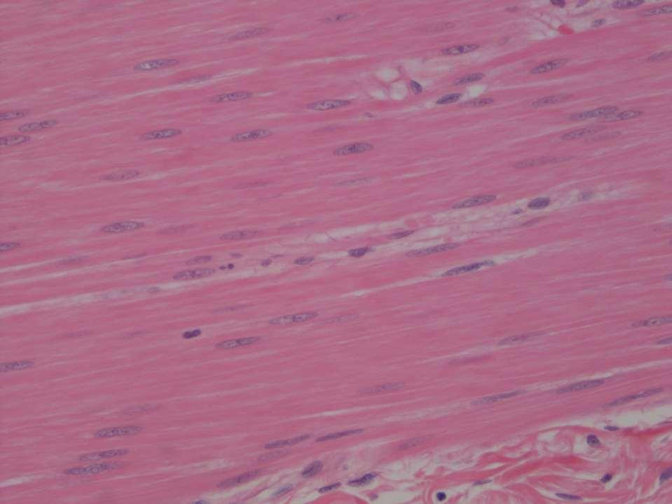
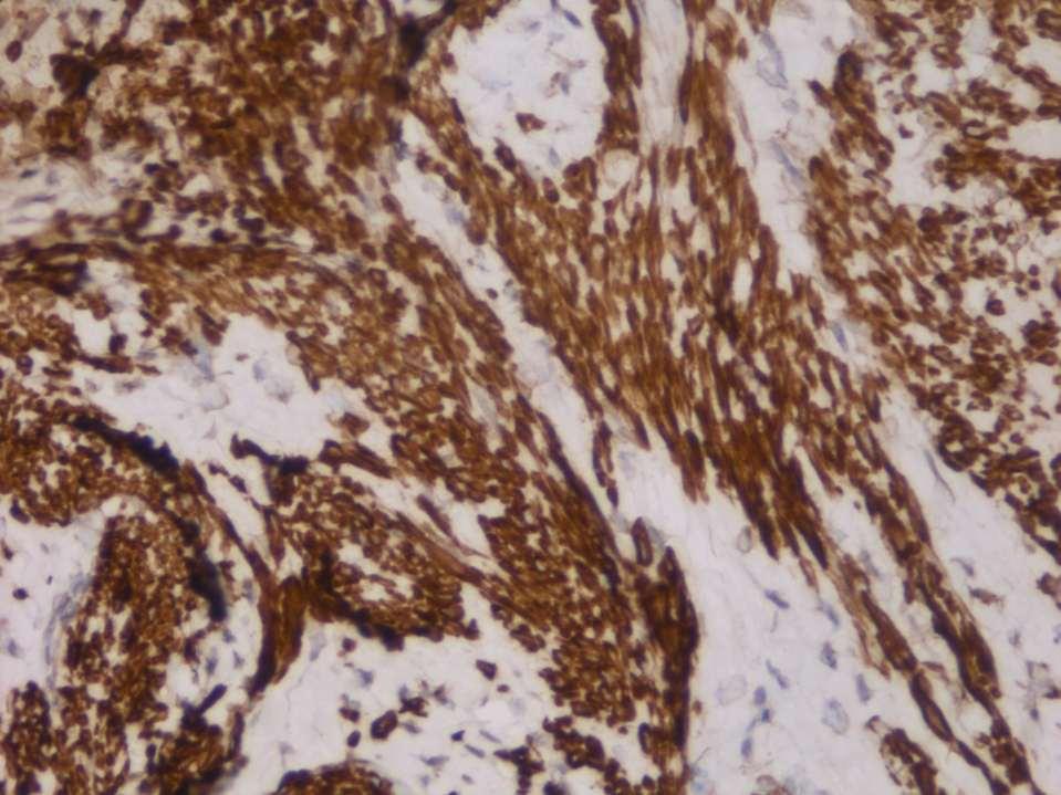
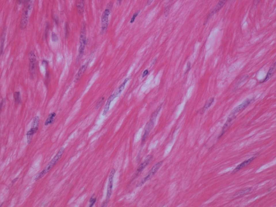
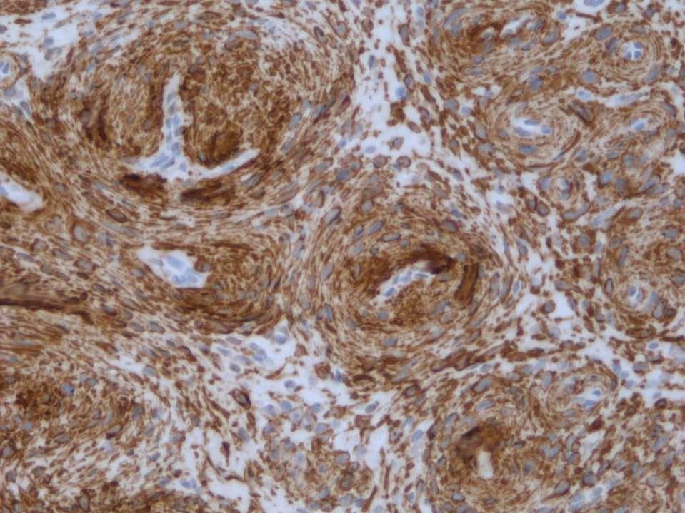
2 NORMAL SMOOTH MUSCLE Cytology Immunohistochemistry Ultrastructure
3
4

5 Masson Trichrome
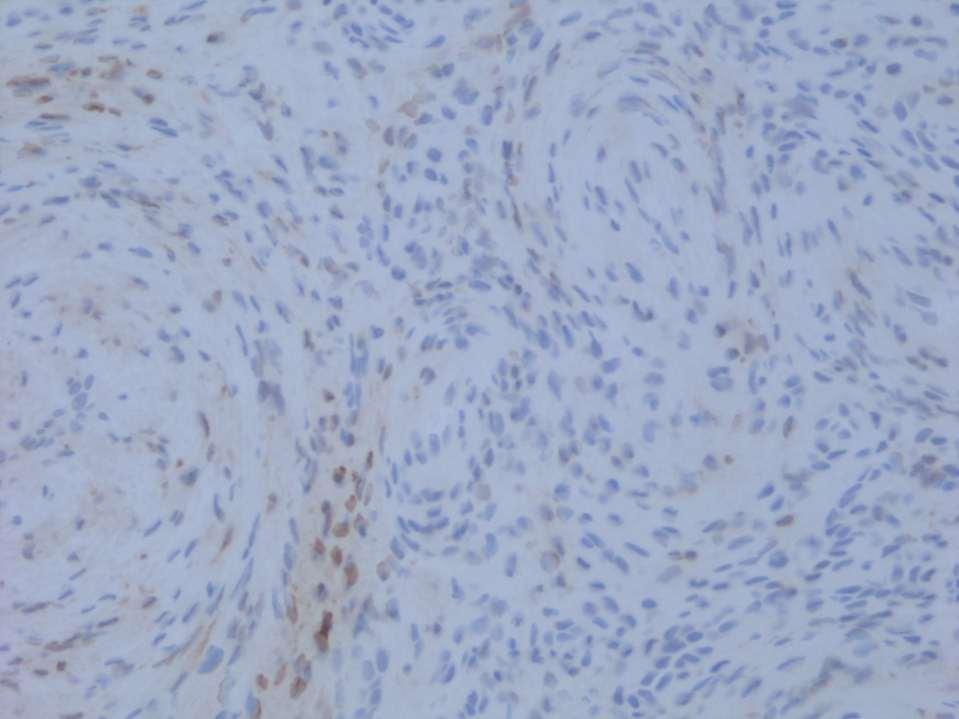
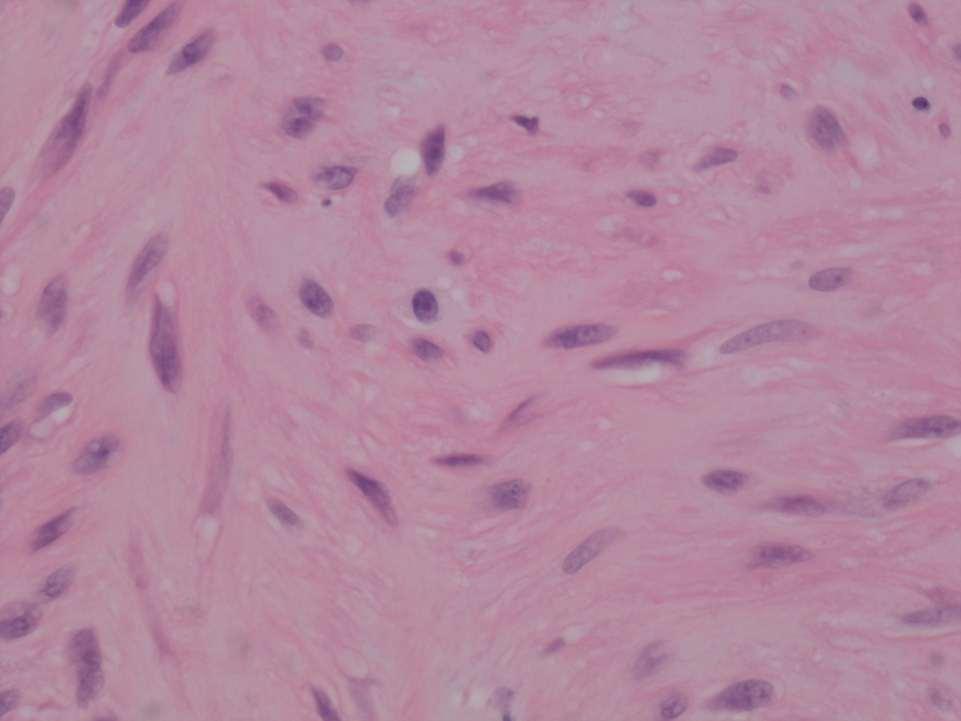
6 Smooth Muscle Ultrastructure Many myofilaments running parallel to the long axis of the smooth muscle cell Myofilaments are of three types (thick myosin filaments, 12nm; these surrounded by thin actin filaments, 9nm) Actin and myosin filaments are contractile and aggregate into a myofibril. The intermediate filament desmin (10nm) is non contractile and is located in the dense body. There is a delicate basal lamina and many surface pinocytotic vesicles
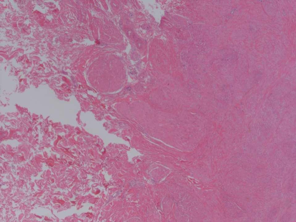
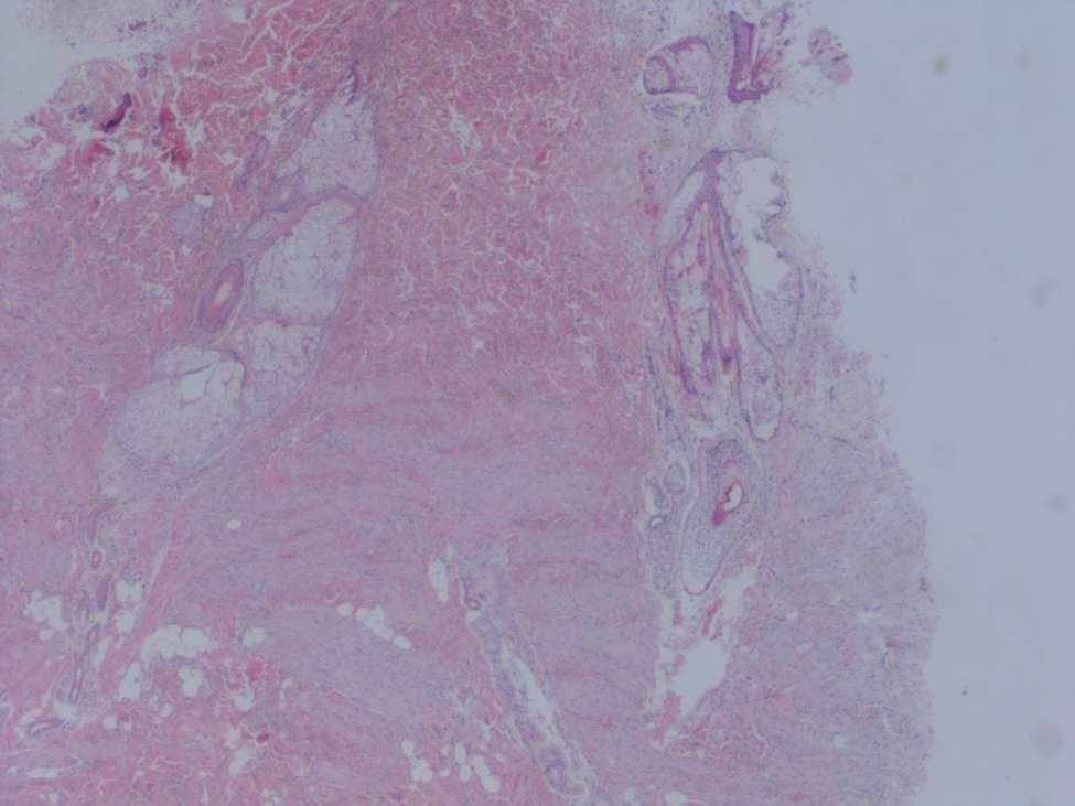
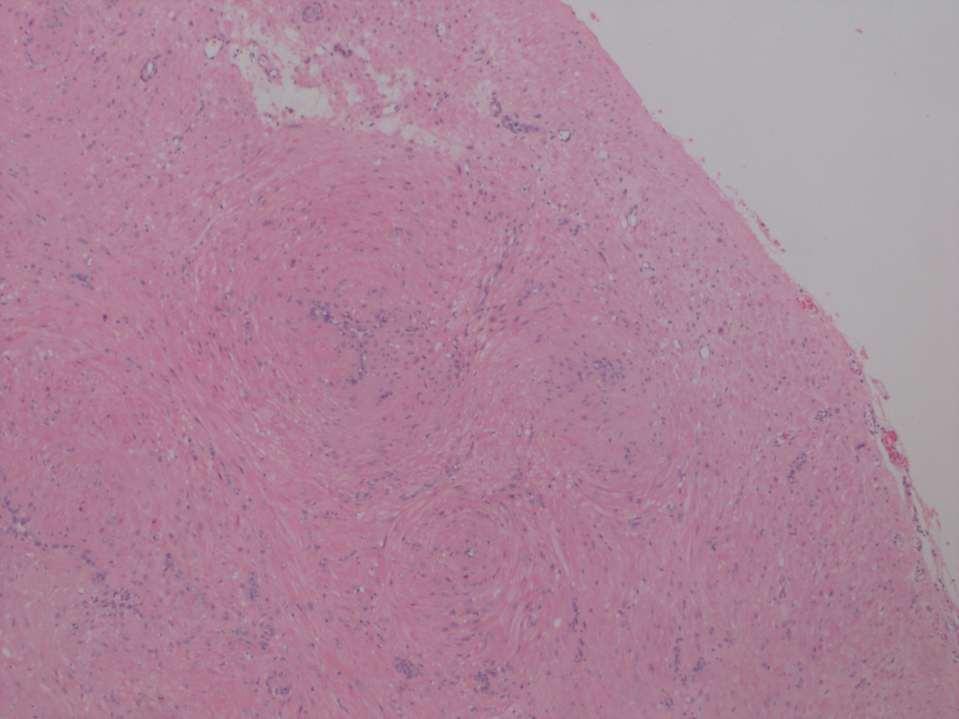
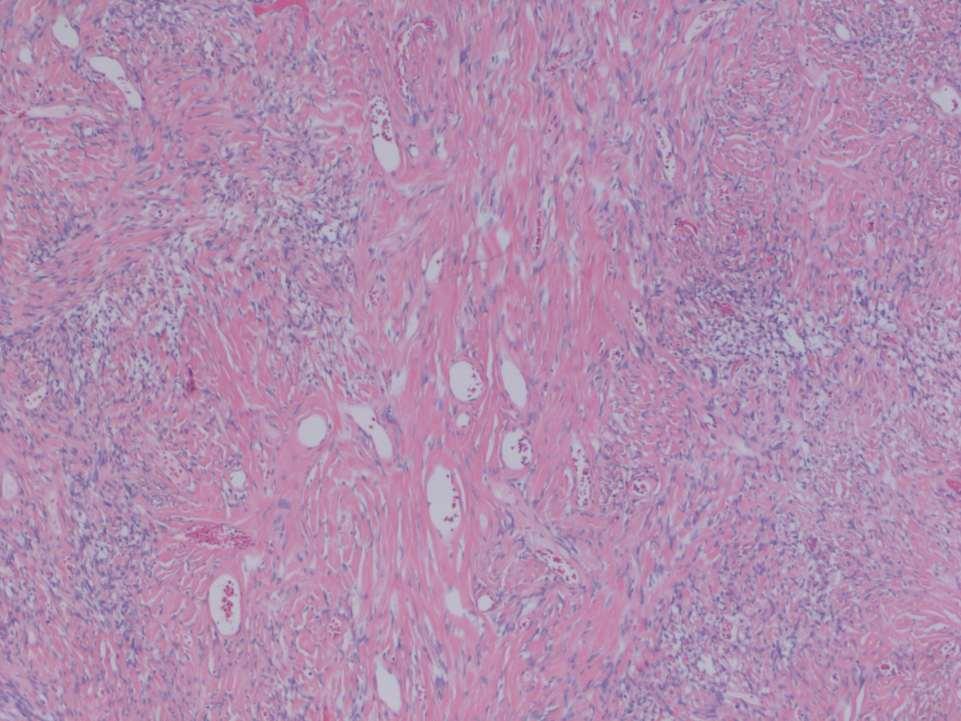
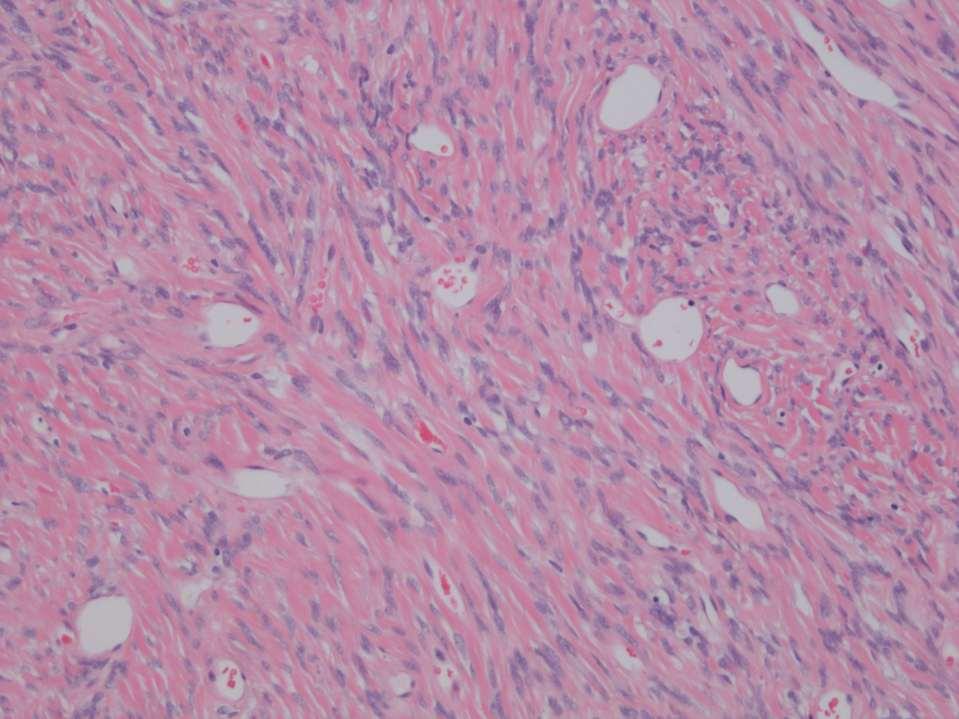
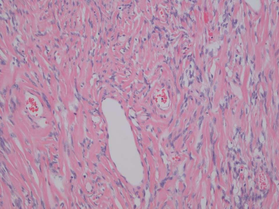
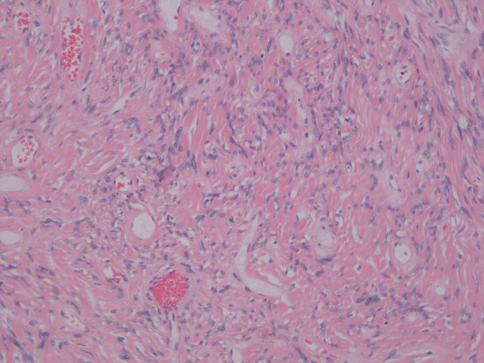
7 LEIOMYOMAS Superficial: pilar (from arrector pilae muscle) or genital (vulva, nipple, scrotum) Deep
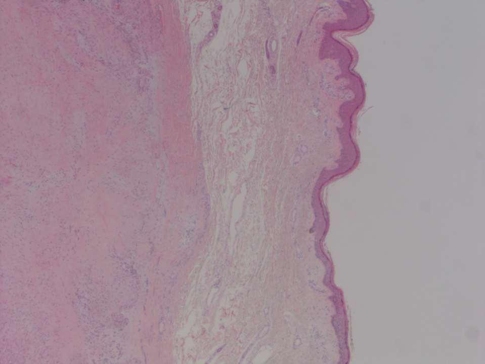
8 Pilar Leiomyoma Adolescents / young adults Solitary or multiple Extensor surfaces of limbs Confined to dermis Small (<20mm) Often painful
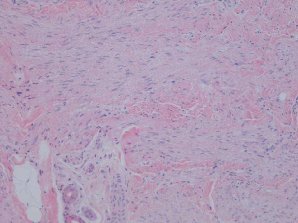
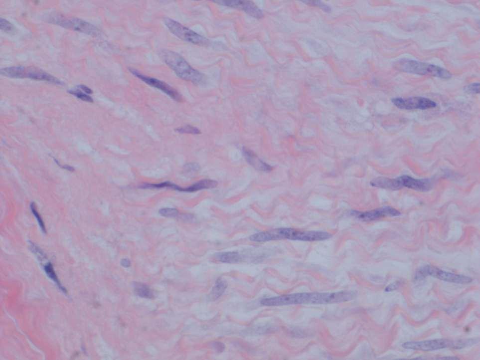
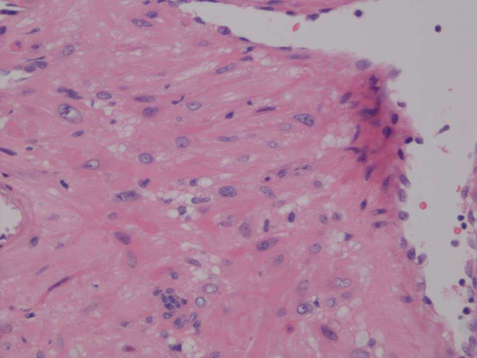
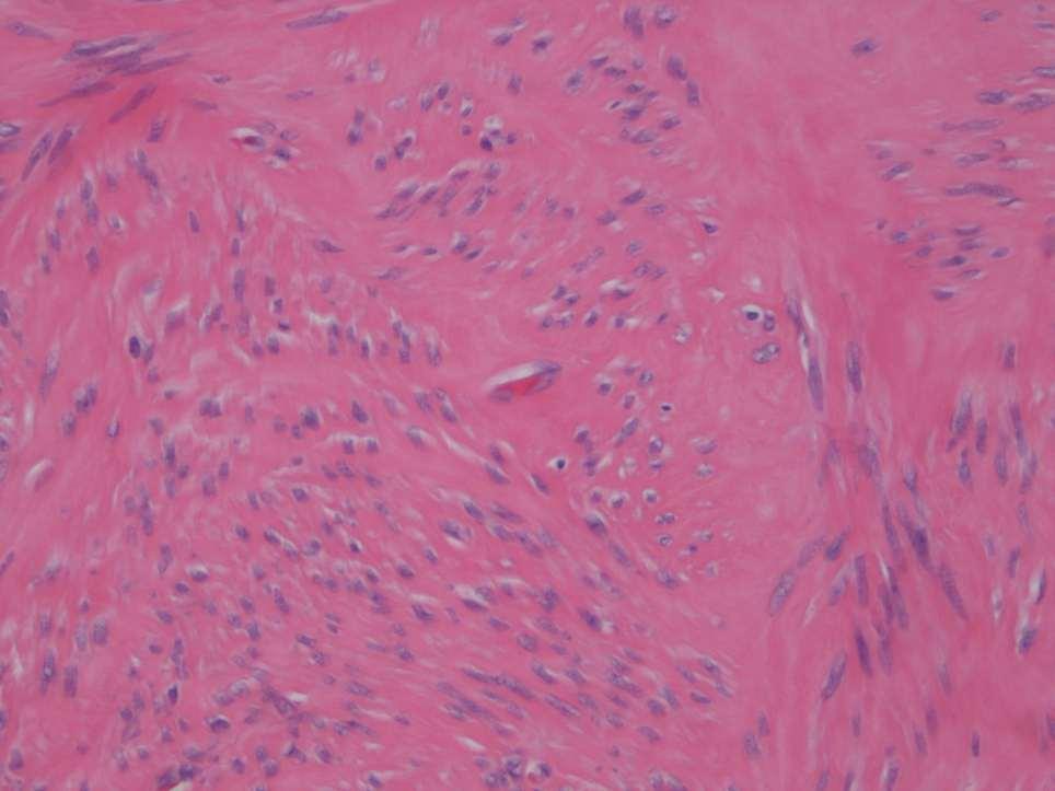
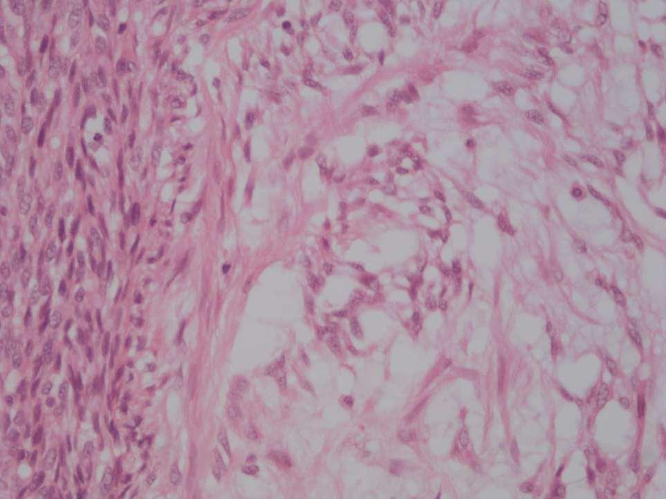
9 Pilar Leiomyoma Histology Confined to dermis Bundles / fascicles of smooth muscle Irregular border, non-encapsulated Occasionally mild degenerative atypia Very occasional mitosis (up to 1/10 hpf) Often epidermal hyperplasia
 Often epidermal")
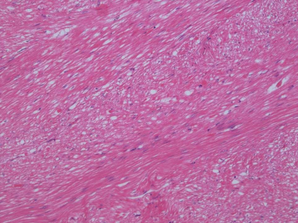
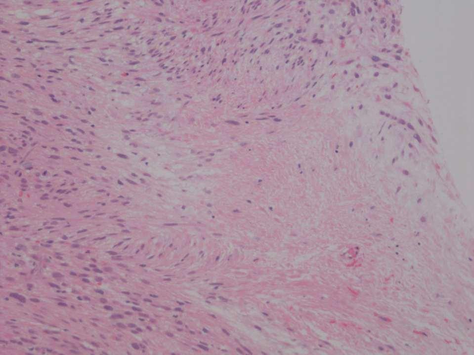
10 Pilar Leiomyoma Histology Confined to dermis Irregular border, not encapsulated Bundles of mature smooth muscle Occasional degenerative atypia Very occasional mitosis (up to 1 per 50 hpf) Often epidermal hyperplasia
11
12
13
14 Familial Pilar Leiomyomas Germline fumarate hydratase mutation (Krebs cycle enzyme) Autosomal dominant inheritance, variable penetrance Multiple cutaneous and uterine leiomyomas Renal cell carcinoma (non-clear cell)
15 Differential Diagnosis Dermatomyofibroma Superficial dermal plaque Shoulder / trunk Spindle cells parallel to epidermis Adnexal structures preserved Myofibroblastic morphology Lacks desmin
16
17
18
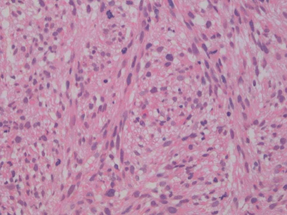
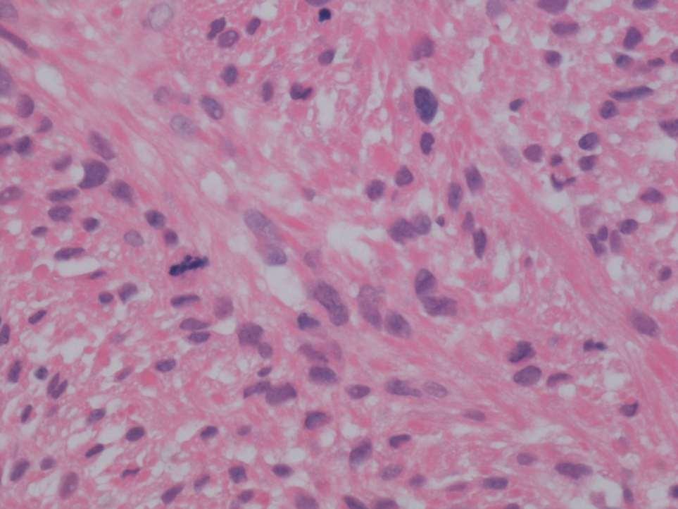
19 Differential Diagnosis Leiomyosarcoma Bigger (often > 20mm) Often infiltrates subcutis More atypia More mitoses Atypical mitoses Sometimes necrosis
20 Angioleiomyoma / Angiomyoma Middle aged adults, F>M Usually solitary Limbs, especially leg Painful Well demarcated Thick-walled blood vessels
21
22

23
24 Deep Leiomyomas Deep leiomyomas will make more sense if we consider uterine smooth muscle tumours first
25 Uterine Leiomyomas Spindled Some epithelioid Some have fat (lipo-leiomyma) Often hyalinised Occasional atypia (degenerative) Some mitoses ER and PR Positive
26 Intravenous leiomyomatosis In pre-menopausal women Benign neoplastic smooth muscle invades myometrial veins Some extend into extra-uterine veins (or IVC or heart) Usually good prognosis with hysterectomy Persistent disease in 30% Occasionally fatal
27 Benign Metastasising Exclusively females Leiomyoma Benign smooth muscle tumour in lungs of women with uterine (or previous uterine) leiomyoma. A few are associated with intravenous leiomyoma Generally good prognosis. A few die of respiratory failure.
28 Leiomyomatosis Peritonealis Exclusively women Child-bearing age Disseminata Black females > white females Associated with hormone production (pregnancy, contraceptive hormone therapy) Myriad of sub-serosal smooth muscle nodules Clinically indolent

29
30
31 H-caldesmon
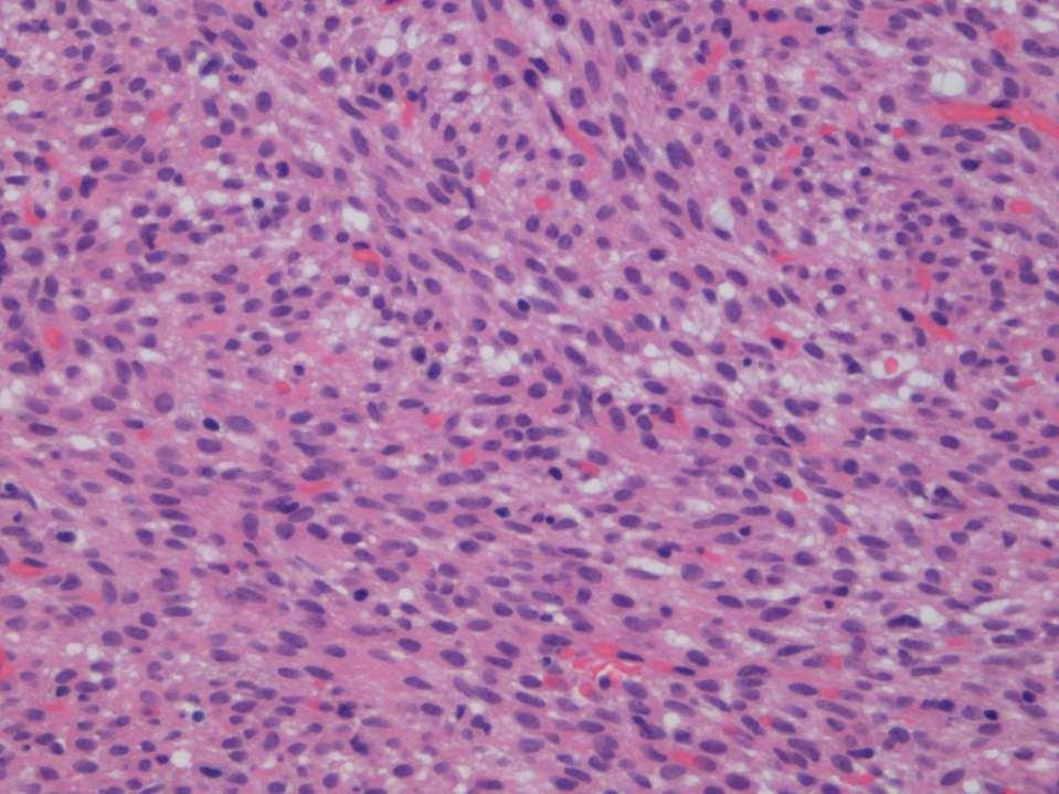
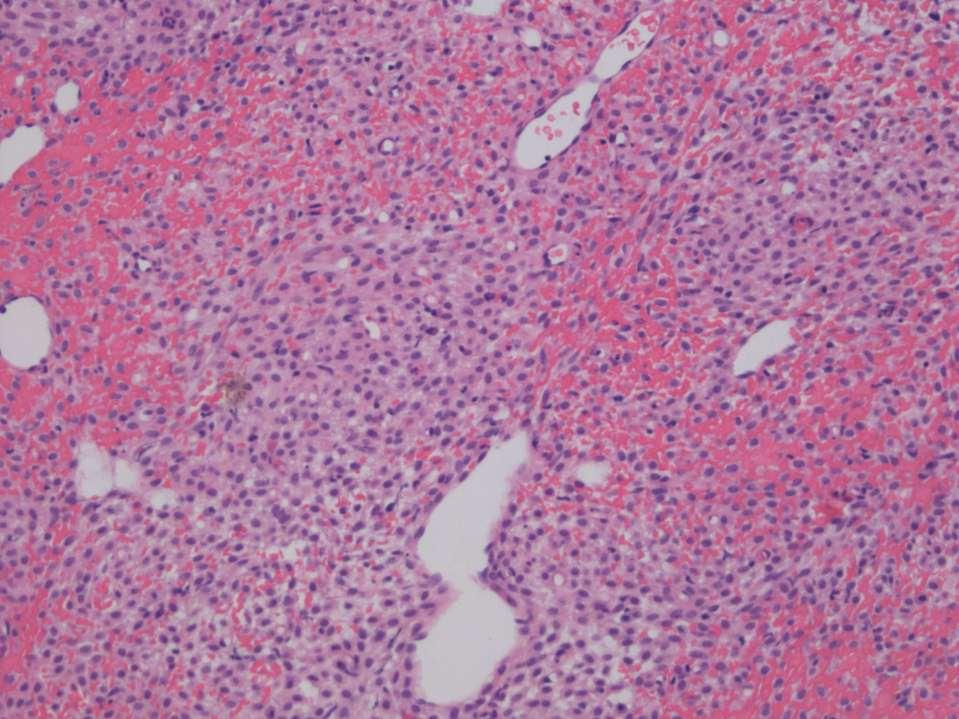
32 Deep Leiomyomas Somatic soft tissue (limbs) (M =F) Virtually no mitotic activity Body cavity LM (pelvis, retroperitoneum, F >>> M) These often hyalinised and ER+ & PR+. Often many typical mitoses. These (particularly in pelvis) of hormonally driven uterine leiomyoma type.
33 Deep Leiomyoma Histology Subcutis / subfascial Bundles / fascicles of smooth muscle at right angles to each other No mitoses or very few (in non-uterine type) Little atypia (and if present degenerative without large nucleoli) No necrosis
34
35
36 Immunosupression related soft tissue tumours?
37 Immunosuppression-related soft tissue tumours Kaposi sarcoma (HHV8) EBV-associated smooth muscle tumour
38 EBV-associated smooth muscle tumour HIV / AIDS Post-transplant immunosuppression Congenital immunosuppression Well-differentiated smooth muscle tumour 50% have foci of round myoid cells Little atypia, few mitoses SMA+ desmin +/-
39 EBV-associated SMT Multifocal Soft tissue Liver, lung, spleen, thyroid Dura Clinically indolent
40 Deep Soft Tissue Leiomyosarcoma Middle-aged adults. Rare in childhood Half arise in retroperitoneum (2/3 of these in women) Vena cava, a common site (3/4 of these in women) Limbs / trunk, M=F
41
42
43
44
45

46
47 INTERMISSION
48 Haemangiopericytoma
49 Solitary Fibrous Tumour Not a smooth muscle or perivascular myocyte tumour. But the cellular subset of SFTs used to be considered a haemangiopericytoma. Actually myofibroblastic (CD34 pos, smooth muscle markers neg)
50
51
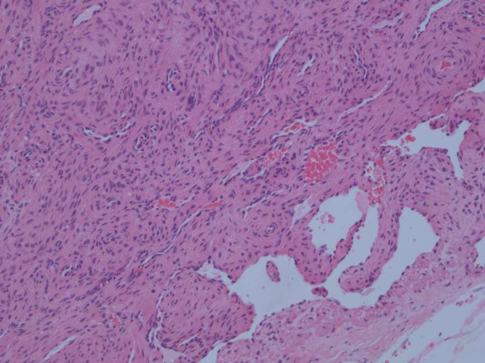
52
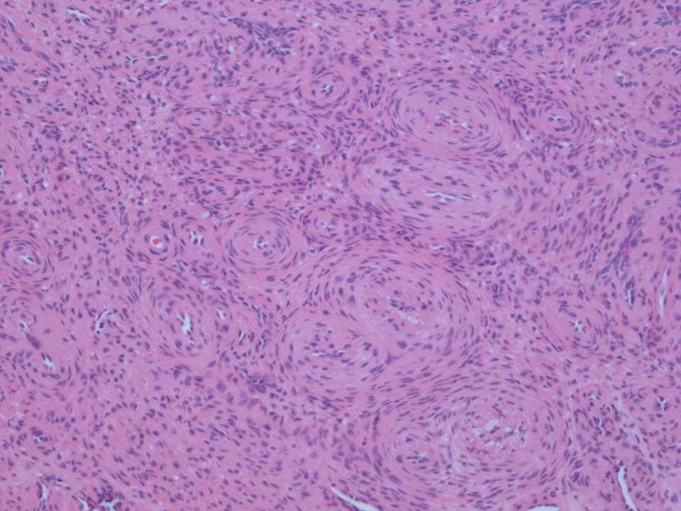
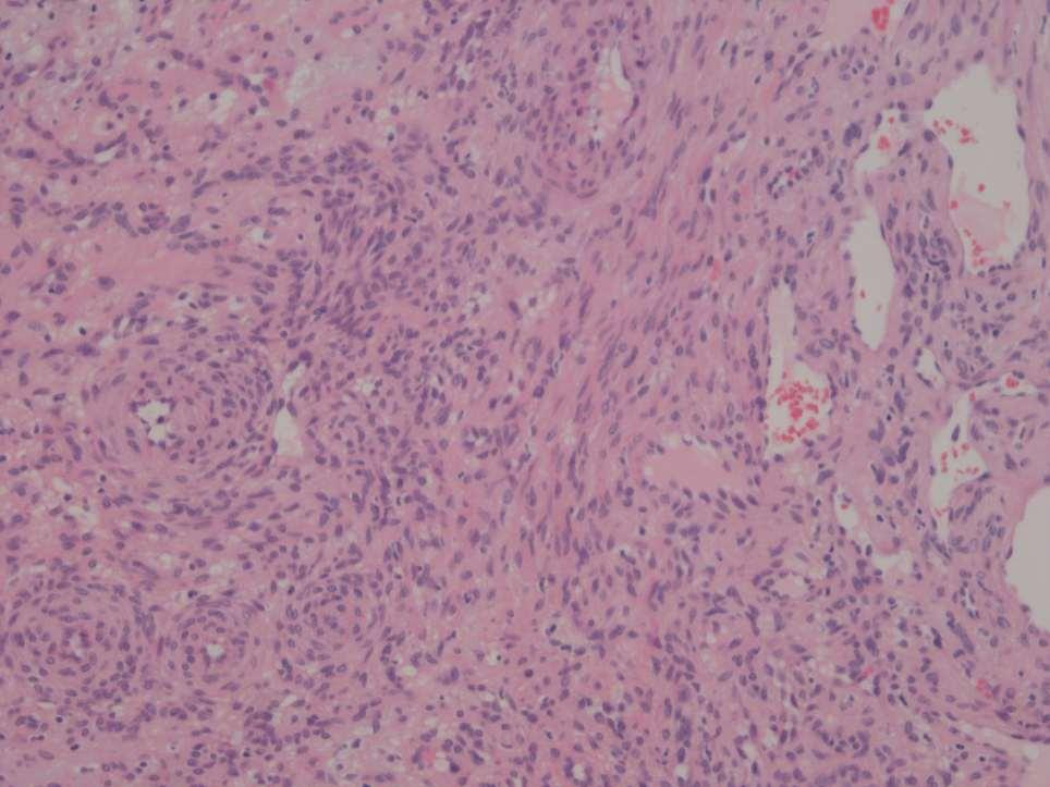
53
54
55 Perivascular Smooth Muscle (Myopericyte) Tumours Sino-nasal haemangiopericytoma Myopericytoma Angiomyoma (Angioleiomyoma) Myofibroma Glomus tumour
56 Sinonasal Haemangiopericytoma

57
58
59
60 Myopericyte tumours (myopericytoma, angiomyoma, myofibroma, glomus tumour) Superficial tumours May show some degenerative atypia Smooth muscle marker positive (SMA and H-caldesmon) Tend to be desmin negative
61 Myopericytoma Perivascular concentric growth Haemangiopericytic vasculature SMA pos, H-caldesmon pos Desmin neg Clinically indolent
62
63
64 H-caldesmon
65 DESMIN
66 Myofibroma
67
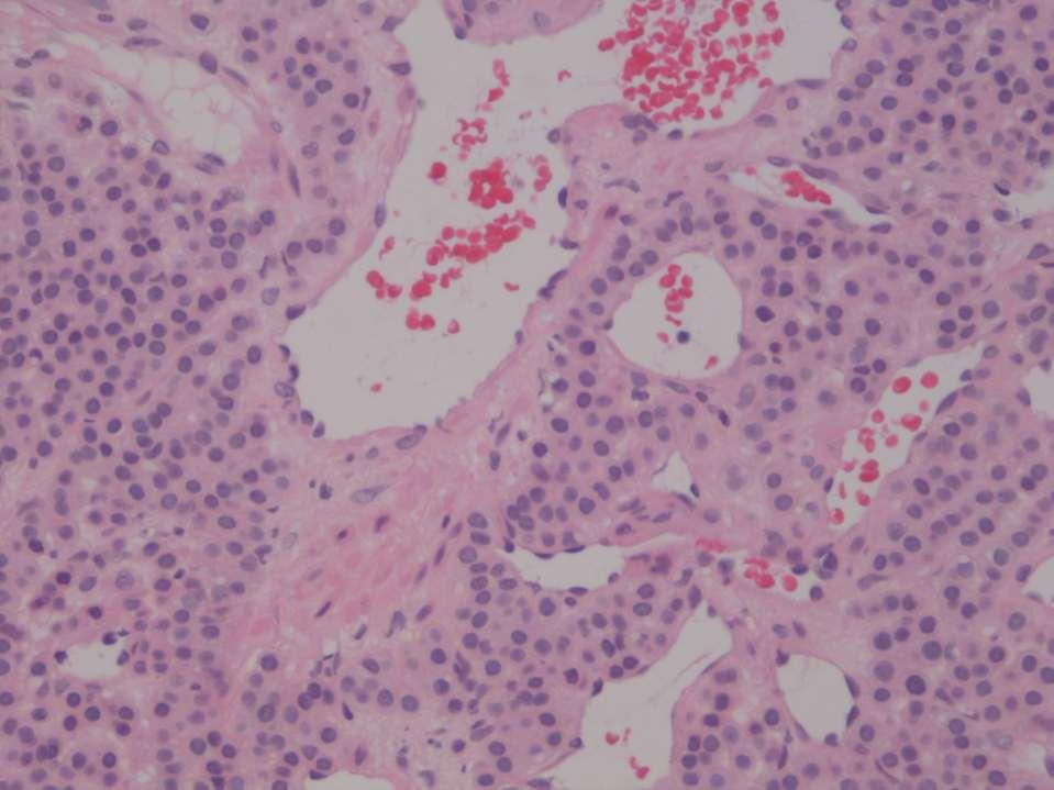
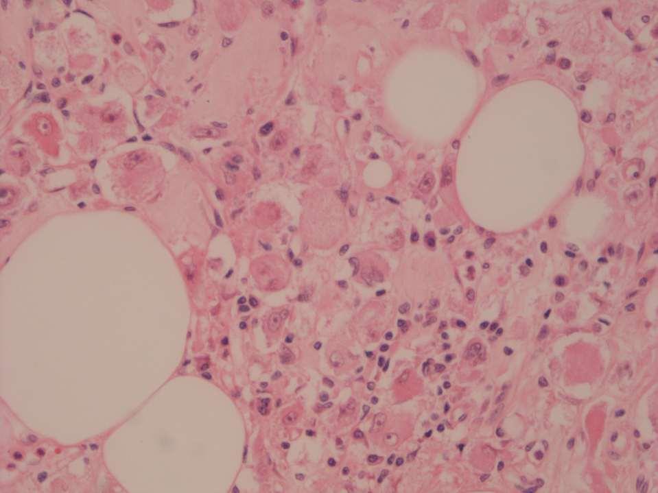
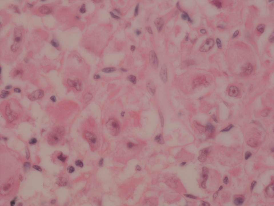
68
69
70
71 Glomus Tumours

72 Gnorma normal glomus body
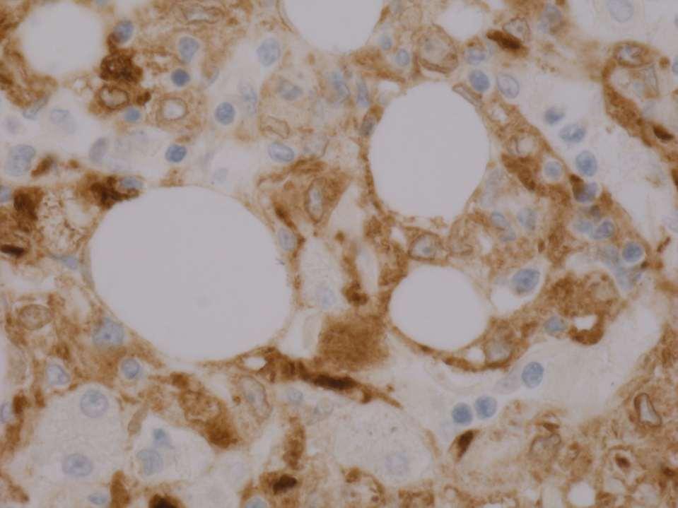
73
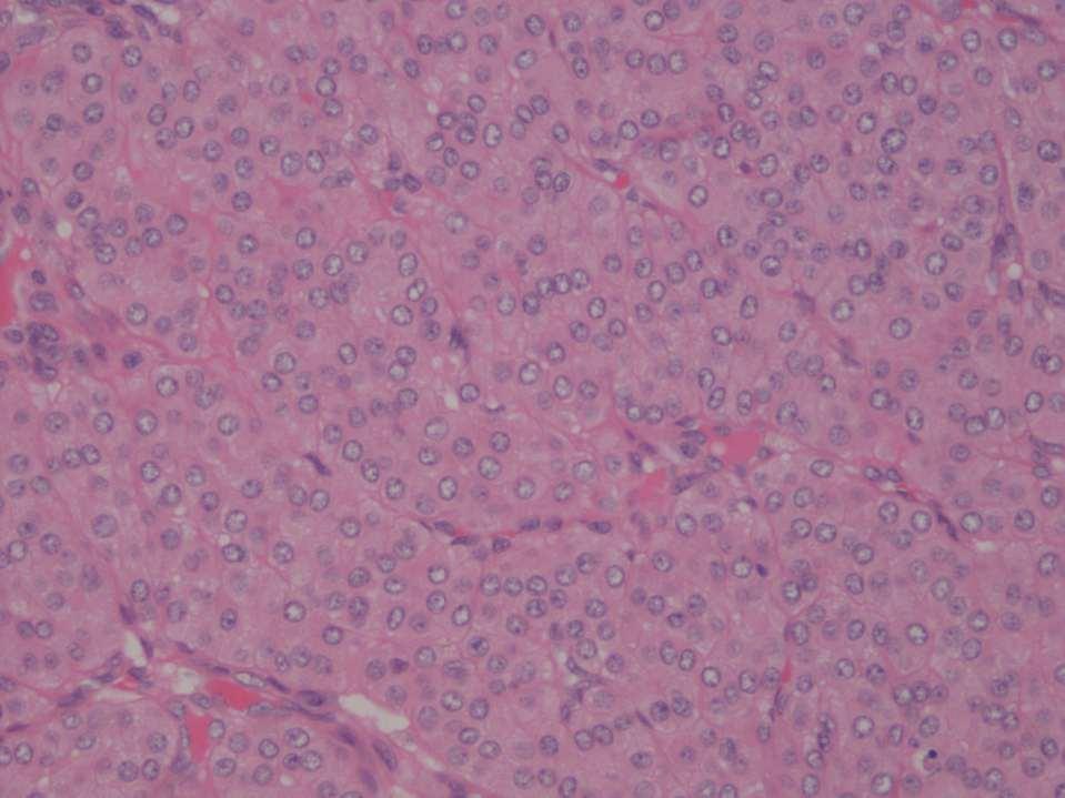
74
75 Glomangiomyoma
76 Classic Angiomyolipoma AML benign mesenchymal tumour comprising variable mixtures of fat spindled/epithelioid smooth muscle cells and thick-walled blood vessels Kidney is classic location Some related to tuberous sclerosis (M=F), in sporadic cases F>M (4:1) The muscle is of perivascular epithelioid type i.e. HMB45+ and melan A+
77 Classic AMLs (interesting facts) Clinically benign (but may result in catastrophic retroperitoneal haemorrhage) May be multifocal (especially in tuberous sclerosis) Occasional degenerative nuclear atypia Can invade renal vein / IVC Can be present in LNs (multifocal growth)
78
79
80
81 Melan-A
82 Epithelioid AML >50% are associated with tuberous sclerosis Clinically more aggressive than classic AML Larger, sometimes necrotic, infiltrative margins, renal vein / IVC extension Approximately 1/3 metastasise (LN, liver, lungs, spine) Differential diagnosis (RCC, metastatic MM)
83
84
85 Perivascular epithelioid cell proliferations (HMB45 pos, melan A pos) PECOMA Angiomyoplipoma Lymphangioleiomyomatosis
أملس عضلي غرن = Leiomyosarcoma. Leiomyosarcoma 1 / 5
 Leiomyosarcoma 1 / 5 EPIDEMIOLOGY Exact incidence is unknown, but older studies suggest that leiomyosarcomas comprise approximately 3 percent of soft-tissue sarcomas. Superficial leiomyosarcoma occurs
CHAPTER 4. Smooth Muscle Tumours
 CHAPTER 4 Smooth Muscle Tumours Smooth muscle tumours arising at non-cutaneous, non-uterine locations have been the focus of a considerable conceptual shift in recent years and this is ongoing. Specifically,
Case 27 Male 42. Painless, static, well-circumscribed, subcutaneous nodule right lower leg,?lipoma. The best diagnosis is:
 Case 27 Male 42. Painless, static, well-circumscribed, subcutaneous nodule right lower leg,?lipoma. The best diagnosis is: A. Angiosarcoma B. Haemangiopericytoma C.Myopericytoma D.Myofibroma E. Angioleiomyoma
Endometrial Stromal Tumors
 Endometrial Stromal Tumors WHO Categories: Endometrial Stromal Nodule (ESN) Endometrial Stromal Sarcoma, low grade (LGESS) Endometrial Stromal Sarcoma, high grade (HGESS) Undifferentiated Uterine Sarcoma
Diagnostic problems in uterine smooth muscle tumors
 Diagnostic problems in uterine smooth muscle tumors Marina Kos Ljudevit Jurak Clinical Department of Pathology, Clinical Hospital Center Sestre milosrdnice, Zagreb Institute of Pathology, University of
An Overview of Genital Stromal Tumors
 An Overview of Genital Stromal Tumors By Konstantinos Linos MD, FCAP, FASDP Bone, Soft Tissue and Dermatopathology Assistant Professor of Pathology Dartmouth-Hitchcock Medical Center Geisel School of Medicine
Uterine Mesenchymal Tumors from a Gynaecological Point of View: A Mini-Review
 EC Gynaecology Special Issue - 2017 Uterine Mesenchymal Tumors from a Gynaecological Point of View: A Mini-Review Mini Review Dr. Huseyin Aydogmus, Dr. Servet Gencdal, Dr. Nihan Gencdal and Dr. Serpil
What really matters When and Why. Pathology of Uterine Mesenchymal Lesions. Nafisa Wilkinson London
 What really matters When and Why Pathology of Uterine Mesenchymal Lesions Nafisa Wilkinson London Patient centred approach immunohistochemistry Histological diagnosis Next generation sequencing Genetic
Financial disclosures
 Mesenchymal Neoplasms with Melanocytic Differentiation By Konstantinos Linos MD, FCAP, FASDP Bone, Soft Tissue and Dermatopathology Assistant Professor of Pathology Dartmouth-Hitchcock Medical Center Geisel
Malignant Peripheral Nerve Sheath Tumor
 C H A P T E R 120 Malignant Peripheral Nerve Sheath Tumor Currently, malignant peripheral nerve sheath tumor (MPNST) is the most commonly used generic name for the neoplasms known in the past as neurosarcoma,
Enterprise Interest Nothing to declare
 Enterprise Interest Nothing to declare Diagnoses one would not like to miss in soft tissue pathology early in your career Marta Sbaraglia, MD Department of Pathology Hospital of Treviso University of Padua
59 yo male with past medical history of prostate carcinoma, presented with upper abdominal pain
 December 2016 59 yo male with past medical history of prostate carcinoma, presented with upper abdominal pain Contributed by: Divya Sharma, MD. Fellow, Gastrointestinal Pathology, Department of Pathology
The Relevance of Cytologic Atypia in Cutaneous Neural Tumors
 The Relevance of Cytologic Atypia in Cutaneous Neural Tumors Recent Findings - New Developments New Problems Zsolt B. Argenyi, M.D. Professor of Pathology & Dermatology Director of Dermatopathology Department
05/07/2018. Types of challenges. Challenging cases in uterine pathology. Case 1 ` 65 year old female Post menopausal bleeding Uterine Polyp
 Types of challenges Challenging cases in uterine pathology Nafisa Wilkinson Gynaecological Pathologist UCLH London Lack of complete history often, NO clinical history at all! Cases from other centres often
Mody. AIS vs. Invasive Adenocarcinoma of the Cervix
 Common Problems in Gynecologic Pathology Michael T. Deavers, M.D. Houston Methodist Hospital, Houston, Texas Common Problems in Gynecologic Pathology Adenocarcinoma in-situ (AIS) of the Cervix vs. Invasive
Case Based Learning Program
 Case Based Learning Program The Department of Urology Glickman Urological & Kidney Institute Cleveland Clinic Case Number 5 CBULP 2010 001 Case Based Urology Learning Program Editor: Associate Editor:
ACCME/Disclosures ALK FUSION-POSITIVE MESENCHYMAL TUMORS. Tumor types with ALK rearrangements. Anaplastic Lymphoma Kinase. Jason L.
 Companion Meeting of the International Society of Bone and Soft Tissue Pathology The Evolving Concept of Mesenchymal Tumors ALK FUSION-POSITIVE MESENCHYMAL TUMORS Jason L. Hornick, MD, PhD March 13, 2016
Solitary Fibrous Tumor of the Kidney with Massive Retroperitoneal Recurrence. A Case Presentation
 246) Prague Medical Report / Vol. 113 (2012) No. 3, p. 246 250 Solitary Fibrous Tumor of the Kidney with Massive Retroperitoneal Recurrence. A Case Presentation Sfoungaristos S., Papatheodorou M., Kavouras
Desmoplastic Melanoma R/O BCC. Clinical Information. 74 y.o. man with lesion on left side of neck r/o BCC
 R/O BCC Sabine Kohler, M.D. Professor of Pathology and Dermatology Dermatopathology Service Stanford University School of Medicine Clinical Information 74 y.o. man with lesion on left side of neck r/o
Neoplasia 2018 Lecture 2. Dr Heyam Awad MD, FRCPath
 Neoplasia 2018 Lecture 2 Dr Heyam Awad MD, FRCPath ILOS 1. List the differences between benign and malignant tumors. 2. Recognize the histological features of malignancy. 3. Define dysplasia and understand
Uterine mesenchymal tumors: Hereditary perspectives
 Uterine mesenchymal tumors: Hereditary perspectives Two hereditary syndromes are known to be related to uterine mesenchymal tumors: Hereditary Leiomyomatosis and Renal Cell Carcinoma syndrome (HLRCC) and
Spindle Cell Lesions Of The Breast. Emad Rakha Professor of Breast Pathology and Consultant Pathologist
 Spindle Cell Lesions Of The Breast Emad Rakha Professor of Breast Pathology and Consultant Pathologist * SCLs comprise a wide spectrum of diseases, ranging from reactive processes to aggressive malignant
Diplomate of the American Board of Pathology in Anatomic and Clinical Pathology
 A 33-year-old male with a left lower leg mass. Contributed by Shaoxiong Chen, MD, PhD Assistant Professor Indiana University School of Medicine/ IU Health Partners Department of Pathology and Laboratory
Leiomyosarcoma of the inferior vena cava: 1 case. B. Bancel, A. Rode, C. Ducerf. Hôpital CROIX ROUSSE LYON. Case report
 Leiomyosarcoma of the inferior vena cava: 1 case B. Bancel, A. Rode, C. Ducerf Hôpital CROIX ROUSSE LYON Bucharest Nov 2011 Case report 34 yr-old woman, no antecedent Sept 2004: Abdominal upper right quadrant
Benign and malignant epithelial lesions: Seborrheic keratosis: A common benign pigmented epidermal tumor occur in middle-aged or older persons more
 Benign and malignant epithelial lesions: Seborrheic keratosis: A common benign pigmented epidermal tumor occur in middle-aged or older persons more common on the trunk; but extremities, head and neck are
A case of pedunculated intraperitoneal leiomyoma
 Jichi Medical University Journal Chio Shuto Kuniyasu Soda Takayoshi Yoshida Fumio Konishi Abstract We report a very rare case of a pedunculated intraperitoneal leiomyoma in the parietal peritoneum of the
Science & Technologies RETROPERITONEAL TUMOR: DIFFERENTIAL DIAGNOSIS BEYOND THE USUALLY SUSPECTED. Medical University Sofia, Bulgaria
 RETROPERITONEAL TUMOR: DIFFERENTIAL DIAGNOSIS BEYOND THE USUALLY SUSPECTED Vesela Ivanova *, Tihomir Dikov *, Goran Derimachkovski **, Petar Panchev ** * Department of General and Clinical Pathology, Medical
Update on Cutaneous Mesenchymal Tumors. Thomas Brenn
 Update on Cutaneous Mesenchymal Tumors Thomas Brenn Cutaneous Mesenchymal Tumours Wide morphological and biological spectrum Myofibroblastic, smooth muscle, neural, vascular, apidocytic, undifferentiated;
International Journal of Pharma and Bio Sciences CUTANEOUS LEIOMYOMA: A CASE STUDY WITH REVIEW OF LITERATURE ON RARE VARIANTS AND CLINICAL BEHAVIOUR
 Review Article Pathology International Journal of Pharma and Bio Sciences ISSN 0975-6299 CUTANEOUS LEIOMYOMA: A CASE STUDY WITH REVIEW OF LITERATURE ON RARE VARIANTS AND CLINICAL BEHAVIOUR DR.ANANDRAJ
Classification (1) Classification (3) Classification (2) Spindle cell lesions. Spindle cell lesions of bladder (Mills et al.
 Classification (3) Classification (2) Spindle cell lesions. Spindle cell lesions of bladder (Mills et al.") Non-epithelial tumours and nonepithelial tumour-like lesions of the bladder Dr Jonathan H Shanks The Christie NHS Foundation Trust, Manchester, UK Classification (1) Myofibroblastic proliferations and
Non-epithelial tumours and nonepithelial tumour-like lesions of the bladder Dr Jonathan H Shanks The Christie NHS Foundation Trust, Manchester, UK Classification (1) Myofibroblastic proliferations and
Basal cell carcinoma 5/28/2011
 Goal of this Presentation A practical approach to the diagnosis of cutaneous carcinomas and their mimics Thaddeus Mully, MD University of California San Francisco To review common non-melanoma skin cancers
SOFT TISSUE TUMOR PATHOLOGY: AN UPDATE
 SOFT TISSUE TUMOR PATHOLOGY: AN UPDATE Jason L. Hornick, MD, PhD July 18, 2013 Department of Pathology Brigham and Women s Hospital Harvard Medical School Boston, MA, USA I have no disclosures. New Soft
Hereditary Leiomyomatosis and Renal Cell Carcinoma Variant of Reed s Syndrome - A Rare Case Report
 American Research Journal of Urology Volume 1, Issue 1, pp:26-30 Case Hereditary Leiomyomatosis and Renal Cell Carcinoma Variant of Reed s Syndrome - A Rare Case Manas Babu, Devesh Bansal, Sony Mehta,
Gross appearance of peritoneal cysts. They have a thin, translucent wall and contain a clear fluid.
 Gross appearance of peritoneal cysts. They have a thin, translucent wall and contain a clear fluid. So-called multicystic benign mesothelioma. A, Gross appearance. So-called multicystic benign mesothelioma.
Special slide seminar
 Special slide seminar Tomáš Rozkoš The Fingerland Department of Pathology Charles University Medical Faculty and Faculty Hospital in Hradec Králové Czech Republic Case history, 33 years old resistance
Selected Pseudomalignant Soft Tissue Tumors of the Skin and Subcutis
 Selected Pseudomalignant Soft Tissue Tumors of the Skin and Subcutis Andrew L. Folpe, M.D. Professor of Laboratory Medicine and Pathology Mayo Clinic, Rochester, MN folpe.andrew@mayo.edu 2016 MFMER slide-1
Case Presentation. Maha Akkawi, MD, Fatima Obeidat, MD, Tariq Aladily, MD. Department of Pathology Jordan University Hospital Amman, Jordan
 Case Presentation Maha Akkawi, MD, Fatima Obeidat, MD, Tariq Aladily, MD Department of Pathology Jordan University Hospital Amman, Jordan The 25th Annual Congress of the ADIAP The 8/11/2013 1 5th International
MECHANISMS OF HUMAN DISEASE: LABORATORY SESSION PATHOLOGY OF THE SKIN LAB. Friday, February 12, :30 am 11:00 am
 MECHANISMS OF HUMAN DISEASE: LABORATORY SESSION PATHOLOGY OF THE SKIN LAB Friday, February 12, 2012 9:30 am 11:00 am FACULTY COPY GOALS: Describe the basic clinical and morphologic features of various
Female 18. Deeply pigmented lesion on trunk.?warty naevus?seborrhoeic keratosis?malignant melanoma. The best diagnosis is:
 Female 18. Deeply pigmented lesion on trunk.?warty naevus?seborrhoeic keratosis?malignant melanoma. The best diagnosis is: A. deep penetrating naevus B. naevoid malignant melanoma C. pigment synthesising
Dr Sanjiv Manek Oxford. Oxford Pathology Course 2010 for FRCPath Illustration-Cellular Pathology. Oxford Radcliffe NHS Trust
 Dr Sanjiv Manek Oxford Oxford Pathology Course 2010 for FRCPath Illustration-Cellular Pathology. Oxford Radcliffe NHS Trust Ovarian Endometrial Vulvo-vaginal Cervical Illustration-Cellular Pathology. Oxford
case report Oman Medical Journal [2016], Vol. 31, No. 1: 60 64
![case report Oman Medical Journal [2016], Vol. 31, No. 1: 60 64](/thumbs/90/102852192.jpg "case report Oman Medical Journal [2016], Vol. 31, No. 1: 60 64") case report Oman Medical Journal [2016], Vol. 31, No. 1: 60 64 Malignant Gastric Glomus Tumor: A Case Report and Literature Review of a Rare Entity Shaesta Zaidi * and Maha Arafah Department of Histopathology,
case report Oman Medical Journal [2016], Vol. 31, No. 1: 60 64 Malignant Gastric Glomus Tumor: A Case Report and Literature Review of a Rare Entity Shaesta Zaidi * and Maha Arafah Department of Histopathology,
Pathology of Sarcoma ELEANOR CHEN, MD, PHD, ASSISTANT PROFESSOR DEPARTMENT OF PATHOLOGY UNIVERSITY OF WASHINGTON
 Pathology of Sarcoma ELEANOR CHEN, MD, PHD, ASSISTANT PROFESSOR DEPARTMENT OF PATHOLOGY UNIVERSITY OF WASHINGTON Presentation outline Background and epidemiology of sarcomas Sarcoma classification Sarcoma
Circulation: X Case number: 501 Number of responses: 84 Date: 4 MAY 12
 Circulation: X Case number: 500 Number of responses: 81 Date: 4 MAY 12 Female, aged 65 TAH and BSO for G1 endometrioid adenocarcinoma. Tumour positive with inhibin, vimentin, CD56 and SMA. Negative with
Dermatopathology. Dr. Rafael Botella Estrada. Hospital La Fe de Valencia
 Dermatopathology Dr. Rafael Botella Estrada. Hospital La Fe de Valencia Melanoma and mimics Dr. Martin Mihm Malignant lesions result from the accumulation of mutations Class I lesions (benign) Class II
An Overview of Cutaneous Vascular Neoplasms
 An Overview of Cutaneous Vascular Neoplasms By Konstantinos Linos MD, FCAP, FASDP Bone, Soft Tissue and Dermatopathology Assistant Professor of Pathology Dartmouth-Hitchcock Medical Center Geisel School
Evening Specialty Conference Bone and Soft Tissue Pathology. Diagnostic pitfalls in bone and soft tissue pathology
 Evening Specialty Conference Bone and Soft Tissue Pathology. Case 1 Elizabeth G Demicco, MD, PhD Mount Sinai Hospital, New York Disclosure of Relevant Financial Relationships USCAP requires that all planners
1/10/2018. Soft Tissue Tumors Showing Melanocytic Differentiation. Overview. Desmoplastic/ Spindle Cell Melanoma
 2016 MFMER slide-1 2016 MFMER slide-2 2016 MFMER slide-3 Soft Tissue Tumors Showing Melanocytic Differentiation Andrew L. Folpe, M.D. Professor of Laboratory Medicine and Pathology Mayo Clinic, Rochester,
Myxo-inflammatory Fibroblastic sarcoma
 AKA Myxo-inflammatory Fibroblastic sarcoma Acral Myxoinflammatory fibroblastic sarcomaam.j.surg.path1998; 22; 911-924 Inflammatory myxoid tumour of soft parts with bizarre giant cells [Pathol.Res.Pract.
21/07/2017. Hobnail endothelial cells are not the same as epithelioid endothelial cells
 UPDATE IN CUTANEOUS VASCULAR S DERMATOPATHOLOGY SESSION BELFAST PATHOLOGY JUNE 21/2017 Dr E Calonje St John s Institute of Dermatology, London, United Kingdom THE FAMILY OF VASCULAR S WITH EPITHELIOID
Mesenchymal neoplasms of the gastrointestinal tract what s new? Newton ACS Wong Department of Histopathology Bristol Royal Infirmary
 Mesenchymal neoplasms of the gastrointestinal tract what s new? Newton ACS Wong Department of Histopathology Bristol Royal Infirmary Talk plan Summary from 2010 talk. What s happened since 2010. GISTs
Bilateral Renal Angiomyolipomas with Invasion of the Renal Vein: A Case Report
 Case Study TheScientificWorldJOURNAL (2008) 8, 145 148 TSW Urology ISSN 1537-744X; DOI 10.1100/tsw.2008.29 Bilateral Renal Angiomyolipomas with Invasion of the Renal Vein: A Case Report C. Blick, N. Ravindranath,
Clinical History. 29 yo woman with polyhydramnios Cardiac mass at fetal ultrasound At 35 weeks, newborn died 30 minutes after delivery
 CASE 1 a Clinical History 29 yo woman with polyhydramnios Cardiac mass at fetal ultrasound At 35 weeks, newborn died 30 minutes after delivery Interface between tumor and normal myocardium Smaller well-demarcated
Ram M, Rodrigo T, Petkar M. A case of cutaneous angiomyolipoma with review of the literature. OA Case Reports 2013 Nov 15;2(13):130.
:130.") Ram M, Rodrigo T, Petkar M. A case of cutaneous angiomyolipoma with review of the literature. OA Case Reports 2013 Nov 15;2(13):130. Licensee OA Publishing London 2013. Creative Commons Attribution License
Ram M, Rodrigo T, Petkar M. A case of cutaneous angiomyolipoma with review of the literature. OA Case Reports 2013 Nov 15;2(13):130. Licensee OA Publishing London 2013. Creative Commons Attribution License
Evening specialty conference: Liver
 Evening specialty conference: Liver Joseph Misdraji, M.D. Disclosure of Relevant Financial Relationships Disclosure of Relevant Financial Relationships USCAP requires that all planners (Education Committee)
Case Presentation 主治醫師 : 宋文鑫日期 :
 Case Presentation 主治醫師 : 宋文鑫日期 : 2015-2-28 General Data Name:OOO Chart Number:OOOOOOO Date of Admission:2014 年 08 月 04 日 Age: 33 y/o Sex:female Occupation : 會計 Chief Complaint Palpable soft tissue mass
ESS: Pathologic Insights
 GEIS XVI INTERNATIONAL SYMPOSIUM Seville 4th October 2018 ESS: Pathologic Insights Sílvia Bagué The Royal Marsden Hospital London (United Kingdom) I have no conflicts of interest Endometrial stromal sarcoma
Papillary Lesions of the breast
 Papillary Lesions of the breast Emad Rakha Professor of Breast Pathology The University of Nottingham Papillary lesions of the breast are a heterogeneous group of disease, which are characterised by neoplastic
SESSION 1: GENERAL (BASIC) PATHOLOGY CONCEPTS Thursday, October 16, :30am - 11:30am FACULTY COPY
 PATHOLOGY CONCEPTS Thursday, October 16, :30am - 11:30am FACULTY COPY") SESSION 1: GENERAL (BASIC) PATHOLOGY CONCEPTS Thursday, October 16, 2008 9:30am - 11:30am FACULTY COPY GOAL: Describe the basic morphologic (structural) changes which occur in various pathologic conditions.
SESSION 1: GENERAL (BASIC) PATHOLOGY CONCEPTS Thursday, October 16, 2008 9:30am - 11:30am FACULTY COPY GOAL: Describe the basic morphologic (structural) changes which occur in various pathologic conditions.
CASE year old male with a PET avid nodule in the left adrenal gland
 CASE 1 55 year old male with a PET avid nodule in the left adrenal gland Case 1 Adrenal gland parenchyma partly replaced by a spindle cell tumour with mild nuclear pleomorphism Atypical mitoses present
CASE REPORT Benign epithelioid peripheral nerve sheath tumour resembling schwannoma
 Malaysian J Pathol 2014; 36(3) : 217 221 CASE REPORT Benign epithelioid peripheral nerve sheath tumour resembling schwannoma Thejasvi KRISHNAMURTHY MD and SR NIVEDITHA MD, DNB Department of Pathology,
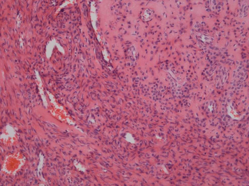
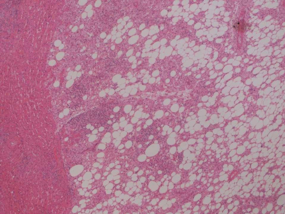
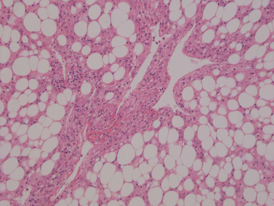
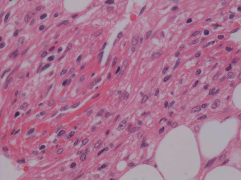
A 42-year-old woman with a liver mass
 April 2016 Case of the Month A 42-year-old woman with a liver mass Contributed by: Natalia I. Rush, MD, Resident Physician, Indiana University School of Medicine, Department of Pathology and Laboratory
64 y.o. F with CLL and leg tumour
 64 y.o. F with CLL and leg tumour Case History Excision with split-skin grafting Histology moderately differentiated squamous cell carcinoma with large areas of necrosis and brisk mitotic activity.
The World Health Organization defines PEComas as mesenchymal
 ORIGINAL ARTICLE Perivascular Epithelioid Cell Neoplasms of Soft Tissue and Gynecologic Origin A Clinicopathologic Study of 26 Cases and Review of the Literature Andrew L. Folpe, MD,* Thomas Mentzel, MD,
Diagnostically Challenging Cases in Gynecologic Pathology
 Diagnostically Challenging Cases in Gynecologic Pathology Eric C. Huang, M.D., Ph.D. Department of Pathology and Laboratory Medicine University of California, Davis Medical Center Case 1 Presentation 38
Mayo Medical Laboratories
 Mayo Medical Laboratories Virtual Lectures 2014 MFMER 2016 MFMER slide-1 Virtual Lectures Planning Committee Disclosure Summary As a provider accredited by ACCME, College of Medicine, Mayo Clinic (Mayo
Case: The patient is a 24 year- old female who was found to have multiple mural nodules within the antrum. Solid and cystic components were noted on
 Case: The patient is a 24 year- old female who was found to have multiple mural nodules within the antrum. Solid and cystic components were noted on imaging. There is no significant past medical history.
Disclosure. Relevant Financial Relationship(s) None. Off Label Usage None MFMER slide-1
 None. Off Label Usage None MFMER slide-1") Disclosure Relevant Financial Relationship(s) None Off Label Usage None 2013 MFMER slide-1 Case Presentation A 43 year old male, with partial nephrectomy for a right kidney mass 2013 MFMER slide-2 2013
Disclosure Relevant Financial Relationship(s) None Off Label Usage None 2013 MFMER slide-1 Case Presentation A 43 year old male, with partial nephrectomy for a right kidney mass 2013 MFMER slide-2 2013
Tumors of Adipose Tissue Tumors Epidemiology Clinical Features. Morphology. Mature Adipocytes Separated by delicate fibrous septa
 Tumors of Adipose Tissue Lipoma Liposarcoma Most commonly happens in female The most common soft tissue tumor o Originates from matured Adipocytes Most commonly happes at the 4 th and 5 th decade of life
International Journal of Case Reports in Medicine
 International Journal of Case Reports in Medicine Vol. 2013 (2013), Article ID 665097, 28 minipages. DOI:10.5171/2013.665097 www.ibimapublishing.com Copyright 2013 Hemalatha A. L., Varna I, Deepthi B.
Bladder Case 1 SURGICAL PATHOLOGY REPORT. Procedure: Cystoscopy, transurethral resection of bladder tumor (TURBT)
") Bladder Case 1 February 17, 2007 Specimen (s) received: Bladder Tumor Pre-operative Diagnosis: Bladder Cancer Post operative Diagnosis: Bladder Cancer Procedure: Cystoscopy, transurethral resection of
Bladder Case 1 February 17, 2007 Specimen (s) received: Bladder Tumor Pre-operative Diagnosis: Bladder Cancer Post operative Diagnosis: Bladder Cancer Procedure: Cystoscopy, transurethral resection of
Malignant Cardiac Tumors Rad-Path Correlation
 Malignant Cardiac Tumors Rad-Path Correlation Vincent B. Ho, M.D., M.B.A. 1 Jean Jeudy, M.D. 2 Aletta Ann Frazier, M.D. 2 1 Uniformed Services University of the Health Sciences 2 University of Maryland
Ocular Neoplasia What s Common? What s New? Richard R Dubielzig
 Ocular Neoplasia What s Common? What s New? Richard R Dubielzig Orbit 288 6% Tumors of the globe make up 3225 out of 6110 total neoplasms = 53%. Tumors of the conjunctiva make up 1192 out of 6110 total
Other New entities in soft tissue tumors.
 Other New entities in soft tissue tumors. Angelo Paolo Dei Tos MD Departments of Pathology and Oncology General Hospital of Treviso, Italy Introduction During the past decade classification schemes have
Various hereditary, acquired and neoplastic conditions can lead to cyst formation in the kidney.
 Dr. Fatima AlAl-Hashimi Hashimi,, MD, FRCPath Salmaniya Medical Complex, Bahrain Various hereditary, acquired and neoplastic conditions can lead to cyst formation in the kidney. The most frequently encountered
All authors abide by the Association for Medical Ethics (AME) ethical rules of disclosure.
 ethical rules of disclosure.") Longo F, Musumeci G, Parenti R, Vecchio G, Magro G. Atypical cell leiomyoma of the uterus with amianthoid-like fibers: A case report. OA Case Reports 2013 Nov 15;2(14):137. Licensee OA Publishing London
Longo F, Musumeci G, Parenti R, Vecchio G, Magro G. Atypical cell leiomyoma of the uterus with amianthoid-like fibers: A case report. OA Case Reports 2013 Nov 15;2(14):137. Licensee OA Publishing London
Slide seminar. Asist. Prof. Jože Pižem, MD, PhD Institute of Pathology Medical Faculty, University of Ljubljana
 Slide seminar Asist. Prof. Jože Pižem, MD, PhD Institute of Pathology Medical Faculty, University of Ljubljana Case 5 A 57-year-old man with a dermal/subcutaneous lesion on the scalp, which was interpreted
Two cases of perivascular epithelioid cell tumor of the uterus: clinical, radiological and pathological diagnostic challenge
 DOI 10.1186/s40001-017-0248-y European Journal of Medical Research CASE REPORT Open Access Two cases of perivascular epithelioid cell tumor of the uterus: clinical, radiological and pathological diagnostic
EPITHELIOID ANGIOMYOLIPOMA OF THE KIDNEY MIMICKING RENAL CELL CARCINOMA: A CLINICOPATHOLOGIC ANALYSIS OF CASES AND LITERATURE REVIEW
 EPITHELIOID ANGIOMYOLIPOMA OF THE KIDNEY MIMICKING RENAL CELL CARCINOMA: A CLINICOPATHOLOGIC ANALYSIS OF CASES AND LITERATURE REVIEW Chia-Chun Tsai, 1 Wen-Jeng Wu, 1,3 Ching-Chia Li, 1,3 Chii-Jye Wang,
Newer soft tissue entities
 Newer soft tissue entities Examples among fibroblastic tumors Turku, May 6, 2010 Markku Miettinen, M.D. AFIP, Washington, DC Fibroblastic neoplasms Solitary fibrous tumor /Hemangiopericytoma Low-grade
BAP-oma & BEYOND MICHAEL A NOWAK, MD
 BAP-oma & BEYOND MICHAEL A NOWAK, MD CONFLICTS No conflicts with the content of this lecture BAP-oma Wiesner 2011: Families with multiple tan dome-shaped papules of head, neck, trunk, and extremities.
IN THE NAME OF GOD Dr. Kheirandish Oral and maxillofacial pathology
 IN THE NAME OF GOD Dr. Kheirandish Oral and maxillofacial pathology ORAL FOCAL MUCINOSIS Uncommon Tumorlike Cutaneous myxoid cyst Overproduction of hyaluronic acid by firoblasts Young adults Female Gingiva
Retroperitoneal Sarcomas - A pictorial review
 Retroperitoneal Sarcomas - A pictorial review Poster No.: C-1409 Congress: ECR 2013 Type: Educational Exhibit Authors: D. Douraghi-Zadeh, K. L. Shahabuddin, R. H. Thomas, E. Moskovic; London/UK Keywords:
CyclinD1 Positive High-Grade Endometrial Stromal Sarcoma: A Fascinating Entity!
 Case Report DOI: 10.21276/APALM.1530 CyclinD1 Positive High-Grade Endometrial Stromal Sarcoma: A Fascinating Entity! Divya Shelly*, Imtiaz Ahmed, Sampath K. Srinivasagowda and Reena Bharadwaj Department
Cutaneous Leiomyoma: Novel Histologic Findings for Classification and Diagnosis
 Cutaneous Leiomyoma: Novel Histologic Findings for Classification and Diagnosis Alireza Ghanadan 1, Ata Abbasi 2, and Kambiz Kamyab Hesari 1 1 Department of Dermatopathology, Razi Hospital, Tehran University
Keywords solitary fibrous tumor, dedifferentiation, dedifferentiated solitary fibrous tumor, STAT6, GRIA2, cytokeratin, rhabdomyosarcomatous
 758452IJSXXX10.1177/1066896918758452International Journal of Surgical PathologyCreytens et al research-article2018 Pitfalls in Pathology Multifocal Cytokeratin Expression in a Dedifferentiated Solitary
Normal endometrium: A, proliferative. B, secretory.
 Normal endometrium: A, proliferative. B, secretory. Nội mạc tử cung Nội mạc tử cung Cyclic changes in endometrium.. Approximate relationship of useful microscopic changes. Arias-Stella reaction in endometrial
PROBLEMS OF PROGNOSTICATION IN SOFT TISSUE TUMOURS. Christopher D.M. Fletcher Brigham and Women s Hospital and Harvard Medical School Boston, MA
 PROBLEMS OF PROGNOSTICATION IN SOFT TISSUE TUMOURS Christopher D.M. Fletcher Brigham and Women s Hospital and Harvard Medical School Boston, MA Dr. Fletcher has no conflict of interest or disclosures to
Papillary Lesions of the Breast A Practical Approach to Diagnosis. (Arch Pathol Lab Med. 2016;140: ; doi: /arpa.
 Papillary Lesions of the Breast A Practical Approach to Diagnosis (Arch Pathol Lab Med. 2016;140:1052 1059; doi: 10.5858/arpa.2016-0219-RA) Papillary lesions of the breast Span the spectrum of benign,
DUSTURBANCES OF GROWTH. MLS Basic histological diagnosis MLS HIST 422 Semester 8- batch 7 L8 Uz: Musa
 DUSTURBANCES OF GROWTH MLS Basic histological diagnosis MLS HIST 422 Semester 8- batch 7 L8 Uz: Musa Agnesia: means complete absence of an organ (Kidney). Aplasia: s defined in general as "defective development
Extrapulmonary Lymphangioleiomyoma: Clinicopathological Analysis of 4 Cases
 The Korean Journal of Pathology 2014; 48: 188-192 ORIGINAL ARTICLE Extrapulmonary Lymphangioleiomyoma: Clinicopathological Analysis of 4 Cases Dae Hyun Song In Ho Choi Sang Yun Ha Kang Min Han Jae Jun
number Done by Corrected by Doctor Maha Shomaf
 number 16 Done by Waseem Abo-Obeida Corrected by Zeina Assaf Doctor Maha Shomaf MALIGNANT NEOPLASMS The four fundamental features by which benign and malignant tumors can be distinguished are: 1- differentiation
3/27/2017. Disclosure of Relevant Financial Relationships
 Ophthalmic Pathology Evening Specialty Conference USCAP 2017 5 th March, 2017 Mukul K. Divatia, MD Assistant Professor Department of Pathology & Genomic Medicine Weill Cornell Medical College Houston Methodist
Case E1. Female aged 63 years Right Nephrectomy Two separate tumours Section of each tumour
 Case E1 Female aged 63 years Right Nephrectomy Two separate tumours Section of each tumour Tumour 1 Upper pole tumour 28mm macro diameter Circumscribed Friable cut surface Tumour 2 Middle pole Part solid
GUT-C 11/30/2017. Debasmita Das, M.D. PGY-1 Danbury Hospital
 GUT-C 11/30/2017 Debasmita Das, M.D. PGY-1 Danbury Hospital CLINICAL SUMMARY 8/2017 59 year old female Presented to the ED with 1 month history of general malaise, fever and weight loss PMH: Significant
CLINICAL SIGNIFICANCE OF BENIGN EPITHELIAL CHANGES
 Papillomas. Papillomas are composed of multiple branching fibrovascular cores, each having a connective tissue axis lined by luminal and myoepithelial cells ( Fig. 23-11 ). Growth occurs within a dilated
The FRCPath Exam. How to pass the short surgicals on the first go By someone who didn t. Plus some tips on frozen sections!
 The FRCPath Exam How to pass the short surgicals on the first go By someone who didn t Plus some tips on frozen sections! Dr Paul Bennett Overview Reminder of the exam format How to format your answer
MECHANISMS OF HUMAN DISEASE: LABORATORY SESSION PATHOLOGY OF THE SKIN LAB. Friday, February 13, :30 am 11:00 am
 MECHANISMS OF HUMAN DISEASE: LABORATORY SESSION PATHOLOGY OF THE SKIN LAB Friday, February 13, 2009 9:30 am 11:00 am FACULTY COPY GOALS: Describe the basic clinical and morphologic features of various
A 25 year old female with a palpable mass in the right lower quadrant of her abdomen
 May 2016 A 25 year old female with a palpable mass in the right lower quadrant of her abdomen Contributed by: Paul Ndekwe, MD, Resident Physician, Indiana University School of Department of Pathology and
6/5/2010. Outline of Talk. Endometrial Alterations That Mimic Cancer & Vice Versa: Metaplastic / reactive changes. Problems in Biopsies/Curettages
 Outline of Talk Endometrial Alterations That Mimic Cancer & Vice Versa: Problems in Biopsies/Curettages Metaplastic / reactive changes Mucinous change Microglandular hyperplasia-like change Squamous metaplasia