Tumores de células pequeñas, redondas y azules: diagnóstico diferencial cuando el tiempo apremia
|
|
|
- Dinah Cummings
- 5 years ago
- Views:
Transcription
1 Tumores de células pequeñas, redondas y azules: diagnóstico diferencial cuando el tiempo apremia Sílvia Bagué Servei de Patologia Hospital de Sant Pau Barcelona
2 Soft tissue sarcomas Heterogeneous group of rare malignancies Both rarity and heterogeneity affects diagnostic accuracy Most of tissue tumors are classified according to the type of normal mesenchymal cell or tissue that they most closely recapitulate ( differentiation )
3 Approach to diagnosis Clinical data & radiology H-E (morphology) Immunohistochemistry Molecular and cytogenetics


4 Clinical & radiological data: essential!! 1. Age, symptoms, site, size, previous history (cancer, RT ) 2. Associated syndromes or genetic disorders(li Fraumeni, Ollier, AFP, NF-1, RB1 )


5 Role of core biopsy (H&E) Is the lesion benign or malignant? if malignant, is it a sarcoma (mesenchymal)? and what type of sarcoma is it? (histologic type & grading)
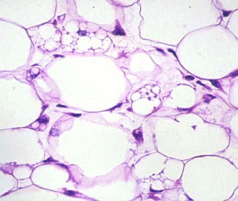
6 Cellular morphology & stroma Spindle Pleomorphic Lipomatous Epithelioid Round cell Myxoid
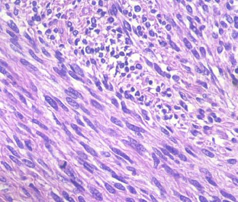
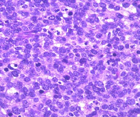
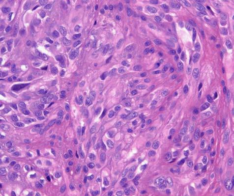
7 Small round & blue cell tumors

8 Small round & blue cell tumors Rhabdomyosarcoma Ewing sarcoma Ewing-like sarcomas Desmoplastic SRCT PD Syn S Myoepithelial tumors Mesenchymal Ch Neuroblastoma Lymphoma Small cell carcinoma Mainly in children & adolescents High-grade by definition Often translocated-sarcomas

9 Incidence Rates per Million Population, Children (0-14), Great Britain Bone and Soft Tissue Sarcomas: [50%] [15%]
10


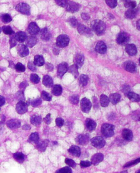
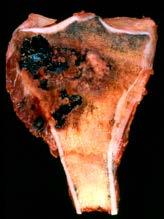
11 Neuroblastoma Clinical history Newborn, 7 m. Abdominal mass

12 Clinical history Boy, 12 y-o. Intraabdominal masses DSRCT


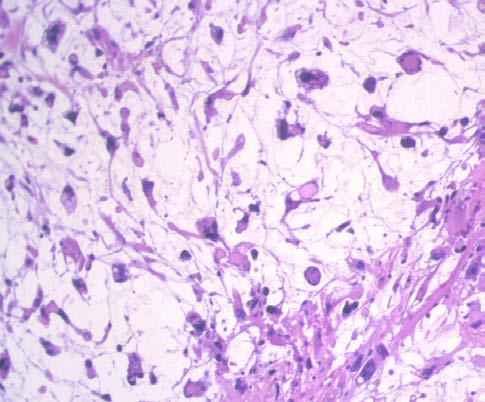
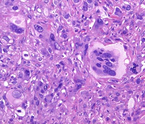
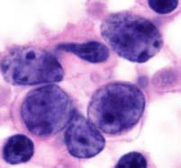
13 Mesenchymal chondrosarcoma Young adults (15-35 yrs) <10% chondrosarc Bone: maxilla, ribs, pelvis, vertebra 20-30%: soft tissue Biphasic pattern
 Standard method for establishing the line of differentiation (vascular, neural, smooth muscle, skeletal")

14 Immunohistochemistry Diferential diagnosis between mesenchymal/non-mesenchymal tumor (carcinoma, melanoma, lymphoma) Standard method for establishing the line of differentiation (vascular, neural, smooth muscle, skeletal muscle, myofibroblastic) Facilitates the differential diagnosis and classification of a spindle and / or round cell neoplasms Detection of underlying molecular alterations Do not provide information about benign or malignant LMS h-caldesmon
15 Immunohistochemistry of SRCT CD99 Desmin Myogenin CK20 LMWK TdT SYN EMA a-rms 15% 95% >90% Ewing sarc 95% 20% 20% 5% PD Syn Sarc 95% 50% 90% DSRCT WT1+ 10% 80% % - 5% 95% Mesenchymal Ch 80% Neuroblastoma % - TCLL 90% <1% 90% - -
")

 Ker")
16 M1 small cell carcinoma (lung) - Age & clinica data! - > 45 yrs; IMAGING! - CD99 (-) Ker AE1/AE3 TTF1
")

 CAM 5.")
17 Merkel cell carcinoma (skin) - Age & clinica data! - Elderly; superficial - CD99 (-) CAM 5.2 Ker 20


18 Ewing Sarcoma -3rd most common sarcoma in children % extraskeletal

19 Ewing Sarcoma


20 Immunohistochemical findings CD99 FLI-1 FLI-1 Membranous ~ 100% cases low specificity! Nuclear 71-84% cases Vimentin, synaptophysin, HNK1, CAV1, NSE: focally LMW cytokeratins in up to 30% of ES! Folpe et al. AJSP 2005; 29;
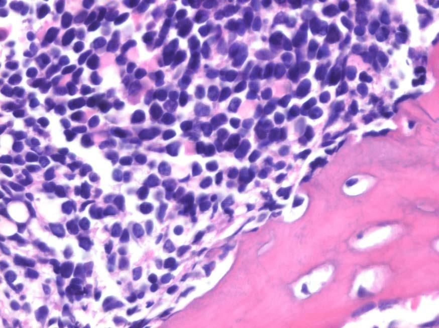
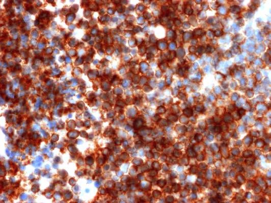

21 Lymphoblastic lymphoma Clinical history 2 year-old, female. Proximal left humerus CD99 TdT


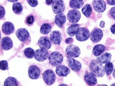
22 Clinical history 17 y-o, male Thigh mass + inguinal lymph node
23 Clinical history 17 y-o, male Thigh mass + inguinal lymph node FNA rhabdoid cells rhabdomyosarcoma??
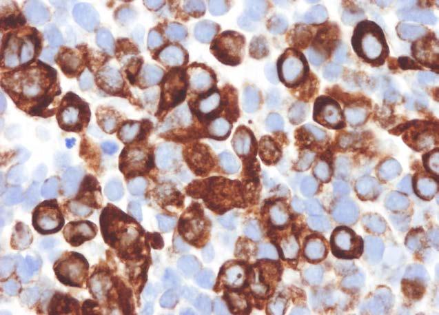
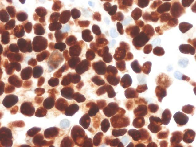

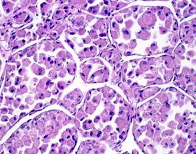
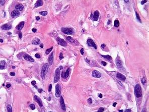
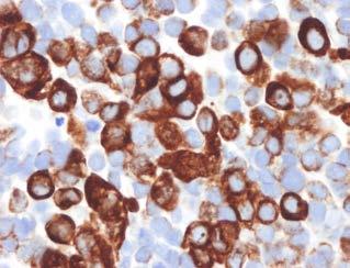
24 Rhabdomyosarcoma (skeletal muscle differentiation) Desmin Myogenin


(p13;q12) and")
25 DSRCT: IHC & Genetics Malignant small round cell tumor associated with prominent stromal desmoplasia and polyphenotypic differentiation with a consistent translocation t(11;22)(p13;q12) and EWSR1-WT1 gene fusion. WT1 CAM 5.2 Vim Desmin
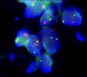
26 Molecular and cytogenetics
(q24;q12) EWSR1-FLI1 fusion gene <")

(q13;q12) t (4;19)(q35;q13)")
27 Ewing s sarcoma: Genetics 95% t(11;22)(q24;q12) EWSR1-FLI1 fusion gene < 5% t(21;22)(q22;q12) EWSR1-ERG fusion gene Sarcomas thouhgt to be Ewing but EWSR1 negative: Ewing-like sarcomas t(20;22)(q13;q12) t (4;19)(q35;q13) EWSR1-NFATC2 CIC-DUX4 BCOR-CCNB3


28 Clinical history 46 y-o female. Right thigh mass + pulmonary mets Small round cell tumor with hemangiopericytoid pattern FISH: SS18 gene rearrangement


29 Clinical history 17 y-o, male Thigh mass + inguinal lymph node FNA Genetics bp PAX FKHR + - Desm PGK + - PAX FKHR + - Myogenin 50% a-rms: t(2;13)(q35;q14) PAX3-FOXO1 25% a-rms: t(1;13)(p36;q14) PAX7-FOXO1 25% fusion-neg cases biologically similar to e-rms PGK FOXO1 rearrangement by FISH
30 Classification of Rhabdomyosarcoma Superior prognosis - Botryoid rhabdomyosarcoma - Spindle cell rhabdomyosarcoma Intermediate prognosis - Embryonal rhabdomyosarcoma Poor prognosis - Alveolar rhabdomyosarcoma - Anaplastic rhabdomyosarcoma Required for patient management and tumor prognostication!


31 Desmoplastic small round cell tumor Clinical history Boy, 12 y-o. Intraabdominal masses
(p13;q12) and EWSR1-WT1 gene fusion.")
32 DSRCT: IHC & Genetics Malignant small round cell tumor associated with prominent stromal desmoplasia and polyphenotypic differentiation with a consistent translocation t(11;22)(p13;q12) and EWSR1-WT1 gene fusion. FISH: EWSR1 rearrangement
33 Soft tissue tumors: Molecular pathology STT other than Ewing carrying EWS translocation Ewing sarcoma t(11;22)(q24;q12) EWSR1-FLI1 DSRCT t(11;22)(p13;q12) EWSR1-WT1 AFH CCS t(2;22)(q33;q12) t(12;22)(q13;q12) t(12;22)(q13;q12) t(2;22)(q33;q12) EWSR1-CREB1 EWSR1-ATF1 EWSR1-ATF1 EWSR1-CREB1 EMC t(9;22)(q22;q12) EWSR1-NR4A3 Myoepithelioma t(19;22)(q13;q12) EWSR1-POU5F1 Mixoid liposarc t(12;16)(q13;p11) t(12;22)(q13;q12) FUS-DDIT3 EWSR1-DDIT3


34 Clinical data & imaging CNB / FNA Specimen work up FNA ME sample CNB H&E Frozen (biobanking) IHC Molecular pathology Final diagnosis
35 In summary: Accurate diagnosis provides appropriate clinical decision making Histotype: predictor of outcome Conventional morphology still powerful tool.although overlapping histologic features create diagnostic challenges Integration with immunohistochemistry is a diagnostic standard Molecular genetics helpful in increasing situations - only in context with morphology! - same genetic alterations in unrelated entities
36 Muchas gracias por su atención
Cutaneous Mesenchymal Neoplasms with EWSR1 Rearrangement
 Cutaneous Mesenchymal Neoplasms with EWSR1 Rearrangement By Konstantinos Linos MD, FCAP, FASDP Bone, Soft Tissue and Dermatopathology Assistant Professor of Pathology Dartmouth-Hitchcock Medical Center
Disclosures. An update on ancillary techniques in the diagnosis of soft tissue tumors. Ancillary techniques. Introduction
 Disclosures An update on ancillary techniques in the diagnosis of soft tissue tumors. I have nothing to disclose. Andrew Horvai, MD, PhD Clinical Professor, Pathology Introduction Ancillary techniques
Surgical Pathology Evening Specialty Conference USCAP 2015
 Surgical Pathology Evening Specialty Conference USCAP 2015 John R. Goldblum, M.D. Chairman, Department of Pathology, Cleveland Clinic Professor of Pathology, Cleveland Clinic Lerner College of Medicine
Financial disclosures
 Cutaneous Mesenchymal Neoplasms with EWSR1 Rearrangement By Konstantinos Linos MD, FCAP, FASDP Bone, Soft Tissue and Dermatopathology Assistant Professor of Pathology Dartmouth-Hitchc Geisel School of
Klinisch belang van chromosomale translocatie detectie in sarcomen
 Translocations in sarcomas Klinisch belang van chromosomale translocatie detectie in sarcomen Judith V.M.G. Bovée, M.D., Ph.D. Department of Pathology Leiden University Medical Center RNA binding DNA binding
Disclosures. An update on ancillary techniques in the diagnosis of soft tissue tumors. Ancillary techniques. Introduction
 Disclosures An update on ancillary techniques in the diagnosis of soft tissue tumors. I have nothing to disclose. Andrew Horvai, MD, PhD Clinical Professor, Pathology Introduction Ancillary techniques
Disclosure of Relevant Financial Relationships
 Ewing and Ewing like sarcomas Using Genetic Signatures in Refining Small Blue Round Cell Tumor Classification Cristina Antonescu, MD Department of Pathology Disclosure of Relevant Financial Relationships
Case 2. Dr. Sathima Natarajan M.D. Kaiser Permanente Medical Center Sunset
 Case 2 Dr. Sathima Natarajan M.D. Kaiser Permanente Medical Center Sunset History 24 year old male presented with a 3 day history of right flank pain, sharp in nature Denies fever, chills, hematuria or
Immunohistochemistry in Bone and Soft Tissue Tumors. Sahar Rassi Zankoul, MD
 Immunohistochemistry in Bone and Soft Tissue Tumors Sahar Rassi Zankoul, MD Introduction Bone tumors represent a wide variety of tumors of various origins and malignant potentials. These different tumor
Clinical History. Pediatric Tumors with Involvement of the Head & Neck
 Pediatric Tumors with Involvement of the Head & Neck John Hicks Texas Children s Hospital Baylor College of Medicine Houston, TX NO DISCLOSURES Clinical History 10 Yr-Old Hispanic Male From Mexico with
Financial disclosures
 An update on immunohistochemical markers in mesenchymal neoplasms By Konstantinos Linos MD, FCAP, FASDP Assistant Professor of Pathology Geisel School of Medicine at Dartmouth Dartmouth-Hitchcock Medical
Recent Advances In Select Round Cell Sarcomas
 Recent Advances In Select Round Cell Sarcomas Rajiv M. Patel, M.D. Associate Professor of Pathology & Dermatology University of Michigan, Ann Arbor, MI rajivpat@med.umich.edu 1 Translocation Associated
Molecular pathology in soft tissue tumors. Sylvia Höller Pathologie
 Molecular pathology in soft tissue tumors Sylvia Höller Pathologie When do we perform molecular testing? Morphology and IHC are not clearly fitting with an entity some translocations are entity specific
The Genetics of Myoepithelial Tumors: salivary glands, soft tissue and bone
 The Genetics of Myoepithelial Tumors: salivary glands, soft tissue and bone Cristina Antonescu, MD Memorial Sloan-Kettering Cancer Center, New York Nothing to declare Disclosure Spectrum of Myoepithelial
Mojca Velikonja Jože Pižem
 Mojca Velikonja Jože Pižem An 81-year old woman presented with an exophytic, wart-like skin lesion on her neck that she had observed for one year. Cryotherapy had been applied twice, but proved unsuccessful.
Molecular Diagnosis of Soft Tissue Tumors: Avoid Pitfalls
 Molecular Diagnosis of Soft Tissue Tumors: Avoid Pitfalls Cristina Antonescu, MD Department of Pathology Memorial Sloan-Kettering Cancer Center, New York Overview I. When should we rely on the help of
An unusual superficial small round cell sarcoma
 An unusual superficial small round cell sarcoma - Diagnostic problems - Differential diagnosis Antonio Llombart Bosch Isidro Machado Dep. Pathology Univ. Medical School Valencia, Institute of Oncology
57th Annual HSCP Spring Symposium 4/16/2016
 An Unusual Malignant Spindle Cell Lesion to Involve the Breast Erinn Downs-Kelly, D.O. Associate Professor of Pathology University of Utah & ARUP Laboratories No disclosures Case 39 y/o female with no
Lung Tumor Cases: Common Problems and Helpful Hints
 Lung Tumor Cases: Common Problems and Helpful Hints Brandon T. Larsen, MD, PhD Senior Associate Consultant Department of Laboratory Medicine and Pathology Mayo Clinic Arizona Arizona Society of Pathologists
Enterprise Interest Nothing to declare
 Enterprise Interest Nothing to declare Diagnoses one would not like to miss in soft tissue pathology early in your career Marta Sbaraglia, MD Department of Pathology Hospital of Treviso University of Padua
Part 1. Slides 1-38, Rita Alaggio Soft tissue tumors Trondheim 14. mars 2013
 Part 1 Slides 1-38, Rita Alaggio Soft tissue tumors Trondheim 14. mars 2013 Pediatric Pathology Soft Tissue Tumors AN UPDATE Rita Alaggio Azienda Ospedaliera Università di Padova Soft Tissue Tumors More
Pediatric Soft Tissue Tumors
 Pediatric Soft Tissue Tumors Jerzy Klijanienko MD PhD MIAC Institut Curie Paris, France 1 - - General 2 - - Cancer incidence in children Type of malignancy % Hematology 38.6 CNS 19 Neuroblastoma 9.2 Kidney
Financial disclosures
 Mesenchymal Neoplasms with Melanocytic Differentiation By Konstantinos Linos MD, FCAP, FASDP Bone, Soft Tissue and Dermatopathology Assistant Professor of Pathology Dartmouth-Hitchcock Medical Center Geisel
From Morphology to Molecular Pathology: A Practical Approach for Cytopathologists Part 1-Cytomorphology. Songlin Zhang, MD, PhD LSUHSC-Shreveport
 From Morphology to Molecular Pathology: A Practical Approach for Cytopathologists Part 1-Cytomorphology Songlin Zhang, MD, PhD LSUHSC-Shreveport I have no Conflict of Interest. FNA on Lymphoproliferative
ESS: Pathologic Insights
 GEIS XVI INTERNATIONAL SYMPOSIUM Seville 4th October 2018 ESS: Pathologic Insights Sílvia Bagué The Royal Marsden Hospital London (United Kingdom) I have no conflicts of interest Endometrial stromal sarcoma
Diagnostic Value of Immunohistochemistry in Soft Tissue Tumors
 Original Article DOI: 10.21276/APALM.1637 Diagnostic Value of Immunohistochemistry in Soft Tissue Tumors Sridevi. V*., Susruthan Muralitharan., and Thanka. J Dept of Pathology, SriMuthukumaran Medical
Breast - ductal carcinoma CK7 ER PR GATA3 Mammaglobin (50-70%) GCDFP-15 (50-70%) E-cadherin HMWCK CK20 PAX2 ER/PR/HER2 on all newly diagnosed cases
 GCDFP-15 (50-70%) E-cadherin HMWCK CK20 PAX2 ER/PR/HER2 on all newly diagnosed cases") Adrenal cortical carcinoma Inhibin Synap Melan-A Calretinin Vimentin Chromogr CK7 CK20 Breast - ductal carcinoma CK7 ER PR GATA3 Mammaglobin (50-70%) GCDFP-15 (50-70%) E-cadherin HMWCK CK20 PAX2 ER/PR/HER2
Adrenal cortical carcinoma Inhibin Synap Melan-A Calretinin Vimentin Chromogr CK7 CK20 Breast - ductal carcinoma CK7 ER PR GATA3 Mammaglobin (50-70%) GCDFP-15 (50-70%) E-cadherin HMWCK CK20 PAX2 ER/PR/HER2
Protocol for the Examination of Biopsy Specimens From Pediatric Patients With Ewing Sarcoma
 Protocol for the Examination of Specimens From Pediatric Patients With Ewing Sarcoma Version: EwingSarcoma 4.0.0.0 Protocol Posting Date: February 2019 Accreditation Requirements The use of this protocol
Classification (1) Classification (3) Classification (2) Spindle cell lesions. Spindle cell lesions of bladder (Mills et al.
 Classification (3) Classification (2) Spindle cell lesions. Spindle cell lesions of bladder (Mills et al.") Non-epithelial tumours and nonepithelial tumour-like lesions of the bladder Dr Jonathan H Shanks The Christie NHS Foundation Trust, Manchester, UK Classification (1) Myofibroblastic proliferations and
Non-epithelial tumours and nonepithelial tumour-like lesions of the bladder Dr Jonathan H Shanks The Christie NHS Foundation Trust, Manchester, UK Classification (1) Myofibroblastic proliferations and
Small Round Cell Tumors in Soft Tissue and Bone. ACCME/Disclosure. Adult Small Round Cell Tumors. Ewing Sarcoma 4/13/2016
 Ultrastructural Pathology Society, Seattle, March, 2016 Small Round Cell Tumors in Soft Tissue and Bone ACCME/Disclosure Dr. Fisher has nothing to disclose Cyril Fisher MA MD DSc FRCPath Professor of Tumor
1/10/2018. Soft Tissue Tumors Showing Melanocytic Differentiation. Overview. Desmoplastic/ Spindle Cell Melanoma
 2016 MFMER slide-1 2016 MFMER slide-2 2016 MFMER slide-3 Soft Tissue Tumors Showing Melanocytic Differentiation Andrew L. Folpe, M.D. Professor of Laboratory Medicine and Pathology Mayo Clinic, Rochester,
Soft Tissue High Grade Myoepithelial Carcinoma With Round Cell Morphology: Report Of A Newly Described Entity With EWSR1 Gene Rearrangement
 Soft Tissue High Grade Myoepithelial Carcinoma With Round Cell Morphology: Report Of A Newly Described Entity With EWSR1 Gene Rearrangement Abstract M. El-Kabany, R. Al-Abdulghani, A. E. Ali, I. M. Francis,
Newer soft tissue entities
 Newer soft tissue entities Examples among fibroblastic tumors Turku, May 6, 2010 Markku Miettinen, M.D. AFIP, Washington, DC Fibroblastic neoplasms Solitary fibrous tumor /Hemangiopericytoma Low-grade
Shintaro Sugita *, Hiroko Asanuma and Tadashi Hasegawa
 Sugita et al. Diagnostic Pathology (2016) 11:37 DOI 10.1186/s13000-016-0486-2 RESEARCH Open Access Diagnostic use of fluorescence in situ hybridization in expert review in a phase 2 study of trabectedin
Update on Cutaneous Mesenchymal Tumors. Thomas Brenn
 Update on Cutaneous Mesenchymal Tumors Thomas Brenn Cutaneous Mesenchymal Tumours Wide morphological and biological spectrum Myofibroblastic, smooth muscle, neural, vascular, apidocytic, undifferentiated;
Fluorescence In Situ Hybridization in the Diagnosis of Soft Tissue Neoplasms: A Review. Munir R. Tanas, MD and John R.
 REVIEW ARTICLE Fluorescence In Situ Hybridization in the Diagnosis of Soft Tissue Neoplasms: A Review Munir R. Tanas, MD and John R. Goldblum, MD Abstract: This paper presents an overview of the role of
DIFFERENTIAL DIAGNOSIS OF SMALL ROUND CELL TUMOURS (SRCT), FLUORESCENCE IN SITU HYBRIDIZATION (FISH)
, FLUORESCENCE IN SITU HYBRIDIZATION (FISH)") POL J PATHOL 2009; 4: 151-162 DIFFERENTIAL DIAGNOSIS OF SMALL ROUND CELL TUMOURS (SRCT), FLUORESCENCE IN SITU HYBRIDIZATION (FISH) AND IMMUNOHISTOCHEMICAL (IHC) STUDY KONRAD PTASZYŃSKI 1, ANNA SZUMERA-CIEĆKIEWICZ
POL J PATHOL 2009; 4: 151-162 DIFFERENTIAL DIAGNOSIS OF SMALL ROUND CELL TUMOURS (SRCT), FLUORESCENCE IN SITU HYBRIDIZATION (FISH) AND IMMUNOHISTOCHEMICAL (IHC) STUDY KONRAD PTASZYŃSKI 1, ANNA SZUMERA-CIEĆKIEWICZ
Slide seminar: Soft tissue and bone pathology
 Slide seminar: Soft tissue and bone pathology Unusual tumors of bone and soft tissue or unusual presentations of common ones Gunhild Mechtersheimer Institute of Pathology, Heidelberg/DE (Sylvia Höller,
Pathology of Sarcoma ELEANOR CHEN, MD, PHD, ASSISTANT PROFESSOR DEPARTMENT OF PATHOLOGY UNIVERSITY OF WASHINGTON
 Pathology of Sarcoma ELEANOR CHEN, MD, PHD, ASSISTANT PROFESSOR DEPARTMENT OF PATHOLOGY UNIVERSITY OF WASHINGTON Presentation outline Background and epidemiology of sarcomas Sarcoma classification Sarcoma
Interesting Cases in Gynecologic Pathology. Michael Ward, MD Surgical Pathology Fellow University of Utah Health Sciences Center Salt Lake City, UT
 Interesting Cases in Gynecologic Pathology Michael Ward, MD Surgical Pathology Fellow University of Utah Health Sciences Center Salt Lake City, UT Case 1 History: 50 year old woman with a uterine mass
Slide Seminar Spanish Society of Pathology
 Slide Seminar Spanish Society of Pathology John R. Goldblum, M.D. Chairman, Department of Anatomic Pathology Cleveland Clinic Professor of Pathology Cleveland Clinic Lerner College of Medicine 1921 Original
IMMUNOHISTOCHEMISTRY IN THE DIAGNOSIS OF SOFT TISSUE TUMORS
 IMMUNOHISTOCHEMISTRY IN THE DIAGNOSIS OF SOFT TISSUE TUMORS Nicolas de Saint Aubain Somerhausen Institut Jules Bordet / Hôpital Erasme nicolas.desaintaubain@synet.be ForPath 2005 1 I. Ancillary techniques
Molecular pathology/genetics of sarcomas
 Molecular pathology/genetics of sarcomas Gunhild Mechtersheimer Institute of Pathology, University of Heidelberg Sarkomkonferenz: 17.03.2011 Berlin Characterization of soft tissue sarcomas / STS (~ 1%
Molecular Genetics of Paediatric Tumours. Gino Somers MBBS, BMedSci, PhD, FRCPA Pathologist-in-Chief Hospital for Sick Children, Toronto, ON, CANADA
 Molecular Genetics of Paediatric Tumours Gino Somers MBBS, BMedSci, PhD, FRCPA Pathologist-in-Chief Hospital for Sick Children, Toronto, ON, CANADA Financial Disclosure NanoString - conference costs for
ASCP Resident Review Mini-Course Series Session 2: Renal and Soft Tissue Pathology. Carole Vogler MD, FASCP Ema Dragoescu MD
 178 2011 ASCP Resident Review Mini-Course Series Session 2: Renal and Soft Tissue Pathology Carole Vogler MD, FASCP Ema Dragoescu MD 2011 Annual Meeting Las Vegas, NV AMERICAN SOCIETY FOR CLINICAL PATHOLOGY
Role of immunohistochemistry in the differential diagnosis of malignant small round cell tumor: a study of 38 cases
 International Journal of Research in Medical Sciences Patel A et al. Int J Res Med Sci. 2015 Dec;3(12):3833-3839 www.msjonline.org pissn 2320-6071 eissn 2320-6012 Research Article DOI: http://dx.doi.org/10.18203/2320-6012.ijrms20151452
LOOK-ALIKES IN SPINDLE AND EPITHELIOID TUMORS: Immunohistochemistry. Cytogenetics Flow cytometry Molecular diagnostics
 LOOK-ALIKES IN SPINDLE AND EPITHELIOID TUMORS: Ultrastructural value and pitfalls in diagnosis Guillermo A Herrera Department of Pathology and Translational Pathobiology Louisiana State University Health
Paediatric small round cell sarcomas an update
 1 Paediatric small round cell sarcomas an update International Society of Bone and Soft Tissue Pathology meeting, Baltimore, MD March 3, 2013 David M. Parham, MD Oklahoma University Health Science Center
Presentation material is for education purposes only. All rights reserved URMC Radiology Page 1 of 98
 Presentation material is for education purposes only. All rights reserved. 2011 URMC Radiology Page 1 of 98 Radiology / Pathology Conference February 2011 Brooke Koltz, Cytopathology Resident Presentation
Case 1. Disclosure. Imaging. Clinical history 5/10/2016. USCAP 2016 Annual Meeting Evening Specialty Conference Bone and Soft tissue Pathology
 Disclosure Dr. Agaram has nothing to disclose Case 1 Narsi Agaram, MBBS USCAP 2016 Annual Meeting Evening Specialty Conference Bone and Soft tissue Pathology Clinical history Imaging 1998 A three month
Keywords solitary fibrous tumor, dedifferentiation, dedifferentiated solitary fibrous tumor, STAT6, GRIA2, cytokeratin, rhabdomyosarcomatous
 758452IJSXXX10.1177/1066896918758452International Journal of Surgical PathologyCreytens et al research-article2018 Pitfalls in Pathology Multifocal Cytokeratin Expression in a Dedifferentiated Solitary
ACCME/Disclosures ALK FUSION-POSITIVE MESENCHYMAL TUMORS. Tumor types with ALK rearrangements. Anaplastic Lymphoma Kinase. Jason L.
 Companion Meeting of the International Society of Bone and Soft Tissue Pathology The Evolving Concept of Mesenchymal Tumors ALK FUSION-POSITIVE MESENCHYMAL TUMORS Jason L. Hornick, MD, PhD March 13, 2016
Rhabdomyomas and Rhabdomyosarcomas (RMS) David M. Parham, MD Chief of Anatomic Pathology
 David M. Parham, MD Chief of Anatomic Pathology") Rhabdomyomas and Rhabdomyosarcomas (RMS) David M. Parham, MD Chief of Anatomic Pathology Tumors of skeletal muscle: Rhabdomyomas and rhabdomyosarcomas Embryonal muscle 2 3 4 5 6 7 8 Rhabdomyoma Benign
Rhabdomyomas and Rhabdomyosarcomas (RMS) David M. Parham, MD Chief of Anatomic Pathology Tumors of skeletal muscle: Rhabdomyomas and rhabdomyosarcomas Embryonal muscle 2 3 4 5 6 7 8 Rhabdomyoma Benign
Schedule of Accreditation issued by United Kingdom Accreditation Service 2 Pine Trees, Chertsey Lane, Staines-upon-Thames, TW18 3HR, UK
 2 Pine Trees, Chertsey Lane, Staines-upon-Thames, TW18 3HR, UK Royal National Orthopaedic Hospital NHS Trust Brockley Hill Stanmore Middlesex HA7 4LP Contact: Professor Adrienne Flanagan Tel: +44 (0)20
4/12/2018. MUSC Pathology Symposium Kiawah Island April 18, Jesse K. McKenney, MD
 MUSC Pathology Symposium Kiawah Island April 18, 2018 Jesse K. McKenney, MD 1 Urothelial Carcinoma with Alternative Differentiation 2 Urothelial Carcinoma with Alternative Differentiation Recognition as
Case Report Solid-pattern desmoplastic small round cell tumor of the orbit: a case report
 Int J Clin Exp Pathol 2018;11(5):2864-2868 www.ijcep.com /ISSN:1936-2625/IJCEP0071553 Case Report Solid-pattern desmoplastic small round cell tumor of the orbit: a case report Pei Wang 1, Yixiong Liu 2,
Diplomate of the American Board of Pathology in Anatomic and Clinical Pathology
 A 33-year-old male with a left lower leg mass. Contributed by Shaoxiong Chen, MD, PhD Assistant Professor Indiana University School of Medicine/ IU Health Partners Department of Pathology and Laboratory
Journal of Solid Tumors, April 2012, Vol. 2, No. 2
 ORIGINAL ARTICLE Utility of fluorescence in situ hybridization in subclassifying unclassified high-grade sarcomas: A study of 40 cases using break-apart probes of EWSR1, FOXO1A, SS18 and DDIT3 genes Alfredo
Aspen conference on pediatric disease. July through August Bone and Soft Tissue Update. David M. Parham, MD. Rhabdomyoma and rhabdomyosarcoma
 Aspen conference on pediatric disease July through August 2014 Bone and Soft Tissue Update David M. Parham, MD Rhabdomyoma and rhabdomyosarcoma Embryonic rhabdomyogenesis is a highly conserved process
Enterprise Interest No disclosures.
 Enterprise Interest No disclosures. Secondary Tumours in Uropathology Case 2 PRESENTED AT: EUROPEAN CONGRESS OF PATHOLOGY 18 #ECP2018 Slides are the property of the author. Permission required for reuse.
What is New in the 2015 WHO Lung Cancer Classification? Zhaolin Xu, MD, FRCPC, FCAP
 What is New in the 2015 WHO Lung Cancer Classification? Zhaolin Xu, MD, FRCPC, FCAP Professor, Dept of Pathology, Dalhousie University, Canada Pulmonary Pathologist and Cytopathologist, QEII HSC Senior
No financial or other disclosures
 Case 2014-5 Esther N. Bit-Ivan, DO Northwestern University Jason Wang, MD Jason Park, MD Korgun Koral, MD Children s Medical Center Charles Timmons, MD Veena Rajaram, MD No financial or other disclosures
The role of immunohistochemistry in surgical pathology of the uterine corpus and cervix
 The role of immunohistochemistry in surgical pathology of the uterine corpus and cervix Prof. Ben Davidson, MD PhD Department of Pathology, Norwegian Radium Hospital, Oslo University Hospital, Oslo, Norway
Enterprise Interest None
 Enterprise Interest None Pauline M. Chou, M.D Professor of Pathology Ann and Robert H. Lurie Children s Hospital Lurie Cancer Center Northwestern University Feinberg School of Medicine, Chicago European
Salivary Gland FNA ATYPICAL : Criteria and Controversies
 Salivary Gland FNA ATYPICAL : Criteria and Controversies W.C. Faquin, M.D., Ph.D. Director, Head and Neck Pathology Massachusetts General Hospital Massachusetts Eye and Ear Infirmary Harvard Medical School
Spindle Cell Lesions Of The Breast. Emad Rakha Professor of Breast Pathology and Consultant Pathologist
 Spindle Cell Lesions Of The Breast Emad Rakha Professor of Breast Pathology and Consultant Pathologist * SCLs comprise a wide spectrum of diseases, ranging from reactive processes to aggressive malignant
Intrarenal Extension. sinus
 Intrarenal Extension into sinus Document Capsular Penetration sinus 16 Pediatric Renal Tumor Staging Stage I Limited to Kidney & Completely Resected Intact Renal Capsule No Previous Rupture or Biopsy Renal
Original Articles. Utilization of Fluorescence In Situ Hybridization in the Diagnosis of 230 Mesenchymal Neoplasms. An Institutional Experience
 Original Articles Utilization of Fluorescence In Situ Hybridization in the Diagnosis of 230 Mesenchymal Neoplasms An Institutional Experience Munir R. Tanas, MD; Brian P. Rubin, MD, PhD; Raymond R. Tubbs,
Case Report A SMARCB1-deficient vulvar neoplasm with prominent myxoid stroma: report of a case showing ERG and FLI1 expression
 Int J Clin Exp Pathol 2015;8(6):7526-7532 www.ijcep.com /ISSN:1936-2625/IJCEP0008225 Case Report A SMARCB1-deficient vulvar neoplasm with prominent myxoid stroma: report of a case showing ERG and FLI1
Problem 1: Differential of Neuroendocrine Carcinoma 3/23/2017. Disclosure of Relevant Financial Relationships
 Differential of Neuroendocrine Carcinoma Alain C. Borczuk,MD Weill Cornell Medicine Disclosure of Relevant Financial Relationships USCAP requires that all faculty in a position to influence or control
Small (and large) Blue Cell Tumors of the Skull Base
 Blue Cell Tumors of the Skull Base") Small (and large) Blue Cell Tumors of the Skull Base Jennifer L. Hunt, MD, MEd Aubrey J. Hough Jr, MD, Endowed Professor of Pathology Chair of Pathology and Laboratory Medicine University of Arkansas for
Small (and large) Blue Cell Tumors of the Skull Base Jennifer L. Hunt, MD, MEd Aubrey J. Hough Jr, MD, Endowed Professor of Pathology Chair of Pathology and Laboratory Medicine University of Arkansas for
Case 4 Diagnosis 2/21/2011 TGB
 Case 4 22 year old female presented with asymmetric enlargement of the left lobe of the thyroid gland. No information available relative to a prior fine needle aspiration biopsy. A left lobectomy was performed.
Case year old female presented with asymmetric enlargement of the left lobe of the thyroid
 Case 4 22 year old female presented with asymmetric enlargement of the left lobe of the thyroid gland. No information available relative to a prior fine needle aspiration biopsy. A left lobectomy was performed.
Institut für Pathologie, Friedrich-Alexander Universität Erlangen-Nürnberg, Erlangen, Germany
 DOI: https://doi.org/10.5114/pjp.2017.71535p pol J Pathol 2017; 68 (3): 261-267 Case report EWSR1-fusion-negative, SMARCB1-deficient primary pulmonary myxoid sarcoma Abbas Agaimy 1, Thomas Duell 2, Alicia
Published in: British Journal of Cancer. Document Version: Publisher's PDF, also known as Version of record
 at a tertiary sarcoma centre; assessing 772 tumour specimens and the value of current ancillary molecular diagnostic modalities Noujaim, J., Jones, R. L., Swansbury, J., Gonzalez, D., Benson, C., Judson,
Protocol for the Examination of Specimens From Pediatric Patients With Ewing Sarcoma*
 Protocol for the Examination of Specimens From Pediatric Patients With Ewing Sarcoma* Version: Protocol Posting Date: August 2016 This protocol is NOT required for accreditation purposes. *This protocol
Current and future applications of Molecular Pathology. Kathy Walsh Clinical Scientist NHS Lothian
 Current and future applications of Molecular Pathology Kathy Walsh Clinical Scientist NHS Lothian Molecular Pathology in Solid tumours Cancer type Genes tested Purpose Associated treatments Non small cell
La chemioterapia neoadiuvante nei sarcomi: novità e attuali indicazioni Lorenzo D Ambrosio, MD PhD Divisione di Oncologia Medica Istituto di Candiolo
 La chemioterapia neoadiuvante nei sarcomi: novità e attuali indicazioni Lorenzo D Ambrosio, MD PhD Divisione di Oncologia Medica Istituto di Candiolo Fondazione del Piemonte per l Oncologia. IRCCS 12 CONGRESSO
SOFT TISSUE TUMOR PATHOLOGY: AN UPDATE
 SOFT TISSUE TUMOR PATHOLOGY: AN UPDATE Jason L. Hornick, MD, PhD July 18, 2013 Department of Pathology Brigham and Women s Hospital Harvard Medical School Boston, MA, USA I have no disclosures. New Soft
5/10. Pathology Soft tissue tumors. Farah Bhani. Mohammed Alorjani
 5/10 Pathology Soft tissue tumors Mohammed Alorjani Farah Bhani Slides are included in this sheet. Objectives: Soft tissue tumors 1. Describe soft tissue tumors. 2. Understand the classification of soft
Objectives. Salivary Gland FNA: The Milan System. Role of Salivary Gland FNA 04/26/2018
 Salivary Gland FNA: The Milan System Dr. Jennifer Brainard Section Head Cytopathology Cleveland Clinic Objectives Introduce the Milan System for reporting salivary gland cytopathology Define cytologic
Applications of IHC. Determination of the primary site in metastatic tumors of unknown origin
 Applications of IHC Determination of the primary site in metastatic tumors of unknown origin Classification of tumors that appear 'undifferentiated' by standard light microscopy Precise classification
PITFALLS AND TRAPS IN THE DIAGNOSIS AND STAGING OF RENAL TUMOURS OF CHILDHOOD. Gordan M. Vujanić Cardiff, U.K.
 PITFALLS AND TRAPS IN THE DIAGNOSIS AND STAGING OF RENAL TUMOURS OF CHILDHOOD Gordan M. Vujanić Cardiff, U.K. RENAL TUMOURS OF CHILDHOOD - CLASSIFICATION (2016) Nephroblastic tumours Mesenchymal tumours
ACCME/Disclosures. Diagnosing Mesothelioma in Limited Tissue Samples. Papanicolaou Society of Cytopathology Companion Meeting March 12 th, 2016
 Diagnosing Mesothelioma in Limited Tissue Samples Papanicolaou Society of Cytopathology Companion Meeting March 12 th, 2016 Sanja Dacic, MD, PhD University of Pittsburgh ACCME/Disclosures GENERAL RULES
Technology from Abcam
 CD2 (EP222) CD2 is one of the earliest T-cell lineage restricted antigens to appear during T-cell differentiation and only rare CD2+ cells can be found in the bone marrow. Anti-CD2 is a pan-t-cell antigen
Minimally invasive adenocarcinoma. 5mm or less = microinvasion No necrosis No lymphatic or pleural invasion No spread through air-spaces (STAS)
") Minimally invasive adenocarcinoma 5mm or less = microinvasion No necrosis No lymphatic or pleural invasion No spread through air-spaces (STAS) 2b lepidic acinar 2c papillary micropapillary solid 2d cribriform
Minimally invasive adenocarcinoma 5mm or less = microinvasion No necrosis No lymphatic or pleural invasion No spread through air-spaces (STAS) 2b lepidic acinar 2c papillary micropapillary solid 2d cribriform
Evening Specialty Conference Bone and Soft Tissue Pathology. Diagnostic pitfalls in bone and soft tissue pathology
 Evening Specialty Conference Bone and Soft Tissue Pathology. Case 1 Elizabeth G Demicco, MD, PhD Mount Sinai Hospital, New York Disclosure of Relevant Financial Relationships USCAP requires that all planners
Disclosure of Relevant Financial Relationships
 Evening Specialty Conference - Genitourinary Pathology Case 2 Disclosure of Relevant Financial Relationships Sean R Williamson, MD Henry Ford Health System, Detroit, MI @Williamson_SR USCAP requires that
HOW MAY THE CLASSIFICATION OF SOFT TISSUE TUMORS EVOLVE?
 Spanish Society of Pathology Zaragoza, May 2011 ARTHUR PURDY STOUT SYMPOSIUM HOW MAY THE CLASSIFICATION OF SOFT TISSUE TUMORS EVOLVE? Christopher D.M. Fletcher, M.D., FRCPath Brigham and Women s Hospital
A 25 year old female with a palpable mass in the right lower quadrant of her abdomen
 May 2016 A 25 year old female with a palpable mass in the right lower quadrant of her abdomen Contributed by: Paul Ndekwe, MD, Resident Physician, Indiana University School of Department of Pathology and
GUT-C 11/30/2017. Debasmita Das, M.D. PGY-1 Danbury Hospital
 GUT-C 11/30/2017 Debasmita Das, M.D. PGY-1 Danbury Hospital CLINICAL SUMMARY 8/2017 59 year old female Presented to the ED with 1 month history of general malaise, fever and weight loss PMH: Significant
Update: Morphologic Considerations in Mesothelioma within the Pleural and Peritoneal Cavities. Douglas J. Hartman, MD June 7, 2018
 Update: Morphologic Considerations in Mesothelioma within the Pleural and Peritoneal Cavities Douglas J. Hartman, MD June 7, 2018 Objectives Review Historical Features associated with prognosis Present
Mayo Medical Laboratories
 Mayo Medical Laboratories Virtual Lectures 2014 MFMER 2016 MFMER slide-1 Virtual Lectures Planning Committee Disclosure Summary As a provider accredited by ACCME, College of Medicine, Mayo Clinic (Mayo
Mesothelioma: diagnostic challenges from a pathological perspective. Naseema Vorajee August 2016
 Mesothelioma: diagnostic challenges from a pathological perspective Naseema Vorajee August 2016 Naseema.vorajee@nhls.ac.za Pleural diseases (whether neoplastic, reactive or infective) may have similar
USCAP COMPANION MEETING INTERNATIONAL SOCIETY OF BONE AND SOFT TISSUE PATHOLOGY DENVER, March 2 nd 2008
 1 USCAP COMPANION MEETING INTERNATIONAL SOCIETY OF BONE AND SOFT TISSUE PATHOLOGY DENVER, March 2 nd 2008 THE EVOLUTION OF SOFT TISSUE TUMOUR TAXONOMY: WHAT STILL NEEDS TO BE DONE? Christopher D.M. Fletcher,
Renal tumours: use of immunohistochemistry & molecular pathology. Dr Lisa Browning John Radcliffe Hospital Oxford
 Renal tumours: use of immunohistochemistry & molecular pathology Dr Lisa Browning John Radcliffe Hospital Oxford Renal tumours: the use of immunohistochemistry & molecular pathology Classification of RCC
Biopsy Interpretation of Spindle cell proliferations of the Serosa
 Biopsy Interpretation of Spindle cell proliferations of the Serosa Richard Attanoos, Cardiff. U.K. Disclosure of Relevant Financial Relationships USCAP requires that all planners (Education Committee)
CASO 1. Xavier Matias-Guiu Hospital Universitari Arnau de Vilanova, Universitat de Lleida, IRBLLEIDA.
 CASO 1 Xavier Matias-Guiu Hospital Universitari Arnau de Vilanova, Universitat de Lleida, IRBLLEIDA. ASSESSMENT OF PRIMARY ORIGIN IN PATIENTS WITH A PREVIOUS HISTORY OF CANCER Primary Tumor T1 T5 T6 T13
Charles Halsey, DVM, PhD, DACVP Pfizer, Inc. IHC Resources
 Charles Halsey, DVM, PhD, DACVP Pfizer, Inc. IHC Resources 1 IHC Identification Targets Specimens Controls 2 Tissue controls Trouble Spots 3 The Key to Description IHC Description 4 Intermediate Filaments
Differential diagnosis of hematolymphoid tumors composed of medium-sized cells. Brian Skinnider B.C. Cancer Agency, Vancouver General Hospital
 Differential diagnosis of hematolymphoid tumors composed of medium-sized cells Brian Skinnider B.C. Cancer Agency, Vancouver General Hospital Lymphoma classification Lymphoma diagnosis starts with morphologic