Receptor-interacting Protein Kinases Mediate Necroptosis In Neural Tissue Damage After Spinal Cord Injury
|
|
|
- Tyler Neil Dawson
- 5 years ago
- Views:
Transcription
1 Receptor-interacting Protein Kinases Mediate Necroptosis In Neural Tissue Damage After Spinal Cord Injury Haruo Kanno, M.D., Ph.D., Hiroshi Ozawa, M.D., Ph.D., Satoshi Tateda, M.D., Kenichiro Yahata, M.D., Eiji Itoi, MD, PhD. Tohoku University School of Medicine, Sendai, Japan. Disclosures: H. Kanno: None. H. Ozawa: None. S. Tateda: None. K. Yahata: None. E. Itoi: None. Introduction: Necrosis was originally thought to be a purely passive and uncontrolled type of cell death. Necrosis, which is marked by cell swelling and membrane rupture, leads to inflammation via the release of intracellular signals [1,2]. In contrast, apoptosis, which is characterized by the activation of caspases and DNA fragmentation, was once considered the sole form of programmed cell death. Chromatin condensation, nuclear shrinkage and fragmentation and membrane blebbing are the result of the proteolytic activity of caspases and define the morphological characteristics of apoptosis [1-3]. Necroptosis is a newly identified type of programmed cell death regulated by caspase-independent pathway and has the morphological features of necrosis [4]. Recent studies have shown that the receptor-interacting protein (RIP) kinase family is specifically involved in regulating necroptosis [2]. Various stimuli to cells can induce the formation of necrotic complexes that contain RIP1, TRADD, FADD and caspase-8. The interaction of RIP3 with RIP1 in the necrotic complex is an important step required for the execution of necroptosis [2]. RIP3 and RIP1 are known as a key mediator of necroptosis [5]. The amounts of RIP3 and RIP1 expressions correlate with necroptosis induction in various types of cells. Recent studies demonstrated that the increased expression of RIP3 and RIP1 in lesions and the induction of necroptosis in various disease models [6]. However, to our knowledge, there has been no study to investigate the expression of RIP proteins and the involvement of necroptosis in damaged neural tissue after spinal cord injury (SCI). In the present study, we used a spinal cord hemisection model in mice to study the alterations of RIP3 and RIP1 expressions and the involvement of necroptosis in SCI. Methods: Animals Adult female C57BL/6J mice between 8 and 10 weeks of age were used in this study. Surgical procedures The T10 vertebra was laminectomized to expose the spinal cord. With a scalpel, the cord was transected on the left side only [7]. Immunohistochemistry At different time points (4, 24 hours, 3, 7 and 21 days) after hemisection and at 24 hours after sham operation, the spinal cords at the lesion were fixed with 4% paraformaldehyde, embedded in OCT-compound and sectioned. Tissue sections were incubated with rabbit anti-rip3 antibody (1:100; Sigma-Aldrich) or mouse anti-rip1 antibody (1:100; BD Transduction Laboratories) and visualized by Alexa Fluor 594 goat anti-rabbit IgG antibody or Alexa Fluor 594 goat anti-mouse IgG antibody (1:500; Molecular Probes). Counting of RIP3- and RIP1-positive cells The numbers of RIP3- and RIP1-positive cells were counted in the injured and the contralateral sides of transverse sections at the different time points (4, 24 hours, 3, 7 and 21 days) after hemisection, and compared with those of the sham control. Western blot analysis The protein expressions of RIP3 in the injured spinal cord and the uninjured spinal cord after sham operation were determined using Western blot analysis.
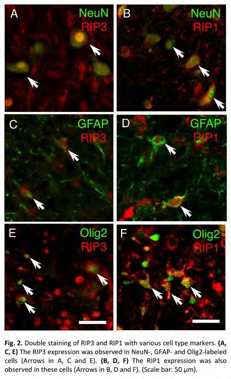
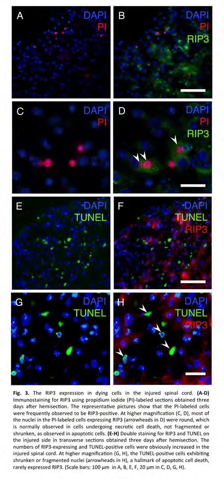
2 Double staining of RIP3 and RIP1 with various cell type markers To examine the expressions of RIP3 and RIP1 in a specific population of cells, the transverse sections at 3 days after hemisection were costained for RIP3 and RIP1 with various cell type markers: NeuN for neurons, GFAP for astrocytes and Olig2 for oligodendrocytes. RIP3 staining in propidium iodide-labeled sections To investigate the RIP3 expression in dying cells, RIP3 staining was performed using propidium iodide (PI)-labeled spinal cord sections. Double staining for RIP3 and TUNEL To detect DNA fragmentation in the RIP3-positive cells, the sections were co-stained with RIP3 and TUNEL. Results: Immunohistochemical stainings of RIP3 and RIP1 Representative pictures showed the RIP3- and RIP1-expressing cells on the injured side obviously increased compared to on the contralateral side at 3 days after hemisection (Fig. 1A-D). Counting of RIP3- and RIP1-positive cells The number of RIP3-positive cells on the injured side was significantly higher than those on the contralateral side and sham control at 24 hours, 3, 7 and 21days (p < 0.05) (Fig. 1E). The number of RIP1-positive cells on the injured side was significantly higher than those on the contralateral side and sham control at 4, 24 hours, 3, 7 and 21 days (p < 0.05) (Fig. 1F). The maximum number of these cells in the injured side was observed at 3 days, and it decreased after 7 days. Western blot analysis The protein expression of RIP3 in the injured spinal cord at 3 days was significantly increased compared to the uninjured spinal cord (p < 0.05) (Fig. 1G, H). Double staining of RIP3 and RIP1 with various cell type markers Double staining with various cell type markers demonstrated the increased expression of RIP3 in neurons, astrocytes and oligodendrocytes on the injured side at 3 days after hemisection (Fig. 2A, C, E). The increased expression of RIP1 was also observed in neurons, astrocytes and oligodendrocytes on the injured side (Fig. 2B, D, F). RIP3 staining in propidium iodide-labeled sections The representative pictures show that the PI-labeled cells were frequently observed to be RIP3-positive (Fig. 3A-D). At higher magnification (Fig. 3C, D), most of the nuclei in the PI-labeled cells expressing RIP3 were round, which is normally observed in cells undergoing necrotic cell death, not fragmented or shrunken, as observed in apoptotic cells. Double staining for RIP3 and TUNEL The numbers of RIP3-expressing and TUNEL-positive cells were obviously increased in the injured spinal cord (Fig. 3E-H). At higher magnification (Fig. 3G, H), the TUNELpositive cells exhibiting shrunken or fragmented nuclei, a hallmark of apoptotic cell death, rarely expressed RIP3. Discussion: The present study demonstrated that the expressions of RIP3 and RIP1 were significantly increased in various neural cells at the lesion site after SCI. In addition, the dying cells expressing RIP3 in the injured spinal cord displayed a similar morphology to that associated with necrotic cell death but not apoptosis. These results first provided the evidence to support the involvement of necroptosis in neural tissue damage after SCI. To date, it has been considered that the secondary neural tissue damage following SCI is caused by apoptosis but not necrosis. Most previous studies related to the cell death in the injured spinal cord focused on apoptosis. In our study, the upregulation of RIP3 and RIP1 expressions was observed starting from 4-24 hours, peaked at 3 days. Importantly, the time course of RIP3 and RIP1 expressions was quite similar to that of the secondary damage following SCI [8]. These results suggest necroptosis contributes to the secondary damage in the injured spinal cord.

3 Significance: The expressions of RIP3 and RIP1 were dramatically increased at the lesion site and may contribute to induction of necroptosis as novel cell death mechanism causing secondary neural tissue damage after SCI. Therefore, necroptosis can be a novel therapeutic target to reduce neural tissue damage following SCI.
4
5
6 ORS 2015 Annual Meeting Poster No: 0689
Department of Orthopaedic Surgery, Tohoku University Graduate School of Medicine, Sendai, Japan, 2
 Low-energy Extracorporeal Shock Wave Therapy Promotes VEGF Expression and Angiogenesis and Improve Locomotor and Sensory Functions after spinal cord injury Kenichiro Yahata 1, Hiroshi Ozawa, M.D., Ph.D.
Rapamycin Suppresses Astrocytic and Microglial Activation and Reduced Development of Neuropathic Pain after Spinal Cord Injury in Mice.
 Rapamycin Suppresses Astrocytic and Microglial Activation and Reduced Development of Neuropathic Pain after Spinal Cord Injury in Mice. Satoshi Tateda, M.D., Haruo Kanno, M.D., Ph.D., Hiroshi Ozawa, M.D.,
Rapamycin suppresses astrocytic and microglial activation and reduces development of neuropathic pain after spinal cord injury in mice.
 Rapamycin suppresses astrocytic and microglial activation and reduces development of neuropathic pain after spinal cord injury in mice. Satoshi Tateda, MD, Haruo Kanno, MD, PhD, Hiroshi Ozawa, MD, PhD,
Page 39 of 44. 8h LTA & AT h PepG & AT h LTA
 Page 39 of 44 Fig. S1 A: B: C: D: 8h LTA 8h LTA & AT7519 E: F: 8h PepG G: 8h PepG & AT7519 Fig. S1. AT7519 overrides the survival effects of lipoteichoic acid (LTA) and peptidoglycan (PepG). (A) Human
Programmed necrosis, not apoptosis, is a key mediator of cell loss and DAMP-mediated inflammation in dsrna-induced retinal degeneration
 Programmed necrosis, not apoptosis, is a key mediator of cell loss and DAMP-mediated inflammation in dsrna-induced retinal degeneration The Harvard community has made this article openly available. Please
Cell cycle and apoptosis
 Cell cycle and apoptosis Cell cycle Definition Stages and steps Cell cycle Interphase (G1/G0, S, and G2) Mitosis (prophase, metaphase, anaphase, telophase, karyokinesis, cytokinesis) Control checkpoints
Reactive astrocytes undergo M1 microglia/ macrohpages-induced necroptosis in spinal cord injury
 Fan et al. Molecular Neurodegeneration (2016) 11:14 DOI 10.1186/s13024-016-0081-8 RESEARCH ARTICLE Open Access Reactive astrocytes undergo M1 microglia/ macrohpages-induced necroptosis in spinal cord injury
Mass Histology Service
 Mass Histology Service A complete anatomical pathology laboratory www.masshistology.com Telephone: (877) 286-6004 Report on Pathology A Time Course Study of the Local Effects of Intramuscular XXXXXXX Injection
Supplementary Figure 1
 Supplementary Figure 1 The average sigmoid parametric curves of capillary dilation time courses and average time to 50% peak capillary diameter dilation computed from individual capillary responses averaged
Caractérisation et méthodes d études de la mort cellulaire par cytométrie en flux
 Caractérisation et méthodes d études de la mort cellulaire par cytométrie en flux Université de Bourgogne Gérard LIZARD - Inserm EA7270 - Equipe Biochimie du Peroxysome, inflammation et Métabolisme Lipidique
Is Discography or Discoblock Safe for Human Intervertebral Disc cells?
 Is Discography or Discoblock Safe for Human Intervertebral Disc cells? KOJI IWASAKI 1, Hideki Sudo 2, Katsuhisa Yamada 1, Norimasa Iwasaki 1. 1 Department of Orthopedic Surgery, Hokkaido University Graduate
- 1 - Cell types Monocytes THP-1 cells Macrophages. LPS Treatment time (Hour) IL-6 level (pg/ml)
 IL-6 level (pg/ml)") Supplementary Table ST1: The dynamic effect of LPS on IL-6 production in monocytes and THP-1 cells after GdA treatment. Monocytes, THP-1 cells and macrophages (5x10 5 ) were incubated with 10 μg/ml of
Supplementary Table ST1: The dynamic effect of LPS on IL-6 production in monocytes and THP-1 cells after GdA treatment. Monocytes, THP-1 cells and macrophages (5x10 5 ) were incubated with 10 μg/ml of
Bone marrow-derived mesenchymal stem cells improve diabetes-induced cognitive impairment by
 Nakano et al. Supplementary information 1. Supplementary Figure 2. Methods 3. References Bone marrow-derived mesenchymal stem cells improve diabetes-induced cognitive impairment by exosome transfer into
SUPPLEMENTARY FIG. S2. Representative counting fields used in quantification of the in vitro neural differentiation of pattern of dnscs.
 Supplementary Data SUPPLEMENTARY FIG. S1. Representative counting fields used in quantification of the in vitro neural differentiation of pattern of anpcs. A panel of lineage-specific markers were used
APOPTOSIS, NECROSIS AND CANCER. Dr. S. P. Pattanayak
 APOPTOSIS, NECROSIS AND CANCER Dr. S. P. Pattanayak LEARNING OBJECTIVES At the end of the lecture, students should be able to: Know the importance of cell death. Define various modes of cell death. Identify
Neuronal Death After Hemorrhagic Stroke In Vitro and In Vivo Shares Features of Ferroptosis and Necroptosis
 Neuronal Death After Hemorrhagic Stroke In Vitro and In Vivo Shares Features of Ferroptosis and Necroptosis Marietta Zille, PhD Burke Medical Research Institute Weill Cornell Medicine White Plains, NY
Impact factor: Reporter:4A1H0019 Chen Zi Hao 4A1H0023 Huang Wan ting 4A1H0039 Sue Yi Zhu 4A1H0070 Lin Guan cheng 4A1H0077 Chen Bo xuan
 Curcumin Protects Neonatal Rat Cardiomyocytes against High Glucose-Induced Apoptosis via PI3K/Akt Signalling Pathway Wei Yu,1,2 Wenliang Zha,1 Zhiqiang Ke,1 Qing Min,2 Cairong Li,1 Huirong Sun,3 and Chao
Supplementary Information
 Supplementary Information Title Degeneration and impaired regeneration of gray matter oligodendrocytes in amyotrophic lateral sclerosis Authors Shin H. Kang, Ying Li, Masahiro Fukaya, Ileana Lorenzini,
Supplementary Figure S I: Effects of D4F on body weight and serum lipids in apoe -/- mice.
 Supplementary Figures: Supplementary Figure S I: Effects of D4F on body weight and serum lipids in apoe -/- mice. Male apoe -/- mice were fed a high-fat diet for 8 weeks, and given PBS (model group) or
Circulating Microrna Expression As A Biomarker For Early Diagnosis Of Severity In Spinal Cord Injury
 Circulating Microrna Expression As A Biomarker For Early Diagnosis Of Severity In Spinal Cord Injury Susumu Hachisuka 1, Naosuke Kamei, MD, PhD 2, Satoshi Ujigo 3, Shigeru Miyaki 3, Mitsuo Ochi 3. 1 Hiroshima
To determine the effect of over-expression and/or ligand activation of. PPAR / on cell cycle, cell lines were cultured as described above until ~80%
 Supplementary Materials and Methods Cell cycle analysis To determine the effect of over-expression and/or ligand activation of PPAR / on cell cycle, cell lines were cultured as described above until ~80%
The Annexin V Apoptosis Assay
 The Annexin V Apoptosis Assay Development of the Annexin V Apoptosis Assay: 1990 Andree at al. found that a protein, Vascular Anticoagulant α, bound to phospholipid bilayers in a calcium dependent manner.
Supplementary Figure 1: Hsp60 / IEC mice are embryonically lethal (A) Light microscopic pictures show mouse embryos at developmental stage E12.
 Light microscopic pictures show mouse embryos at developmental stage E12.") Supplementary Figure 1: Hsp60 / IEC mice are embryonically lethal (A) Light microscopic pictures show mouse embryos at developmental stage E12.5 and E13.5 prepared from uteri of dams and subsequently genotyped.
Supplementary Figure 1: Hsp60 / IEC mice are embryonically lethal (A) Light microscopic pictures show mouse embryos at developmental stage E12.5 and E13.5 prepared from uteri of dams and subsequently genotyped.
Genesis of cerebellar interneurons and the prevention of neural DNA damage require XRCC1.
 Genesis of cerebellar interneurons and the prevention of neural DNA damage require XRCC1. Youngsoo Lee, Sachin Katyal, Yang Li, Sherif F. El-Khamisy, Helen R. Russell, Keith W. Caldecott and Peter J. McKinnon.
Introduction to pathology lecture 5/ Cell injury apoptosis. Dr H Awad 2017/18
 Introduction to pathology lecture 5/ Cell injury apoptosis Dr H Awad 2017/18 Apoptosis = programmed cell death = cell suicide= individual cell death Apoptosis cell death induced by a tightly regulated
marker. DAPI labels nuclei. Flies were 20 days old. Scale bar is 5 µm. Ctrl is
 Supplementary Figure 1. (a) Nos is detected in glial cells in both control and GFAP R79H transgenic flies (arrows), but not in deletion mutant Nos Δ15 animals. Repo is a glial cell marker. DAPI labels
Siglec-15 Is A Potential Therapeutic Target For Postmenopausal Osteoporosis
 Siglec-15 Is A Potential Therapeutic Target For Postmenopausal Osteoporosis Yusuke Kameda, Masahiko Takahata, Tomohiro Shimizu, Hiroki Hamano, Norimasa Iwasaki. Department of Orthopedic Surgery, Hokkaido
Sestrin2 and BNIP3 (Bcl-2/adenovirus E1B 19kDa-interacting. protein3) regulate autophagy and mitophagy in renal tubular cells in. acute kidney injury
 regulate autophagy and mitophagy in renal tubular cells in. acute kidney injury") Sestrin2 and BNIP3 (Bcl-2/adenovirus E1B 19kDa-interacting protein3) regulate autophagy and mitophagy in renal tubular cells in acute kidney injury by Masayuki Ishihara 1, Madoka Urushido 2, Kazu Hamada
Sestrin2 and BNIP3 (Bcl-2/adenovirus E1B 19kDa-interacting protein3) regulate autophagy and mitophagy in renal tubular cells in acute kidney injury by Masayuki Ishihara 1, Madoka Urushido 2, Kazu Hamada
Studying apoptosis in DT40 cells
 Studying apoptosis in DT40 cells Sandrine Ruchaud E12.5! Acridine Orange! Role of apoptosis in sculpting the mouse paw E13.5! E14.5! gift of William Wood & Paul Martin! University College, London! Normal
Supplemental Figure 1. Intracranial transduction of a modified ptomo lentiviral vector in the mouse
 Supplemental figure legends Supplemental Figure 1. Intracranial transduction of a modified ptomo lentiviral vector in the mouse hippocampus targets GFAP-positive but not NeuN-positive cells. (A) Stereotaxic
Effect of low temperatures on BAX and BCL2 proteins in rats with spinal cord ischemia reperfusion injury
 Effect of low temperatures on BAX and BCL2 proteins in rats with spinal cord ischemia reperfusion injury P. Zhu 1 *, M.Y. Zhao 1,2 *, X.H. Li 1 *, Q. Fu 1, Z.F. Zhou 1, C.F. Huang 1, X.S. Zhang 1, H.L.
Supplemental Table 1. Primers used for RT-PCR analysis of inflammatory cytokines Gene Primer Sequence
 Supplemental Table 1. Primers used for RT-PCR analysis of inflammatory cytokines Gene Primer Sequence IL-1α Forward primer 5 -CAAGATGGCCAAAGTTCGTGAC-3' Reverse primer 5 -GTCTCATGAAGTGAGCCATAGC-3 IL-1β
SUPPLEMENTARY FIGURES
 SUPPLEMENTARY FIGURES 1 Supplementary Figure 1, Adult hippocampal QNPs and TAPs uniformly express REST a-b) Confocal images of adult hippocampal mouse sections showing GFAP (green), Sox2 (red), and REST
Apoptosis Chapter 9. Neelu Yadav PhD
 Apoptosis Chapter 9 Neelu Yadav PhD Neelu.Yadav@Roswellpark.org 1 Apoptosis: Lecture outline Apoptosis a programmed cell death pathway in normal homeostasis Core Apoptosis cascade is conserved Compare
Figure S 1. S1. Histological evaluation of lateral hemisection.
 Dorsal Central Ventral Figure S1. Histological evaluation of lateral hemisection. Schematic Figure S1. Histological evaluation of lateral hemisection. Schematic representation of hemisection at. Dashed
PE Annexin V Apoptosis Detection Kit User Manual KT40001
 PE Annexin V Apoptosis Detection Kit User Manual KT40001 For research use only. Not intended for diagnostic testing. a WuXi AppTec company www.abgent.com.cn PE Annexin-V Apoptosis Detection Kit Product
c Ischemia (30 min) Reperfusion (8 w) Supplementary Figure bp 300 bp Ischemia (30 min) Reperfusion (4 h) Dox 20 mg/kg i.p.
 Reperfusion (8 w) Supplementary Figure bp 300 bp Ischemia (30 min) Reperfusion (4 h) Dox 20 mg/kg i.p.") a Marker Ripk3 +/ 5 bp 3 bp b Ischemia (3 min) Reperfusion (4 h) d 2 mg/kg i.p. 1 w 5 w Sacrifice for IF size A subset for echocardiography and morphological analysis c Ischemia (3 min) Reperfusion (8
a Marker Ripk3 +/ 5 bp 3 bp b Ischemia (3 min) Reperfusion (4 h) d 2 mg/kg i.p. 1 w 5 w Sacrifice for IF size A subset for echocardiography and morphological analysis c Ischemia (3 min) Reperfusion (8
ERK1/2/MAPK pathway-dependent regulation of the telomeric factor TRF2
 ERK1/2/MAPK pathway-dependent regulation of the telomeric factor TRF2 SUPPLEMENTARY FIGURES AND TABLE Supplementary Figure S1: Conservation of the D domain throughout evolution. Alignment of TRF2 sequences
Original Article Protective effects of LM22A-4 on injured spinal cord nerves
 Int J Clin Exp Pathol 2015;8(6):6526-6532 www.ijcep.com /ISSN:1936-2625/IJCEP0008748 Original Article Protective effects of LM22A-4 on injured spinal cord nerves Guangzhe Yu 1, Wenbo Wang 2 Departments
B-cell. Astrocyte SCI SCI. T-cell
 RF #2015 P-01 PI: Azizul Haque, PhD Grant Title: Targeting Enolase in Spinal Cord Injury 12-month Technical Progress Report Progress Report (First Six Months): Enolase is one of the most abundantly expressed
Product Datasheet. Caspase-3 Antibody - (active/cleaved) NB Unit Size: 0.05 ml. Store at -20C. Avoid freeze-thaw cycles.
 NB Unit Size: 0.05 ml. Store at -20C. Avoid freeze-thaw cycles.") Product Datasheet Caspase-3 Antibody - (active/cleaved) NB100-56113 Unit Size: 0.05 ml Store at -20C. Avoid freeze-thaw cycles. Publications: 16 Protocols, Publications, Related Products, Reviews, Research
Product Datasheet Caspase-3 Antibody - (active/cleaved) NB100-56113 Unit Size: 0.05 ml Store at -20C. Avoid freeze-thaw cycles. Publications: 16 Protocols, Publications, Related Products, Reviews, Research
Supplementary Figure 1 P53 is degraded following Chlamydia infection independent of the cell lysis and protein sample preparation procedure applied.
 Supplementary Figure 1 P53 is degraded following Chlamydia infection independent of the cell lysis and protein sample preparation procedure applied. (a) Western blotting analysis showing degradation of
Key Words spinal cord injury; extracorporeal shock wave therapy; vascular endothelial growth factor
 Laboratory investigation J Neurosurg Spine 25:745 755, 2016 Low-energy extracorporeal shock wave therapy for promotion of vascular endothelial growth factor expression and angiogenesis and improvement
Name Animal source Vendor Cat # Dilutions
 Supplementary data Table S1. Primary and Secondary antibody sources Devi et al, TXNIP in mitophagy A. Primary Antibodies Name Animal source Vendor Cat # Dilutions 1. TXNIP mouse MBL KO205-2 1:2000 (WB)
Probe. Hind III Q,!?R'!! /0!!!!D1"?R'! vector. Homologous recombination
 Supple-Zhang Page 1 Wild-type locus Targeting construct Targeted allele Exon Exon3 Exon Probe P1 P P3 FRT FRT loxp loxp neo vector amh I Homologous recombination neo P1 P P3 FLPe recombination Q,!?R'!!
Chapter 1 CELL INJURY CELL DEATH CELL ADAPTATIONS. M.G.Rajanandh, Dept. of Pharmacy Practice, SRM College of Pharmacy, SRM University.
 Chapter 1 CELL INJURY CELL DEATH CELL ADAPTATIONS M.G.Rajanandh, Dept. of Pharmacy Practice, SRM College of Pharmacy, SRM University. CONCEPTS IN CELL INJURY The clinical signs and symptoms are several
Cord blood monocytes as a source of cell therapy products for treatment of brain injuries ISCT/CBA 2015 Cord Blood Workshop Wednesday, May 27, 2015
 Cord blood monocytes as a source of cell therapy products for treatment of brain injuries ISCT/CBA 2015 Cord Blood Workshop Wednesday, May 27, 2015 Andrew E. Balber, PhD Senior Scientific Advisor CT 2,
Chapter 7: Modes of Cell Death
 Chapter 7: Modes of Cell Death 7.1. Background Cell death can follow one of two distinct pathways, apoptosis or necrosis, and can occur in response to severe stress conditions or after exposure to toxic
PREPARED BY P.DHARANI PRASAD II YEAR B.PHARM II SEM SUB:PATHOPHYSIOLOGY
 CELL INJURY UNIT I PREPARED BY P.DHARANI PRASAD II YEAR B.PHARM II SEM SUB:PATHOPHYSIOLOGY DETECTION OF CELLULAR CHANGES AFTER INJURY BY: LIGHT MICROSCOPY OR GROSS EXAMINATION DETECT CHANGES HOURS TO DAYS
Neocortex Zbtb20 / NFIA / Sox9
 Neocortex / NFIA / Sox9 Supplementary Figure 1. Expression of, NFIA, and Sox9 in the mouse neocortex at. The lower panels are higher magnification views of the oxed area. Arrowheads indicate triple-positive
Multi-Parameter Apoptosis Assay Kit
 Multi-Parameter Apoptosis Assay Kit Catalog Number KA1335 5 x 96 assays Version: 05 Intended for research use only www.abnova.com Table of Contents Introduction... 3 Background... 3 Principle of the Assay...
Erzsebet Kokovay, Susan Goderie, Yue Wang, Steve Lotz, Gang Lin, Yu Sun, Badrinath Roysam, Qin Shen,
 Cell Stem Cell, Volume 7 Supplemental Information Adult SVZ Lineage Cells Home to and Leave the Vascular Niche via Differential Responses to SDF1/CXCR4 Signaling Erzsebet Kokovay, Susan Goderie, Yue Wang,
PK15 48 (1) :58 63, (intermediate filament, IF) D ( Traub et al., 1994) IF P K15 D. ( Fuchs et al., 1994), , ( Fuchs et al., 1992) (apoptosis)
 :58 63, (intermediate filament, IF) D ( Traub et al., 1994) IF P K15 D. ( Fuchs et al., 1994), , ( Fuchs et al., 1992) (apoptosis)") 48 (1) :58 63, 2002 A cta Zoologica S inica 3 PK15 (, 100871) D P K15 ( Porcrne Kidney215) DNA, DNA ladder ;,,,, P K15 D (intermediate filament, IF) D ( Traub et al., 1994) IF P K15 ( Porcine Kidney215),,
48 (1) :58 63, 2002 A cta Zoologica S inica 3 PK15 (, 100871) D P K15 ( Porcrne Kidney215) DNA, DNA ladder ;,,,, P K15 D (intermediate filament, IF) D ( Traub et al., 1994) IF P K15 ( Porcine Kidney215),,
An unconventional role for mirna: let-7 activates Toll-like receptor 7 and causes neurodegeneration
 An unconventional role for mirna: let-7 activates Toll-like receptor 7 and causes neurodegeneration Sabrina M. Lehmann, Christina Krüger, Boyoun Park, Katja Derkow, Karen Rosenberger, Jan Baumgart, Thorsten
Fang et al. NMuMG. PyVmT unstained Anti-CCR2-PE MDA-MB MCF MCF10A
 A NMuMG PyVmT 16.5+.5 47.+7.2 Fang et al. unstained Anti-CCR2-PE 4T1 Control 37.6+6.3 56.1+.65 MCF1A 16.1+3. MCF-7 3.1+5.4 MDA-M-231 42.1+5.5 unstained Secondary antibody only Anti-CCR2 SUPPLEMENTAL FIGURE
Nature Neuroscience: doi: /nn Supplementary Figure 1
 Supplementary Figure 1 EGFR inhibition activates signaling pathways (a-b) EGFR inhibition activates signaling pathways (a) U251EGFR cells were treated with erlotinib (1µM) for the indicated times followed
Supplementary Figure 1: si-craf but not si-braf sensitizes tumor cells to radiation.
 Supplementary Figure 1: si-craf but not si-braf sensitizes tumor cells to radiation. (a) Embryonic fibroblasts isolated from wildtype (WT), BRAF -/-, or CRAF -/- mice were irradiated (6 Gy) and DNA damage
Kinase Inhibitor p21 WAF1/CIP1 in Apoptosis and Autophagy
 Pivotal Role of the Cyclin-dependent Kinase Inhibitor p21 WAF1/CIP1 in Apoptosis and Autophagy Keishi Fujiwara, Shigeru Daido, Akitsugu Yamamoto, Ryuji Kobayash, Tomohisa Yokoyama, Hiroshi Aok, Eiji Iwado,
HCC1937 is the HCC1937-pcDNA3 cell line, which was derived from a breast cancer with a mutation
 SUPPLEMENTARY INFORMATION Materials and Methods Human cell lines and culture conditions HCC1937 is the HCC1937-pcDNA3 cell line, which was derived from a breast cancer with a mutation in exon 20 of BRCA1
The Pathogenesis of Chlamydia pneumoniae in Multiple Sclerosis: Current Thoughts and Future Directions
 The Pathogenesis of Chlamydia pneumoniae in Multiple Sclerosis: Current Thoughts and Future Directions Seminars in Pathology March 9, 2010 Charles W. Stratton, M.D. Features of C. pneumoniae Infection
Acute lung injury in children : from viral infection and mechanical ventilation to inflammation and apoptosis Bern, R.A.
 UvA-DARE (Digital Academic Repository) Acute lung injury in children : from viral infection and mechanical ventilation to inflammation and apoptosis Bern, R.A. Link to publication Citation for published
Comparison of Herpes Simplex Virus Reactivation in Ganglia In Vivo and in Explants Demonstrates Quantitative and Qualitative Differences
 JOURNAL OF VIROLOGY, July 2004, p. 7784 7794 Vol. 78, No. 14 0022-538X/04/$08.00 0 DOI: 10.1128/JVI.78.14.7784 7794.2004 Copyright 2004, American Society for Microbiology. All Rights Reserved. Comparison
Supporting Information
 Supporting Information Fujishita et al. 10.1073/pnas.0800041105 SI Text Polyp Scoring. Intestinal polyps were counted as described (1). Briefly, the small and large intestines were excised, washed with
(A) RT-PCR for components of the Shh/Gli pathway in normal fetus cell (MRC-5) and a
 RT-PCR for components of the Shh/Gli pathway in normal fetus cell (MRC-5) and a") Supplementary figure legends Supplementary Figure 1. Expression of Shh signaling components in a panel of gastric cancer. (A) RT-PCR for components of the Shh/Gli pathway in normal fetus cell (MRC-5) and
Supplementary figure legends Supplementary Figure 1. Expression of Shh signaling components in a panel of gastric cancer. (A) RT-PCR for components of the Shh/Gli pathway in normal fetus cell (MRC-5) and
#19 Apoptosis Chapter 9. Neelu Yadav PhD
 #19 Apoptosis Chapter 9 Neelu Yadav PhD Neelu.Yadav@Roswellpark.org Why cells decide to die? - Stress, harmful, not needed - Completed its life span Death stimulation or Stress Cell Survival Death Functions
Nature Medicine: doi: /nm.4322
 1 2 3 4 5 6 7 8 9 10 11 Supplementary Figure 1. Predicted RNA structure of 3 UTR and sequence alignment of deleted nucleotides. (a) Predicted RNA secondary structure of ZIKV 3 UTR. The stem-loop structure
Supplementary Figure 1. Validation of astrocytes. Primary astrocytes were
 Supplementary Figure 1. Validation of astrocytes. Primary astrocytes were separated from the glial cultures using a mild trypsinization protocol. Anti-glial fibrillary acidic protein (GFAP) immunofluorescent
C-Phycocyanin (C-PC) is a n«sjfc&c- waefc-jduble phycobiliprotein. pigment isolated from Spirulina platensis. This water- soluble protein pigment is
 is a n«sjfc&c- waefc-jduble phycobiliprotein. pigment isolated from Spirulina platensis. This water- soluble protein pigment is") ' ^Summary C-Phycocyanin (C-PC) is a n«sjfc&c- waefc-jduble phycobiliprotein pigment isolated from Spirulina platensis. This water- soluble protein pigment is of greater importance because of its various
' ^Summary C-Phycocyanin (C-PC) is a n«sjfc&c- waefc-jduble phycobiliprotein pigment isolated from Spirulina platensis. This water- soluble protein pigment is of greater importance because of its various
Supplementary Figure 1. Expression of CUGBP1 in non-parenchymal liver cells treated with TGF-β
 Supplementary Figures Supplementary Figure 1. Expression of CUGBP1 in non-parenchymal liver cells treated with TGF-β and LPS. Non-parenchymal liver cells were isolated and treated with or without TGF-β
hexahistidine tagged GRP78 devoid of the KDEL motif (GRP78-His) on SDS-PAGE. This
 on SDS-PAGE. This") SUPPLEMENTAL FIGURE LEGEND Fig. S1. Generation and characterization of. (A) Coomassie staining of soluble hexahistidine tagged GRP78 devoid of the KDEL motif (GRP78-His) on SDS-PAGE. This protein was expressed
SUPPLEMENTAL FIGURE LEGEND Fig. S1. Generation and characterization of. (A) Coomassie staining of soluble hexahistidine tagged GRP78 devoid of the KDEL motif (GRP78-His) on SDS-PAGE. This protein was expressed
Mechanisms of Cell Injury
 Causes of Cell Injury 1- oxygen deprivation (anoxia) 2- physical agents 3- chemical agents 4- infections agents 5- immunologic reactions 6- genetic defects 7- nutritional imbalances Mechanisms of Cell
Electrical Stimulation Control Nerve Regeneration via the p38 Mitogen-activated Protein Kinase and CREB
 Electrical Stimulation Control Nerve Regeneration via the p38 Mitogen-activated Protein Kinase and CREB Kenji Kawamura, Yoshio Kano. Kibi International University, Takahashi-city, Japan. Disclosures: K.
OVARY The surface of the ovary is covered with surface epithelium
 OVARY Cow The ovary, or female gonad, is: 1. an exocrine gland, producing oocytes 2. an endocrine gland, secreting hormones, i.e., estrogen and progesterone OVARY OVARY The surface of the ovary is covered
Wnt7a Inhibits Cartilage Matrix Degradation in a Mouse In Vivo Osteoarthritis Model
 Wnt7a Inhibits Cartilage Matrix Degradation in a Mouse In Vivo Osteoarthritis Model Averi Leahy, Andrea Foote, Tomoya Uchimura, Li Zeng, PhD. Tufts University, Boston, MA, USA. Disclosures: A. Leahy: None.
Problem Set 8 Key 1 of 8
 7.06 2003 Problem Set 8 Key 1 of 8 7.06 2003 Problem Set 8 Key 1. As a bright MD/PhD, you are interested in questions about the control of cell number in the body. Recently, you've seen three patients
Supplemental Figure 1. (A) The localization of Cre DNA recombinase in the testis of Cyp19a1-Cre mice was detected by immunohistchemical analyses
 The localization of Cre DNA recombinase in the testis of Cyp19a1-Cre mice was detected by immunohistchemical analyses") Supplemental Figure 1. (A) The localization of Cre DNA recombinase in the testis of Cyp19a1-Cre mice was detected by immunohistchemical analyses using an anti-cre antibody; testes at 1 week (left panel),
Supplemental Figure 1. (A) The localization of Cre DNA recombinase in the testis of Cyp19a1-Cre mice was detected by immunohistchemical analyses using an anti-cre antibody; testes at 1 week (left panel),
CHAPTER 4 RESULTS AND DISCUSSION. chemistry, polar substances would dissolve in polar solvents while non-polar substances
 CHAPTER 4 RESULTS AND DISCUSSION 4.1 Extraction yield of Alpinia scabra Samples of A. scabra were dried in an oven as a method of preservation after which the samples were ground to powder. The latter
NOTES. Apoptosis Plays an Important Role in Experimental Rabies Virus Infection. Received 8 January 1997/Accepted 14 March 1997
 JOURNAL OF VIROLOGY, July 1997, p. 5603 5607 Vol. 71, No. 7 0022-538X/97/$04.00 0 Copyright 1997, American Society for Microbiology NOTES Apoptosis Plays an Important Role in Experimental Rabies Virus
Supplemental Information. Tissue Myeloid Progenitors Differentiate. into Pericytes through TGF-b Signaling. in Developing Skin Vasculature
 Cell Reports, Volume 18 Supplemental Information Tissue Myeloid Progenitors Differentiate into Pericytes through TGF-b Signaling in Developing Skin Vasculature Tomoko Yamazaki, Ani Nalbandian, Yutaka Uchida,
Supporting Information
 Supporting Information Sasportas et al. 10.1073/pnas.0806647106 SI Methods Lentiviral Transduction. MSC and glioma cells were transduced with LVs at varying multiplicity of infection (moi) by incubating
Effective Targeting of Quiescent Chronic Myelogenous
 Cancer Cell, Volume 7 Supplemental Information Effective Targeting of Quiescent Chronic Myelogenous Leukemia Stem Cells by Histone Deacetylase Inhibitors in Combination with Imatinib Mesylate Bin Zhang,
Supplementary Information Supplementary Fig. 1. Elevated Usp9x in melanoma and NRAS mutant melanoma cells are dependent on NRAS for 3D growth.
 Supplementary Information Supplementary Fig. 1. Elevated Usp9x in melanoma and NRAS mutant melanoma cells are dependent on NRAS for 3D growth. a. Immunoblot for Usp9x protein in NRAS mutant melanoma cells
Under the Radar Screen: How Bugs Trick Our Immune Defenses
 Under the Radar Screen: How Bugs Trick Our Immune Defenses Session 8: Apoptosis Marie-Eve Paquet and Gijsbert Grotenbreg Whitehead Institute for Biomedical Research Myxoma virus Poxvirus Infects rabbits
Primary Cilia are Expressed on a Small Fraction of Cells in Trabecular Bone and Marrow
 Primary Cilia are Expressed on a Small Fraction of Cells in Trabecular Bone and Marrow Thomas R. Coughlin 1, Muriel Voisin, Ph.D. 2, Glen L. Niebur, PhD 1, Laoise M. McNamara 2. 1 University of Notre Dame,
Chemical Chaperones Mitigate Experimental Asthma By Attenuating Endoplasmic
 Chemical Chaperones Mitigate Experimental Asthma By Attenuating Endoplasmic Reticulum Stress Lokesh Makhija, BE, Veda Krishnan, MSc, Rakhshinda Rehman, MTech, Samarpana Chakraborty, MSc, Shuvadeep Maity,
Mechanisms of Cell Death
 Mechanisms of Cell Death CELL DEATH AND FORMATION OF THE SEMICIRCULAR CANALS Carol M. Troy August 25, 2008 FROM: Fekete et al., Development 124: 2451 (1997) PHENOMENOLOGY OF CELL DEATH I. DEVELOPMENT A.
SUPPLEMENTARY INFORMATION
 DOI: 10.1038/ncb2535 Figure S1 SOX10 is expressed in human giant congenital nevi and its expression in human melanoma samples suggests that SOX10 functions in a MITF-independent manner. a, b, Representative
Li et al. Journal of Experimental & Clinical Cancer Research (2018) 37:108
 37:108") Li et al. Journal of Experimental & Clinical Cancer Research (2018) 37:108 https://doi.org/10.1186/s13046-018-0774-7 CORRECTION Correction to: Novel smac mimetic APG- 1387 elicits ovarian cancer cell killing
Li et al. Journal of Experimental & Clinical Cancer Research (2018) 37:108 https://doi.org/10.1186/s13046-018-0774-7 CORRECTION Correction to: Novel smac mimetic APG- 1387 elicits ovarian cancer cell killing
Supplementary Information
 Supplementary Information Figure S1. Int6 gene silencing efficiency. (A) Western Blot analysis of Int6 expression at different times after sirna transfection. Int6 expression is strongly silenced in Int6
Supplementary Figures
 Supplementary Figures Figure S1. Validation of kinase regulators of ONC201 sensitivity. Validation and screen results for changes in cell viability associated with the combination of ONC201 treatment (1
Fluorescence Microscopy
 Fluorescence Microscopy Imaging Organelles Mitochondria Lysosomes Nuclei Endoplasmic Reticulum Plasma Membrane F-Actin AAT Bioquest Introduction: Organelle-Selective Stains Organelles are tiny, specialized
Dual role of SIRT1 in UVB-induced skin tumorigenesis
 Dual role of SIRT1 in UVB-induced skin tumorigenesis Mei Ming 1, Keyoumars Soltani 1, Christopher R. Shea 1, Xiaoling Li 2, and Yu-Ying He 1 * 1 Section of Dermatology, Department of Medicine, University
Identification and characterization of genes responsive to apoptosis: Application of DNA chip technology and mrna differential display
 Histol Histopathol (2000) 15: 1271-1 284 http://www.ehu.es/histol-histopathol Histology and H istopat hology Cellular and Molecular Biology Invited Revie W Identification and characterization of genes
In vivo reprogramming reactive glia into ipscs to produce new neurons in the
 In vivo reprogramming reactive glia into ipscs to produce new neurons in the cortex following traumatic brain injury Xiang Gao 1, Xiaoting Wang 1, Wenhui Xiong 1, Jinhui Chen 1, * 1 Spinal Cord and Brain
Impact of Sox9 Dosage and Hes1-mediated Notch Signaling in Controlling the Plasticity of Adult Pancreatic Duct Cells in Mice
 Impact of Sox9 Dosage and Hes1-mediated Notch Signaling in Controlling the Plasticity of Adult Pancreatic Duct Cells in Mice Shinichi Hosokawa 1,3,Kenichiro Furuyama 1,3, Masashi Horiguchi 1,3,Yoshiki
Supporting Information. FADD regulates NF-кB activation and promotes ubiquitination of cflip L to induce. apoptosis
 1 2 Supporting Information 3 4 5 FADD regulates NF-кB activation and promotes ubiquitination of cflip L to induce apoptosis 6 7 Kishu Ranjan and Chandramani Pathak* 8 9 Department of Cell Biology, School
Cell Death & Renewal (part 2)
") 17 Cell Death & Renewal (part 2) Programmed Cell Death A major signaling pathway that promotes cell survival is initiated by the enzyme PI 3-kinase, which phosphorylates PIP2 to form PIP3, which activates
17 Cell Death & Renewal (part 2) Programmed Cell Death A major signaling pathway that promotes cell survival is initiated by the enzyme PI 3-kinase, which phosphorylates PIP2 to form PIP3, which activates
Nature Neuroscience: doi: /nn.2275
 Supplementary Figure S1. The presence of MeCP2 in enriched primary glial cultures from rat or mouse brains is not neuronal. Western blot analysis of protein extracts from (a) rat glial and neuronal cultures.
SCIRF Award #2016 I-03 PI: Azizul Haque, PhD Grant Title: Neuron-specific Enolase and SCI
 SCIRF Award #2016 I-03 PI: Azizul Haque, PhD Grant Title: Neuron-specific Enolase and SCI 10-month Technical Progress Report Enolase is a multifunctional glycolytic enzyme involved in growth control, hypoxia,
number Done by Corrected by Doctor Heyam Awad
 number 4 Done by Waseem Abu Obeida Corrected by Saad Al-Hayek Doctor Heyam Awad Cell injury -in the previous lectures we talked about the causes (etiology) and the mechanism (pathogenesis) of cell injury.