Proliferative Epithelial lesions of the Breast. Sami Shousha, MD, FRCPath Charing Cross Hospital & Imperial College, London
|
|
|
- Gyles Lewis
- 5 years ago
- Views:
Transcription
1 Proliferative Epithelial lesions of the Breast Sami Shousha, MD, FRCPath Charing Cross Hospital & Imperial College, London Amman, November2013
2 Proliferative Epithelial Lesions of the Breast Usual type ductal hyperplasia Columnar cell change Columnar cell Hyperplasia Flat epithelial atypia Atypical ductal hyperplasia Intraductal/ intracystic papillary lesions In situ lobular neoplasia
3 Proliferative Epithelial Lesions of the Breast Usual type ductal hyperplasia Columnar cell change Columnar cell Hyperplasia Flat epithelial atypia Atypical ductal hyperplasia Intraductal/ intracystic papillary lesions In situ lobular neoplasia

4 Florid Regular Hyperplasia The proliferating cells are similar to the normal cells lining the ducts The cells are haphazardly arranged, may overlap or have a streaming arrangement When spaces are present, they are irregular and mostly peripheral An occasional mitotic figure may be present
5
6
7 Florid Regular Hyperplasia vs. Low grade DCIS If still in doubt: Stain for cytokeratin 5

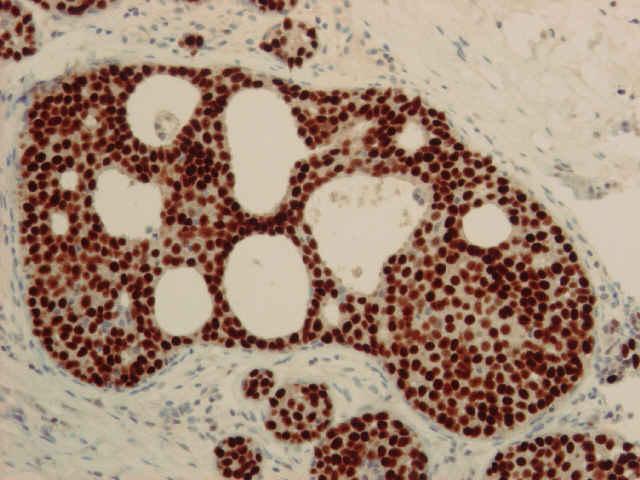
8 CK 5 There is a mixture of positive and negative cells (polyclonal)
9 CK 5 Malignant (& atypical) cells are CK 5 negative (monoclonal)

10 CK5/6 ER Diagnosis: Florid usual type uctal hyperplasia


11 F20, Right breast lump CK5/6 Diagnosis: Complex Sclerosing Lesion with Florid Usual Type Ductal Hyperplasia
12 CK5/6 Diagnosis: DCIS
13 Proliferative Epithelial Lesions of the Breast Usual type ductal hyperplasia Columnar cell change Columnar cell Hyperplasia Flat epithelial atypia Atypical ductal hyperplasia Intraduct/ intracystic papillary lesions In situ lobular neoplasia

14 CCC/CCH/ FEA/ADH Columnar Cell Change These are not new lesions They involve the terminal duct lobular units (TDLUs) Columnar Cell Hyperplasia And were considered in the past as part of the fibrocystic change spectrum Flat Epithelial Atypia They are now being singled out because they have chromosomal abnormalities indicating possible pre-cancerous potentials Atypical Ductal Hyperplasia
15 The simplest form of this group of lesions Columnar Cell Change and the one with least chromosomal abnormalities Usually involves whole TDLUs Which become cystic

16 Lined by one or two layers of columnar epithelial cells, with uniform elongated nuclei, arranged perpendicular to the basement membrane. Columnar Cell Change Nucleoli are not obvious and mitotic figures are rare The cells usually have apical snouts and the lumina contain flocculent material Luminal calcification is common, and is usually the reason for the biopsy

17 Columnar Cell Hyperplasia Dilated glands lined by more than 2 layers of columnar cells, Columnar Cell Hyperplasia with no atypia, i.e. the cells are arranged perpendicular to the basement membrane and have ovoid or elongated nuclei, The proliferating cells may form mounds, tufts, or short micropapillae


18 Flat Epithelial Atypia By low power, TDLUs are usually bluer than usual Glands are lined by 2 or more flat layers of cells showing low grade, monomorphic, cytologic atypia (resembling those seen in low grade DCIS)
19 Flat Epithelial Atypia Nuclei are typically round, hyperchromatic, lack polarity, and may have prominent nucleoli Apical snouts, luminal secretion and calcification are common No complex architecture
20 ADH FEA
21 Columnar Cell Change & Hyperplasia + Flat Epithelial atypia/ Immunohistochemistry ER & PgR strongly positive Cytokeratin 19 positive ER Cytokeratin 5 & 14 negative HER2 negative

22 CK5 FEA HUT ADH LG/ DCIS


23 ER HUT CCC+H FEA ADH LG/DCIS
24 Proliferative Epithelial Lesions of the Breast Usual type ductal hyperplasia Columnar cell change Columnar cell Hyperplasia Flat epithelial atypia Atypical ductal hyperplasia Intraductal/ intracystic papillary lesions In situ lobular neoplasia
25 Papillary lesions I. Benign Benign intraduct papilloma Multiple intraduct papillomas Intraduct papilloma with focal usual type hyperplasia II. Atypical Intraduct papilloma with focal atypical hyperplasia (atypical intraduct papilloma) Intraduct papilloma with focal DCIS III. Malignant Papillary DCIS Intracystic (encapsulated) papillary carcinoma Solid papillary carcinoma* *Collins LC, Schnitt SJ. Histopathology 2008, 52,20-29
26 Papillary lesions I. Benign Benign intraduct papilloma Multiple intraduct papillomas Intraduct papilloma with focal usual type hyperplasia II. Atypical Intraduct papilloma with focal atypical hyperplasia (atypical intraduct papilloma) Intraduct papilloma with focal DCIS III. Malignant Papillary DCIS Intracystic (encapsulated) papillary carcinoma Solid papillary carcinoma* *Collins LC, Schnitt SJ. Histopathology 2008, 52,20-29

27 1. Benign Intraduct Papilloma

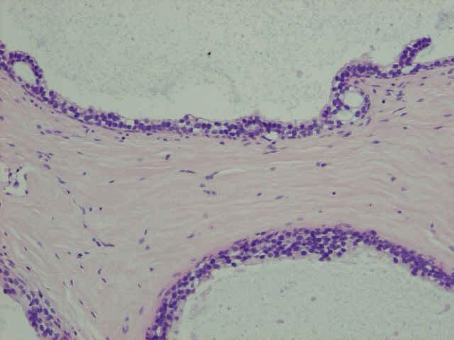
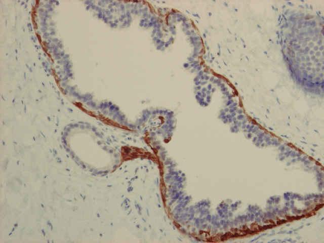
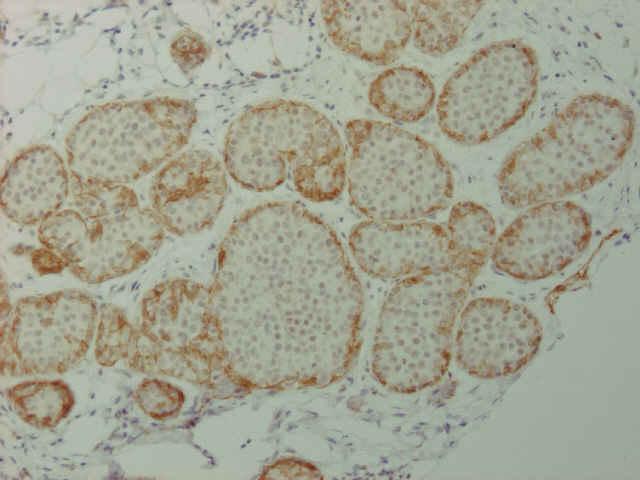
28 Benign Intraduct Papilloma Fronds covered by 2 layers of cells: luminal and myoepithelial Myoepithelial cells surround the dilated duct

29 Benign Intraduct Papilloma SMA

30 2. Multiple Benign intraduct papillomas* *5 or more

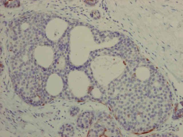
31 3. Intraduct papilloma with focal usual type hyperplasia The fronds are covered by more than 2 layers of epithelial cells With no atypia *Page DL et al. Cancer 1996,78:
 will show 2 populations of cells: negative and")
32 Intraduct papilloma with focal usual type hyperplasia CK 5 CK 5 (& ER) will show 2 populations of cells: negative and positive
33 Papillary lesions I. Benign Benign intraduct papilloma Multiple intraduct papillomas Intraduct papilloma with focal usual type hyperplasia II. Atypical Intraduct papilloma with focal atypical hyperplasia (atypical intraduct papilloma) Intraduct papilloma with focal DCIS III. Malignant Papillary DCIS Intracystic (encapsulated) papillary carcinoma Solid papillary carcinoma* *Collins LC, Schnitt SJ. Histopathology 2008, 52,20-29

34 4. Intraduct papilloma with focal atypical hyperplasia atypical cells occupy an area less than 3mm Atypical cells are relatively large with abundant cytoplasm and large uniform nuclei Cells are CK 5 negative, ER positive

35 5. Intraduct papilloma with focal DCIS Atypical cells occupy an area larger than 3mm
36 Papillary lesions I. Benign Benign intraduct papilloma Multiple intraduct papillomas Intraduct papilloma with focal usual type hyperplasia II. Atypical Intraduct papilloma with focal atypical hyperplasia (atypical intraduct papilloma) Intraduct papilloma with focal DCIS III. Malignant Papillary DCIS Intracystic (encapsulated) papillary carcinoma Solid papillary carcinoma* *Collins LC, Schnitt SJ. Histopathology 2008, 52,20-29


37 6. Papillary DCIS CK5/6

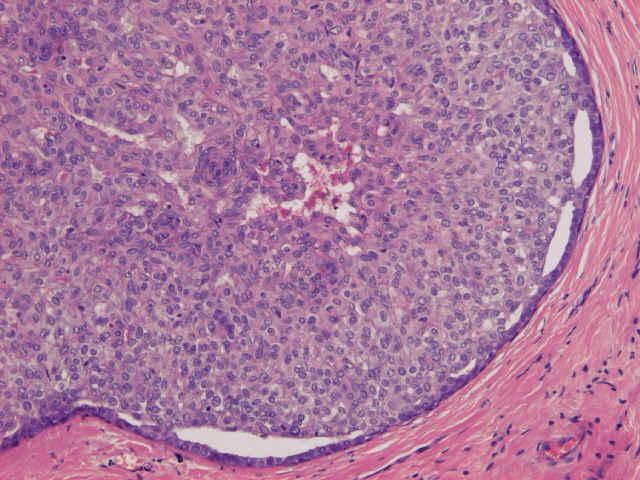
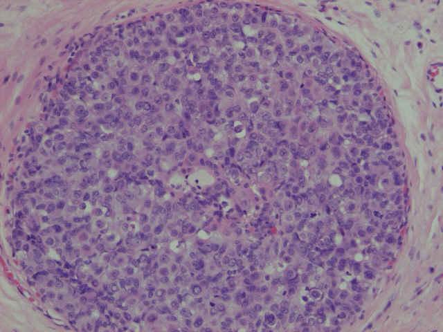
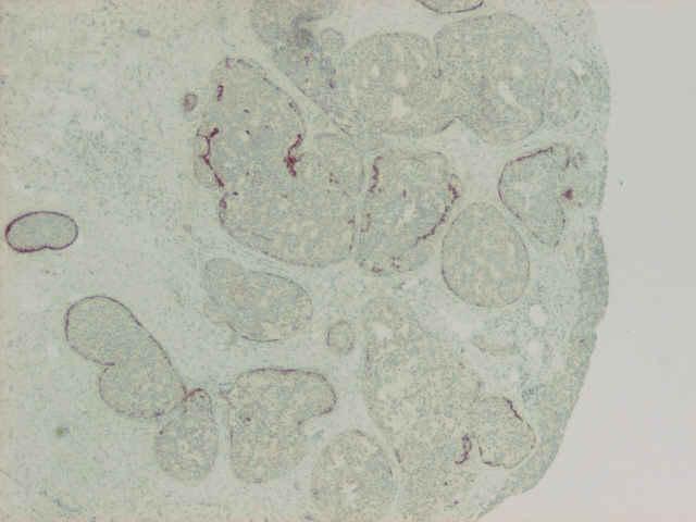
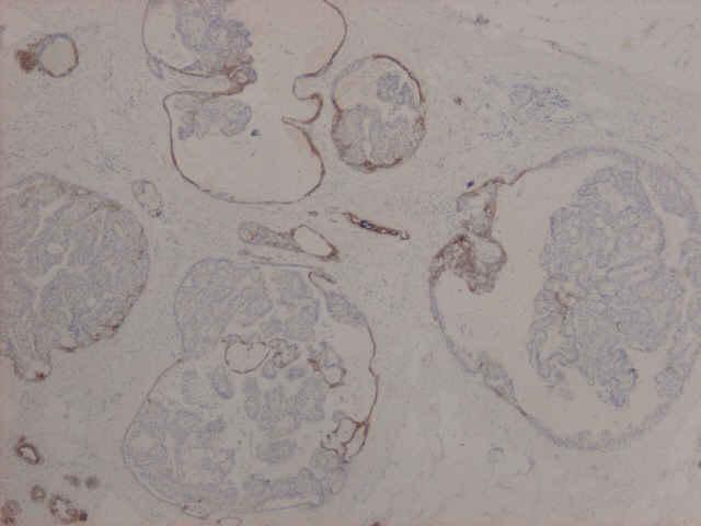
38 7. Intracystic (encapsulated) papillary carcinoma well-defined lesions consisting entirely of malignant cells covering papillary fronds, with no underlying myoepithelial cells, developing in an apparently dilated, usually subareolar, duct, surrounded by a thick fibrous capsule

 papillary")
39 Case No. 152:F 64y, Left Breast, Cystic Lump Intracystic (encapsulated) papillary carcinoma/ IH K5 SMA
40 Intracystic (encapsulated) papillary carcinoma There may be no myoepithethelial cells around the lesion Thus, the lesion may be in fact a form of low grade invasive carcinoma with an expansile growth pattern Or part of progression from in situ to invasive carcinoma

41 Intracystic (encapsulated) papillary carcinoma The lesion may be associated with foci of DCIS or frankly invasive carcinoma In the latter case, Collins & Schnitt recommend considering only the size of the frankly invasive component for staging purposes

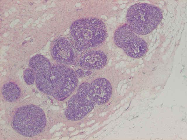
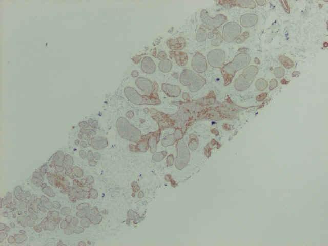
42 8. Solid Papillary carcinoma Circumscribed solid nodule May be associated with adjacent foci of in situ or invasive carcinoma

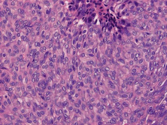
43 Solid Papillary carcinoma Discrete papillae are not present, but the underlying papillary structure is represented by a network of fibrovascular cores among the solid epithelial proliferation

44 Solid Papillary carcinoma There are no myoepithelial cells within the lesion And myoepithelial cells may be also lacking around the lesion in some cases, raising the possibility, as in intracystic papillary lesions, that at least some of these cases also represent low grade expansile invasive cancers CK 5
45 Proliferative Epithelial Lesions of the Breast Usual type ductal hyperplasia Columnar cell change & Hyperplasia Flat epithelial atypia Atypical ductal hyperplasia Intraduct/ intracystic papillary lesions In situ lobular neoplasia
/ Lobular Carcinoma in Situ")
46 In Situ Lobular Neoplasia (Atypical Lobular Hyperplasia (ALH)/ Lobular Carcinoma in Situ (LCIS)
47 1. (Incidental) In Situ Lobular Neoplasia

48 Extensive classic LCIS E-Cadherin
49 Pleomorphic LCIS

50 Pleomorphic LCIS/ E-Cadherin
51 Mixed LCIS

52 In Situ Lobular Neoplasia in core biopsies B3 B5a Extensive LCIS Pleomorphic LCIS Mixed LCIS B5a (If no invasive elements are present) B5a

53 Thank you
Papillary Lesions of the breast
 Papillary Lesions of the breast Emad Rakha Professor of Breast Pathology The University of Nottingham Papillary lesions of the breast are a heterogeneous group of disease, which are characterised by neoplastic
Columnar Cell Lesions. Columnar Cell Lesions and Flat Epithelial Atypia
 Columnar Cell Lesions and Stuart J. Schnitt, M.D. Beth Israel Deaconess Medical Center and Harvard Medical School Boston, MA, USA Columnar Cell Lesions Lesions characterized by columnar epithelial cells
Epithelial Columnar Breast Lesions: Histopathology and Molecular Markers
 29th Annual International Conference Advances in the Application of Monoclonal Antibodies in Clinical Oncology and Symposium on Cancer Stem Cells 25 th -27t h June, 2012, Mykonos, Greece Epithelial Columnar
Papillary Lesions of the Breast A Practical Approach to Diagnosis. (Arch Pathol Lab Med. 2016;140: ; doi: /arpa.
 Papillary Lesions of the Breast A Practical Approach to Diagnosis (Arch Pathol Lab Med. 2016;140:1052 1059; doi: 10.5858/arpa.2016-0219-RA) Papillary lesions of the breast Span the spectrum of benign,
Columnar Cell Lesions
 Columnar Cell Lesions Laura C. Collins, M.D. Department of Pathology Beth Israel Deaconess Medical Center and Harvard Medical School Boston, MA Question? Columnar cell lesions are: a) Annoying lesions
04/10/2018. Intraductal Papillary Neoplasms Of Breast INTRADUCTAL PAPILLOMA
 Intraductal Papillary Neoplasms Of Breast Savitri Krishnamurthy MD Professor of Pathology Deputy Division Head The University of Texas MD Anderson Cancer Center 25 th Annual Seminar in Pathology Pittsburgh,
CLINICAL SIGNIFICANCE OF BENIGN EPITHELIAL CHANGES
 Papillomas. Papillomas are composed of multiple branching fibrovascular cores, each having a connective tissue axis lined by luminal and myoepithelial cells ( Fig. 23-11 ). Growth occurs within a dilated
Proliferative Breast Disease: implications of core biopsy diagnosis. Proliferative Breast Disease
 Proliferative Breast Disease: implications of core biopsy diagnosis Jean F. Simpson, M.D. Breast Pathology Consultants, Inc. Nashville, TN Proliferative Breast Disease Must be interpreted in clinical and
04/10/2018 HIGH RISK BREAST LESIONS. Pathology Perspectives of High Risk Breast Lesions ELEVATED RISK OF BREAST CANCER HISTORICAL PERSPECTIVES
 Pathology Perspectives of High Risk Breast Lesions Savitri Krishnamurthy MD Professor of Pathology Deputy Division Head Director of Clinical Trials, Research and Development The University of Texas MD
Flat Epithelial Atypia
 Flat Epithelial Atypia Richard Owings, M.D. University of Arkansas for Medical Sciences Department of Pathology Flat epithelial atypia can be a difficult lesion May be a subtle diagnosis Lots of changes
Papillary Lesions of the Breast
 Papillary Lesions of the Breast Laura C. Collins, M.D. Associate Professor of Pathology Associate Director, Division of Anatomic Pathology Beth Israel Deaconess Medical Center and Harvard Medical School
Columnar Cell Lesions and Flat Epithelial Atypia
 Columnar Cell Lesions and Flat Epithelial Atypia Laura C. Collins, M.D. Department of Pathology Beth Israel Deaconess Medical Center and Harvard Medical School, Boston, MA Terminology for Columnar Cell
Papillary Lesions of the Breast: WHO Update
 Papillary Lesions of the Breast: WHO Update Stuart J. Schnitt, M.D. Department of Pathology Beth Israel Deaconess Medical Center and Harvard Medical School Boston, MA, USA Papillary Lesions of the Breast
Breast pathology. 2nd Department of Pathology Semmelweis University
 Breast pathology 2nd Department of Pathology Semmelweis University Breast pathology - Summary - Benign lesions - Acute mastitis - Plasma cell mastitis / duct ectasia - Fat necrosis - Fibrocystic change/
Incidence of ductal lesions
 Ductal Proliferative Lesions of the Breast: From FEA to ADH to DCIS Incidence of ductal lesions Pre-mammography: DCIS < 3% of breast cancers, large palpable masses, with invasion Mammography: DCIS 25%
ARTHUR PURDY STOUT SOCIETY COMPANION MEETING: DIFFICULT NEW DIFFERENTIAL DIAGNOSES IN PROSTATE PATHOLOGY. Jonathan I. Epstein.
 1 ARTHUR PURDY STOUT SOCIETY COMPANION MEETING: DIFFICULT NEW DIFFERENTIAL DIAGNOSES IN PROSTATE PATHOLOGY Jonathan I. Epstein Professor Pathology, Urology, Oncology The Reinhard Professor of Urological
Cytyc Corporation - Case Presentation Archive - March 2002
 FirstCyte Ductal Lavage History: 68 Year Old Female Gail Index: Unknown Clinical History: Negative Mammogram in 1995 6 yrs. later presents with bloody nipple discharge Subsequent suspicious mammogram Suspicious
Papillary Lesions of the Breast
 Papillary Lesions of the Breast Texas Society of Pathologists 2013 Laura C. Collins, M.D. Associate Professor of Pathology Associate Director, Division of Anatomic Pathology Beth Israel Deaconess Medical
1 NORMAL HISTOLOGY AND METAPLASIAS
 1 NORMAL HISTOLOGY AND METAPLASIAS, MD Anatomy and Histology 1 Metaplasias 2 ANATOMY AND HISTOLOGY The female breast is composed of a branching duct system, which begins at the nipple with the major lactiferous
Breast Pathology. Breast Development
 Breast Pathology Lecturer: Hanina Hibshoosh, M.D. Reading: Kumar, Cotran, Robbins, Basic Pathology, 6th Edition, pages 623-635 Breast Development 5th week - thickening of the epidermis - milk line 5th
Title malignancy. Issue Date Right 209, 12, (2013)
") NAOSITE: Nagasaki University's Ac Title Author(s) A case of intracystic apocrine papi malignancy Hayashi, Hiroko; Ohtani, Hiroshi; Y Citation Pathology - Research and Practice, Issue Date 2013-12 URL Right
NAOSITE: Nagasaki University's Ac Title Author(s) A case of intracystic apocrine papi malignancy Hayashi, Hiroko; Ohtani, Hiroshi; Y Citation Pathology - Research and Practice, Issue Date 2013-12 URL Right
Case study 1. Rie Horii, M.D., Ph.D. Division of Pathology Cancer Institute Hospital, Japanese Foundation for Cancer Research
 NCCN/JCCNB Seminar in Japan April 15, 2012 Case study 1 Rie Horii, M.D., Ph.D. Division of Pathology Cancer Institute Hospital, Japanese Foundation for Cancer Research Present illness: A 50y.o.premenopausal
Diseases of the breast (1 of 2)
") Diseases of the breast (1 of 2) Introduction A histology introduction Normal ducts and lobules of the breast are lined by two layers of cells a layer of luminal cells overlying a second layer of myoepithelial
Diseases of the breast (1 of 2) Introduction A histology introduction Normal ducts and lobules of the breast are lined by two layers of cells a layer of luminal cells overlying a second layer of myoepithelial
Pathology of Lobular & Ductal Preneoplasia. Syed A Hoda, MD Weill-Cornell, New York, NY
 Pathology of Lobular & Ductal Preneoplasia Syed A Hoda, MD Weill-Cornell, New York, NY Proliferative Epithelial Changes in Breast A wide range of proliferative epithelial changes occur in the breast There
Good afternoon everyone. First of all many thanks to Dr. Bonaventura and Dr. Arn for inviting
 PATHOLOGY IN-SITU CARCINOMA, ROHIT BHARGAVA, MD 1 Good afternoon everyone. First of all many thanks to Dr. Bonaventura and Dr. Arn for inviting me here, it s great to be here and I m going to talk about
6/3/2010. Outline of Talk. Lobular Breast Cancer: Definition of lobular differentiation. Common Problems in Diagnosing LCIS in Core Biopsies
 Outline of Talk Lobular Breast Cancer: Common Problems in Diagnosing LCIS in Core Biopsies Definition of lobular differentiation Variants of LCIS that: carry risk for unsampled invasive cancer mimic DCIS
High risk lesions of the breast : Review of the current diagnostic and management strategies
 High risk lesions of the breast : Review of the current diagnostic and management strategies Poster No.: C-1204 Congress: ECR 2016 Type: Educational Exhibit Authors: P. Jagmohan, F. J. Pool, P. G. Pillay,
COMPANION MEETING BREAST. Auditorium 11:15 1:00 am. Convenor: A/Professor Gelareh Farshid, SA Pathology, SA
 Australasian Division of the International Academy of Pathology Limited ABN 73 008 593 815 36 TH Annual Scientific Meeting Darling Harbour Convention Centre, Sydney, Australia June 3-5, 2011 COMPANION
Treatment options for the precancerous Atypical Breast lesions. Prof. YOUNG-JIN SUH The Catholic University of Korea
 Treatment options for the precancerous Atypical Breast lesions Prof. YOUNG-JIN SUH The Catholic University of Korea Not so benign lesions? Imaging abnormalities(10% recall) lead to diagnostic evaluation,
Disclosures 5/27/2012. Outline of Talk. Outline of Talk. When Is LCIS Clinically Significant? Classic LCIS. Classic LCIS
 When Is LCIS Clinically Significant? Disclosures I have nothing to disclose Yunn-Yi Chen, MD, PhD Professor Outline of Talk Outline of Talk Classic LCIS Classic LCIS Definition of lobular differentiation
Benign Breast Disease and Breast Cancer Risk
 Benign Breast Disease and Breast Cancer Risk Jean F. Simpson, M.D. Vanderbilt University Nashville, Tennessee December 1, 2011 Nashville Nashville Lebanon 1 Cedars of Lebanon State Park The American University
Basement membrane in lobule.
 Bahram Memar, MD Basement membrane in lobule. Normal lobule-luteal phase Normal lobule-follicular phase Lactating breast Greater than 95% are adenocarcinomas in situ carcinomas and invasive carcinomas.
Papillary Lesions in Breast Pathology Practice: Diagnostic Challenges and Practical Approach. A Six- Year Experience from a Tertiary Care Hospital
 Open Access Journal Research Article DOI: 10.23958/ijirms/vol02-i05/12 Papillary Lesions in Breast Pathology Practice: Diagnostic Challenges and Practical Approach. A Six- Year Experience from a Tertiary
In situ lobular neoplasia of the breast with marked myoepithelial proliferation
 In situ lobular neoplasia of the breast with marked myoepithelial proliferation Sami Shousha To cite this version: Sami Shousha. In situ lobular neoplasia of the breast with marked myoepithelial proliferation.
Ductal Proliferations of the Breast: The Good, the Bad, and the Ugly
 Ductal Proliferations of the Breast: The Good, the Bad, and the Ugly Melinda F. Lerwill, MD CRITERIA FOR DISTINGUISHING LOW-GRADE DUCTAL CARCINOMA IN SITU FROM USUAL DUCTAL HYPERPLASIA CYTOLOGY Low-grade
Papillary lesions of the breast: selected diagnostic and management issues
 Histopathology 2008, 52, 20 29. DOI: 10.1111/j.1365-2559.2007.02898.x REVIEW Papillary lesions of the breast: selected diagnostic and management issues L C Collins & S J Schnitt Department of Pathology,
CPC 4 Breast Cancer. Rochelle Harwood, a 35 year old sales assistant, presents to her GP because she has noticed a painless lump in her left breast.
 CPC 4 Breast Cancer Rochelle Harwood, a 35 year old sales assistant, presents to her GP because she has noticed a painless lump in her left breast. 1. What are the most likely diagnoses of this lump? Fibroadenoma
Intraductal carcinoma of the prostate on needle biopsy: histologic features and clinical significance
 & 2006 USCAP, Inc All rights reserved 0893-3952/06 $30.00 www.modernpathology.org Intraductal carcinoma of the prostate on needle biopsy: histologic features and clinical significance Charles C Guo 1 and
The Hot Topic for today is a biopsy from a 58-year-old woman who had worrisome mammographic calcifications on screening.
 The Hot Topic for today is a biopsy from a 58-year-old woman who had worrisome mammographic calcifications on screening. 1 My name is Dan Visscher; I am a consultant in the Division of Anatomic Pathology
Demystifying Endometrial Hyperplasia
 Demystifying Endometrial Hyperplasia A review from Diagnostic Histopathology 19:7 Dr R Hadden ST5 Histopathology Derriford Hospital Plymouth Endometrium Target for sex-steroid hormones Glands Stroma Proliferate
BREAST PATHOLOGY. Fibrocystic Changes
 BREAST PATHOLOGY Lesions of the breast are very common, and they present as palpable, sometimes painful, nodules or masses. Most of these lesions are benign. Breast cancer is the 2 nd most common cause
The management of B3 lesions with emphasis on lobular neoplasia
 The management of B3 lesions with emphasis on lobular neoplasia Abeer Shaaban Queen Elizabeth Hospital Birmingham NHSBSP core biopsy categories B1 - Normal B2 - Benign B3 Uncertain malignant potential
Significance of flat epithelial atypia on mammotome core needle biopsy: should it be excised? B
 Human Pathology (2007) 38, 35 41 www.elsevier.com/locate/humpath Original contribution Significance of flat epithelial atypia on mammotome core needle biopsy: should it be excised? B Lakshmi P. Kunju MD,
Macro- and microacinar proliferations of the prostate
 Macro- and microacinar proliferations of the prostate (with emphasis on cancer mimics) Rodolfo Montironi, MD (IT), FRCPath (UK), IFCAP (USA) Polytechnic University of Marche Region (Ancona) School of Medicine,
Atypical proliferative lesions diagnosed on core biopsy - 6 year review
 Atypical proliferative lesions diagnosed on core biopsy - 6 year review Dr Angela Harris, Dr Julie Weigner & Dr Ricardo Vilain NSW Health Pathology Pathology North, Hunter Anatomical Pathology & Cytology
Immunohistochemical studies (ER & Ki-67) in Proliferative breast lesions adjacent to malignancy
 in Proliferative breast lesions adjacent to malignancy") IOSR Journal of Dental and Medical Sciences (IOSR-JDMS) e-issn: 2279-0853, p-issn: 2279-0861.Volume 13, Issue 3 Ver. IV. (Mar. 2014), PP 84-89 Immunohistochemical studies (ER & Ki-67) in Proliferative
IOSR Journal of Dental and Medical Sciences (IOSR-JDMS) e-issn: 2279-0853, p-issn: 2279-0861.Volume 13, Issue 3 Ver. IV. (Mar. 2014), PP 84-89 Immunohistochemical studies (ER & Ki-67) in Proliferative
Flat Epithelial Atypia: A Review of Current Concepts
 90 The Open Breast Cancer Journal, 2010, 2, 90-94 Flat Epithelial Atypia: A Review of Current Concepts Amy L. Adams * Open Access Department of Pathology and Laboratory Medicine, Emory University School
In Situ Breast Carcinoma. James L. Connolly, M.D Beth Israel Deaconess Medical Center Professor of Pathology Harvard Medical School Boston, MA
 In Situ Breast Carcinoma James L. Connolly, M.D Beth Israel Deaconess Medical Center Professor of Pathology Harvard Medical School Boston, MA Content In Situ Ductal Carcinoma In Situ Lobular Carcinoma
Papillary lesions of the breast - Imaging findings and diagnostic challenges
 Papillary lesions of the breast - Imaging findings and diagnostic challenges Poster No.: R-0146 Congress: RANZCR-AOCR 2012 Type: Educational Exhibit Authors: P. Jagmohan, F. J. Pool Keywords: Breast, Mammography,
Current issues in diagnostic breast pathology
 1 Cancer Studies and Molecular Medicine, University of Leicester, Leicester, UK 2 Academic Unit of Pathology, Leeds University, Leeds, UK 3 Academic Oncology/Breast Pathology, King s College London, London,
Benign Mimics of Malignancy in Breast Pathology
 Arthur Purdy Stout Society of Surgical Pathologists Companion Meeting Benign Mimics of Malignancy in Breast Pathology Stuart J. Schnitt, M.D. Beth Israel Deaconess Medical Center and Harvard Medical School,
Disclosures. Premalignant Lesions of the Breast: What Clinicians Want and Why. NY Times: Prone to Error: Earliest Steps to Find Cancer.
 Disclosures Premalignant Lesions of the Breast: What Clinicians Want and Why I have nothing to disclose Rick Baehner, MD Assistant Professor, UCSF Pathology NY Times: Prone to Error: Earliest Steps to
Enterprise Interest None
 Enterprise Interest None B3 lesions of the breast What are they at surgery? Case 4 Edi Brogi MD PhD Attending Pathologist - Director of Breast Pathology Memorial Sloan Kettering Cancer Center New York
3/28/2017. Disclosure of Relevant Financial Relationships. GU Evening Subspecialty Case Conference. Differential Diagnosis:
 GU Evening Subspecialty Case Conference Rajal B. Shah, M.D. VP, Medical Director, Urologic Pathology Miraca Life Sciences, Irving, Texas Clinical Associate Professor of Pathology Baylor College of Medicine,
Interpretation of Breast Pathology in the Era of Minimally Invasive Procedures
 Shahla Masood, M.D. Professor and Chair Department of Pathology and Laboratory Medicine University of Florida College of Medicine Jacksonville Medical Director, UF Health Breast Center Chief of Pathology
Gross appearance of nodular hyperplasia in material obtained from suprapubic prostatectomy. Note the multinodular appearance and the admixture of
 Tiền liệt tuyến Tiền liệt tuyến Gross appearance of nodular hyperplasia in material obtained from suprapubic prostatectomy. Note the multinodular appearance and the admixture of solid and microcystic areas.
CASE REPORT Malignant transformation of breast ductal adenoma: a diagnostic pitfall
 Malaysian J Pathol 2015; 37(3) : 281 285 CASE REPORT Malignant transformation of breast ductal adenoma: a diagnostic pitfall Hiroko HAYASHI, Hiroshi OHTANI,* Junzo YAMAGUCHI,** and Isao SHIMOKAWA Department
HISTOPATHOLOGICAL EVALUATION OF BENIGN PROLIFERATIVE BREAST LESIONS
 7 ORIGINAL ARTICLE HISTOPATHOLOGICAL EVALUATION OF BENIGN PROLIFERATIVE BREAST LESIONS DR. VIBHUTI H. CHIHLA*, DR. N N. JAGRIT **, DR. JAYASHREE M. SHAH*** *3 rd year Pathology Resident, **Associate Professor,
PROSTATIC ADENOCARCINOMA: DIAGNOSTIC CRITERIA AND IMPORTANT MIMICKERS PROSTATIC ADENOCARCINOMA: DIAGNOSTIC CRITERIA
 PROSTATIC ADENOCARCINOMA: DIAGNOSTIC CRITERIA AND IMPORTANT MIMICKERS PROSTATIC ADENOCARCINOMA: DIAGNOSTIC CRITERIA 1 A good H & E helps! ADENOCARCINOMA DIAGNOSTIC CRITERIA Relatively uniform proliferation
Image guided core biopsies:
 Recommendations on the Surgical, Radiologic and Pathologic Approaches to Breast Disease: Using best practices based on multidisciplinary methodologies developed through the Allina Breast Committee. Image
Case Report Synchronous Bilateral Solid Papillary Carcinomas of the Breast
 Case Reports in Surgery Volume 2013, Article ID 812129, 4 pages http://dx.doi.org/10.1155/2013/812129 Case Report Synchronous Bilateral Solid Papillary Carcinomas of the Breast Noriko Yoshimura, 1 Shigeru
Ana Sofia Preto 19/06/2013
 Ana Sofia Preto 19/06/2013 Understanding the underlying pathophysiologic processes leading to the various types of calcifications Description and illustration of the several types of calcifications, according
3/27/2017. Disclosure of Relevant Financial Relationships. Papilloma???
 Management of Papillary Lesions Diagnosed at Rad Path Concordant Core Biopsy (CNB) Disclosure of Relevant Financial Relationships USCAP requires that all planners (Education Committee) in a position to
Synonyms. Nephrogenic metaplasia Mesonephric adenoma
 Nephrogenic Adenoma Synonyms Nephrogenic metaplasia Mesonephric adenoma Definition Benign epithelial lesion of urinary tract with tubular, glandular, papillary growth pattern Most frequently in the urinary
Management of B3 lesions
 Management of B3 lesions Pathological view Abeer Shaaban Queen Elizabeth Hospital Birmingham FEA AIDP B3 lesions In situ Lobular neoplasia Papilloma Radial scar Fibroaepithelial lesion Mucocoele like lesion
They Do Look Alike : Mimics of Prostate Cancer in Biopsy Samples
 They Do Look Alike : in Biopsy Samples Gladell P. Paner, MD Departments of Pathology and Surgery (Urology) University of Chicago, IL USA Gladell.paner@uchospitals.edu Benign in Needle Biopsy 1. Benign
American Journal of Cancer Case Reports. Invasive Papillary Carcinoma of Male Breast: A Rare Case Report
 American Journal of Cancer Case Reports http://ivyunion.org/index.php/ajccr SantraAetal. American Journal of Cancer Case Reports 2014, 3:56-61 Page 1 of 6 Vol 3 Article ID 20140617, 6 pages Case Report
Ductal Carcinoma in Situ. Laura C. Collins, M.D. Department of Pathology Beth Israel Deaconess Medical Center and Harvard Medical School Boston, MA
 Ductal Carcinoma in Situ Laura C. Collins, M.D. Department of Pathology Beth Israel Deaconess Medical Center and Harvard Medical School Boston, MA Definition of DCIS WHO 2012 A neoplastic proliferation
Protocol for the Examination of Biopsy Specimens From Patients With Invasive Carcinoma of the Breast
 Protocol for the Examination of Specimens From Patients With Invasive Carcinoma of the Breast Version: BreastInvasive 1.0.0.0 Protocol Posting Date: February 2019 Accreditation Requirements The use of
Biliary tract tumors
 Short Course 2010 Annual Fall Meeting of the Korean Society for Pathologists Biliary tract tumors Joon Hyuk Choi, M.D., Ph.D. Professor, Department of Pathology, Yeungnam Univ. College of Medicine, Daegu,
Can True Papillary Neoplasms of Breast and Their Mimickers Be Accurately Classified by Cytology?
 92 CANCER CYTOPATHOLOGY Can True Papillary Neoplasms of Breast and Their Mimickers Be Accurately Classified by Cytology? Claire W. Michael, M.D. 1 Bruce Buschmann, C.T. 2 1 University of Michigan, Department
Atypical Ductal Hyperplasia and Papillomas: A Comparison of Ultrasound Guided Breast Biopsy and Stereotactic Guided Breast Biopsy
 Atypical Ductal Hyperplasia and Papillomas: A Comparison of Ultrasound Guided Breast Biopsy and Stereotactic Guided Breast Biopsy Breast Cancer is the most common cancer diagnosed in women in the United
Pleomorphic adenoma of breast - a case report and distinction with metaplastic carcinoma D Gupta, S Agrawal, N Trivedi, A Tewari
 of breast - a case report and distinction with metaplastic carcinoma D Gupta, S Agrawal, N Trivedi, A Tewari Introduction, also known as mixed tumour, is a benign tumour which typically presents as a painless,
The Diagnosis and Management of Pre-invasive Breast Disease: Flat Epithelial Atypia Classification, Pathologic Features and Clinical Significance
 The Diagnosis and Management of Pre-invasive Breast Disease: Flat Epithelial Atypia Classification, Pathologic Features and Clinical Significance The Harvard community has made this article openly available.
Low-grade Adenosquamous Carcinoma Coexisting with Sclerosing Adenosis of the Breast: A Case Report
 31 Case Report J. St. Marianna Univ. Vol. 8, pp. 31 35, 2017 Low-grade Adenosquamous Carcinoma Coexisting with Sclerosing Adenosis of the Breast: A Case Report Ryoko Oi 1, 2, Ichiro Maeda 1, Yoshio Aida
Diagnosis of Fibroepithelial and Mesenchymal Lesions on Core Needle Biopsy
 Diagnosis of Fibroepithelial and Mesenchymal Lesions on Core Needle Biopsy Emmanuel Agosto-Arroyo, MD Assistant Member Department of Anatomic Pathology 3/3/2018 Disclosure There are no conflicts of interest.
EQA Circulation 43 Educational Cases
 EQA Circulation 43 Educational Cases E1-E2 Monica Agarwal Monklands Hospital E1 38 yrs male Submandibular gland tumour E1 Formal excision following diagnosis of poorly differentiated carcinoma on core
Non-mass Enhancement on Breast MRI. Aditi A. Desai, MD Margaret Ann Mays, MD
 Non-mass Enhancement on Breast MRI Aditi A. Desai, MD Margaret Ann Mays, MD Breast MRI Important screening and diagnostic tool, given its high sensitivity for breast cancer detection Breast MRI - Indications
PLEOMORPHIC ADENOMA ( BENIGN MIXED TUMOR )
") ( BENIGN MIXED TUMOR ) Grossly, the tumor is freely movable, solid, sometimes lobulated and occasionally cystic. If recurrent, multinodular masses are common. Histologically, within a fibrous capsule,
( BENIGN MIXED TUMOR ) Grossly, the tumor is freely movable, solid, sometimes lobulated and occasionally cystic. If recurrent, multinodular masses are common. Histologically, within a fibrous capsule,
FIBROEPITHELIAL LESIONS
 DEFINITIONS FIBROEPITHELIAL LESIONS Suzanne Moore FIBROADENOMA- A discrete benign tumour showing evidence of connective tissue and epithelial proliferation- WHO Fibrous stromal element of these tumours
HISTOMORPHOLOGICAL SPECTRUM OF BREAST LESIONS
 HISTOMORPHOLOGICAL SPECTRUM OF BREAST LESIONS Kiran H. S, Jayaprakash Shetty, Chandrika Rao Assistant Professor, Department of Pathology, Yenepoya Medical College, Mangalore. Professor, Department of Pathology,
Atypical papillary lesions after core needle biopsy and subsequent breast carcinoma
 Asian Biomedicine Vol. 5 No. 2 April 2011; 243-248 DOI: 10.5372/1905-7415.0502.031 Original article Atypical papillary lesions after core needle biopsy and subsequent breast carcinoma Tuenchit Khamapirad
Notice of Faculty Disclosure
 California Society of Pathology Diagnostic Problems in Surgical Pathology December 2015 Case 2 Laura C. Collins, M.D. Associate Professor of Pathology Associate Director of Anatomic Pathology Beth Israel
Breast Evaluation & Management Guidelines
 Breast Evaluation & Management Guidelines Pamela L. Kurtzhals, M.D. F.A.C.S. Head, Dept. of General Surgery Scripps Clinic, La Jolla Objective Review screening & diagnostic guidelines Focused patient complaints
Atypical Hyperplasia/EIN
 EIN Atypical Hyperplasia/EIN Based on scientific and diagnostic advances, in 2014 the WHO moved that the precursor lesion for endometrioid carcinoma be atypical hyperplasia/ein, rather than what was previously
K. M. Sorensen Utah State University, Logan, Utah
 K. M. Sorensen Utah State University, Logan, Utah T. E. Doyle, B. D. Borget, M. Cervantes, J. A. Chappell, B. J. Curtis, M. A. Grover, J. E. Roring, J. E. Stiles, and L. A. Thompson Utah Valley University,
Neoplasia 2018 Lecture 2. Dr Heyam Awad MD, FRCPath
 Neoplasia 2018 Lecture 2 Dr Heyam Awad MD, FRCPath ILOS 1. List the differences between benign and malignant tumors. 2. Recognize the histological features of malignancy. 3. Define dysplasia and understand
Surgical Pathology Issues of Practical Importance
 Surgical Pathology Issues of Practical Importance Anne Moore, MD Medical Oncology Syed Hoda, MD Surgical Pathology The pathologist is central to the team approach needed to manage the patient with breast
COMMON BENIGN DISORDERS AND DISEASES OF THE BREAST
 COMMON BENIGN DISORDERS AND DISEASES OF THE BREAST Aberrations of Normal Development and Involution (ANDI). The basic principles underlying the aberrations of normal development and involution (ANDI) classification
RSNA, /radiol Appendix E1. Methods
 RSNA, 2016 10.1148/radiol.2016151097 Appendix E1 Methods US and Near-infrared Data Acquisition Four optical wavelengths (740 nm, 780 nm, 808 nm, and 830 nm) were used to sequentially deliver the light
LYMPHATIC DRAINAGE AXILLARY (MOSTLY) INTERNAL MAMMARY SUPRACLAVICULAR
 INTERNAL MAMMARY SUPRACLAVICULAR") BREAST LYMPHATIC DRAINAGE AXILLARY (MOSTLY) INTERNAL MAMMARY SUPRACLAVICULAR HISTOLOGY LOBE: (10 in whole breast) LOBULE: (many per lobe) ACINUS/I, aka ALVEOLUS/I: (many per lobule) DUCT(S): INTRA- or
BREAST LYMPHATIC DRAINAGE AXILLARY (MOSTLY) INTERNAL MAMMARY SUPRACLAVICULAR HISTOLOGY LOBE: (10 in whole breast) LOBULE: (many per lobe) ACINUS/I, aka ALVEOLUS/I: (many per lobule) DUCT(S): INTRA- or
Breast Cancer. Dr Rodney Itaki Anatomical Pathology Discipline Division of Pathology
 Breast Cancer Dr Rodney Itaki Anatomical Pathology Discipline Division of Pathology Muscles Muscles underneath the breasts separating them from the ribs Breast has no muscle tissue 2 Female Breast Anatomy
IBCM 2, April 2009, Sarajevo, Bosnia and Herzegovina
 Preoperative diagnosis and treatment planning in breast cancer The pathologist s perspective L. Mazzucchelli Istituto Cantonale di Patologia Locarno, Switzerland IBCM 2, 23-25 April 2009, Sarajevo, Bosnia
Disorders of Cell Growth & Neoplasia. Histopathology Lab
 Disorders of Cell Growth & Neoplasia Histopathology Lab Paul Hanna April 2010 Case #84 Clinical History: 5 yr-old, West Highland White terrier. skin mass from axillary region. has been present for the
Controversies and Problematic Issues in Core Needle Biopsies (To excise or not to excise)
") Controversies and Problematic Issues in Core Needle Biopsies (To excise or not to excise) Laura C. Collins, M.D. Beth Israel Deaconess Medical Center and Harvard Medical School Boston, MA Schematic Representation
Controversies and Problematic Issues in Core Needle Biopsies (To excise or not to excise) Laura C. Collins, M.D. Beth Israel Deaconess Medical Center and Harvard Medical School Boston, MA Schematic Representation
Case Report Endocrine Mucin-Producing Sweat Gland Carcinoma, a Histological Challenge
 Hindawi Volume 2017, Article ID 6343709, 4 pages https://doi.org/10.1155/2017/6343709 Case Report Endocrine Mucin-Producing Sweat Gland Carcinoma, a Histological Challenge Mary Anne Brett, Samih Salama,
Promise of a beautiful day
 Promise of a beautiful day Ductal carcinoma in Situ Lobular Carcinoma in Situ Natural History Manosmed Tartous Oct 2009 Gérard ABADJIAN MD Pathology Department Hôtel-Dieu de France. Associate Professor
Quality ID #263: Preoperative Diagnosis of Breast Cancer National Quality Strategy Domain: Effective Clinical Care
 Quality ID #263: Preoperative Diagnosis of Breast Cancer National Quality Strategy Domain: Effective Clinical Care 2018 OPTIONS FOR INDIVIDUAL MEASURES: REGISTRY ONLY MEASURE TYPE: Process DESCRIPTION:
Salivary Glands 3/7/2017
 Salivary Glands 3/7/2017 Goals and objectives Focus on the entities unique to H&N Common board type facts Information for your future practice Salivary Glands Salivary Glands Major gland. Paratid. Submandibular.