MRI and CT of the CNS
|
|
|
- Mervin Stanley
- 5 years ago
- Views:
Transcription

1 MRI and CT of the CNS Dr.Maha ELBeltagy Assistant Professor of Anatomy Faculty of Medicine The University of Jordan 2018
2 Computed Tomography CT is used for the detection of intracranial lesions. CT relies on the same physics as conventional x-rays. Structures are distinguished from one another by their ability to absorb energy from x-rays. The x-ray tube emits a narrow beam of radiation as it passes in a series of scanning movements through an arc of 180 degrees around the patient's head. The x-rays having passed through the head are collected by a special x-ray detector. The information is fed to a computer that processes the information, which is then displayed as a reconstructed picture on a television like screen. The observer sees an image of a thin slice through the head. The gray matter of the cerebral cortex, white matter, internal capsule, corpus callosum, ventricles, and subarachnoid spaces can all be recognized. An iodine-containing medium can be injected intravascularly, which enhances greatly the contrast between tissues having a different blood flow. Since a CT scan can be performed in 5 to 10 minutes, it is the method of choice in an emergency situation with patients with head trauma or suspected intracranial hemorrhage.

3 CT Machine
4 Magnetic Resonance Imaging The technique of MRI uses the magnetic properties of the hydrogen nucleus excited by radiofrequency radiation transmitted by a coil surrounding the head. The excited hydrogen nuclei emit a signal that is detected as induced electric currents in a receiver coil. it provides better differentiation between gray and white matter. The reason for this is that gray matter contains more hydrogen in the form of water than does white matter, and the hydrogen atoms are less bound in fat. MRI is the best imaging method for detecting low-contrast lesions as brain tumors or small multiple sclerosis plaques. It is also capable of showing clear images of the brain stem, cerebellum, and the pituitary fossa, which in the case of a CT scan are overshadowed by the dense bones of the base of the skull. The spinal cord structure is much more clearly visualized with MRI. Unfortunately, an MRI takes longer and costs two-thirds more than a CT scan

5 MRI Machine
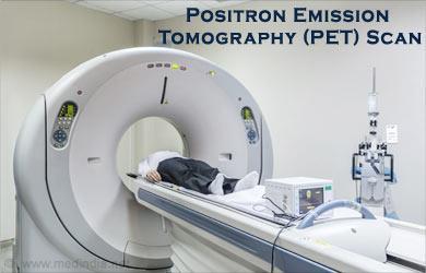
6 Positron Emission Tomography Positron emission tomography (PET) uses radioactive isotopes that decay with the emission of positively charged electrons (positrons) to map the biochemical, physiologic, and pharmacologic processes taking place in the brain.the appropriate isotope is incorporated into molecules of known biochemical behavior in the brain and then is injected into the patient. The metabolic activity of the compound can then be studied by making cross-sectional tomographic images of the brain using the same principles as in CT. By making a series of time-lapse images at different anatomical sites, it is possible to study the variations in brain metabolism at these sites. This technique has been used to study the distribution and activity of neurotransmitters, the variations in oxygen utilization, and cerebral blood flow.
7

8 CT scan showing the structure of the brain. A, B: Horizontal cuts (axial sections)
.")
9 Horizontal (axial) CT scan of the brain (contrast enhanced).

10 MRI showing the structure of the brain. A: Sagittal. B: Coronal.
11 Horizontal (axial) MRI of the brain.

12 Coronal MRI of the brain.

13 A coronal MRI (contrast enhanced) through the hindbrain showing the fourth ventricle and the surrounding neural and bony structures.

14 Anteroposterior (angled) vertebral arteriogram. Woman aged 35 years.
 are seen in the cerebral cortex. The lateral ventricles are also demonstrated.")
15 Axial (horizontal) PET scan of a normal brain following the injection of 18- fluorodeoxyglucose. Regions of active metabolism (yellow areas) are seen in the cerebral cortex. The lateral ventricles are also demonstrated. (Courtesy Dr. Holley Dey.)
 is seen in the region of the tumor.")
16 Coronal PET scan of a 62-year-old male patient with a malignant glioma in the left parietal lobe, following the injection of 18- Fluorodeoxyglucose. A high concentration of the compound (circular yellow area) is seen in the region of the tumor.
 distributed throughout the corpus striatum in both cerebral hemispheres.")
17 Axial (horizontal) positron emission tomography (PET) scans of a normal brain (A) and the brain of a patient with early Parkinson disease (B) following the injection of 18- fluoro-6-ldopa. The normal brain image shows large amounts of the compound (yellow areas) distributed throughout the corpus striatum in both cerebral hemispheres. In the patient with Parkinson disease, the brain image shows that the total amount of the compound is low, and it is unevenly distributed in the corpus striatum. (Courtesy Dr. Holley Dey.)

18 Sagittal MRI of the lumbosacral part of the vertebral column showing several prolapsed intervertebral discs.
19
20

21
22
23

24

25 MRI showing fronto temporal Subdural haemorrage

26
Gross Organization I The Brain. Reading: BCP Chapter 7
 Gross Organization I The Brain Reading: BCP Chapter 7 Layout of the Nervous System Central Nervous System (CNS) Located inside of bone Includes the brain (in the skull) and the spinal cord (in the backbone)
CNS Imaging. Dr Amir Monir, MD. Lecturer of radiodiagnosis.
 CNS Imaging Dr Amir Monir, MD Lecturer of radiodiagnosis www.dramir.net Types of radiological examinations you know Plain X ray X ray with contrast GIT : barium (swallow, meal, follow through, enema) ERCP
Non-Invasive Techniques
 Non-Invasive Techniques Key: Does not hurt the organism Psychology 372 Physiological Psychology Steven E. Meier, Ph.D. Listen to the audio lecture while viewing these slides or view the video presentation
Non-Invasive Techniques
 Many Procedures Non-Invasive Techniques Key: Does not hurt the organism Psychology 372 Physiological Psychology Steven E. Meier, Ph.D. Listen to the audio lecture while viewing these slides or view the
Chapter 6 Section 1. The Nervous System: The Basic Structure
 Chapter 6 Section 1 The Nervous System: The Basic Structure Essential Question: How does studying the biology of the brain give us an understanding of our behavior? Draw or type 2 things you already know
fmri (functional MRI)
") Lesion fmri (functional MRI) Electroencephalogram (EEG) Brainstem CT (computed tomography) Scan Medulla PET (positron emission tomography) Scan Reticular Formation MRI (magnetic resonance imaging) Thalamus
Lesion fmri (functional MRI) Electroencephalogram (EEG) Brainstem CT (computed tomography) Scan Medulla PET (positron emission tomography) Scan Reticular Formation MRI (magnetic resonance imaging) Thalamus
Organization of the nervous system. The withdrawal reflex. The central nervous system. Structure of a neuron. Overview
 Overview The nervous system- central and peripheral The brain: The source of mind and self Neurons Neuron Communication Chemical messengers Inside the brain Parts of the brain Split Brain Patients Organization
III. Studying The Brain and Other Structures
 III. Studying The Brain and Other Structures 1. Accidents (case study) In 1848, a railroad worker named Phineas Gage was involved in an accident that damaged the front part of his brain. Gage s doctor
HEAD AND NECK IMAGING. James Chen (MS IV)
") HEAD AND NECK IMAGING James Chen (MS IV) Anatomy Course Johns Hopkins School of Medicine Sept. 27, 2011 OBJECTIVES Introduce cross sectional imaging of head and neck Computed tomography (CT) Review head
HEAD AND NECK IMAGING James Chen (MS IV) Anatomy Course Johns Hopkins School of Medicine Sept. 27, 2011 OBJECTIVES Introduce cross sectional imaging of head and neck Computed tomography (CT) Review head
HSC Physics. Module 9.6. Medical Physics
 HSC Physics Module 9.6 Medical Physics Contextual Outline 9.6 Medical Physics (28 indicative hours) The use of other advances in technology, developed from our understanding of the electromagnetic spectrum,
Organization of the nervous system. [See Fig. 48.1]
![Organization of the nervous system. [See Fig. 48.1]](/thumbs/90/103926552.jpg "Organization of the nervous system. [See Fig. 48.1]") Nervous System [Note: This is the text version of this lecture file. To make the lecture notes downloadable over a slow connection (e.g. modem) the figures have been replaced with figure numbers as found
Nervous System [Note: This is the text version of this lecture file. To make the lecture notes downloadable over a slow connection (e.g. modem) the figures have been replaced with figure numbers as found
Chapter 6. Body and Behavior
 Chapter 6 Body and Behavior Section 1 The Nervous System: The Basic Structure How the nervous system works Central nervous system (CNS)- the brain and spinal cord Spinal cord- nerves that run up and down
Introduction to the Course and the Techniques. Jeffry R. Alger, PhD Ahmanson-Lovelace Brain Mapping Center Department of Neurology
 Introduction to the Course and the Techniques Jeffry R. Alger, PhD Ahmanson-Lovelace Brain Mapping Center Department of Neurology (jralger@ucla.edu) CTSI Neuroimaging April 2014 Rationale for the Course
Unit 3: The Biological Bases of Behaviour
 Unit 3: The Biological Bases of Behaviour Section 1: Communication in the Nervous System Section 2: Organization in the Nervous System Section 3: Researching the Brain Section 4: The Brain Section 5: Cerebral
Name: Period: Chapter 2 Reading Guide The Biology of Mind
 Name: Period: Chapter 2 Reading Guide The Biology of Mind The Nervous System (pp. 55-58) 1. What are nerves? 2. Complete the diagram below with definitions of each part of the nervous system. Nervous System
Restoring Tumorous MRI Brain Images
 Restoring Tumorous MRI Brain Images Yadavan Varatharajah Summer Ventures 2007 Appalachian State University Abstract Brain tumors are caused by many environmental, lifestyle, and genetic factors. This project
COGNITIVE SCIENCE 17. Peeking Inside The Head. Part 1. Jaime A. Pineda, Ph.D.
 COGNITIVE SCIENCE 17 Peeking Inside The Head Part 1 Jaime A. Pineda, Ph.D. Imaging The Living Brain! Computed Tomography (CT)! Magnetic Resonance Imaging (MRI)! Positron Emission Tomography (PET)! Functional
Myers Psychology for AP*
 Myers Psychology for AP* David G. Myers PowerPoint Presentation Slides by Kent Korek Germantown High School Worth Publishers, 2010 *AP is a trademark registered and/or owned by the College Board, which
1. Processes nutrients and provides energy for the neuron to function; contains the cell's nucleus; also called the soma.
 1. Base of brainstem; controls heartbeat and breathing 2. tissue destruction; a brain lesion is a naturally or experimentally caused destruction of brain tissue 3. A thick band of axons that connects the
biological psychology, p. 40 The study of the nervous system, especially the brain. neuroscience, p. 40
 biological psychology, p. 40 The specialized branch of psychology that studies the relationship between behavior and bodily processes and system; also called biopsychology or psychobiology. neuroscience,
Okami Study Guide: Chapter 2 1
 Okami Study Guide: Chapter 2 1 Chapter Test 1. A cell that receives information and transmits it to other cells via an electrochemical process is called a(n) a. neuron b. hormone c. glia d. endorphin Answer:
Applicable Neuroradiology
 For the Clinical Neurology Clerkship LSU Medical School New Orleans Amy W Voigt, MD Clerkship Director Introduction The field of Radiology first developed following the discovery of X-Rays by Wilhelm Roentgen
Attenuation value in HU From -500 To HU From -10 To HU From 60 To 90 HU. From 200 HU and above
 Brain Imaging Common CT attenuation values Structure Air Fat Water Brain tissue Recent hematoma Calcifications Bone Brain edema and infarction Normal liver parenchyma Attenuation value in HU From -500
STRUCTURAL ORGANIZATION OF THE NERVOUS SYSTEM
 STRUCTURAL ORGANIZATION OF THE NERVOUS SYSTEM STRUCTURAL ORGANIZATION OF THE BRAIN The central nervous system (CNS), consisting of the brain and spinal cord, receives input from sensory neurons and directs
COMENIUS-Project: SM&CLIL Radiation & Medicine
 Medical imaging refers to the techniques and processes used to create images of the human body (or parts thereof) for clinical purposes. Thanks to modern mathematics and computer technology, medical imaging
Sincerely, Ms. Paoloni and Mrs. Whitney
 Dear Students, Welcome to AP Psychology! We will begin our course of study focusing on the nervous system with a particular emphasis on how the brain and neurotransmitters influence our behaviors. In preparation
Ways we Study the Brain. Accidents Lesions CAT Scan PET Scan MRI Functional MRI
 The Brain Ways we Study the Brain Accidents Lesions CAT Scan PET Scan MRI Functional MRI Accidents Phineas Gage Story Personality changed after the accident. What this this tell us? That different part
Medical imaging X-ray, CT, MRI, scintigraphy, SPECT, PET Györgyi Műzes
 Medical imaging X-ray, CT, MRI, scintigraphy, SPECT, PET Györgyi Műzes Semmelweis University, 2nd Dept. of Medicine Medical imaging: definition technical process of creating visual representations about
The Brain Studying & Structures. Unit 3
 The Brain Studying & Structures Unit 3 Modified PowerPoint from: Aneeq Ahmad -- Henderson State University. Worth Publishers 2007 Learning Objectives Describe the nervous system and its subdivisions and
Brain Tumors. What is a brain tumor?
 Scan for mobile link. Brain Tumors A brain tumor is a collection of abnormal cells that grows in or around the brain. It poses a risk to the healthy brain by either invading or destroying normal brain
Ways to Study Brain Structures and Functioning. Can physically trace connections. Ablation. Is the most primitive Can be done with any structures
 Ways to Study Brain Structures and Functioning Can physically trace connections Is the most primitive Can be done with any structures Ablation Can remove a piece of the brain and see what happens If the
Chapter 3 Biological Psychology
 Chapter 3 Biological Psychology Introduction Reductionism? Scientists in many fields use a strategy called reductionism; they attempt to explain complex phenomena by reducing them to combinations of simpler
Acetylcholine (ACh) Action potential. Agonists. Drugs that enhance the actions of neurotransmitters.
 Action potential. Agonists. Drugs that enhance the actions of neurotransmitters.") Acetylcholine (ACh) The neurotransmitter responsible for motor control at the junction between nerves and muscles; also involved in mental processes such as learning, memory, sleeping, and dreaming. (See
Acetylcholine (ACh) The neurotransmitter responsible for motor control at the junction between nerves and muscles; also involved in mental processes such as learning, memory, sleeping, and dreaming. (See
The Biological Level of Analysis: Studying the Brain
 The Biological Level of Analysis: Studying the Brain In the past the study of the brain was limited to people suffering from head injuries and the effects of accidental damage. It was only possible to
Outline. Biological Psychology: Research Methods. Dr. Katherine Mickley Steinmetz
 Biological Psychology: Research Methods Dr. Katherine Mickley Steinmetz Outline Neuroscience Methods Histology Electrophysiological Recordings Lesion Neuroimaging Neuroanatomy Histology: Brain structure
CEREBRAL BLOOD FLOW AND METABOLISM
 Supported by: HURO/0901/069/2.3.1 HU-RO-DOCS CEREBRAL BLOOD FLOW AND METABOLISM Part 3 Modern imaging methods SPECT, PET, nmri History of Nuclear Medicine Starts with the invention of the X-ray 1946: radioactive
Brain and behaviour (Wk 6 + 7)
") Brain and behaviour (Wk 6 + 7) What is a neuron? What is the cell body? What is the axon? The basic building block of the nervous system, the individual nerve cell that receives, processes and transmits
Brain and behaviour (Wk 6 + 7) What is a neuron? What is the cell body? What is the axon? The basic building block of the nervous system, the individual nerve cell that receives, processes and transmits
Anatomy & Physiology Central Nervous System Worksheet
 1. What are the two parts of the CNS? 2. What are the four functions of the CNS Anatomy & Physiology Central Nervous System Worksheet 3. What are the four functions of the meninges? (p430) 4. Starting
FOR CMS (MEDICARE) MEMBERS ONLY NATIONAL COVERAGE DETERMINATION (NCD) FOR MAGNETIC RESONANCE IMAGING:
 MEMBERS ONLY NATIONAL COVERAGE DETERMINATION (NCD) FOR MAGNETIC RESONANCE IMAGING:") National Imaging Associates, Inc. Clinical guidelines SINUS MRI Original Date: November 2007 Page 1 of 5 CPT Codes: 70540, 70542, 70543 Last Review Date: July 2014 NCD 220.2 MRI Last Effective Date: July
National Imaging Associates, Inc. Clinical guidelines SINUS MRI Original Date: November 2007 Page 1 of 5 CPT Codes: 70540, 70542, 70543 Last Review Date: July 2014 NCD 220.2 MRI Last Effective Date: July
Methods of Visualizing the Living Human Brain
 Methods of Visualizing the Living Human Brain! Contrast X-rays! Computerized Tomography (CT)! Magnetic Resonance Imaging (MRI)! Positron Emission Tomography (PET)! Functional MRI! Magnetoencephalography
FOR CMS (MEDICARE) MEMBERS ONLY NATIONAL COVERAGE DETERMINATION (NCD) FOR MAGNETIC RESONANCE IMAGING:
 MEMBERS ONLY NATIONAL COVERAGE DETERMINATION (NCD) FOR MAGNETIC RESONANCE IMAGING:") National Imaging Associates, Inc. Clinical guidelines BONE MARROW MRI Original Date: July 2008 Page 1 of 5 CPT Codes: 77084 Last Review Date: September 2014 NCD 220.2 MRI Last Effective Date: July 2011
National Imaging Associates, Inc. Clinical guidelines BONE MARROW MRI Original Date: July 2008 Page 1 of 5 CPT Codes: 77084 Last Review Date: September 2014 NCD 220.2 MRI Last Effective Date: July 2011
Keep Imaging Simple: An Introduction To Neuroimaging
 Keep Imaging Simple: An Introduction To Neuroimaging Meghan Elkins, OD, FAAO Please silence all mobile devices and remove items from chairs so others can sit. Unauthorized recording of this session is
Principles Arteries & Veins of the CNS LO14
 Principles Arteries & Veins of the CNS LO14 14. Identify (on cadaver specimens, models and diagrams) and name the principal arteries and veins of the CNS: Why is it important to understand blood supply
The neurvous system senses, interprets, and responds to changes in the environment. Two types of cells makes this possible:
 NERVOUS SYSTEM The neurvous system senses, interprets, and responds to changes in the environment. Two types of cells makes this possible: the neuron and the supporting cells ("glial cells"). Neuron Neurons
Introduction to Radiology
 Introduction - Lecture 1 436 Teams Introduction to Radiology Objectives Introduce the various Medical Imaging Modalities. Understand the basics of image generation. Relate imaging to gross anatomy. Appreciate
To understand AD, it is important to
 To understand AD, it is important to know a bit about the brain. This part of Unraveling the Mystery gives an inside view of the normal brain, how it works, and what happens during aging. The brain is
LESSON 1.3 WORKBOOK. How can we study the behaving brain?
 LESSON 1.3 WORKBOOK How can we study the behaving brain? We are in the middle of a technological revolution when it comes to how closely we can look at the behaving brain. Scientists and doctors now have
Central Nervous System Practical Exam. Chapter 12 Nervous System Cells. 1. Please identify the flagged structure.
 Central Nervous System Practical Exam Chapter 12 Nervous System Cells 1. Please identify the flagged structure. 2. Please identify the flagged structure. 3. Please identify the flagged structure. 4. A
IV. The Divisions of the Brain. Slide # 1
 IV. The Divisions of the Brain Slide # 1 The Hindbrain Hindbrain, located at the rear base of the skull, controlling automatic functions Contains: Cerebellum (balance & coordination) Medulla (heartbeat,
Neuroimaging and Assessment Methods
 Psych 2200, Lecture 5 Experimental Design and Brain Imaging Methods Tues Sept 15, 2015 Revised TA office hours (Sam), today 4-5p, and wed 11:30-1:30. I will not have office hours this thurs but you should
Molecular Imaging and the Brain
 Molecular imaging technologies are playing an important role in neuroimaging, a branch of medical imaging, by providing a window into the living brain. Where CT and conventional MR imaging provide important
Department of Cognitive Science UCSD
 Department of Cognitive Science UCSD Verse 1: Neocortex, frontal lobe, Brain stem, brain stem, Hippocampus, neural node, Right hemisphere, Pons and cortex visual, Brain stem, brain stem, Sylvian fissure,
Psychology Unit II: The Brain and Biology
 Psychology Unit II: The Brain and Biology NATURE or NURTURE What are the effects of biochemistry on behavior? VOCABULARY Central Nervous System (CNS): The central nervous system is the part of the nervous
Medical Use of Radioisotopes
 Medical Use of Radioisotopes Therapy Radioisotopes prove to be useful in the application of brachytherapy, the procedure for using temporary irradiation close to the area of disease (i.e. cancer) 10% Medical
Nuclear neurology. Zámbó Katalin Department of Nuclear Medicine
 Nuclear neurology Zámbó Katalin Department of Nuclear Medicine To refresh your memory Brain has a high rate of oxidative metabolism. It has no reserves either of oxygen or of glucose and has a very limited
Radiologic Imaging Magnetic Resonance Imaging (MRI)
") Radiologic Imaging X-ray has always been the golden rule in diagnosing and treating podiatric patients. Unfortunately, for some patients the diagnosis is not as evident. That is when we need to utilize
Radiologic Imaging X-ray has always been the golden rule in diagnosing and treating podiatric patients. Unfortunately, for some patients the diagnosis is not as evident. That is when we need to utilize
Slide 1. Slide 2. Slide 3. Tomography vs Topography. Computed Tomography (CT): A simplified Topographical review of the Brain. Learning Objective
: A simplified Topographical review of the Brain. Learning Objective") Slide 1 Computed Tomography (CT): A simplified Topographical review of the Brain Jon Wheiler, ACNP-BC Slide 2 Tomography vs Topography Tomography: A technique for displaying a representation of a cross
Slide 1 Computed Tomography (CT): A simplified Topographical review of the Brain Jon Wheiler, ACNP-BC Slide 2 Tomography vs Topography Tomography: A technique for displaying a representation of a cross
Chapter 3. Biological Processes
 Biological Processes Psychology, Fifth Edition, James S. Nairne What s It For? Biological Solutions Communicating internally Initiating and coordinating behavior Regulating growth and other internal functions
Cerebral Cortex 1. Sarah Heilbronner
 Cerebral Cortex 1 Sarah Heilbronner heilb028@umn.edu Want to meet? Coffee hour 10-11am Tuesday 11/27 Surdyk s Overview and organization of the cerebral cortex What is the cerebral cortex? Where is each
BHARAT SEVAK SAMAJ NATIONAL DEVELOPMENT AGENCY, PROMOTED BY GOVERNMENT OF INDIA BSS DIPLOMA IN MRI SCAN
 BHARAT SEVAK SAMAJ NATIONAL DEVELOPMENT AGENCY, PROMOTED BY GOVERNMENT OF INDIA CENTRAL BOARD OF EXAMINATIONS BSS NATION TIONAL VOCA OCATION TIONAL EDUCATION MISSION BSS DIPLOMA IN MRI SCAN TWO YEAR COURSE
Biocomputer Wired for Action MWABBYH CTBIR LOBES
 Biocomputer Wired for Action MWABBYH CTBIR LOBES 100 100 100 100 100 200 200 200 200 200 300 300 300 300 300 400 400 400 400 400 500 500 500 500 500 Biocomputer Wired for Action MWABBYH CTBIR LOBES 100
10/3/2016. T1 Anatomical structures are clearly identified, white matter (which has a high fat content) appears bright.
 appears bright.") H2O -2 atoms of Hydrogen, 1 of Oxygen Hydrogen just has one single proton and orbited by one single electron Proton has a magnetic moment similar to the earths magnetic pole Also similar to earth in that
H2O -2 atoms of Hydrogen, 1 of Oxygen Hydrogen just has one single proton and orbited by one single electron Proton has a magnetic moment similar to the earths magnetic pole Also similar to earth in that
CISC 3250 Systems Neuroscience
 CISC 3250 Systems Neuroscience Levels of organization Central Nervous System 1m 10 11 neurons Neural systems and neuroanatomy Systems 10cm Networks 1mm Neurons 100μm 10 8 neurons Professor Daniel Leeds
THE ESSENTIAL BRAIN INJURY GUIDE
 THE ESSENTIAL BRAIN INJURY GUIDE Neuroanatomy & Neuroplasticity Section 2 Contributors Erin D. Bigler, PhD Michael R. Hoane, PhD Stephanie Kolakowsky-Hayner, PhD, CBIST, FACRM Dorothy A. Kozlowski, PhD
Chapter 2 Test. 1. Evolutionary structures within the are the most primitive. *a. hindbrain b. thalamus c. forebrain d. midbrain e.
 Cognitive Psychology In and Out of the Laboratory 5th Edition Galotti TEST BANK Full clear download (no formatting errors) at: https://testbankreal.com/download/cognitive-psychology-laboratory-5thedition-galotti-test-bank/
CT - Brain Examination
 CT - Brain Examination Submitted by: Felemban 1 CT - Brain Examination The clinical indication of CT brain are: a) Chronic cases (e.g. headache - tumor - abscess) b) ER cases (e.g. trauma - RTA - child
Unit Three. The brain includes: cerebrum, diencephalon, brain stem, & cerebellum. The brain lies within the cranial cavity of the skull.
 Human Anatomy & Physiology 11 Divisions of the Nervous System Karen W. Smith, Instructor Unit Three BRAIN & SPINAL CORD Refer to the following URLs. Be sure to study these along with your book. http://www.sirinet.net/~jgjohnso/nervous.html
Option D: Medicinal Chemistry
 Option D: Medicinal Chemistry Basics - unstable radioactive nuclei emit radiation in the form of smaller particles alpha, beta, positron, proton, neutron, & gamma are all used in nuclear medicine unstable
Announcement. Danny to schedule a time if you are interested.
 Announcement If you need more experiments to participate in, contact Danny Sanchez (dsanchez@ucsd.edu) make sure to tell him that you are from LIGN171, so he will let me know about your credit (1 point).
Chapter 2 Neuroscience, Genetics and Behavior. Neural Communication. Neural Communication. Myers PSYCHOLOGY (7th Ed)
") Myers PSYCHOLOGY (7th Ed) Chapter 2 Neuroscience, Genetics and Behavior James A. McCubbin, PhD Clemson University Worth Publishers Neural Biological Psychology branch of psychology concerned with the links
Myers PSYCHOLOGY (7th Ed) Chapter 2 Neuroscience, Genetics and Behavior James A. McCubbin, PhD Clemson University Worth Publishers Neural Biological Psychology branch of psychology concerned with the links
Neurons. Biological Basis of Behavior. Three Types of Neurons. Three Types of Neurons. The Withdrawal Reflex. Transmission of message 10/2/2017
 Neurons Basic units of the nervous system Receive, integrate, and transmit information Biological Basis of Behavior Chapter 2 The adult human brain has ~180 BILLION cells ~ 80 billion neurons Three Types
Brain and Cognition. Cognitive Neuroscience. If the brain were simple enough to understand, we would be too stupid to understand it
 Brain and Cognition Cognitive Neuroscience If the brain were simple enough to understand, we would be too stupid to understand it 1 The Chemical Synapse 2 Chemical Neurotransmission At rest, the synapse
The Nervous System. Biological School. Neuroanatomy. How does a Neuron fire? Acetylcholine (ACH) TYPES OF NEUROTRANSMITTERS
 TYPES OF NEUROTRANSMITTERS") Biological School The Nervous System It is all about the body!!!! It starts with an individual nerve cell called a NEURON. Synapse Neuroanatomy Neurotransmitters (chemicals held in terminal buttons that
Biological School The Nervous System It is all about the body!!!! It starts with an individual nerve cell called a NEURON. Synapse Neuroanatomy Neurotransmitters (chemicals held in terminal buttons that
The Nervous System PART B
 7 The Nervous System PART B PowerPoint Lecture Slide Presentation by Jerry L. Cook, Sam Houston University ESSENTIALS OF HUMAN ANATOMY & PHYSIOLOGY EIGHTH EDITION ELAINE N. MARIEB The Reflex Arc Reflex
Chapter 3. Structure and Function of the Nervous System. Copyright (c) Allyn and Bacon 2004
 Allyn and Bacon 2004") Chapter 3 Structure and Function of the Nervous System 1 Basic Features of the Nervous System Neuraxis: An imaginary line drawn through the center of the length of the central nervous system, from the
Chapter 3 Structure and Function of the Nervous System 1 Basic Features of the Nervous System Neuraxis: An imaginary line drawn through the center of the length of the central nervous system, from the
A U. Methods of Cognitive Neuroscience 2/8/2016. Neat Stuff! Cognitive Psychology
 Methods of Cognitive Neuroscience Neat Stuff! Optogenetics http://spie.org/newsroom/technical-articles-archive/videos/0411-boyden Stimulating the brain with light Cognitive Psychology Mental Representation
Central Nervous System (CNS) -> brain and spinal cord. Major Divisions of the nervous system:
 -> brain and spinal cord. Major Divisions of the nervous system:") Central Nervous System (CNS) -> brain and spinal cord Major Divisions of the nervous system: Afferent (sensory input) -> cell bodies outside of the central nervous system (CNS), carry info into the CNS
Central Nervous System (CNS) -> brain and spinal cord Major Divisions of the nervous system: Afferent (sensory input) -> cell bodies outside of the central nervous system (CNS), carry info into the CNS
Neuroanatomy. Assistant Professor of Anatomy Faculty of Medicine The University of Jordan Dr Maha ELBeltagy
 Neuroanatomy Dr. Maha ELBeltagy Assistant Professor of Anatomy Faculty of Medicine The University of Jordan 2018 Development of the Central Nervous System Development of the nervous system Development
Biological Process 9/7/10. (a) Anatomy: Neurons have three basic parts. 1. The Nervous System: The communication system of your body and brain
 Anatomy: Neurons have three basic parts. 1. The Nervous System: The communication system of your body and brain") Biological Process Overview 1. The Nervous System: s (a) Anatomy, (b) Communication, (c) Networks 2. CNS/PNS 3. The Brain (a) Anatomy, (b) Localization of function 4. Methods to study the brain (Dr. Heidenreich)
Biological Process Overview 1. The Nervous System: s (a) Anatomy, (b) Communication, (c) Networks 2. CNS/PNS 3. The Brain (a) Anatomy, (b) Localization of function 4. Methods to study the brain (Dr. Heidenreich)
Magnetic Resonance Imaging on Soft Tissue. Jiten K. Mistry Calvin Gan
 Magnetic Resonance Imaging on Soft Tissue 1 Jiten K. Mistry Calvin Gan Outline Background of Medical Imaging Introduction to MRI How MRI works MRI of Soft Tissue Benefits & Risks Recent Advances 2 The
Bio11: The Nervous System. Body control systems. The human brain. The human brain. The Cerebrum. What parts of your brain are you using right now?
 Bio11: The Nervous System Body control systems Nervous system Quick Sends message directly to target organ Endocrine system Sends a hormone as a messenger to the target organ Can target several organs
Biological Bases of Behavior. 3: Structure of the Nervous System
 Biological Bases of Behavior 3: Structure of the Nervous System Neuroanatomy Terms The neuraxis is an imaginary line drawn through the spinal cord up to the front of the brain Anatomical directions are
Wetware: The Biological Basis of Intellectual Giftedness
 Wetware: The Biological Basis of Intellectual Giftedness Why is "giftedness" such a puzzle for parents? Why is there so much confusion? The most common plea heard on TAGFAM is "my child is different; please
Regional and Lobe Parcellation Rhesus Monkey Brain Atlas. Manual Tracing for Parcellation Template
 Regional and Lobe Parcellation Rhesus Monkey Brain Atlas Manual Tracing for Parcellation Template Overview of Tracing Guidelines A) Traces are performed in a systematic order they, allowing the more easily
Test 3. Module 5 & 6
 Test 3 Module 5 & 6 Questions from the GVLS website Define the terms: Muscle- Involuntary- Voluntary- Striated- Smooth- Cardiac- Sarcomere - Actin - Myosin - Myofibril - Muscle Contraction - A-band - I-band
10/15/2010. Biology and Behavior Behavioral neuroscience: Biology and Behavior. The Nervous System
 Biology and Behavior Behavioral neuroscience: 2-1 Biology and Behavior To survive, human beings must be able to perform three interrelated activities: sensing events, or stimuli; processing stimuli; and
PSYC& 100: Biological Psychology (Lilienfeld Chap 3) 1
 1") PSYC& 100: Biological Psychology (Lilienfeld Chap 3) 1 1 What is a neuron? 2 Name and describe the functions of the three main parts of the neuron. 3 What do glial cells do? 4 Describe the three basic
PSYC& 100: Biological Psychology (Lilienfeld Chap 3) 1 1 What is a neuron? 2 Name and describe the functions of the three main parts of the neuron. 3 What do glial cells do? 4 Describe the three basic
8.3 The Central Nervous System. SBI4U Ms. Ho-Lau
 8.3 The Central Nervous System SBI4U Ms. Ho-Lau The Central Nervous System the structural and functional centre for the entire nervous system the site of neural integration and processing The Central
RADIATION PROTECTION OF THE PATIENT IN PAEDIATRIC RADIOLOGY. Bahnarel Ion, Dimov Nicolae, Coretchi Liuba, Cujba Natalia
 RADIATION PROTECTION OF THE PATIENT IN PAEDIATRIC RADIOLOGY Bahnarel Ion, Dimov Nicolae, Coretchi Liuba, Cujba Natalia Medical Diagnostic Centre Magnific Chisinau, Republic of Moldova, e-mail: Ndimov@mail.ru
Announcements. Exam 1. VII. Imaging techniques of the brain. Anatomical/Structural Scans. Structural Scans: CT. Structural Scans: CT 2/17/2014
 Exam 1 None at the moment! Announcements Mean 78.0% Median 80% Mode 86% Min 26% Max 98% Std Dev 12.6% VII. Imaging techniques of the brain A. CT: anatomical B. MRI: anatomical C. fmri: functional D. SPECT
BRAIN STEM AND CEREBELLUM..
 Lecture Title: BRAIN STEM AND CEREBELLUM.. (CNS Block, Radiology) Dr. Hamdy Hassan Ass.Prof. Consultant Radiology Department KKHU King Saud University Lecture Objectives.. Students at the end of the lecture
Yin-Hui Siow MD, FRCPC Director of Nuclear Medicine Southlake Regional Health Centre
 Yin-Hui Siow MD, FRCPC Director of Nuclear Medicine Southlake Regional Health Centre Today Introduction to CT Introduction to MRI Introduction to nuclear medicine Imaging the dementias The Brain ~ 1.5
Exam 1. Mean 78.0% Median 80% Mode 86% Min 26% Max 98% Std Dev 12.6%
 Exam 1 Mean 78.0% Median 80% Mode 86% Min 26% Max 98% Std Dev 12.6% None at the moment! Announcements VII. Imaging techniques of the brain A. CT: anatomical B. MRI: anatomical C. fmri: functional D. SPECT
TABLE OF CONTINENTS. PSYC1002 Notes. Neuroscience.2. Cognitive Processes Learning and Motivation. 37. Perception Mental Abilities..
 TABLE OF CONTINENTS Neuroscience.2 Cognitive Processes...21 Learning and Motivation. 37 Perception.....54 Mental Abilities.. 83 Abnormal Psychology....103 1 Topic 1: Neuroscience Outline 1. Gross anatomy
Meninges and Ventricles
 Meninges and Ventricles Irene Yu, class of 2019 LEARNING OBJECTIVES Describe the meningeal layers, the dural infolds, and the spaces they create. Name the contents of the subarachnoid space. Describe the
RADIOLOGIC TECHNOLOGY (526)
") RADIOLOGIC TECHNOLOGY (526) 526-133 DMS General Procedures 2 Radiologic Technology (526) 1 526-130 Introduction to Diagnostic Medical Sonography This course introduces the student to the history of ultrasound
RADIOLOGIC TECHNOLOGY (526) 526-133 DMS General Procedures 2 Radiologic Technology (526) 1 526-130 Introduction to Diagnostic Medical Sonography This course introduces the student to the history of ultrasound
Assessing Brain Volumes Using MorphoBox Prototype
 MAGNETOM Flash (68) 2/207 33 Assessing Brain Volumes Using MorphoBox Prototype Alexis Roche,2,3 ; Bénédicte Maréchal,2,3 ; Tobias Kober,2,3 ; Gunnar Krueger 4 ; Patric Hagmann ; Philippe Maeder ; Reto
Fig.1: A, Sagittal 110x110 mm subimage close to the midline, passing through the cingulum. Note that the fibers of the corpus callosum run at a
 Fig.1 E Fig.1:, Sagittal 110x110 mm subimage close to the midline, passing through the cingulum. Note that the fibers of the corpus callosum run at a slight angle are through the plane (blue dots with
Sample Copyright. Academic Group SELF 1 2. Syllabus Checklist. On completion of this chapter you should be able to understand:
 SELF 1 2 Syllabus Checklist On completion of this chapter you should be able to understand: 2.1 Biological influences/bases of behaviour functions of the major parts of the brain hindbrain midbrain forebrain