Bone Tumors: A Practical Approach Based on Clinical and Radiographic Correlation
|
|
|
- Loraine Carter
- 5 years ago
- Views:
Transcription
1 Bone Tumors: A Practical Approach Based on Clinical and Radiographic Correlation Josefine Heim-Hall MD General practitioner/ ER physician etc The Clinical Team Orthopedic surgeon Radiologist Patient Pathologist/ Pathology Assistant Role of the clinical team in bone tumor diagnosis Orthopedic surgeon: provide clinical information, explain the type of surgical procedure; orient the specimen Radiologist: describe the exact location and tumor extent and narrow the differential PA/pathology resident: correctly describe, dissect and sample Histotechnologist: provide good histology Pathologist: render final diagnosis 1
2 Type of Surgical Procedures Incisional biopsy Excisional biopsy Intralesional excision: curetting, piecemeal debulking Marginal excision: tumor removed in one piece, cut through pseudocapsule or reactive rind ( shelled out ) Wide excision; en bloc, cut beyond reactive tissue Radical: entire bone or compartment plus adjacent joint and part of adjacent bone Importance of clinico-pathological correlation in bone tumors Clinical history is essential! Age Location Symptoms Duration X-ray appearance: always review films Patterns of bone destruction Periosteal reactions Matrix mineralization Role of imaging Detect and localize lesion Characterize lesion Aid in diagnosis Staging local/distant Guide management biopsy/surgical Evaluate treatment response Follow-up 2


3 Greenspan: Orthopedic Radiology Greenspan: Orthopedic Radiology Handling bone specimens: Technical aspects Frozen section Grossing Description Dissection Use of bone saw Fixation and decalcification Sampling for microscopic examination 3
4 Frozen Section in Bone and Soft TissueTumors: Purpose Has diagnostic tissue been obtained? Is the tumor benign of malignant? Allocation of tissue for ancillary studies Snap freeze tissue whenever possible! Cytogenetics Molecular diagnostics Flow cytometry (r/o lymphoma) (EM) Touch imprints (for possible FISH): important: do NOT use alcohol to fix slide; instead air dry (needs to be taken to lab within 24 hours) or fix in formalin Bone saws Handsaw ( striker saw): need vise or clamp Oscillating hand held saw Butcher type band saw: needed for large specimens Safety!! Circular diamond saw: Cuts through metal Suitable for small specimens Expensive blades Decalcification process Necessary for any specimen containing mineralized bone Ideal decalcification method/solution Speed Optimal histology Antigen preservation for immunohistochemistry Preservation of DNA/RNA for molecular analysis 4
5 Decalcifyers Acid based: Most commonly used Rapid Harsh (risk of overdecalcification) Possible loss of antigen reactivity Hampers DNA/RNA based procedure Chelating agents (EDTA): Gentle but slow Less effect on DNA/RNA Commonly used decalcifiers RDO rapid decalcifier RBD Rapid Bone Decalcifyer Versenate (EDTA) Decalcification solutions Acid: Commercial preparation Self prepared: diluted hydrochloric or nitric acid Neutralizing agent used to stop decalcification EDTA (ethylene-diamine-tetraacetate): Solution must be changed frequently 5
 tumors Bone forming (osteogenic) tumors Fibrogenic/fibrohistiocytic tumors Hematopoietic tumors Osteoclastic giant cell rich tumors")
6 Biological Behavior of Bone Tumors (WHO classification) Benign Intermediate Locally aggressive Rarely metastasizing Malignant WHO classification of bone tumours (2013) WHO Classification of bone tumors by presumed histogenesis Cartilage forming (chondrogenic) tumors Bone forming (osteogenic) tumors Fibrogenic/fibrohistiocytic tumors Hematopoietic tumors Osteoclastic giant cell rich tumors Notochordal tumors Vascular tumors Myogenic tumors Lipogenic tumors Tumors of undefined neoplastic nature Miscellaneous tumors 6


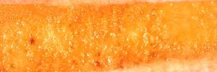
7 Case Aneurysmal Bone Cyst 13 year old with knee pain after a fall. MR features: fluid-fluid levels Aneurysmal bone cyst 7
8 Gross features: Usually a curettage (or core bx) specimen Small fragments of bone admixed with blood, variable amount of some hemorrhagic or fibrous soft tissue Decalcify if bony fragments present Submit entirely ABC: Summary All ages, most common <20 (median 13) Site: metaphysis of any bone Symptoms: Pain/swelling/fracture Recurrence: 20-70% following curettage Case Unicameral (simple) bone cyst UBC 8

9 12 yr old male with one month history of right ankle swelling. Recent twisting injury to ankle. Obvious fullness of lateral malleolus. No edema, warmth or skin discoloration over fullness. Nontender over fullness. No fevers, weight loss or night pain Gross features: Remarkably small specimen (compared to size of lesion on X- ray) Small spicules of bone, scant fibrous tissue, usually not very bloody Submit entirely after decal Scant tissue 9

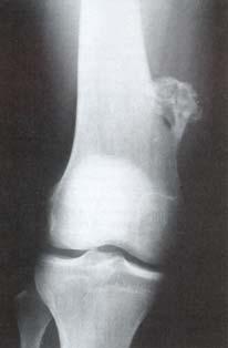
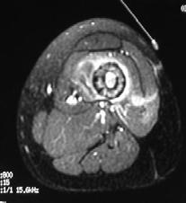
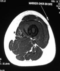
10 Simple bone cyst Age: 85% <20 Site: long bones (proximal humerus, femur, tibia, also pelvis, calcaneus) Recurrence: 10-20% Case Giant cell tumor of bone 23 yr old female with three month history of wrist pain, worse after a fall one month ago. Pain interferes with sleep Fullness at distal ulna region with overlying warmth Very tender to palpation Atrophy of hypothenar muscles 10
 Often abundant tissue,")


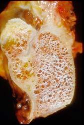
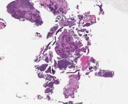
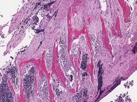
11 Recurrence Gross features: Usually curettage (rarely en-bloc) Often abundant tissue, especially when excisional biopsy Brown-red hemorrhagic tissue, mostly soft with minor bony component Decalcify only portion that contains bone Submit liberally (might be too large to submit entirely) 11
 Epiphyseal")


12 Giant cell tumor of bone Age: (very rare before skeletal maturity) Epiphyseal sites: (distal femur, prox. tibia, distal radius, prox. humerus, also flat and small tubular bones, vertebrae Local recurrence 25%, lung metastases 2% (slow growing or regressing) 12
13 Bone forming tumors Osteosarcoma and its variants General Approach to Handling Bone Tumor Specimens Do not freeze the limb (freezing artifact!) Ink soft tissue margins (if tumor is close) Sample margins, LN s, vessels Dissect soft tissue off bone except for soft tissue extension of tumor Obtain cross section of the bone margin Disarticulate joint if free of tumor Section bone longitudinally (avoid smearing ink into bone saw) 13
")

 Obtain")

14 cont.: Handling Bone Tumor Specimens Clean cut surface (rinse, gently brush) Photograph Obtain parallel cut (2-3mm) Fix in formalin, then decalcify Submit entire tumor surface of slab (make drawing, dictate accurately) Obtain additional sections as needed AKA for proximal tibia osteosarcoma Proximal Margin (bone, soft tissue, skin) 14


15 Ink margin (may be omitted if location of tumor is known), and sample soft tissue and vascular (venous!) margin Limb sparing excision: ink entire margin Identify neurovascular bundle, sample vascular margin (vein!), soft tissue and bone margin 15


16 Remove skin and dissect soft tissue (sample previous biopsy site), look for lymph nodes Inspect joint cavity for tumor involvement Disarticulate Transect the long bone to a manageable length Sample synovium 16


17 Inspect interosseous soft tissue Inspect tib/fib joint After sampling bone margins, obtain longitudinal section (usually coronal) 17
 Decalcification Use relatively thin sections Fix in formalin!")

18 Obtain additional sections as indicated clinically (X-rays, clinical info) Cut thin slab (few mm) Decalcification Use relatively thin sections Fix in formalin! (never decalcify before the bone is well fixed) Decalcify and check for appropriate decalcification, then submit Wash decalcifying solution off before definite processing Do not overdecalcify Decalcify only when necessary! 18
 Limb sparing resection: Sections to take from the slab From: Dorfman and")
19 Handling of Post-chemo Bone tumors More sections required to assess % tumor necrosis (may be 30-60) Sample sanctuary sites: tumor periphery, soft tissue extension, areas of hemorrhage Percent necrosis is assessed microscopically by comparing cellularity to original tumor (critical value is usually 90% necrosis) Limb sparing resection: Sections to take from the slab From: Dorfman and Czerniak s Bone Tumors Mosby 1998 Case High Grade Osteosarcoma, conventional type 19


20 17 yr old female with R knee atraumatic pain/vague swelling x 4-5 months. Acute worsening of night pain x 2-3 weeks. Xray AP view Xray lateral view Bone scan CT without and with contrast Sagittal slab Coronal slab 20


21 Histology Treatment of Osteosarcoma Pre-operative Chemotherapy Surgical Excision (Radiation) Post Operative Chemotherapy Treated osteosarcoma 21


22 Epidemiology of Osteosarcoma Most common primary malignancy of the skeleton 20% of primary sarcoma of bone Incidence is 4-5 per million newly diagnosed cases in US Osteosarcoma 60 M:F 1:6 1.6: % % 4% 2% 8% 8% 33% Age in Decades 20% Classification of Osteosarcoma Intramedullary Conventional: osteoblastic chondroblastic fibroblastic Telangiectatic Small cell Giant cell Well differentiated Juxtacortical Well-differentiated (Parosteal) Chondroblastic, low to intermediate (periosteal) Osteoblastic, high grade surface 22


23 Parosteal OS Courtesy of Dr. Gunnlaugur P. Nielsen Periosteal OS Courtesy of Dr. Gunnlaugur P. Nielsen 23

24 Telangiectatic OS Courtesy of Dr. Gunnlaugur P. Nielsen Cartilage tumors Chondrosarcoma and its DD Osteochondroma Enchondroma Conventional chondrosarcoma Dedifferentiated chondrosarcoma Chondroblastic osteosarcoma 24
25 Osteochondroma Courtesy of Dr. Gunnlaugur P. Nielsen Osteochondroma 25
 Peripheral portion consists of cartilage cap Enchondral ossification beneath (inner portion of the lesion and stalk consist of")
26 Gross features: May be intact or fragmented Orientation: Recognize margin of resection (base of exophytic lesion)and cartilage cap Describe thickness of cap and sample Decalcification required Osteochondroma Osteochondroma Mushroom shaped with stalk (or sessile) Peripheral portion consists of cartilage cap Enchondral ossification beneath (inner portion of the lesion and stalk consist of bone) similar to growth plate Cortex of the stalk merges with cortex of the bone it arose from (medullary cavity of the osteochondroma and bone are connected) 26


27 Large osteochondroma removed in fragments Enchondroma Enchondroma small bones of the hand > feet (60%) > long tubular bones asymptomatic? pain 7:1 27


28 Gross features: Usually curettage Sample liberally if clinical suspicious for malignancy CT enchondroma Enchondroma 28
29 Enchondroma Gray-blue and translucent (like normal cartilage) Well-circumscribed nodules of benign cartilage (low cellularity) Periphery of nodules with ossification No cortical erosion or soft tissue extension Case Osteochondroma.Later revised to low grade chondrosarcoma 14 yo girl with left arm mass since infancy now interfering with her sleep Family h/o multiple osteochondromatosis 29


30 Fragmented exophytic lesion, thick irregular cartilage cap Conventional Chondrosarcoma 2nd most common primary bone sarcoma 1/4 of all primary bone sarcomas ilium>femur>humerus 6th-7th decade of life 20% 15% 10% Intramedullary Chondrosarcoma Proximal Femur 30
 Gross features: Often large Low grade tumors resemble")


31 Example of grade 2 CS Consistency softer (more gelatinous = myxoid) Gross features: Often large Low grade tumors resemble hyaline cartilage (glistening grey-white, often lobulated) Focal gritty calcifications Higher grade tumors often myxoid with hemorrhage and necrosis CS grade 1-2/3 Permeative growth with destruction of bony trabeculae Residual trabecula Soft tissue extension 31


32 Case Dedifferentiated Chondrosarcoma 44 yr old female presents with left shoulder pain for 10 years Recently enlarging, painful left shoulder mass and night pain Scapula Tumor 32


33 Dedifferentiated CHSA transformation of a low grade CHSA into a high grade sarcoma (OSA, MFH) 10% of all CHSA Femur > pelvis > humerus 6th - 7th decade 90% die within the first year 20% 30% 15% Small round cell tumors 33


34 Bone lesions with a permeative pattern by X-ray Osteomyelitis Ewing s sarcoma Lymphoma (Non-Hodgkin s) Myeloma Langerhans histiocytosis Bone metastases Osteomyelitis: Radiographic mimicker of malignancy Small round cell tumors of bone Lymphoma/Leukemia Ewings/PNET Mesenchymal Chondrosarcoma Osteosarcoma (small cell variant) (Metastatic small round cell tumors, i.e. rhabdomyosarcoma, neuroblastoma, Wilms tumor, melanoma, small cell carcinoma..) 34
 Molecular diagnostics (snap frozen or paraffin block) Flow cytometry (r/o lymphoma), fresh tissue Electron microscopy,")
 or fix in formalin Case Ewing sarcoma 18 yr old female with several month history of left thigh pain Xrays Bone scan")
35 Allocation of tissue for ancillary studies important in round cell tumors DD Snap freeze tissue whenever possible Cytogenetics (fresh tissue in cell culture medium) Molecular diagnostics (snap frozen or paraffin block) Flow cytometry (r/o lymphoma), fresh tissue Electron microscopy, in glutaraldehyde fixative Touch imprints (for possible FISH): important: do NOT use alcohol to fix slide; instead air dry (needs to be taken to lab within 24 hours) or fix in formalin Case Ewing sarcoma 18 yr old female with several month history of left thigh pain Xrays Bone scan 35
36 CT scans Incisional biopsy 36


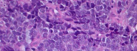
37 Small round cell tumor Additional studies required 37

 in")


38 Immunohistochemistry results CD99 LCA S-100 Keratin Ewing sarcoma: Ancillary Tests Immunohistochemistry: CD99 etc. Molecular: Tumor specific translocation t(11;22) in 85% gene fusion gene EWS/Fli-1 (oncogene) Diagnostic tests used to identify fusion gene: conventional cytogenetics (requires live cells) RT-PCR FISH Ewing s Sarcoma/PNET Round cell sarcoma with varying degree of neuroectodermal differentiation 38

")
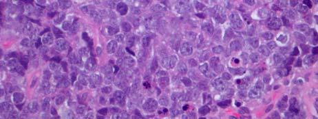
39 Ewing Sarcoma/PNET: Primary bone sarcoma (majority) 2 nd most common primary malignant bone tumor in children Extraskeletal Ewing s: 2 nd most common soft tissue sarcoma in children Age: (80% <20) Symptoms: pain, mass, fever, ESR, anemia, leukocytosis, large soft tissue mass common Resection post-chemotherapy Case Diffuse Large Cell (B-cell) Lymphoma 39


40 69 yr old male with a 6 month history of right elbow pain and swelling. The pain is present at all times and interferes with sleep. Originally treated for tennis elbow with PT. Case #2 Incisional Biopsy 40
 CD20 Control Bone Metastases")
")

41 Lymphoma Allocate tissue for lymphoma workup whenever lymphoma is suspected (clinically or at the time of frozen section) CD20 Control Bone Metastases Most common bone malignancy overall 3 rd most common metastatic site Rare in children (may be seen in neuroblastoma, RMS, clear cells sarcoma of kidney) Sites: bones rich in red marrow (vertebrae, proximal femur, ribs, skull, pelvis, sternum, shoulder Symptoms: pain, swelling, fracture, neural symptoms, hypercalcemia 41
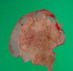
42 Metastatic carcinoma: Radiographic features Periosteal reaction is uncommon Lytic Pure lysis and blow out: renal cell Mixed lytic and blastic: breast, lung, GI Pure blastic: prostate, medulloblastoma Intracortical: lung Hands and feet (acral metastasis): lung Bone Metastases: Pathology Osteoblastic (dense on Xray): prostate, some breast, lung Osteolytic (hypodense on Xray): renal cell, most metastases in general Micro: recapitulates the original tumor Immunohistochemistry often required to confirm the tumor origin Fresh tissue may be required for special studies (chemosensitivity, molecular analysis) Case Metastatic breast cancer 42


43 Distorted femur head Ill-defined hemorrhagic lesion Metastatic carcinoma: touch prep during frozen section 43

.")

44 Metastasis (left) and previous breast biopsy (right). Notice that the histology is very similar Thank you! 44
Primary bone tumors > metastases from other sites Primary bone tumors widely range -from benign to malignant. Classified according to the normal cell
 Primary bone tumors > metastases from other sites Primary bone tumors widely range -from benign to malignant. Classified according to the normal cell counterpart and line of differentiation. Among the
The Radiology Assistant : Bone tumor - ill defined osteolytic tumors and tumor-like lesions
 Bone tumor - ill defined osteolytic tumors and tumor-like lesions Henk Jan van der Woude and Robin Smithuis Radiology department of the Onze Lieve Vrouwe Gasthuis, Amsterdam and the Rijnland hospital,
Malignant bone tumors. Incidence Myeloma 45% Osteosarcoma 24% Chondrosarcoma 12% Lyphoma 8% Ewing s Sarcoma 7%
 Malignant bone tumors Incidence Myeloma 45% Osteosarcoma 24% Chondrosarcoma 12% Lyphoma 8% Ewing s Sarcoma 7% Commonest primary bone sarcoma is osteosarcoma X ray Questions to ask 1. Solitary or Multiple
MARK D. MURPHEY MD, FACR. Physician-in-Chief, AIRP. Chief, Musculoskeletal Imaging
 ALPHABET SOUP AND CYSTIC LESIONS OF THE BONE MARK D. MURPHEY MD, FACR Physician-in-Chief, AIRP Chief, Musculoskeletal Imaging ALPHABET SOUP AND CYSTIC LESIONS OF THE BONE Giant cell tumor (GCT) Unicameral
Bone Tumours - a synopsis. Dr Zena Slim SpR in Histopathology QAH 2009
 Bone Tumours - a synopsis Dr Zena Slim SpR in Histopathology QAH 2009 Aims General approach to diagnosis Common entities.and not so common ones. Mini quiz Challenge of bone tumour diagnosis Bone tumours
Bone Tumors Clues and Cues
 William Herring, M.D. 2002 Bone Tumors Clues and Cues In Slide Show mode, advance the slides by pressing the spacebar All Photos Retain the Copyright of their Authors Clues by Appearance of Lesion Patterns
The Radiology Assistant : Bone tumor - well-defined osteolytic tumors and tumor-like lesions
 Bone tumor - well-defined osteolytic tumors and tumor-like lesions Henk Jan van der Woude and Robin Smithuis Radiology department of the Onze Lieve Vrouwe Gasthuis, Amsterdam and the Rijnland hospital,
APMA 2018 Radiology Track Bone Tumors When to say Gulp!
 APMA 2018 Radiology Track Bone Tumors When to say Gulp! DANIEL P. EVANS, DPM, FACFAOM Professor, Department of Podiatric Medicine and Radiology Dr. Wm. Scholl College of Podiatric Medicine Conflict of
Musculoskeletal Sarcomas
 Musculoskeletal Sarcomas Robert C. Orth, M.D., Ph.D. Edward B. Singleton Department of Pediatric Radiology Texas Children s Hospital Page 0 xxx00.#####.ppt 9/23/2012 9:01:18 AM No disclosures Page 1 xxx00.#####.ppt
Disclosures. Giant Cell Rich Tumors of Bone. Outline. The osteoclast. Giant cell rich tumors 5/21/11
 Disclosures Giant Cell Rich Tumors of Bone Andrew Horvai, MD, PhD Associate Clinical Professor, Pathology This lecture discusses "off label" uses of a number of pharmaceutical agents. The speaker is describing
Introduction to Musculoskeletal Tumors. James C. Wittig, MD Orthopedic Oncologist Sarcoma Surgeon
 Introduction to Musculoskeletal Tumors James C. Wittig, MD Orthopedic Oncologist Sarcoma Surgeon www.tumorsurgery.org Definitions Primary Bone / Soft tissue tumors Mesenchymally derived tumors (Mesodermal)
LAC + USC.
 Jeff McDavit,, M.D. LAC + USC mcdavit@usc.edu Clinical History 55 year old male with large, deep, non- tender left thigh mass. Seen at LAC+USC Med Ctr FNA clinic No h/o trauma or radiation Vimentin
Bone tumors. RMG: jan
 Bone tumors RMG: jan 217. @Kijohs KIZZA JOHN KIJOHS Diseases arising in bone Lipoma Fibrous cortical defects Non-ossifying fibroma Bone island Benign simple cysts Enchondroma Osteochondroma Osteoid osteoma
Grading of Bone Tumors
 Grading of Bone Tumors Joon Hyuk Choi, M.D. Department of Pathology College of Medicine, Yeungnam University Introduction to grading system of bone tumor used at Mayo Clinic WHO Histologic Classification
USCAP 2014 Common problems in bone and soft tissue pathology: Cartilage tumors
 USCAP 2014 Common problems in bone and soft tissue pathology: Cartilage tumors Andrew Horvai MD PhD Clinical Professor, Pathology UCSF, San Francisco, CA Outline Common intramedullary tumors Enchondroma
Kidney Case 1 SURGICAL PATHOLOGY REPORT
 Kidney Case 1 Surgical Pathology Report February 9, 2007 Clinical History: This 45 year old woman was found to have a left renal mass. CT urography with reconstruction revealed a 2 cm medial mass which
COPYRIGHT 2004 BY THE JOURNAL OF BONE AND JOINT SURGERY, INCORPORATED
 84 COPYRIGHT 2004 BY THE JOURNAL BONE AND JOINT SURGERY, INCORPORATED Radiographic Evaluation of Pathological Bone Lesions: Current Spectrum of Disease and Approach to Diagnosis BY BENJAMIN G. DOMB, MD,
STAGING, BIOPSY AND NATURAL HISTORY OF TUMORS SCOTT D WEINER MD
 STAGING, BIOPSY AND NATURAL HISTORY OF TUMORS SCOTT D WEINER MD WHAT DO YOU DO WHEN THIS SHOWS UP IN YOUR OFFICE? besides panicking KEY PRINCIPLE!!! Reactive zone is the edema, neovascularity and inflammation
University Journal of Surgery and Surgical Specialities
 University Journal of Surgery and Surgical Specialities Volume 1 Issue 1 2015 EXTRA SKELETAL MESENCHYMAL CHONDROSARCOMA :A CASE REPORT Rajaraman R Subbiah S Navin Naushad Kilpaulk Medical College Abstract:
MRI XR, CT, NM. Principal Modality (2): Case Report # 2. Date accepted: 15 March 2013
: Case Report # 2. Date accepted: 15 March 2013") Radiological Category: Musculoskeletal Principal Modality (1): Principal Modality (2): MRI XR, CT, NM Case Report # 2 Submitted by: Hannah Safia Elamir, D.O. Faculty reviewer: Naga R. Chinapuvvula, M.D.
Radiological Category: Musculoskeletal Principal Modality (1): Principal Modality (2): MRI XR, CT, NM Case Report # 2 Submitted by: Hannah Safia Elamir, D.O. Faculty reviewer: Naga R. Chinapuvvula, M.D.
CONSULTATION DURING SURGERY / NOT A FINAL DIAGNOSIS. FROZEN SECTION DIAGNOSIS: - A. High grade sarcoma. Wait for paraffin sections results.
 Pathology Report Date: 3/5/02 A, B. Biopsy right distal femur- high grade spindle cell sarcoma Immunohistochemistry studies are pending to further classify the nature of the tumor. CONSULTATION DURING
What Do You Need to Know About Bone Pathology? Benjamin L. Hoch M.D. Associate Professor Department of Pathology University of Washington
 What Do You Need to Know About Bone Pathology? Benjamin L. Hoch M.D. Associate Professor Department of Pathology University of Washington What s Do You Need To Know About Bone Pathology? Reactive/pseudosarcomatous
FRACTURE CALLUS ASSOCIATED WITH BENIGN AND MALIGNANT BONE LESIONS AND MIMICKING OSTEOSARCOMA
 THE AMERICAN JOURNAL OF CLINICAL PATHOLOGY Vol. 52, No. 1 Copyright 1969 by The Williams & Wilkins Co. Printed in U.S.A. FRACTURE CALLUS ASSOCIATED WITH BENIGN AND MALIGNANT BONE LESIONS AND MIMICKING
Typical skeletal location and differential diagnosis of bone tumors.
 Typical skeletal location and differential diagnosis of bone tumors. Poster No.: C-2418 Congress: ECR 2015 Type: Educational Exhibit Authors: M. Barros, L. A. Ferreira, Y. Costa, P. J. V. Coelho, F. Caseiro
SMALL ROUND BLUE CELL LESION OF BONE
 DISCLOSURE SMALL ROUND BLUE CELL LESION OF BONE Dr. Alistair Jordan University of South Alabama No financial support or endorsement OBJECTIVES Describe the more common small round cell lesions of bone
Malignant Bone Tumours. PathoBasic, Daniel Baumhoer
 Malignant Bone Tumours PathoBasic, 20.03.18 Daniel Baumhoer FNCLCC Grading The differentiation score is defined as the extent to which a tumor resembles adult mesenchymal tissue (score 1), the extent to
Immunohistochemistry in Bone and Soft Tissue Tumors. Sahar Rassi Zankoul, MD
 Immunohistochemistry in Bone and Soft Tissue Tumors Sahar Rassi Zankoul, MD Introduction Bone tumors represent a wide variety of tumors of various origins and malignant potentials. These different tumor
Advertisement. Osteochondroma
 Advertisement Osteochondroma An osteochondroma is a benign (noncancerous) tumor that develops during childhood or adolescence. It is an abnormal growth that forms on the surface of a bone near the growth
Skeletal metastases are the most common variety of bone tumors and should always be considered in the differential diagnosis, particularly in older
 Dr Brajesh Nandan Skeletal metastases are the most common variety of bone tumors and should always be considered in the differential diagnosis, particularly in older patients. Cancers of the breast, prostate,
Effective local and systemic therapy is necessary for the cure of Ewing tumor Most chemotherapy regimens are a combination of cyclophosphamide,
 Ewing Tumor Perez Ewing tumor is the second most common primary tumor of bone in childhood, and also occurs in soft tissues Ewing tumor is uncommon before 8 years of age and after 25 years of age In the
RESEARCH INFORMATION AWARENESS SUPPORT PRIMARY BONE CANCER CHONDROSARCOMA. Visit bcrt.org.uk for more information
 RESEARCH INFORMATION AWARENESS SUPPORT PRIMARY BONE CANCER CHONDROSARCOMA Visit bcrt.org.uk for more information CONTENTS What is it? Who does it affect? Symptoms Types of Chondrosarcoma Cause and Risk
Fluid-fluid levels in bone tumors: A pictorial review
 Fluid-fluid levels in bone tumors: A pictorial review Poster No.: C-578 Congress: ECR 2009 Type: Educational Exhibit Topic: Musculoskeletal Authors: L. Figueroa Nasra, C. Martín Hervás, M. Tapia-Viñé,
36 1 The Skeletal System Slide 1 of 40
 1 of 40 The Skeleton All organisms need structural support. Unicellular organisms have a cytoskeleton. Multicellular animals have either an exoskeleton (arthropods) or an endoskeleton (vertebrates). 2
LYMPH NODE: BIOPSY Guideline & Procedure: Lymph Node Biopsy
 LYMPH NODE: BIOPSY Guideline & Procedure: Lymph Node Biopsy (Discuss with hematopathology attending for lymphoma work up case prior to proceed to gross at LBJ) 1. Measure the specimen (three dimensions).
Chapter 6 & 7 The Skeleton
 Chapter 6 & 7 The Skeleton Try this Make clockwise circles with your RIGHT foot, while doing this, draw the number 6 in the air with you RIGHT hand what happens to your foot???? Bony Background Adult body
Bone/Osteoid Producing Lesions
 Chapter 2 Bone/Osteoid Producing Lesions Introduction There are many lesions that are associated with reactive new bone formation; this chapter predominantly covers those in which deposition of osteoid/bone
GIANT CELL-RICH OSTEOSARCOMA: A CASE REPORT
 Nagoya J. Med. Sci. 59. 151-157, 1996 CASE REPORTS GIANT CELL-RICH OSTEOSARCOMA: A CASE REPORT KEIJI SATO!, SHIGEKI YAMAMURA!, HISASHI IWATA!, HIDESHI SUGIURA 2, NOBUO NAKASHIMA 3 and TETSURO NAGASAKA
Due in Lab. Due next week in lab - Scientific America Article Select one article to read and complete article summary
 Due in Lab 1. Skeletal System 33-34 2. Skeletal System 26 3. PreLab 6 Due next week in lab - Scientific America Article Select one article to read and complete article summary Cell Defenses and the Sunshine
JMSCR Vol 3 Issue 11 Page November 2015
 www.jmscr.igmpublication.org Impact Factor 3.79 Index Copernicus Value: 5.88 ISSN (e)-2347-176x ISSN (p) 2455-0450 DOI: http://dx.doi.org/10.18535/jmscr/v3i11.36 Diagnostic Dilemmas in Cytodiagnosis of
Review Course «Musculoskeletal Oncology» October 6, 2011 UNIKLINIK BALGRIST. Imaging of Bone and Soft Tissue. Tumors
 Imaging of Bone and Soft Tissue Tumors Approach from a radiologist s point of view Florian Buck Radiology Radio- Radio- Oncologist Oncologist Orthopedist Orthopedist Patient Management Oncologist Oncologist
Pediatric Pathology Grossing Guidelines
 When you get a case that should/could be for Pediatric Pathology, show it to the gross room supervisor or page Dr. Goldstein at 31418 before you cut it in. If it is the weekend or late at night, page the
Human Skeletal System Glossary
 Acromegaly Apatite Acromegaly - is a condition which involves excessive growth of the jaw, hands, and feet. It results from overproduction of somatotropin in adults (after fusion of the ossification centres
The Skeletal System:Bone Tissue
 The Skeletal System:Bone Tissue Dynamic and ever-changing throughout life Skeleton composed of many different tissues cartilage, bone tissue, epithelium, nerve, blood forming tissue, adipose, and dense
Monophasic Synovial Carcinoma of knee joint- A Case Report and Review of Literature
 IOSR Journal of Dental and Medical Sciences (IOSR-JDMS) e-issn: 2279-0853, p-issn: 2279-0861.Volume 17, Issue 3 Ver.5 March. (2018), PP 13-17 www.iosrjournals.org Monophasic Synovial Carcinoma of knee
Functions of the Skeletal System. Chapter 6: Osseous Tissue and Bone Structure. Classification of Bones. Bone Shapes
 Chapter 6: Osseous Tissue and Bone Structure Functions of the Skeletal System 1. Support 2. Storage of minerals (calcium) 3. Storage of lipids (yellow marrow) 4. Blood cell production (red marrow) 5. Protection
Klinisch belang van chromosomale translocatie detectie in sarcomen
 Translocations in sarcomas Klinisch belang van chromosomale translocatie detectie in sarcomen Judith V.M.G. Bovée, M.D., Ph.D. Department of Pathology Leiden University Medical Center RNA binding DNA binding
Primary bone tumors according to the WHO classification: a review of 13 years with illustrative examples
 Primary bone tumors according to the WHO classification: a review of 13 years with illustrative examples Poster No.: C-1741 Congress: ECR 2015 Type: Educational Exhibit Authors: J. Silva, M. A. Ramírez
Suspecting Tumors, or Could it be cancer?
 Suspecting Tumors, or Could it be cancer? Donna E. Reece, M.D. Princess Margaret Cancer Centre University Health Network Toronto, ON CANADA 07 February 2018 Background Low back pain is common However,
Solitary Bone Cyst of the Lunate: A Case Report
 Cronicon OPEN ACCESS ORTHOPAEDICS Case Report Solitary Bone Cyst of the Lunate: A Case Report MihirDesai* and Shivanand Bandekar Department of Orthopedics, Goa Medical College, Goa, India *Corresponding
What is bone? Specialized form of connective tissue: mineralized collagen matrix, therefore very rigid and strong while still retaining some degree of
 Bone What is bone? Specialized form of connective tissue: mineralized collagen matrix, therefore very rigid and strong while still retaining some degree of flexibility Other types of connective tissue:
Downloaded from by on 11/21/17 from IP address Copyright ARRS. For personal use only; all rights reserved
 Downloaded from www.ajronline.org by 46.3.196.1 on 11/21/17 from IP address 46.3.196.1. opyright RRS. For personal use only; all rights reserved T he scapula is a small bone in which many neoplasms can
Skeletal Radiology. Solitary (unicameral) bone cyst. The fallen fragment sign revisited
 bone cyst. The fallen fragment sign revisited") Skeletal Radiol (1989) 18:261-265 Skeletal Radiology Solitary (unicameral) bone cyst The fallen fragment sign revisited S. Struhl, M.D., C. Edelson, M.D., H. Pritzker, M.D., L.P. Seimon, M.D., and H.D.
Skeletal Radiol (1989) 18:261-265 Skeletal Radiology Solitary (unicameral) bone cyst The fallen fragment sign revisited S. Struhl, M.D., C. Edelson, M.D., H. Pritzker, M.D., L.P. Seimon, M.D., and H.D.
Primary Tumors of Ribs
 Primary Tumors of Ribs Frank E. Schmidt, M.D., and Max J. Trummer, Capt, MC, USN ABSTRACT An analysis of 50 consecutive patients with primary rib tumors operated on at the U.S. Naval Hospital, San Diego,
Primary Bone Tumors: Spine Surgery Live -Video Techniques Mobile Spine
 Primary Bone Tumors: Spine Surgery Live -Video Techniques Mobile Spine Christopher Ames MD Professor of Neurosurgery and Orthopedic Surgery Director of Spine Tumor And Deformity Surgery UCSF Department
Radiology Pathology Conference
 Radiology Pathology Conference Sharlin Johnykutty,, MD, Cytopathology Fellow Sara Majewski, MD, Radiology Resident Friday, August 28, 2009 Presentation material is for education purposes only. All rights
ORTHOPAEDIC ONCOLOGY OITE REVIEW COURSE
 ORTHOPAEDIC ONCOLOGY OITE REVIEW COURSE Richard D. Lackman, MD FACS Director, Orthopaedic Oncology Center Cancer Institute Introduction In the evaluation of a patient with a bone tumor, there are several
Skeletal System. Chapter 6.1 Human Anatomy & Physiology
 Skeletal System Chapter 6.1 Human Anatomy & Physiology Overview of Skeletal System Bones Joints Skeletal System Cartilage Tendons (bone to muscle) Ligaments (bone to bone) Function of the Skeletal System
A 64 y.o. man presents to the hospital with persistent cough and hemoptysis. Fernando Mut Montevideo - Uruguay
 A 64 y.o. man presents to the hospital with persistent cough and hemoptysis Fernando Mut Montevideo - Uruguay Teaching case Bone # 1 A 64 y.o. man presents to the hospital with persistent cough and hemoptysis.
* I have no disclosures or any
 Howard Rosenthal, M.D. Associate Professor of Orthopedic Surgery University of Kansas Sarcoma Center I have no disclosures or any conflicts related to the content of this presentation. Objectives 1. Describe
BONE TISSUE. Dr. Heba Kalbouneh Associate Professor of Anatomy and Histology
 BONE TISSUE Dr. Heba Kalbouneh Associate Professor of Anatomy and Histology BONE FUNCTION Support Protection (protect internal organs) Movement (provide leverage system for skeletal muscles, tendons, ligaments
Prof. Dr. NAGUI M. ABDELWAHAB,M.D.; MARYSE Y. AWADALLAH, M.D. AYA M. BASSAM, Ms.C.
 Role of Whole-body Diffusion MR in Detection of Metastatic lesions Prof. Dr. NAGUI M. ABDELWAHAB,M.D.; MARYSE Y. AWADALLAH, M.D. AYA M. BASSAM, Ms.C. Cancer is a potentially life-threatening disease,
VALORACIÒN RADIOLÓGICA DE LA LESIÒN ÒSEA SOLITARIA IMAGENOLOGIA MEDICA UNIVERSIDAD HISPANOAMERICANA
 VALORACIÒN RADIOLÓGICA DE LA LESIÒN ÒSEA SOLITARIA IMAGENOLOGIA MEDICA UNIVERSIDAD HISPANOAMERICANA TUMORES ÓSEOS SE PRESENTAN POR RANGOS DE EDAD, PRINCIPALMENTE: MENORES DE 20 AÑOS 20 A 40 AÑOS MAYORES
Associated Terms: Osteosarcoma, Bone Cancer, Limb Salvage, Appendicular Osteosarcoma, Pathologic Fracture, Chondrosarcoma
 1 of 9 9/29/2014 8:25 PM Associated Terms: Osteosarcoma, Bone Cancer, Limb Salvage, Appendicular Osteosarcoma, Pathologic Fracture, Chondrosarcoma The term "ACVS Diplomate" refers to a veterinarian who
Case Scenario 1 History and Physical 3/15/13 Imaging Pathology
 Case Scenario 1 History and Physical 3/15/13 The patient is an 84 year old white female who presented with an abnormal mammogram. The patient has a five year history of refractory anemia with ringed sideroblasts
Case Report Intramedullary Chondrosarcoma of Proximal Humerus
 Hindawi Publishing Corporation Case Reports in Radiology Volume 2012, Article ID 642062, 7 pages doi:10.1155/2012/642062 Case Report Intramedullary Chondrosarcoma of Proximal Humerus Pratiksha Yadav, Dolly
ISSN: DISTRIBUTION OF BONE AND CARTILAGINOUS TUMORS IN PEDIATRIC AGE GROUP IN WESTERN UTTAR-PRADESH: AN EVALUATIVE STUDY
 : 289-295 ISSN: 2277 4998 DISTRIBUTION OF BONE AND CARTILAGINOUS TUMORS IN PEDIATRIC AGE GROUP IN WESTERN UTTAR-PRADESH: AN EVALUATIVE STUDY QADRI S, HASAN M, AKHTAR K * AND SHERWANI RK The Departments
The Skeletal System PART A
 5 The Skeletal System PART A PowerPoint Lecture Slide Presentation by Jerry L. Cook, Sam Houston University ESSENTIALS OF HUMAN ANATOMY & PHYSIOLOGY EIGHTH EDITION ELAINE N. MARIEB The Skeletal System
Bone Composition. Bone is very strong for its relatively light weight The major components of bone are:
 Human Bones Bone Composition Bone is very strong for its relatively light weight The major components of bone are: Calcium carbonate Calcium phosphate Collagen Water Cortical Bone Spongy Bone Medullary
The Skeletal System. Chapter 4
 The Skeletal System Chapter 4 FUNCTIONS OF THE SKELETAL SYSTEM Support o Provides shape Protection o Internal organs Movement o Provides structure for muscle to act upon Storage o Minerals & fat Blood
General Approach to Lytic Bone Lesions D. Lee Bennett, MD, MA, Georges Y. El Khoury, MD Appl Radiol. 2004;33(5)
") General Approach to Lytic Bone Lesions D. Lee Bennett, MD, MA, Georges Y. El Khoury, MD Appl Radiol. 2004;33(5) www.medscape.com Abstract and Introduction Abstract When interpreting musculoskeletal radiographs,
General Approach to Lytic Bone Lesions D. Lee Bennett, MD, MA, Georges Y. El Khoury, MD Appl Radiol. 2004;33(5) www.medscape.com Abstract and Introduction Abstract When interpreting musculoskeletal radiographs,
Interesting case. Vikas Kundra, M.D., Ph.D. October Vikas Kundra, M.D., Ph.D.
 Interesting case October 2012 Disclosure Information Vikas Kundra, M.D, Ph.D. I have no financial relationships to disclose. I WILL NOT include discussion of investigational or off-label use of a product
Case 8 Soft tissue swelling
 Case 8 Soft tissue swelling 26-year-old female presented with a swelling on the back of the left knee joint since the last 6 months and chronic pain in the calf and foot since the last 2 months. Pain in
Periosteal Osteosarcoma Arising from the Rib and Scapula: Imaging Features in Two Cases
 Case Report Musculoskeletal Imaging http://dx.doi.org/10.3348/kjr.2014.15.3.370 pissn 1229-6929 eissn 2005-8330 Korean J Radiol 2014;15(3):370-375 Periosteal Osteosarcoma Arising from the Rib and Scapula:
Lytic Lesion in the Distal Phalanx of the Hand
 Shafa Ortho J. 2015 February; 2(1):e441. Published online 2015 February 15. DOI: 10.5812/soj.441 Research Article Lytic Lesion in the Distal Phalanx of the Hand Khodamorad Jamshidi 1 ; Farid Najd Mazhar
The Skeletal System. Chapter 7a. Skeletal System Introduction Functions of the skeleton Framework of bones The skeleton through life
 The Skeletal System Skeletal System Introduction Functions of the skeleton Framework of bones The skeleton through life Chapter 7a Support Protection Movement Storage areas Minerals Lipids Hemopoiesis
UK Musculoskeletal Oncology: Something for All Ages. Lars Wagner, MD Pediatric Hematology/Oncology University of Kentucky
 UK Musculoskeletal Oncology: Something for All Ages Lars Wagner, MD Pediatric Hematology/Oncology University of Kentucky Pediatric-Type Sarcomas of Bone and Soft Tissue The incidence of sarcoma continues
Bubbly Lesions of Bone
 Residents Section Pattern of the Month w79 08.18.09 Eisenberg Residents Section Pattern of the Month Residents inradiology Ronald L. Eisenberg 1 Eisenberg RL Keywords: bubbly lesions, fegnomashic, skeletal
04/27/2017. The Spectrum of Cartilaginous Tumors
 The Spectrum of Cartilaginous Tumors L I S A E R C O L A N O, M D M U S C U L O S K E L E T A L O N C O L O G Y D E P A R T M E N T O F O R T H O P A E D I C S A L L E G H E N Y H E A L T H N E T W O R
Hths 2231 Laboratory 13 Alterations in Musculoskeletal
 Watch Movie: Osteoporosis Answer the movie questions on the worksheet. Complete activities 1-4. Activity #1: Click on the website link in activity 1 to review the structure and function of bone. Activity
Chapter 5 The Skeletal System
 Chapter 5 The Skeletal System The Skeletal System Parts of the skeletal system Bones (skeleton) Joints Cartilages Ligaments (bone to bone)(tendon=bone to muscle) Divided into two divisions Axial skeleton:
From Morphology to Molecular Pathology: A Practical Approach for Cytopathologists Part 1-Cytomorphology. Songlin Zhang, MD, PhD LSUHSC-Shreveport
 From Morphology to Molecular Pathology: A Practical Approach for Cytopathologists Part 1-Cytomorphology Songlin Zhang, MD, PhD LSUHSC-Shreveport I have no Conflict of Interest. FNA on Lymphoproliferative
Lymphoma Read with the experts
 Lymphoma Read with the experts Marc Seltzer, MD Associate Professor of Radiology Geisel School of Medicine at Dartmouth Director, PET-CT Course American College of Radiology Learning Objectives Recognize
FORMATION OF BONE. Intramembranous Ossification. Bone-Lec-10-Prof.Dr.Adnan Albideri
 FORMATION OF BONE All bones are of mesodermal origin. The process of bone formation is called ossification. We have seen that formation of most bones is preceded by the formation of a cartilaginous model,
Disseminated Primary Non-Hodgkin s Lymphoma of Bone : A Case Re p o r t 1
 Disseminated Primary Non-Hodgkin s Lymphoma of Bone : A Case Re p o r t 1 Hee-Jin Park, M.D., Sung-Moon Lee, M.D., Hee-Jung Lee, M.D., Jung-Sik Kim, M.D., Hong Kim, M.D. Primary lymphoma of bone is uncommon
Pediatric Pathology Grossing Guidelines
 When you get a case that should/could be for Pediatric Pathology, show it to the gross room supervisor or page Dr. Goldstein at 31418 before you cut it in. If it is the weekend or late at night, page the
PowerPoint Lecture Slides. Prepared by Patty Bostwick-Taylor, Florence-Darlington Technical College. The Skeletal System Pearson Education, Inc.
 PowerPoint Lecture Slides Prepared by Patty Bostwick-Taylor, Florence-Darlington Technical College CHAPTER 5 The Skeletal System 2012 Pearson Education, Inc. Title Classification of Bones and Gross Anatomy
BIOH111. o Cell Module o Tissue Module o Integumentary system o Skeletal system o Muscle system o Nervous system o Endocrine system
 BIOH111 o Cell Module o Tissue Module o Integumentary system o Skeletal system o Muscle system o Nervous system o Endocrine system Endeavour College of Natural Health endeavour.edu.au 1 TEXTBOOK AND REQUIRED/RECOMMENDED
MSK Interesting Cases. Dr Yap Sheau Huey
 MSK Interesting Cases Dr Yap Sheau Huey Case 1: History 41 y.o man, surf skier C/o pain over anterior left 5 th to 8 th ribs. Worse after sport activity. Chest Radiograph US Periostitis and early callus
History. 33 y/o F with hx of palpable anterior tibial mass x 2 years, only painful with palpation
 History 33 y/o F with hx of palpable anterior tibial mass x 2 years, only painful with palpation Imaging Photo Album Patient also had a smaller lesion 1 cm proximal to this lesion, not seen radiographically.
1/25/13 Right partial nephrectomy followed by completion right radical nephrectomy.
 History and Physical Case Scenario 1 45 year old white male presents with complaints of nausea, weight loss, and back pain. A CT of the chest, abdomen and pelvis was done on 12/8/12 that revealed a 12
Common Primary Tumors of Bone
 Special Report Common Primary Tumors of Bone Primary bone tumors are a relatively rare occurrence, however, they can have serious deleterious consequences. Many possess the ability to degenerate into malignant
ORTHOPAEDIC TUMOURS DJM FRANTZEN 2012 AUGUST TUMOURS 1
 ORTHOPAEDIC TUMOURS DJM FRANTZEN 2012 AUGUST TUMOURS 1 PRINCIPLES STAGING WORKUP RADIOLOGY BIOPSY PROCEDURES CHEMOTHERAPY RADIOTHERAPY 2012 AUGUST TUMOURS 2 STAGING ENNEKING'S SURGICAL STAGES (ENNEKING)
SKELETAL TISSUES CHAPTER 7 INTRODUCTION TO THE SKELETAL SYSTEM TYPES OF BONES
 SKELETAL TISSUES CHAPTER 7 By John McGill Supplement Outlines: Beth Wyatt Original PowerPoint: Jack Bagwell INTRODUCTION TO THE SKELETAL SYSTEM STRUCTURE Organs: Bones Related Tissues: Cartilage and Ligaments
Giant cell tumour of the sternum-two cases
 Giant cell tumour of the sternum-two cases Nishaa.P 1, Raghuram.P 2, Navin patil 3, Jaipal B.R 4 Akkamahadevi patel 5 Assistant Professor ESIC medical college and PGIMSR 1 Professor and HOD, 2 Professor
OSTEOMYELITIS. If it occurs in adults, then the axial skeleton is the usual site.
 OSTEOMYELITIS Introduction Osteomyelitis is an acute or chronic inflammatory process of the bone and its structures secondary to infection with pyogenic organisms. Pathophysiology Osteomyelitis may be
CASE STUDY: PRO-DENSE Injectable Regenerative Graft Used to Backfill a Bone Cavity Following Resection of a Giant Cell Tumor
 : PRO-DENSE Used to Backfill a Bone Cavity Following Resection of a Giant Cell Tumor Contributed by: Matthew J. Seidel, MD* Lauren A. Schwartz, NP Scottsdale, AZ *Dr. Seidel is a paid consultant for Wright
Pediatric Retroperitoneal Masses Radiologic-Pathologic Correlation
 Acta Radiológica Portuguesa, Vol.XVIII, nº 70, pág. 61-70, Abr.-Jun., 2006 Pediatric Retroperitoneal Masses Radiologic-Pathologic Correlation Marilyn J. Siegel Mallinckrodt Institute of Radiology, Washington
The formation of blood cells is called. hemopoiesis. What does our bone store? Where do our bones store fat? yellow marrow.
 What are the 5/6 functions of the skeletal system? support, protection, movement, blood cell formation, storage, homeostasis The formation of blood cells is called hemopoiesis What does our bone store?
DIAGNOSING EWING S SARCOMA OF THE RIB IN CHILDREN IS IT QUITE CHALLENGING??
 Indian Journal of Medical s ISSN: 2319 3832(Online) DIAGNOSING EWING S SARCOMA OF THE RIB IN CHILDREN IS IT QUITE CHALLENGING?? *Arvind Kumar S.M. and Shyam Sundar V, Dinakar B.K. and SandhyaV. and Kailash
Principles of Surgical Oncology. Winnie Achilles Tierklinik Hollabrunn Lastenstrasse Hollabrunn
 Principles of Surgical Oncology Winnie Achilles Tierklinik Hollabrunn Lastenstrasse 2 2020 Hollabrunn boexi@gmx.de The first surgery provides the best chance for a cure in an animal with a tumor Clinical