Fun with Fat. General Rules. Case
|
|
|
- Bethanie Neal
- 5 years ago
- Views:
Transcription
1 Fun with Fat General Rules Imaging: location (deep vs. superficial) Superficial lesions are seldom liposarcomas Deep lesions may be benign or malignant Myxoid stroma is common in benign and malignant lesions Lipoblasts can be seen in benign and malignant tumors Well diff. liposarcoma is a 4x diagnosis Case 34 year old female with a 2cm mass lesion located on the chest, adjacent to the armpit. The lesion was excised and sent to pathology.

2


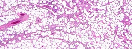
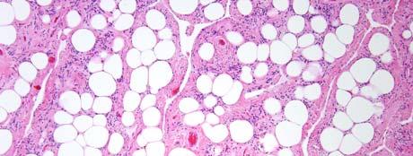
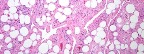
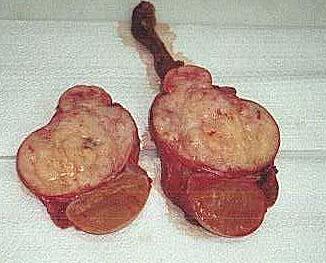
3 Diagnosis Ruptured silicone breast implant Case 40 year old male with a 2 cm neck mass. Lesion was excised and sent to pathology.
4 Diagnosis Benign Lipoma with fat necrosis

 Simple excision.")


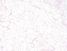
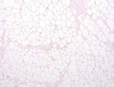
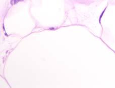
5 Lipoma Benign tumors composed of cells resembling mature adipocytes Most common soft tissue tumors of adulthood (40-60 yrs) Simple excision. Rarely recur. Many subtypes; none with prognostic significance May be superficial or deep, intra- or inter-muscular Main differential: Well diff. liposarcoma Lipoma with focal fat necrosis: May mimic fibrous septa seen in WDLS. Lipoma with focal fat necrosis: May mimic variation in adipocyte size as in WDLS.


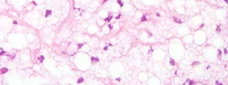
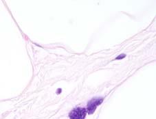
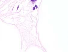
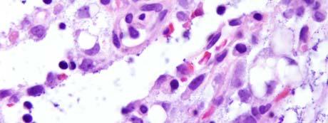
6 Lipoma with fat necrosis: Sclerosis and variation in adipocyte size Lipoma with fat necrosis; histiocytes look pink Lochkern intranuclear lipid invagination

7 Hyperchromatic ugly cell in a well differentiated liposarcoma Good 4X Bad 4X Good - Oil Bad - Oil
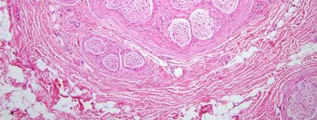
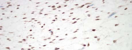
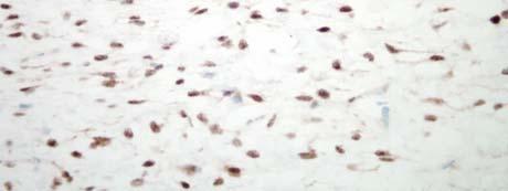
![Intra-muscular Lipoma: Prone to recur [infiltrative]](/docs-images/86/93469240/images/8-0.jpg "Fibrolipomatous Hamartoma of Nerve Lipoma-like mass")


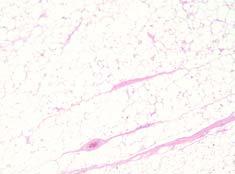
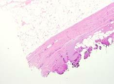
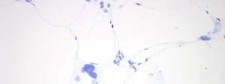
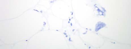
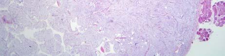
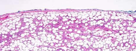
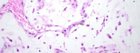
8 Intra-muscular Lipoma: Prone to recur [infiltrative] Fibrolipomatous Hamartoma of Nerve Lipoma-like mass lesion attached to a large nerve with fibrosis Often median or ulnar nerves on hand, wrist, or fingers. Macrodactyly. Nerve is surrounded by ring of concentric fibrosis Benign FLPH of nerve: Fat surrounds a nerve


9 FLPH of nerve: Fat surrounds a nerve with concentric fibrosis FLPH of nerve: Fat surrounds a nerve with concentric fibrosis FLPH of nerve: Fibrosis-rich lesion


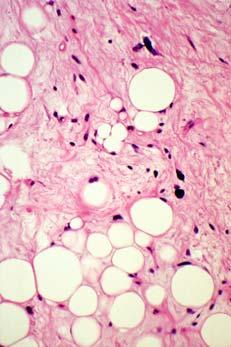
10 Case 2 year old male with a slowly growing mass lesion on the upper left arm. The lesion was excised and sent to pathology.
.")
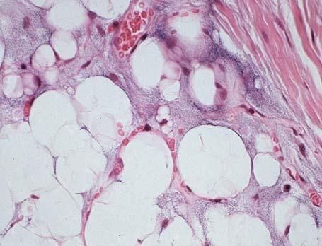
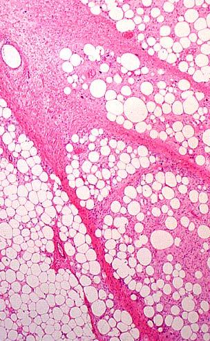
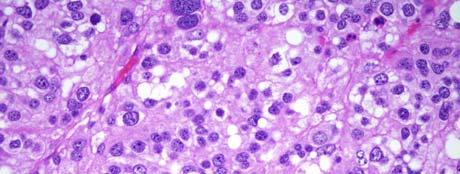
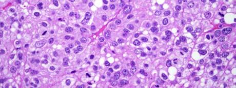
11 Diagnosis Lipoblastoma Lipoblastoma Benign tumor, typically arises during the first three years of life Slow growing painless mass most often on subcutaneous extremities Lipoblastomatosis: multiple poorly circumscribed masses, may be deep Local resection. May recur locally (9-22%). Does not metastasize Has features in common with lipoma (lobules of mature fat), WDLS (fibrous septa), myxoid liposarcoma (myxoid stroma with branching vessels, lipoblasts) Lipoblastoma (mature areas): lobules, fibrous septa
:")


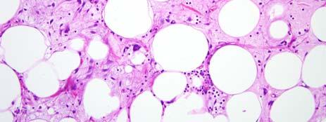
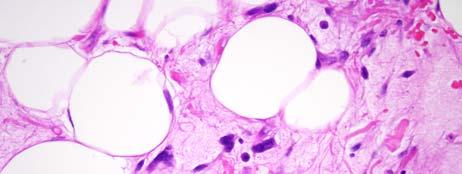
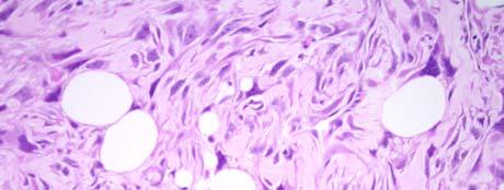
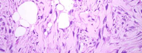
12 Lipoblastoma (immature): Myxoid stroma with branching vessels Lipoblastoma Myxoid Stroma with prominent vessels Lipoblastoma - Lipoblasts


13 Lipoblastoma: peripheral immature cellular areas Case 43 year old male with a 3 cm mass on the back of the neck. Lesion was excised and sent to pathology.
14 MDM2
15 Diagnosis Pleomorphic Lipoma Lipoma Variants Many Variants, we will cover the most common entities All behave similar to regular lipomas Stromal collagen bands are very helpful to distinguish benign from malignant adipose tumors MDM2 immunostains/fish Angiolipoma Usually young adults Often on forearms Painful and multiple Numerous small capillaries with admixed fat in a lobular configuration Capillary supporting elements and fibrin thrombi


16 Angiolipoma: Lobular at low power. Fat, blood, and hypercellularity. Angiolipoma: Lobular arrangements of small vessels with admixed adipocytes Angiolipoma: Stromal support elements surround and invest capillary proliferations.


17 Angiolipoma: Fibrin thrombi Chondroid Lipoma Usually small, subcutaneous mass on extremity of an adult (female predominance) Well-circumscribed, lobulated, cellular Well-defined cells with intracytoplasmic vacuoles growing in chondroid matrix S100 protein, occasional focal Keratin Characteristic 11;16 translocation Chondroid lipoma small, well circumscribed subcutaneous lesion


18 Chondroid lipoma lobulated, cellular Chondroid lipoma numerous vacuolated cells in an eosinophilic matrix that appears chondroid Chondroid lipoma Vacuolated cells resemble lipoblasts

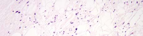
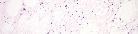
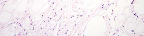
19 Spindle Cell Lipoma Usually in males years Solitary, painless mass on back of neck, shoulders, elsewhere Circumscribed and subcutaneous Spindle Cell Lipoma Variable numbers of mature adipocytes, bland spindle cells, mast cells Wiry collagen fibers embedded in myxoid stroma, cleft-like spaces CD34+, S100- Spindle Cell Lipoma


20 Spindle Cell Lipoma: Well-circumscribed. Fibrotic and myxoid with fat at low power. Spindle cell lipoma 1.Mature fat 2. Small hyperchromatic spindle cells 3. Myxoid matrix 4. Wiry Collagen 5. Mast cells Spindle Cell Lipoma: Mast Cells


21 Spindle cell lipoma myxoid with cleft-like spaces Spindle cell lipoma clefts, myxoid, wiry collagen


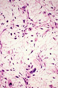
22 Spindle cell lipoma clefts, myxoid, wiry collagen Spindle cell lipoma mast cells Pleomorphic Lipoma Basically a spindle cell lipoma with atypia, thicker collagen bundles Hyperchromatic multinucleated cells in a floret arrangement Lipoblasts may also be present How to distinguish from WDLS - collagen pattern, MACROSCOPIC appearance, MDM2(-)


23 Pleomorphic lipoma circumscribed, fat, fibrotic and myxoid stroma with large cells at low power Pleomorphic Lipoma: Atypical Hyperchromatic cells Pleomorphic Lipoma: Bizarre Atypia


24 Pleomorphic Lipoma: Lipoblasts Pleomorphic Lipoma: Mast Cells and Collagen Pleomorphic Lipoma: dense eosinophilic collagen bands and cellular myxoid areas

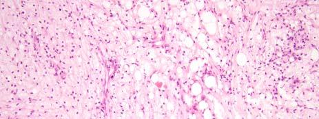
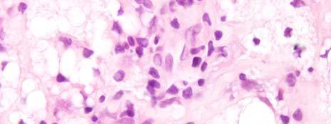
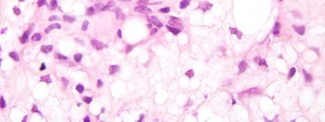
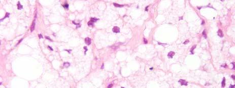
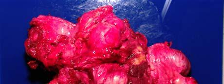
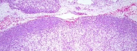
25 Pleomorphic Lipoma: cellular area with collagen fibrils and mast cells Hibernoma Uncommon benign tumor with features of brown fat; younger patients than lipoma (late 30s). Arise in several locations, including abdominal cavity and retroperitoneum. Usually 9-10cm. Benign. Three cell types: mature adipose, finely vacuolated, granular eosinophilic. Small branching capillaries. Hibernoma: Large tan-red mass lesion


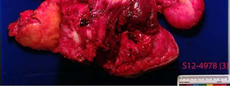
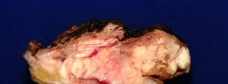
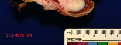
26 Hibernoma: Lobulated with clear and eosinophilic cells Hibernoma: Three distinct cell types Case 41 year old male with a large ileoinguinal mass lesion. The lesion was resected and sent to pathology.
27
28
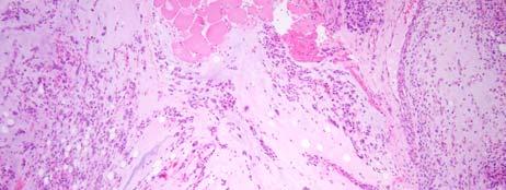
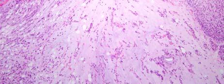
29 Diagnosis Dedifferentiated liposarcoma Liposarcomas Three general types Well-Differentiated /Dedifferentiated Myxoid/High grade myxoid (Round Cell Liposarcoma) Pleomorphic Liposarcoma Well-Differentiated Liposarcoma 40-45% of liposarcomas Middle aged adults 6 th decade, M=F Deep soft tissues of limbs [esp thighs where the fat is] followed by retroperitoneum and groin Metastasis is a risk in lesions with dedifferentiation (28% of retroperitoneal lesions) Wide Local excision % recurrence rate for extremities. Retroperitoneal and groin lesions nearly always recur (80-90%)

30 Atypical Lipomatous Tumor ALT terminology is applied to superficial/ extremity lesions Same histology and genetic alterations as deep lesions Different natural history: much lower likelihood of dedifferentiation (5% vs. 28%) and metastasis Well-Differentiated Liposarcoma: Lipoma-like Thick fibrous bands Variation in mature adipocyte size Pleomorphic hyperchromatic cells visible within fibrous bands at 4x scanning magnification +/- Lipoblasts: Not necessary for diagnosis. Retroperitoneal well-diff liposarcoma
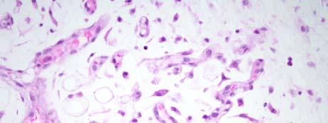
31 Paratesticular well-diff liposarcoma Thick fibrous bands with hyperchromatic cells WDLS: Hyperchromatic cells at low power


32 Hyperchromatic cells with no lipoblasts Pleomorphic cells in thick collagen bands -The eosinophilic fibrillar collagen bands seen in pleomorphic lipoma are absent. WDLS: Needle Biopsy at 4x mag. Fat, hyperchromatic cells, fibrous septa


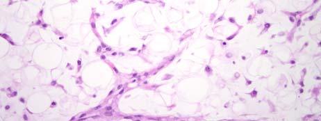
33 WDLS: Myxoid Stroma WDLS: MDM2 Immunostain Well-Differentiated Liposarcoma: Inflammatory type Extensive infiltration of tumor with lymphoid cells with follicles and germinal centers May also have eosinophils and neutrophils Atypical cells of WDLS may cause confusion with lymphoma Focal inflammatory-type areas may be seen in other subtypes


34 Inflammatory WDLS: Fat, fibrous bands, myxoid areas, Inflammation, lymphoid follicles. Well-Differentiated Liposarcoma: Sclerosing Type May not appear adipocytic on imaging Mostly fibrotic or hyalinized stroma with hyperchromatic cells in sclerotic areas Infrequent mitoses, moderate to low cellularity, no fascicles Sclerosing WDLS: Hyperchromatic and pleomorphic Cells in sclerotic stroma with few adipocytes
. May occur at any site and also increases risk of local recurrence.")

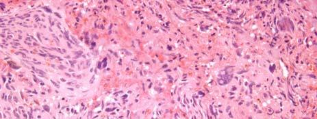
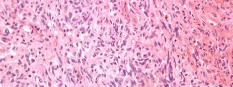
35 Dedifferentiated Liposarcoma A confluent focus of high grade sarcoma arising within or adjacent to a WDLS. No size criteria. Bestows metastatic capability (15-30% metastatic rate). May occur at any site and also increases risk of local recurrence. Dedifferentiated Liposarcoma May present as any non-adipocytic sarcoma, MFH most common Most retroperitoneal MFHs represent dediff. liposarcoma Classically abrupt transition from WDLS to dediff. component Hypercellularity without adipocytes, nuclear pleomorphism, high mitotic rate Dediff. liposarcoma Lipoma-like WDLS


36 Lipoma-like WDLS Sclerosing WDLS Dediff. liposarcoma MFH-like Myxofibrosarcoma-like Dedifferentiated Liposarcoma Dediff. Liposarcoma: MDM2 immunostain


37 Low grade Dedifferentiation >1 low power field with no fat More cellular and fascicular than sclerosing WDLS Basically a low grade fibrosarcoma component Does not improve prognosis Chief differential: Sclerosing WDLS Low-grade dediff. Sclerosing WDLS Fooler for Atypical Lipomatous Tumor/Well Diff LPS Massive localized lymphedema

:560-4. Review. PubMed PMID: 21518378.")


38 Shon W, Ida CM, Boland-Froemming JM, Rose PS, Folpe A. Cutaneous angiosarcoma arising in massive localized lymphedema of the morbidly obese: a report of five cases and review of the literature. J Cutan Pathol Jul;38(7): Review. PubMed PMID: Manduch M, Oliveira AM, Nascimento AG, Folpe AL. Massive localized lymphoedema: a clinicopathological study of 22 cases and review of the literature. J Clin Pathol Sep;62(9): Review. PubMed PMID: Farshid G, Weiss SW. Massive localized lymphedema in the morbidly obese: a histologically distinct reactive lesion simulating liposarcoma. Am J Surg Pathol Oct;22(10): Review. PubMed PMID: Massive localized lymphedema in the morbidly obese


39 Massive localized lymphedema in the morbidly obese Massive localized lymphedema in the morbidly obese Reactive fibroblasts in septa appear similar to the cells in atypical lipomatous tumor Massive localized lymphedema in the morbidly obese Reactive fibroblasts in septa appear similar to the cells in atypical lipomatous tumor CONTEXT of superficial mass with ectatic lymphatic vessels is key


40 Massive localized lymphedema in the morbidly obese Reactive fibroblasts in septa appear similar to the cells in atypical lipomatous tumor note the hemosiderin Massive localized lymphedema in the morbidly obese MDM2 stain negative nuclei Case 43 year old male with a 7cm soft tissue mass deep in the thigh. The lesion was biopsied and sent to pathology.

41 T-2 weighted MRI


42 Diagnosis Myxoid Liposarcoma Case An incidental plaque-like nodule on the abdominal wall of a 78 year old female was noticed during an exploratory laparotomy. It was excised and sent to pathology.

43 Diagnosis Fat Atrophy
 Liposarcoma About a third of liposarcs,")
, younger patients (4 th decade) ~30% metastasize.")


44 Cytomegalovirus infection with fat atrophy in an HIV/AIDS patient Cytomegalovirus infection with fat atrophy in an HIV/AIDS patient Myxoid/Round Cell (high grade form) Liposarcoma About a third of liposarcs, deep extremities (esp. thigh), younger patients (4 th decade) ~30% metastasize. Peculiar metastatic tropism to other soft tissue sites, lung, and bone A translocation sarcoma: bland, uniform oval to spindled tumor cells in a myxoid matrix with finely branching vasculature Cells often cluster at periphery of lobules or around vessels. Lipoblasts present Round cell is high grade variant of myxoid
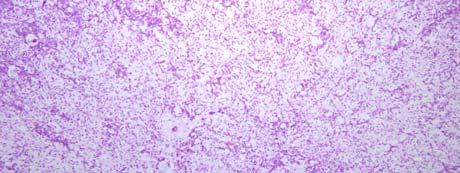
45 Myxoid Liposarcoma: Lobules with clustering of tumor cells at periphery and around vessels in a myxoid background.

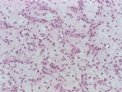
46 Myxoid Liposarcoma: Cyst-like areas Myxoid Liposarcoma: Rich Vascularity Myxoid Liposarcoma: Lipoblasts


 TLS(FUS) CHOP")
47 Myxoid Liposarcoma: Uniform oval cells Myxoid Liposarcoma Uniform spindled cells. Children may present with purely spindled cells with few to no fat or lipoblasts. Round Cell Liposarcoma t(12;22) TLS(FUS) CHOP (DDIT3) T(12;22) EWSR1-CHOP (DDIT3)


 Variable areas of pleomorphic lipoblasts and MFH-like morphology,")
48 Transitional Myxoid/Round Cell Liposarcoma Partial preservation of background stroma Pleomorphic Liposarcoma Rare and aggressive. M=F, >50yrs Deep soft tissues of the thigh, trunk or retroperitoneum Typically exceed 10.0 cm High incidence of metastases and tumor related mortality (5yr survival of 50-63%) Variable areas of pleomorphic lipoblasts and MFH-like morphology, some resemble myxofibrosarcoma Pleomorphic Liposarcoma: Numerous atypical Lipoblasts.


49 Pleomorphic Liposarcoma MFH-like areas Lipoblast areas Pleomorphic liposarcoma: Epithelioid lipoblasts Pleomorphic liposarcoma: Myxoid stroma


50 Case 63 year old female with a history of myelofibrosis with a 3cm perirenal mass lesion. The lesion was biopsied and sent to pathology.


51 Diagnosis Sclerosing extramedullary hematopoietic tumor SEMHT WDLPS Case 76 year old female with a 5cm subcutaneous mass on the foot. The lesion was excised and sent to pathology.

52 Diagnosis Myxofibrosarcoma Myxofibrosarcoma: Fake Lipoblast Pleo. Liposarcoma: Real Lipoblast


53 Myxofibrosarcoma (Myxoid MFH) - One of the most common sarcomas during 6 th -8 th decade; typically limbs, ~50:50 superficial:deep - Multilobulated with incomplete fibrous septa; multiple infiltrative nodules in superficial locations vs. a solitary mass deep - Spindled and pleomorphic cells in myxoid matrix with curvilinear vessels, pseudolipoblasts - Graded primarily on cellularity: low grade lesions do not metastasize, 21-38% of intermediate and high grade lesions do - All grades have high recurrence rates, and low grade lesions may recur as a higher grade Myxofibrosarcoma subcutaneous very common but unusual for myxoid liposarcoma Myxofibrosarcoma: multilobulated, infiltrative, subcutaneous


54 Myxofibrosarcoma: pleomorphism, myxoid stroma, prominent vascularity Myxofibrosarcoma: curvilinear vessels Myxofibrosarcoma: pleomorphism Myxoid LPS: Uniform cells
55 Who needs DDIT3 with good H&E???? Thank you!!!!
Update On Lipomatous Tumors: Old Standbys and New Concepts
 Update On Lipomatous Tumors: Old Standbys and New Concepts John R. Goldblum, M.D. Chairman, Department of Anatomic Pathology Cleveland Clinic Professor of Pathology Cleveland Clinic Lerner College of Medicine
Mayo Medical Laboratories
 Mayo Medical Laboratories Virtual Lectures 2014 MFMER 2016 MFMER slide-1 Virtual Lectures Planning Committee Disclosure Summary As a provider accredited by ACCME, College of Medicine, Mayo Clinic (Mayo
A 25 year old female with a palpable mass in the right lower quadrant of her abdomen
 May 2016 A 25 year old female with a palpable mass in the right lower quadrant of her abdomen Contributed by: Paul Ndekwe, MD, Resident Physician, Indiana University School of Department of Pathology and
Newer soft tissue entities
 Newer soft tissue entities Examples among fibroblastic tumors Turku, May 6, 2010 Markku Miettinen, M.D. AFIP, Washington, DC Fibroblastic neoplasms Solitary fibrous tumor /Hemangiopericytoma Low-grade
Malignant Peripheral Nerve Sheath Tumor
 C H A P T E R 120 Malignant Peripheral Nerve Sheath Tumor Currently, malignant peripheral nerve sheath tumor (MPNST) is the most commonly used generic name for the neoplasms known in the past as neurosarcoma,
The Relevance of Cytologic Atypia in Cutaneous Neural Tumors
 The Relevance of Cytologic Atypia in Cutaneous Neural Tumors Recent Findings - New Developments New Problems Zsolt B. Argenyi, M.D. Professor of Pathology & Dermatology Director of Dermatopathology Department
Diplomate of the American Board of Pathology in Anatomic and Clinical Pathology
 A 33-year-old male with a left lower leg mass. Contributed by Shaoxiong Chen, MD, PhD Assistant Professor Indiana University School of Medicine/ IU Health Partners Department of Pathology and Laboratory
Selected Pseudomalignant Soft Tissue Tumors of the Skin and Subcutis
 Selected Pseudomalignant Soft Tissue Tumors of the Skin and Subcutis Andrew L. Folpe, M.D. Professor of Laboratory Medicine and Pathology Mayo Clinic, Rochester, MN folpe.andrew@mayo.edu 2016 MFMER slide-1
21/07/2017. Hobnail endothelial cells are not the same as epithelioid endothelial cells
 UPDATE IN CUTANEOUS VASCULAR S DERMATOPATHOLOGY SESSION BELFAST PATHOLOGY JUNE 21/2017 Dr E Calonje St John s Institute of Dermatology, London, United Kingdom THE FAMILY OF VASCULAR S WITH EPITHELIOID
Tumors of Adipose Tissue Tumors Epidemiology Clinical Features. Morphology. Mature Adipocytes Separated by delicate fibrous septa
 Tumors of Adipose Tissue Lipoma Liposarcoma Most commonly happens in female The most common soft tissue tumor o Originates from matured Adipocytes Most commonly happes at the 4 th and 5 th decade of life
Musculoskeletal Sarcomas
 Musculoskeletal Sarcomas Robert C. Orth, M.D., Ph.D. Edward B. Singleton Department of Pediatric Radiology Texas Children s Hospital Page 0 xxx00.#####.ppt 9/23/2012 9:01:18 AM No disclosures Page 1 xxx00.#####.ppt
IN THE NAME OF GOD Dr. Kheirandish Oral and maxillofacial pathology
 IN THE NAME OF GOD Dr. Kheirandish Oral and maxillofacial pathology ORAL FOCAL MUCINOSIS Uncommon Tumorlike Cutaneous myxoid cyst Overproduction of hyaluronic acid by firoblasts Young adults Female Gingiva
Essential Dermatopathology. Jinah Kim, MD, PhD Department of Pathology and Dermatology Stanford University Medical Center
 Essential Dermatopathology Jinah Kim, MD, PhD Department of Pathology and Dermatology Stanford University Medical Center OBJECTIVES Review clinical, pathologic and molecular aspects of bone and fat tumors
Myxo-inflammatory Fibroblastic sarcoma
 AKA Myxo-inflammatory Fibroblastic sarcoma Acral Myxoinflammatory fibroblastic sarcomaam.j.surg.path1998; 22; 911-924 Inflammatory myxoid tumour of soft parts with bizarre giant cells [Pathol.Res.Pract.
We are IntechOpen, the world s leading publisher of Open Access books Built by scientists, for scientists. International authors and editors
 We are IntechOpen, the world s leading publisher of Open Access books Built by scientists, for scientists 3,900 116,000 120M Open access books available International authors and editors Downloads Our
أملس عضلي غرن = Leiomyosarcoma. Leiomyosarcoma 1 / 5
 Leiomyosarcoma 1 / 5 EPIDEMIOLOGY Exact incidence is unknown, but older studies suggest that leiomyosarcomas comprise approximately 3 percent of soft-tissue sarcomas. Superficial leiomyosarcoma occurs
5/10. Pathology Soft tissue tumors. Farah Bhani. Mohammed Alorjani
 5/10 Pathology Soft tissue tumors Mohammed Alorjani Farah Bhani Slides are included in this sheet. Objectives: Soft tissue tumors 1. Describe soft tissue tumors. 2. Understand the classification of soft
3/27/2017. Disclosure of Relevant Financial Relationships
 Ophthalmic Pathology Evening Specialty Conference USCAP 2017 5 th March, 2017 Mukul K. Divatia, MD Assistant Professor Department of Pathology & Genomic Medicine Weill Cornell Medical College Houston Methodist
Adipocytic Tumours in children
 Università degli Studi di Padova Dipartimento di Medicina Sezione di Anatomia Patologica Generale e Citopatologia Adipocytic Tumours in children Rita Alaggio Basel Seminars in Pathology Paediatric Pathology
Desmoplastic Melanoma R/O BCC. Clinical Information. 74 y.o. man with lesion on left side of neck r/o BCC
 R/O BCC Sabine Kohler, M.D. Professor of Pathology and Dermatology Dermatopathology Service Stanford University School of Medicine Clinical Information 74 y.o. man with lesion on left side of neck r/o
Evening Specialty Conference Bone and Soft Tissue Pathology. Diagnostic pitfalls in bone and soft tissue pathology
 Evening Specialty Conference Bone and Soft Tissue Pathology. Case 1 Elizabeth G Demicco, MD, PhD Mount Sinai Hospital, New York Disclosure of Relevant Financial Relationships USCAP requires that all planners
SESSION 1: GENERAL (BASIC) PATHOLOGY CONCEPTS Thursday, October 16, :30am - 11:30am FACULTY COPY
 PATHOLOGY CONCEPTS Thursday, October 16, :30am - 11:30am FACULTY COPY") SESSION 1: GENERAL (BASIC) PATHOLOGY CONCEPTS Thursday, October 16, 2008 9:30am - 11:30am FACULTY COPY GOAL: Describe the basic morphologic (structural) changes which occur in various pathologic conditions.
SESSION 1: GENERAL (BASIC) PATHOLOGY CONCEPTS Thursday, October 16, 2008 9:30am - 11:30am FACULTY COPY GOAL: Describe the basic morphologic (structural) changes which occur in various pathologic conditions.
Cellular Neurothekeoma
 Cellular Neurothekeoma Scott W Binder, MD Pritzker Professor of Pathology & Dermatology Sr. Vice Chair Director, Pathology Clinical Services Chief, Dermatopathology Geffen/UCLA School of Medicine Clinical
POORLY DIFFERENTIATED, HIGH GRADE AND ANAPLASTIC CARCINOMAS: WHAT IS EVERYONE TALKING ABOUT?
 POORLY DIFFERENTIATED, HIGH GRADE AND ANAPLASTIC CARCINOMAS: WHAT IS EVERYONE TALKING ABOUT? AGGRESSIVE THYROID CANCERS PAPILLARY CARCINOMA CERTAIN SUBTYPES POORLY DIFFERENTIATED CARCINOMA HIGH GRADE DIFFERENTIATED
57th Annual HSCP Spring Symposium 4/16/2016
 An Unusual Malignant Spindle Cell Lesion to Involve the Breast Erinn Downs-Kelly, D.O. Associate Professor of Pathology University of Utah & ARUP Laboratories No disclosures Case 39 y/o female with no
ACCME/Disclosures ALK FUSION-POSITIVE MESENCHYMAL TUMORS. Tumor types with ALK rearrangements. Anaplastic Lymphoma Kinase. Jason L.
 Companion Meeting of the International Society of Bone and Soft Tissue Pathology The Evolving Concept of Mesenchymal Tumors ALK FUSION-POSITIVE MESENCHYMAL TUMORS Jason L. Hornick, MD, PhD March 13, 2016
WHAT IS MDM2? (MDMTWOMICS) MDM2 IN SARCOMAS? (MDMTWOMAS) MDM2MICS? NO CONFLICT OF INTERESTS 5/07/2018 MDM2 IN SOFT TISSUE AND BONE SARCOMAS
 MDM2 IN SARCOMAS? (MDMTWOMAS) MDM2MICS? NO CONFLICT OF INTERESTS 5/07/2018 MDM2 IN SOFT TISSUE AND BONE SARCOMAS") IN SOFT TISSUE AND BONE SARCOMAS WHAT IS? (MDMTWOMICS) Raf Sciot, M.D., PhD. Department of Pathology, University Hospitals Katholieke Universiteit Leuven, LEUVEN, Belgium IN SARCOMAS? (MDMTWOMAS) MICS?
IN SOFT TISSUE AND BONE SARCOMAS WHAT IS? (MDMTWOMICS) Raf Sciot, M.D., PhD. Department of Pathology, University Hospitals Katholieke Universiteit Leuven, LEUVEN, Belgium IN SARCOMAS? (MDMTWOMAS) MICS?
Gross appearance of peritoneal cysts. They have a thin, translucent wall and contain a clear fluid.
 Gross appearance of peritoneal cysts. They have a thin, translucent wall and contain a clear fluid. So-called multicystic benign mesothelioma. A, Gross appearance. So-called multicystic benign mesothelioma.
CASE REPORT PLEOMORPHIC LIPOSARCOMA OF PECTORALIS MAJOR MUSCLE IN ELDERLY MAN- CASE REPORT & REVIEW OF LITERATURE.
 PLEOMORPHIC LIPOSARCOMA OF PECTORALIS MAJOR MUSCLE IN ELDERLY MAN- CASE REPORT & REVIEW OF LITERATURE. M. Madan 1, K. Nischal 2, Sharan Basavaraj. C. J 3. HOW TO CITE THIS ARTICLE: M. Madan, K. Nischal,
Spindle Cell Lesions Of The Breast. Emad Rakha Professor of Breast Pathology and Consultant Pathologist
 Spindle Cell Lesions Of The Breast Emad Rakha Professor of Breast Pathology and Consultant Pathologist * SCLs comprise a wide spectrum of diseases, ranging from reactive processes to aggressive malignant
Case: The patient is a 62 year old woman with a history of renal cell carcinoma that was removed years ago. A 2.4 cm liver mass was found on CT
 Case: The patient is a 62 year old woman with a history of renal cell carcinoma that was removed years ago. A 2.4 cm liver mass was found on CT during follow- up. ALT, AST, Alk Phos and bilirubin were
Atypical Palisaded Myofibroblastoma of Lymph Node: Report of a rare case.
 ISPUB.COM The Internet Journal of Pathology Volume 10 Number 1 Atypical Palisaded Myofibroblastoma of Lymph Node: Report of a rare case. V Kinnera, R Nandyala, M Yootla, K Mandyam Citation V Kinnera, R
Note: The cause of testicular neoplasms remains unknown
 - In the 15- to 34-year-old age group, they are the most common tumors of men. - Tumors of the testis are a heterogeneous group of neoplasms that include: I. Germ cell tumors : 95%; all are malignant.
Case Report Spindle cell lipoma of the wrist, occurring in a distinctly rare location: a case report with review of literature
 Int J Clin Exp Pathol 2015;8(3):3299-3303 www.ijcep.com /ISSN:1936-2625/IJCEP0004992 Case Report Spindle cell lipoma of the wrist, occurring in a distinctly rare location: a case report with review of
Pleomorphic Liposarcoma: A Clinicopathologic Analysis Of 19 Cases
 Pleomorphic Liposarcoma: A Clinicopathologic Analysis Of 19 Cases Katharine A. Downes, M.D., John R. Goldblum, M.D., Elizabeth A. Montgomery, M.D., Cyril Fisher, M.D., F.R.C.Path. Departments of Anatomic
Salivary Glands 3/7/2017
 Salivary Glands 3/7/2017 Goals and objectives Focus on the entities unique to H&N Common board type facts Information for your future practice Salivary Glands Salivary Glands Major gland. Paratid. Submandibular.
Endometrial Stromal Tumors
 Endometrial Stromal Tumors WHO Categories: Endometrial Stromal Nodule (ESN) Endometrial Stromal Sarcoma, low grade (LGESS) Endometrial Stromal Sarcoma, high grade (HGESS) Undifferentiated Uterine Sarcoma
Case Report Primary Small Bowel Liposarcoma (Atypical Lipomatous Tumour) with Myogenic Differentiation
 with Myogenic Differentiation") Sarcoma Volume 2010, Article ID 807981, 4 pages doi:10.1155/2010/807981 Case Report Primary Small Bowel Liposarcoma (Atypical Lipomatous Tumour) with Myogenic Differentiation J. Patel, R. Deb, W. Speake,
Sarcoma Volume 2010, Article ID 807981, 4 pages doi:10.1155/2010/807981 Case Report Primary Small Bowel Liposarcoma (Atypical Lipomatous Tumour) with Myogenic Differentiation J. Patel, R. Deb, W. Speake,
SOFT TISSUE TUMOR PATHOLOGY: AN UPDATE
 SOFT TISSUE TUMOR PATHOLOGY: AN UPDATE Jason L. Hornick, MD, PhD July 18, 2013 Department of Pathology Brigham and Women s Hospital Harvard Medical School Boston, MA, USA I have no disclosures. New Soft
Soft tissue lipomas, lipoma variants and liposarcomas: MRI evaluation and review of literature
 Soft tissue lipomas, lipoma variants and liposarcomas: MRI evaluation and review of literature Poster No.: R-0122 Congress: RANZCR-AOCR 2012 Type: Authors: Keywords: DOI: Educational Exhibit A. A. Tandon,
Diagnostic problems in uterine smooth muscle tumors
 Diagnostic problems in uterine smooth muscle tumors Marina Kos Ljudevit Jurak Clinical Department of Pathology, Clinical Hospital Center Sestre milosrdnice, Zagreb Institute of Pathology, University of
Takayuki Ohguri 1 Takatoshi Aoki 1 Masanori Hisaoka 2 Hideyuki Watanabe 1 Katsumi Nakamura 1 Hiroshi Hashimoto 2 Toshitaka Nakamura 3 Hajime Nakata 1
 Takayuki Ohguri 1 Takatoshi Aoki 1 Masanori Hisaoka 2 Hideyuki Watanabe 1 Katsumi Nakamura 1 Hiroshi Hashimoto 2 Toshitaka Nakamura 3 Hajime Nakata 1 Received July 1, 2002; accepted after revision November
Special slide seminar
 Special slide seminar Tomáš Rozkoš The Fingerland Department of Pathology Charles University Medical Faculty and Faculty Hospital in Hradec Králové Czech Republic Case history, 33 years old resistance
Part 1. Slides 1-38, Rita Alaggio Soft tissue tumors Trondheim 14. mars 2013
 Part 1 Slides 1-38, Rita Alaggio Soft tissue tumors Trondheim 14. mars 2013 Pediatric Pathology Soft Tissue Tumors AN UPDATE Rita Alaggio Azienda Ospedaliera Università di Padova Soft Tissue Tumors More
Case year female. Routine Pap smear
 Case 1 57 year female Routine Pap smear Diagnosis? 1. Atypical glandular cells of unknown significance (AGUS) 2. Endocervical AIS 3. Endocervical adenocarcinoma 4. Endometrial adenocarcinoma 5. Adenocarcinoma
Normal endometrium: A, proliferative. B, secretory.
 Normal endometrium: A, proliferative. B, secretory. Nội mạc tử cung Nội mạc tử cung Cyclic changes in endometrium.. Approximate relationship of useful microscopic changes. Arias-Stella reaction in endometrial
Disclosures. Parathyroid Pathology. Objectives. The normal parathyroid 11/10/2012
 Disclosures Parathyroid Pathology I have nothing to disclose Annemieke van Zante MD/PhD Assistant Professor of Clinical Pathology Associate Chief of Cytopathology Objectives 1. Review the pathologic features
Disclosures. Giant Cell Rich Tumors of Bone. Outline. The osteoclast. Giant cell rich tumors 5/21/11
 Disclosures Giant Cell Rich Tumors of Bone Andrew Horvai, MD, PhD Associate Clinical Professor, Pathology This lecture discusses "off label" uses of a number of pharmaceutical agents. The speaker is describing
CYSTIC TUMORS OF THE KIDNEY JOHN N. EBLE, M.D. CYSTIC NEPHROMA
 Page 1 CYSTIC TUMORS OF THE KIDNEY JOHN N. EBLE, M.D. Department of Pathology & Laboratory Medicine Phone (317) 274-4806 Medical Science A-128 FAX: (317) 278-2018 635 Barnhill Drive jeble @iupui.edu Indianapolis,
Lách
 Lách Lách Lách Lách Splenogonadal fusion. Splenic tissue is attached to testicular tissue. Pseudocyst (false or secondary cyst). A, Outer aspect. Pseudocyst (false or secondary cyst). B, Inner surface.
Chondroid lipoma: A rare recently described benign lipomatous tumor
 Al-Malki et al. 7 case report peer Reviewed open OPEN ACCESS Chondroid lipoma: A rare recently described benign lipomatous tumor Salman T. Al-Malki, Abdullah S. Al-Khamiss Abstract Introduction: lipomatous
Contents Part I Introduction 1 General Description 2 Natural History: Importance of Size, Site, Histopathology
 Contents Part I Introduction 1 General Description... 3 1.1 Introduction... 3 1.2 Incidence and Prevalence... 5 1.3 Predisposing and Genetic Factors... 8 References... 16 2 Natural History: Importance
Primary Breast Liposarcoma
 Primary Breast Liposarcoma Bhagyam Nagarajan 1*, GayatriAutkar 1, Keyuri Patel 1, Meghal Sanghvi 1 1. Department of Radiology, Wockhardt Hospital, Mumbai, India * Correspondence: Dr Bhagyam Nagarajan,
PLEOMORPHIC ADENOMA ( BENIGN MIXED TUMOR )
") ( BENIGN MIXED TUMOR ) Grossly, the tumor is freely movable, solid, sometimes lobulated and occasionally cystic. If recurrent, multinodular masses are common. Histologically, within a fibrous capsule,
( BENIGN MIXED TUMOR ) Grossly, the tumor is freely movable, solid, sometimes lobulated and occasionally cystic. If recurrent, multinodular masses are common. Histologically, within a fibrous capsule,
A case of fat-free pleomorphic lipoma occurring in the upper back and axilla simultaneously
 Wang et al. World Journal of Surgical Oncology 2013, 11:145 WORLD JOURNAL OF SURGICAL ONCOLOGY CASE REPORT Open Access A case of fat-free pleomorphic lipoma occurring in the upper back and axilla simultaneously
Case 1 10/2/17. Myxoid Soft Tissue Tumors & Tumor-like Lesions. Myxofibro- or Fibromyxo-?: Myxoid Soft Tissue Tumours We Are All Mixed Up About
 Myxoid Soft Tissue Tumors & Tumor-like Lesions Myxofibro- or Fibromyxo-?: Myxoid Soft Tissue Tumours We Are All Mixed Up About Rajiv M. Patel, M.D. RCPA NZ ASM 2017 (4:15-5:00pm, Saturday, 23-09-17) Heterogenous
USCAP 2011: ASDP companion meeting. Steven D. Billings 1
 USCAP 2011: ASDP companion meeting. Steven D. Billings (billins@ccf.org) 1 Spindle cell tumors that make you say, Oh $*&%! This lecture will focus on examples of cutaneous tumors that present particular
Case year old Chinese female. Radiological echo-distortion in the right breast at o clock. Core biopsy of the o clock lesion.
 Case 3 64 year old Chinese female. Radiological echo-distortion in the right breast at 10-12 o clock. Core biopsy of the 11-12 o clock lesion. Division of Pathology Courtesty of Dr Lester Leong ill-defined,
Division of Pathology
 Case 38 Adult woman with a 35mm right breast lump at the 10 o clock position. Excision performed. (Case contributed by Dr Mihir Gudi, KKH) Division of Pathology Merlion, One Fullerton Singapore Diagnosis
Case 27 Male 42. Painless, static, well-circumscribed, subcutaneous nodule right lower leg,?lipoma. The best diagnosis is:
 Case 27 Male 42. Painless, static, well-circumscribed, subcutaneous nodule right lower leg,?lipoma. The best diagnosis is: A. Angiosarcoma B. Haemangiopericytoma C.Myopericytoma D.Myofibroma E. Angioleiomyoma
USCAP 2014 Common problems in bone and soft tissue pathology: Cartilage tumors
 USCAP 2014 Common problems in bone and soft tissue pathology: Cartilage tumors Andrew Horvai MD PhD Clinical Professor, Pathology UCSF, San Francisco, CA Outline Common intramedullary tumors Enchondroma
1/10/2018. Soft Tissue Tumors Showing Melanocytic Differentiation. Overview. Desmoplastic/ Spindle Cell Melanoma
 2016 MFMER slide-1 2016 MFMER slide-2 2016 MFMER slide-3 Soft Tissue Tumors Showing Melanocytic Differentiation Andrew L. Folpe, M.D. Professor of Laboratory Medicine and Pathology Mayo Clinic, Rochester,
Lung Tumor Cases: Common Problems and Helpful Hints
 Lung Tumor Cases: Common Problems and Helpful Hints Brandon T. Larsen, MD, PhD Senior Associate Consultant Department of Laboratory Medicine and Pathology Mayo Clinic Arizona Arizona Society of Pathologists
Diseases of the breast (1 of 2)
") Diseases of the breast (1 of 2) Introduction A histology introduction Normal ducts and lobules of the breast are lined by two layers of cells a layer of luminal cells overlying a second layer of myoepithelial
Diseases of the breast (1 of 2) Introduction A histology introduction Normal ducts and lobules of the breast are lined by two layers of cells a layer of luminal cells overlying a second layer of myoepithelial
Solitary Fibrous Tumor of the Kidney with Massive Retroperitoneal Recurrence. A Case Presentation
 246) Prague Medical Report / Vol. 113 (2012) No. 3, p. 246 250 Solitary Fibrous Tumor of the Kidney with Massive Retroperitoneal Recurrence. A Case Presentation Sfoungaristos S., Papatheodorou M., Kavouras
Review of the AP Part II Practical Examination. Dr David Clift Co Chief Examiner
 Review of the AP Part II Practical Examination Dr David Clift Co Chief Examiner General Remarks The part II practical examination involved 15 cases which were presented with sufficient clinical data to
Hemangioendothelioma with a Prominent Lymphoid Infiltrate Mimicking Follicular Dendritic Cell Tumor: Report of a Case
 Journal of Cancer Research Updates, 2013, 2, 135-139 135 Hemangioendothelioma with a Prominent Lymphoid Infiltrate Mimicking Follicular Dendritic Cell Tumor: Report of a Case Justin Kerstetter 1, Mia Perez
Case Report Dendritic fibromyxolipoma in the latissimus dorsi: a case report and review of the literature
 Int J Clin Exp Pathol 2015;8(7):8650-8654 www.ijcep.com /ISSN:1936-2625/IJCEP0010580 Case Report Dendritic fibromyxolipoma in the latissimus dorsi: a case report and review of the literature Shuguang Liu
A case of pedunculated intraperitoneal leiomyoma
 Jichi Medical University Journal Chio Shuto Kuniyasu Soda Takayoshi Yoshida Fumio Konishi Abstract We report a very rare case of a pedunculated intraperitoneal leiomyoma in the parietal peritoneum of the
A case of giant cell tumour of soft parts in a horse Francesco Cian 1, Sarah Whiteoak 2, Jennifer Stewart 1
 A case of giant cell tumour of soft parts in a horse Francesco Cian 1, Sarah Whiteoak 2, Jennifer Stewart 1 1 Animal Health Trust, Newmarket, UK 2 608 Equine and Farm Vets, Rowington, UK Signalment: Horse,
Respiratory Tract Cytology
 Respiratory Tract Cytology 40 th European Congress of Cytology Liverpool, UK Momin T. Siddiqui M.D. Professor of Pathology and Laboratory Medicine Director of Cytopathology Emory University Hospital, Atlanta,
Papillary Lesions of the Breast A Practical Approach to Diagnosis. (Arch Pathol Lab Med. 2016;140: ; doi: /arpa.
 Papillary Lesions of the Breast A Practical Approach to Diagnosis (Arch Pathol Lab Med. 2016;140:1052 1059; doi: 10.5858/arpa.2016-0219-RA) Papillary lesions of the breast Span the spectrum of benign,
Early View Article: Online published version of an accepted article before publication in the final form.
 Early View Article: Online published version of an accepted article before publication in the final form. Journal Name: Journal of Case Reports and Images in Pathology Type of Article: Case Report Title:
Inflammatory pseudotumor
 Inflammatory pseudotumor Inflammatory pseudotumor (IPT) Heterogeneous group of lesions of obscure etiology On physical and radiographic examination often confused with malignancy Synonyms Plasma cell granuloma
Basal cell carcinoma 5/28/2011
 Goal of this Presentation A practical approach to the diagnosis of cutaneous carcinomas and their mimics Thaddeus Mully, MD University of California San Francisco To review common non-melanoma skin cancers
Neoplasia 2018 Lecture 2. Dr Heyam Awad MD, FRCPath
 Neoplasia 2018 Lecture 2 Dr Heyam Awad MD, FRCPath ILOS 1. List the differences between benign and malignant tumors. 2. Recognize the histological features of malignancy. 3. Define dysplasia and understand
1 NORMAL HISTOLOGY AND METAPLASIAS
 1 NORMAL HISTOLOGY AND METAPLASIAS, MD Anatomy and Histology 1 Metaplasias 2 ANATOMY AND HISTOLOGY The female breast is composed of a branching duct system, which begins at the nipple with the major lactiferous
An Overview of Cutaneous Vascular Neoplasms
 An Overview of Cutaneous Vascular Neoplasms By Konstantinos Linos MD, FCAP, FASDP Bone, Soft Tissue and Dermatopathology Assistant Professor of Pathology Dartmouth-Hitchcock Medical Center Geisel School
Not the usual liposarcoma... could it be a fatty benign tumor?
 Not the usual liposarcoma... could it be a fatty benign tumor? Poster No.: C-0635 Congress: ECR 2017 Type: Educational Exhibit Authors: A. Cano Rodríguez, J. M. Morales Pérez, C. Le Cacheux, J. I. 1 1
* I have no disclosures or any
 Howard Rosenthal, M.D. Associate Professor of Orthopedic Surgery University of Kansas Sarcoma Center I have no disclosures or any conflicts related to the content of this presentation. Objectives 1. Describe
My Journey into the World of Salivary Gland Sebaceous Neoplasms
 My Journey into the World of Salivary Gland Sebaceous Neoplasms Douglas R. Gnepp Warren Alpert Medical School at Brown University Rhode Island Hospital Pathology Department Providence RI Asked to present
AGGRESSIVE VARIANTS OF PAPILLARY THYROID CARCINOMA DIAGNOSIS AND PROGNOSIS
 AGGRESSIVE VARIANTS OF PAPILLARY THYROID CARCINOMA DIAGNOSIS AND PROGNOSIS PAPILLARY THYROID CARCINOMA Clinical Any age Microscopic to large Female: Male= 2-4:1 Radiation history Lymph nodes Prognosis
Hsuan-Ying Huang 1, Murray F Brennan 2, Samuel Singer 2 and Cristina R Antonescu 1
 & 2005 USCAP, Inc All rights reserved 0893-3952/05 $30.00 www.modernpathology.org Distant metastasis in retroperitoneal dedifferentiated liposarcoma is rare and rapidly fatal: a clinicopathological study
Objectives. Atypical Glandular Cells. Atypical Endocervical Cells. Reactive Endocervical Cells
 2013 California Society of Pathologists 66 th Annual Meeting San Francisco, CA Atypical Glandular Cells to Early Invasive Adenocarcinoma: Cervical Cytology and Histology Christina S. Kong, MD Associate
Breast pathology. 2nd Department of Pathology Semmelweis University
 Breast pathology 2nd Department of Pathology Semmelweis University Breast pathology - Summary - Benign lesions - Acute mastitis - Plasma cell mastitis / duct ectasia - Fat necrosis - Fibrocystic change/
number Done by Corrected by Doctor Maha Shomaf
 number 16 Done by Waseem Abo-Obeida Corrected by Zeina Assaf Doctor Maha Shomaf MALIGNANT NEOPLASMS The four fundamental features by which benign and malignant tumors can be distinguished are: 1- differentiation
Neoplasia literally means "new growth.
 NEOPLASIA Neoplasia literally means "new growth. A neoplasm, defined as "an abnormal mass of tissue the growth of which exceeds and is uncoordinated with that of the normal tissues and persists in the
Grading of Bone Tumors
 Grading of Bone Tumors Joon Hyuk Choi, M.D. Department of Pathology College of Medicine, Yeungnam University Introduction to grading system of bone tumor used at Mayo Clinic WHO Histologic Classification
Diagnostically Challenging Cases in Gynecologic Pathology
 Diagnostically Challenging Cases in Gynecologic Pathology Eric C. Huang, M.D., Ph.D. Department of Pathology and Laboratory Medicine University of California, Davis Medical Center Case 1 Presentation 38
Imaging in breast cancer. Mammography and Ultrasound Donya Farrokh.MD Radiologist Mashhad University of Medical Since
 Imaging in breast cancer Mammography and Ultrasound Donya Farrokh.MD Radiologist Mashhad University of Medical Since A mammogram report is a key component of the breast cancer diagnostic process. A mammogram
Update in Salivary Gland Pathology. Benjamin L. Witt University of Utah/ARUP Laboratories February 9, 2016
 Update in Salivary Gland Pathology Benjamin L. Witt University of Utah/ARUP Laboratories February 9, 2016 Objectives Review the different appearances of a selection of salivary gland tumor types Establish
THYMIC CARCINOMAS AN UPDATE
 THYMIC CARCINOMAS AN UPDATE Mark R. Wick, M.D. University of Virginia Medical Center Charlottesville, VA CARCINOMA OF THE THYMUS General Clinical Features No apparent gender predilection Age range of 35-75
An Overview of Genital Stromal Tumors
 An Overview of Genital Stromal Tumors By Konstantinos Linos MD, FCAP, FASDP Bone, Soft Tissue and Dermatopathology Assistant Professor of Pathology Dartmouth-Hitchcock Medical Center Geisel School of Medicine
Notice of Faculty Disclosure
 Mesenchymal Tumors of the Vulva: Old, New, Something(s) Different Napa Valley Conference Pathology Education Partners Inc May 15, 2018 Teri A. Longacre, M.D. longacre@stanford.edu Stanford University,
Pathology of Sarcoma ELEANOR CHEN, MD, PHD, ASSISTANT PROFESSOR DEPARTMENT OF PATHOLOGY UNIVERSITY OF WASHINGTON
 Pathology of Sarcoma ELEANOR CHEN, MD, PHD, ASSISTANT PROFESSOR DEPARTMENT OF PATHOLOGY UNIVERSITY OF WASHINGTON Presentation outline Background and epidemiology of sarcomas Sarcoma classification Sarcoma
Benign and malignant epithelial lesions: Seborrheic keratosis: A common benign pigmented epidermal tumor occur in middle-aged or older persons more
 Benign and malignant epithelial lesions: Seborrheic keratosis: A common benign pigmented epidermal tumor occur in middle-aged or older persons more common on the trunk; but extremities, head and neck are
Classification (1) Classification (3) Classification (2) Spindle cell lesions. Spindle cell lesions of bladder (Mills et al.
 Classification (3) Classification (2) Spindle cell lesions. Spindle cell lesions of bladder (Mills et al.") Non-epithelial tumours and nonepithelial tumour-like lesions of the bladder Dr Jonathan H Shanks The Christie NHS Foundation Trust, Manchester, UK Classification (1) Myofibroblastic proliferations and
Non-epithelial tumours and nonepithelial tumour-like lesions of the bladder Dr Jonathan H Shanks The Christie NHS Foundation Trust, Manchester, UK Classification (1) Myofibroblastic proliferations and
Enterprise Interest Nothing to declare
 Enterprise Interest Nothing to declare Diagnoses one would not like to miss in soft tissue pathology early in your career Marta Sbaraglia, MD Department of Pathology Hospital of Treviso University of Padua
Case Presentation. Maha Akkawi, MD, Fatima Obeidat, MD, Tariq Aladily, MD. Department of Pathology Jordan University Hospital Amman, Jordan
 Case Presentation Maha Akkawi, MD, Fatima Obeidat, MD, Tariq Aladily, MD Department of Pathology Jordan University Hospital Amman, Jordan The 25th Annual Congress of the ADIAP The 8/11/2013 1 5th International
Rhabdomyomas and Rhabdomyosarcomas (RMS) David M. Parham, MD Chief of Anatomic Pathology
 David M. Parham, MD Chief of Anatomic Pathology") Rhabdomyomas and Rhabdomyosarcomas (RMS) David M. Parham, MD Chief of Anatomic Pathology Tumors of skeletal muscle: Rhabdomyomas and rhabdomyosarcomas Embryonal muscle 2 3 4 5 6 7 8 Rhabdomyoma Benign
Rhabdomyomas and Rhabdomyosarcomas (RMS) David M. Parham, MD Chief of Anatomic Pathology Tumors of skeletal muscle: Rhabdomyomas and rhabdomyosarcomas Embryonal muscle 2 3 4 5 6 7 8 Rhabdomyoma Benign