BILATERAL OPTIC MALIGNANT ASTROCYTOMA IN A 3 YEAR OLD CHILD WITH NFI CASE PRESENTATION
|
|
|
- Simon Hood
- 5 years ago
- Views:
Transcription
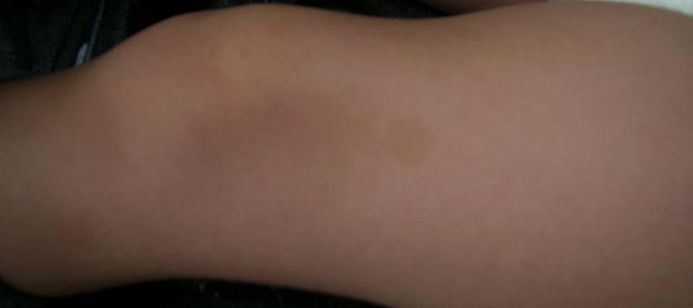
1 BILATERAL OPTIC MALIGNANT ASTROCYTOMA IN A 3 YEAR OLD CHILD WITH NFI CASE PRESENTATION BOGDAN ILIESCU 1, M. VUKIC 2, ZIYAD FAIYAD 1, RAMONA FILIPESCU*, ION POEATA 1 1 3rd Neurosurgery Department, Prof. Dr. N. Oblu Clinical Emergency Hospital 2 Department of Neurosurgery, Medical School University of Zagreb, Zagreb, Croatia *Neurosurgery Department, St Mary Children s Hospital We present a rare case of a 3 year old child with a fast-growing bilateral exophtalmus in the context of neurofibromatosis type 1. We analyze the particularities of the clinical and imagistic aspects of the case, and discuss them in the light of pathological diagnosis (malignant astrocytoma) and imagistic follow-up. Keywords: malignant astrocytoma, neurofibromatosis type 1, optic nerve, optic chiasm INTRODUCTION Neurofibromatosis type 1(NF I) is a common autosomal dominant condition that affects about 1 in 5000 people. Optic nerve glioma is one of the landmark lesions characterizing this disease. They usually occur in children during the first decade of life and are seen in 11 30% of patients with NF I. When associated with NF1, the tumors are more often benign, can be multifocal and bilateral, and are usually found within the optic nerves although they can occur anywhere along the optic pathway, all the way to the visual cortex. Although the unilateral occurrence is a finding quite common in this population (up to 30%) bilateral involvement of the optic nerves is rare. Further, the optic gliomas in NF1 are self-limiting lesions with a very slow growth (which stops in adolescence). As a result exophtalmus is a very rare finding and they require surgical intervention in a very small number of cases. CASE REPORT A 3 year old boy was addressed to our department for a fast-growing, grotesque exophtalmus, bilateral but more prominent on the left side. From apparently normal to present status the evolution occurred during a period of roughly 6 months (as documented by family photographs. He lost vision in both eyes 8 months before presentation, after a short period of amblyopia with a rapid course (7 days). The clinical exam revealed café-au-lait spots of various sizes and locations (trunk and limbs) and important of left eye conjunctiva. Both eyes were deviated from the normal position towards inferior left corner. No other clinical sign was apparent. A brain and orbit MRI exam was ordered with contrast enhancement. Axial T1-weighted magnetic resonance images showed isointense masses developing within both optic nerves, occupying most of the retro-ocular orbit and extended to the optic chiasm. Both lesions enhanced homogeneously after contrast administration. Decision was made to remove both tumors in order to restore facial cosmetics and to prevent further deterioration of the eyes (the left eye was already affected by a severe conjunctivitis). The two gliomas were approached in one surgery through a bifrontal- left pterional approach, orbit ceiling perforation, dural flap and complete tumor removal. The desired cosmetic result was achieved, but third day 32
 and immediate antibiotic therapy was started with Vancomycin.")

2 BILATERAL OPTIC MALIGNANT ASTROCYTOMA postoperatively the patient developed high fever, vomiting and presented signs of meningeal irritation. Lumbar puncture revealed a high concentration of white blood cells (5100/ml) and immediate antibiotic therapy was started with Vancomycin. The initial microscopic pathological exam indicated the two tumors as being pilocytic astrocytoma and suggested further investigation through immunohistochemistry. The suggestion was followed and the second exam revealed the tumors to be malignant astrocytomas with a KI67 index of 12%. At three months postoperatively a MRI follow-up was performed. The new scan revealed a mass occupying lesion within the optic chiasm with the same characteristic as the previous optic nerve tumors. The lesion was, visible on the initial scan, showed an accelerated growth reaching 1,6 cm in diameter. The patient was addressed to gamma-knife center for radiosurgery. DISCUSSION Although optic nerve glioma represents a common finding in patients with type 1 neurofibromatosis, either unilateral or bilateral, our case posed a clinical challenge from the start as to the reasons behind a very fast growth rate (from normal to massive exophtalmus in about 6 months). The classic tumor found in NFI is known to be a slow growing tumor that has a self-limiting evolution with variable influence on visual function. Our patient lost vision in both eyes in a very short period (7 days). Furthermore, the MRI aspect of both optic nerve tumors didn t present any particular characteristics to set them apart form the usual optic nerve glioma or to suggest malignancy. Although the initial surgery removed successfully the optic nerve tumors to a good cosmetic result the immunohistochemical diagnostic, the rapid clinical course, and the imagistic documentation of a relapse of the disease at the level of optic chiasm all concur to a reserved vital prognostic. We believe that this case is illustrative for the need to put together all the available data in diagnosing, treating and counseling a case with salient tumors in the context of NFI, never overseeing the most rare possibilities. A) B) FIG. 1 Clinical aspect in A) November 2008, and B) February

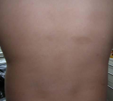
3 BOGDAN ILIESCU FIG. 2 Café-au-lait spots on runk and limbs 34
 FIG.")

 February 2009, native")

4 BILATERAL OPTIC MALIGNANT ASTROCYTOMA FIG. 3 A) FIG. 3 B) FIG. 3 Comparative MRI aspects in A) November 2008, and B) February 2009, native and after contrast enhancement on the left and right images respectively 35


5 BOGDAN ILIESCU FIG. 4 CT control 1st day postop FIG. 5 Mri aspects at 3 months postoperatively 36
Year 2003 Paper two: Questions supplied by Tricia
 question 43 A 42-year-old man presents with a two-year history of increasing right facial numbness. He has a history of intermittent unsteadiness, mild hearing loss and vertigo but has otherwise been well.
NON MALIGNANT BRAIN TUMOURS Facilitator. Ros Taylor Advanced Neurosurgical Nurse Practitioner Southmead Hospital Bristol
 NON MALIGNANT BRAIN TUMOURS Facilitator Ros Taylor Advanced Neurosurgical Nurse Practitioner Southmead Hospital Bristol Neurosurgery What will be covered? Meningioma Vestibular schwannoma (acoustic neuroma)
Spinal and para-spinal plexiform neurofibromas in NF1 patients, a clinical-radiological correlation study
 Spinal and para-spinal plexiform neurofibromas in NF1 patients, a clinical-radiological correlation study Poster No.: C-1846 Congress: ECR 2015 Type: Scientific Exhibit Authors: M. Mauda-Havakuk, B. Shofty,
A Case of Carotid-Cavernous Fistula
 A Case of Carotid-Cavernous Fistula By : Mohamed Elkhawaga 2 nd Year Resident of Ophthalmology Alexandria University A 19 year old male patient came to our outpatient clinic, complaining of : -Severe conjunctival
Background Principles and Technical Development
 Contents Part I Background Principles and Technical Development 1 Introduction and the Nature of Radiosurgery... 3 Definitions of Radiosurgery... 5 Consequences of Changing Definitions of Radiosurgery...
Imaging in neurofibromatosis type 1: An original research article with focus on spinal lesions
 Original Research Article Imaging in neurofibromatosis type 1: An original research article with focus on spinal lesions Kalpesh Patel 1*, Siddharth Zala 2, C. Raychaudhuri 3 1 Assistant Professor, 2 1
Advances In Orbital Neuropathology
 Advances In Orbital Neuropathology Charles G. Eberhart, MD PhD Associate Professor of Pathology, Ophthalmology and Oncology Johns Hopkins University School of Medicine Overview Non-neoplastic lesions Microphthalmos/pseudoglioma
Metastasis. 57 year old with progressive Headache and Right Sided Visual Loss
 Metastasis 1% of sellar/parasellar masses Usually occurs with known primary Can involve third ventricle, hypothalamus, infundibular stalk May be both supra-, intrasellar 57 year old with progressive Headache
No Financial Interest
 Pituitary Apoplexy Michael Vaphiades, D.O. Professor Department of Ophthalmology, Neurology, Neurosurgery University of Alabama at Birmingham, Birmingham, AL No Financial Interest N E U R O L O G I C
Brain Tumors. What is a brain tumor?
 Scan for mobile link. Brain Tumors A brain tumor is a collection of abnormal cells that grows in or around the brain. It poses a risk to the healthy brain by either invading or destroying normal brain
Case Report Malignant Peripheral Nerve Sheath Tumor of the Inguinum and Angiosarcoma of the Scalp in a Child with Neurofibromatosis Type 1
 Hindawi Case Reports in Pathology Volume 2017, Article ID 7542825, 4 pages https://doi.org/10.1155/2017/7542825 Case Report Malignant Peripheral Nerve Sheath Tumor of the Inguinum and Angiosarcoma of the
Dosimetry, see MAGIC; Polymer gel dosimetry. Fiducial tracking, see CyberKnife radiosurgery
 Subject Index Acoustic neuroma, neurofibromatosis type 2 complications 103, 105 hearing outcomes 103, 105 outcome measures 101 patient selection 105 study design 101 tumor control 101 105 treatment options
Capt. Nazim ATA Aerospace Medicine Specialist Turkish Air Force AAMIMO 2013
 F-15 Pilot with ACOUSTIC NEUROMA Capt. Nazim ATA Aerospace Medicine Specialist Turkish Air Force AAMIMO 2013 Disclosure Information 84 th Annual AsMA Scientific Meeting Nazim ATA I have no financial relationships
CLINICAL PEARLS IN OCULAR ONCOLOGY
 CLINICAL PEARLS IN OCULAR ONCOLOGY IRIS NEVUS - Two kinds circumscribed and diffuse - Photodocumentation important to monitor growth - Risk Factors for iris nevus growth to melanoma (ABCDEF) A Age (young),
University of Washington Radiology Review Course: Strange and Specific Diagnoses. Case #1
 University of Washington Radiology Review Course: Strange and Specific Diagnoses Katherine E. Dee, MD Seattle Breast Center Via Radiology 2014 Case #1 37 year old presents with bilateral palpable lumps.
LOW GRADE ASTROCYTOMAS
 LOW GRADE ASTROCYTOMAS This article was provided to us by David Schiff, MD, Associate Professor of Neurology, Neurosurgery, and Medicine at University of Virginia, Charlottesville. We appreciate his generous
Brain Tumors. Andrew J. Fabiano, MD FAANS. Associate Professor of Neurosurgery Roswell Park Cancer Institute SUNY at Buffalo School of Medicine
 Brain Tumors Andrew J. Fabiano, MD FAANS Associate Professor of Neurosurgery Roswell Park Cancer Institute SUNY at Buffalo School of Medicine Brain Tumors Brain Tumor Basics Types of Tumors Cases Brain
doi: /j.anl
 doi: 10.1016/j.anl.2006.07.001 Synchronous unilateral parotid gland neoplasms of three different histological types Shuho Tanaka 1, Keiji Tabuchi 1, Keiko Oikawa 1, Rika Kohanawa 1, Hideki Okubo 1, Dai
Coding Companion for Ophthalmology. A comprehensive illustrated guide to coding and reimbursement
 Coding Companion for Ophthalmology A comprehensive illustrated guide to coding and reimbursement 2015 Contents Getting Started with Coding Companion...i General Integumentary/Skin...1 Introduction...8
Linking Optic Nerve Sheath Diameters and Glioblastoma Multiforme: A Retrospective Analysis
 Linking Optic Nerve Sheath Diameters and Glioblastoma Multiforme: A Retrospective Analysis Andrew Teh, DO Alysha Vartevan, DO Aswin Kumar, DO Department of Radiology, Larkin Community Hospital/Lake Erie
Sphenoid rhinosinusitis associated with abducens nerve palsy Case report
 Romanian Journal of Rhinology, Volume 8, No. 30, April-June 2018 CASE REPORT Sphenoid rhinosinusitis associated with abducens nerve palsy Case report Lucian Lapusneanu 1, Marlena Radulescu 1, Florin Ghita
Dr. T. Venkat Kishan Asst. Prof Department of Radiodiagnosis
 Dr. T. Venkat Kishan Asst. Prof Department of Radiodiagnosis Schwannomas (also called neurinomas or neurilemmomas) constitute the most common primary cranial nerve tumors. They are benign slow-growing
Adult Brain and Spinal Cord Tumors
 Adult Brain and Spinal Cord Tumors An adult central nervous system (CNS) tumor is a disease in which abnormal cells form in the tissues of the brain and or the spinal cord. Major Parts of the Brain Anatomy
Neurofibromatosis Type One: A Guide for Educators by Bruce R. Korf, M.D., Ph.D.
 Neurofibromatosis Type One: A Guide for Educators by Bruce R. Korf, M.D., Ph.D. The diagnosis of neurofibromatosis (NF) in a child raises many important issues for care whether at home, at school, or in
Information for patients. Acoustic Neuroma. Neurosurgery: Neurosciences. Supported by
 Information for patients Acoustic Neuroma Neurosurgery: Neurosciences Supported by What is an Acoustic Neuroma You have been diagnosed as having an acoustic neuroma. An acoustic neuroma also known as a
Supratentorial multiple little meningiomas with transitory stroke symptoms like. MRI case presentation
 114 Romanian Neurosurgery (2010) XVII 1: 114-121 Supratentorial multiple little meningiomas with transitory stroke symptoms like. MRI case presentation E. Moldovanu 1,2, Adriana Moldovanu 1,2, Carmen Gherman
ACOUSTIC NEUROMAS. Being invited to Florence, Italy to address an international medical meeting about our work for
 ACOUSTIC NEUROMAS Being invited to Florence, Italy to address an international medical meeting about our work for head and neck tumors was a great honor. The symposium organized under the auspices of the
Brain Tumor Treatment
 Scan for mobile link. Brain Tumor Treatment Brain Tumors Overview A brain tumor is a group of abnormal cells that grows in or around the brain. Tumors can directly destroy healthy brain cells. They can
Stereotactic Radiosurgery. Extracranial Stereotactic Radiosurgery. Linear accelerators. Basic technique. Indications of SRS
 Stereotactic Radiosurgery Extracranial Stereotactic Radiosurgery Annette Quinn, MSN, RN Program Manager, University of Pittsburgh Medical Center Using stereotactic techniques, give a lethal dose of ionizing
GENETIC TESTING FOR NEUROFIBROMATOSIS
 GENETIC TESTING FOR NEUROFIBROMATOSIS Non-Discrimination Statement and Multi-Language Interpreter Services information are located at the end of this document. Coverage for services, procedures, medical
What is cancer? l Cancer is a group of several diseases caused by the abnormal growth of cells.
 Cancer Biology What is cancer? l Cancer is a group of several diseases caused by the abnormal growth of cells. What is cancer? l Cancer is a group of several diseases caused by the abnormal growth of cells.
Plexiform Tumor of the Orbit
 Plexiform Tumor of the Orbit Anat Stemmer-Rachamimov, MD Department of Pathology Massachusetts General Hospital Harvard Medical School Disclosure of Relevant Financial Relationships USCAP requires that
Contralateral clipping of bilateral middle cerebral artery aneurysms. Case report
 Romanian Neurosurgery Volume XXXI Number 1 2017 January - March Article Contralateral clipping of bilateral middle cerebral artery aneurysms. Case report Georgiana Ion, Alexandru Chiriac, Ziyad Faiyad,
COPYRIGHT 2012 THE TRANSVERSE MYELITIS ASSOCIATION. ALL RIGHTS RESERVED
 The Transverse Myelitis Association...advocating for those with acute disseminated encephalomyelitis, neuromyelitis optica, optic neuritis and transverse myelitis ACUTE DISSEMINATED ENCEPHALOMYELITIS (ADEM)
Meningioma tumor. Meningiomas are named according to their location (Fig. 1) and cause various symptoms: > 1
 and cause various symptoms: > 1") Meningioma tumor Overview A meningioma is a type of tumor that grows from the protective membranes, called meninges, which surround the brain and spinal cord. Most meningiomas are benign (not cancer) and
Meningioma tumor Overview A meningioma is a type of tumor that grows from the protective membranes, called meninges, which surround the brain and spinal cord. Most meningiomas are benign (not cancer) and
THE EFFECTIVE OF BRAIN CANCER AND XAY BETWEEN THEORY AND IMPLEMENTATION. Mustafa Rashid Issa
 THE EFFECTIVE OF BRAIN CANCER AND XAY BETWEEN THEORY AND IMPLEMENTATION Mustafa Rashid Issa ABSTRACT: Illustrate malignant tumors that form either in the brain or in the nerves originating in the brain.
MD (Ophthalmology) May 2007 Examination Paper I MD (Ophthalmology) May 2007 Examination Paper II
 May 2007 Examination Paper I MD (Ophthalmology) May 2007 Examination Paper II") All India Institute of Medical Science MD Ophthalmology Time: 3 hours Max. Marks: 100 Attempt all the questions briefly with labeled diagrams wherever possible Q1. Discuss the mechanisms of accommodation
All India Institute of Medical Science MD Ophthalmology Time: 3 hours Max. Marks: 100 Attempt all the questions briefly with labeled diagrams wherever possible Q1. Discuss the mechanisms of accommodation
Brain and Spine Tumors
 Brain and Spine Tumors Andrew J. Fabiano, MD FAANS Associate Professor of Neurosurgery Roswell Park Cancer Institute SUNY at Buffalo School of Medicine Brain Tumors Brain Tumor Basics Types of Tumors Cases
Case Report Carbon Ion Beam Radiotherapy for Sinonasal Malignant Tumors Invading Skull Base
 Case Reports in Otolaryngology, Article ID 241856, 4 pages http://dx.doi.org/10.1155/2014/241856 Case Report Carbon Ion Beam Radiotherapy for Sinonasal Malignant Tumors Invading Skull Base Nobuo Ohta,
Von Recklinghausen s Disease with a Giant Lipoma
 Von Recklinghausen s Disease with a Giant Lipoma Daiki Iwana¹( ) Kazutaka Izawa¹ Mitsuhiro Kawamura¹ Takaharu Nabeshima¹ Hideki Yoshikawa² ¹Department of Orthopaedic Surgery, Toneyama National Hospital,
NANOS Patient Brochure
 NANOS Patient Brochure Pituitary Tumor Copyright 2015. North American Neuro-Ophthalmology Society. All rights reserved. These brochures are produced and made available as is without warranty and for informational
Gastrointestinal Stromal Tumor Causes, Risk Factors, and Prevention
 Gastrointestinal Stromal Tumor Causes, Risk Factors, and Prevention Risk Factors A risk factor is anything that affects your chance of getting a disease such as cancer. Learn more about the risk factors
THE CHILD WITH NEUROFIBROMATOSIS TYPE 1 (NF1) A GUIDE FOR
 A GUIDE FOR") THE CHILD WITH NEUROFIBROMATOSIS TYPE 1 (NF1) A GUIDE FOR HEALTH CARE PROFESSIONALS Neurofibromatosis is actually a term that encompasses at least two distinct disorders, Neurofibromatosis Type 1 (NF1)
THE CHILD WITH NEUROFIBROMATOSIS TYPE 1 (NF1) A GUIDE FOR HEALTH CARE PROFESSIONALS Neurofibromatosis is actually a term that encompasses at least two distinct disorders, Neurofibromatosis Type 1 (NF1)
Brain tumors: tumor types
 Brain tumors: tumor types Tumor types There are more than 120 types of brain tumors. Today, most medical institutions use the World Health Organization (WHO) classification system to identify brain tumors.
Early detection of Retinoblastoma in children. Max Mantik
 Early detection of Retinoblastoma in children Max Mantik Introduction The most common primary intraocular malignancy of childhood 10 to 15 % of cancers that occur within the first year of life Typical
Prevalence of venous sinus stenosis in Pseudotumor cerebri(ptc) using digital subtraction angiography (DSA)
 using digital subtraction angiography (DSA)") Prevalence of venous sinus stenosis in Pseudotumor cerebri(ptc) using digital subtraction angiography (DSA) Dr.Mohamed hamdy ibrahim MBBC,MSc,MD, PhD Neurology Degree Kings lake university (USA). Fellow
Prevalence of venous sinus stenosis in Pseudotumor cerebri(ptc) using digital subtraction angiography (DSA) Dr.Mohamed hamdy ibrahim MBBC,MSc,MD, PhD Neurology Degree Kings lake university (USA). Fellow
PORT WINE STAINS AND STURGE-WEBER SYNDROME
 PORT WINE STAINS AND STURGE-WEBER SYNDROME Ong Hian Tat It is important for general practitioners to recognize cutaneous port-wine stains as these could signify important association with Sturge Weber
Five Most Common Problems in Surgical Neuropathology
 Five Most Common Problems in Surgical Neuropathology If the brain were so simple that we could understand it, we would be so simple that we couldn t Emerson Pugh What is your greatest difficulty in neuropathology?
Living with Neurofibromatosis Type 1: A Guide for Adults
 Living with Neurofibromatosis Type 1: A Guide for Adults by Linda Piersall, M.S. David H. Gutmann, M.D., Ph.D. Neurofibromatosis Program St. Louis Children s Hospital St. Louis, Missouri Rosalie Ferner,
Partial vertebrectomy with vertebral shortening for. Key words: thoraco-lumbar fracturedislocation,
 Romanian Neurosurgery (2014) XXI 2: 183-187 183 Partial vertebrectomy with vertebral shortening for thoraco-lumbar fracture-dislocation. Case report and technical note Bogdan Costachescu 1,2, Cezar E.
Neurofibromatosis type 1: a review of MRI findings
 Neurofibromatosis type 1: a review of MRI findings Poster No.: C-2198 Congress: ECR 2014 Type: Authors: Keywords: DOI: Educational Exhibit J. L. Ortega Garcia 1, M. A. MACEDO PASCUAL 2, A. A. García Ortega
Vision Health: Conditions, Disorders & Treatments EYELID DISORDERS
 Vision Health: Conditions, Disorders & Treatments EYELID DISORDERS There are a number of disorders that can affect the eyelid. Entropion Entropion is an inward turning of the eyelid and lashes toward the
Our experience in surgical treatment of intraorbital tumors
 Romanian Neurosurgery (2012) XIX 4: 289 293 289 Our experience in surgical treatment of intraorbital tumors A. Chiriac, S. Daja 1, B. Iliescu 1, I. Poeata Grigore T. Popa University of Medicine and Pharmacy,
Corporate Medical Policy
 Corporate Medical Policy Genetic Testing for Neurofibromatosis File Name: Origination: Last CAP Review: Next CAP Review: Last Review: genetic_testing_for_neurofibromatosis 4/2016 7/2017 7/2018 1/2018 Description
DISORDERS OF THE SALIVARY GLANDS Neoplasms Dr.M.Baskaran Selvapathy S IV
 DISORDERS OF THE SALIVARY GLANDS Neoplasms Dr.M.Baskaran Selvapathy S IV NEOPLASMS A) Epithelial I. Benign Pleomorphic adenoma( Mixed tumour) Adenolymphoma (Warthin s tumour) Oxyphil adenoma (Oncocytoma)
Adult Central Nervous System Tumors Treatment (PDQ )
") 1 di 20 28/06/2016 11.18 NCBI Bookshelf. A service of the National Library of Medicine, National Institutes of Health. PDQ Cancer Information Summaries [Internet]. Bethesda (MD): National Cancer Institute
1 di 20 28/06/2016 11.18 NCBI Bookshelf. A service of the National Library of Medicine, National Institutes of Health. PDQ Cancer Information Summaries [Internet]. Bethesda (MD): National Cancer Institute
Pediatric optic ner-glioma: A case study
 Case Study Prasuna Jelly 1,*, SK Mohana Sundari 2 1 AIIMS, Rishikesh, Uttarakhand, 2 AIIMS, Jodhpur, Rajasthan *Corresponding Author: Email: prasunajelly@gmail.com Abstract An optic nerve glioma is a type
Fractionated Stereotactic Radiotherapy. Rationale, indications, & treatment techniques
 Fractionated Stereotactic Radiotherapy Rationale, indications, & treatment techniques Radiobiological principles The BED (Gy) = D(1 + d/α/β) Assume BED 1 = BED 2 for tissue of an unknown α/β: Optic
General Imaging. Imaging modalities. Incremental CT. Multislice CT Multislice CT [ MDCT ]
![General Imaging. Imaging modalities. Incremental CT. Multislice CT Multislice CT [ MDCT ]](/thumbs/76/74079340.jpg "General Imaging. Imaging modalities. Incremental CT. Multislice CT Multislice CT [ MDCT ]") General Imaging Imaging modalities Conventional X-rays Ultrasonography [ US ] Computed tomography [ CT ] Radionuclide imaging Magnetic resonance imaging [ MRI ] Angiography conventional, CT,MRI Interventional
General Imaging Imaging modalities Conventional X-rays Ultrasonography [ US ] Computed tomography [ CT ] Radionuclide imaging Magnetic resonance imaging [ MRI ] Angiography conventional, CT,MRI Interventional
BRITISH BIOMEDICAL BULLETIN
 Journal Home Page www.bbbulletin.org BRITISH BIOMEDICAL BULLETIN Case Report Neurofibromatosis Type 1 with Chiari Malformation Type 1 in Child: A Case Report Mallesh Kariyappa*, Febna A. Rahiman and Jayanthi
AMERICAN BRAIN TUMOR ASSOCIATION. Oligodendroglioma and Oligoastrocytoma
 AMERICAN BRAIN TUMOR ASSOCIATION Oligodendroglioma and Oligoastrocytoma ACKNOWLEDGEMENTS ABOUT THE AMERICAN BRAIN TUMOR ASSOCIATION Founded in 1973, the American Brain Tumor Association (ABTA) was the
Neuro-Ophthalmic Masqueraders
 Neuro-Ophthalmic Masqueraders Leonid Skorin, Jr., OD, DO, MS, FAAO, FAOCO Mayo Clinic Health System in Albert Lea Denise Goodwin, OD, FAAO Pacific University College of Optometry Please silence all mobile
Outstanding Questions from Webinar: Long-Term Survival Outcomes for LGG Patients, May 5 th, 2014
 Outstanding Questions from Webinar: Long-Term Survival Outcomes for LGG Patients, May 5 th, 2014 Answers provided as a courtesy by Dana Farber Cancer Institute experts: Dr. Mark Kieran Dr. Peter Manley
The Investigation of Proptosis in Paediatric Practice.
 The Investigation of Proptosis in Paediatric Practice. Ms Sayantani Ghosh 1, Mr Saugat Dey 1 1 Bankura Sammilani Medical College and Hospital, 136, Dr. Meghnad Saha Road, PRATYASHA APARTMENT Flat-4C, KOLKATA-
Otolaryngologist s Perspective of Stereotactic Radiosurgery
 Otolaryngologist s Perspective of Stereotactic Radiosurgery Douglas E. Mattox, M.D. 25 th Alexandria International Combined ORL Conference April 18-20, 2007 Acoustic Neuroma Benign tumor of the schwann
ADVANCE APPROACH FOR IDENTIFICATION WHITE MATTER FROM BRAIN MRI IMAGES AND CLASSIFICATION
 ADVANCE APPROACH FOR IDENTIFICATION WHITE MATTER FROM BRAIN MRI IMAGES AND CLASSIFICATION Alkesh M. Kaba 1, Reena P. Parmar 2, 1 Student, Computer Department, Swamminarayan College of Engg. & Tech, Gujarat,
Management of pediatric brain tumors, strategies and long term outcome
 Management of pediatric brain tumors, strategies and long term outcome SAN The Sudanese association of neurosurgeons By Dr. Abubakr Darrag Salim Ahmed Dr. Mohammed Awad Elzain Khartoum Sudan Pediatric
Neurofibromatosis type 1 and RASopathies
 Neurofibromatosis type 1 and RASopathies Dawn Siegel, MD Medical College of Wisconsin American Academy of Dermatology San Diego, CA February 19 th, 2018 Neurofibromatosis Type 1 NF1- diagnostic criteria
N EOPLASMS of the optic nerves occur
 Tumors of the optic nerve and optic chiasm COLLINS. MAcCARTY~ M.D., ALLEN S. BOYD, JR., M.D., AND DONALD S. CHILDS, JR,, M.D. Departments of Neurologic Surgery and Therapeutic Radiology, Mayo Clinic and
Neurofibromatosis (NF) Center
 Center") Washington University Neurofibromatosis (NF) Center THE NF CENTER: EXCEPTIONAL CARE THROUGH GROUNDBREAKING RESEARCH An international leader in research and treatment of neurofibromatosis (NF), the Washington
Washington University Neurofibromatosis (NF) Center THE NF CENTER: EXCEPTIONAL CARE THROUGH GROUNDBREAKING RESEARCH An international leader in research and treatment of neurofibromatosis (NF), the Washington
Adrenal masses in infancy and childhood: A clinical and radiological overview M. Mearadji
 Adrenal masses in infancy and childhood: A clinical and radiological overview M. Mearadji International Foundation for Pediatric Imaging Aid Introduction Neoplastic adrenal masses usually originate from
Stereotactic Radiosurgery/Fractionated Stereotactic Radiotherapy for Acoustic Neuroma (Vestibular Schwannomas)
") Strategic Commissioning Group West Midlands Commissioning Policy (WM/38) Stereotactic Radiosurgery/Fractionated Stereotactic Radiotherapy for Acoustic Neuroma (Vestibular Schwannomas) Version 1 July 2010
Strategic Commissioning Group West Midlands Commissioning Policy (WM/38) Stereotactic Radiosurgery/Fractionated Stereotactic Radiotherapy for Acoustic Neuroma (Vestibular Schwannomas) Version 1 July 2010
Case Report Atypical Presentation of Idiopathic Bilateral Optic Perineuritis in a Young Patient
 Case Reports in Ophthalmological Medicine Volume 2016, Article ID 6741925, 4 pages http://dx.doi.org/10.1155/2016/6741925 Case Report Atypical Presentation of Idiopathic Bilateral Optic Perineuritis in
Spinal meningioma imaging
 Spinal meningioma imaging Poster No.: C-0448 Congress: ECR 2018 Type: Educational Exhibit Authors: M. Smoljan, D. Zadravec ; Zagreb/HR, Zageb/HR Keywords: Neoplasia, Imaging sequences, Education, MR, CT,
Optic neuritis. Information for patients Ophthalmology
 Optic neuritis Information for patients Ophthalmology page 2 of 8 What is optic neuritis? Optic neuritis is a common cause of acute visual loss in young people. The optic nerve is like a cable of electric
Childhood Craniopharyngioma Treatment (PDQ )
") 1 di 12 27/06/2016 09.45 NCBI Bookshelf. A service of the National Library of Medicine, National Institutes of Health. PDQ Cancer Information Summaries [Internet]. Bethesda (MD): National Cancer Institute
1 di 12 27/06/2016 09.45 NCBI Bookshelf. A service of the National Library of Medicine, National Institutes of Health. PDQ Cancer Information Summaries [Internet]. Bethesda (MD): National Cancer Institute
ERN services: A Patient Case Study. Matt Johnson, EURORDIS Potential ERN Services Second Workshop, PWC, Brussels 24 May 2016
 ERN services: A Patient Case Study Matt Johnson, EURORDIS Potential ERN Services Second Workshop, PWC, Brussels 24 May 2016 1 Collective knowledge and expertise A European Reference Network is more than
section 2 What Is Neurofibromatosis Type 1?
 section 2 What Is Neurofibromatosis Type 1? What Is Neurofibromatosis Type 1? The term neurofibromatosis covers three different genetic disorders that cause tumors to form around the nerves: neurofibromatosis
Anaplastic Pilocytic Astrocytoma: The fusion of good and bad
 Anaplastic Pilocytic Astrocytoma: The fusion of good and bad Alexandrina Nikova 1, Charalampos-Chrysovalantis Chytoudis-Peroudis 2, Penelope Korkolopoulou 3 and Dimitrios Kanakis 4 Abstract 5 Pilocytic
PRINCESS MARGARET CANCER CENTRE CLINICAL PRACTICE GUIDELINES
 PRINCESS MARGARET CANCER CENTRE CLINICAL PRACTICE GUIDELINES CENTRAL NERVOUS SYSTEM MENINGIOMA CNS Site Group Meningioma Author: Dr. Norm Laperriere Date: February 20, 2018 1. INTRODUCTION 3 2. PREVENTION
Tumors of the Nervous System
 Tumors of the Nervous System Peter Canoll MD. PhD. What I want to cover What are the most common types of brain tumors? Who gets them? How do they present? What do they look like? How do they behave? 1
Penetration of the Optic Nerve or Chiasm by Anterior Communicating Artery Aneurysms. - Three Case Reports-
 Penetration of the Optic Nerve or Chiasm by Anterior Communicating Artery Aneurysms. - Three Case Reports- Tetsuyoshi Horiuchi 1, Toshiya Uchiyama 1, Yoshikazu Kusano 1, Maki Okada 1, Kazuhiro Hongo 1,
PUBLISHED VERSION.
 PUBLISHED VERSION Shay Keren, Gad Dotan, Leah Leibovitch, Dinesh Selva, and Igal Leibovitch Indocyanine green assisted removal of orbital lacrimal duct cysts in children Ophthalmology, 2015; 2015:130215-1-130215-5
Brain Injuries. Presented By Dr. Said Said Elshama
 Brain Injuries Presented By Dr. Said Said Elshama Types of head injuries 1- Scalp injuries 2- Skull injuries 3- Intra Cranial injuries ( Brain ) Anatomical structure of meninges Intra- Cranial Injuries
Leksell Gamma Knife Icon. Treatment information
 Leksell Gamma Knife Icon Treatment information You may be feeling frightened or overwhelmed by your recent diagnosis. It can be confusing trying to process a diagnosis, understand a new and challenging
Chapter 5 Section 3.1
 Radiology Chapter 5 Section 3.1 Issue Date: March 27, 1991 Authority: 32 CFR 199.4(b)(2), (b)(2)(x), (c)(2)(viii), and (g)(15) 1.0 CPT 1 PROCEDURE CODES 37243, 61793, 61795, 77261-77421, 77427-77799, 0073T
Collection of Recorded Radiotherapy Seminars
 IAEA Human Health Campus Collection of Recorded Radiotherapy Seminars http://humanhealth.iaea.org The Role of Radiosurgery in the Treatment of Gliomas Luis Souhami, MD Professor Department of Radiation
Enhancement of Optic Nerve in Leukemic Patients: Leukemic Infiltration of Optic Nerve versus Optic Neuritis
 pissn 2384-1095 eissn 2384-1109 imri 2016;20:167-174 http://dx.doi.org/10.13104/imri.2016.20.3.167 Enhancement of Optic Nerve in Patients: Infiltration of Optic Nerve versus Optic Neuritis Yo Han Ra 1,
ROTATOR CUFF INJURIES / IMPINGEMENT SYNDROME
 ROTATOR CUFF INJURIES / IMPINGEMENT SYNDROME Shoulder injuries are common in patients across all ages, from young, athletic people to the aging population. Two of the most common problems occur in the
Biology 3201 Nervous System # 7: Nervous System Disorders
 Biology 3201 Nervous System # 7: Nervous System Disorders Alzheimer's Disease first identified by German physician, Alois Alzheimer, in 1906 most common neurodegenerative disease two thirds of cases of
Problems of Neurological Function
 Problems of Neurological Function Unit 10 Independent Student Review Brain Anatomy and physiology of cerebral hemispheres, diencephalon, brain stem, and cerebellum Meninges, ventricles, flow of CSF Blood
Oligodendrogliomas & Oligoastrocytomas
 Oligodendrogliomas & Oligoastrocytomas ABOUT THE AMERICAN BRAIN TUMOR ASSOCIATION Founded in 1973, the American Brain Tumor Association (ABTA) was the first national nonprofit organization dedicated solely
Problems of Neurological Function. Unit 10
 Problems of Neurological Function Unit 10 Independent Student Review Brain Anatomy and physiology of cerebral hemispheres, diencephalon, brain stem, and cerebellum Meninges, ventricles, flow of CSF Blood
Hypothalamic glioma masquerading as craniopharyngioma
 1 di 7 24/01/2014 18.58 J Neurosci Rural Pract. 2013 Jul-Sep; 4(3): 323 325. doi: 10.4103/0976-3147.118790 PMCID: PMC3821425 Hypothalamic glioma masquerading as craniopharyngioma Sameer Vyas, Nidhi Prabhakar,
Pediatr Blood Cancer 2014
 Low grade Glioma! 40% of pediatric brain tumors Pathologically, anatomically, clinically and biologically heterogeneous Leptomeningeal metastases in 5% Frequently protracted clinical course Long-Term Outcome
Selected radiosurgery cases from the Rotating Gamma Institute Debrecen, Hungary
 Selected radiosurgery cases from the Rotating Gamma Institute Debrecen, Hungary László Bognár M.D., Ph.D., József G. Dobai M.D., Gábor Csiky and Imre Fedorcsák M.D., Ph.D. Department of Neurosurgery, Medical
SOLID TUMOURS IN CHILDHOOD
 SOLID TUMOURS IN CHILDHOOD Fareed Omar Paediatric Oncology Steve Biko Academic hospital Introduction 1 Introduction Lymphomas and Leukemias make up about 40% of all childhood Cancers (Systemic cancers)
North Oaks Trauma Symposium Friday, November 3, 2017
 + Evaluation and Management of Facial Trauma D Antoni Dennis, MD North Oaks ENT an Allergy November 3, 2017 + Financial Disclosure I do not have any conflicts of interest or financial interest to disclose
CLINICAL INFORMATION SHEET NEUROFIBROMATOSIS (NF)
") CLINICAL INFORMATION SHEET NEUROFIBROMATOSIS (NF) NNFF International NF1 Genetic Mutation Analysis Consortium Submission Form FORM USE Name of investigator (required field): Institution (required field):
CLINICAL INFORMATION SHEET NEUROFIBROMATOSIS (NF) NNFF International NF1 Genetic Mutation Analysis Consortium Submission Form FORM USE Name of investigator (required field): Institution (required field):
Orbital Tumors - A Clinico Pathological Study
 Orbital Tumors - A Clinico Pathological Study Radha. J. DO, Ani Sreedhar. MS. Little Flower Hospital, Angamaly, Kerala ORIGINAL ARTICLES Abstract: Aim. To study the clinical and histopathological profiles