Computer based delineation and follow-up multisite abdominal tumors in longitudinal CT studies
|
|
|
- Cathleen Pierce
- 5 years ago
- Views:
Transcription
1 Research plan submitted for approval as a PhD thesis Submitted by: Refael Vivanti Supervisor: Professor Leo Joskowicz School of Engineering and Computer Science, The Hebrew University of Jerusalem Computer based delineation and follow-up multisite abdominal tumors in longitudinal CT studies
2 Introduction (1) Radiological follow-up of solid tumors is the cornerstone of modern oncology. Cause of abdominal cancer death (2008): Lung cancer: 1.4 million deaths per year Stomach cancer: 740,000 deaths per year Liver cancer: 700,000 deaths per year For any treatment a series of abdominal scans are needed To evaluate the progression of the tumor To evaluate the effectiveness of the treatment
3 Introduction (2) Currently, radiologists perform the initial diagnosis and follow-up using simple guidelines RECIST The product of the largest distance between in-tumor points and the maximum perpendicular diameter Only a rough approximation from a single 2D projection image For 3D CT, the tumor volume is estimated with ellipsoid formula: max-length max-depth max-width Research shows that 3D volumetric measurements provide the best information for tumor progress monitoring.


4 Linear measurements [Schwartz and Zhao. 2010] Pre Therapy Long axis: 25.0mm Short axis: 20.4mm Volume: 3,420 mm 3 After 24 days Long axis: 25.9mm Short axis: 19.8mm Volume: 4,608 mm 3 Change in Long axis 4% Change in Volume 35%
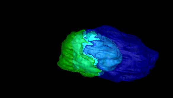
![Volumetric measurements: Study: [Weltens 2001] Axial MRI slice Nine independent observers](/docs-images/86/94555293/images/5-1.jpg "Repeated delineations Inter/intra observer variability: 30% Key issue: fuzzy tumor")
5 Volumetric measurements: Study: [Weltens 2001] Axial MRI slice Nine independent observers Repeated delineations Inter/intra observer variability: 30% Key issue: fuzzy tumor boundaries
6 Introduction (3) Measuring volume manually / semi-automaticly. Time-consuming: done on each slice separately Requires expert knowledge User dependent: intra- and inter- observer variability. Current methods focus on a single organ The patient might have tumors in multiple sites A multisite segmentation method is desirable Can give a better understanding of the disease course
7 Introduction (4) The abdominal sites: the liver and the lungs. Liver difficulties Ambiguity of the liver and tumors boundaries Complexity of the tumors surfaces Contrast variability between the parenchyma, vessels, and tumors Different tumor sizes and shapes Possible presence of many small metastases. Lungs difficulties Similarity to normal anatomies such as the lung nodules, Small size of the tumors.
8 Previous work (1) Medical image seg. is widely researched. However, abdominal tumor segmentation in follow-up study framework is a new territory. Relevant research: Liver tumor segmentation. Lung-tumors segmentation. Follow-up studies in medical image processing.
9 Previous work (2) Liver tumor segmentation Segmentation by threshold, followed by deformable model refinement. Markov Random Field estimation coupled with deformable models. Interactive region-growing. Classify 1D intensity profiles of the tumors. Solving an energy function describing the propagation of the classified voxels MICCAI2008 challenge: Liver tumor segmentation
10 Previous work (3) Lung-tumors segmentation [AUTHOR1, 2009] All on registered PET/CT scans Thresholding and connected-components analysis Unsupervised Maximum A Posterior MRF extended to a vectorial approach. Classify tumors to 5 shape-categories, segmentation based on a mixed internal/external force and on a cluster function.

11 Previous work (4) Follow-up studies in medical image processing Brain tumors rigid registration: Registration and change detection Find which boundaries were changed, and redefine them. [Weizman et al ] Lung tumors non-rigid registration: Jiajing et al - adaptive region growing and clustering Opfer et al - model-based segmentation Both on the less common PET/CT scans
12 Goals A generic methodology for nearly automatic tumor mass delineation on a baseline abdominal CT scan A clinician-oriented, fully automatic analysis method for patient-specific multi-mass and multiorgan, longitudinal lung and liver studies To conduct retrospective preliminary comparative clinical validation studies based on clinical scans and radiologist ground truth tumor delineations.

13 Method overview
14 Method components (1) Baseline scan analysis Semi-automatic: user initial markings and corrections.
15 Method components (2) Region Of Interest Identification To reduce running time and increase accuracy Lungs: using threshold for low intensity. Liver: between the lungs and the kidney.
16 Method components (3) Registration of new scan and baseline Deformable registration model. Mutual information registration grade. Multi-stage registration scheme: Global and then local Pure translation, affine registration and then deformable registration
17 Method components (4) Segmentation of tumors in new scan The best model is the patient itself The transformed segmentation is a good prior: Statistical approach Morphological approach
18 Method components (5) Detection of new tumors in new scan Statistical information from old tumors in the same scan A prior on the size and shape of young tumors
19 Method components (6) Validation study Database of follow-up studies Ground Truth - delineations made by a radiologist analysis of the tumor progression over time
20 Preliminary results (1) Prototypes of some methods are implemented Initial delineation Automatic follow-up study We tested them on datasets: Liver follow-up dataset: 10 cases Lungs follow-up dataset: currently 1 case

21 Preliminary results (2) Initial delineation semi-automatic Inputs: Seg. on central slice Thresholds and ROI Algorithms: Moving segmentation from slice to slice Connected components analysis. Graphic User Interface: Input markers Post-process tools

22 Preliminary results (3) Automatic follow-up study Prototype of main stages: Registration Segmentation Thresholds Connected components Leaks correction Tested on database Proof of concept Work needed for robustness and accuracy
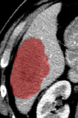
23 Base scan of lung tumor from 14/10/2010
24 Base scan with our semi-automatic delineation

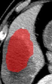
25 Follow-up scan from 3/1/2011 (2.5 moths later)
26 Follow-up scan with our automatic delineation


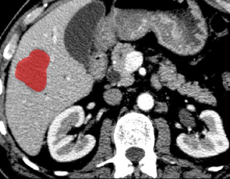
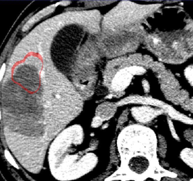
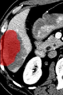
27 Liver tumor 5 May May 2011
28 Before registration
29 Before registration
30 Before registration

31 Global Affine Registration

32 Local B-spline Registration
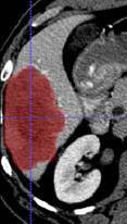
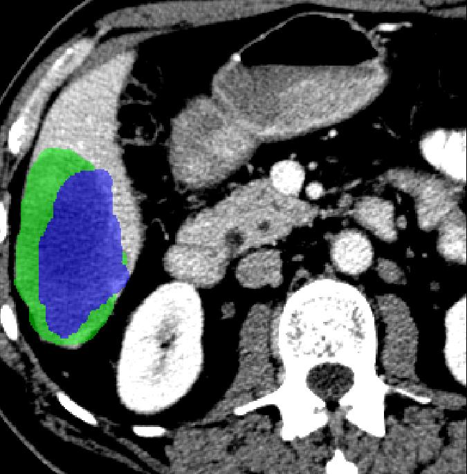
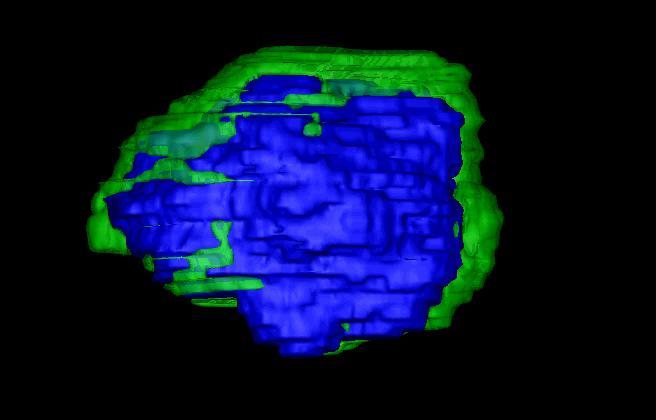
33 Segmentation results
34 Follow-up comparison
35 Follow-up comparison
36 Research Plan 1. A semi-automatic method for initial delineation of abdominal tumors. 2. An automatic Region Of Interest (ROI) identification. 3. Registration 4. Segmentation 5. New lesions 6. Implementation 7. Validation
A Novel Fully Automatic Technique for Liver Tumor Segmentation from CT Scans with knowledge-based constraints
 A Novel Fully Automatic Technique for Liver Tumor Segmentation from CT Scans with knowledge-based constraints Nader H. Abdel-massieh, Mohiy M. Hadhoud, Khalid M. Amin Faculty of Computers and Information
Semiautomatic segmentation and follow-up of multicomponent low-grade tumors in longitudinal brain MRI studies
 Semiautomatic segmentation and follow-up of multicomponent low-grade tumors in longitudinal brain MRI studies Lior Weizman a) School of Engineering and Computer Science, The Hebrew University of Jerusalem,
Comparative Study of K-means, Gaussian Mixture Model, Fuzzy C-means algorithms for Brain Tumor Segmentation
 Comparative Study of K-means, Gaussian Mixture Model, Fuzzy C-means algorithms for Brain Tumor Segmentation U. Baid 1, S. Talbar 2 and S. Talbar 1 1 Department of E&TC Engineering, Shri Guru Gobind Singhji
POC Brain Tumor Segmentation. vlife Use Case
 Brain Tumor Segmentation vlife Use Case 1 Automatic Brain Tumor Segmentation using CNN Background Brain tumor segmentation seeks to separate healthy tissue from tumorous regions such as the advancing tumor,
ANALYSIS AND DETECTION OF BRAIN TUMOUR USING IMAGE PROCESSING TECHNIQUES
 ANALYSIS AND DETECTION OF BRAIN TUMOUR USING IMAGE PROCESSING TECHNIQUES P.V.Rohini 1, Dr.M.Pushparani 2 1 M.Phil Scholar, Department of Computer Science, Mother Teresa women s university, (India) 2 Professor
Lung Region Segmentation using Artificial Neural Network Hopfield Model for Cancer Diagnosis in Thorax CT Images
 Automation, Control and Intelligent Systems 2015; 3(2): 19-25 Published online March 20, 2015 (http://www.sciencepublishinggroup.com/j/acis) doi: 10.11648/j.acis.20150302.12 ISSN: 2328-5583 (Print); ISSN:
PET in Radiation Therapy. Outline. Tumor Segmentation in PET and in Multimodality Images for Radiation Therapy. 1. Tumor segmentation in PET
 Tumor Segmentation in PET and in Multimodality Images for Radiation Therapy Wei Lu, Ph.D. Department of Radiation Oncology Mallinckrodt Institute of Radiology Washington University in St. Louis Outline
8/10/2016. PET/CT Radiomics for Tumor. Anatomic Tumor Response Assessment in CT or MRI. Metabolic Tumor Response Assessment in FDG-PET
 PET/CT Radiomics for Tumor Response Evaluation August 1, 2016 Wei Lu, PhD Department of Medical Physics www.mskcc.org Department of Radiation Oncology www.umaryland.edu Anatomic Tumor Response Assessment
ABAS Atlas-based Autosegmentation
 ABAS Atlas-based Autosegmentation Raising contouring to the next level Simultaneous Truth and Performance Level Estimation (STAPLE) Unique to ABAS, the STAPLE calculation is more robust than simple averaging.
Classification of Alzheimer s disease subjects from MRI using the principle of consensus segmentation
 Classification of Alzheimer s disease subjects from MRI using the principle of consensus segmentation Aymen Khlif and Max Mignotte 1 st September, Maynooth University, Ireland Plan Introduction Contributions
Classifying Breast Masses in Volumetric Whole Breast Ultrasound Data: A 2.5-Dimensional Approach
 Classifying Breast Masses in Volumetric Whole Breast Ultrasound Data: A 2.5-Dimensional Approach Gobert N. Lee 1,*, Toshiaki Okada 2, Daisuke Fukuoka 3, Chisako Muramatsu 2, Takeshi Hara 2, Takako Morita
Segmentation of Tumor Region from Brain Mri Images Using Fuzzy C-Means Clustering And Seeded Region Growing
 IOSR Journal of Computer Engineering (IOSR-JCE) e-issn: 2278-0661,p-ISSN: 2278-8727, Volume 18, Issue 5, Ver. I (Sept - Oct. 2016), PP 20-24 www.iosrjournals.org Segmentation of Tumor Region from Brain
EARLY STAGE DIAGNOSIS OF LUNG CANCER USING CT-SCAN IMAGES BASED ON CELLULAR LEARNING AUTOMATE
 EARLY STAGE DIAGNOSIS OF LUNG CANCER USING CT-SCAN IMAGES BASED ON CELLULAR LEARNING AUTOMATE SAKTHI NEELA.P.K Department of M.E (Medical electronics) Sengunthar College of engineering Namakkal, Tamilnadu,
Streamlined workflow for review and analysis of oncology patients
 Streamlined workflow for review and analysis of oncology patients Philips IntelliSpace Portal Multi-modality Tumor Tracking application (MMTT) Ekta Dharaiya, MS, Philips Healthcare, Cleveland, OH Cancer
Liver metastases: treatment planning. PJ Valette
 Liver metastases: treatment planning PJ Valette Liver metastases removal December 2010 April 2011 : after chemotherapy June 2011 : after resection of left lobe mets & portal embol. Sept 2011 : 1 year after
Computer-based 3d Puzzle Solving For Pre-operative Planning Of Articular Fracture Reductions In The Ankle, Knee, And Hip
 Computer-based 3d Puzzle Solving For Pre-operative Planning Of Articular Fracture Reductions In The Ankle, Knee, And Hip Andrew M. Kern, MS, Donald Anderson. University of Iowa, Iowa City, IA, USA. Disclosures:
Chapter 6. Hester Gietema Cornelia Schaefer-Prokop Willem Mali Gerard Groenewegen Mathias Prokop. Accepted for publication in Radiology
 Chapter 6 Interscan variability of semiautomated volume measurements in intraparenchymal pulmonary nodules using multidetector-row computed tomography: Influence of inspirational level, nodule size and
Copyright 2008 Society of Photo Optical Instrumentation Engineers. This paper was published in Proceedings of SPIE, vol. 6915, Medical Imaging 2008:
 Copyright 2008 Society of Photo Optical Instrumentation Engineers. This paper was published in Proceedings of SPIE, vol. 6915, Medical Imaging 2008: Computer Aided Diagnosis and is made available as an
Copyright 2007 Society of Photo Optical Instrumentation Engineers. This paper was published in Proceedings of SPIE, volume 6514, Medical Imaging
 Copyright 2007 Society of Photo Optical Instrumentation Engineers. This paper was published in Proceedings of SPIE, volume 6514, Medical Imaging 2007: Computer Aided Diagnosis and is made available as
Tumor cut segmentation for Blemish Cells Detection in Human Brain Based on Cellular Automata
 Tumor cut segmentation for Blemish Cells Detection in Human Brain Based on Cellular Automata D.Mohanapriya 1 Department of Electronics and Communication Engineering, EBET Group of Institutions, Kangayam,
Confidence-based Ensemble for GBM brain tumor segmentation
 Confidence-based Ensemble for GBM brain tumor segmentation Jing Huo 1, Eva M. van Rikxoort 1, Kazunori Okada 2, Hyun J. Kim 1, Whitney Pope 1, Jonathan Goldin 1, Matthew Brown 1 1 Center for Computer vision
LUNG NODULE SEGMENTATION IN COMPUTED TOMOGRAPHY IMAGE. Hemahashiny, Ketheesan Department of Physical Science, Vavuniya Campus
 LUNG NODULE SEGMENTATION IN COMPUTED TOMOGRAPHY IMAGE Hemahashiny, Ketheesan Department of Physical Science, Vavuniya Campus tketheesan@vau.jfn.ac.lk ABSTRACT: The key process to detect the Lung cancer
Developing ML Models for semantic segmentation of medical images
 Developing ML Models for semantic segmentation of medical images Raj Jena University of Cambridge Microsoft Research Cambridge Disclosures I perform consultancy work for InnerEye team at Microsoft Research
Classification and Statistical Analysis of Auditory FMRI Data Using Linear Discriminative Analysis and Quadratic Discriminative Analysis
 International Journal of Innovative Research in Computer Science & Technology (IJIRCST) ISSN: 2347-5552, Volume-2, Issue-6, November-2014 Classification and Statistical Analysis of Auditory FMRI Data Using
ISSUES ON COMPUTATIONAL MODELING FOR COMPUTATION-AIDED DIAGNOSIS 臨床診断支援ツールのための計算力学モデリング
 ISSUES ON COMPUTATIONAL MODELING FOR COMPUTATION-AIDED DIAGNOSIS 臨床診断支援ツールのための計算力学モデリング Hao LIU Advanced Computer and Information Division, RIKEN 2-1, Hirosawa, Wako-shi, Saitama 351-0198 JAPAN e-mail:
Automated Brain Tumor Segmentation Using Region Growing Algorithm by Extracting Feature
 Automated Brain Tumor Segmentation Using Region Growing Algorithm by Extracting Feature Shraddha P. Dhumal 1, Ashwini S Gaikwad 2 1 Shraddha P. Dhumal 2 Ashwini S. Gaikwad ABSTRACT In this paper, we propose
Automatic Ascending Aorta Detection in CTA Datasets
 Automatic Ascending Aorta Detection in CTA Datasets Stefan C. Saur 1, Caroline Kühnel 2, Tobias Boskamp 2, Gábor Székely 1, Philippe Cattin 1,3 1 Computer Vision Laboratory, ETH Zurich, 8092 Zurich, Switzerland
Automated Volumetric Cardiac Ultrasound Analysis
 Whitepaper Automated Volumetric Cardiac Ultrasound Analysis ACUSON SC2000 Volume Imaging Ultrasound System Bogdan Georgescu, Ph.D. Siemens Corporate Research Princeton, New Jersey USA Answers for life.
Pulmonary Nodule Volumetric Measurement Variability as a Function of CT Slice Thickness and Nodule Morphology
 CT of Pulmonary Nodules Chest Imaging Original Research Myria Petrou 1 Leslie E. Quint 1 in Nan 2 Laurence H. aker 3 Petrou M, Quint LE, Nan, aker LH Keywords: chest, lung disease, MDCT, oncologic imaging,
Robust Estimation for Brain Tumor Segmentation
 Robust Estimation for Brain Tumor Segmentation 1 Marcel Prastawa, 2 Elizabeth Bullitt, 1 Sean Ho, 1,3 Guido Gerig 1 Dept. of Computer Science, 2 Dept. of Surgery, 3 Dept. of Psychiatry University of North
Automatic cardiac contour propagation in short axis cardiac MR images
 International Congress Series 1281 (2005) 351 356 www.ics-elsevier.com Automatic cardiac contour propagation in short axis cardiac MR images G.L.T.F. Hautvast a,b, T, M. Breeuwer a, S. Lobregt a, A. Vilanova
Early Detection of Lung Cancer
 Early Detection of Lung Cancer Aswathy N Iyer Dept Of Electronics And Communication Engineering Lymie Jose Dept Of Electronics And Communication Engineering Anumol Thomas Dept Of Electronics And Communication
Unsupervised MRI Brain Tumor Detection Techniques with Morphological Operations
 Unsupervised MRI Brain Tumor Detection Techniques with Morphological Operations Ritu Verma, Sujeet Tiwari, Naazish Rahim Abstract Tumor is a deformity in human body cells which, if not detected and treated,
Detection of Mild Cognitive Impairment using Image Differences and Clinical Features
 Detection of Mild Cognitive Impairment using Image Differences and Clinical Features L I N L I S C H O O L O F C O M P U T I N G C L E M S O N U N I V E R S I T Y Copyright notice Many of the images in
Combined Radiology and Pathology Classification of Brain Tumors
 Combined Radiology and Pathology Classification of Brain Tumors Rozpoznanie guza mózgu na podstawie obrazu radiologicznego i patologicznego Piotr Giedziun Supervisor: dr hab. inż. Henryk Maciejewski 4
Image Fusion, Contouring, and Margins in SRS
 Image Fusion, Contouring, and Margins in SRS Sarah Geneser, Ph.D. Department of Radiation Oncology University of California, San Francisco Overview Review SRS uncertainties due to: image registration contouring
Ultralow Dose Chest CT with MBIR
 Ultralow Dose Chest CT with MBIR Ella A. Kazerooni, M.D. Professor & Director Cardiothoracic Radiology Associate Chair for Clinical Affairs University of Michigan Disclosures Consultant: GE Healthcare
NIH Public Access Author Manuscript Proc SPIE. Author manuscript; available in PMC 2014 February 07.
 NIH Public Access Author Manuscript Published in final edited form as: Proc SPIE. 2007 March 5; 6512: 651236. doi:10.1117/12.708950. Semi-Automatic Parcellation of the Corpus Striatum Ramsey Al-Hakim a,
Diagnosis of Liver Tumor Using 3D Segmentation Method for Selective Internal Radiation Therapy
 Diagnosis of Liver Tumor Using 3D Segmentation Method for Selective Internal Radiation Therapy K. Geetha & S. Poonguzhali Department of Electronics and Communication Engineering, Campus of CEG Anna University,
Fetal Dose Calculations and Impact on Patient Care
 Fetal Dose Calculations and Impact on Patient Care Matt Hough, MS, DABR, DABMP Florida Hospital Diagnostic Medical Physics and Radiation Safety Resource ACR-SPR Practice Parameter for Imaging Pregnant
COMPUTERIZED SYSTEM DESIGN FOR THE DETECTION AND DIAGNOSIS OF LUNG NODULES IN CT IMAGES 1
 ISSN 258-8739 3 st August 28, Volume 3, Issue 2, JSEIS, CAOMEI Copyright 26-28 COMPUTERIZED SYSTEM DESIGN FOR THE DETECTION AND DIAGNOSIS OF LUNG NODULES IN CT IMAGES ALI ABDRHMAN UKASHA, 2 EMHMED SAAID
COMPUTER AIDED DIAGNOSTIC SYSTEM FOR BRAIN TUMOR DETECTION USING K-MEANS CLUSTERING
 COMPUTER AIDED DIAGNOSTIC SYSTEM FOR BRAIN TUMOR DETECTION USING K-MEANS CLUSTERING Urmila Ravindra Patil Tatyasaheb Kore Institute of Engineering and Technology, Warananagar Prof. R. T. Patil Tatyasaheb
Analyse d'images médicales pour les maladies cardiovasculaires
 25 juin 2015 Workshop VIVABRAIN Paris, France Analyse d'images médicales pour les maladies cardiovasculaires Dr. Hortense A. Kirisli Project Manager / Advanced SW developer AQUILAB, Lille, France Cardiovascular
GE Healthcare. LOGIQ E9 XDclear 2.0. Complete Ultrasound HEAD TO TOE OBESE TO THIN NEONATE TO GERIATRIC
 GE Healthcare LOGIQ E9 XDclear 2.0 Complete Ultrasound HEAD TO TOE OBESE TO THIN NEONATE TO GERIATRIC In addition to extraordinary image quality, the system has been further enhanced to meet the needs
Automatic Definition of Planning Target Volume in Computer-Assisted Radiotherapy
 Automatic Definition of Planning Target Volume in Computer-Assisted Radiotherapy Angelo Zizzari Department of Cybernetics, School of Systems Engineering The University of Reading, Whiteknights, PO Box
Construction of 4D Left Ventricle and Coronary Artery Models
 1 Construction of 4D Left Ventricle and Coronary Artery Models Dong Ping Zhang Supervisors: Prof Daniel Rueckert, Dr Eddie Edwards 2 1 Background Conventional heart surgery is one of the most invasive
AUTOMATIC MEASUREMENT ON CT IMAGES FOR PATELLA DISLOCATION DIAGNOSIS
 AUTOMATIC MEASUREMENT ON CT IMAGES FOR PATELLA DISLOCATION DIAGNOSIS Qi Kong 1, Shaoshan Wang 2, Jiushan Yang 2,Ruiqi Zou 3, Yan Huang 1, Yilong Yin 1, Jingliang Peng 1 1 School of Computer Science and
A Reliable Method for Brain Tumor Detection Using Cnn Technique
 IOSR Journal of Electrical and Electronics Engineering (IOSR-JEEE) e-issn: 2278-1676,p-ISSN: 2320-3331, PP 64-68 www.iosrjournals.org A Reliable Method for Brain Tumor Detection Using Cnn Technique Neethu
MR-US Fusion. Image-guided prostate biopsy. Richard E Fan Department of Urology Stanford University
 MR-US Fusion Image-guided prostate biopsy Richard E Fan Department of Urology Stanford University Who am I? An instructor in the Department of Urology Quick plug for MED 275B Intro to Biodesign for Undergraduates
A New Approach For an Improved Multiple Brain Lesion Segmentation
 A New Approach For an Improved Multiple Brain Lesion Segmentation Prof. Shanthi Mahesh 1, Karthik Bharadwaj N 2, Suhas A Bhyratae 3, Karthik Raju V 4, Karthik M N 5 Department of ISE, Atria Institute of
Interobserver variation in cervical cancer tumor delineation for image-based radiotherapy planning among and within different specialties
 JOURNAL OF APPLIED CLINICAL MEDICAL PHYSICS, VOLUME 6, NUMBER 4, FALL 2005 Interobserver variation in cervical cancer tumor delineation for image-based radiotherapy planning among and within different
Assessment of Adipose Tissue from Whole Body 3T MRI Scans
 Assessment of Adipose Tissue from Whole Body 3T MRI Scans Ting Song 1, Jing An 2, Qun Chen 2, Vivian Lee 2, Andrew Laine 1 1 Department of Biomedical Engineering, Columbia University, New York, NY, USA
IEEE TRANSACTIONS ON MEDICAL IMAGING, VOL. 25, NO. 4, APRIL
 IEEE TRANSACTIONS ON MEDICAL IMAGING, VOL. 25, NO. 4, APRIL 2006 435 On Measuring the Change in Size of Pulmonary Nodules Anthony P. Reeves*, Senior Member, IEEE, Antoni B. Chan, David F. Yankelevitz,
Cover Page. Author: Wang, Ancong Title: Automatic quantification of intravascular optical coherence tomography Issue Date:
 Cover Page The handle http://hdl.handle.net/1887/29690 holds various files of this Leiden University dissertation Author: Wang, Ancong Title: Automatic quantification of intravascular optical coherence
Contributions to Brain MRI Processing and Analysis
 Contributions to Brain MRI Processing and Analysis Dissertation presented to the Department of Computer Science and Artificial Intelligence By María Teresa García Sebastián PhD Advisor: Prof. Manuel Graña
Brain tissue and white matter lesion volume analysis in diabetes mellitus type 2
 Brain tissue and white matter lesion volume analysis in diabetes mellitus type 2 C. Jongen J. van der Grond L.J. Kappelle G.J. Biessels M.A. Viergever J.P.W. Pluim On behalf of the Utrecht Diabetic Encephalopathy
Compute-aided Differentiation of Focal Liver Disease in MR Imaging
 1063 Compute-aided Differentiation of Focal Liver Disease in MR Imaging Xuejun Zhang a, Masayuki Kanematsu b, Hiroshi Fujita c, Takeshi Hara c, Hiroshi Kondo b, Xiangrong Zhou c, Wenguang Li a and Hiroaki
1st Turku Traumatic Brain Injury Symposium Turku, Finland, January 2014
 The TBIcare decision support tool aid for the clinician Jyrki Lötjönen & Jussi Mattila, VTT Technical Research Centre of Finland Validation of the decision support tool Ari Katila University of Turku 1st
Lung Detection and Segmentation Using Marker Watershed and Laplacian Filtering
 International Journal of Biomedical Engineering and Clinical Science 2015; 1(2): 29-42 Published online October 20, 2015 (http://www.sciencepublishinggroup.com/j/ijbecs) doi: 10.11648/j.ijbecs.20150102.12
Copyright 2007 IEEE. Reprinted from 4th IEEE International Symposium on Biomedical Imaging: From Nano to Macro, April 2007.
 Copyright 27 IEEE. Reprinted from 4th IEEE International Symposium on Biomedical Imaging: From Nano to Macro, April 27. This material is posted here with permission of the IEEE. Such permission of the
Automatic Classification System for Lumbar Spine X-ray Images
 Automatic Classification System for Lumbar Spine X-ray Images Soontharee Koompairojn Kien A. Hua School of Electrical Engineering and Computer Science University of Central Florida Orlando, FL 32816 {soonthar,
5/28/2015. The need for MRI in radiotherapy. Multiparametric MRI reflects a more complete picture of the tumor biology
 Ke Sheng, Ph.D., DABR Professor of Radiation Oncology University of California, Los Angeles The need for MRI in radiotherapy T1 FSE CT Tumor and normal tissues in brain, breast, head and neck, liver, prostate,
International Journal of Research (IJR) Vol-1, Issue-6, July 2014 ISSN
 Vol-1, Issue-6, July 2014 ISSN") Developing an Approach to Brain MRI Image Preprocessing for Tumor Detection Mr. B.Venkateswara Reddy 1, Dr. P. Bhaskara Reddy 2, Dr P. Satish Kumar 3, Dr. S. Siva Reddy 4 1. Associate Professor, ECE Dept,
Developing an Approach to Brain MRI Image Preprocessing for Tumor Detection Mr. B.Venkateswara Reddy 1, Dr. P. Bhaskara Reddy 2, Dr P. Satish Kumar 3, Dr. S. Siva Reddy 4 1. Associate Professor, ECE Dept,
Mammogram Analysis: Tumor Classification
 Mammogram Analysis: Tumor Classification Term Project Report Geethapriya Raghavan geeragh@mail.utexas.edu EE 381K - Multidimensional Digital Signal Processing Spring 2005 Abstract Breast cancer is the
MRI-Based Classification Techniques of Autistic vs. Typically Developing Brain
 MRI-Based Classification Techniques of Autistic vs. Typically Developing Brain Presented by: Rachid Fahmi 1 2 Collaborators: Ayman Elbaz, Aly A. Farag 1, Hossam Hassan 1, and Manuel F. Casanova3 1Computer
Review of Longitudinal MRI Analysis for Brain Tumors. Elsa Angelini 17 Nov. 2006
 Review of Longitudinal MRI Analysis for Brain Tumors Elsa Angelini 17 Nov. 2006 MRI Difference maps «Longitudinal study of brain morphometrics using quantitative MRI and difference analysis», Liu,Lemieux,
Organ Contour Adaptor to create new structures to use for adaptive radiotherapy of cervix cancer using Matlab Bridge and 3DSlicer / SlicerRT
 Organ Contour Adaptor to create new structures to use for adaptive radiotherapy of cervix cancer using Matlab Bridge and 3DSlicer / SlicerRT Y. Seppenwoolde 1,2, M. Daniel 2, H. Furtado 2,3, D. Georg 1,2
Framework for 3D TransRectal Ultrasound (TRUS) Image-Based Tracking - Example of Use Evaluation of 2D TRUS Prostate Biopsies Mapping
 Image-Based Tracking - Example of Use Evaluation of 2D TRUS Prostate Biopsies Mapping") Author manuscript, published in "Johns Hopkins University "Prostate Day", Baltimore : United States (2008)" Framework for 3D TransRectal Ultrasound (TRUS) Image-Based Tracking - Example of Use Evaluation
Author manuscript, published in "Johns Hopkins University "Prostate Day", Baltimore : United States (2008)" Framework for 3D TransRectal Ultrasound (TRUS) Image-Based Tracking - Example of Use Evaluation
Lymph Node Detection in IASLC-defined zones on PET/CT Images
 Lymph Node Detection in IASLC-defined zones on PET/CT Images Yihua Song a, b, Jayaram K. Udupa* b, Dewey Odhner b, Yubing Tong b, Drew A. Torigian b a Key Laboratory of Medical Image Computing Ministry
Lung structure recognition: a further study of thoracic organ recognitions based on CT images
 Lung structure recognition: a further study of thoracic organ recognitions based on CT images X. Zhou a, S. Kobayashi a, T. Hayashi a, N. Murata a, T. Hara a, H. Fujita a, R. Yokoyama b, T. Kiryu b, H.
Primary Level Classification of Brain Tumor using PCA and PNN
 Primary Level Classification of Brain Tumor using PCA and PNN Dr. Mrs. K.V.Kulhalli Department of Information Technology, D.Y.Patil Coll. of Engg. And Tech. Kolhapur,Maharashtra,India kvkulhalli@gmail.com
Proceedings of the UGC Sponsored National Conference on Advanced Networking and Applications, 27 th March 2015
 Brain Tumor Detection and Identification Using K-Means Clustering Technique Malathi R Department of Computer Science, SAAS College, Ramanathapuram, Email: malapraba@gmail.com Dr. Nadirabanu Kamal A R Department
Shape Modeling of the Corpus Callosum for Neuroimaging Studies of the Brain (Part I) Dongqing Chen, Ph.D.
 Dongqing Chen, Ph.D.") The University of Louisville CVIP Lab Shape Modeling of the Corpus Callosum for Neuroimaging Studies of the Brain (Part I) Dongqing Chen, Ph.D. Computer Vision & Image Processing (CVIP) Laboratory Department
The University of Louisville CVIP Lab Shape Modeling of the Corpus Callosum for Neuroimaging Studies of the Brain (Part I) Dongqing Chen, Ph.D. Computer Vision & Image Processing (CVIP) Laboratory Department
Automatic Hemorrhage Classification System Based On Svm Classifier
 Automatic Hemorrhage Classification System Based On Svm Classifier Abstract - Brain hemorrhage is a bleeding in or around the brain which are caused by head trauma, high blood pressure and intracranial
Four Tissue Segmentation in ADNI II
 Four Tissue Segmentation in ADNI II Charles DeCarli, MD, Pauline Maillard, PhD, Evan Fletcher, PhD Department of Neurology and Center for Neuroscience, University of California at Davis Summary Table of
Cancer Cells Detection using OTSU Threshold Algorithm
 Cancer Cells Detection using OTSU Threshold Algorithm Nalluri Sunny 1 Velagapudi Ramakrishna Siddhartha Engineering College Mithinti Srikanth 2 Velagapudi Ramakrishna Siddhartha Engineering College Kodali
By choosing to view this document, you agree to all provisions of the copyright laws protecting it.
 Copyright 2009 IEEE. Reprinted from 31 st Annual International Conference of the IEEE Engineering in Medicine and Biology Society, 2009. EMBC 2009. Sept. 2009. This material is posted here with permission
Background Information
 Background Information Erlangen, November 26, 2017 RSNA 2017 in Chicago: South Building, Hall A, Booth 1937 Artificial intelligence: Transforming data into knowledge for better care Inspired by neural
3D ultrasound applied to abdominal aortic aneurysm: preliminary evaluation of diameter measurement accuracy
 3D ultrasound applied to abdominal aortic aneurysm: preliminary evaluation of diameter measurement accuracy Poster No.: C-0493 Congress: ECR 2011 Type: Authors: Keywords: DOI: Scientific Paper A. LONG
Automatic Classification of Calcification in the Coronary Vessel Tree
 Automatic Classification of Calcification in the Coronary Vessel Tree R. Shahzad 1,2,3, L. J. van Vliet 2, W. J. Niessen 2,3, and T. van Walsum 3 1 Division of Image Processing, Department of Radiology,
Segmentation and Analysis of Cancer Cells in Blood Samples
 Segmentation and Analysis of Cancer Cells in Blood Samples Arjun Nelikanti Assistant Professor, NMREC, Department of CSE Hyderabad, India anelikanti@gmail.com Abstract Blood cancer is an umbrella term
Automatic Segmentation of Jaw Tissues in CT Using Active Appearance Models and Semi-automatic Landmarking
 Automatic Segmentation of Jaw Tissues in CT Using Active Appearance Models and Semi-automatic Landmarking Sylvia Rueda 1,JoséAntonioGil 1, Raphaël Pichery 2,andMarianoAlcañiz 1 1 Medical Image Computing
Automated Tumour Delineation Using Joint PET/CT Information
 Automated Tumour Delineation Using Joint PET/CT Information Vaclav Potesil 1,2, Xiaolei Huang 1,3, Xiang Sean Zhou 1, 1 Siemens Medical Solutions USA, Computer Aided Diagnosis and Therapy Solutions; 2
UCLA UCLA UCLA 7/10/2015. The need for MRI in radiotherapy. Multiparametric MRI reflects a more complete picture of the tumor biology
 Ke Sheng, Ph.D., DABR Professor of Radiation Oncology University of California, Los Angeles The need for MRI in radiotherapy T1 FSE CT Tumor and normal tissues in brain, breast, head and neck, liver, prostate,
A new Method on Brain MRI Image Preprocessing for Tumor Detection
 2015 IJSRSET Volume 1 Issue 1 Print ISSN : 2395-1990 Online ISSN : 2394-4099 Themed Section: Engineering and Technology A new Method on Brain MRI Preprocessing for Tumor Detection ABSTRACT D. Arun Kumar
TWO HANDED SIGN LANGUAGE RECOGNITION SYSTEM USING IMAGE PROCESSING
 134 TWO HANDED SIGN LANGUAGE RECOGNITION SYSTEM USING IMAGE PROCESSING H.F.S.M.Fonseka 1, J.T.Jonathan 2, P.Sabeshan 3 and M.B.Dissanayaka 4 1 Department of Electrical And Electronic Engineering, Faculty
Copyright 2007 Society of Photo Optical Instrumentation Engineers. This paper was published in Proceedings of SPIE, volume 6514, Medical Imaging
 Copyright 2007 Society of Photo Optical Instrumentation Engineers. This paper was published in Proceedings of SPIE, volume 6514, Medical Imaging 2007: Computer Aided Diagnosis and is made available as
CALCULATION of the CEREBRAL HEMORRHAGE VOLUME USING ANALYSIS of COMPUTED TOMOGRAPHY IMAGE
 CALCULATION of the CEREBRAL HEMORRHAGE VOLUME USING ANALYSIS of COMPUTED TOMOGRAPHY IMAGE Cory Amelia* Magister of Physics, Faculty of Science and Mathematics, Diponegoro University, Semarang, Indonesia
A deep learning model integrating FCNNs and CRFs for brain tumor segmentation
 A deep learning model integrating FCNNs and CRFs for brain tumor segmentation Xiaomei Zhao 1,2, Yihong Wu 1, Guidong Song 3, Zhenye Li 4, Yazhuo Zhang,3,4,5,6, and Yong Fan 7 1 National Laboratory of Pattern
IJESRT. Scientific Journal Impact Factor: (ISRA), Impact Factor: 1.852
, Impact Factor: 1.852") IJESRT INTERNATIONAL JOURNAL OF ENGINEERING SCIENCES & RESEARCH TECHNOLOGY Performance Analysis of Brain MRI Using Multiple Method Shroti Paliwal *, Prof. Sanjay Chouhan * Department of Electronics & Communication
IJESRT INTERNATIONAL JOURNAL OF ENGINEERING SCIENCES & RESEARCH TECHNOLOGY Performance Analysis of Brain MRI Using Multiple Method Shroti Paliwal *, Prof. Sanjay Chouhan * Department of Electronics & Communication
A System for Simulating the Orthodontic Treatment Plan
 A System for Simulating the Orthodontic Treatment Plan Lamia N. Omran*, Ahmed M. El-Bialy**, Ahmed H. Kandil*** & Sahar Ali Fawzy**** *Assistant Lecturer, System and Biomedical Engineering Department,
Automatic Lung Cancer Detection Using Volumetric CT Imaging Features
 Automatic Lung Cancer Detection Using Volumetric CT Imaging Features A Research Project Report Submitted To Computer Science Department Brown University By Dronika Solanki (B01159827) Abstract Lung cancer
Predicting Heart Attack using Fuzzy C Means Clustering Algorithm
 Predicting Heart Attack using Fuzzy C Means Clustering Algorithm Dr. G. Rasitha Banu MCA., M.Phil., Ph.D., Assistant Professor,Dept of HIM&HIT,Jazan University, Jazan, Saudi Arabia. J.H.BOUSAL JAMALA MCA.,M.Phil.,
International Journal of Engineering Trends and Applications (IJETA) Volume 4 Issue 2, Mar-Apr 2017
 Volume 4 Issue 2, Mar-Apr 2017") RESEARCH ARTICLE OPEN ACCESS Knowledge Based Brain Tumor Segmentation using Local Maxima and Local Minima T. Kalaiselvi [1], P. Sriramakrishnan [2] Department of Computer Science and Applications The Gandhigram
RESEARCH ARTICLE OPEN ACCESS Knowledge Based Brain Tumor Segmentation using Local Maxima and Local Minima T. Kalaiselvi [1], P. Sriramakrishnan [2] Department of Computer Science and Applications The Gandhigram
Brain Tumor Detection using Watershed Algorithm
 Brain Tumor Detection using Watershed Algorithm Dawood Dilber 1, Jasleen 2 P.G. Student, Department of Electronics and Communication Engineering, Amity University, Noida, U.P, India 1 P.G. Student, Department
Joint Myocardial Registration and Segmentation of Cardiac BOLD MRI
 Joint Myocardial Registration and Segmentation of Cardiac BOLD MRI Ilkay Oksuz 1,2, Rohan Dharmakumar 3, and Sotirios A. Tsaftaris 2 1 IMT Institute for Advanced Studies Lucca, 2 The University of Edinburgh,
Development of Novel Approach for Classification and Detection of Brain Tumor
 International Journal of Latest Technology in Engineering & Management (IJLTEM) www.ijltem.com ISSN: 245677 Development of Novel Approach for Classification and Detection of Brain Tumor Abstract This paper
Noise-Robust Speech Recognition Technologies in Mobile Environments
 Noise-Robust Speech Recognition echnologies in Mobile Environments Mobile environments are highly influenced by ambient noise, which may cause a significant deterioration of speech recognition performance.
Automatic recognition of lung lobes and fissures from multi-slice CT images
 Automatic recognition of lung lobes and fissures from multi-slice CT images Xiangrong Zhou* a, Tatsuro Hayashi a, Takeshi Hara a, Hiroshi Fujita a, Ryujiro Yokoyama b, Takuji Kiryu b, Hiroaki Hoshi b a
2D-Sigmoid Enhancement Prior to Segment MRI Glioma Tumour
 2D-Sigmoid Enhancement Prior to Segment MRI Glioma Tumour Pre Image-Processing Setyawan Widyarto, Siti Rafidah Binti Kassim 2,2 Department of Computing, Faculty of Communication, Visual Art and Computing,