MRI Based treatment planning for with focus on prostate cancer. Xinglei Shen, MD Department of Radiation Oncology KUMC
|
|
|
- Christina Day
- 5 years ago
- Views:
Transcription
1 MRI Based treatment planning for with focus on prostate cancer Xinglei Shen, MD Department of Radiation Oncology KUMC
2 Overview How magnetic resonance imaging works (very simple version) Indications for MRI in diagnosis and treatment planning MRI improves upon CT in prostate cancer Technique for incorporating MRI into prostate cancer treatment planning (If time allows) MRI in cervical brachytherapy
 results in alignment of the atoms to the magnetic field and the nuclei in atoms to precess (spin) This generates a net")
3 How MRI works (the very dummy version) Tissues are ultimately made of atoms (Hydrogen is by far most common) Because they spin, hydrogen atoms have tiny currents and thus tiny magnetic fields Putting atoms in a huge magnetic field (1 Tesla = 20,000 times earth s magnetic field) results in alignment of the atoms to the magnetic field and the nuclei in atoms to precess (spin) This generates a net magnetic field along direction of applied magnetic field (i.e. a human magnet)
4 How MRI works (the very dummy version) Hydrogen atoms precess at a specific frequency. Adding energy at the specific frequency to align hydrogen atoms Effect on hydrogen can be measured based on radiofrequency signal emitted by these atoms as they relax Relaxation characteristics after a energy pulse in different directions (longitudinal or perpendicular to main magnetic field) is affected by tissue composition T1 = time for longitudinal relaxation T2 = time for perpendicular relaxation
5 MRI Sequences Different tissue compositions have characteristic appearances on MRI sequences T1 or T2 weighted sequences show different properties of the tissue T1 (in line relaxation) related to energy transfer between molecules Free hydrogens in water and hydrogens bound to large molecules do not exchange energy well Bound water (i.e. attached to proteins) and hydrogens in fat exchange energy well T2 (transverse relaxation) related to interactions from nearby small magnetic fields from tissue More molecules in tissue = more interaction Free water has interactions than organized tissues

6 CT versus MRI

7 CT versus MRI

8 CT versus MRI

9 CT versus MRI
10 MRI vs CT in diagnostic imaging MRI Slower acquisition/sensitive to motion Cannot use with certain metal/pacemakers etc. Looks at multiple characteristics of tissues Better for soft tissue (i.e. tumor within soft tissue) CT Fast/less sensitive to motion No restrictions Looks at electron density only Better for density (i.e. calcification) Cannot use directly for RT Treatment Planning Can use directly for RT Treatment Planning
11 Use of MRI in Treatment planning Location CNS Head and neck Lung/thoracic Breast GI (esophagus, pancreas, gastric) Liver Spine Pelvis Role of MRI Important Not necessary (except for skull base) Not necessary Not necessary Not necessary Important Important Not necessary but nice to have
12 Take home point MRI demonstrates better soft tissue differentiation compared to CT MRI important where we treat gross tumor (GTV) which is surrounded by OAR with soft tissue of similar density
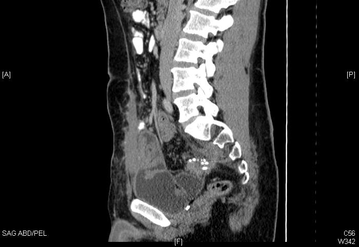
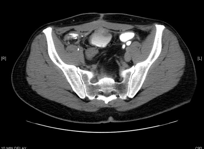
13 How does MRI help patients with pelvic cancers? Prostate cancer Staging (extracapsular invasion, seminal vesicle invasion, bladder neck invasion etc) Precise location of gland, urethra, bladder, rectum Cervical cancer Staging (parametrial invasion, local invasion etc) Brachytherapy boost tumor extent Sarcoma Delineating tumor from muscle
14 Current standards CT based volume definition is standard for pelvic cancers No requirement for MRI based treatment planning Prostate cancer MRI not in routine use Cervical cancer brachytherapy: GEC-ESTRO guidelines strongly recommends MRI based 3D-volume definition for cervical brachytherapy ABS (2011) guidelines also recommend 3D-volume definition with ultrasound, CT or MRI
 Gao et al. Radiotherapy and Oncology. 2007.")
15 Prostate contouring is not easy Visible Human Project Normal male cadaver CT at 3 mm slices Anatomy slices at 1 mm Six experience radiation oncologists asked to contour on CT Compare to true anatomy (tissue) Gao et al. Radiotherapy and Oncology :

 on average And")

16 Problem with CT based volume definition Inter-observer variability 18.8% Contoured volume exceeded true prostate volume (30%) on average And did not include 9-21% of true prostate Gao et al. Radiotherapy and Oncology :
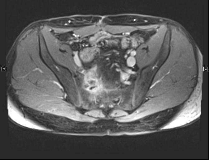
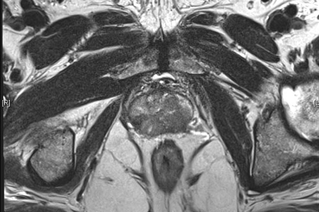
17 Prostate MRI Impossible to see prostate cancer on CT MRI allows to distinguish: Minor extracapsular extention Minor seminal vesicle invasion Location of anterior fibrous stroma Location of GU diaphragm Bladder neck


18 MRI in prostate cancer
 McLaughlin et al.")
19 Common areas of mistakes on contouring (avoidable by MRI) McLaughlin et al. IJROBP 2010: 78:

20 How much does this matter? PTV expansion covers areas of inadequate CTV definition Over-treatment laterally, anteriorly do not necessarily have much consequence Superior bladder neck Inferior penile bulb/erectile tissues Posterior rectum
21 Ali et al. PRO. 2013: e1-9. MRI vs CT Target Delineation for PC Multi-institutional study of 155 consecutive patients CT vs CT + MRI Prostate volume smaller on MRI 43 cc vs 55.7 cc DVH parameters better Less acute GU toxicity with MRI
22 MRI vs CT Target Delineation for PC MRI planning had 21.5% smaller prostate volume (40.9 cc vs 52.1 cc) No difference in PSA outcome? Difference in late toxicity Sander et al. Acta oncologica. 2014: 53:
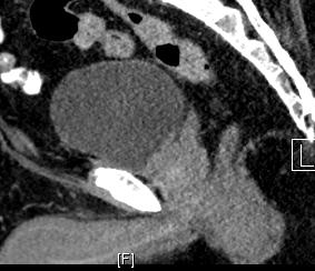
23 Areas where MRI is especially helpful Rectal prostate interface Hip arthroplasty artifact Small margins (SBRT) Delineation of target and OAR structures within the prostate Avoid under-coverage of base Delineate SV from prostate (spare rectum) Focal therapy Urethra
24 Target delineation rectal interface
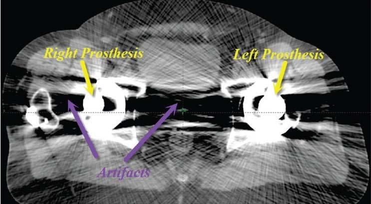
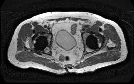
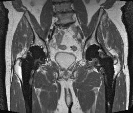
25 Hip prostheses
26 SBRT Selected RTOG 0938 prostate SBRT clinical trial guidelines: Margins: 3mm posteriorly/5 mm on sides Urethral dose < 107% hot spot

27 Avoid over contouring

28 Urethral sparing
29 Take home point MRI improves upon CT based planning Amount of benefit is not large in most cases and CT based planning is adequate MRI very helpful in select situations

 Contouring on CT/MRI Fusion/Registration Planning")
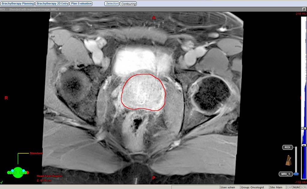
30 Process for Incorporation of MRI in treatment planning for pelvic malignancy CT-simulation MRI simulation (future?) Contouring on CT/MRI Fusion/Registration Planning on CT MRI Other diagnostic studies (CT, PET)
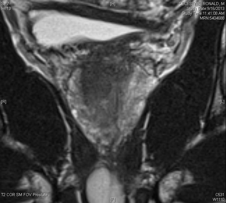
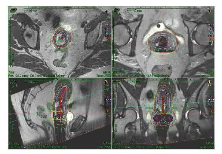
31 How to incorporate MRI for PC Imaging characterstics: Hypodense on T2 Early enhancement and washout Restricted diffusion MR-Spect with decreased citrate, increased choline Typically use T2 images to fuse Fuse with axial and sagittal and coronal images
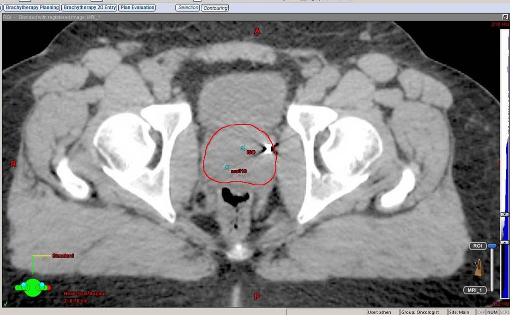
32 How I fuse MRI Auto-fusion software ALWAYS VERIFY Over-relies on bony anatomy Set appropriately small ROI Easy fusions: skull, spine, pelvis (regions limited by bony anatomy) Hard fusions: soft tissue (i.e. abdomen), altered anatomy (i.e. curve in spine) Contour on CT and match to appropriate structure on MRI Look at fusion in multiple planes Only need to make sure ROI fusion is good


33 Fusion using fixed match points (fiducials)
34 Iterative refinement May need to keep adjusting fusion between CT and MRI until fusion satisfactory Sometimes fusion simply does not work because of misalignment between scans or distortion but this is rare
35 Common pitfalls Changes in MRI based volume due to ADT Using autofusion (relies too much on bone) Not looking on multiple planes Not all MRI are the same need 3T multiparametric imaging for prostate cancer
36 Take home points For fusion to MRI: do not rely on bony landmarks and use iterative review
37 Future Directions for MRI in prostate cancer
38 Focal therapy Classically prostate cancer has always been treated to the whole prostate Treatment of only a portion of the prostate gland may reduce toxicity Potential to re-treat other parts of the gland Multiple modalities: HIFU, cryo, thermal ablation, brachytherapy etc.

39 Comparison of MRI with whole mount to detect prostate cancer mpmri: For Gleason > 6 or tumor size > 1 cm sensitivity = 72% Largest tumor detected in 80% Le et al. Eur Urology. 2015: 67:
40 Comparison of MRI with whole mount to detect prostate cancer Le et al. Eur Urology. 2015: 67:
41 Use of MRI in focal therapy MRI can identify target lesions (largest, most aggressive) MRI tends to underestimate size of lesions need 5-9mm margins Not ready for prime time yet!
42 MRI only treatment planning Challenges: Need to convert to electron density (already have in CT information) for dose calculation Geometric distortion (magnetic field, tissue susceptibility changes) in MRI Potential solutions: Automatic segmentation of bone and assign all other tissue water density (dose calc within 1-2%) External or internal reference markers Minimal distortion at isocenter 3 mm distortion at 9 cm from isocenter Kapanen et al. Magnetic Resonance in Medicine. 2013:

43 MRI only treatment planning Commercially available system!
44 Take away points MRI becoming more important in treatment planning in prostate cancer Not yet standard, but will become a greater part of treatment planning.
45 Thank you!
46 MRI brachytherapy in cervical cancer
47 Impact of image guidance in cervical brachytherapy: 2D vs 3D Prospective non-randomized French trial of 2D vs 3D brachytherapy in cervical cancer Better local control with 3D Less toxicity with 3D Charra-Brunaud et al. Radiotherapy and Oncology. 2012:
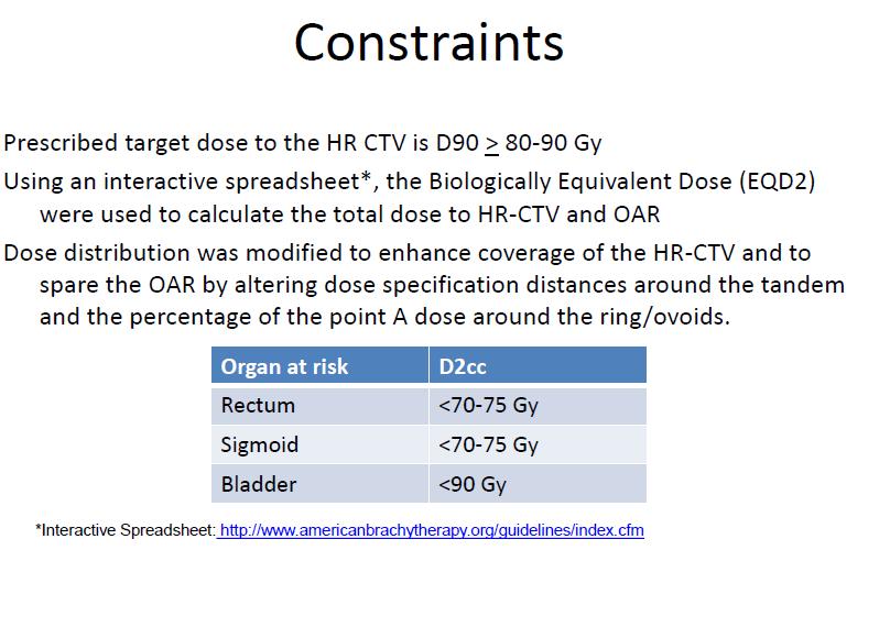
48 GEC-ESTRO Guidelines moving toward volume based implants High risk CTV = Whole cervix PLUS residual extra-cervical tumor at time of implant Large tumors difficult to see extra-cervical extension on CT American Brachytherapy society guidelines still allow point A dosing, but are moving towards 3D implants

49 MRI base brachytherapy ARRO Cervical cancer lecture
50 MRI based brachytherapy
51 MRI based brachytherapy

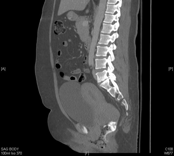
52 CT vs MRI cervical brachytherapy
53 Take away points MRI replacing point A dosing For most situations, CT is good enough in 3D treatment planning, but MRI is the best
2
 1 2 3 4 5 6 7 The RTOG contouring recommendations state the femurs are to be contourned to the bottom of the ischial tuberosity. 8 This slide shows the hourglass configuration. It is only present in about
A Comparison of IMRT and VMAT Technique for the Treatment of Rectal Cancer
 A Comparison of IMRT and VMAT Technique for the Treatment of Rectal Cancer Tony Kin Ming Lam Radiation Planner Dr Patricia Lindsay, Radiation Physicist Dr John Kim, Radiation Oncologist Dr Kim Ann Ung,
MRI Applications in Radiation Oncology:
 MRI Applications in Radiation Oncology: Physician s Perspective Jeff Olsen, MD Department of Radiation Oncology Washington University, St. Louis, MO Disclosures Washington University has research and service
IMRT - the physician s eye-view. Cinzia Iotti Department of Radiation Oncology S.Maria Nuova Hospital Reggio Emilia
 IMRT - the physician s eye-view Cinzia Iotti Department of Radiation Oncology S.Maria Nuova Hospital Reggio Emilia The goals of cancer therapy Local control Survival Functional status Quality of life Causes
Image Fusion, Contouring, and Margins in SRS
 Image Fusion, Contouring, and Margins in SRS Sarah Geneser, Ph.D. Department of Radiation Oncology University of California, San Francisco Overview Review SRS uncertainties due to: image registration contouring
Herlev radiation oncology team explains what MRI can bring
 Publication for the Philips MRI Community Issue 46 2012/2 Herlev radiation oncology team explains what MRI can bring The radiotherapy unit at Herlev University Hospital investigates use of MRI for radiotherapy
Johannes C. Athanasios Dimopoulos
 BrachyNext Symposium Miami Beach, USA, May 30 31, 2014 Imaging Modalities: Current Challenges and Future Directions Johannes C. Athanasios Dimopoulos Imaging Modalities: Current Challenges and Future Directions
Outline - MRI - CT - US. - Combinations of imaging modalities for treatment planning
 Imaging Outline - MRI - CT - US - Combinations of imaging modalities for treatment planning Imaging Part 1: MRI MRI for cervical cancer high soft tissue contrast multiplanar imaging MRI anatomy: the normal
REVISITING ICRU VOLUME DEFINITIONS. Eduardo Rosenblatt Vienna, Austria
 REVISITING ICRU VOLUME DEFINITIONS Eduardo Rosenblatt Vienna, Austria Objective: To introduce target volumes and organ at risk concepts as defined by ICRU. 3D-CRT is the standard There was a need for a
Pitfalls in SBRT Treatment Planning for a Moving Target
 Pitfalls in SBRT Treatment Planning for a Moving Target Cynthia F. Chuang, Ph.D. Department of Radiation Oncology University of California-San Francisco I have no conflicts of interests to disclose In
Dosimetric Analysis of 3DCRT or IMRT with Vaginal-cuff Brachytherapy (VCB) for Gynaecological Cancer
 for Gynaecological Cancer") Dosimetric Analysis of 3DCRT or IMRT with Vaginal-cuff Brachytherapy (VCB) for Gynaecological Cancer Tan Chek Wee 15 06 2016 National University Cancer Institute, Singapore Clinical Care Education Research
Dosimetric Analysis of 3DCRT or IMRT with Vaginal-cuff Brachytherapy (VCB) for Gynaecological Cancer Tan Chek Wee 15 06 2016 National University Cancer Institute, Singapore Clinical Care Education Research
CT Guided Contouring: Challenges and Pitfalls
 CT Guided Contouring: Challenges and Pitfalls Dr Umesh Mahantshetty, Associate Professor, GYN & Urology Disease Management Group (DMG) Member Tata Memorial Hospital, Mumbai, India GYN GEC ESTRO NETWORK
Feasibility of 4D IMRT Delivery for Hypofractionated High Dose Partial Prostate Treatments
 Feasibility of 4D IMRT Delivery for Hypofractionated High Dose Partial Prostate Treatments R.A. Price Jr., Ph.D., J. Li, Ph.D., A. Pollack, M.D., Ph.D.*, L. Jin, Ph.D., E. Horwitz, M.D., M. Buyyounouski,
MRI Guided GYN Brachytherapy: Clinical Considerations
 MRI Guided GYN Brachytherapy: Clinical Considerations AAPM Junzo Chino MD Duke Radiation Oncology 8/8/2013 Disclosures none Learning Objectives Historical Context: Film based Brachytherapy Advantages of
The Evolution of RT Techniques for Gynaecological Cancers in a developing country context
 The Evolution of RT Techniques for Gynaecological Cancers in a developing country context Hannah Simonds Stellenbosch University/ Tygerberg Academic Hospital ESMO Africa 2017 I have no disclosures External
IMRT for Prostate Cancer
 IMRT for Cancer All patients are simulated in the supine position. Reproducibility is achieved using a custom alpha cradle cast that extends from the mid-back to mid-thigh. The feet are positioned in a
From position verification and correction to adaptive RT Adaptive RT and dose accumulation
 From position verification and correction to adaptive RT Adaptive RT and dose accumulation Hans de Boer Move away from Single pre-treatment scan Single treatment plan Treatment corrections by couch shifts
CT Guided Contouring: Challenges and Pitfalls
 CT Guided Contouring: Challenges and Pitfalls Dr Umesh Mahantshetty, Associate Professor, GYN & Urology Disease Management Group (DMG) Member Tata Memorial Hospital, Mumbai, India GYN GEC ESTRO NETWORK
Clinical Implementation of a New Ultrasound Guidance System. Vikren Sarkar Bill Salter Martin Szegedi
 Clinical Implementation of a New Ultrasound Guidance System Vikren Sarkar Bill Salter Martin Szegedi Disclosure The University of Utah has research agreements with Elekta Agenda Historical Review Trans-Abdominal
3D ANATOMY-BASED PLANNING OPTIMIZATION FOR HDR BRACHYTHERAPY OF CERVIX CANCER
 SAUDI JOURNAL OF OBSTETRICS AND GYNECOLOGY VOLUME 11 NO. 2 1430 H - 2009 G 3D ANATOMY-BASED PLANNING OPTIMIZATION FOR HDR BRACHYTHERAPY OF CERVIX CANCER DR YASIR BAHADUR 1, DR CAMELIA CONSTANTINESCU 2,
UCLA UCLA UCLA 7/10/2015. The need for MRI in radiotherapy. Multiparametric MRI reflects a more complete picture of the tumor biology
 Ke Sheng, Ph.D., DABR Professor of Radiation Oncology University of California, Los Angeles The need for MRI in radiotherapy T1 FSE CT Tumor and normal tissues in brain, breast, head and neck, liver, prostate,
IGRT Solution for the Living Patient and the Dynamic Treatment Problem
 IGRT Solution for the Living Patient and the Dynamic Treatment Problem Lei Dong, Ph.D. Associate Professor Dept. of Radiation Physics University of Texas M. D. Anderson Cancer Center Houston, Texas Learning
Patient Safety Focused QA. LDR Brachytherapy Vrinda Narayana
 Patient Safety Focused QA LDR Brachytherapy Vrinda Narayana D < 2 Gy/h Old LDR Brachytherapy? Ra-226; Cs-137; Ir-192 New Gynecological; interstitial Pd-103; I-125; Cs-131 Prostate implants Eye plaques
Multiparametric MR Imaging of the Prostate after Treatment of Prostate Cancer
 Multiparametric MR Imaging of the Prostate after Treatment of Prostate Cancer RadioGraphics 2018; 38:437 449 Pritesh Patel, MD Melvy S. Mathew, MD Igor Trilisky, MD Aytekin Oto, MD, MBA Jeffrey S. Klein,
The Physics of Oesophageal Cancer Radiotherapy
 The Physics of Oesophageal Cancer Radiotherapy Dr. Philip Wai Radiotherapy Physics Royal Marsden Hospital 1 Contents Brief clinical introduction Imaging and Target definition Dose prescription & patient
SUPERIORITY OF A REAL TIME PLANNING TECHNIQUE OVER IMAGE GUIDED RADIATION THERAPY FOR THE TREATMENT OF PRIMARY PROSTATE CANCERS
 SUPERIORITY OF A REAL TIME PLANNING TECHNIQUE OVER IMAGE GUIDED RADIATION THERAPY FOR THE TREATMENT OF PRIMARY PROSTATE CANCERS Authors: Scott Merrick James Wong MD, Mona Karim MD, Yana Goldberg MD DISCLOSURE
Long Term Clinical Experience Using Ultrasound Alignment. Overview. Ultrasound Alignment Experience at Fox Chase
 Long Term Clinical Experience Using Ultrasound Alignment Shawn McNeeley M.S.. Fox Chase Cancer Center Department of Radiation Oncology Philadelphia, PA Overview Discuss various technique, and patient related,
New Technologies for the Radiotherapy of Prostate Cancer
 Prostate Cancer Meyer JL (ed): IMRT, IGRT, SBRT Advances in the Treatment Planning and Delivery of Radiotherapy. Front Radiat Ther Oncol. Basel, Karger, 27, vol. 4, pp 315 337 New Technologies for the
Proton Therapy for Prostate Cancer. Andrew K. Lee, MD, MPH Director Proton Therapy Center
 Proton Therapy for Prostate Cancer Andrew K. Lee, MD, MPH Director Proton Therapy Center Disclosures No relevant financial disclosures This presentation will not discuss off-label or investigational treatments
Andrew K. Lee, MD, MPH Associate Professor Department tof fradiation Oncology M.D. Anderson Cancer Center
 Proton Therapy for Prostate Cancer Andrew K. Lee, MD, MPH Associate Professor Department tof fradiation Oncology M.D. Anderson Cancer Center Seungtaek Choi, MD Assistant Professor Department tof fradiation
Defining Target Volumes and Organs at Risk: a common language
 Defining Target Volumes and Organs at Risk: a common language Eduardo Rosenblatt Section Head Applied Radiation Biology and Radiotherapy (ARBR) Section Division of Human Health IAEA Objective: To introduce
5/28/2015. The need for MRI in radiotherapy. Multiparametric MRI reflects a more complete picture of the tumor biology
 Ke Sheng, Ph.D., DABR Professor of Radiation Oncology University of California, Los Angeles The need for MRI in radiotherapy T1 FSE CT Tumor and normal tissues in brain, breast, head and neck, liver, prostate,
Chapters from Clinical Oncology
 Chapters from Clinical Oncology Lecture notes University of Szeged Faculty of Medicine Department of Oncotherapy 2012. 1 RADIOTHERAPY Technical aspects Dr. Elemér Szil Introduction There are three possibilities
Presentation Outline. Patient Setup Imaging in RT: Getting the Most Bang for your Buck. Main Errors in RT. Hypothetical Patient Examples
 Patient Setup Imaging in RT: Getting the Most Bang for your Buck Olivier Morin, PhD UC SF Comprehensive Cancer Center San Francisco, CA Presentation Outline Errors in RT. Radiographic films in RT. On-board
Definitions. Brachytherapy in treatment of cancer. Implantation Techniques and Methods of Dose Specifications. Importance of Brachytherapy in GYN
 Implantation Techniques and Methods of Dose Specifications Brachytherapy Course Lecture V Krishna Reddy, MD, PhD Assistant Professor, Radiation Oncology Brachytherapy in treatment of cancer GYN Cervical
FOR CMS (MEDICARE) MEMBERS ONLY NATIONAL COVERAGE DETERMINATION (NCD) FOR MAGNETIC RESONANCE IMAGING:
 MEMBERS ONLY NATIONAL COVERAGE DETERMINATION (NCD) FOR MAGNETIC RESONANCE IMAGING:") National Imaging Associates, Inc. Clinical guidelines BONE MARROW MRI Original Date: July 2008 Page 1 of 5 CPT Codes: 77084 Last Review Date: September 2014 NCD 220.2 MRI Last Effective Date: July 2011
National Imaging Associates, Inc. Clinical guidelines BONE MARROW MRI Original Date: July 2008 Page 1 of 5 CPT Codes: 77084 Last Review Date: September 2014 NCD 220.2 MRI Last Effective Date: July 2011
MR-Guided Brachytherapy
 MR-Guided Brachytherapy Joann I. Prisciandaro, Ph.D. The Department of Radiation Oncology University of Michigan Outline Traditional 2D technique for brachytherapy treatment planning Transition to MR-guided
8/1/2017. Clinical Indications and Applications of Realtime MRI-Guided Radiotherapy
 Clinical Indications and Applications of Realtime MRI-Guided Radiotherapy Michael F Bassetti MD PhD Assistant Professor, Department of Human Oncology University of Wisconsin, Madison. Carbone Cancer Center
Salvage HDR Brachytherapy. Amit Bahl Consultant Clinical Oncologist The Bristol Cancer Institute, UK
 Salvage HDR Brachytherapy Amit Bahl Consultant Clinical Oncologist The Bristol Cancer Institute, UK Disclosures Still No financial disclosures! Limited personal experience of HDR Brachy as salvage option
11/10/2015. Prostate cancer in the U.S. Multi-parametric MRI of Prostate Diagnosis and Treatment Planning. NIH estimates for 2015.
 Multi-parametric MRI of Prostate Diagnosis and Treatment Planning Temel Tirkes, M.D. Associate Professor of Radiology Director, Genitourinary Radiology Indiana University School of Medicine Department
Optimal Imaging and Technical Aspects of Prostate SRT
 Optimal Imaging and Technical Aspects of Prostate SRT Maris Mezeckis Dr., MBA, Vladislav Buryk Dr., PhD Sigulda Hospital Stereotactic Radiosurgery centre Homogeneous planning: PTV=prostate + 5 mm, 3 mm
FOR CMS (MEDICARE) MEMBERS ONLY NATIONAL COVERAGE DETERMINATION (NCD) FOR MAGNETIC RESONANCE IMAGING:
 MEMBERS ONLY NATIONAL COVERAGE DETERMINATION (NCD) FOR MAGNETIC RESONANCE IMAGING:") National Imaging Associates, Inc. Clinical guidelines SINUS MRI Original Date: November 2007 Page 1 of 5 CPT Codes: 70540, 70542, 70543 Last Review Date: July 2014 NCD 220.2 MRI Last Effective Date: July
National Imaging Associates, Inc. Clinical guidelines SINUS MRI Original Date: November 2007 Page 1 of 5 CPT Codes: 70540, 70542, 70543 Last Review Date: July 2014 NCD 220.2 MRI Last Effective Date: July
Quality Assurance of Ultrasound Imaging in Radiation Therapy. Zuofeng Li, D.Sc. Murty S. Goddu, Ph.D. Washington University St.
 Quality Assurance of Ultrasound Imaging in Radiation Therapy Zuofeng Li, D.Sc. Murty S. Goddu, Ph.D. Washington University St. Louis, Missouri Typical Applications of Ultrasound Imaging in Radiation Therapy
Helical Tomotherapy Experience. TomoTherapy Whole Brain Head & Neck Prostate Lung Summary. HI-ART TomoTherapy System. HI-ART TomoTherapy System
 The Challenges Associated with Differential Dose Delivery using IMRT Chester Ramsey, Ph.D. Director of Medical Physics Thompson Cancer Center Knoxville, Tennessee, U.S.A Collaborators Chester Ramsey, Ph.D.
The objective of this lecture is to integrate our knowledge of the differences between 2D and 3D planning and apply the same to various clinical
 The objective of this lecture is to integrate our knowledge of the differences between 2D and 3D planning and apply the same to various clinical sites. The final aim will be to be able to make out these
Radiation Therapy for Metastatic Non-Small Cell Lung Carcinoma of the Right Hip
 Angela Kempen June Case Study June 15 th, 2012 Radiation Therapy for Metastatic Non-Small Cell Lung Carcinoma of the Right Hip History of Present Illness: HT is a 66 year-old Caucasian male who was diagnosed
Outline. Contour quality control. Dosimetric impact of contouring errors and variability in Intensity Modulated Radiation Therapy (IMRT)
") Dosimetric impact of contouring errors and variability in Intensity Modulated Radiation Therapy (IMRT) James Kavanaugh, MS DABR Department of Radiation Oncology Division of Medical Physics Outline Importance
Dosimetric impact of contouring errors and variability in Intensity Modulated Radiation Therapy (IMRT) James Kavanaugh, MS DABR Department of Radiation Oncology Division of Medical Physics Outline Importance
Changing Paradigms in Radiotherapy
 Changing Paradigms in Radiotherapy Marco van Vulpen, MD, PhD Mouldroomdag-2015 Towards the elimination of invasion 1 NIH opinion on the future of oncology Twenty-five years from now,i hope that we won
Linac Based SBRT for Low-intermediate Risk Prostate Cancer in 5 Fractions: Preliminary Report of a Phase II Study with FFF Delivery
 Linac Based SBRT for Low-intermediate Risk Prostate Cancer in 5 Fractions: Preliminary Report of a Phase II Study with FFF Delivery FILIPPO ALONGI MD Radiation Oncology & Radiosurgery Istituto Clinico
f) DATTOLI CANCER CENTER
 DATTOLI CANCER CENTER") f) DATTOLI CANCER CENTER Why Focal Therapies Won't Work Michael Dattoli, MD New prostate cancer treatment theories, therapies and approaches seem to surface every month or so these days. It is a challenge
f) DATTOLI CANCER CENTER Why Focal Therapies Won't Work Michael Dattoli, MD New prostate cancer treatment theories, therapies and approaches seem to surface every month or so these days. It is a challenge
BLADDER RADIOTHERAPY PLANNING DOCUMENT
 A 2X2 FACTORIAL RANDOMISED PHASE III STUDY COMPARING STANDARD VERSUS REDUCED VOLUME RADIOTHERAPY WITH AND WITHOUT SYNCHRONOUS CHEMOTHERAPY IN MUSCLE INVASIVE BLADDER CANCER (ISRCTN 68324339) BLADDER RADIOTHERAPY
MRI in Cervix and Endometrial Cancer
 28th Congress of the Hungarian Society of Radiologists RCR Session Budapest June 2016 MRI in Cervix and Endometrial Cancer DrSarah Swift St James s University Hospital Leeds, UK Objectives Cervix and endometrial
HDR vs. LDR Is One Better Than The Other?
 HDR vs. LDR Is One Better Than The Other? Daniel Fernandez, MD, PhD 11/3/2017 New Frontiers in Urologic Oncology Learning Objectives Indications for prostate brachytherapy Identify pros/cons of HDR vs
Prostate Fossa Contouring Guide. Jill Gunther, MD Modified by the econtour Team
 Prostate Fossa Contouring Guide Jill Gunther, MD Modified by the econtour Team You want to contour: Post-op Prostate What now? Find your references RTOG Prostate Fossa Contouring Atlas hdps://www.rtog.org/corelab/contouringatlases/
Treatment Planning & IGRT Credentialing for NRG SBRT Trials
 Treatment Planning & IGRT Credentialing for NRG SBRT Trials Hania Al Hallaq, Ph.D. Department of Radiation & Cellular Oncology The University of Chicago Learning Objectives Explain rationale behind credentialing
New research in prostate brachytherapy
 New research in prostate brachytherapy Dr Ann Henry Associate Professor in Clinical Oncology University of Leeds and Leeds Cancer Centre PIVOTAL boost opening 2017 To evaluate - The benefits of pelvic
Index. B Biologically effective dose (BED), 158
, 158") Index B Biologically effective dose (BED), 158 C Catheter displacement, 113, 114 rectal probe, 114 self-anchoring catheters, 113 Catheter fixation, HDR, 106 107 Catheter insertion, HDR sagittal ultrasound
Index B Biologically effective dose (BED), 158 C Catheter displacement, 113, 114 rectal probe, 114 self-anchoring catheters, 113 Catheter fixation, HDR, 106 107 Catheter insertion, HDR sagittal ultrasound
HALF. Who gets radiotherapy? Who gets radiotherapy? Half of all cancer patients get radiotherapy. By 1899 X rays were being used for cancer therapy
 The Physical and Biological Basis of By 1899 X rays were being used for cancer therapy David J. Brenner, PhD, DSc Center for Radiological Research Department of Radiation Oncology Columbia University Medical
It s All About Margins. Maaike Milder, Ph.D. Accuray Symposium April 21 st 2018
 It s All About Margins Maaike Milder, Ph.D. Accuray Symposium April 21 st 2018 Why margins? The smaller the better! Short Introduction Erasmus MC has been using the CyberKnife Robotic Radiosurgery System
Role of Belly Board Device in the Age of Intensity Modulated Radiotherapy for Pelvic Irradiation
 Role of Belly Board Device in the Age of Intensity Modulated Radiotherapy for Pelvic Irradiation 2017 AAMD 42 nd Annual Meeting Neil C. Estabrook, MD 6 / 14 / 2017 7/5/2017 1 Conflicts of Interest None
Strengthening the weakest link: improving tumour definition
 Strengthening the weakest link: improving tumour definition Marianne C Aznar marianne.aznar@manchester.ac.uk University of Manchester / The Christie NHS Clinical Trial Service Unit, University of Oxford
Trina Lynd, M.S. Medical Physicist Lifefirst Imaging & Oncology Cullman, AL Tri-State Alabama, Louisiana and Mississippi Spring 2016 Meeting April
 Trina Lynd, M.S. Medical Physicist Lifefirst Imaging & Oncology Cullman, AL Tri-State Alabama, Louisiana and Mississippi Spring 2016 Meeting April 17, 2016 Discuss permanent prostate brachytherapy and
Linac or Non-Linac Demystifying And Decoding The Physics Of SBRT/SABR
 Linac or Non-Linac Demystifying And Decoding The Physics Of SBRT/SABR PhD, FAAPM, FACR, FASTRO Department of Radiation Oncology Indiana University School of Medicine Indianapolis, IN, USA Indra J. Das,
Using MRI only in prostate cancer radiotherapy workflow
 Using MRI only in prostate cancer radiotherapy workflow FieldStrength MRI magazine User experiences - February 2018 www.philips.com/fieldstrength MR-only simulation streamlines workflow for prostate cancer
Radiotherapy physics & Equipments
 Radiotherapy physics & Equipments RAD 481 Lecture s Title: An Overview of Radiation Therapy for Health Care Professionals Dr. Mohammed Emam Vision :IMC aspires to be a leader in applied medical sciences,
8/3/2016. Outline. Site Specific IGRT Considerations for Clinical Imaging Protocols. Krishni Wijesooriya, PhD University of Virginia
 Site Specific IGRT Considerations for Clinical Imaging Protocols Krishni Wijesooriya, PhD University of Virginia Outline Image registration accuracies for different modalities What imaging modality best
MRI to fit your planning. Philips Panorama HFO Oncology Configuration
 MRI to fit your planning Philips Panorama HFO Oncology Configuration MR Imaging that fits Philips Panorama HFO Oncology Configuration allows radiation oncologists to take full advantage of MRI s excellent
FROM ICARO1 TO ICARO2: THE MEDICAL PHYSICS PERSPECTIVE. Geoffrey S. Ibbott, Ph.D. June 20, 2017
 FROM ICARO1 TO ICARO2: THE MEDICAL PHYSICS PERSPECTIVE Geoffrey S. Ibbott, Ph.D. June 20, 2017 1 DISCLOSURES My institution holds Strategic Partnership Research Agreements with Varian, Elekta, and Philips
Jean Pouliot, PhD Professor and Vice Chair, Department of Radiation Oncology, Director of Physics Division
 IMRT / Tomo / VMAT / Cyberknife / HDR Brachytherapy: Jean Pouliot, PhD Professor and Vice Chair, Department of Radiation Oncology, Director of Physics Division Should Choices be Based on Dosimetric and
Feasibility and initial dosimetric findings for a randomized trial using dose painted multi-parametric-mri defined targets in prostate cancer
 Feasibility and initial dosimetric findings for a randomized trial using dose painted multi-parametric-mri defined targets in prostate cancer Thoughts on the use of MRI in the treatment of prostate cancer
CBCT of the patient in the treatment position has gained wider applications for setup verification during radiotherapy.
 Gülcihan CÖDEL Introduction The aim of this study is to evaluate the changes in bladder doses during the volumetric modulated arc therapy (VMAT) treatment of prostate cancer patients using weekly cone
Using Task Group 137 to Prescribe and Report Dose. Vrinda Narayana. Department of Radiation Oncology University of Michigan. The
 Using Task Group 137 to Prescribe and Report Dose Vrinda Narayana The Department of Radiation Oncology University of Michigan TG137 AAPM Recommendations on Dose Prescription and Reporting Methods for Permanent
NEWER RADIATION (3 D -CRT, IMRT, IGRT) TECHNIQUES FOR CERVICAL CANCERS (COMMON PELVIC TUMORS)
 TECHNIQUES FOR CERVICAL CANCERS (COMMON PELVIC TUMORS)") NEWER RADIATION (3 D -CRT, IMRT, IGRT) TECHNIQUES FOR CERVICAL CANCERS (COMMON PELVIC TUMORS) Umesh Mahantshetty, DMRT, MD, DNBR Associate Professor, Radiation Oncology Convener: Urology Disease Management
NEWER RADIATION (3 D -CRT, IMRT, IGRT) TECHNIQUES FOR CERVICAL CANCERS (COMMON PELVIC TUMORS) Umesh Mahantshetty, DMRT, MD, DNBR Associate Professor, Radiation Oncology Convener: Urology Disease Management
Evaluation of Whole-Field and Split-Field Intensity Modulation Radiation Therapy (IMRT) Techniques in Head and Neck Cancer
 Techniques in Head and Neck Cancer") 1 Charles Poole April Case Study April 30, 2012 Evaluation of Whole-Field and Split-Field Intensity Modulation Radiation Therapy (IMRT) Techniques in Head and Neck Cancer Abstract: Introduction: This study
1 Charles Poole April Case Study April 30, 2012 Evaluation of Whole-Field and Split-Field Intensity Modulation Radiation Therapy (IMRT) Techniques in Head and Neck Cancer Abstract: Introduction: This study
FEMALE PELVIS Normal Tissue RTOG Consensus Contouring Guidelines
 FEMALE PELVIS Normal Tissue RTOG Consensus Contouring Guidelines Hiram A. Gay, M.D., H. Joseph Barthold, M.D., Elizabeth O Meara, C.M.D., Walter R. Bosch, Ph.D., Issam El Naqa, Ph.D., Rawan Al-Lozi, Seth
Comparison of high and low energy treatment plans by evaluating the dose on the surrounding normal structures in conventional radiotherapy
 Turkish Journal of Cancer Volume 37, No. 2, 2007 59 Comparison of high and low energy treatment plans by evaluating the dose on the surrounding normal structures in conventional radiotherapy MUHAMMAD BASIM
Interstitial Brachytherapy. Low dose rate brachytherapy. Brachytherapy alone cures some cervical cancer. Learning Objectives
 Interstitial Learning Objectives To discuss practical aspects of selection and insertion techniques for interstitial brachytherapy and their relation to clinical trials Akila Viswanathan, MD MPH Johns
PINPOINTING RADIATION THERAPY WITH THE PRECISION OF MR.
 GE Healthcare PINPOINTING RADIATION THERAPY WITH THE PRECISION OF MR. MR Radiation Oncology Suite MAXIMIZE YOUR PRECISION. HELP MINIMIZE PATIENT COMPLICATIONS. Our goal in MR radiation oncology is to
Subject: Image-Guided Radiation Therapy
 04-77260-19 Original Effective Date: 02/15/10 Reviewed: 01/25/18 Revised: 01/01/19 Subject: Image-Guided Radiation Therapy THIS MEDICAL COVERAGE GUIDELINE IS NOT AN AUTHORIZATION, CERTIFICATION, EXPLANATION
Intensity modulated radiotherapy (IMRT) for treatment of post-operative high grade glioma in the right parietal region of brain
 for treatment of post-operative high grade glioma in the right parietal region of brain") 1 Carol Boyd March Case Study March 11, 2013 Intensity modulated radiotherapy (IMRT) for treatment of post-operative high grade glioma in the right parietal region of brain History of Present Illness:
1 Carol Boyd March Case Study March 11, 2013 Intensity modulated radiotherapy (IMRT) for treatment of post-operative high grade glioma in the right parietal region of brain History of Present Illness:
Disclosure. Acknowledgement. What is the Best Workup for Rectal Cancer Staging: US/MRI/PET? Rectal cancer imaging. None
 What is the Best Workup for Rectal Cancer Staging: US/MRI/PET? Zhen Jane Wang, MD Assistant Professor in Residence UC SF Department of Radiology Disclosure None Acknowledgement Hueylan Chern, MD, Department
Department of Radiotherapy & Nuclear Medicine, National Cancer Institute, Cairo University, Cairo, Egypt.
 Original article Res. Oncol. Vol. 12, No. 1, Jun. 2016:10-14 Dosimetric comparison of 3D conformal conventional radiotherapy versus intensity-modulated radiation therapy both in conventional and high dose
Page 1. Helical (Spiral) Tomotherapy. UW Helical Tomotherapy Unit. Helical (Spiral) Tomotherapy. MVCT of an Anesthetized Dog with a Sinus Tumor
 Tomotherapy. UW Helical Tomotherapy Unit. Helical (Spiral) Tomotherapy. MVCT of an Anesthetized Dog with a Sinus Tumor") Helical (Spiral) Tomotherapy Novel Clinical Applications of IMRT Linac Ring Gantry CT Detector X-Ray Fan Beam Binary Multileaf Collimator Binary MLC Leaves James S Welsh, MS, MD Department of Human Oncology
Helical (Spiral) Tomotherapy Novel Clinical Applications of IMRT Linac Ring Gantry CT Detector X-Ray Fan Beam Binary Multileaf Collimator Binary MLC Leaves James S Welsh, MS, MD Department of Human Oncology
ART for Cervical Cancer: Dosimetry and Technical Aspects
 ART for Cervical Cancer: Dosimetry and Technical Aspects D.A. Jaffray, Ph.D. Radiation Therapy Physics Princess Margaret Cancer Centre/Techna/Ontario Cancer Institute Professor Departments of Radiation
PROSTATE CANCER BRACHYTHERAPY. Kazi S. Manir MD,DNB,PDCR RMO cum Clinical Tutor Department of Radiotherapy R. G. Kar Medical College
 PROSTATE CANCER BRACHYTHERAPY Kazi S. Manir MD,DNB,PDCR RMO cum Clinical Tutor Department of Radiotherapy R. G. Kar Medical College Risk categorization Very Low Risk Low Risk Intermediate Risk High Risk
Prof. Dr. NAGUI M. ABDELWAHAB,M.D.; MARYSE Y. AWADALLAH, M.D. AYA M. BASSAM, Ms.C.
 Role of Whole-body Diffusion MR in Detection of Metastatic lesions Prof. Dr. NAGUI M. ABDELWAHAB,M.D.; MARYSE Y. AWADALLAH, M.D. AYA M. BASSAM, Ms.C. Cancer is a potentially life-threatening disease,
Which Planning CT Should be Used for Lung SBRT? Ping Xia, Ph.D. Head of Medical Physics in Radiation Oncology Cleveland Clinic
 Which Planning CT Should be Used for Lung SBRT? Ping Xia, Ph.D. Head of Medical Physics in Radiation Oncology Cleveland Clinic Outline Image quality and image dose Free breathing CT, 4DCT, and synthetic
Basic Concepts in Image Based Brachytherapy (GEC-ESTRO Target Concept & Contouring)
") Basic Concepts in Image Based Brachytherapy (GEC-ESTRO Target Concept & Contouring) Dr Umesh Mahantshetty, Professor, Radiation Oncology GYN & Urology Disease Management Group (DMG) Member Tata Memorial
Basic Concepts in Image Based Brachytherapy (GEC-ESTRO Target Concept & Contouring) Dr Umesh Mahantshetty, Professor, Radiation Oncology GYN & Urology Disease Management Group (DMG) Member Tata Memorial
UltrasoundeCT fusion compared with MReCT fusion for postimplant dosimetry in permanent prostate brachytherapy
 Brachytherapy 12 (2013) 38e43 UltrasoundeCT fusion compared with MReCT fusion for postimplant dosimetry in permanent prostate brachytherapy David Bowes 1, Juanita M. Crook 2, *, Cynthia Araujo 3, Deidre
Prostate Cancer: Low Dose Rate (Seed) Brachytherapy. Information for patients, families and friends
 Brachytherapy. Information for patients, families and friends") Prostate Cancer: Low Dose Rate (Seed) Brachytherapy Information for patients, families and friends About this booklet This booklet is designed to give you information about low dose-rate (seed) brachytherapy
Prostate Cancer: Low Dose Rate (Seed) Brachytherapy Information for patients, families and friends About this booklet This booklet is designed to give you information about low dose-rate (seed) brachytherapy
Anatomical Terminology
 Anatomical Terminology Dr. A. Ebneshahidi Anatomy Anatomy : is the study of structures or body parts and their relationships to on another. Anatomy : Gross anatomy - macroscopic. Histology - microscopic.
3D Conformal Radiation Therapy for Mucinous Carcinoma of the Breast
 1 Angela Kempen February Case Study February 22, 2012 3D Conformal Radiation Therapy for Mucinous Carcinoma of the Breast History of Present Illness: JE is a 45 year-old Caucasian female who underwent
Medical Dosimetry Graduate Certificate Program IU Graduate School & The Department of Radiation Oncology IU Simon Cancer Center
 Medical Dosimetry Graduate Certificate Program IU Graduate School & The Department of Radiation Oncology IU Simon Cancer Center All students accepted into the Medical Dosimetry Graduate Certificate Program
MR-US Fusion. Image-guided prostate biopsy. Richard E Fan Department of Urology Stanford University
 MR-US Fusion Image-guided prostate biopsy Richard E Fan Department of Urology Stanford University Who am I? An instructor in the Department of Urology Quick plug for MED 275B Intro to Biodesign for Undergraduates
Patient Information. Prostate Tissue Ablation. High Intensity Focused Ultrasound for
 High Intensity Focused Ultrasound for Prostate Tissue Ablation Patient Information CAUTION: Federal law restricts this device to sell by or on the order of a physician CONTENT Introduction... 3 The prostate...
S Crouzet, O Rouvière, JY Chapelon, F Mege, X martin, A Gelet
 S Crouzet, O Rouvière, JY Chapelon, F Mege, X martin, A Gelet Why HIFU? Efficacy demonstrated Real time control of the target Early control of the necrosis area is possible with MRI or TRUS using contrast
Prostate Cancer Local or distant recurrence?
 Prostate Cancer Local or distant recurrence? Diagnostic flowchart Vanessa Vilas Boas Urologist VFX Hospital FEBU PSA - only recurrence PSA recurrence: 27-53% of all patients undergoing treatment with curative
Partial Breast Irradiation using adaptive MRgRT
 Partial Breast Irradiation using adaptive MRgRT Shyama Tetar, radiation-oncologist VUmc Amsterdam 15-12-2017 5 th Vumc SBRT symposium 2017 Current practice Breast conserving treatment (BCT) Breast conserving
Prostate cancer staging and datasets: The Nitty-Gritty. What determines our pathological reports? 06/07/2018. Dan Berney Maastricht 2018
 Prostate cancer staging and datasets: The Nitty-Gritty What determines our pathological reports? Dan Berney Maastricht 2018 Biopsy reporting. How not to do it. The TNM 8 th edition. Changes good and bad
Radiotherapy Advances
 Radiotherapy Advances Not Radiotherapy Principles IMRT IGRT Image Fusion Planning Introduction IMRT = Intensity Modulated RadioTherapy Restriction: IMRT with photon beams IMRT: Highly conformal technique