Abdulrahman Alhanbali. Bahaa Najjar. Maha shomaf
|
|
|
- Loreen Pierce
- 5 years ago
- Views:
Transcription

1 14 Abdulrahman Alhanbali Bahaa Najjar Maha shomaf 1
2 Neoplasia In this lecture we will talk about neoplasia, its features and the nomenclature of different types of tumors. Neoplasia (neo: new and plasia: growth) is a very important topic nowadays. Neoplastic tissue is characterized by being a new abnormal growth of cells that are normally present in the body. There are some differences between neoplastic growth and normal growth such as: We know that normal cells are divided to 3 groups according to their ability to grow and divide: labile cells which are continuously dividing, quiescent cells which divide when it is needed and permanent cells that don t divide. On the other hand, neoplastic cells can divide regardless of their type. Unlike normal cells, the growth of neoplastic cells is not coordinated; they don t need a stimulus to start growing and dividing, and they don t respond to inhibitors. Neoplastic cells can escape from cell death mechanisms by different way. Those differences will lead to the accumulation of neoplastic cells forming masses commonly known as tumors. Note: tumors can be inflammatory, hyperplastic, clotting Types of neoplasm: neoplasms are classified according to their behavior to benign and malignant. -Benign tumors are localized and can t infiltrate to other tissues and that s why they can be cured by surgery and are usually incapsulated. -malignant tumors on the other hand can metastasize (infiltrate to other tissues). Malignant tumors are called cancer (the Latin word for crab) because they adhere tightly to the tissue, so it is hard to remove them by surgery. Regardless of the other differences between benign and malignant tumors, metastasis is the most differential manifestation of malignant tumors. (metastasis will be discussed in detail in the next lectures). 2
3 Some characteristics of tumor cells: Cells that make tumors are abnormal because they acquire some genetic changes(mutations). Those cells are called transformed cells. During the process of tumor progression these cells acquire more and more genetic changes and that will change the characteristics of the tumor. (e.g. some tumors are responsive to therapy in initial stages but in terminal stages they become immune to that therapy). These cells are called subclones. Tumor cells are very active cells so their nutritional needs increase; that s why tumor cells can make new blood vessels. In addition to that, tumor cells will consume all nutrients of the tissue, so the patient can be losing weight, yet the tumor is still growing rapidly. Tumors are composed of two types of cells: 1- Neoplastic cells called parenchyma. 2- the stroma: Supportive background of the tumor (connective tissue, blood vessels and inflammatory cells). Note: stroma can be the epithelial tissue in sarcoma case. Note: sarcoma mean cancer "malignant" in C.T. Nomenclature: When we want to designate a tumor, we depend on the classification of that tumor. -Tumors can be malignant or benign. -Tumors can arise from epithelial or non-epithelial tissue. -We don t depend on stroma in nomenclature because it is nearly the same for all tumors. 1- Benign tumors: a- Non-epithelial: 3
4 For naming benign tumors from non-epithelial origin we use the tissue name followed by the suffix -oma. (e.g. fibroma, lipoma etc.). -sometimes we use the Latin name of the tissue (e.g. chondroma, osteoma, hemangioma). -there are some exceptions (names that end in oma but are actually describing malignant tumors) such as: lymphoma, melanoma (melanocytes tumor), dysgerminoma (in ovaries), seminoma and all brain tumors that end with oma (neurocytoma, astrocytoma, ependymoma), also all tumors that end with blastoma (e.g. nephroblastoma). b- epithelial: we depend on the microscopic and macroscopic appearance of the tumor to name the benign tumors from epithelial origin. -a benign tumor that forms a gland is called an adenoma. However, tumors that arise from cells that secrete anything even if they didn t form a complete gland are also called adenoma (e.g. thyroid adenoma, adrenal adenoma etc.). -Sometimes in adenoma some of the glands dilate and form a cystlike structure. We call those tumors cystadenoma; these are common in ovaries. -we sometimes describe the tumor according to the gross structure such as papilloma and polyps. -papilloma and polyps are masses that project over surfaces. -a polyp is a tumor that arises from superficial epithelium forming a lump that projects over the surface (remember that this is a gross description and it is not specific like microscopic descriptions. So, to know exact type and behavior of polyp it must be resected and sent to the microscopic examination). The term polyp is commonly used in the GI tract. -papillomas are usually larger than polyps and their surfaces show some invaginations (finger-like projections over the surface of the tumor, e.g. nasal papilloma). 4
5 c-mixed tumors: tumors are usually formed from one type of cells, but some tumors are composed from more than one type and those are called mixed tumors. -a pleomorphic adenoma is a mixed tumor of the salivary gland. It is composed from epithelial and nonepithelial components. It can contain islets of bone or cartilage. -fibroadenoma is a benign mixed tumor that arises in the female breast. It is composed of epithelial ducts surrounded by nonepithelial fibrous tissue. Sometimes the fibrous tissue can outgrow the epithelial cells, so the ducts will be compressed and will appear as lines under the microscope. - Teratomas originate from totipotential stem cells such as those normally present in the ovary and testis and sometimes abnormally present in sequestered midline embryonic rests. -teratoma can contain any type of body tissues (e.g. it can contain bone, eye, hair etc). In order to differentiate whether the tumor is benign or malignant we have to look for mature tissues in the teratoma; if all the tissues are mature then the teratoma is benign, if there is an immature tissue and there are some stem cells that are capable of dividing and differentiating then the tumor is malignant. 2- Malignant tumors: a- Epithelial: Epithelial malignant tumors are called carcinoma. If the tumor is forming in a gland it is called adenocarcinoma. b- Non-epithelial: Malignant tumors originate from non-epithelial cells are called sarcomas. (e.g. fibrosarcoma, chondrosarcoma etc.). 5
6 Some masses are not considered neoplastic masses such as: -hamartoma which are malformation of the tissue that presents as a mass of disorganized tissue indigenous to the site (e.g. liver hamartoma, lung hamartoma) **note: according to the book they are considered to be neoplastic, but the doctor said they aren t. :\ -choristoma: we might find a well-developed tissue far from its original place (ectopic) those are called choristoma (e.g. Pancreatic or adrenal rest). The doctor said that the tables in the slides are very important, so you should memorize them. Now some of note: "very important" - Any mass, any swelling is tumor. - Tumor can be plastic "neoplasm" or non-plastic. - Neoplasm is clonal that's mean it comes from 1 cell من الدكتورة هيام : في عنا خلية وحدة صار فيها فصارت تنقسم كثير فبصير عنا tumor Accumulation of mutations فهاي الخلية انجنت - To say a clonal should be a parent from 1 cell - Hyperplasia can cause a tumor, but it s hyperplastic not clonal and not neoplastic. neoplasm "حميد" Benign "خبيث" Malignant 6
7 - Both are uncontrolled but malignant is more uncontrolled. - Benign, innocent, localized and usually incapsulated. - Malignant can invade and destroy adjacent structure and can metastasize "spread to distant sites". - The main characteristic of malignant is genetically unstable, so they accumulate mutations. - Benign is more genetically stable. Q) can benign transform to malignant? Benign, have a mutation and are clonal but genetically more stable " they don t accumulate more mutation. the final answer: in general, they don t but in very rare cases they do transform. ** neoplasm is autonomy: means they keep growing regardless of normal growth regulatory mechanisms. 7
8 8
9 9
Neoplasia literally means "new growth.
 NEOPLASIA Neoplasia literally means "new growth. A neoplasm, defined as "an abnormal mass of tissue the growth of which exceeds and is uncoordinated with that of the normal tissues and persists in the
Neoplasia 2018 Lecture 1. Dr Heyam Awad MD, FRCPath
 Neoplasia 2018 Lecture 1 Dr Heyam Awad MD, FRCPath Dear All Welcome to this part of your course ( introduction to pathology) where we will study neoplasia in detail. Please note that each lecture builds
ONCOLOGY. Csaba Bödör. Department of Pathology and Experimental Cancer Research november 19., ÁOK, III.
 ONCOLOGY Csaba Bödör Department of Pathology and Experimental Cancer Research 2018. november 19., ÁOK, III. bodor.csaba1@med.semmelweis-univ.hu ONCOLOGY Characteristics of Benign and Malignant Neoplasms
Neoplasia part I. Dr. Mohsen Dashti. Clinical Medicine & Pathology nd Lecture
 Neoplasia part I By Dr. Mohsen Dashti Clinical Medicine & Pathology 316 2 nd Lecture Lecture outline Review of structure & function. Basic definitions. Classification of neoplasms. Morphologic features.
NEOPLASIA-I CANCER. Nam Deuk Kim, Ph.D.
 NEOPLASIA-I CANCER Nam Deuk Kim, Ph.D. 1 2 Tumor in the hieroglyphics of the Edwin Smith papyrus (1,600 B.C., Breasted s translation 1930) 3 War on Cancer (National Cancer Act, 1971) 4 Cancer Acts in Korea
Dr Rodney Itaki Lecturer Anatomical Pathology Discipline. University of Papua New Guinea School of Medicine & Health Sciences Division of Pathology
 Neoplasia Dr Rodney Itaki Lecturer Anatomical Pathology Discipline University of Papua New Guinea School of Medicine & Health Sciences Division of Pathology General Considerations Overview: Neoplasia uncontrolled,
A neoplasm is defined as "an abnormal tissue proliferation, which exceeds that of adjacent normal tissue. This proliferation continues even after
 NEOPLASIA Neoplasia is a very important topic in pathology because neoplasms are both common and serious diseases. A neoplasm literally means a new growth, and this term is used interchangeably with a
MVST BOD & NST PART IB Thurs. 2 nd & Fri. 3 rd March 2017 Pathology Practical Class 23
 MVST BOD & NST PART IB Thurs. 2 nd & Fri. 3 rd March 2017 Pathology Practical Class 23 Neoplasia I Neoplasia I: Benign and malignant neoplasms in glandular epithelium and mesenchyme 1.0. Aims 1. To understand
Neoplasia. Fatima Obeidat, MD Assistant Professor of Neuropathology
 Neoplasia Fatima Obeidat, MD Assistant Professor of Neuropathology 1 - Neoplasia literally means "new growth - Neoplastic cells are transformed because they continue to replicate, independent of normal
Neoplasia 2018 Lecture 2. Dr Heyam Awad MD, FRCPath
 Neoplasia 2018 Lecture 2 Dr Heyam Awad MD, FRCPath ILOS 1. List the differences between benign and malignant tumors. 2. Recognize the histological features of malignancy. 3. Define dysplasia and understand
For more information about how to cite these materials visit
 Author(s): Gerald Abrams, M.D., 2009 License: Unless otherwise noted, this material is made available under the terms of the Creative Commons Attribution Non-commercial Share Alike 3.0 License: http://creativecommons.org/licenses/by-nc-sa/3.0/
DUSTURBANCES OF GROWTH. MLS Basic histological diagnosis MLS HIST 422 Semester 8- batch 7 L8 Uz: Musa
 DUSTURBANCES OF GROWTH MLS Basic histological diagnosis MLS HIST 422 Semester 8- batch 7 L8 Uz: Musa Agnesia: means complete absence of an organ (Kidney). Aplasia: s defined in general as "defective development
NEOPLASIA! Terminology and Classification of Neoplastic cells! Objectives: Asst. Prof. Prasit Suwannalert, Ph.D. Leading Questions
 NEOPLASIA! Asst. Prof. Prasit Suwannalert, Ph.D. (Email: prasit.suw@mahidol.ac.th)! Department of Pathobiology Faculty of Science, Mahidol University! Objectives: After learning, students should be able
Tumour Structure and Nomenclature. Paul Edwards. Department of Pathology and Cancer Research UK Cambridge Institute, University of Cambridge
 Tumour Structure and Nomenclature Paul Edwards Department of Pathology and Cancer Research UK Cambridge Institute, University of Cambridge Malignant Metastasis Core idea of cancer Normal Cell Slightly
Introduction to Basic Oncology
 Introduction to Basic Oncology Cancer Cell AHS 102 Med Term Dr. Susie Turner 1/3/13 General Oncology Study of Tumors Neoplasms/Tumors Abnormal growth of new tissue Are either; Benign or Malignant Onc/o
CODING TUMOUR MORPHOLOGY. Otto Visser
 CODING TUMOUR MORPHOLOGY Otto Visser INTRODUCTION The morphology describes the tissue of the tumour closest to normal tissue Well differentiated tumours are closest to normal Undifferentiated tumours show
NEOPLASIA! Terminology and Classification of Neoplastic cells! Asst. Prof. Prasit Suwannalert, Ph.D. Objectives:
 NEOPLASIA! Asst. Prof. Prasit Suwannalert, Ph.D. (SCPA 202: Feb 20, 2018) (Email: prasit.suw@mahidol.ac.th)! Department of Pathobiology Faculty of Science, Mahidol University! 1! Topic: Neoplasia Lecturer
the urinary system pathology Dr. Fairoz A Eltorgman
 the urinary system pathology Dr. Fairoz A Eltorgman Tumors of the renal pelvis & kidney Benign tumors of the renal pelvis: Hemangioma Leiomyoma Malignant tumors: Transitional cell carcinoma Squamous cell
Cancer arises from the mutation of a normal gene. A factor which brings about a mutation is called a mutagen.
 Cancer Single cells divide by mitosis to form many cells. This cells undergo physical and chemical changes in order to perform specific functions. (we say the cells have Differentiated) in this way we
-The cause of testicular neoplasms remains unknown
 - In the 15- to 34-year-old age group, they are the most common tumors of men. - include: I. Germ cell tumors : (95%); all are malignant. II. Sex cord-stromal tumors: from Sertoli or Leydig cells; usually
Epithelial tumors. Dr. F.F. Khuzin, PhD Dr. M.O. Mavlikeev
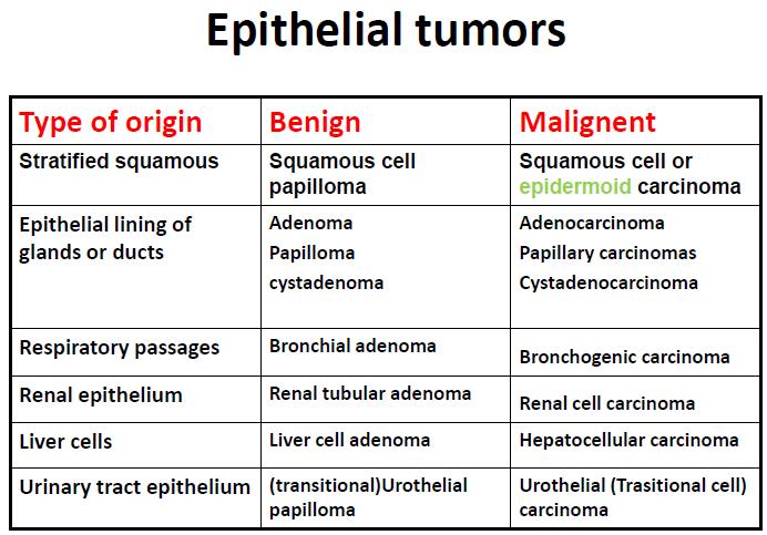
 Epithelial tumors Dr. F.F. Khuzin, PhD Dr. M.O. Mavlikeev Epithelial tumors Tumors from the epithelium are the most frequent among tumors. There are 2 group features of these tumors: The presence in most
Note: The cause of testicular neoplasms remains unknown
 - In the 15- to 34-year-old age group, they are the most common tumors of men. - Tumors of the testis are a heterogeneous group of neoplasms that include: I. Germ cell tumors : 95%; all are malignant.
TUMOR,NEOPLASM. Pathology Department, Zhejiang University School of Medicine,
 TUMOR,NEOPLASM Pathology Department, Zhejiang University School of Medicine, 马丽琴,maliqin198@zju.edu.cn The points in this chapter What is a neoplasm (conception) Morphology of neoplasm Macroscopy of Neoplasm
Health Reference Series. Seventh Edition. Cancer
 Health Reference Series Seventh Edition Cancer SOURCEBOOK Basic Consumer Health Information about Major Forms and Stages of Cancer, Featuring Facts about Head and Neck Cancers, Lung Cancers, Gastrointestinal
number Done by Corrected by Doctor Maha Shomaf
 number 16 Done by Waseem Abo-Obeida Corrected by Zeina Assaf Doctor Maha Shomaf MALIGNANT NEOPLASMS The four fundamental features by which benign and malignant tumors can be distinguished are: 1- differentiation
NEOPLASIA. 3. Which of the following tumour is benign a. Chondrosarcoma b. Osteochondroma c. Chondroblastoma d. Ewing s tumour e.
 NEOPLASIA 1. malignant neoplasms a. are independent of hormonal influence b. are always composed of homogenous cell lines c. arise from differentiated cells by a process of anaplasia d. display abnormal
Chapter 9, Part 1: Biology of Cancer and Tumor Spread
 PATHOPHYSIOLOGY Name Chapter 9, Part 1: Biology of Cancer and Tumor Spread I. Cancer Characteristics and Terminology Neoplasm new growth, involves the overgrowth of tissue to form a neoplastic mass (tumor).
number Done by Corrected by Doctor Maha shomaf
 number 17 Done by Ahmad rawajbeh Corrected by أسامة الخضر Doctor Maha shomaf 0 P a g e In this lecture, we are going to: complete the differentiation between benign and malignant tumors. - -start to study
3 cell types in the normal ovary
 Ovarian tumors 3 cell types in the normal ovary Surface (coelomic epithelium) the origin of the great majority of ovarian tumors (neoplasms) 90% of malignant ovarian tumors Totipotent germ cells Sex cord-stromal
Learning Outcomes: The following list provides the learning objectives that will be covered in the lectures, and tutorials of each week:
 Course Code Course Title ECTS Credits MED-309 Pathology II 6 School Semester Prerequisites Medical School Spring (Semester 6) MED-304 Pathology I Type of Course Field Language of Instruction Required Medicine
Biochemistry of Carcinogenesis. Lecture # 35 Alexander N. Koval
 Biochemistry of Carcinogenesis Lecture # 35 Alexander N. Koval What is Cancer? The term "cancer" refers to a group of diseases in which cells grow and spread unrestrained throughout the body. It is difficult
Differential Diagnosis of Oral Masses. Palatal Lesions
 Differential Diagnosis of Oral Masses Palatal Lesions Palatal Masses Periapical Abscess Torus Palatinus Mucocele Lymphoid Hyperplasia Adenomatous Hyperplasia Benign Salivary Neoplasms Malignant Salivary
SESSION 1: GENERAL (BASIC) PATHOLOGY CONCEPTS Thursday, October 16, :30am - 11:30am FACULTY COPY
 PATHOLOGY CONCEPTS Thursday, October 16, :30am - 11:30am FACULTY COPY") SESSION 1: GENERAL (BASIC) PATHOLOGY CONCEPTS Thursday, October 16, 2008 9:30am - 11:30am FACULTY COPY GOAL: Describe the basic morphologic (structural) changes which occur in various pathologic conditions.
SESSION 1: GENERAL (BASIC) PATHOLOGY CONCEPTS Thursday, October 16, 2008 9:30am - 11:30am FACULTY COPY GOAL: Describe the basic morphologic (structural) changes which occur in various pathologic conditions.
number Done by Corrected by Doctor Maha Shomaf
 number 19 Done by Waseem Abo-Obeida Corrected by Abdullah Zreiqat Doctor Maha Shomaf Carcinogenesis: the molecular basis of cancer. Non-lethal genetic damage lies at the heart of carcinogenesis and leads
Test Bank for Robbins and Cotran Pathologic Basis of Disease 9th Edition by Kumar
 Link full download: http://testbankair.com/download/test-bank-for-robbins-cotran-pathologic-basis-of-disease-9th-edition-bykumar-abbas-and-aster Test Bank for Robbins and Cotran Pathologic Basis of Disease
Chapter 3. Neoplasms. Copyright 2015 Cengage Learning.
 Chapter 3 Neoplasms Terminology Related to Neoplasms and Tumors Neoplasm New growth Tumor Swelling or neoplasm Leukemia Malignant disease of bone marrow Hematoma Bruise or contusion Classification of Neoplasms
DISORDERS OF THE SALIVARY GLANDS Neoplasms Dr.M.Baskaran Selvapathy S IV
 DISORDERS OF THE SALIVARY GLANDS Neoplasms Dr.M.Baskaran Selvapathy S IV NEOPLASMS A) Epithelial I. Benign Pleomorphic adenoma( Mixed tumour) Adenolymphoma (Warthin s tumour) Oxyphil adenoma (Oncocytoma)
Disorders of Cell Growth & Neoplasia. Histopathology Lab
 Disorders of Cell Growth & Neoplasia Histopathology Lab Paul Hanna April 2010 Case #84 Clinical History: 5 yr-old, West Highland White terrier. skin mass from axillary region. has been present for the
BY Mrs. K.SHAILAJA., M. PHARM., LECTURER DEPT OF PHARMACY PRACTICE, SRM COLLEGE OF PHARMACY
 BY Mrs. K.SHAILAJA., M. PHARM., LECTURER DEPT OF PHARMACY PRACTICE, SRM COLLEGE OF PHARMACY Cancer is a group of more than 100 different diseases that are characterized by uncontrolled cellular growth,
Spontaneous Neoplasms and Survival in Wistar Han Rats: Compilation of Control Group Data. March, 2003
 Spontaneous Neoplasms and Survival in Wistar Han Rats: Compilation of Control Group Data March, 2003 Information Prepared by Mary L.A. Giknis Ph.D Charles B. Clifford D.V.M, Ph.D TABLE OF CONTENTS INTRODUCTION...1
Test Bank for Robbins and Cotran Pathologic Basis of Disease 9th Edition by Kumar
 Link full download:https://getbooksolutions.com/download/test-bank-for-robbinsand-cotran-pathologic-basis-of-disease-9th-edition-by-kumar Test Bank for Robbins and Cotran Pathologic Basis of Disease 9th
4Ps LUMPS AND BUMPS B.L.&T. BUMPS, LUMPS, AND TATTOOS. Most Common BUMP in the oral cavity Fibroma INTERDENTAL PAPILLAE LESIONS
 B.L.&T. BUMPS, LUMPS, AND TATTOOS LUMPS AND BUMPS DIFFERENTIAL DIAGNOSIS FOR LUMPS AND BUMPS Traumatic Fibroma Papilloma Epulis Fissuratum Inflammatory Papillary Hyperplasia Lesions of Attached Gingiva
Part I. An Introduction to Cancer
 Part I An Introduction to Cancer 2 Chapter 1 Cancer: Descriptive Overview Cancer is a disease in which cells propagate uncontrollably. These cells can come from many different parts of the body and the
Tinh hoàn
 Tinh hoàn Tinh hoàn Tinh hoàn Tiền liệt tuyến Tiền liệt tuyến Mào tinh hoàn Mào tinh hoàn Túi tinh Túi tinh Túi tinh Túi tinh So-called cystadenoma of seminal vesicle. Gross appearance of granulomatous
BIT 120. Copy of Cancer/HIV Lecture
 BIT 120 Copy of Cancer/HIV Lecture Cancer DEFINITION Any abnormal growth of cells that has malignant potential i.e.. Leukemia Uncontrolled mitosis in WBC Genetic disease caused by an accumulation of mutations
Diagnostic Cytology of Cancer Cases
 Diagnostic Cytology of Cancer Cases Somporn Techangamsuwan Companion Animal Cancer Research Unit (CAC-RU) Department of Pathology, Faculty of Veterinary Science, Chulalongkorn University 1 Tumor or Non-tumor
Carcinogens and Carcinogenesis
 Special topics in tumor biochemistry Carcinogens and Carcinogenesis Speaker: Prof. Jiunn-Jye Chuu E-Mail: jjchuu@mail.stust.edu.tw What Is Cancer? Cancer is the unregulated multiplication of specific cells
EQA circulation 35 educational cases. Dr. A Graham Aberdeen Royal Infirmary
 EQA circulation 35 educational cases Dr. A Graham Aberdeen Royal Infirmary Case E1 Female 52 Polypoid mass right side of cervix, adjacent to os 70 Biphasic lesion 4 No answer 3 Prolapsed tube 2 Endometriosis
Mousa. Israa Ayed. Abdullah AlZibdeh. 0 P a g e
 1 Mousa Israa Ayed Abdullah AlZibdeh 0 P a g e Breast pathology The basic histological units of the breast are called lobules, which are composed of glandular epithelial cells (luminal cells) resting on
7 Mousa. Obada Zalat. Mohammad Badi
 7 Mousa Obada Zalat Mohammad Badi Tumors of the ovaries Last lecture we talked about surface epithelial tumors of the ovaries (the most common type). But there are many other types of tumors of germ cell
Definitions, Terminology, and Morphology
 Neoplasia I Definitions, Terminology, and Morphology Patrice Spitalnik, MD pfs2101@columbia.edu Cancer - second leading cause of deaths in the US after CV disease 1 Nomenclature Neoplasia new growth Neoplasms
TUMOR,NEOPLASM. Pathology Department, Zhejiang University School of Medicine,
 TUMOR,NEOPLASM Pathology Department, Zhejiang University School of Medicine, 马丽琴,maliqin198@zju.edu.cn Precancerous lesions, dysplasia, IN and carcinoma in situ 1. precancerous lesions: certain clinical
CONTINUING EDUCATION IN TOXICOLOGIC PATHOLOGY REPRODUCTIVE SYSTEM
 CONTINUING EDUCATION IN TOXICOLOGIC PATHOLOGY REPRODUCTIVE SYSTEM ORGANIZED BY SOCIETY FOR TOXICOLOGIC PATHOLOGY IN INDIA (STPI) OCTOBER 29-31, 2010 The Atria Hotel, # 1, Palace Road, Bangalore - 560 001
Spontaneous Neoplastic Lesions in the Crl:CD-1 (ICR)BR Mouse. March, 2000
BR Mouse. March, 2000") Spontaneous Neoplastic Lesions in the Crl:CD-1 (ICR)BR Mouse March, 2000 Information Prepared by Mary L. A. Giknis, Ph.D. Charles B. Clifford, D.V.M., Ph.D. CHARLES RIVER LABORATORIES TABLE OF CONTENTS
Spontaneous Neoplastic Lesions in the Crl:CD-1 (ICR)BR Mouse March, 2000 Information Prepared by Mary L. A. Giknis, Ph.D. Charles B. Clifford, D.V.M., Ph.D. CHARLES RIVER LABORATORIES TABLE OF CONTENTS
Table of Contents. Preface xi. Acknowledgments xiii. Part I Overview of the Diagnostic Process 1. 1 Overview of Grading and Staging 3
 Table of Contents Preface xi Acknowledgments xiii Part I Overview of the Diagnostic Process 1 1 Overview of Grading and Staging 3 Identification of the process 3 Identification of tumor types 5 Grading
number Done by Corrected by Doctor مها شوماف
 number 15 Done by Ali Yaghi Corrected by Waseem Alhaj Doctor مها شوماف 1 P a g e Epidemiology Epidemiology is the study of the incidence of a disease. It can give us information about the possible causes
XX. Tumours of the nasal cavity *
 XX. Tumours of the nasal cavity * H. STONZI 1 & B. HAUSER2 Tumours of the nasal cavity are rare in domestic animals, most cases occurring in the dog. Epithelial tumours are the most common type in carnivores
Cancer and Oncogenes Bioscience in the 21 st Century. Linda Lowe-Krentz
 Cancer and Oncogenes Bioscience in the 21 st Century Linda Lowe-Krentz December 1, 2010 Just a Few Numbers Becoming Cancer Genetic Defects Drugs Our friends and family 25 More mutations as 20 you get older
Select problems in cystic pancreatic lesions
 Disclosure Select problems in cystic pancreatic lesions Five Prime Therapeutics shareholder Adicet Bio shareholder Bristol-Meyer Squibb advisory board grace.kim@ucsf.edu Pancreatic cystic lesions Intraductal
CANCER Uncontrolled Cell Division
 CANCER Uncontrolled Cell Division What is cancer? Why does it occur? Where does it occur? Benign vs. Malignant? Types of Cancer (3 main groups) There are over 200 different types of cancer 1) Carcinomas
Serous borderline tumor icd 10
 Note. May be used as an additional code to identify functional activity associated with a carcinoid tumor. Free, official information about 2012 (and also 2013-2015) ICD -9-CM diagnosis code 220, including
3 cell types in the normal ovary
 Ovarian tumors 3 cell types in the normal ovary Surface (coelomic epithelium) the origin of the great majority of ovarian tumors 90% of malignant ovarian tumors Totipotent germ cells Sex cord-stromal cells
Chapter 10-3 Regulating the Cell Cycle
 Chapter 10-3 Regulating the Cell Cycle Vocabulary: Cyclin Cancer Key Concepts: How is the cell cycle regulated? How are cancer cells different from other cells? I. Introduction A. An Interesting Fact About
LYMPHATIC DRAINAGE AXILLARY (MOSTLY) INTERNAL MAMMARY SUPRACLAVICULAR
 INTERNAL MAMMARY SUPRACLAVICULAR") BREAST LYMPHATIC DRAINAGE AXILLARY (MOSTLY) INTERNAL MAMMARY SUPRACLAVICULAR HISTOLOGY LOBE: (10 in whole breast) LOBULE: (many per lobe) ACINUS/I, aka ALVEOLUS/I: (many per lobule) DUCT(S): INTRA- or
BREAST LYMPHATIC DRAINAGE AXILLARY (MOSTLY) INTERNAL MAMMARY SUPRACLAVICULAR HISTOLOGY LOBE: (10 in whole breast) LOBULE: (many per lobe) ACINUS/I, aka ALVEOLUS/I: (many per lobule) DUCT(S): INTRA- or
Gynaecological Malignancies
 Gynaecological Malignancies Dr Rodney Itaki Lecturer Anatomical Pathology Discipline University of Papua New Guinea Division of Pathology School of Medicine & Health Sciences Overview Genital tract tumors
Gastrointestinal pathology 2018 lecture 4. Dr Heyam Awad FRCPath
 Gastrointestinal pathology 2018 lecture 4 Dr Heyam Awad FRCPath Topics to be covered Peptic ulcer disease Hiatal hernia Gastric neoplasms Peptic ulcer disease (PUD)= chronic gastric ulcer Causes H pylori
PLEOMORPHIC ADENOMA OF LATERAL WALL OF NOSE A RARE PRESENTATION
 ISSN: 2250-0359 Volume 4 Issue 1 2014 PLEOMORPHIC ADENOMA OF LATERAL WALL OF NOSE A RARE PRESENTATION *USHA KUMAR MAHESH *RATNAKAR MADHAVARAO POTEKAR * B.L.D.E UNIVERSITY ABSTRACT: The aim of the article
Spontaneous Neoplastic Lesions in the CrI:CD-1(ICR) Mouse in Control Groups from 18 Month to 2 year Studies. March, 2005
 Mouse in Control Groups from 18 Month to 2 year Studies. March, 2005") Spontaneous Neoplastic Lesions in the CrI:CD-1(ICR) Mouse in Control Groups from 18 Month to 2 year Studies March, 2005 Information Prepared by Mary L.A. Giknis Ph.D Charles B. Clifford D.V.M, Ph.D 1063
Spontaneous Neoplastic Lesions in the CrI:CD-1(ICR) Mouse in Control Groups from 18 Month to 2 year Studies March, 2005 Information Prepared by Mary L.A. Giknis Ph.D Charles B. Clifford D.V.M, Ph.D 1063
DISORDERS OF THE BREAST Dated. FIBROADENOSIS Other common names: mastitis, fibrocystic disease, cystic mammary dysplasia.
 DISORDERS OF THE BREAST Dated BENIGN BREAST DISORDERS (Essential Surg 2 nd Ed, pp 540) FIBROADENOSIS Other common names: mastitis, fibrocystic disease, cystic mammary dysplasia. Fibroadenosis is the distortion
Neoplasias Quisticas del Páncreas
 SEAP -Aproximación Práctica a la Patología Gastrointestinal- Madrid, 26 de mayo, 2006 Neoplasias Quisticas del Páncreas Gregory Y. Lauwers, M.D. Director, Service Massachusetts General Hospital Harvard
Aberrant cell Growth. Younas Masih New Life College of Nursing Karachi. 3/4/2016 Younas Masih ( NLCON)
") Aberrant cell Growth Younas Masih New Life College of Nursing Karachi 1 Objectives By the end of this session the learners will be able to, Define the characteristics of the normal cell Describe the characteristics
Aberrant cell Growth Younas Masih New Life College of Nursing Karachi 1 Objectives By the end of this session the learners will be able to, Define the characteristics of the normal cell Describe the characteristics
Contents. Basic Ultrasound Principles and Terminology. Ultrasound Nodule Characteristics
 Contents Basic Ultrasound Principles and Terminology Basic Ultrasound Principles... 1 Ultrasound System... 2 Linear Transducer for Superficial Images and Ultrasound-Guided FNA... 3 Scanning Planes... 4
Institute of Pathology First Faculty of Medicine Charles University. Ovary
 Ovary Barrett esophagus ph in vagina between 3.8 and 4.5 ph of stomach varies from 1-2 (hydrochloric acid) up to 4-5 BE probably results from upward migration of columnar cells from gastroesophageal junction
CNS TUMORS. D r. Ali Eltayb ( U. of Omdurman. I ). M. Path (U. of Alexandria)
. M. Path (U. of Alexandria)") CNS TUMORS D r. Ali Eltayb ( U. of Omdurman. I ). M. Path (U. of Alexandria) CNS TUMORS The annual incidence of intracranial tumors of the CNS ISmore than intraspinal tumors May be Primary or Secondary
CNS TUMORS D r. Ali Eltayb ( U. of Omdurman. I ). M. Path (U. of Alexandria) CNS TUMORS The annual incidence of intracranial tumors of the CNS ISmore than intraspinal tumors May be Primary or Secondary
Methoden / Methods inc. ICCC-3 105
 Methoden / Methods inc. ICCC-3 105 Internationale Klassifikation der Krebserkrankungen bei Kindern (ICCC-3) Zuordnung von ICD-O-3-Codes für Morphologie und Topographie zu diagnostischen Kategorien International
Radiotherapy in small animals. How is it realized? How does it work? For which patient is it indicated?
 Radiotherapy in small animals How is it realized? How does it work? For which patient is it indicated? Radiotherapy Our linear accelerator can generate photons or electrons. The electron beam is used to
Appendix 4: WHO Classification of Tumours of the pancreas 17
 S3.01 The WHO histological tumour type must be recorded. CS3.01a The histological type of the tumour should be recorded based on the current WHO classification 17 (refer to Appendices 4-7). Appendix 4:
Cancer Fundamentals. Julie Randolph-Habecker, Ph.D. Director, Experimental Histopathology Shared Resource
 Cancer Fundamentals Julie Randolph-Habecker, Ph.D. Director, Experimental Histopathology Shared Resource Cancer Overview Leading cause of death in US 1.2 million diagnosed each year More common after age
Pelvic tumor in childhood Classification, imaging approach and radiological findings
 Pelvic tumor in childhood Classification, imaging approach and radiological findings M. Mearadji International Foundation for Pediatric Imaging Aid Rotterdam, The Netherlands Solid pelvic masses in childhood
B. Environmental Factors. a. The major risk factor to papillary thyroid cancer is exposure to ionizing radiation, during the first 2 decades of life.
 B. Environmental Factors. a. The major risk factor to papillary thyroid cancer is exposure to ionizing radiation, during the first 2 decades of life. b. Deficiency of dietary iodine: - Is linked with a
Oncology 101. Cancer Basics
 Oncology 101 Cancer Basics What Will You Learn? What is Cancer and How Does It Develop? Cancer Diagnosis and Staging Cancer Treatment What is Cancer? Cancer is a group of more than 100 different diseases
Radiology Pathology Conference
 Radiology Pathology Conference Nadia F. Yusaf, M.D. PGY-3 1/29/2010 Presentation material is for education purposes only. All rights reserved. 2010 URMC Radiology Page 1 of 90 Case 1 60 year- old man presents
The Vocabulary of Cancer - By Patricia Long November 20, 2002
 The Vocabulary of Cancer - By Patricia Long November 20, 2002 Websites to bookmark: www.cancer.gov www.cancer.org www.vetcancersociety.org www.oncolink.com/templates/types/section.cfm?c=22&s=69 Like any
Diseases of the vulva
 Diseases of the vulva 1. Bartholin Cyst - Infection of the Bartholin gland produces an acute inflammation within the gland (adenitis) and may result in an abscess. Bartholin duct cysts - Are relatively
Neoplasms. Nomenclature. Cellular atypia/anaplasia. Benign (locally malignant or rarely metastasizing) Benign v/s malignant neoplasm.
 Benign v/s malignant neoplasm.") Neoplasms-3 1 - Leiomyoma 2 - Lipoma 3-4 - Chondrosarcoma Non-epithelial tumors Neoplasms Benign (locally malignant or rarely metastasizing) borderline/intermediate malignant 5 - Meningioma 6 Glioblastoma
Neoplasms-3 1 - Leiomyoma 2 - Lipoma 3-4 - Chondrosarcoma Non-epithelial tumors Neoplasms Benign (locally malignant or rarely metastasizing) borderline/intermediate malignant 5 - Meningioma 6 Glioblastoma
Mousa. Najat kayed &Renad Al-Awamleh. Nizar Alkhlaifat
 6 Mousa Najat kayed &Renad Al-Awamleh Nizar Alkhlaifat P a g e 1 This sheet written based on record 13 on website Cover slide( 95-117 ) No need to go back to slide FALLOPIAN TUBE PATHOLOGY In general fallopian
Clinical indications for positron emission tomography
 Clinical indications for positron emission tomography Oncology applications Brain and spinal cord Parotid Suspected tumour recurrence when anatomical imaging is difficult or equivocal and management will
Summary of Inhalation Carcinogenicity Study. of Isopropyl Acetate. in F344 Rats
 Summary of Inhalation Carcinogenicity Study of Isopropyl Acetate in F344 Rats March 2009 Japan Bioassay Research Center Japan Industrial Safety and Health Association PREFACE The tests were contracted
- is a common disease - 1 person in 3 can expect to contract cancer at some stage in their life -1 person in 5 can expect to die from it
 MBB157 Dr D Mangnall The Molecular Basis of Disease CANCER Lecture 1 One of the simpler (and better) definitions of cancer comes from the American Cancer Society, who define cancer as; 'Cancer is a group
Malignant Cardiac Tumors Rad-Path Correlation
 Malignant Cardiac Tumors Rad-Path Correlation Vincent B. Ho, M.D., M.B.A. 1 Jean Jeudy, M.D. 2 Aletta Ann Frazier, M.D. 2 1 Uniformed Services University of the Health Sciences 2 University of Maryland
A factor which brings about a mutation is called a mutagen. Any agent that causes cancer is called a carcinogen and is described as carcinogenic.
 Cancer Cancer is one of the most common diseases in the developed world: 1 in 4 deaths are due to cancer 1 in 17 deaths are due to lung cancer Lung cancer is the most common cancer in men Breast cancer
Gross appearance of nodular hyperplasia in material obtained from suprapubic prostatectomy. Note the multinodular appearance and the admixture of
 Tiền liệt tuyến Tiền liệt tuyến Gross appearance of nodular hyperplasia in material obtained from suprapubic prostatectomy. Note the multinodular appearance and the admixture of solid and microcystic areas.
CELL AND TISSUE INJURY COURSE-II PATHOLOGY LABORATORY. PATHOLOGY of MASS LESIONS and TISSUE DEFECTS -MACROSCOPY Assoc. Professor Rengin Ahıskalı
 CELL AND TISSUE INJURY COURSE-II PATHOLOGY LABORATORY PATHOLOGY of MASS LESIONS and TISSUE DEFECTS -MACROSCOPY Assoc. Professor Rengin Ahıskalı M1 - RENAL TUBERCULOSIS cavitary areas caseous necrosis fibrous
Department of Radiology, University of Szeged. Imaging of the skeleton
 Imaging of the skeleton Methods of examination: plain x-ray (radiography, densitometry) x-ray with contrast material (fistulography, angiography) ultrasound (b-mode, Doppler, color, duplex) computed tomography
Other Sites. Table 2 Continued. MPH Rules 11/8/07. NAACCR Webinar Series 1
 MPH s 11/8/07 Other s 1 Table 2 Continued Use this two-page table to select combination histology codes. Compare the terms in the diagnosis to the terms in Columns 1 and 2. If the terms match, code the
2 to 3% of All New Visceral Cancers Peak Incidence is 6th Decade M:F = 2:1 Grossly is a Bright Yellow, Necrotic Mass with a Pseudocapsule
 GENITOURINARY PATHOLOGY Kathleen M. O Toole, M.D. Renal Cell Carcinoma 2 to 3% of All New Visceral Cancers Peak Incidence is 6th Decade M:F = 2:1 Grossly is a Bright Yellow Necrotic Mass Grossly is a Bright
CHAPTER. Host reactions to Biomaterials and Their Evaluation
 CHAPTER 2 Host reactions to Biomaterials and Their Evaluation IMMUNOLOGIC TOXICITY OF MEDICAL DEVICES Immunologic toxicity to surface medical devices and external communicating devices (dialyzers, laparoscopes,
Culture Advantage. Anatomy and Medical Terminology for Interpreters ONCOLOGY TERMINOLOGY
 1 Culture Advantage Anatomy and Medical Terminology for Interpreters ONCOLOGY TERMINOLOGY Marlene V. Obermeyer, MA, RN marlene@culture-advantage.com 2 Oncology Case Study for the PowerPoint Presentation
doi: /j.anl
 doi: 10.1016/j.anl.2006.07.001 Synchronous unilateral parotid gland neoplasms of three different histological types Shuho Tanaka 1, Keiji Tabuchi 1, Keiko Oikawa 1, Rika Kohanawa 1, Hideki Okubo 1, Dai
Mast Cell Tumors in Dogs
 Mast Cell Tumors in Dogs 803-808-7387 www.gracepets.com These notes are provided to help you understand the diagnosis or possible diagnosis of cancer in your pet. For general information on cancer in pets