Disclosures. The Thin Red Line Between Neuropathology and Head & Neck Pathology. Introduction CASE 1. Current Issues Tihan
|
|
|
- Agatha Powell
- 5 years ago
- Views:
Transcription

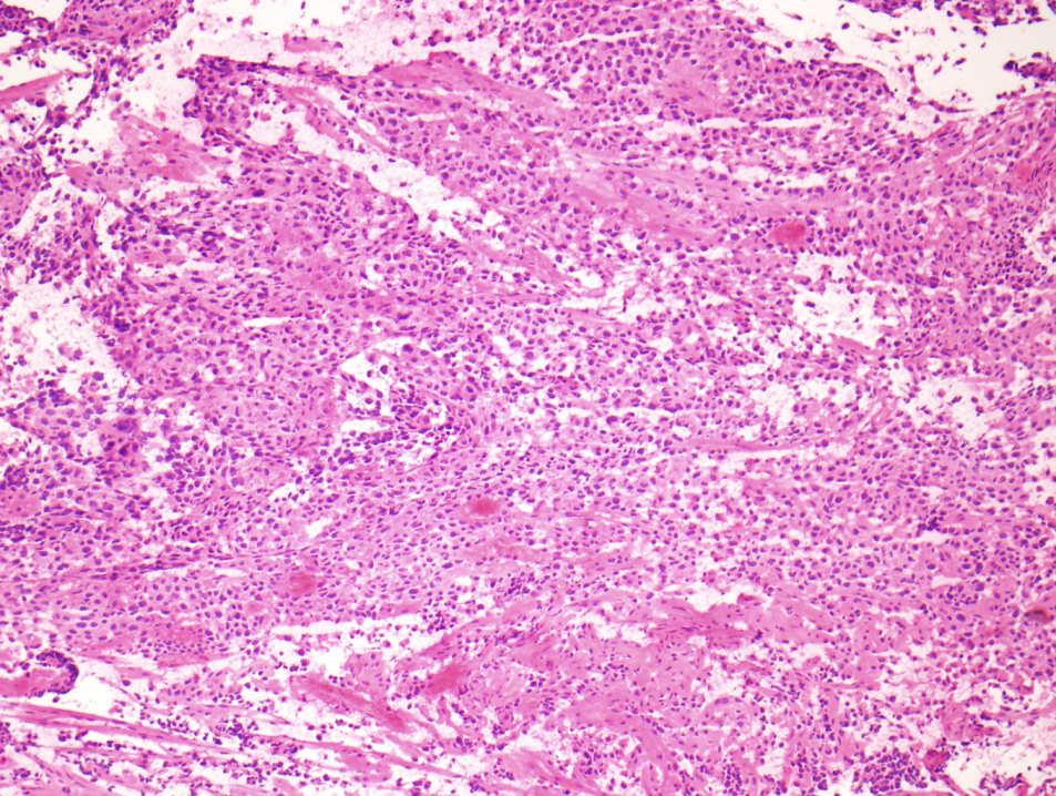
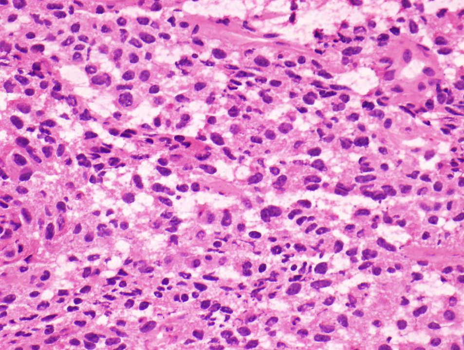
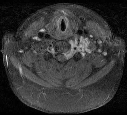
1 Disclosures I have nothing to disclose The Thin Red Line Between Neuropathology and Head & Neck Pathology Tarik Tihan, MD, PhD UCSF, Department of Pathology Neuropathology Division Introduction Three cases that straddle the boundary between Neuropathology and Head & Neck Pathology Importance of recognizing different perspectives that are often complementary in reaching the correct diagnosis The importance of thinking out of the box of a specific subspecialty Recognition of the differences in the literature from different subspecialties, and the need to reconcile these differences in real life CASE 1 Dear Doctor I had the pleasure of evaluating this patient, a very pleasant 73-year-old male who has a history of nasal congestion for years. In November 2007, he developed some epistaxis for which he went to the emergency room and a workup revealed a suggestion of sinusitis on CT scan. He was referred to Dr. from Otolaryngology who found an intranasal mass and performed a biopsy on February The biopsy was consistent with esthesioneuroblastoma. He was referred to UCSF for surgical resection with a plan for postoperative radiation therapy. Past medical history includes diabetes and abnormal electrocardiogram. 1


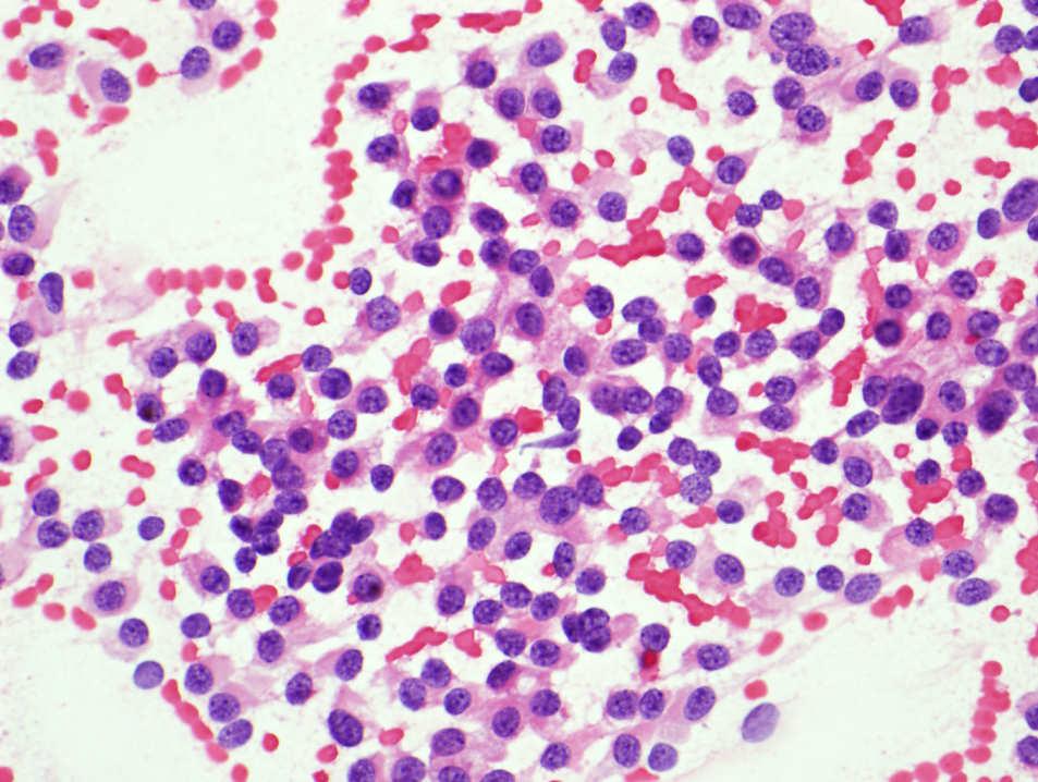
2 AXIAL T1-gad AXIAL T1-gad Smear Frozen 2
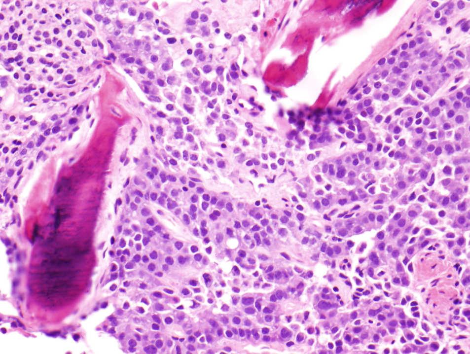
3 Frozen 3


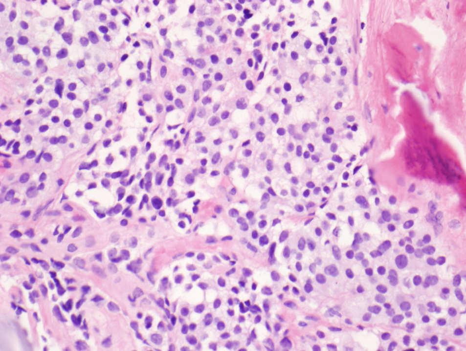
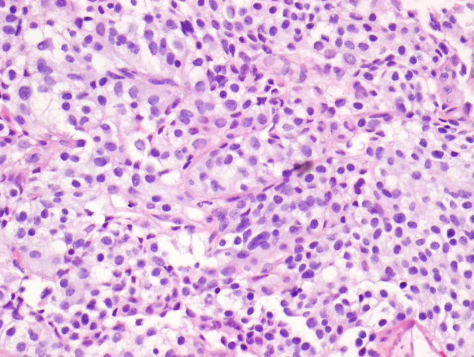
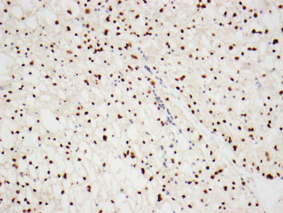
4 Synaptophysin Chromogranin MIB-1 BUT WAIT!!! ISN T THERE ANYTHING UNUSUAL HERE? 4


5 Cytokeratin ACTH Answer Case 1= Pituitary Adenoma Clinical: Typical visual field defect and endocrinological symptoms are helpful if present. Often a long-standing clinical history Radiological: Involvement of the sella turcica and sphenoid prior to nasal or ethmoid involvement Histological: Ample, sometimes clear cytoplasm, rare mitoses. Otherwise similar to carcinoid tumors Immunohistochemistry: CHR, SYN, Pituitary Transcription Factors or Hormones FEATURE Lobular pattern Uniform nuclei Mitotic Figures Pituitary Adenoma Olfactory Neuroblastoma Low Grade Olfactory Neuroblastoma High Grade Sinonasal Undifferentiated Carcinoma Common Common Focal or Rare Rare Typical Typical Focal or Absent Absent Rare Rare Frequent Frequent Necrosis Absent Absent Rare Frequent Rosettes Absent Present Rare/Absent Absent 5
6 FEATURE Cytokeratins Pituitary Adenoma Mostly Positive Olfactory Neuroblastoma Low Grade Olfactory Neuroblastoma High Grade Sinonasal Undifferentiated Carcinoma Negative Negative Positive S100 protein Negative Positive Positive/focal Negative/Rare NSE Positive Positive Positive Positive (50%) PIT1/SF- 1/TPIT Or Pit Hormones Positive Negative Negative Negative Follow-up 7 years later Dear Doctor I am delighted to report that the MRI showed no evidence whatsoever of a recurrent pituitary tumor. This is excellent news! I would recommend that you repeat the MRI again in two years. You could work with at to make the arrangements for the follow-up MRI and the appointment. Synaptophysin Positive Positive Positive/Focal Negative Chromogranin Often positive Often positive Occasionally positive Rare positive cells CASE 2 A 21 year old man presented with dysphagia and a change in his voice. He has also lost 15 lb over the last few months. An MRI revealed a cervical mass. He underwent a biopsy of the lesion, followed by a radical resection. The tumor appeared to have encased the vertebral artery and involved the neural foramen and partially compressed the cervical spinal cord. 6

7 SAGITTAL T2 AXIAL T1-gad 7
8 8


9 AE1-AE3 CAM5.2 EMA Brachyury 9


10 Brachyury Answer Case 2 = Chordoma Most common location sacrum, followed by skull base/clivus Midline with contrast enhancement Epithelial differentiation, typically EMA positive, and also cytokeratin positive S100 protein often strongly positive along with Vimentin Brachyury is the marker of choice for the diagnosis of Chordomas FEATURES CHORDOMA CHONDROSARCOMA Localization Midline Clivus Lateralized, Temporal bone Physalliphorous cells YES NO Cytokeratin Positive Negative S100 protein Positive Positive EMA Positive Negative Brachyury Positive Negative IDH1 or IDH2 mutations Absent Present 10


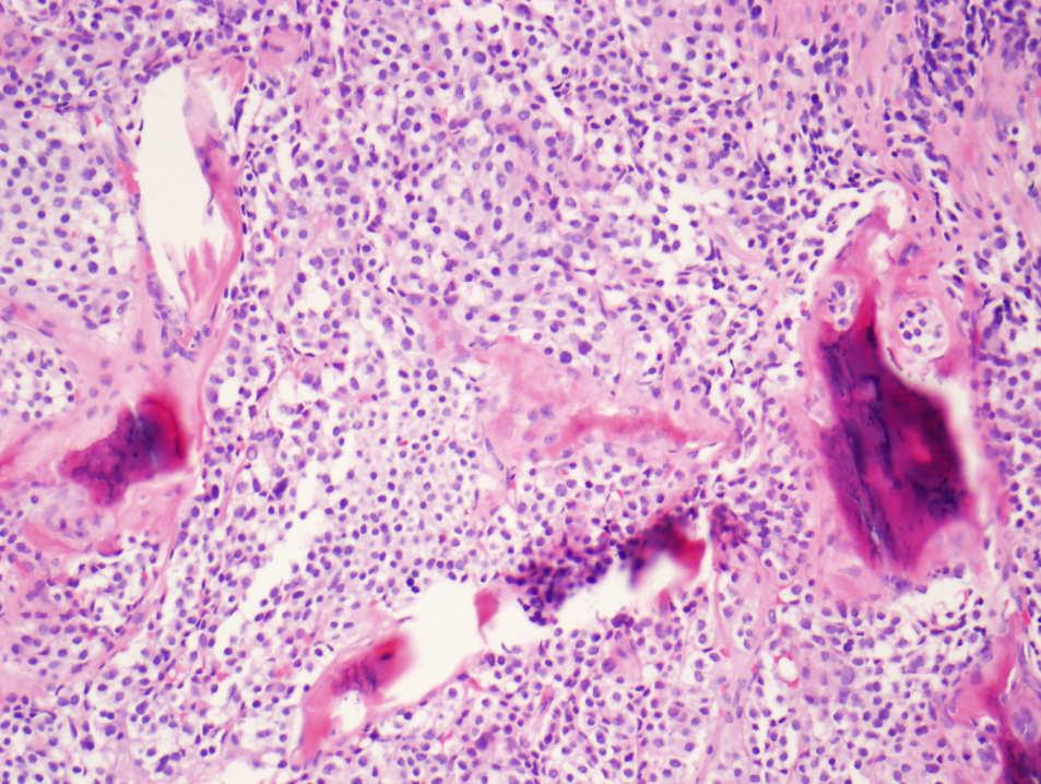
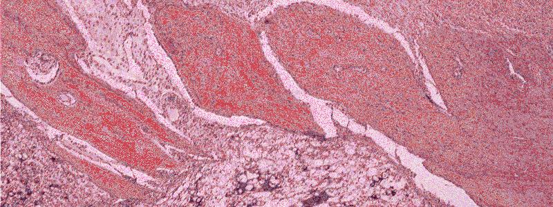
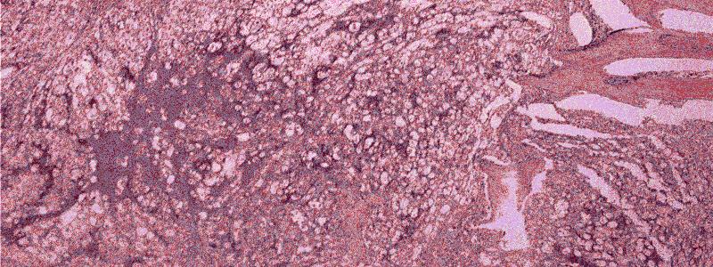
11 CASE 3 A 43-year-old man presented with significant weight loss, postural instability and difficulty in walking. He also suffered from occasional nausea and vomiting. A recent audiogram demonstrated left severe mixed hearing loss. An MRI revealed a mass that distorted the fourth ventricle with significant hydrocephalus. 11


12 Current 5/21/2015 Issues Tihan Trichrome Type IV Collagen EMA 12


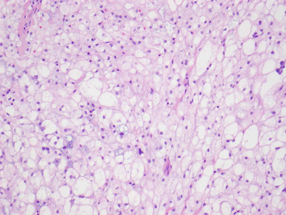
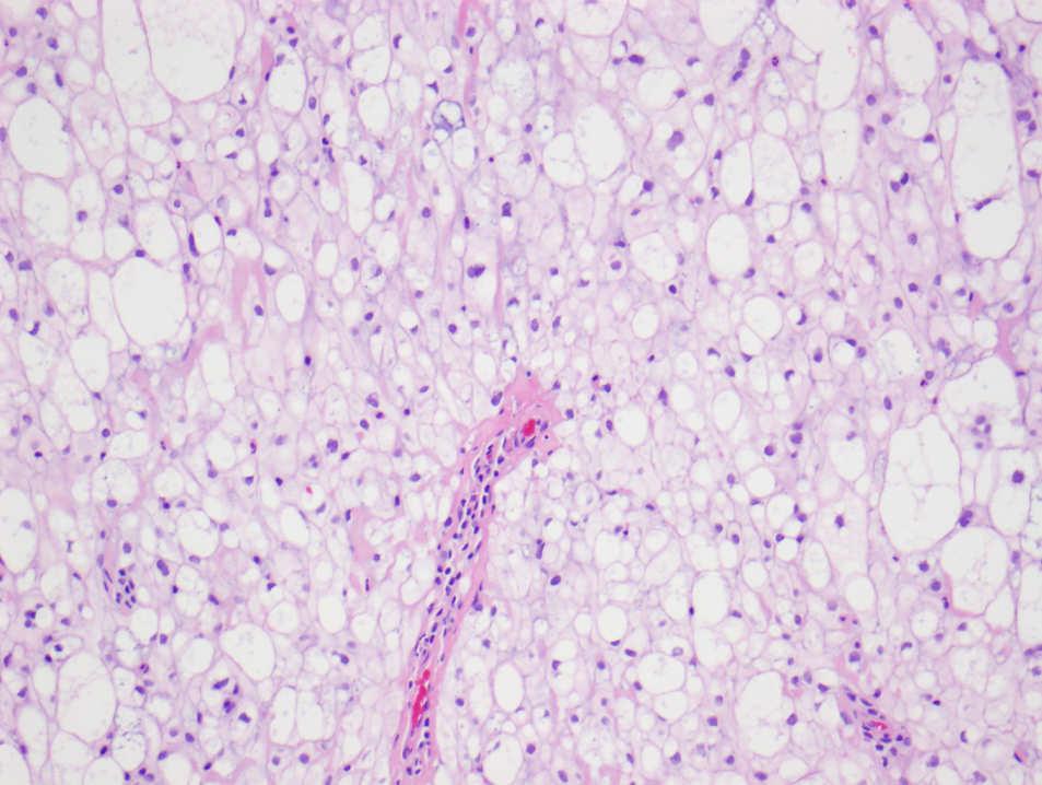
13 Current 5/21/2015 Issues Tihan CD34 BCL-2 STAT6 BUT WAIT!!! ISN T THERE ANYTHING UNUSUAL HERE? 13


")

 NAB2/STAT6 fusion YES (ex4-ex6")
14 Answer Case 2 = Solitary Fibrous Tumor Unification FEATURE Solitary Fibrous Tumor Hemangiopericytoma Collagen-rich YES NO HPC-like vasculature YES YES Reticulin Stain Focal positive & vascular pattern Strongly positive CD34 staining Diffuse Strong Focal or Negative BCL-2 staining Diffuse Strong Diffuse Strong STAT-6 staining Diffuse Strong (nuclear) Diffuse Strong (nuclear) Biphasic architecture Common Uncommon Local Recurrence Rare Common (~60%) Extracranial metastasis Exceptional Common (~30%) NAB2/STAT6 fusion YES (ex4-ex6 fusion)* YES (ex6-ex16 fusion)* 14
15 THANK YOU 15
Small (and large) Blue Cell Tumors of the Skull Base
 Blue Cell Tumors of the Skull Base") Small (and large) Blue Cell Tumors of the Skull Base Jennifer L. Hunt, MD, MEd Aubrey J. Hough Jr, MD, Endowed Professor of Pathology Chair of Pathology and Laboratory Medicine University of Arkansas for
Small (and large) Blue Cell Tumors of the Skull Base Jennifer L. Hunt, MD, MEd Aubrey J. Hough Jr, MD, Endowed Professor of Pathology Chair of Pathology and Laboratory Medicine University of Arkansas for
Case 2. Dr. Sathima Natarajan M.D. Kaiser Permanente Medical Center Sunset
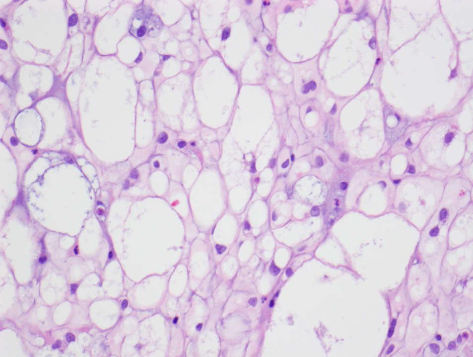
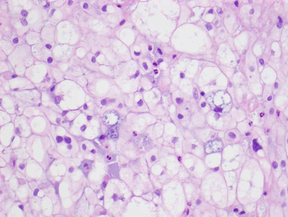
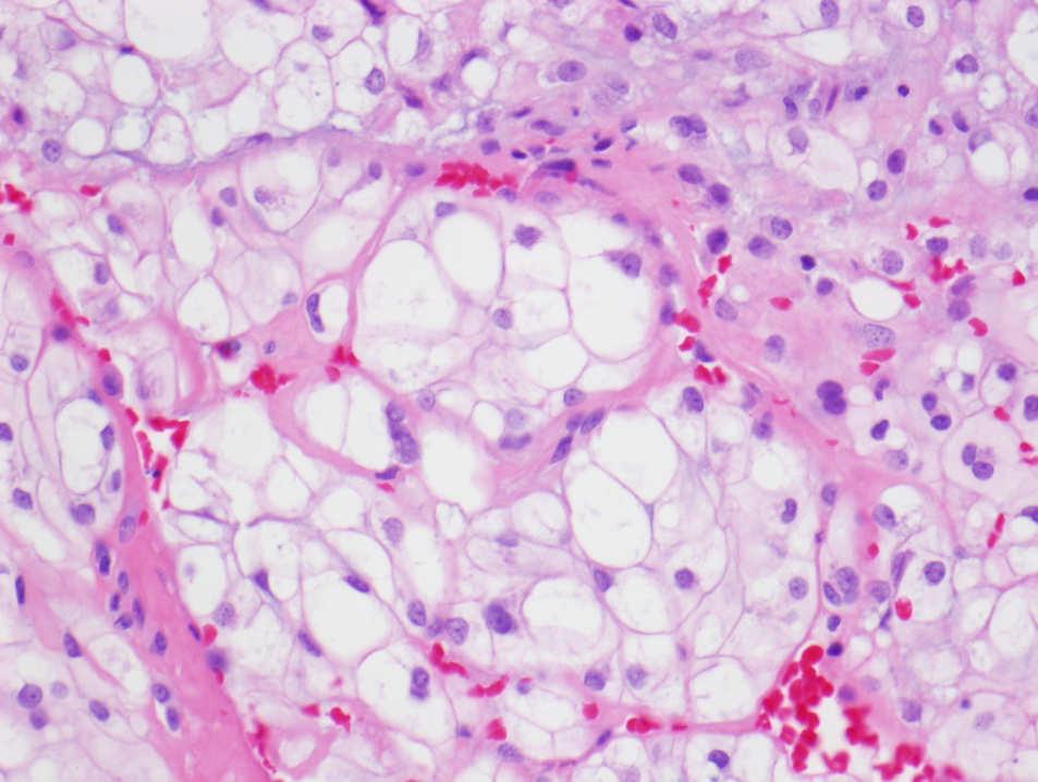
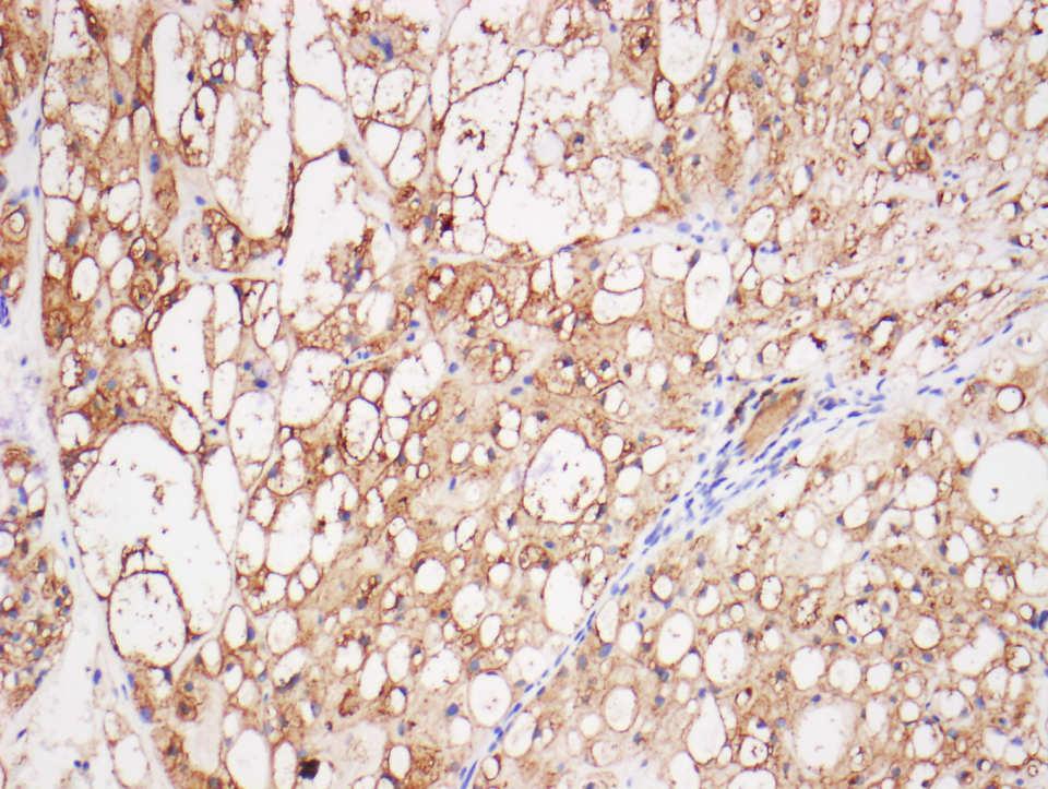
 Case 2 Dr. Sathima Natarajan M.D. Kaiser Permanente Medical Center Sunset History 24 year old male presented with a 3 day history of right flank pain, sharp in nature Denies fever, chills, hematuria or
Five Most Common Problems in Surgical Neuropathology
 Five Most Common Problems in Surgical Neuropathology If the brain were so simple that we could understand it, we would be so simple that we couldn t Emerson Pugh What is your greatest difficulty in neuropathology?
Solitary Fibrous Tumor of the Kidney with Massive Retroperitoneal Recurrence. A Case Presentation
 246) Prague Medical Report / Vol. 113 (2012) No. 3, p. 246 250 Solitary Fibrous Tumor of the Kidney with Massive Retroperitoneal Recurrence. A Case Presentation Sfoungaristos S., Papatheodorou M., Kavouras
Problem 1: Differential of Neuroendocrine Carcinoma 3/23/2017. Disclosure of Relevant Financial Relationships
 Differential of Neuroendocrine Carcinoma Alain C. Borczuk,MD Weill Cornell Medicine Disclosure of Relevant Financial Relationships USCAP requires that all faculty in a position to influence or control
Nutn but a Small Biopsy How to approach small biopsy samples of the sinonasal tract
 California Society of Pathology Saturday Slide Seminar Nutn but a Small Biopsy How to approach small biopsy samples of the sinonasal tract Lester D. R. Thompson www.lester-thompson.com Learning Objectives
3/27/2017. Disclosure of Relevant Financial Relationships
 Ophthalmic Pathology Evening Specialty Conference USCAP 2017 5 th March, 2017 Mukul K. Divatia, MD Assistant Professor Department of Pathology & Genomic Medicine Weill Cornell Medical College Houston Methodist
Introduction to Neurosurgical Subspecialties:
 Introduction to Neurosurgical Subspecialties: Tumor and Skull Base Neurosurgery Brian L. Hoh, MD 1 and Gregory J. Zipfel, MD 2 1 University of Florida, 2 Washington University Tumor / Skull Base Neurosurgery
HEAD AND NECK IMAGING. James Chen (MS IV)
") HEAD AND NECK IMAGING James Chen (MS IV) Anatomy Course Johns Hopkins School of Medicine Sept. 27, 2011 OBJECTIVES Introduce cross sectional imaging of head and neck Computed tomography (CT) Review head
HEAD AND NECK IMAGING James Chen (MS IV) Anatomy Course Johns Hopkins School of Medicine Sept. 27, 2011 OBJECTIVES Introduce cross sectional imaging of head and neck Computed tomography (CT) Review head
Case year old female presented with asymmetric enlargement of the left lobe of the thyroid
 Case 4 22 year old female presented with asymmetric enlargement of the left lobe of the thyroid gland. No information available relative to a prior fine needle aspiration biopsy. A left lobectomy was performed.
Case 4 Diagnosis 2/21/2011 TGB
 Case 4 22 year old female presented with asymmetric enlargement of the left lobe of the thyroid gland. No information available relative to a prior fine needle aspiration biopsy. A left lobectomy was performed.
Neuroendocrine Carcinoma. Lebanon Neuroendocrine Neoplasms of H&N Nov /7/2011. Broad Classification:
 H&N Neuroendocrine Neoplasms: Classification and Diagnostic Considerations Adel K. El-Naggar, M.D., Ph.D. The University of Texas MD Anderson Cancer Center, Houston, Texas Broad Classification: A. Epithelial:
Carbon Ion Radiotherapy for Skull Base and Paracervical Chordomas
 Carbon Ion Radiotherapy for Skull Base and Paracervical Chordomas Azusa Hasegawa, Jun-etsu Mizoe and Hirohiko Tsujii Research Center Hospital for Charged Particle Therapy National Institute of Radiological
The View through the Nose: ENT considerations for Pituitary/Skull Base Surgery
 The View through the Nose: ENT considerations for Pituitary/Skull Base Surgery Edsel Kim, M.D. Otolaryngology-Head and Neck Surgery The Oregon Clinic Providence Brain and Spine Institute Pituitary, Thyroid
CHONDROID CHORDOMA OF NASAL SEPTUM: A CASE REPORT Gayattre Kailas 1, Muniraju M 2, Archana Mathri 3
 CHONDROID CHORDOMA OF NASAL SEPTUM: A Gayattre Kailas 1, Muniraju M 2, Archana Mathri 3 HOW TO CITE THIS ARTICLE: Gayattre Kailas, Muniraju M, Archana Mathri. Chondroid Chordoma of Nasal Septum: A Case
Evening Specialty Conference Bone and Soft Tissue Pathology. Diagnostic pitfalls in bone and soft tissue pathology
 Evening Specialty Conference Bone and Soft Tissue Pathology. Case 1 Elizabeth G Demicco, MD, PhD Mount Sinai Hospital, New York Disclosure of Relevant Financial Relationships USCAP requires that all planners
Case Scenario 1: Thyroid
 Case Scenario 1: Thyroid History and Physical Patient is an otherwise healthy 80 year old female with the complaint of a neck mass first noticed two weeks ago. The mass has increased in size and is palpable.
Disclosures. Parathyroid Pathology. Objectives. The normal parathyroid 11/10/2012
 Disclosures Parathyroid Pathology I have nothing to disclose Annemieke van Zante MD/PhD Assistant Professor of Clinical Pathology Associate Chief of Cytopathology Objectives 1. Review the pathologic features
Case Studies in the Skull Base
 Case Studies in the Skull Base Amy C Tsai, MD Neuroradiology Fellow Department of Radiology and Imaging Sciences University of Utah Health Sciences Center Salt Lake City, Utah, USA No disclosures related
Nasal Cavity and Paranasal Sinuses
 Chapter 2 Nasal Cavity and Paranasal Sinuses Introduction Included in this chapter are nasal cavities, frontal sinus, ethmoid complex, sphenoid sinus, and maxillary sinuses. These cavities and sinuses
Case Presentation. Maha Akkawi, MD, Fatima Obeidat, MD, Tariq Aladily, MD. Department of Pathology Jordan University Hospital Amman, Jordan
 Case Presentation Maha Akkawi, MD, Fatima Obeidat, MD, Tariq Aladily, MD Department of Pathology Jordan University Hospital Amman, Jordan The 25th Annual Congress of the ADIAP The 8/11/2013 1 5th International
An Unexpected Cause of Hypoglycemia
 An Unexpected Cause of Hypoglycemia Stacey A. Milan, MD FACS Surgical Oncology Nothing to disclose Disclosures Objectives Identify indications for workup of hypoglycemia Define work up for hypoglycemic
My personal experience at University of Toronto and recent updates of
 My personal experience at University of Toronto and recent updates of Endocrine Pathology Toshitetsu Hayashi M.D. Ph.D. ¹Department of Diagnostic Pathology, Takamatsu Red Cross Hospital, Japan ²Laboratory
What s New in Adrenal Gland Pathology. Marina Scarpelli
 What s New in Adrenal Gland Pathology Marina Scarpelli Background Histological criteria for adrenocortical proliferative lesions Immunohistochemical markers Molecular markers Histological Criteria for
3/22/2017. Disclosure of Relevant Financial Relationships. Disclosure of Relevant Financial Relationships
 Ki67 in Pituitary Tumors: What Information Could We Obtain? Robert Y. Osamura, MD International University of Health and Welfare Ki67 in Pituitary Tumors: What Information Could We Obtain? Robert Y. Osamura,
Respiratory Tract Cytology
 Respiratory Tract Cytology 40 th European Congress of Cytology Liverpool, UK Momin T. Siddiqui M.D. Professor of Pathology and Laboratory Medicine Director of Cytopathology Emory University Hospital, Atlanta,
General: Brain tumors are lesions that have mass effect distorting the normal tissue and often result in increased intracranial pressure.
 1 Lecture Objectives Know the histologic features of the most common tumors of the CNS. Know the differences in behavior of the different tumor types. Be aware of the treatment modalities in the various
Skull-2. Norma Basalis Interna. Dr. Heba Kalbouneh Assistant Professor of Anatomy and Histology
 Skull-2 Norma Basalis Interna Dr. Heba Kalbouneh Assistant Professor of Anatomy and Histology Norma basalis interna Base of the skull- superior view The interior of the base of the skull is divided into
Insulinoma-associated protein (INSM1) is a sensitive and specific marker for lung neuroendocrine tumors in cytologic and surgical specimens
 is a sensitive and specific marker for lung neuroendocrine tumors in cytologic and surgical specimens") Insulinoma-associated protein (INSM1) is a sensitive and specific marker for lung neuroendocrine tumors in cytologic and surgical specimens Kartik Viswanathan, M.D., Ph.D New York Presbyterian - Weill
Insulinoma-associated protein (INSM1) is a sensitive and specific marker for lung neuroendocrine tumors in cytologic and surgical specimens Kartik Viswanathan, M.D., Ph.D New York Presbyterian - Weill
57th Annual HSCP Spring Symposium 4/16/2016
 An Unusual Malignant Spindle Cell Lesion to Involve the Breast Erinn Downs-Kelly, D.O. Associate Professor of Pathology University of Utah & ARUP Laboratories No disclosures Case 39 y/o female with no
A 25 year old female with a palpable mass in the right lower quadrant of her abdomen
 May 2016 A 25 year old female with a palpable mass in the right lower quadrant of her abdomen Contributed by: Paul Ndekwe, MD, Resident Physician, Indiana University School of Department of Pathology and
Importance of initial aggressive treatment for pineal parenchymal tumor of intermediate differentiation: A case report and review of literature
 Practical Radiation Oncology (2013) 3, e29 e34 www.practicalradonc.org Teaching Case Importance of initial aggressive treatment for pineal parenchymal tumor of intermediate differentiation: A case report
Polymorphous Low-Grade. December 5 th, 2008
 Polymorphous Low-Grade Adenocarcinoma December 5 th, 2008 Epidemiology Represents 2 nd or 3 rd most common minor salivary gland malignancy (17-26%) 1 st mucoepidermoid carcinoma Rare in reported Asian
CNS pathology Third year medical students. Dr Heyam Awad 2018 Lecture 12: CNS tumours 2/3
 CNS pathology Third year medical students Dr Heyam Awad 2018 Lecture 12: CNS tumours 2/3 Pilocytic astrocytoma Relatively benign ( WHO grade 1) Occurs in children and young adults Mostly: in the cerebellum
Spinal Cord Compression caused by Metastatic Epithelial Myoepithelial Carcinoma of the Parotid Gland
 Spinal Cord Compression caused by Metastatic Epithelial Myoepithelial Carcinoma of the Parotid Gland Pages with reference to book, From 249 To 250 Irshad N. Soomro,Akber S. Hussainy,Rashida Ahmed,Sheema
Case Report An Extremely Rare Case of Advanced Metastatic Small Cell Neuroendocrine Carcinoma of Sinonasal Tract
 Case Reports in Oncological Medicine Volume 2016, Article ID 1496916, 5 pages http://dx.doi.org/10.1155/2016/1496916 Case Report An Extremely Rare Case of Advanced Metastatic Small Cell Neuroendocrine
Disclosure. Case. Mixed Tumors of the Uterine Corpus and Cervix. I have nothing to disclose
 Mixed Tumors of the Uterine Corpus and Cervix Marisa R. Nucci, M.D. Division of Women s and Perinatal Pathology Department of Pathology Brigham and Women s Hospital Boston, MA UCSF Current Issues in Anatomic
Cervical paraspinal chordoma with left vertebral artery encasement
 www.edoriumjournals.com CASE REPORT PEER REVIEWED OPEN ACCESS Cervical paraspinal chordoma with left vertebral artery encasement Chi-Man Yip, Ping-Hong Lai, Hui-Hwa Tseng, Shu-Shong Hsu ABSTRACT Introduction:
Newer soft tissue entities
 Newer soft tissue entities Examples among fibroblastic tumors Turku, May 6, 2010 Markku Miettinen, M.D. AFIP, Washington, DC Fibroblastic neoplasms Solitary fibrous tumor /Hemangiopericytoma Low-grade
Intracranial Solitary Fibrous Tumor with Pseudopapillary Architecture: An Uncommon Tumor with Unusual Histopathology
 North American Journal of Medicine and Science Oct 2016 Vol 9 No.4 181 Case Report Intracranial Solitary Fibrous Tumor with Pseudopapillary Architecture: An Uncommon Tumor with Unusual Histopathology Guang
Kidney Case 1 SURGICAL PATHOLOGY REPORT
 Kidney Case 1 Surgical Pathology Report February 9, 2007 Clinical History: This 45 year old woman was found to have a left renal mass. CT urography with reconstruction revealed a 2 cm medial mass which
Brain Tumors. Medulloblastoma. Pilocytic astrocytoma: Ahmed Koriesh, MD. Pathological finding
 NeuroPathology Page 8 Brain Tumors Pathological finding Pseudorosette Rosenthal fibers Rosettes Wet Keratin Psammoma bodies Fried egg Tumor Ependymoma, SEGA Pilocytic astrocytoma Medulloblastoma Craniopharyngioma
No financial or other disclosures
 Case 2014-5 Esther N. Bit-Ivan, DO Northwestern University Jason Wang, MD Jason Park, MD Korgun Koral, MD Children s Medical Center Charles Timmons, MD Veena Rajaram, MD No financial or other disclosures
PLEOMORPHIC ADENOMA ( BENIGN MIXED TUMOR )
") ( BENIGN MIXED TUMOR ) Grossly, the tumor is freely movable, solid, sometimes lobulated and occasionally cystic. If recurrent, multinodular masses are common. Histologically, within a fibrous capsule,
( BENIGN MIXED TUMOR ) Grossly, the tumor is freely movable, solid, sometimes lobulated and occasionally cystic. If recurrent, multinodular masses are common. Histologically, within a fibrous capsule,
3/27/2017. Pulmonary Pathology Specialty Conference. Disclosure of Relevant Financial Relationships. Clinical History:
 Pulmonary Pathology Specialty Conference Saul Suster, M.D. Medical College of Wisconsin Disclosure of Relevant Financial Relationships USCAP requires that all planners (Education Committee) in a position
Pitfalls in the diagnosis of well-differentiated hepatocellular lesions
 2013 Colorado Society of Pathology Pitfalls in the diagnosis of well-differentiated hepatocellular lesions Sanjay Kakar, MD University of California, San Francisco Outline Hepatocellular adenoma: new WHO
05/07/2018. Types of challenges. Challenging cases in uterine pathology. Case 1 ` 65 year old female Post menopausal bleeding Uterine Polyp
 Types of challenges Challenging cases in uterine pathology Nafisa Wilkinson Gynaecological Pathologist UCLH London Lack of complete history often, NO clinical history at all! Cases from other centres often
Tumors of the Central Nervous System
 Tumors of the Central Nervous System 1 Financial Disclosures I have NO SIGNIFICANT FINANCIAL, GENERAL, OR OBLIGATION INTERESTS TO REPORT Introduction General: Brain tumors are lesions that have mass effect
PRINCESS MARGARET CANCER CENTRE CLINICAL PRACTICE GUIDELINES
 PRINCESS MARGARET CANCER CENTRE CLINICAL PRACTICE GUIDELINES CENTRAL NERVOUS SYSTEM MEDULLOBLASTOMA AND PNET CNS Site Group Medulloblastoma and PNET Author: Dr. Norm Laperriere 1. INTRODUCTION 3 2. PREVENTION
DIAGNOSTIC DILEMMA. Case Reports Clinical history. Materials and Methods
 DIAGNOSTIC DILEMMA A Metastatic Renal Carcinoid Tumor Presenting as Breast Mass: A Diagnostic Dilemma Farnaz Hasteh, M.D., 1 Robert Pu, M.D., Ph.D., 2 and Claire W. Michael, M.D. 2 * We present clinicopathological
Skull-2. Norma Basalis Interna Norma Basalis Externa. Dr. Heba Kalbouneh Associate Professor of Anatomy and Histology
 Skull-2 Norma Basalis Interna Norma Basalis Externa Dr. Heba Kalbouneh Associate Professor of Anatomy and Histology Norma basalis interna Base of the skull- superior view The interior of the base of the
Head & Neck Clinical Sub Group. Network Agreed Imaging Guidelines for UAT and Thyroid Cancer. Measure Nos: 11-1C-105i & 11-1C-106i
 Greater Manchester, Lancashire & South Cumbria Strategic Clinical Network & Senate Head & Neck Clinical Sub Group Network Agreed Imaging Guidelines for UAT and Thyroid Cancer Measure Nos: 11-1C-105i &
Pitfalls in Sinonasal Pathology
 Pitfalls in Sinonasal Pathology Jennifer L. Hunt, MD, MEd Aubrey J. Hough Jr, MD, Endowed Professor of Pathology Chair of Pathology and Laboratory Medicine University of Arkansas for Medical Sciences jhunt2@uams.edu
APPENDICULAR SKELETON 126 AXIAL SKELETON SKELETAL SYSTEM. Cranium. Skull. Face. Skull and associated bones. Auditory ossicles. Associated bones.
 SKELETAL SYSTEM 206 AXIAL SKELETON 80 APPENDICULAR SKELETON 26 Skull Skull and associated s 29 Cranium Face Auditory ossicles 8 4 6 Associated s Hyoid Thoracic cage 25 Sternum Ribs 24 Vertebrae 24 column
Rapid recurrence of a malignant meningioma: case report
 Romanian Neurosurgery Volume XXXI Number 2 2017 April-June Article Rapid recurrence of a malignant meningioma: case report Oguz Baran, Sima Sayyahmeli, Taner Tanriverdi, Pamir Erdincler TURKEY DOI: 10.1515/romneu-2017-0027
Anatomy and Physiology 1 Chapter 7 self quiz Pro, Dima Darwish,MD.
 Anatomy and Physiology 1 Chapter 7 self quiz Pro, Dima Darwish,MD. 1) How many bones make up the axial skeleton? A) 50 B) 60 C) 70 D) 80 E) 90 2) Which of the following is a function of the axial skeleton?
CASE REPORT What Is It? A Rare Presentation of a Meningioma
 CASE REPORT What Is It? A Rare Presentation of a Meningioma Matthew A. Applebaum, MS, a Connor Barnes, MD, b and Michael Harrington, MD MPH b a University of South Florida Morsani College of Medicine,
Radiotherapy approaches to pituitary tumors
 Disclosures No relevant disclosures Radiotherapy approaches to pituitary tumors Pituitary Disorders: Advances in Diagnosis and Management Steve Braunstein, MD, PhD UCSF Department of Radiation Oncology
NCCN GUIDELINES ON PROTON THERAPY (AS OF 4/23/18) BONE (Version , 03/28/18)
 BONE (Version , 03/28/18)") BONE (Version 2.2018, 03/28/18) NCCN GUIDELINES ON PROTON THERAPY (AS OF 4/23/18) Radiation Therapy Specialized techniques such as intensity-modulated RT (IMRT); particle beam RT with protons, carbon ions,
BONE (Version 2.2018, 03/28/18) NCCN GUIDELINES ON PROTON THERAPY (AS OF 4/23/18) Radiation Therapy Specialized techniques such as intensity-modulated RT (IMRT); particle beam RT with protons, carbon ions,
Merkel Cell Carcinoma Case # 2
 DISCHARGE SUMMARY Admitted: 10/11/2010 Discharged: 10/13/2010 Merkel Cell Carcinoma Case # 2 Chief Compliant: A 79 year old lady status post tumor on the scalp excision and left neck likely dissection
Neoplasia 2018 lecture 11. Dr H Awad FRCPath
 Neoplasia 2018 lecture 11 Dr H Awad FRCPath Clinical aspects of neoplasia Tumors affect patients by: 1. their location 2. hormonal secretions 3. paraneoplastic syndromes 4. cachexia Tumor location Even
Combined Large Cell Neuroendocrine Carcinoma and Spindle Cell Carcinoma of the Lung
 Case Reports Jpn J Clin Oncol 2011;41(6)797 802 doi:10.1093/jjco/hyr034 Advance Access Publication 16 March 2011 Combined Large Cell Neuroendocrine Carcinoma and Spindle Cell Carcinoma of the Lung Taichiro
Immunohistochemistry in Bone and Soft Tissue Tumors. Sahar Rassi Zankoul, MD
 Immunohistochemistry in Bone and Soft Tissue Tumors Sahar Rassi Zankoul, MD Introduction Bone tumors represent a wide variety of tumors of various origins and malignant potentials. These different tumor
Meningioma tumor. Meningiomas are named according to their location (Fig. 1) and cause various symptoms: > 1
 and cause various symptoms: > 1") Meningioma tumor Overview A meningioma is a type of tumor that grows from the protective membranes, called meninges, which surround the brain and spinal cord. Most meningiomas are benign (not cancer) and
Meningioma tumor Overview A meningioma is a type of tumor that grows from the protective membranes, called meninges, which surround the brain and spinal cord. Most meningiomas are benign (not cancer) and
Pineal region tumors
 Case Series Pineal region tumors Meena Patil, Manjiri Karandikar Abstract The pineal gland is located near the center of the brain, between two hemispheres in between the two thalamic bodies. It is activated
Neoplasms of the Canine, Feline and Lemur Liver:
 Neoplasms of the Canine, Feline and Lemur Liver: Classification and Prognosis Annual Seminar of the French Society of Veterinary Pathology John M. Cullen VMD PhD DACVP North Carolina State University Primary
Case Report Renal Cell Carcinoma Metastatic to Thyroid Gland, Presenting Like Anaplastic Carcinoma of Thyroid
 Case Reports in Urology Volume 2013, Article ID 651081, 4 pages http://dx.doi.org/10.1155/2013/651081 Case Report Renal Cell Carcinoma Metastatic to Thyroid Gland, Presenting Like Anaplastic Carcinoma
NEURO PROTOCOLS MRI NEURO PROTOCOLS (SIEMENS SCANNERS)
") Page 1 NEURO PROTOCOLS Brain Stroke Brain Brain with contrast Brain for seizures Brain for MS Brain for Pineal gland Sella FAST Scan for hydrocephalus MRA/MRV Brain MRA carotids 8 th nerve Cranial nerves
Page 1 NEURO PROTOCOLS Brain Stroke Brain Brain with contrast Brain for seizures Brain for MS Brain for Pineal gland Sella FAST Scan for hydrocephalus MRA/MRV Brain MRA carotids 8 th nerve Cranial nerves
DIAGNOSTIC SLIDE SEMINAR: PART 1 RENAL TUMOUR BIOPSY CASES
 DIAGNOSTIC SLIDE SEMINAR: PART 1 RENAL TUMOUR BIOPSY CASES Dr. Andrew J. Evans MD, PhD, FACP, FRCPC Consultant in Genitourinary Pathology University Health Network, Toronto, ON Case 1 43 year-old female,
RADIOLOGY TEACHING CONFERENCE
 RADIOLOGY TEACHING CONFERENCE John Athas, MD Monica Tadros, MD Columbia University, College of Physicians & Surgeons Department of Otolaryngology- Head & Neck Surgery September 27, 2007 CT SCAN IMAGING
October 13, Surgical Nuances to Managing Cushing s Disease. Cortisol Regulation. Cushing s Syndrome Excess Cortisol. Sandeep Kunwar, M.D.
 Surgical Nuances to Managing Cushing s Disease Cortisol Regulation Sandeep Kunwar, M.D. Surgical Director, California Center for Pituitary Disorders Associate Clinical Professor, University of California,
Radiology Pathology Conference
 Radiology Pathology Conference Sharlin Johnykutty,, MD, Cytopathology Fellow Sara Majewski, MD, Radiology Resident Friday, August 28, 2009 Presentation material is for education purposes only. All rights
Understanding general brain tumor pathology, Part I: The basics. Craig Horbinski, M.D., Ph.D. Department of Pathology University of Kentucky
 Understanding general brain tumor pathology, Part I: The basics Craig Horbinski, M.D., Ph.D. Department of Pathology University of Kentucky plan of attack what IS a pathologist, anyway? what s so special
Biology 218 Human Anatomy. Adapted from Martini Human Anatomy 7th ed. Chapter 6 The Skeletal System: Axial Division
 Adapted from Martini Human Anatomy 7th ed. Chapter 6 The Skeletal System: Axial Division Introduction The axial skeleton: Composed of bones along the central axis of the body Divided into three regions:
Lesions Mimicking Adenoid Cystic Carcinoma. Diagnostic Problems in Salivary Gland Pathology An Update 5/29/2009
 Diagnostic Problems in Salivary Gland Pathology An Update Lesions Mimicking Adenoid Cystic Carcinoma Stacey E. Mills, M.D. W.S. Royster Professor of Pathology Director of Surgical and Cytopathology University
Imaging: When to get MRI, CT or PET-CT?
 Imaging: When to get MRI, CT or PET-CT? Alina Uzelac, D.O. Assistant Clinical Professor Neuroradiology UCSF Department of Radiology and Biomedical Imaging San Francisco General Hospital Overview CT MRI
CNS TUMORS. D r. Ali Eltayb ( U. of Omdurman. I ). M. Path (U. of Alexandria)
. M. Path (U. of Alexandria)") CNS TUMORS D r. Ali Eltayb ( U. of Omdurman. I ). M. Path (U. of Alexandria) CNS TUMORS The annual incidence of intracranial tumors of the CNS ISmore than intraspinal tumors May be Primary or Secondary
CNS TUMORS D r. Ali Eltayb ( U. of Omdurman. I ). M. Path (U. of Alexandria) CNS TUMORS The annual incidence of intracranial tumors of the CNS ISmore than intraspinal tumors May be Primary or Secondary
Pathologic Analysis of CNS Surgical Specimens
 2015 Kenneth M. Earle Memorial Neuropathology Review Pathologic Analysis of CNS Surgical Specimens Peter C. Burger, MD Interdisciplinary Quality Control Familiarity with entities Use of diagnostic algorithm
CHAPTER 11 Tumors Originating in the Brain Medulloblastomas, PNETs and Ependymomas
 Tumors Originating in the Brain Medulloblastomas, PNETs and Ependymomas Foolishly, I waited 7 months before I joined this (or any) group. By that time, my son had radiation, chemo, and a recurrence of
Case Scenario #1 Larynx
 Case Scenario #1 Larynx 56 year old white female who presented with a 2 month history of hoarseness treated with antibiotics, but with no improvement. In the last 3 weeks, she has had a 15 lb weight loss,
Diplomate of the American Board of Pathology in Anatomic and Clinical Pathology
 A 33-year-old male with a left lower leg mass. Contributed by Shaoxiong Chen, MD, PhD Assistant Professor Indiana University School of Medicine/ IU Health Partners Department of Pathology and Laboratory
ANATOMY & PHYSIOLOGY I Laboratory Version B Name Section. REVIEW SHEET Exercise 10 Axial Skeleton
 ANATOMY & PHYSIOLOGY I Laboratory Version B Name Section REVIEW SHEET Exercise 10 Axial Skeleton 1 POINT EACH. THE SKULL MULTIPLE CHOICE 1. The major components of the axial skeleton include the 7. The
Sinonasal Tumors. Objectives. Objectives. Incidence of Paranasal Sinus Tumors. Demographics of Paranasal Sinus Tumors. Paranasal Sinus Tumors
 Sinonasal Tumors Objectives Incidence and demographics of sinonasal tumors Separating tumors from inflammatory changes Common and notable histologic types of sinonasal tumors Staging of sinonasal tumors
Skeletal system. Prof. Abdulameer Al-Nuaimi. E. mail:
 Skeletal system Prof. Abdulameer Al-Nuaimi E-mail: a.al-nuaimi@sheffield.ac.uk E. mail: abdulameerh@yahoo.com Functions of Bone and The Skeletal System Support: The skeleton serves as the structural framework
Case of the month. Dr Charles Bénière, Institut universitaire de pathologie, Lausanne
 Case of the month Dr Charles Bénière, Institut universitaire de pathologie, Lausanne Clinical history 39 years old male, smoker (19 pack-year) without any prior medical record nor professional exposure.
Rare Parotid Myxofibrosarcoma: Discussion of Radiologic- Pathological Correlation
 Rare Parotid Myxofibrosarcoma: Discussion of Radiologic- Pathological Correlation Sara Boyd, D.O. PGY-IV Julia Cameron-Morrison, D.O. PGY-IV Amber Berry, D.O., Department of Otolaryngology Head and Neck
Desmoplastic Melanoma R/O BCC. Clinical Information. 74 y.o. man with lesion on left side of neck r/o BCC
 R/O BCC Sabine Kohler, M.D. Professor of Pathology and Dermatology Dermatopathology Service Stanford University School of Medicine Clinical Information 74 y.o. man with lesion on left side of neck r/o
Spindle Cell Lesions Of The Breast. Emad Rakha Professor of Breast Pathology and Consultant Pathologist
 Spindle Cell Lesions Of The Breast Emad Rakha Professor of Breast Pathology and Consultant Pathologist * SCLs comprise a wide spectrum of diseases, ranging from reactive processes to aggressive malignant
DISCLOSURES LEARNING OBJECTIVES WE WILL NOT DISCUSS. CSB: Birdseye View MESSAGE NAVIGATING THE SELLA AND CENTRAL SKULL BASE
 NAVIGATING THE SELLA AND CENTRAL SKULL BASE Christopher P. Hess, M.D., Ph.D. DISCLOSURES Research Support, General Electric SLIDES: http://www.radiology.ucsf.edu/research/meetings/rsna LEARNING OBJECTIVES
Basement membrane in lobule.
 Bahram Memar, MD Basement membrane in lobule. Normal lobule-luteal phase Normal lobule-follicular phase Lactating breast Greater than 95% are adenocarcinomas in situ carcinomas and invasive carcinomas.
(CYLINDROMA) ATLAS OF HEAD AND NECK PATHOLOGY ADENOID CYSTIC CARCINOMA
 ATLAS OF HEAD AND NECK PATHOLOGY ADENOID CYSTIC CARCINOMA") (CYLINDROMA) This malignant tumor is poorly encapsulated and while seemingly well defined within the affected gland, there is usually infiltration of surrounding tissue on closer examination. The cut surface
(CYLINDROMA) This malignant tumor is poorly encapsulated and while seemingly well defined within the affected gland, there is usually infiltration of surrounding tissue on closer examination. The cut surface
Case Report Endocrine Mucin-Producing Sweat Gland Carcinoma, a Histological Challenge
 Hindawi Volume 2017, Article ID 6343709, 4 pages https://doi.org/10.1155/2017/6343709 Case Report Endocrine Mucin-Producing Sweat Gland Carcinoma, a Histological Challenge Mary Anne Brett, Samih Salama,
PRINCESS MARGARET CANCER CENTRE CLINICAL PRACTICE GUIDELINES
 PRINCESS MARGARET CANCER CENTRE CLINICAL PRACTICE GUIDELINES CENTRAL NERVOUS SYSTEM MENINGIOMA CNS Site Group Meningioma Author: Dr. Norm Laperriere Date: February 20, 2018 1. INTRODUCTION 3 2. PREVENTION
Rare Presentation Of Adenoidcystic Carcinoma Of External Auditory Canal With Subcutaneous Metastasis In Temporal Region
 ISPUB.COM The Internet Journal of Otorhinolaryngology Volume 13 Number 2 Rare Presentation Of Adenoidcystic Carcinoma Of External Auditory Canal With Subcutaneous Metastasis In Temporal Region S Kaushik,
FINALIZED SEER SINQ QUESTIONS
 0076 Source 1: WHO Class CNS Tumors pgs: 33 MP/H Rules/Histology--Brain and CNS: What is the histology code for a tumor originating in the cerebellum and extending into the fourth ventricle described as
Neuroendocrine Lung Tumors Myers
 Diagnosis and Classification of Neuroendocrine Lung Tumors Jeffrey L. Myers, M.D. A. James French Professor Director, Anatomic Pathology & MLabs University of Michigan, Ann Arbor, MI myerjeff@umich.edu
Impact of Gamma Knife Radiosurgery on the neurosurgical management of skull-base lesions: The Combined Approach
 Radiosurgery as part of the neurosurgical armamentarium: Educational Symposium November 24 th 2011 Impact of Gamma Knife Radiosurgery on the neurosurgical management of skull-base lesions: The Combined
Differential diagnosis of HCC
 Hepatocellular Carcinoma Quest for an Ideal Immunohistochemical Panel Sanjay Kakar, MD UCSF Differential diagnosis of HCC Hepatocellular lesions Adenoma, FNH, HG dysplasia Adenocarcinoma CholangioCA, metastasis
A712(18)- Test slide, Breast cancer tissues with corresponding normal tissues
- Test slide, Breast cancer tissues with corresponding normal tissues") A712(18)- Test slide, Breast cancer tissues with corresponding normal tissues (formalin fixed) For research use only Specifications: No. of cases: 12 Tissue type: Breast cancer tissues with corresponding
A712(18)- Test slide, Breast cancer tissues with corresponding normal tissues (formalin fixed) For research use only Specifications: No. of cases: 12 Tissue type: Breast cancer tissues with corresponding
3/24/2017. Disclosure of Relevant Financial Relationships. Mixed Epithelial Endometrial Carcinoma. ISGyP Endometrial Cancer Project
 Disclosure of Relevant Financial Relationships USCAP requires that all planners (Education Committee) in a position to influence or control the content of CME disclose any relevant financial relationship