Bibliography. Serous Tumors of the Ovary. Nomenclature
|
|
|
- Elinor Wilkerson
- 5 years ago
- Views:
Transcription
1 Bibliography Serous Tumors of the Ovary Nomenclature 1. Allison KH, Swisher EM, Kerkering KM, et al. Defining an appropriate threshold for the diagnosis of serous borderline tumor of the ovary: when is a full staging procedure unnecessary? Int J Gynecol Pathol 1993; 12: Lee KR, Tavassoli FA, Prat J, et al. Surface epithelial-stromal tumours. In: Tavassoli FA, Devilee P, ed. WHO Classification of Tumours: Pathology & Genetics of Tumours of the Breast and Female Genital Organs Lyon: IARC Press, 2003: Longacre TA, McKenney JK, Tazelaar HD, et al. Ovarian serous tumors of low malignant potential (borderline tumors): outcome-based study of 276 patients with long-term ( 5-year) follow up. Am J Surg Pathol 2005; 29: Seidman JD, Soslow RA, Vang R, et al. Borderline ovarian tumors: diverse contemporary viewpoints on terminology and diagnostic criteria with illustrative images. Hum Pathol 2004; 35: Silverberg, SG, Bell DA, Kurman RJ, et al. Borderline ovarian tumors: key points and workshop summary. Hum Pathol 2004; 35: Extraovarian lesions in ovarian serous neoplasms of low malignant potential 1. Bell DA, Weinstock MA, Scully RE. Peritoneal implants of ovarian serous borderline tumors. Histologic features and prognosis. Cancer 1988; 62: Bell KA, Smith Sehdev AE, Kurman RJ. Refined diagnostic criteria for implants associated with ovarian atypical prolifeartive serous tumors (borderline) and micropapillary serous carcinoma. Am J Surg Pathol 2001;25:
2 3. Camatte S, Morice P, Atallah D, et al. Lymph node disorders and prognostic value of nodal involvement in patients treated for a borderline ovarian tumor: an analysis of a series of 42 lymphadenectomies. J Am Coll Surg 2002; 195: Djordjevic B, Clement-Kruzel S, Atkinson NE, et al. Nodal endosalpingiosis in ovarian serous tumors of low malignant potential with lymph node involvement: a case for a precursor lesion. Am J Surg Pathol 2010; 34(10): Djordjevic B, Malpica A. Lymph node involvement in ovarian serous tumors of low malignant potential: a clinicopathologic study of thrity-six cases. Am J Surg Pathol 2010; 34(1): McKenney JK, Balzer BL, Longacre TA. Lymph node involvement in ovarian serous tumors of low malignant potential (borderline tumors): pathology, prognosis, and proposed classification. Am J Surg Pathol 2006; 30(5): Silva EG, Gershenson DM, Malpica A, et al. The recurrence and the overall survival rates of ovarian serous borderline neoplasms with noninvasive implants is time dependent. Am J Surg Pathol 2006; 30(11): Seidman JD, Kurman RJ. Ovarian serous borderline tumors: a critical review of the literature with emphasis on prognostic indicators. Hum Pathol 2000; 31(5): Binary system for grading ovarian carcinoma 1. Brustmann H. Epidermal growth factor receptor expression in serous ovarian carcinoma: an immunohistochemical study with Galectin-3 and Cyclin D1 and outcome. Int J Gynecol Pathol 2008; 27: Gershenson DM, Sun CC, Malpica A, et al. Clinical behavior of stage II-IV low-grade serous carcinoma of the ovary. Obstet Gynecol 2006; 108(2): Kobel M, Huntsman D, Gilks B. Critical molecular abnormalities in highgrade serous carcinoma of the ovary. Expert Rev Mol Med 2008; 10:e Malpica A, Deavers MT, Lu K, et al. Grading ovarian serous carcinoma using a two-tier system. Am J Surg Pathol 2004; 28:
3 5. Malpica A, Deavers MT, Tornos C, et al. Inter-observer and intra-observer variability of a two-tier system for grading ovarian serous carcinoma. Am J Surg Pathol 2007; 31(8): O Neill CJ, Deavers MT, Malpica A, et al. An immunohistochemical comparison between low-grade and high-grade ovarian serous carcinomas: significantly higher expression of p53, MIB1, BCL2, HER- 2/neu, and C-kit in high-grade neoplasms. Am J Surg Pathol 2005; 29: Plaxe SC. Epidemiology of low-grade serous ovarian cancer. Am J Obstet Gynecol 2008; 198:459.e1-459.e9. 8. Shih L and Kurman RJ. Ovarian tumorigenesis. A proposed model based on morphological and molecular genetic analysis. Am J Surg Pathol 2004; 164: Wong KK, Tsang-Lee YT, Deavers MT, Mok S, Zu Z, Sun CC, Malpica A, Wolf JK, Lu KH, Gershenson DM. BRAF mutation is rare in advanced stage low-grade ovarian serous carcinomas. AM J Pathol. 177(4):1611-7, Mucin in Serous Tumors 1. Che M, Tornos C, Deavers MT, et al. Ovarian mixed-epithelial carcinomas with a microcystic pattern and signet-ring cells. Int J Gynecol Pathol 2001; 20: Mooney J, Silva E, Tornos C, et al. Unusual features of serous neoplasms of low malignant potential during pregnancy. Gynecol Oncol 1997;65: Mucinous neoplasms in the ovary 1. Chiesa AG, Deavers MT, Veras E, Silva EG, Gershenson D, Malpica A. Ovarian intestinal type mucinous borderline tumors: are we ready for a nomenclature change? Int J Gynecol Pathol 29(2):108-12,
4 2. Hart WR. Mucinous tumors of the ovary: a review. Int J Gynecol Pathol 2004; 24: Lee KR, Tavassoli FA, Prat J, et al. Surface epithelial-stromal tumours. In: Tavassoli FA, Devilee P, ed. WHO Classification of Tumours: Pathology & Genetics of Tumours of the Breast and Female Genital Organs Lyon: IARC Press, 2003: Lee KR, Young RH. The distinction between primary and metastatic mucinous carcinomas of the ovary. Gross and histologic findings in 50 cases. Am J Surg Pathol 2003; 27: Lewis MR, Deavers MT, Silva EG, et al. Ovarian involvement by metastatic colorectal adenocarcinoma. Still a diagnostic challenge. Am J Surg Pathol 2006; 30: McKenney JK, Soslow RA, Longacre TA. Ovarian mature teratomas with mucinous epithelial neoplasms: morphologic heterogeneity and and association with pseudomyxoma peritonei. Am J Surg Pathol 2008; 32: Meridan Z, Yemelyanova AV, Vang R, et al. Ovarian metastases of pancreaticobiliary tract adenocarcinomas: analysis of 35 cases, emphasis on the ability of metastases to simulate primary ovarian mucinous tumors. Am J Surg Pathol 2011; 35(2): Riopel MA, Ronnett BM, Kurman RJ. Evaluation of diagnostic criteria and behavior of avarian intestinal-type mucinous tumors. Atypical proliferative (borderline) tumors and intraepithelial, microinvasive, invasive, and metastatic carcinomas. Am J Surg Pathol 1999; 23: Ronnett BM, Kajdacsy-Balla A, Gilks CB, et al. Mucinous borderline ovarian tumors: points of general agreement and persistent controversies regarding nomenclature, diagnostic criteria, and behavior. Hum Pathol 2004; 35: Ronnett BM, Seidman JD. Mucinous tumors arising in ovarian mature cystic teratomas. Relationship to the clinical syndrome of pseudomyxoma peritonei. Am J Surg Pathol 2003; 27: Ronnett BM, Yemelyanova AV, Vang R, et al. Endocervical adenocarcinomas with ovarian metastases: analysis of 29 cases with emphasis on minimally invasive cervical tumors and the ability of the metastases to simulate primary ovarian neoplasms. Am J Surg Pathol 2008; 32:
5 12. Seidman JD, Kurman RJ, Ronnett BM. Primary and metastatic mucinous adenocarcinomas in the ovaries. Incidence in routine practice with a new approach to improve intraoperative diagnosis. Am J Surg Pathol 2003; 27: Seidman JD, Soslow RA, Vang R, et al. Borderline ovarian tumors: diverse contemporary viewpoints on terminology and diagnostic criteria with illustrative images. Hum Pathol 2004; 335: Vang R, Gown AM, Wu LSF, et al. Immunohistochemical expression of CDX2 in primary ovarian mucinous tumors and metastatic mucinous carcinomas involving the ovary: comparison with CK20 and correlation with coordinate expression of CK7. Mod Pathol 2006; 19:


6 Mucinous and Serous Tumors in the Ovary: The Most Frequently Asked Questions The International Society of Gynecological Pathologists Meeting USCAP 2011 Annual Meeting Anaís Malpica, M.D. Professor of Pathology and Gynecologic Oncology
7 The Most Frequently Asked Questions Ovarian Mucinous Tumors: Nomenclature issue Mucinous neoplasm of low malignant potential (LMP)/borderline mucinous tumor versus atypical proliferative mucinous tumor Sampling of mucinous tumors in the ovary Metastasis versus primary
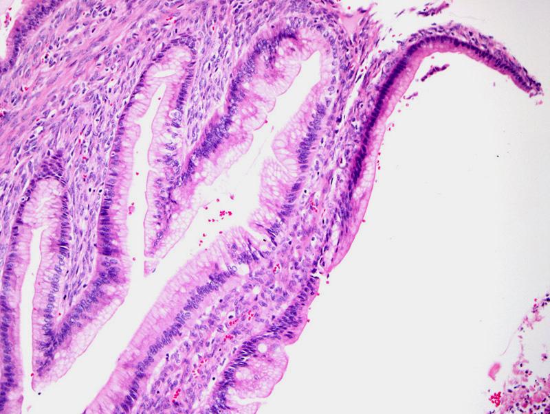
8 Heterogeneous Tumors, with Areas with Columnar Cells with Nuclear Stratification, Oval Nuclei and Goblet Cells
9 Columnar Cells with Nuclear Stratification, Oval Nuclei and Goblet Cells
10 WHO (2003) Borderline Mucinous Tumor (Mucinous Tumor of Low Malignant Potential) In our opinion it is not advisable to use the term Atypical Proliferative Tumor Problems to obtain staging Some cases can contain carcinoma
11 Review of 33 FIGO stage 1 cases with at least 5 years of follow-up Two pts with recurrences at 12 and 14 months, respectively (first patient with tumor incompletely excised due to adhesion to the ileum and sigmoid; second patient treated with cystectomy only) After the completion of this study, we have encountered rare patients with tumors initially diagnosed as a mucinous tumors of low malignant potential, intestinal type, who developed pelvic carcinomatosis or lung metastasis within a few years after dx, 1-3 years (SAMPLING ARTIFACT ISSUE) In our opinion, the current nomenclature should be retained to ensure the follow-up of patients affected by this disease until an evidence based sampling protocol becomes available
12


13
14 Primary Ovarian Mucinous Tumors How many sections do we submit for microscopic examination? Tumor < 10 cm 1 section per cm considering the largest dimension of the tumor Tumor 10 cm or tumors of any size with worrisome features (i.e., intraepithelial carcinoma, microinvasion) 2 sections per cm considering the largest dimension of the tumor
15 Mucinous Carcinoma Metastatic to the Ovary More frequent than primary ovarian mucinous carcinoma Primary Ovarian Invasive Mucinous Carcinoma is usually Unilateral FIGO stage I Expansile type IHC In situ hybridization for HPV Clinical correlation is of utmost importance to determine with certainty the origin of a subset of cases
16 Gross Features of Mucinous Carcinoma in the Ovary Metastasis Ovarian Primary Bilateral 10 cm With Tumor Outside the Ovary Unilateral > 10 cm Tumor Limited to the Ovary Seidman JD et al., 2003
17 Bilateral Ovarian Involvement


18 Multinodular Growth Pattern Separated by Normal Ovary
19 Tumor Invasion into Normal Structures of the Ovary
20 Prominent Desmoplastic Response
21 Ovarian Surface Involvement
22 Signet-Ring Cell Pattern
23 Numerous Pools of Mucin Dissecting the Stroma
24 Mucinous Carcinoma Metastatic to the Ovary with Areas Resembling a Mucinous Cystadenoma, note Numerous Mitoses
25 Mucinous Carcinoma Metastatic to the Ovary with Areas Resembling a Mucinous Cystadenoma, note Numerous Mitoses
 Metastatic Carcinoma of")
26 Ovary, Papillary Tumor with Unusual Features (Intracellular Mucin, Cribriform Pattern and Marked Cytologic Atypia) Metastatic Carcinoma of Cervical Origin
27 The Most Frequently Asked Questions Ovarian Serous Tumors: Nomenclature issue Benign vs. serous neoplasm of low malignant potential (borderline serous tumor) Serous neoplasm of low malignant potential (borderline serous tumor) vs. atypical proliferative serous tumor
28 The Most Frequently Asked Questions Ovarian Serous Tumors: Extraovarian findings in ovarian serous tumors of low malignant potential Endosalpingiosis Implants, non-invasive vs. invasive Serous carcinoma Lymph node involvement
29 The Most Frequently Asked Questions Ovarian Serous Tumors: The binary system for grading serous carcinoma Diagnostic criteria Is this system useful? Heterogeneity How to make the diagnosis of serous carcinoma? Ovarian serous tumors with extracellular or intracellular mucin
30 Serous LMP tumor Epithelial stratification with tufting and cell detachment Hierarchical branching
31 What percentage of the tumor has to have this pattern to use the term serous tumor of low malignant potential? Many authors consider 10% as the cut-off point However, in our practice we have encountered rare cases with less than 10% of this pattern that have recurred or had extraovarian disease
32

33
34


35 Therefore, if the serous neoplasm of low malignant potential area represents less than 10% of the tumor, the diagnosis of serous neoplasm of low malignant potential is made and the word focal is added to the diagnosis i.e., Focal Serous Neoplasm of Low Malignant Potential in a Serous Cystadenoma (Cystadenofibroma)
 It is not advisable")

36 WHO (2003) Borderline Serous Tumor (Serous Tumor of Low Malignant Potential) It is not advisable to use the term Atypical Proliferative Tumor Problems to obtain staging Some cases evolve to carcinoma
37 Extraovarian Findings in Ovarian Serous Tumors of Low Malignant Potential Endosalpingiosis
38 Endosalpingiosis in Ovarian Serous Tumors of Low Malignant Potential Peritoneal It can be seen in 50% to 71% of the cases Lymph node It can be seen in up to 33% of the cases (significantly higher than in the control group) Djordjevic B, et al, 2010 It is seen in 58% to 63% of the cases with lymph node involvement
39 Extraovarian Findings in Ovarian Serous Tumors of Low Malignant Potential Non-invasive vs. invasive implants Conventional approach Emergent approach Prognosis Treatment

40 Non-invasive implant, epithelial type

41 Non-invasive implant, desmoplastic type
42 Invasive implant

43 Invasive implant
44 Micropapillary Pattern Emergent Approach Invasive Implants Clusters of Cells Surrounded by Clefts Devoid of Lining Epithelium
45 Implants Non-invasive implants excellent prognosis in most studies Overall survival: 95.3% Invasive implants poor prognosis Overall survival: 66% Seidman and Kurman, 2000
46
47 Extraovarian Findings in Ovarian Serous Tumors of Low Malignant Potential Low Grade Serous Carcinoma
48 Extraovarian Findings in Ovarian Serous Tumors of Low Malignant Potential Lymph Node Involvement Limited Experience Incidence 21% to 42% of the cases No significant difference in the survival of cases with or without lymph node involvement Upstaged 13% of the cases McKenney JK et al., 2006
49 supradiaphragmatic mesenteric/ omental, paraaortic pelvic
50 Ovarian Serous Tumor of Low Malignant Potential with Lymph Node Involvement 87% of the cases associated with peritoneal implants Nodular pattern (epithelial aggregate >1 mm) associated with a higher risk of recurrence (equivalent to invasive implant) McKenney JK et al., 2006
51
52 Lymph node involvement (LNI) is associated with a higher incidence of implants LNI is not an independent predictor of disease free-survival or overall survival LNI can be the only site of extra-ovarian involvement (22% of the cases) Grossly unremarkable lymph nodes are not at a decreased risk of LNI (LN size in cases with LNI, 0.2 to 4.0 cm comparable to control group)


53 Lymph Node Involvement by Serous Neoplasm of Low Malignant Potential What does it look like? Individual cells Clusters of cells Papillae and small papillae Papillae and micropapillae Glandular proliferation Intraglandular proliferation Djordjevic B,and Malpica A, 2010


54 Focal Desmoplastic Reaction Djordjevic B, Malpica A, 2010
55 What is the Correct Term to Designate this Lymph Node Serous Lesion Associated with an Ovarian Serous Neoplasm of Low Malignant Potential? Low Grade Serous Carcinoma Djordjevic B,and Malpica A, 2010
56 Grading of Ovarian Serous Carcinoma The two-tier grading system is based: Primarily on the assessment of cytologic atypia Secondary feature, the mitotic rate User friendly Reproducible Meaningful segregation of cases
57 Low grade serous carcinoma: tumor with uniform cells with mild to moderate cytologic atypia and usually a low mitotic index ( 12 mitoses per 10 HPFs)
, and a high mitotic index (>12 mitoses per 10")
58 High grade serous carcinoma: tumor with pleomorphic cells with marked nuclear atypia ( 3:1 variation in size and shape), and a high mitotic index (>12 mitoses per 10 HPFs)
59 Ovarian High vs. Low Grade Serous Carcinoma-Differences Incidence High grade serous carcinoma is more common than low grades serous carcinoma Ovarian Tumorigenesis Model Prototypic type I: High grade serous carcinoma P53 mutation Prototypic type II: Low grade serous carcinoma KRAS and BRAF mutation KRAS mutation (MDACC experience)
60 Ovarian High vs. Low Grade Serous Carcinoma-Differences Immunohistochemistry High grade: higher expression of p53, Bcl-2, c-kit,p16, and EGFR Low grade: higher expression of ER, PR, and ECAD Genetic Risk BRCA1/2 germline mutations have been found in high grade serous carcinoma
61 Ovarian High vs. Low Grade Serous Carcinoma-Differences Behavior Low grade serous carcinoma appears to be less responsive to conventional chemotherapy than high grade serous carcinoma Patients with low grade serous carcinoma have a longer overall survival than patients with high grade serous carcinoma Patients with low grade serous carcinoma are younger and have a better survival than patients with high grade serous carcinoma

62 Low vs. High Grade Serous Carcinoma, Tumor Heterogeneity Uniform Cells
63 Low vs. High Grade Serous Carcinoma, Tumor Heterogeneity Nuclear Pleomorphism
64 Diagnostic Criteria for Ovarian Serous Carcinoma A serous tumor with low grade cytology Invasion is required = Low Grade Serous Carcinoma Invasion is identified by Size, measuring more than 3.0 mm or The presence of unequivocal desmoplasia A serous tumor with definitive high grade cytology Invasion is not required = High Grade Serous Carcinoma Note: Attention has to be paid to the cytologic features not to mistake a clear cell carcinoma for a serous neoplasm of low malignant potential
65 Serous Tumor of Low Malignant Potential, Hierarchical Branching, Epithelial Tufting and Cell Detachment


66 Papillae infiltrating the stroma
67 Tangentially cut cystic space, note the lining epithelium Spaces in areas of invasion lack lining epithelium
68 > 3mm, single dimension
69 Desmoplasia
70 Desmoplasia
71 A Serous Tumor with a Low Malignant Potential Pattern
72 High Grade Cytology


73 Clear Cell Carcinoma


74 Clear Cells, Nuclear Atypia, and Hyalinized Stroma
75 Focal Low Grade Serous Carcinoma in a Background of Serous LMP Note gelatinous papillae
76 Abundant Extracellular Mucin in a Serous Neoplasm of LMP This is a common finding in tumors detected during pregnancy
77 Serous Carcinoma with a Microcystic Pattern
78 Serous Carcinoma with a Microcystic Pattern - Mucicarmine +
79 Signet-ring Cells in Serous Carcinoma
80 Signet-ring Cells in Serous Carcinoma, WT-1 (+)


81
International Society of Gynecological Pathologists Symposium 2007
 International Society of Gynecological Pathologists Symposium 2007 Anais Malpica, M.D. Department of Pathology The University of Texas M.D. Anderson Cancer Center Grading of Ovarian Cancer Histologic grade
Low-grade serous neoplasia. Robert A. Soslow, MD
 Low-grade serous neoplasia Robert A. Soslow, MD soslowr@mskcc.org Outline Orientation Ovarian tumor overview Non serous borderline tumors Serous borderline tumors Clinical summary Morphologic description
Mucinous Tumors of the Ovary Beirut, Lebanon. Anaís Malpica, M.D. Professor Department of Pathology
 Mucinous Tumors of the Ovary Beirut, Lebanon Anaís Malpica, M.D. Professor Department of Pathology Primary Mucinous Tumors of the Ovary Cystadenoma Borderline (Tumor of Low Malignant Potential/Atypical
Survival Analysis and Prognosis for Patients with Serous and Mucinous Borderline Ovarian Tumors: 14-Year Experience from a Tertiary Center in Iran
 ORIGINAL ARTICLE Survival Analysis and Prognosis for Patients with Serous and Mucinous Borderline Ovarian Tumors: 14-Year Experience from a Tertiary Center in Iran Katayoun Ziari, Ebrahim Soleymani, and
Low-Grade Serous Ovarian Tumors Debra A. Bell, MD Mayo Clinic and Mayo Medical School Rochester, MN
 1 Low-Grade Serous Ovarian Tumors Debra A. Bell, MD Mayo Clinic and Mayo Medical School Rochester, MN It is very appropriate to discuss low-grade ovarian serous neoplasms in a symposium in honor of Dr.
Borderline tumors. Borderline tumors. Serous borderline tumor are NOT benign. Low grade serous carcinoma: pathogenesis. Serous carcinoma: pathogenesis
 Serous borderline tumor are NOT benign Robert A. Soslow, MD Memorial Sloan-Kettering Cancer Center soslowr@mskcc.org Borderline tumors Serous BTs and seromucinous BTs are both histopathologically borderline
Original contribution
 Human Pathology (2012) 43, 747 752 www.elsevier.com/locate/humpath Original contribution The presence and location of epithelial implants and implants with epithelial proliferation may predict a higher
Ovarian carcinoma classification. Robert A. Soslow, MD
 Ovarian carcinoma classification Robert A. Soslow, MD soslowr@mskcc.org WHO classification Serous Mucinous Endometrioid Clear cell Transitional Squamous Mixed epithelial Undifferentiated Introduction Rationale
A Survay on Appendiceal Involvement in Ovarian Mucinous Tumors
 http://www.ijwhr.net Open Access doi 10.15296/ijwhr.2018.33 Original Article International Journal of Women s Health and Reproduction Sciences Vol. 6, No. 2, April 2018, 199 203 ISSN 2330-4456 A Survay
USCAP 2013: THE ORIGINS OF OVARIAN CANCER: MUCINOUS TUMORS
 USCAP 2013: THE ORIGINS OF OVARIAN CANCER: MUCINOUS TUMORS Russell Vang, M.D. Associate Professor Division of Gynecologic Pathology The Johns Hopkins Hospital Table of contents: Powerpoint handout Text
Ovarian serous tumors of low malignant potential
 ORIGINAL ARTICLE Lymph Node Involvement in Ovarian Serous Tumors of Low Malignant Potential (Borderline Tumors) Pathology, Prognosis, and Proposed Classification Jesse K. McKenney, MD, Bonnie L. Balzer,
Mody. AIS vs. Invasive Adenocarcinoma of the Cervix
 Common Problems in Gynecologic Pathology Michael T. Deavers, M.D. Houston Methodist Hospital, Houston, Texas Common Problems in Gynecologic Pathology Adenocarcinoma in-situ (AIS) of the Cervix vs. Invasive
Mucinous Adenocarcinoma Involving the Ovary: Comparative Evaluation of the Classification Algorithms using Tumor Size and Laterality
 J Korean Med Sci 2010; 25: 220-5 ISSN 1011-8934 DOI: 10.3346/jkms.2010.25.2.220 Mucinous Adenocarcinoma Involving the Ovary: Comparative Evaluation of the Classification Algorithms using Tumor Size and
Case # 4 Low-Grade Serous Carcinoma (Macropapillary) of the Ovary Arising in an Atypical Proliferative Serous Tumor
 of the Ovary Arising in an Atypical Proliferative Serous Tumor") Case # 4 Low-Grade Serous Carcinoma (Macropapillary) of the Ovary Arising in an Atypical Proliferative Serous Tumor Robert J Kurman, M.D. Johns Hopkins University School of Medicine Case History A 53 year
Case # 4 Low-Grade Serous Carcinoma (Macropapillary) of the Ovary Arising in an Atypical Proliferative Serous Tumor Robert J Kurman, M.D. Johns Hopkins University School of Medicine Case History A 53 year
Current Concept in Ovarian Carcinoma: Pathology Perspectives
 Current Concept in Ovarian Carcinoma: Pathology Perspectives Rouba Ali-Fehmi, MD Professor of Pathology The Karmanos Cancer Institute, Wayne State University School of Medicine Current Concept in Ovarian
The Origin of Pelvic Low-Grade Serous Proliferative Lesions
 The Origin of Pelvic Low-Grade Serous Proliferative Lesions Ovarian Atypical Proliferative (Borderline) Serous Tumors, Noninvasive Implants and Endosalpingiosis Robert J. Kurman, M.D. Kurman RJ, Vang R,
Serous Borderline Tumors of the Ovary: Implants, Manifestations, Biology & New Insights in Progression
 Serous Borderline Tumors of the Ovary: Implants, Manifestations, Biology & New Insights in Progression Stanley J. Robboy, MD Professor of Pathology Professor of Obstetrics & Gynecology Vice Chairman for
Borderline Ovarian Mucinous Tumors: Consensus Points and Persistent Controversies Regarding Nomenclature, Diagnostic Criteria, and Behavior
 Borderline Ovarian Mucinous Tumors: Consensus Points and Persistent Controversies Regarding Nomenclature, Diagnostic Criteria, and Behavior Brigitte M. Ronnett, M.D.; C. Blake Gilks, M.D., Maria J. Merino,
David Nunns on behalf of the Gynae Guidelines Group Date:
 Title of Guideline (must include the word Guideline (not protocol, policy, procedure etc) Borderline tumours of the ovary management and follow-up Author: Contact Name and Job Title Directorate & Speciality
Section 1. Biology of gynaecological cancers: our current understanding
 Section 1 Biology of gynaecological cancers: our current understanding Chapter 1 Morphological sub-types of ovarian carcinoma: new developments and pathogenesis W Glenn McCluggage 1 Introduction In most
Urinary Bladder: WHO Classification and AJCC Staging Update 2017
 Urinary Bladder: WHO Classification and AJCC Staging Update 2017 Houston Society of Clinical Pathologists 58 th Annual Spring Symposium Houston, TX April 8, 2017 Jesse K. McKenney, MD Classification
of 20 to 80 and subsequently declines [2].
![of 20 to 80 and subsequently declines [2].](/thumbs/80/81450506.jpg "of 20 to 80 and subsequently declines [2].") - - According to the 2014 World Health Organization (WHO) classification and tumor morphology, primary ovarian tumors are subdivided into three categories: epithelial (60%), germ cell (30%), and sex-cord
- - According to the 2014 World Health Organization (WHO) classification and tumor morphology, primary ovarian tumors are subdivided into three categories: epithelial (60%), germ cell (30%), and sex-cord
GOBLET CELL CARCINOID. Hanlin L. Wang, MD, PhD University of California Los Angeles
 GOBLET CELL CARCINOID Hanlin L. Wang, MD, PhD University of California Los Angeles Disclosure of Relevant Financial Relationships USCAP requires that all planners (Education Committee) in a position to
GOBLET CELL CARCINOID
 GOBLET CELL CARCINOID Hanlin L. Wang, MD, PhD University of California Los Angeles Disclosure of Relevant Financial Relationships USCAP requires that all planners (Education Committee) in a position to
5/26/2016. Pelvic Serous Carcinoma: 2014 W.H.O. Update. Outline of Talk. Changes to 2014 WHO system for pelvic serous tumors
 Pelvic Serous Carcinoma: 2014 W.H.O. Update Outline of Talk Practical Implications for Pathologists Changes to 2014 WHO system for pelvic serous tumors High grade serous carcinoma versus low grade serous
SEROUS TUMORS. Dr. Jaime Prat. Hospital de la Santa Creu i Sant Pau. Universitat Autònoma de Barcelona
 SEROUS TUMORS Dr. Jaime Prat Hospital de la Santa Creu i Sant Pau Universitat Autònoma de Barcelona Serous Borderline Tumors (SBTs) Somatic genetics Clonality studies have attempted to dilucidate whether
Case Report Ovarian Seromucinous Borderline Tumor and Clear Cell Carcinoma: An Unusual Combination
 Case Reports in Obstetrics and Gynecology Volume 2015, Article ID 690891, 5 pages http://dx.doi.org/10.1155/2015/690891 Case Report Ovarian Seromucinous Borderline Tumor and Clear Cell Carcinoma: An Unusual
A Serous Borderline Tumor of the Fallopian Tube Detected Incidentally
 A Serous Borderline Tumor of the Fallopian Tube Detected Incidentally Imrana Tanvir, Ghania Ali, Haseeb Ahmed Khan and Ahmed Nasir Hanifi* Dept. of Histopathology, FMH College of Medicine & Dentistry,
Mucinous tumours of appendix and ovary: an overview and evaluation of current practice
 Department of Pathology, Laboratory Medicine Program, University Health Network, University of Toronto, Toronto, Canada Correspondence to Dr Marjan Rouzbahman, Department of Pathology, 11th floor, Eaton
The Diagnostic Challenges of Low Grade and High Grade Tubo-Ovarian Serous Carcinomas. W Glenn McCluggage Belfast, Northern Ireland
 The Diagnostic Challenges of Low Grade and High Grade Tubo-Ovarian Serous Carcinomas W Glenn McCluggage Belfast, Northern Ireland Enterprise Interest None OVARIAN SEROUS CARCINOMA (OSC) RECENT DEVELOPMENTS
Adenocarcinoma of the Cervix
 Question 1. Each of the following statements about cervical adenocarcinoma is true except: Adenocarcinoma of the Cervix SAMS a) A majority of women with cervical adenocarcinoma have stage I tumors at diagnosis.
Borderline Ovarian Tumours. Andreas Obermair Brisbane
 Borderline Ovarian Tumours Andreas Obermair Brisbane Definition First described in 1929 Cellular features of malignancy Cellular atypia Mitotic activity No stromal invasion An entity per se??? (or precursor
3 cell types in the normal ovary
 Ovarian tumors 3 cell types in the normal ovary Surface (coelomic epithelium) the origin of the great majority of ovarian tumors (neoplasms) 90% of malignant ovarian tumors Totipotent germ cells Sex cord-stromal
How to Recognize Gynecologic Cancer Cells from Pelvic Washing and Ascetic Specimens
 How to Recognize Gynecologic Cancer Cells from Pelvic Washing and Ascetic Specimens Wenxin Zheng, M.D. Professor of Pathology and Gynecology University of Arizona zhengw@email.arizona.edu http://www.zheng.gynpath.medicine.arizona.edu/index.html
4/12/2018. MUSC Pathology Symposium Kiawah Island April 18, Jesse K. McKenney, MD
 MUSC Pathology Symposium Kiawah Island April 18, 2018 Jesse K. McKenney, MD 1 Urothelial Carcinoma with Alternative Differentiation 2 Urothelial Carcinoma with Alternative Differentiation Recognition as
Introduction. Agenda. Pathogenesis. Pathogenesis and Clinical Implications. Pathogenesis and Clinical Implications
 Surface Epithelial Tumors of the Ovary Part II Invasive Carcinomas Moderator Robert J. Kurman, M.D. Discussants Kathleen R. Cho, M.D. Anais Malpica, M.D. Patricia A. Shaw, M.D. Robert A. Soslow, M.D. C.
Ovarian Clear Cell Carcinoma
 Ovarian Clear Cell Carcinoma Rouba Ali-Fehmi, MD Professor of Pathology The Karmanos Cancer Institute, Wayne State University School of Medicine 50 year old woman with chief complaint of shortness of breath
Session Number: 1020 Session: Adenocarcinoma of the Cervix: Diagnostic Pitfalls and New Prognostic Implications. Andres A. Roma, MD Cleveland Clinic
 Session Number: 1020 Session: Adenocarcinoma of the Cervix: Diagnostic Pitfalls and New Prognostic Implications Andres A. Roma, MD Cleveland Clinic No Disclosures In the past 12 months, I have not had
Follicular Derived Thyroid Tumors
 Follicular Derived Thyroid Tumors Jennifer L. Hunt, MD, MEd Aubrey J. Hough Jr, MD, Endowed Professor of Pathology Chair of Pathology and Laboratory Medicine University of Arkansas for Medical Sciences
Invited Re vie W. Molecular genetics of ovarian carcinomas. Histology and Histo pathology
 Histol Histopathol (1 999) 14: 269-277 http://www.ehu.es/histol-histopathol Histology and Histo pathology Invited Re vie W Molecular genetics of ovarian carcinomas J. Diebold Pathological Institute, Ludwig-Maximilians-University
Carcinoma mammario: le istologie non frequenti. Valentina Guarneri Università di Padova IOV-IRCCS
 Carcinoma mammario: le istologie non frequenti Valentina Guarneri Università di Padova IOV-IRCCS Histological diversity of breast adenocarcinomas Different histological types are defined according to specific
Papillary Lesions of the breast
 Papillary Lesions of the breast Emad Rakha Professor of Breast Pathology The University of Nottingham Papillary lesions of the breast are a heterogeneous group of disease, which are characterised by neoplastic
Joseph Misdraji, M.D. GI pathology Unit Massachusetts General Hospital
 Joseph Misdraji, M.D. GI pathology Unit Massachusetts General Hospital jmisdraji@partners.org Low-grade appendiceal mucinous neoplasm (LAMN) High-grade appendiceal mucinous neoplasm (HAMN) Adenocarcinoma
3 cell types in the normal ovary
 Ovarian tumors 3 cell types in the normal ovary Surface (coelomic epithelium) the origin of the great majority of ovarian tumors 90% of malignant ovarian tumors Totipotent germ cells Sex cord-stromal cells
New Developments in Immunohistochemistry for Gynecologic Pathology
 New Developments in Immunohistochemistry for Gynecologic Pathology Michael T. Deavers, M.D. Professor, Departments of Pathology and Gynecologic Oncology Immunohistochemistry in Gynecologic Pathology Majority
Objectives. Atypical Glandular Cells. Atypical Endocervical Cells. Reactive Endocervical Cells
 2013 California Society of Pathologists 66 th Annual Meeting San Francisco, CA Atypical Glandular Cells to Early Invasive Adenocarcinoma: Cervical Cytology and Histology Christina S. Kong, MD Associate
Wendy L Frankel. Chair and Distinguished Professor
 1 Wendy L Frankel Chair and Distinguished Professor Case 1 59 y/o woman Abdominal pain No personal or family history of cancer History of colon polyps Colonoscopy Polypoid rectosigmoid mass Biopsy 3 4
Case 1. Pathology of gynecological cancer. What do we need to know (Case 1) Luca Mazzucchelli Istituto cantonale di patologia Locarno
 Luca Mazzucchelli Istituto cantonale di patologia Locarno") Case 1 Pathology of gynecological cancer. What do we need to know (Case 1) Luca Mazzucchelli Istituto cantonale di patologia Locarno SAMO Interdisciplinary Workshop on Gynecological Tumors Lucern, October
Case 1 Pathology of gynecological cancer. What do we need to know (Case 1) Luca Mazzucchelli Istituto cantonale di patologia Locarno SAMO Interdisciplinary Workshop on Gynecological Tumors Lucern, October
Case year old female presented with asymmetric enlargement of the left lobe of the thyroid
 Case 4 22 year old female presented with asymmetric enlargement of the left lobe of the thyroid gland. No information available relative to a prior fine needle aspiration biopsy. A left lobectomy was performed.
Case 4 Diagnosis 2/21/2011 TGB
 Case 4 22 year old female presented with asymmetric enlargement of the left lobe of the thyroid gland. No information available relative to a prior fine needle aspiration biopsy. A left lobectomy was performed.
ACCME/Disclosures. Case History 4/13/2016. USCAP GU Specialty Conference Case 3. Ann Arbor, MI
 USCAP GU Specialty Conference Case 3 March 2016 L. Priya Kunju, M.D. University of Michigan Health System Ann Arbor, MI University of Michigan Health System ACCME/Disclosures The USCAP requires that anyone
64 YO lady THBSO for prolapse At gross : A 3 cm endometrial polyp in the fundus
 Case 6 64 YO lady THBSO for prolapse At gross : A 3 cm endometrial polyp in the fundus Numerous irregular, large glands with leaf-like pattern Large glands with broad-based papillary infolding into the
Important Recent Advances in Gynaecological Pathology
 Important Recent Advances in Gynaecological Pathology Sanjiv Manek Consultant Gynaecological Pathologist Oxford, UK In recent years there have been a significant number of changes in gynaecological pathology
Salivary Glands 3/7/2017
 Salivary Glands 3/7/2017 Goals and objectives Focus on the entities unique to H&N Common board type facts Information for your future practice Salivary Glands Salivary Glands Major gland. Paratid. Submandibular.
Pathology of the Thyroid
 Pathology of the Thyroid Thyroid Carcinoma Arising from Follicular Cells 2015-01-19 Prof. Dr. med. Katharina Glatz Pathologie Carcinomas Arising from Follicular Cells Differentiated Carcinoma Papillary
INTRADUCTAL LESIONS OF THE PROSTATE. Jonathan I. Epstein
 INTRADUCTAL LESIONS OF THE PROSTATE Jonathan I. Epstein Topics Prostatic intraepithelial neoplasia (PIN) Intraductal adenocarcinoma (IDC-P) Intraductal urothelial carcinoma Ductal adenocarcinoma High Prostatic
Clinical History USCAP Specialty Conference. Gynecologic Pathology Case 3
 2010 USCA Specialty Conference Gynecologic athology Case Kathleen R. Cho, M.D. Department of athology Clinical History 46 yo woman presented with bilateral ovarian masses and elevated CA-125 TAH/BSO, pelvic
Borderline Brenner tumor of the ovary: a case report with immunohistochemical and molecular study
 De Cecio et al. Journal of Ovarian Research 2014, 7:101 CASE REPORT Open Access Borderline Brenner tumor of the ovary: a case report with immunohistochemical and molecular study Rossella De Cecio 1, Monica
Note: The cause of testicular neoplasms remains unknown
 - In the 15- to 34-year-old age group, they are the most common tumors of men. - Tumors of the testis are a heterogeneous group of neoplasms that include: I. Germ cell tumors : 95%; all are malignant.
05/07/2018. Types of challenges. Challenging cases in uterine pathology. Case 1 ` 65 year old female Post menopausal bleeding Uterine Polyp
 Types of challenges Challenging cases in uterine pathology Nafisa Wilkinson Gynaecological Pathologist UCLH London Lack of complete history often, NO clinical history at all! Cases from other centres often
Atypical Hyperplasia/EIN
 EIN Atypical Hyperplasia/EIN Based on scientific and diagnostic advances, in 2014 the WHO moved that the precursor lesion for endometrioid carcinoma be atypical hyperplasia/ein, rather than what was previously
Fertility Preservation Is Safe for Serous Borderline Ovarian Tumors
 ORIGINAL STUDY Fertility Preservation Is Safe for Serous Borderline Ovarian Tumors Eveline Vancraeynest, MD,* Philippe Moerman, MD, PhD,Þ Karin Leunen, MD, PhD,* Frédéric Amant, MD, PhD,* Patrick Neven,
Unknown Slides Conference
 Unknown Slides Conference Jae Y. Ro, MD, PhD Weill Medical College of Cornell Univ. The Methodist Hospital, and UT MD Anderson Cancer Center Houston, TX November 9, 2013 Amman, Jordan 25 th Congress of
ACCME/Disclosures. Cribriform Lesions of the Prostate. Case
 Cribriform Lesions of the Prostate Ming Zhou, MD, PhD Departments of Pathology and Urology New York University Langone Medical Center New York, NY Ming.Zhou@NYUMC.ORG ACCME/Disclosures The USCAP requires
Papillary Lesions of the Breast: WHO Update
 Papillary Lesions of the Breast: WHO Update Stuart J. Schnitt, M.D. Department of Pathology Beth Israel Deaconess Medical Center and Harvard Medical School Boston, MA, USA Papillary Lesions of the Breast
Syllabus. Appendiceal GCC and LAMN Navigating the Alphabet Soup in the Appendix. Appendiceal tumors. Summary provided Complete presentation
 2016 Current Issues in Surgical Pathology Appendiceal GCC and LAMN Navigating the Alphabet Soup in the Appendix Syllabus Summary provided Complete presentation sanjay.kakar@ucsf.edu Sanjay Kakar, MD University
Basement membrane in lobule.
 Bahram Memar, MD Basement membrane in lobule. Normal lobule-luteal phase Normal lobule-follicular phase Lactating breast Greater than 95% are adenocarcinomas in situ carcinomas and invasive carcinomas.
Article begins on next page
 Pseudopapillary Granulosa Cell Tumor: A Case of This Rare Subtype Rutgers University has made this article freely available. Please share how this access benefits you. Your story matters. [https://rucore.libraries.rutgers.edu/rutgers-lib/50622/story/]
Papillary Lesions of the Breast A Practical Approach to Diagnosis. (Arch Pathol Lab Med. 2016;140: ; doi: /arpa.
 Papillary Lesions of the Breast A Practical Approach to Diagnosis (Arch Pathol Lab Med. 2016;140:1052 1059; doi: 10.5858/arpa.2016-0219-RA) Papillary lesions of the breast Span the spectrum of benign,
Mammary analogue secretory carcinoma of salivary gland A case report of new entity
 Case Report Mammary analogue secretory carcinoma of salivary gland A case report of new entity Vaibhav Bhika Bari 1*, Sandhya Unmesh Bholay 2 1 Assistant Professor, 2 Associate Professor Rajiv Gandhi Medical
بسم هللا الرحمن الرحيم. Prof soha Talaat
 بسم هللا الرحمن الرحيم Ovarian tumors The leading indication for gynecologic surgery. Preoperative characterization of complex solid and cystic adnexal masses is crucial for informing patients about possible
Pathology of Ovarian Tumours. Dr. Jyothi Ranganathan MD ( Path) AFMC Pune PDCC (Cytopathology) PGI Chandigarh
 AFMC Pune PDCC (Cytopathology) PGI Chandigarh") Pathology of Ovarian Tumours Dr. Jyothi Ranganathan MD ( Path) AFMC Pune PDCC (Cytopathology) PGI Chandigarh Outline Incidence Risk factors Classification Pathology of tumours Tumour markers Prevention
Pathology of Ovarian Tumours Dr. Jyothi Ranganathan MD ( Path) AFMC Pune PDCC (Cytopathology) PGI Chandigarh Outline Incidence Risk factors Classification Pathology of tumours Tumour markers Prevention
R J M E Romanian Journal of Morphology & Embryology
 Rom J Morphol Embryol 2012, 53(4):967 973 ORIGINAL PAPER R J M E Romanian Journal of Morphology & Embryology http://www.rjme.ro/ The immunohistochemical expression of p53 and Ki67 in ovarian epithelial
Neoplasia 2018 Lecture 2. Dr Heyam Awad MD, FRCPath
 Neoplasia 2018 Lecture 2 Dr Heyam Awad MD, FRCPath ILOS 1. List the differences between benign and malignant tumors. 2. Recognize the histological features of malignancy. 3. Define dysplasia and understand
Mousa. Najat kayed &Renad Al-Awamleh. Nizar Alkhlaifat
 6 Mousa Najat kayed &Renad Al-Awamleh Nizar Alkhlaifat P a g e 1 This sheet written based on record 13 on website Cover slide( 95-117 ) No need to go back to slide FALLOPIAN TUBE PATHOLOGY In general fallopian
Select problems in cystic pancreatic lesions
 Disclosure Select problems in cystic pancreatic lesions Five Prime Therapeutics shareholder Adicet Bio shareholder Bristol-Meyer Squibb advisory board grace.kim@ucsf.edu Pancreatic cystic lesions Intraductal
Histological pattern of ovarian tumors and their age distribution
 Original Article Nepal Med Coll J 2008; 10(2): 81-85 Histological pattern of ovarian s and their age distribution R Jha and S Karki Department of Pathology, TUTH, Maharajgunj, Kathmandu, Nepal Corresponding
6/5/2010. Outline of Talk. Endometrial Alterations That Mimic Cancer & Vice Versa: Metaplastic / reactive changes. Problems in Biopsies/Curettages
 Outline of Talk Endometrial Alterations That Mimic Cancer & Vice Versa: Problems in Biopsies/Curettages Metaplastic / reactive changes Mucinous change Microglandular hyperplasia-like change Squamous metaplasia
Pathobiology of ovarian carcinomas
 Chinese Journal of Cancer Review Mojgan Devouassoux-Shisheboran 1 and Catherine Genestie 2 Abstract Ovarian tumors comprise a heterogeneous group of lesions, displaying distinct tumor pathology and oncogenic
Dr Agata T Kochman Wishaw General Hospital
 Dr Agata T Kochman Wishaw General Hospital Case E1 84 year old male Symptoms: R shoulder pain CT = thymic mass and (R) LL nodules + (L) lung nodule Clinically metastatic lesions in lung with primary thymic
What s (new) and Important in Reporting of Uterine Cancers Katherine Vroobel The Royal Marsden
 and Important in Reporting of Uterine Cancers Katherine Vroobel The Royal Marsden") What s (new) and Important in Reporting of Uterine Cancers Katherine Vroobel The Royal Marsden Maastricht Pathology 2018 Wednesday 20 th June Endometrioid adenocarcinoma High grade carcinomas (common)
What s (new) and Important in Reporting of Uterine Cancers Katherine Vroobel The Royal Marsden Maastricht Pathology 2018 Wednesday 20 th June Endometrioid adenocarcinoma High grade carcinomas (common)
04/10/2018. Intraductal Papillary Neoplasms Of Breast INTRADUCTAL PAPILLOMA
 Intraductal Papillary Neoplasms Of Breast Savitri Krishnamurthy MD Professor of Pathology Deputy Division Head The University of Texas MD Anderson Cancer Center 25 th Annual Seminar in Pathology Pittsburgh,
Appendix cancer mimicking ovarian cancer
 Int J Gynecol Cancer 2002, 12, 768 772 CORRESPONDENCE AND BRIEF REPORTS Appendix cancer mimicking ovarian cancer P. A. GEHRIG *, J. F. BOGGESS*, D. W. OLLILA, P. A. GROBEN & L. VAN LE* *Division of Gynecologic
When Immunostains Can Get You in Trouble: Gynecologic Pathology p16: Panacea or Pandora s Box?
 When Immunostains Can Get You in Trouble: Gynecologic Pathology p16: Panacea or Pandora s Box? Teri A. Longacre, MD Stanford Medicine Stanford California pi6 in Gynecologic Pathology: Panacea or Pandora
GOBLET CELL CARCINOID. Hanlin L. Wang, MD, PhD University of California Los Angeles
 GOBLET CELL CARCINOID Hanlin L. Wang, MD, PhD University of California Los Angeles hanlinwang@mednet.ucla.edu Goblet cell carcinoid (GCC) is a unique type of mixed endocrine-exocrine neoplasm, almost exclusively
Diagnostic problems in uterine smooth muscle tumors
 Diagnostic problems in uterine smooth muscle tumors Marina Kos Ljudevit Jurak Clinical Department of Pathology, Clinical Hospital Center Sestre milosrdnice, Zagreb Institute of Pathology, University of
Villoglandular adenocarcinoma of cervix a tumour with bland cytological features: report of a case missed on cytology
 Malaysian J Pathol 2003; 25(2) : CERVICAL 139 143 VILLOGLANDULAR ADENOCARCINOMA CYTOLOGY CASE REPORT Villoglandular adenocarcinoma of cervix a tumour with bland cytological features: report of a case missed
Slide seminar. Asist. Prof. Jože Pižem, MD, PhD Institute of Pathology Medical Faculty, University of Ljubljana
 Slide seminar Asist. Prof. Jože Pižem, MD, PhD Institute of Pathology Medical Faculty, University of Ljubljana Case 5 A 57-year-old man with a dermal/subcutaneous lesion on the scalp, which was interpreted
ONCOLOGY. Csaba Bödör. Department of Pathology and Experimental Cancer Research november 19., ÁOK, III.
 ONCOLOGY Csaba Bödör Department of Pathology and Experimental Cancer Research 2018. november 19., ÁOK, III. bodor.csaba1@med.semmelweis-univ.hu ONCOLOGY Characteristics of Benign and Malignant Neoplasms
ENODMETRIAL CARCINOMA: SPECIAL & NOT SO SPECIAL VARIANTS
 ENODMETRIAL CARCINOMA: SPECIAL & NOT SO SPECIAL VARIANTS Pacific Northwest Society of Pathologists Vancouver, B.C. September 26, 2015 Teri A. Longacre, M.D. longacre@stanford.edu Stanford University, Stanford,
Maram Abdaljaleel, MD Dermatopathologist and Neuropathologist University of Jordan, School of Medicine
 Maram Abdaljaleel, MD Dermatopathologist and Neuropathologist University of Jordan, School of Medicine The most common non-skin malignancy of women 2 nd most common cause of cancer deaths in women, following
Biliary tract tumors
 Short Course 2010 Annual Fall Meeting of the Korean Society for Pathologists Biliary tract tumors Joon Hyuk Choi, M.D., Ph.D. Professor, Department of Pathology, Yeungnam Univ. College of Medicine, Daegu,
CASE 1A DIAGNOSIS. Problems in the Evaluation of Surface Epithelial-Stromal Tumors of the Ovary: an Update
 Problems in the Evaluation of Surface Epithelial-Stromal Tumors of the Ovary: an Update John H. Eichhorn C. Blake Gilks CASE 1A A 27-year-old woman with primary infertility had a negative work-up, except
Advanced Stage Mucinous Adenocarcinoma of the Ovary Is Both Rare and Highly Lethal
 Advanced Stage Mucinous Adenocarcinoma of the Ovary Is Both Rare and Highly Lethal A Gynecologic Oncology Group Study Richard J. Zaino, MD 1 ; Mark F. Brady, PhD 2 ; Subodh M. Lele, MD 3 ; Helen Michael,
ACCME/Disclosures. Diagnosing Mesothelioma in Limited Tissue Samples. Papanicolaou Society of Cytopathology Companion Meeting March 12 th, 2016
 Diagnosing Mesothelioma in Limited Tissue Samples Papanicolaou Society of Cytopathology Companion Meeting March 12 th, 2016 Sanja Dacic, MD, PhD University of Pittsburgh ACCME/Disclosures GENERAL RULES
Solid pseudopapillary tumour of the pancreas: Report of five cases
 ISPUB.COM The Internet Journal of Pathology Volume 8 Number 2 Solid pseudopapillary tumour of the pancreas: Report of five cases P Srilatha, V Manna, P Kanthilatha Citation P Srilatha, V Manna, P Kanthilatha..
Applications of IHC. Determination of the primary site in metastatic tumors of unknown origin
 Applications of IHC Determination of the primary site in metastatic tumors of unknown origin Classification of tumors that appear 'undifferentiated' by standard light microscopy Precise classification
Interpretation of p53 Immunostains. P53 Mutations are Ubiquitous in High Grade Serous Carcinoma. Diffuse strong positive nuclear staining
 Stains for Tumor Classification p53 p16 WT1 HMGA2 P53 Mutations are Ubiquitous in High Grade Serous Carcinoma Source Ahmed et al Australian Ovarian Cancer Study Cancer Genome Atlas Research Network Cases
Non Small Cell Lung Cancer Histopathology ד"ר יהודית זנדבנק
 Non Small Cell Lung Cancer Histopathology ד"ר יהודית זנדבנק 26.06.09 Lecture outlines WHO histological classification Macro/Micro assessment Early diagnosis Minimal pathology Main subtypes SCC, AdCa, LCLC
Presenter: Yeh-Han Wang M.D.
 Korea-Taiwan-Japan Joint Meeting for Gynecological Pathology Mini-lecture Female Adnexal Tumor of Probable Wolffian Origin (FATWO) in Taiwan: A Small Case Series and Literature Review Presenter: Yeh-Han