Review Course «Musculoskeletal Oncology» October 6, 2011 UNIKLINIK BALGRIST. Imaging of Bone and Soft Tissue. Tumors
|
|
|
- Anissa Garrison
- 5 years ago
- Views:
Transcription
1 Imaging of Bone and Soft Tissue Tumors Approach from a radiologist s point of view Florian Buck Radiology


2 Radio- Radio- Oncologist Oncologist Orthopedist Orthopedist Patient Management Oncologist Oncologist General General Practitioner Practitioner Radiologist Radiologist Pathologist Pathologist
3 Radio- Radio- Oncologist Oncologist Orthopedist Orthopedist Patient Management Oncologist Oncologist General General Practitioner Practitioner... Radiologist Radiologist Pathologist Pathologist
4 Patient Management Jigsaw pieces provided by Radiologists Consultant for state of the art imaging protocols and completeness of imaging studies to ensure optimal diagnostic yield Dignity of a lesion Differential diagnosis Resection planning, biopsy planning Image guided biopsy
5 Radiologic-Orthopedic Interface What s that? When can you biopsy that? T1-weighted fat-saturated post KM
6 Radiologic-Orthopedic Interface Correct Question would be: I have a patient with a history of... -Is the imaging sufficient? -What s the (differential) diagnosis? -Is a biopsy needed? -Is a biopsy feasible?
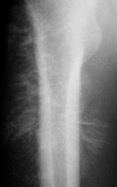
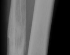
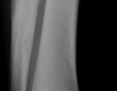
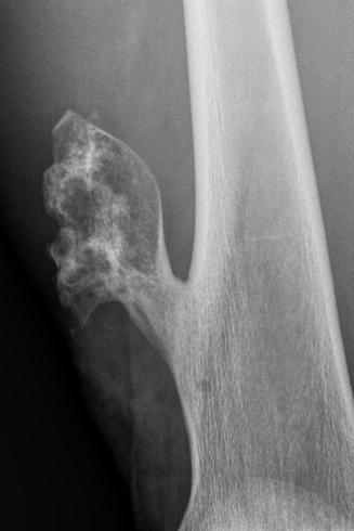
7 Radiologic-Orthopedic Interface Diagnosis: Stress Fracture with typical history of excessive sports training. Teaching points: - Stress fractures can mimick bone tumors and should never be biopsied. - History is key! - Imaging is always influenced by the patients history/differential diagnosis.
 do not imply more diagnostic security More images can divert from the important findings on plain films Well known benign lesion diagnosed on plain")
8 Radiologic-Orthopedic Interface Imaging overkill Radiologists know certain pathologies almost exclusively from plain radiographs! More images (MRI) do not imply more diagnostic security More images can divert from the important findings on plain films Well known benign lesion diagnosed on plain films can look malignant on MR images.
9 Radiologic-Orthopedic Interface Imaging overkill T1-weighted fat-saturated post KM T1-weighted

10 State of the art imaging 1. Plain films Mandatory in 2 planes! 2. MRI Soft tissue 3. CT scan Osteolysis, matrix mineralization 4. PET-CT Whole body, staging, tumor viability 5. PET-MRI Research, indications to be defined


11 State of the art MR imaging Robust, old, well-known sequences Optimal planes depending on anatomy: axial second best Tagging with capsule on the skin if there is a small nodule or the like T1 T2 T1 fs CM STIR


12 State of the art MR imaging T1 axial T2 axial T1 sagittal STIR coronal Post KM fat-sat Post KM fat-sat T1 KM T2 T1 axial - Anatomy - Compartment - Muscles - Vessel & Nerves - Fat Tissue T1 sagittal

13 State of the art MR imaging T1 axial T2 axial T1 sagittal STIR coronal Post KM fat-sat KM T1 axial Post KM fat-sat T1 sagittal - Additional information about aforementioned anatomical questions - Fluid collections - Skip lesions T1 STIR
14 State of the art MR imaging T1 axial T2 axial T1 sagittal STIR coronal Post KM fat-sat T1 KM T1 fs post KM T1 axial - Tumor tissue - Tumor size - Necrosis Post KM fat-sat T1 sagittal - Infiltration in adjacent structures

15 State of the art MR imaging T1 axial T2 axial T1 sagittal STIR coronal Post KM fat-sat KM T1 axial - Tumor tissue - Tumor size - Necrosis Post KM fat-sat T1 sagittal - Infiltration in adjacent structures
16 Posttreatment (follow-up) MR imaging Main focus on Follow-up MRI because of high rate of recurrence (about 50%) more examinations are performed as follow-up examinations than for treatment planning early recognition of recurrent disease is crucial for treatment Ref.: Garner HW, Kransdorf MJ, Bancroft LW, Peterson JJ, Berquist TH, Murphey MD. Benign and malignant soft-tissue tumors: posttreatment MR imaging. Radiographics. 2009;29(1):
17 Posttreatment (follow-up) MR imaging Compare with pre-treatment images As a general rule, the signal intensitiy and enhancement characteristics of a recurrent tumor typically mimics those of the original tumor. At least primarily calcifying tumors should be imaged using radiographs also Ref.: Garner HW, Kransdorf MJ, Bancroft LW, Peterson JJ, Berquist TH, Murphey MD. Benign and malignant soft-tissue tumors: posttreatment MR imaging. Radiographics. 2009;29(1):


18 Posttreatment (follow-up) MR imaging Importance of patient history Radiation therapy or reconstructive surgery Alteration of imaging appearance of surgical bed in a time-dependent manner Many different types of changes. Ref.: Garner HW, Kransdorf MJ, Bancroft LW, Peterson JJ, Berquist TH, Murphey MD. Benign and malignant soft-tissue tumors: posttreatment MR imaging. Radiographics. 2009;29(1):
19 Posttreatment MR imaging Ref.: Garner HW, Kransdorf MJ, Bancroft LW, Peterson JJ, Berquist TH, Murphey MD. Benign and malignant soft-tissue tumors: posttreatment MR imaging. Radiographics. 2009;29(1):
20 Posttreatment MR imaging Radiation therapy Subcutaneous edema: Peaking at month, in 50% normal over the ensuing 2-3 years Bone marrow change: 1-6 weeks after initiation of therapy, regeneration of bone marrow is rare Radiation osteitis: 8-49 months post initiation Muscle edema, Fibrosis, Seroma Chemotherapy Initially chemotherapy may cause an increase in tumor size because of intralesional edema, hemorrhage, and necrosis Ref.: Garner HW, Kransdorf MJ, Bancroft LW, Peterson JJ, Berquist TH, Murphey MD. Benign and malignant soft-tissue tumors: posttreatment MR imaging. Radiographics. 2009;29(1):
21 Posttreatment imaging Recurrent tumor Presence of discrete nodule or mass with signal characteristics that typically mirror those of the original tumor Post-surgery Post-radiation Metallic implants Hematoma Seroma Strategies - Full clinical history / surgery report - Baseline MRI postsurgery for comparison purposes - Advanced imaging techniques Osteitis - bone marrow edema - fatty change Soft tissue edema Tumor necrosis Myocutaneous flaps
:1424 1429.")
22 Posttreatment: Advances CT 64 kev 69 kev 88 kev 105 kev optimal kev Dual-energy CT: Monoenergetic extrapolation Ref.: Bamberg F, et al. Metal artifact reduction by dual energy computed tomography using monoenergetic extrapolation. Eur Radiol Jul.;21(7):


23 Posttreatment: Advances CT Dual-energy CT: Monoenergetic extrapolation Ref.: Bamberg F, et al. Metal artifact reduction by dual energy computed tomography using monoenergetic extrapolation. Eur Radiol Jul.;21(7):
24 Posttreatment: Optimal MRI 80 Hz/Pixel Increasing Bandwidth 390 Hz/Pixel


25 Posttreatment imaging: Optimal MRI e» s y r g u o l o o C c n w e O i l v a e t 1 e R l 1 e 0 k 2 T s, S o l I 6 u R r c e G s b L u o t A M c B «O NIK I L K I UNIncreasing Bandwidth 80 Hz/Pixel 390 Hz/Pixel Ref.: Buck FM, et al. Shoulder arthroplasty. Eur Radiol Dec.;18(12):

")
26 Posttreatment: Advances MRI standard sequence (turbo spin echo, high bandwidth) WARP SPACE

")
27 Posttreatment: Advances MRI standard STIR (high bandwidth) WARP TSE


")
28 Posttreatment: Advances MRI e» s y r g u o l o o C c n w e O i l v a e t 1 e R l 1 e 0 k 2 T s, S o l I 6 u R r c e G s b L u o t A M c B «O NIK I L K I new STIR IR N standard STIR U (high bandwidth)
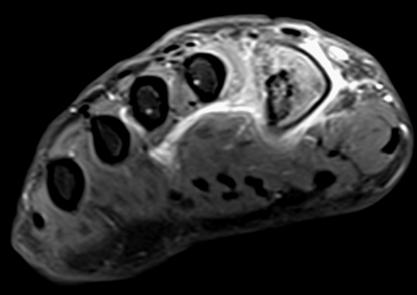
29 Periprosthetic Osteolysis?
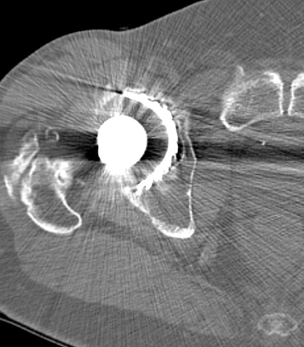
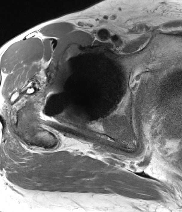
30 Periprosthetic Osteolysis? CT MRI
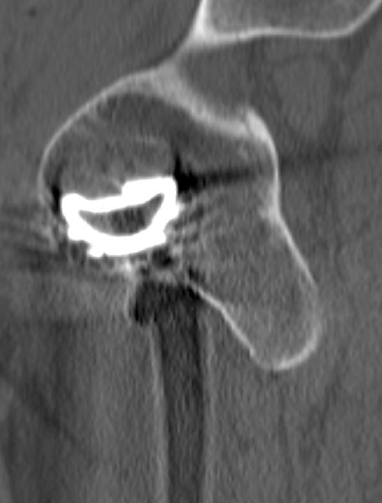
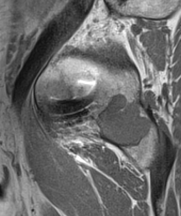
31 Periprosthetic Osteolysis? CT MRI
32 Dignity of a Lesion primarily evaluated on plain films NOT on MR images! Two most important aspects of evaluating a bone tumor are: Tumor location Patient age NOT radiologic appearance! This information alone is enough to narrow the differential diagnosis without even looking at any images! Ref.: Miller TT. Bone tumors and tumorlike conditions: analysis with conventional radiography. Radiology Mar. 1;246(3):
 Margins Zone of transition Periosteal reaction Presence of a soft-tissue")
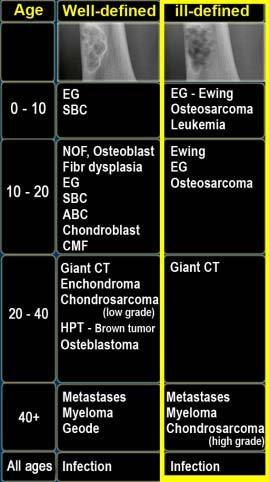
33 Dignity of a Lesion Criteria Tumor localization (in the body) Patient Age Specific radiographic features: Tumor location (inside the bone) Margins Zone of transition Periosteal reaction Presence of a soft-tissue component

34 Tumor Localization Most bone tumors often occur in a characteristic location in the skeleton - axial versus appendicular skeleton - long versus flat bones - predilection for sites of rapid bone growth usually the metaphyseal region, e.g. osteosarcoma - follow the distribution of red bone marrow, e.g. Ewing sarcoma Ref.:


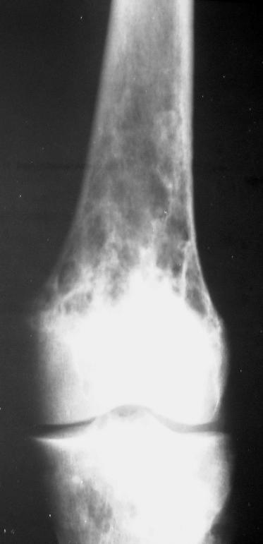
35 Zone of Transition Non-ossifying Fibroma Ewing's Sarcoma well-defined lesion ill-defined lesion

36 Margins: Lodwick's Classification Type 1a Geographic Lesion: Well-defined lucency with sclerotic rim. Intraosseous lipoma Ref.: Lodwick GS et al. Determining growth rates of focal bone lesions. Radiology 1980;134: Miller TT et al. Bone tumors and tumorlike conditions... Radiology 2008 Mar. 1;246(3):

37 Lodwick's Classification Type 1b Geographic Lesion: Well-defined lucency without sclerotic rim and endosteal scalloping. Myeloma Ref.: Lodwick GS et al. Determining growth rates of focal bone lesions. Radiology 1980;134: Miller TT et al. Bone tumors and tumorlike conditions... Radiology 2008 Mar. 1;246(3):

38 Lodwick's Classification Type 1c Geographic Lesion: Ill-defined lytic lesion with cortical disrupton. Codman triangle. New bone formation. Osteosarcoma Ref.: Lodwick GS et al. Determining growth rates of focal bone lesions. Radiology 1980;134: Miller TT et al. Bone tumors and tumorlike conditions... Radiology 2008 Mar. 1;246(3):

39 Lodwick's Classification Type 2 Moth-eaten Lesion: Ill-defined patchy lytic lesion with multilamellated periosteal reaction. Osteosarcoma Ref.: Lodwick GS et al. Determining growth rates of focal bone lesions. Radiology 1980;134: Miller TT et al. Bone tumors and tumorlike conditions... Radiology 2008 Mar. 1;246(3):

40 Lodwick's Classification Type 3 Permeated Lytic Lesion: Fine permeated pattern with ill-defined small patchy lucencies. Ewing's Sarcoma Ref.: Lodwick GS et al. Determining growth rates of focal bone lesions. Radiology 1980;134: Miller TT et al. Bone tumors and tumorlike conditions... Radiology 2008 Mar. 1;246(3):
41 The most important piece of clinical information when assessing a bone tumor is the patient s age. For example: - Simple simple bone cysts and chondroblastomas occur in skeletally immature people - Giant cell tumors in skeletally mature people Patient Age - Conventional osteosarcoma have two age peaks: In teenagers (de novo), in pagetic or previously irradiated bone, in adults older than 50 years Ref.: Miller TT. Bone tumors and tumorlike conditions: analysis with conventional radiography. Radiology Mar. 1;246(3):

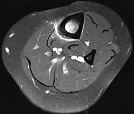
42 The most important piece of clinical information when assessing a bone tumor is the patient s age. For example: Patient Age - Malignant bone lesion in an adult over 40 years old is much more likely to be: metastatic carcinoma myeloma metastatic non-hodgkin lymphoma rather than a primary bone sarcoma. Ref.: Miller TT. Bone tumors and tumorlike conditions: analysis with conventional radiography. Radiology Mar. 1;246(3):

43 Radiographic features: Location A lesion in a long bone may be characterized by its: longitudinal location epipyseal metaphyseal diaphyseal transverse location medullary cortical juxtacortical Ref.:

44 Periosteal reaction Hypertrophic osteoarthropathy in a patient with lung cancer Ref.:

45 Periosteal reaction Osteosarcoma Ref.:

46 Radiographic features: Margin The margin of a lesion and type of periosteal reaction are indicators of lesion aggressiveness, but not necessarily of whether it is benign or malignant. A well-defined lesion with a sclerotic rim and thick unilamellar periosteal reaction is the most innocuous appearance A permeated pattern with spiculated periosteal reaction is the most aggressive. Ref.:


47 Radiographic features: Margin Ref.:


48 Radiographic features: Margin Non-ossifying Fibroma Brown tumor - HPT Ref.:
49 Radiographic features: Margin Ewing's Sarcoma Metastases Ref.:
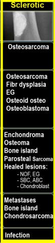
50 Radiographic features: Margin Osteoid Osteoma LSMFT liposclerosing myxofibrous tumor Ref.:


51 Soft-Tissue Component The presence of a soft-tissue component: suggests a malignant process typical tumors that often have a soft-tissue component: - osteosarcoma -Ewing'ssarcoma - lymphoma
52 So What?!? Do we need these criteria... We need biospy anyway...


53 Leave me alone lesions enchondroma metaphyseal cortical irregularity melorheostosis osteochondroma non-ossifying fibroma bone infarct junvenile bone cyst osteoidosteoma inclusion cyst of finger myositis ossificans Ref.: Hamers S. "Leave me alone lesions" des Knochens. Radiologie up2date 2002 Jul. 2;1-30.


54 So What?!? Yes, we need these criteria to: obviate biopsy of lesions that should not be biopsied And also. know where biopsy should be performed (vital tumor tissue) plan the biopsy direction plan surgery

55 Surgery Planning - T1-weighted & T2-weighted sequences - axial plane medial T1 T2 Leiomyosarcoma lateral
56 Surgery Planning - Identify compartments -Vessels -Nerves medial T1 T2 Leiomyosarcoma lateral
57 Surgery Planning - Identify compartments -Vessels -Nerves medial T1 T2 Leiomyosarcoma lateral

58 Surgery Planning - Identify compartments -Vessels -Nerves medial T1 T2 Leiomyosarcoma lateral


59 Surgery Planning - Scrolling through the images - Define compartments, vessels and nerves involved medial T1 T2 Leiomyosarcoma lateral


60 Image-guided Biopsy Bonopty Trapsystem Spirotome Coaxial Achieve "Snapper" SOFT TISSUE BONE
61 Thank you for your attention!
Bone Tumors Clues and Cues
 William Herring, M.D. 2002 Bone Tumors Clues and Cues In Slide Show mode, advance the slides by pressing the spacebar All Photos Retain the Copyright of their Authors Clues by Appearance of Lesion Patterns
MRI XR, CT, NM. Principal Modality (2): Case Report # 2. Date accepted: 15 March 2013
: Case Report # 2. Date accepted: 15 March 2013") Radiological Category: Musculoskeletal Principal Modality (1): Principal Modality (2): MRI XR, CT, NM Case Report # 2 Submitted by: Hannah Safia Elamir, D.O. Faculty reviewer: Naga R. Chinapuvvula, M.D.
Radiological Category: Musculoskeletal Principal Modality (1): Principal Modality (2): MRI XR, CT, NM Case Report # 2 Submitted by: Hannah Safia Elamir, D.O. Faculty reviewer: Naga R. Chinapuvvula, M.D.
Imaging Findings Of Bone Tumors: A Pictorial Review
 Imaging Findings Of Bone Tumors: A Pictorial Review Poster No.: C-2511 Congress: ECR 2015 Type: Educational Exhibit Authors: M. Limeme, N. Benzina, A. BelKhiria, H. Zaghouani, S. Majdoub, N. Mallat, H.
Primary bone tumors > metastases from other sites Primary bone tumors widely range -from benign to malignant. Classified according to the normal cell
 Primary bone tumors > metastases from other sites Primary bone tumors widely range -from benign to malignant. Classified according to the normal cell counterpart and line of differentiation. Among the
Malignant bone tumors. Incidence Myeloma 45% Osteosarcoma 24% Chondrosarcoma 12% Lyphoma 8% Ewing s Sarcoma 7%
 Malignant bone tumors Incidence Myeloma 45% Osteosarcoma 24% Chondrosarcoma 12% Lyphoma 8% Ewing s Sarcoma 7% Commonest primary bone sarcoma is osteosarcoma X ray Questions to ask 1. Solitary or Multiple
The Radiology Assistant : Bone tumor - ill defined osteolytic tumors and tumor-like lesions
 Bone tumor - ill defined osteolytic tumors and tumor-like lesions Henk Jan van der Woude and Robin Smithuis Radiology department of the Onze Lieve Vrouwe Gasthuis, Amsterdam and the Rijnland hospital,
APMA 2018 Radiology Track Bone Tumors When to say Gulp!
 APMA 2018 Radiology Track Bone Tumors When to say Gulp! DANIEL P. EVANS, DPM, FACFAOM Professor, Department of Podiatric Medicine and Radiology Dr. Wm. Scholl College of Podiatric Medicine Conflict of
The Radiology Assistant : Bone tumor - well-defined osteolytic tumors and tumor-like lesions
 Bone tumor - well-defined osteolytic tumors and tumor-like lesions Henk Jan van der Woude and Robin Smithuis Radiology department of the Onze Lieve Vrouwe Gasthuis, Amsterdam and the Rijnland hospital,
Recommendations for cross-sectional imaging in cancer management, Second edition
 www.rcr.ac.uk Recommendations for cross-sectional imaging in cancer management, Second edition Musculoskeletal tumours Faculty of Clinical Radiology www.rcr.ac.uk Contents Primary bone tumours 3 Clinical
A Modified Lodwick-Madewell Grading System for the Evaluation of Lytic Bone Lesions
 Musculoskeletal Imaging Original Research Caracciolo et al. Evaluation of Lytic one Lesions Musculoskeletal Imaging Original Research Jamie T. Caracciolo 1 H. Thomas Temple 2 G. Douglas Letson 3 Mark J.
Typical skeletal location and differential diagnosis of bone tumors.
 Typical skeletal location and differential diagnosis of bone tumors. Poster No.: C-2418 Congress: ECR 2015 Type: Educational Exhibit Authors: M. Barros, L. A. Ferreira, Y. Costa, P. J. V. Coelho, F. Caseiro
Key points in the evaluation of focal bone lesions: from plain film to multidetector CT
 Key points in the evaluation of focal bone lesions: from plain film to multidetector CT Poster No.: C-2060 Congress: ECR 2011 Type: Educational Exhibit Authors: I. Rubio Marco, M. Arraiza Sarasa, H. Gómez
Imaging of bone metastases
 Imaging of bone metastases Antoine Feydy Service de Radiologie B Hôpital Cochin APHP Université Paris Descartes antoine.feydy@aphp.fr MEXICO 2016 INTRODUCTION Diagnostic Imaging Imaging Modalities Strengths,
Bone tumors. RMG: jan
 Bone tumors RMG: jan 217. @Kijohs KIZZA JOHN KIJOHS Diseases arising in bone Lipoma Fibrous cortical defects Non-ossifying fibroma Bone island Benign simple cysts Enchondroma Osteochondroma Osteoid osteoma
Topics. Musculoskeletal Infection Extremities. Detection of Infection. Role of Imaging in Extremity Infection. Detection of Infection
 Topics Musculoskeletal Infection Extremities Nuttaya Pattamapaspong M.D. Department of Radiology, Faculty of Medicine, Chiang Mai University, Chiang Mai, Thailand Role of imaging in extremity infection
Radiologic Pathologic Correlation of Intraosseous Lipomas. Tim Propeck 1, Mary Anne Bullard 1, John Lin 1, Kei Doi 2, William Martel 1
 Downloaded from www.ajronline.org by 148.251.232.83 on 04/10/18 from IP address 148.251.232.83. opyright RRS. For personal use only; all rights reserved Radiologic Pathologic orrelation of Intraosseous
General Approach to Lytic Bone Lesions D. Lee Bennett, MD, MA, Georges Y. El Khoury, MD Appl Radiol. 2004;33(5)
") General Approach to Lytic Bone Lesions D. Lee Bennett, MD, MA, Georges Y. El Khoury, MD Appl Radiol. 2004;33(5) www.medscape.com Abstract and Introduction Abstract When interpreting musculoskeletal radiographs,
General Approach to Lytic Bone Lesions D. Lee Bennett, MD, MA, Georges Y. El Khoury, MD Appl Radiol. 2004;33(5) www.medscape.com Abstract and Introduction Abstract When interpreting musculoskeletal radiographs,
Musculoskeletal Sarcomas
 Musculoskeletal Sarcomas Robert C. Orth, M.D., Ph.D. Edward B. Singleton Department of Pediatric Radiology Texas Children s Hospital Page 0 xxx00.#####.ppt 9/23/2012 9:01:18 AM No disclosures Page 1 xxx00.#####.ppt
Radiography in the Initial Diagnosis of Primary Bone Tumors
 Residents Section Structured Review Costelloe and Madewell Radiography of Primary Bone Tumors Residents Section Structured Review Colleen M. Costelloe 1 John E. Madewell Costelloe CM, Madewell JE Keywords:
Bone Tumours - a synopsis. Dr Zena Slim SpR in Histopathology QAH 2009
 Bone Tumours - a synopsis Dr Zena Slim SpR in Histopathology QAH 2009 Aims General approach to diagnosis Common entities.and not so common ones. Mini quiz Challenge of bone tumour diagnosis Bone tumours
VALORACIÒN RADIOLÓGICA DE LA LESIÒN ÒSEA SOLITARIA IMAGENOLOGIA MEDICA UNIVERSIDAD HISPANOAMERICANA
 VALORACIÒN RADIOLÓGICA DE LA LESIÒN ÒSEA SOLITARIA IMAGENOLOGIA MEDICA UNIVERSIDAD HISPANOAMERICANA TUMORES ÓSEOS SE PRESENTAN POR RANGOS DE EDAD, PRINCIPALMENTE: MENORES DE 20 AÑOS 20 A 40 AÑOS MAYORES
COPYRIGHT 2004 BY THE JOURNAL OF BONE AND JOINT SURGERY, INCORPORATED
 84 COPYRIGHT 2004 BY THE JOURNAL BONE AND JOINT SURGERY, INCORPORATED Radiographic Evaluation of Pathological Bone Lesions: Current Spectrum of Disease and Approach to Diagnosis BY BENJAMIN G. DOMB, MD,
STAGING, BIOPSY AND NATURAL HISTORY OF TUMORS SCOTT D WEINER MD
 STAGING, BIOPSY AND NATURAL HISTORY OF TUMORS SCOTT D WEINER MD WHAT DO YOU DO WHEN THIS SHOWS UP IN YOUR OFFICE? besides panicking KEY PRINCIPLE!!! Reactive zone is the edema, neovascularity and inflammation
Contents. Basic Ultrasound Principles and Terminology. Ultrasound Nodule Characteristics
 Contents Basic Ultrasound Principles and Terminology Basic Ultrasound Principles... 1 Ultrasound System... 2 Linear Transducer for Superficial Images and Ultrasound-Guided FNA... 3 Scanning Planes... 4
Fluid-fluid levels in bone tumors: A pictorial review
 Fluid-fluid levels in bone tumors: A pictorial review Poster No.: C-578 Congress: ECR 2009 Type: Educational Exhibit Topic: Musculoskeletal Authors: L. Figueroa Nasra, C. Martín Hervás, M. Tapia-Viñé,
Department of Radiology, University of Szeged. Imaging of the skeleton
 Imaging of the skeleton Methods of examination: plain x-ray (radiography, densitometry) x-ray with contrast material (fistulography, angiography) ultrasound (b-mode, Doppler, color, duplex) computed tomography
* I have no disclosures or any
 Howard Rosenthal, M.D. Associate Professor of Orthopedic Surgery University of Kansas Sarcoma Center I have no disclosures or any conflicts related to the content of this presentation. Objectives 1. Describe
MARK D. MURPHEY MD, FACR. Physician-in-Chief, AIRP. Chief, Musculoskeletal Imaging
 ALPHABET SOUP AND CYSTIC LESIONS OF THE BONE MARK D. MURPHEY MD, FACR Physician-in-Chief, AIRP Chief, Musculoskeletal Imaging ALPHABET SOUP AND CYSTIC LESIONS OF THE BONE Giant cell tumor (GCT) Unicameral
Index. Note: Page numbers of article titles are in boldface type.
 Magn Reson Imaging Clin N Am 12 (2004) 185 189 Index Note: Page numbers of article titles are in boldface type. A Acromioclavicular joint, MR imaging findings concerning, 161 Acromion, types of, 77 79
Disseminated Primary Non-Hodgkin s Lymphoma of Bone : A Case Re p o r t 1
 Disseminated Primary Non-Hodgkin s Lymphoma of Bone : A Case Re p o r t 1 Hee-Jin Park, M.D., Sung-Moon Lee, M.D., Hee-Jung Lee, M.D., Jung-Sik Kim, M.D., Hong Kim, M.D. Primary lymphoma of bone is uncommon
Primary bone tumors according to the WHO classification: a review of 13 years with illustrative examples
 Primary bone tumors according to the WHO classification: a review of 13 years with illustrative examples Poster No.: C-1741 Congress: ECR 2015 Type: Educational Exhibit Authors: J. Silva, M. A. Ramírez
Disclosures. Giant Cell Rich Tumors of Bone. Outline. The osteoclast. Giant cell rich tumors 5/21/11
 Disclosures Giant Cell Rich Tumors of Bone Andrew Horvai, MD, PhD Associate Clinical Professor, Pathology This lecture discusses "off label" uses of a number of pharmaceutical agents. The speaker is describing
Grading of Bone Tumors
 Grading of Bone Tumors Joon Hyuk Choi, M.D. Department of Pathology College of Medicine, Yeungnam University Introduction to grading system of bone tumor used at Mayo Clinic WHO Histologic Classification
Introduction to Musculoskeletal Tumors. James C. Wittig, MD Orthopedic Oncologist Sarcoma Surgeon
 Introduction to Musculoskeletal Tumors James C. Wittig, MD Orthopedic Oncologist Sarcoma Surgeon www.tumorsurgery.org Definitions Primary Bone / Soft tissue tumors Mesenchymally derived tumors (Mesodermal)
Mark D. Murphey, MD, FACR
 Fundamental Concepts of Musculoskeletal Neoplasm: CT and MRI Mark D. Murphey, MD, FACR Important Features in Evaluation of Musculoskeletal Masses Differential diagnosis Preoperative assessment and staging
Unusual location of bone sarcoma in children
 Unusual location of bone sarcoma in children Poster No.: C-1517 Congress: ECR 2014 Type: Educational Exhibit Authors: S. JERBI, A. Khalfalli, G. Abid, O. Bradai, N. chouchane, H. HAMZA; Mahdia/TN Keywords:
Downloaded from by on 11/21/17 from IP address Copyright ARRS. For personal use only; all rights reserved
 Downloaded from www.ajronline.org by 46.3.196.1 on 11/21/17 from IP address 46.3.196.1. opyright RRS. For personal use only; all rights reserved T he scapula is a small bone in which many neoplasms can
Evaluation of Bone tumors with Magnetic Resonance Imaging and correlation with surgical and gross pathological findings.
 9-137 611 Evaluation of Bone tumors with Magnetic Resonance Imaging and correlation with surgical and gross pathological findings. S BAWEJA, R ARORA, S SINGH, A SHARMA, P NARANG, S GHUMAN, SK KAPOOR, S
The term bone tumor is a broad category, encompassing benign and malignant neoplasms, reactive focal abnormalities, metabolic abnormalities, and misce
 Note: This copy is for your personal, non-commercial use only. To order presentation-ready copies for distribution to your colleagues or clients, use the Radiology Reprints form at the end of this article.
Pictorial Essay Benign and Malignant Bone Tumors: Radiological Diagnosis and Imaging Features
 Clinical Orthopedic Imaging Pictorial Essay Benign and Malignant Bone Tumors: Radiological Diagnosis and Imaging Features Katharina Grünberg, M.D.; Christoph Rehnitz, M.D.; Marc-André Weber, M.D., M.Sc.
Malignant Bone Tumors - Part I: a brief revision of diagnostic aspects with conventional radiology
 Malignant Bone Tumors - Part I: a brief revision of diagnostic aspects with conventional radiology Poster No.: C-2473 Congress: ECR 2013 Type: Educational Exhibit Authors: I. Candelaria, L. B. Barbosa,
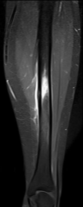
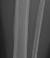
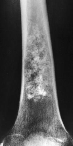
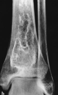
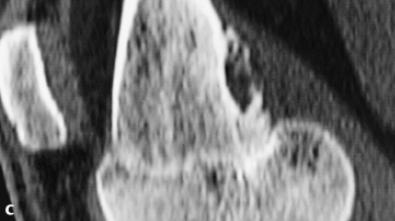
Chealon Miller, HMS IV Gillian Lieberman, MD. November Stress Fractures. Chealon Miller, Harvard Medical School Year IV Gillian Lieberman, MD
 November 2005 Stress Fractures Chealon Miller, Harvard Medical School Year IV Our Patient G.F. 29 year old female runner c/o left shin pain and swelling Evaluated at OSH with MRI showing a mass Referred
Case Report Giant Cell Tumor of Bone: Documented Progression over 4 Years from Its Origin at the Metaphysis to the Articular Surface
 Volume 2016, Article ID 9786925, 5 pages http://dx.doi.org/10.1155/2016/9786925 Case Report Giant Cell Tumor of Bone: Documented Progression over 4 Years from Its Origin at the Metaphysis to the Articular
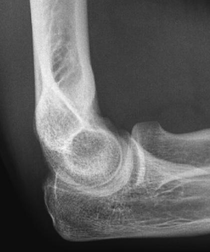
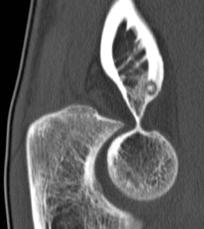
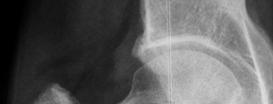
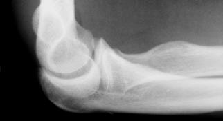
Olecranon lesions: Radiographic Appearances with Cross Sectional Imaging Correlation
 Olecranon lesions: Radiographic Appearances with Cross Sectional Imaging Correlation Poster No.: P-0107 Congress: ESSR 2014 Type: Educational Poster Authors: U. Kularatne, N. Evans, S. L. J. James ; Nottingham/UK,
Publication for the Philips MRI Community
 FieldStrength Publication for the Philips MRI Community Issue 38 Summer 2009 Pediatric MSK imaging benefits from tailored scan protocols Vanderbilt University Children s Hospital builds dedicated scans
GIANT CELL-RICH OSTEOSARCOMA: A CASE REPORT
 Nagoya J. Med. Sci. 59. 151-157, 1996 CASE REPORTS GIANT CELL-RICH OSTEOSARCOMA: A CASE REPORT KEIJI SATO!, SHIGEKI YAMAMURA!, HISASHI IWATA!, HIDESHI SUGIURA 2, NOBUO NAKASHIMA 3 and TETSURO NAGASAKA
Focal Sclerosis in a Vertebra: Differential Diagnosis of a Solitary Osteoblastic Metastasis
 July 2001 Focal Sclerosis in a Vertebra: Differential Diagnosis of a Solitary Osteoblastic Metastasis Alice L. Fisher, Harvard Medical School Year IV Review of the Normal Anatomy of a Lumbar Vertebra:
Orthopedic Hardware Imaging Part II: MRI v. Metal
 Orthopedic Hardware Imaging Trent Roth, MD And Lauren Ladd, MD Indiana University School of Medicine IU Health Physicians-Radiology Recap: Imaging Techniques Radiography Standard for initial and surveillance
Imaging of Pediatric MSK Tumors
 Imaging of Pediatric MSK Tumors Kirsten Ecklund, M.D. Boston Children s Hospital Harvard Medical School kirsten.ecklund@childrens.harvard.edu Tumor Imaging Goals Diagnosis Lesion characterization Benign
Associated Terms: Osteosarcoma, Bone Cancer, Limb Salvage, Appendicular Osteosarcoma, Pathologic Fracture, Chondrosarcoma
 1 of 9 9/29/2014 8:25 PM Associated Terms: Osteosarcoma, Bone Cancer, Limb Salvage, Appendicular Osteosarcoma, Pathologic Fracture, Chondrosarcoma The term "ACVS Diplomate" refers to a veterinarian who
MRI and CT Evaluation of Primary Bone and Soft- Tissue Tumors
 749 Alex M. Aisen1 William Martel1 Ethan M. Braunstein1 Kim I. McMillin1 William A. Phillips2 Thomas F. KIing2 Received June 10, 1985; accepted after revision December 23, 1985. Presented at the annu meeting
Bone and Joint Part 2. Leslie G Dodd, MD
 Bone and Joint Part 2 Leslie G Dodd, MD Relative rates of cancer Sarcomas are relatively uncommon tumors New cancer cases 2007 All sites 1.4 million prostate 218,890 lung 213,380 breast 180,510 Soft tissue
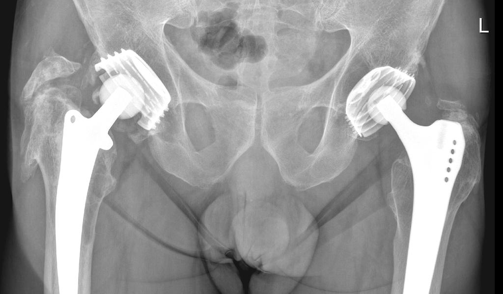
Soft Tissue Imaging in. Total Hip Arthroplasty
 FDA Orthopaedic Rehabilitation Devices Panel Medical Devices Advisory Committee Meeting Thursday June 28th 2012 Soft Tissue Imaging in Metal-on on-metal Total Hip Arthroplasty Young-Min Kwon MD, PhD, FRCS,
Recognizing Cartilaginous Tumors: Spectrum of Imaging Characteristics with Radiologic-Pathologic correlation.
 Recognizing Cartilaginous Tumors: Spectrum of Imaging Characteristics with Radiologic-Pathologic correlation. Poster No.: C-1451 Congress: ECR 2012 Type: Educational Exhibit Authors: E. Barcina García,
A peculiar location of a rare bone tumor: sternal lipoma
 A peculiar location of a rare bone tumor: sternal lipoma Poster No.: P-0033 Congress: ESSR 2016 Type: Authors: Keywords: DOI: Scientific Poster Z. Akkaya, C. Uzun, S. Enon, G. Kocaman, G. Sahin; Ankara/TR
LAC + USC.
 Jeff McDavit,, M.D. LAC + USC mcdavit@usc.edu Clinical History 55 year old male with large, deep, non- tender left thigh mass. Seen at LAC+USC Med Ctr FNA clinic No h/o trauma or radiation Vimentin
History. 33 y/o F with hx of palpable anterior tibial mass x 2 years, only painful with palpation
 History 33 y/o F with hx of palpable anterior tibial mass x 2 years, only painful with palpation Imaging Photo Album Patient also had a smaller lesion 1 cm proximal to this lesion, not seen radiographically.
SMALL ROUND BLUE CELL LESION OF BONE
 DISCLOSURE SMALL ROUND BLUE CELL LESION OF BONE Dr. Alistair Jordan University of South Alabama No financial support or endorsement OBJECTIVES Describe the more common small round cell lesions of bone
Plain Film CT. Principal Modality (2): Case Report # [] Date accepted: 15 March 2014
![Plain Film CT. Principal Modality (2): Case Report # [] Date accepted: 15 March 2014](/thumbs/89/100531147.jpg "Plain Film CT. Principal Modality (2): Case Report # [] Date accepted: 15 March 2014") Radiological Category: Musculoskeletal Principal Modality (1): Principal Modality (2): Plain Film CT Case Report # [] Submitted by: Dr. Jason E. Lally, M.D. Faculty reviewer: Dr. Naga Ramesh Chinapuvvula,
Radiological Category: Musculoskeletal Principal Modality (1): Principal Modality (2): Plain Film CT Case Report # [] Submitted by: Dr. Jason E. Lally, M.D. Faculty reviewer: Dr. Naga Ramesh Chinapuvvula,
CASE REPORT PLEOMORPHIC LIPOSARCOMA OF PECTORALIS MAJOR MUSCLE IN ELDERLY MAN- CASE REPORT & REVIEW OF LITERATURE.
 PLEOMORPHIC LIPOSARCOMA OF PECTORALIS MAJOR MUSCLE IN ELDERLY MAN- CASE REPORT & REVIEW OF LITERATURE. M. Madan 1, K. Nischal 2, Sharan Basavaraj. C. J 3. HOW TO CITE THIS ARTICLE: M. Madan, K. Nischal,
Radiologic approach to pediatric lytic bone lesions
 Radiologic approach to pediatric lytic bone lesions Poster No.: C-1177 Congress: ECR 2016 Type: Educational Exhibit Authors: J. L. LERMA GALLARDO, I. de la Pedraja, A. Lancharro 1 1 1 2 1 1 Zapata, J.
Musculoskeletal Imaging What to order? Brian Cole, MD
 Musculoskeletal Imaging What to order? Brian Cole, MD my background: 1994 University of Illinois 1998 MD University of Illinois College of Medicine 1999-2003 Diagnostic Radiology Mayo Clinic 2004 Fellowship
ORTHOPAEDIC ONCOLOGY OITE REVIEW COURSE
 ORTHOPAEDIC ONCOLOGY OITE REVIEW COURSE Richard D. Lackman, MD FACS Director, Orthopaedic Oncology Center Cancer Institute Introduction In the evaluation of a patient with a bone tumor, there are several
Department of Radiology, Yeungnam University College of Medicine, Yeungnam University Medical Center, Daegu, Korea 4
 Original Article pissn 1738-2637 / eissn 2288-2928 http://dx.doi.org/10.3348/jksr.2015.73.4.240 Bone Tumors with an Associated Pathologic Fracture: Differentiation between Benign and Malignant Status Using
Skeletal metastases are the most common variety of bone tumors and should always be considered in the differential diagnosis, particularly in older
 Dr Brajesh Nandan Skeletal metastases are the most common variety of bone tumors and should always be considered in the differential diagnosis, particularly in older patients. Cancers of the breast, prostate,
Osteonecrosis - Spectrum of imaging findings
 Osteonecrosis - Spectrum of imaging findings Poster No.: C-1861 Congress: ECR 2016 Type: Educational Exhibit Authors: P. Ninitas, A. L. Amado Costa, A. Duarte, I. Távora ; Lisbon/ 1 1 2 1 1 2 PT, Costa
Case Studies in the Skull Base
 Case Studies in the Skull Base Amy C Tsai, MD Neuroradiology Fellow Department of Radiology and Imaging Sciences University of Utah Health Sciences Center Salt Lake City, Utah, USA No disclosures related
A Journey Down The Canal
 A Journey Down The Canal Radiological Assessment of Spinal Cord Masses John Berry-Candelario HMS III Gillian Lieberman, MD BIDMC Objectives Patient review Anatomy of the spine Imaging techniques Classification
MSK Interesting Cases. Dr Yap Sheau Huey
 MSK Interesting Cases Dr Yap Sheau Huey Case 1: History 41 y.o man, surf skier C/o pain over anterior left 5 th to 8 th ribs. Worse after sport activity. Chest Radiograph US Periostitis and early callus
Case Report Intramedullary Chondrosarcoma of Proximal Humerus
 Hindawi Publishing Corporation Case Reports in Radiology Volume 2012, Article ID 642062, 7 pages doi:10.1155/2012/642062 Case Report Intramedullary Chondrosarcoma of Proximal Humerus Pratiksha Yadav, Dolly
The role of Imaging in Ewing sarcoma
 The role of Imaging in Ewing sarcoma Poster No.: P-0109 Congress: ESSR 2014 Type: Educational Poster Authors: D. Beomonte Zobel, C. Dell'atti, M. Bartocci, V. Martinelli, N. 1 2 2 2 1 1 1 2 Magarelli,
Why Talk About Technique? MRI of the Knee:
 Why Talk About Technique? MRI of the Knee: Part 1 - Imaging Techniques Mark Anderson, M.D. University of Virginia Health Sciences Center Charlottesville, Virginia Always had an interest teach our fellows
Laura M. Fayad, MD. Associate Professor of Radiology, Orthopaedic Surgery & Oncology The Johns Hopkins University
 Society of Pediatric Radiology, May 2013 Laura M. Fayad, MD Associate Professor of Radiology, Orthopaedic Surgery & Oncology The Johns Hopkins University Describes surgical techniques that resect and reconstruct
Primary periosteal lymphoma rare and unusual
 Skeletal Radiol DOI 10.1007/s00256-006-0096-2 CASE REPORT Ibrahim Fikry Abdelwahab Benjamin Hoch George Hermann Stefano Bianchi Michael J. Klein Dempsey S. Springfield Primary periosteal lymphoma rare
A review of Tumoral lesions of the shoulder
 A review of Tumoral lesions of the shoulder Poster No.: P-0109 Congress: ESSR 2013 Type: Scientific Exhibit Authors: M. M. Milán Rodríguez, Á. E. Moreno Puertas, J. M. Giménez, 1 1 1 1 2 1 A. Rubio Fernández,
This presentation is the intellectual property of the author. Contact them for permission to reprint and/or distribute.
 MRI of the Knee Jennifer Swart, M.D. Musculoskeletal Radiology South Texas Radiology Group Outline Coils, Patient Positioning Acquisition Parameters, Planes and Pulse Sequences Knee Arthrography Normal
This presentation is the intellectual property of the author. Contact them at for permission to reprint and/or distribute.
 MRI of the Knee Jennifer Swart, M.D. Musculoskeletal Radiology South Texas Radiology Group Financial Disclosure Dr. Jennifer Swart has no relevant financial relationships with commercial interests to disclose.
Disclosure. Acknowledgement. What is the Best Workup for Rectal Cancer Staging: US/MRI/PET? Rectal cancer imaging. None
 What is the Best Workup for Rectal Cancer Staging: US/MRI/PET? Zhen Jane Wang, MD Assistant Professor in Residence UC SF Department of Radiology Disclosure None Acknowledgement Hueylan Chern, MD, Department
FIRST COAST SERVICE OPTIONS FLORIDA MEDICARE PART B LOCAL COVERAGE DETERMINATION
 FIRST COAST SERVICE OPTIONS FLORIDA MEDICARE PART B LOCAL COVERAGE DETERMINATION CPT/HCPCS Codes 78300 Bone and/or joint imaging; limited area 78305 multiple areas 78306 whole body 78315 three phase study
Scrotum-like protrusion of lipoma arising from the proximal thigh
 Upsala J Med sci 109: 261 265, 2004 Scrotum-like protrusion of lipoma arising from the proximal thigh Report of two cases Koshi Hattori, 1 Masahito Hatori, 1 Mika Watanabe, 2 Toshihisa Osanai, 3 Shoichi
MRI evaluation of the shoulder: Beyond rotator cuff
 MRI evaluation of the shoulder: Beyond rotator cuff Poster No.: C-2447 Congress: ECR 2015 Type: Educational Exhibit Authors: C. Rumie, A. Vasquez, J. A. Abreu, A. P. Guarnizo, O. Rivero, 1 1 2 3 1 1 1
Section II Musculoskeletal Radiology
 Section II Musculoskeletal Radiology Figure 1 25. You are shown a noncontrast CT (Figure 1) of the thigh. What is the MOST LIKELY diagnosis? A. Synovial sarcoma B. Hemangioma C. Organizing hematoma D.
Pseudotumor Deltoideus in the Left Humerus of a Young Adult Female Patient with Acute Lateral Shoulder Pain: A Case Report
 of a Young Adult Female Patient with Acute Lateral Shoulder Pain: A Gulsen Aykol 1, Senol Fatih Elbir 2 1 Physical Medicine and Rehabilitation, Private Sevgi Medical Centre, Malatya, Turkey 2 Radiology
Case 8 Soft tissue swelling
 Case 8 Soft tissue swelling 26-year-old female presented with a swelling on the back of the left knee joint since the last 6 months and chronic pain in the calf and foot since the last 2 months. Pain in
How accurate is MRI in prediction of musculoskeletal tumors -A prospective evaluation
 Mary Hazarika Bhuyan and R K Bhuyan / International Journal of Biomedical Research 2015; 6(12): 942-946. 942 International Journal of Biomedical Research ISSN: 0976-9633 (Online); 2455-0566 (Print) Journal
FOR CMS (MEDICARE) MEMBERS ONLY NATIONAL COVERAGE DETERMINATION (NCD) FOR MAGNETIC RESONANCE IMAGING:
 MEMBERS ONLY NATIONAL COVERAGE DETERMINATION (NCD) FOR MAGNETIC RESONANCE IMAGING:") National Imaging Associates, Inc. Clinical guidelines BONE MARROW MRI Original Date: July 2008 Page 1 of 5 CPT Codes: 77084 Last Review Date: September 2014 NCD 220.2 MRI Last Effective Date: July 2011
National Imaging Associates, Inc. Clinical guidelines BONE MARROW MRI Original Date: July 2008 Page 1 of 5 CPT Codes: 77084 Last Review Date: September 2014 NCD 220.2 MRI Last Effective Date: July 2011
Clinical Study Enchondroma versus Low-Grade Chondrosarcoma in Appendicular Skeleton: Clinical and Radiological Criteria
 Oncology Volume 2012, Article ID 437958, 6 pages doi:10.1155/2012/437958 Clinical Study Enchondroma versus Low-Grade Chondrosarcoma in Appendicular Skeleton: Clinical and Radiological Criteria Eugenio
Primary Intraosseus Xanthoma Involving the Proximal Femur in a Normolipidemic Patient: A Case Report
 CASE REPORT Hip Pelvis 28(3): 182-186, 2016 http://dx.doi.org/10.5371/hp.2016.28.3.182 Print ISSN 2287-3260 Online ISSN 2287-3279 Primary Intraosseus Xanthoma Involving the Proximal Femur in a Normolipidemic
Soft Tissue Tumour & Sarcoma Imaging Guidelines 2012
 Soft Tissue Tumour & Sarcoma Imaging Guidelines 2012 Version Control This is a controlled document please destroy all previous versions on receipt of a new version. Date Approved: March 2011 reissued April
Dr Sneha Shah Tata Memorial Hospital, Mumbai.
 Dr Sneha Shah Tata Memorial Hospital, Mumbai. Topics covered Lymphomas including Burkitts Pediatric solid tumors (non CNS) Musculoskeletal Ewings & osteosarcoma. Neuroblastomas Nasopharyngeal carcinomas
Takayuki Ohguri 1 Takatoshi Aoki 1 Masanori Hisaoka 2 Hideyuki Watanabe 1 Katsumi Nakamura 1 Hiroshi Hashimoto 2 Toshitaka Nakamura 3 Hajime Nakata 1
 Takayuki Ohguri 1 Takatoshi Aoki 1 Masanori Hisaoka 2 Hideyuki Watanabe 1 Katsumi Nakamura 1 Hiroshi Hashimoto 2 Toshitaka Nakamura 3 Hajime Nakata 1 Received July 1, 2002; accepted after revision November
Ethan M. Braunstein, M.D. 1, Steven A. Goldstein, Ph.D. 2, Janet Ku, M.S. 2, Patrick Smith, M.D. 2, and Larry S. Matthews, M.D. 2
 Skeletal Radiol (1986) 15:27-31 Skeletal Radiology Computed tomography and plain radiography in experimental fracture healing Ethan M. Braunstein, M.D. 1, Steven A. Goldstein, Ph.D. 2, Janet Ku, M.S. 2,
Periosteal Osteosarcoma Arising from the Rib and Scapula: Imaging Features in Two Cases
 Case Report Musculoskeletal Imaging http://dx.doi.org/10.3348/kjr.2014.15.3.370 pissn 1229-6929 eissn 2005-8330 Korean J Radiol 2014;15(3):370-375 Periosteal Osteosarcoma Arising from the Rib and Scapula:
Pathology of Sarcoma ELEANOR CHEN, MD, PHD, ASSISTANT PROFESSOR DEPARTMENT OF PATHOLOGY UNIVERSITY OF WASHINGTON
 Pathology of Sarcoma ELEANOR CHEN, MD, PHD, ASSISTANT PROFESSOR DEPARTMENT OF PATHOLOGY UNIVERSITY OF WASHINGTON Presentation outline Background and epidemiology of sarcomas Sarcoma classification Sarcoma
FEGNOMASHIC: from x-ray to MRI
 FEGNOMASHIC: from x-ray to MRI Poster No.: C-2441 Congress: ECR 2015 Type: Educational Exhibit Authors: S. Fouassier, A. L. C. Duarte, C. Ruivo, J. Velez ; Évora/PT, 1 2 1 2 3 1 3 Coimbra/PT, PT Keywords:
Lesion Imaging Characteristics Mass, Favoring Benign Circumscribed Margins Intramammary Lymph Node
 Lesion Imaging Characteristics Mass, Favoring Benign Circumscribed Margins Intramammary Lymph Node Oil Cyst Mass, Intermediate Concern Microlobulated Margins Obscured Margins Mass, Favoring Malignant Indistinct
Bone Tumors: In 1 Simple Chart
 Bone Tumors with PowerPoint Interactivity Download this entire slideshow from When running this on your own computer you can jump from slide to slide using these buttons at bottom of each slide: Last slide
The role of CT and MRI in evaluation of Osteoid Oteoma
 The role of CT and MRI in evaluation of Osteoid Oteoma Elene Iordanishvili Tbilisi Sate Medical University Instructor: Prof. Dr. Ketevan Kotetishvili Department of Physics Georgian Technical University
Incidental bone tumors are asymptomatic lesions that are. Incidental Bone Lesions. When to Refer to the Tumor Specialist
 Bulletin of the NYU Hospital for Joint Diseases 2012;70(4):235-40 235 Incidental Bone Lesions When to Refer to the Tumor Specialist LT Suezie Kim, M.D., M.C., U.S.N., Catherine N. Laible, M.D., Leon D.
Common Primary Tumors of Bone
 Special Report Common Primary Tumors of Bone Primary bone tumors are a relatively rare occurrence, however, they can have serious deleterious consequences. Many possess the ability to degenerate into malignant