CINtec p16 INK4a Staining Atlas
|
|
|
- Brice Earl Norton
- 5 years ago
- Views:
Transcription
1 CINtec p16 INK4a Staining Atlas
2 Rating


3 Rating Positive The rating positive will be assigned if the p16 INK4a -stained slide shows a continuous staining of cells of the basal and parabasal cell layers of the squamous cervical epithelium, with or without staining of cells of superficial cell layers ( diffuse staining pattern ). Diffuse Staining Pattern: CIN2 CIN3 Invasive carcinoma 3


, or a staining of")


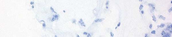
4 Rating Negative The rating negative will be assigned if the p16 INK4a -stained slide shows either a negative staining reaction in the squamous epithelium ( negative staining pattern ), or a staining of isolated cells or small cell clusters; i.e., a non-continuous staining, particularly not of the basal and parabasal cells ( focal staining pattern ). Negative Staining Pattern: Normal squamous epithelium Squamous metaplasia, immature Cervicitis 4

, or a staining of")

.")
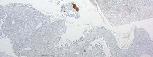
5 Rating Negative The rating negative will be assigned if the p16 INK4a -stained slide shows either a negative staining reaction in the squamous epithelium ( negative staining pattern ), or a staining of isolated cells or small cell clusters; i.e., a non-continuous staining, particularly not of the basal and parabasal cells ( focal staining pattern ). Focal Staining Pattern: Squamous metaplasia, mature Squamous metaplasia, mature 5


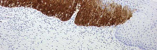
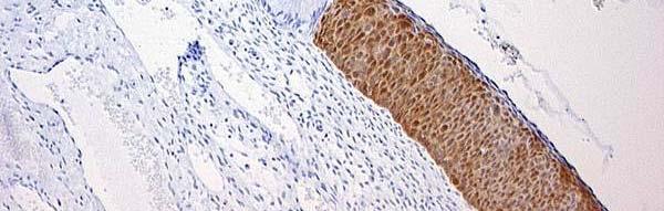
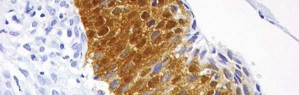
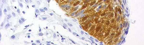
6 CINtec p16 INK4a Histology Strong diffuse p16 INK4a -specific immuno-reactivity in dysplastic cervical lesions CIN1 CIN2 CIN3 6

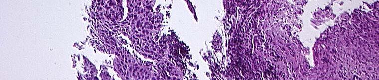

7 CINtec p16 INK4a Histology Conventional H&E CINtec p16 INK4a CIN3 severe dysplasia 7


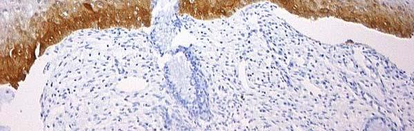
8 CINtec p16 INK4a Histology Potential increased reproducibility in diagnosing cervical lesions by p16 INK4a IHC Consensus diagnosis: no lesion 25x 25x High-grade g lesion detected by p16 INK4a IHC 400x 400x 8
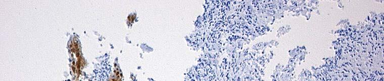
9 Dysplasia, a, caused by HR-HPV p16 INK4a Immunostain: diffuse distribution of (+) cells in immature layers Stained cells may reach the surface of the epithelium 9


10 Invasive squamous carcinoma Fig. 2a: Tumor cells with marked variation of nuclear size and shape. Marked inflammatory background. Fig. 2b: Strong positivity for p16 INK4a in cytoplasm and nuclei. 10 Cytology


11 Invasive squamous carcinoma H&E p16 INK4a Fig. 2c: Irregular infiltrates of tumor cells in connective tissue with inflammatory reaction. Fig. 2d: Strong cytoplasmic and nuclear positivity for p16 INK4a of carcinoma cells. 11 Histology
 Tumor cells of")


.")


12 Endocervical adenocarcinoma (in situ) Tumor cells of endocervical adenocarcinoma in situ: Fig. 3a: Pseudostratified complex of malignant glandular cells with marked nuclear enlargement and feathering (here:endocervical adenocarcinoma in situ). Tumor cells of endocervical adenocarcinoma: Fig. 3b: Strong positivity for p16 INK4a in cytoplasm and nuclei of malignant glandular cells. 12 Cytology
: Fig.")


13 Endocervical adenocarcinoma Endocervical adenocarcinoma (Grade I): Fig. 3c: Neoplastic endocervical glands are lined by a pseudostratified epithelium, consisting of neoplastic columnar cells with hyperchromatic, irregularly shaped nuclei. Endocervical adenocarcinoma (Grade I): Fig. 3d: Strong cytoplasmic and nuclear positivity for p16 INK4a with a diffuse distribution. 13 Histology


14 Cervical Intraepithelial Neoplasia Grade 3 Cells from CIN3 (HSIL): Fig. 4a: Several undifferentiated neoplastic cells. Marked hyperchromasia and anisonucleosis. Marked increase of nuclear-cytoplasmic ratio. Cells from CIN3 (HSIL): Fig. 4b: Strong cytoplasmic and nuclear positivity for p16 INK4a. 14 Cytology
 H&E")
 extending")


15 Cervical Intraepithelial Neoplasia Grade 3 (Carcinoma in situ) H&E Carcinoma in situ (CIN3): Fig. 5a: (Carcinoma in situ) extending into endocervical gland. Neoplastic cells with relatively uniform, hyperchromatic nuclei throughout the epithelium. Carcinoma in situ (CIN3): Fig. 5b: Carcinoma in situ extending into endocervical gland. Strong nuclear and cytoplasmic p16 INK4a positivity in neoplastic epithelium in a diffuse distribution. 15 Histology
 Dysplastic")


16 Cervical Intraepithelial Neoplasia Grade 3 (Severe Dysplasia) Dysplastic cells from severe dysplasia (HSIL): Fig. 6a: Dysplastic cells with variation of nuclear and cytoplasmic shape. Markedly increased nuclear-cytoplasmic ratio. Coarsely granular chromatin. No nuceoli. Dysplastic cells from severe dysplasia (HSIL): Fig. 6b: Nuclear and cytoplasmic positivity for p16 INK4a. 16 Cytology
 Severe dysplasia")


17 Cervical Intraepithelial Neoplasia Grade 3 (Severe Dysplasia) Severe dysplasia (CIN3)Fig. 6c: Dysplastic cells with large, hyperchromatic nuclei almost throughout the entire epithelium. Severe dysplasia (CIN3) Fig. 6d: Mostly strong, nuclear and cytoplasmic positivity for p16 INK4a in dysplastic epithelium with a diffuse distribution. 17 Histology
: Cells from CIN2")


18 Cervical Intraepithelial Neoplasia Grade 2 Cells from CIN2 (HSIL): Cells from CIN2 (HSIL): Fig. 7a: Sheet of dysplastic cells of intermediate Fig. 7b: Several dysplastic yp cells of deeper layers. cell layers. Variation of nuclear shape. Nuclear Marked variation of nuclear and cytoplasmic shape. enlargement. Coarsely granular chromatin. Increased nuclear-cytoplasmic ratio. Hyperchromasia. Chromocenters. No nucleoli. No p16 INK4a Coarsely granular chromatin. Cytoplasmic and nuclear positivity. positivity for p16 INK4a in dysplastic cells. 18 Cytology


19 Cervical Intraepithelial Neoplasia Grade 2 CIN2 without koilocytosis: CIN2 without koilocytosis: Fig. 7c: Dysplastic cells in the lower half of the Fig. 7d: In the lower half of the epithelium epithelium. dysplastic cells show cytoplasmic and nuclear p16 INK4a positivity in a diffuse distribution. The cells in the superficial layers show no or only weak p16 INK4a positivity. 19 Histology


20 Koilocytosis Koilocytes: Fig. 8a: Large intermediate cells with perinuclear cavity. Relatively dense rim of peripheral cytoplasm. Mild nuclear irregularity with hyperchromasia. Koilocyte: Fig. 8b: No staining reaction for p16 INK4a in koilocyte with a double nucleus. 20 Cytology


21 Koilocytosis CIN2 with Koilocytosis: CIN2 with koilocytosis: Fig. 8c: Moderate dysplasia showing koilocytes Fig. 8d: p16 INK4a positivity only in dysplastic cells with perinuclear cavity and mildly irregular, of fbasal and parabasal llayers, koilocytes in hyperchromatic nuclei in upper epithelial cell upper layers are mostly p16 INK4a negative. layers (Arrowhead: Mitosis). 21 Histology
22 22 Fig. 9: Scheme of p16 INK4a immunostaining, stratified squamous epithelium


23 Superficial cells Superficial cells from stratified squamous epithelium: Fig. 10a: Large polygonal flat cell. Distinct cell borders. Pyknotic nuclei. Superficial cells from stratified squamous epithelium: Fig. 10b: Negative for p16ink4a. 23 Cytology


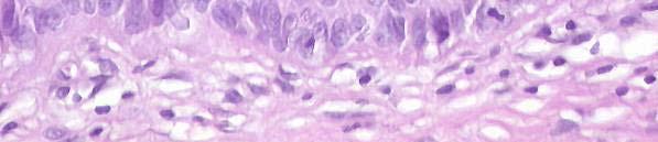
24 Squamous epithelium, upper layers Normal stratified squamous epithelium, Normal stratified squamous epithelium, superficial cell layers: superficial cell layers: Fig. 11 a: Superficial i differentiated t d squamous Fig. 11b: Negative for p16 INK4a. cells with large and flat cell bodies. Pyknotic nuclei. 24 Histology


25 Squamous epithelium, intermediate layers Intermediate cells from stratified squamous Intermediate cells from stratified squamous epithelium: epithelium: Fig. 12a: Large to medium m large polygonal cells. Fig. 12b: Negative for p16 INK4a. Distinct cell borders. Vesicular nuclei. Finely granular chromatin. Occasionally chromocenters. Open arrowheads: Superficial cells. 25 Cytology


26 Squamous epithelium, intermediate layers Stratified squamous epithelium, intermediate cell layer: Fig. 12c: Medium large, differentiating polygonal l cells. Vesicular nuclei. Uniformely fine granular chromatin. Vacuolic cell body due to glycogen content. Stratified squamous epithelium, intermediate cell layer: Fig. 12d: Negative for p16 INK4a. 26 Histology


27 Squamous epithelium, parabasal cells Parabasal cells from stratified squamous epithelium: Fig. 13a: Relatively small squamous cells with round to oval shape. Round to oval vesicular nuclei. Evenly distributed and finely granular chromatin. Chromocenters. Parabasal cells from stratified squamous epithelium: Fig. 13b: Negative for p16ink4a. 27 Cytology


28 Squamous epithelium, lower layers Stratified squamous epithelium, basal and parabasal cell layer: Fig. 14a: Relatively small squamous cells with round to oval shape. Round to oval vesicular nuclei. Arrowhead: Clearly delineated row of palisading basal cells. Bracket: Parabasal cell layers. Stratified squamous epithelium, basal and parabasal cell layer: Fig. 14b: Negative for p16 INK4a. Arrowhead: Clearly delineated row of palisading basal cells. Bracket: Parabasal cell layers. 28 Histology


29 Atrophy Cells from ectocervical atrophy: Fig. 15a: Atrophic cell cluster, small rounded to slightly elongated cells with small round to oval regular nuclei, some of them slightly hyperchromatic. Some small intermediate cells. Cells from ectocervical atrophy: Fig. 15b: Negative for p16ink4a with occasional positive cells in superficial layers (Arrowheads). 29 Cytology


.")
30 Squamous epithelium, Atrophy Atrophic squamous epithelium: Atrophic squamous epithelium: Fig. 16a: Stratified squamous epithelium with Fig. 16b: Negative for p16 INK4a with occasional little maturation. Epithelium is composed almost positive cells in superficial layers (Arrow). entirely of basal and parabasal cells. 30 Histology


31 Endocervical columnar cells Columnar cells from endocervical epithelium: Fig. 17a : Mucus producing cells in palisading and also in honeycomb arrangement. Distinct cell borders. Round to oval, basally located nuclei of uniform size. Finely granular chromatin. Chromocenters. Columnar cells from endocervical epithelium: Fig. 17b: Single layered endocervical cells, columnar cells mostly negative for p16ink4, occasionally a positive p16ink4a reaction in cytoplasm and nuclei of columnar cells is seen. 31 Cytology


32 Endocervical epithelium Endocervical mucosa, columnar cells: Fig. 18a: Mucus producing cells in a single layer lining an endocervical gland. Distinct cell borders. Round to oval, basally located nuclei of uniform size. Mucus in glandular lumen. Endocervical mucosa, columnar cells: Fig. 18b: Overwhelming majority of cells negative for p16 INK4, interspersed single cells may occasionally show positive p16 INK4a reaction in cytoplasm and nuclei. 32 Histology


33 Endometrium Cluster of endometrial cells: Fig. 19a: Three-dimensional cluster of endometrial cells with rounded outline from the endometrial cavity. Tightly packed nuclei, obscuring nuclear detail. Round to oval nuclear shape. Uniformely fine chromatin. Chromocenters. Fig. 19b: Single cells positive for p16ink4a. 33 Cytology


34 Endometrium Endometrium during menstruation: Fig. 19c: Glands and stromal cells of endometrial tissue shrunken and partly dissociated with cellular aggregates between fresh hemorrhages. Endometrium during menstruation: Fig. 19d: Single cells may occasionally show p16 INK4a positivity. 34 Histology


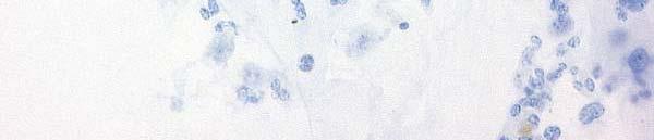
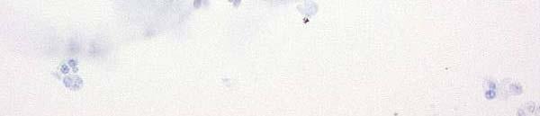
35 Inflammation Neutrophil granulocytes: Fig. 20a: Polymorphonuclear leukocytes with typical nuclear shape. Neutrophil granulocytes: Fig. 20b: Negative for p16ink4a. 35 Cytology


36 Inflammation Histiocytes: Histiocytes: Fig. 20c: Open arrowheads: Several large Fig. 20d: Open arrowheads: Histiocytes are histiocytes with typical foamy cytoplasm and negative for p16 INK4a. indistinct cell borders (conventional smear). 36 Cytology


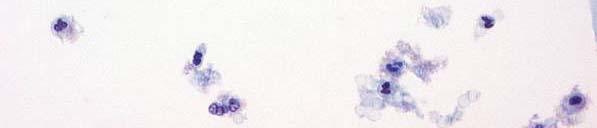
37 Microorganisms Bacteria: Fig. 21a: Small rod-shaped bacteria of Döderlein type. Fig. 21b: Positive staining reaction of bacteria in less than 5% of the cases, always easily discriminated from cellular staining. 37 Cytology


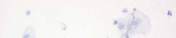
38 Microorganisms Trichomoniasis: Trichomoniasis: Fig. 21c: Oval to pear shaped trichomonads Fig. 21d: Negative staining reaction with CINtec with flagelli. Small, comma-shaped nucleus. Cytology Kit. Staining with other antibodies Some granulocytes. against the antigen p16 INK4a may result in falsepositive reaction. 38 Cytology

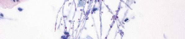
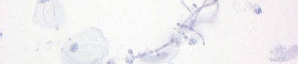

39 Microorganisms Candida albicans: Fig. 21e: Tubular hyphae with segmentation, branching and budding. Candida albicans: Fig. 21f: Negative staining reaction. 39 Cytology
40 Fig. 22: Scheme of p16 INK4a immunostaining, transformation zone with squamous metaplasia / tissue repair 40


41 Metaplasic Changes I Cells from immature metaplasia and normal endocervical cells: Fig. 23a: Group of columnar cells with small subcolumnar reserve cells. Relatively dense cytoplasm. Round to oval nuclei with finely granular chromatin. No nucleoli. Cells from immature metaplasia and normal endocervical cells: Fig. 23b: Subcolumnar reserve cells negative for p16ink4a. 41 Cytology


42 Metaplasic Changes II Maturing squamous cell metaplasia / tissue repair: Fig. 24a: Polygonal cells of intermediate cell type with rounded corners. Uniformly finely granular chromatin. Maturing squamous cell metaplasia / tissue repair: Fig. 24b: Group of relatively mature squamous epithelial cells shows positivity for p16ink4a in some cells, whereas others are negative. 42 Cytology


43 Metaplasic Changes II Transformation zone, maturing squamous Transformation zone, maturing squamous cell metaplasia: cell metaplasia: Fig. 24c: Regularly l maturing squamous cells Fig. 24d: Some of the maturing squamous cells bordering to endocervical columnar cells show cytoplasmic and nuclear positivity for (arrowhead). p16 INK4a in a sporadic distribution of the positive cells in superficial layers. 43 Histology


44 Tubal metaplasia H&E Pap p16ink4a p16 INK4a Tubal metaplasia Fig. 25a: Ciliated columnar cells in a single cell layer lining an endocervical gland. Metaplastic cells show positivity for p16 INK4a only in part of the epithelial cells, neighboring cells are negative (arrowheads). Insert: Higher Magnification to demonstrate t ciliated cells (H&E). Fig. 25b: Small group of endocervical ciliated columnar cells. Chromatin somewhat coarser than in normal endocervical cells. No nucleoli. 44


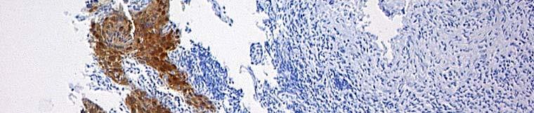
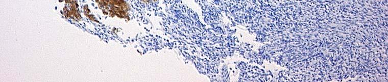
45 Squamous Metaplasia Immature squamous metaplasia: Fig. 26a: Reserve cells, partly covered by a single layer of endocervical columnar cells (arrowheads) with focal differentiation to metaplastic squamous cells. Round to oval nuclei with finely granular chromatin. Immature squamous metaplasia: Fig. 26b: No positivity for p16 INK4a in subcolumnar reserve cells. Arrowhead: Endocervical columnar cells. 45 Histology
SQUAMOUS CELLS: Atypical squamous cells (ASC) - of undetermined significance (ASC-US) - cannot exclude HSIL (ASC-H)
 - of undetermined significance (ASC-US) - cannot exclude HSIL (ASC-H)") SQUAMOUS CELLS: Atypical squamous cells (ASC) - of undetermined significance (ASC-US) - cannot exclude HSIL (ASC-H) ASC refers to cytologic changes suggestive of SIL, which are qualitativley or quantitatively
SQUAMOUS CELLS: Atypical squamous cells (ASC) - of undetermined significance (ASC-US) - cannot exclude HSIL (ASC-H) ASC refers to cytologic changes suggestive of SIL, which are qualitativley or quantitatively
Table of Contents. 1. Overview. 2. Interpretation Guide. 3. Staining Gallery Cases Negative for CINtec PLUS
 Staining Atlas Table of Contents 1. Overview 1.1 Introduction 1.2 Role of p16 INK4a 1.3 Role of Ki-67 1.4 Molecular Pathogenesis 1.5 p16 INK4a Expression in Cervical Dysplasia 1.6 The Concept of CINtec
LGM International, Inc.
 Liqui-PREP TM Cytology Atlas Preface The following pictures are examples with descriptions of cytology slides processed with the Liqui-PREP TM System.. The descriptions are reviewed by Pathologists. It
Prepared By Jocelyn Palao and Layla Faqih
 Prepared By Jocelyn Palao and Layla Faqih The structure of the suspected atypical cell should always be compared to the structure of other similar, benign, cells which are present in the smears. The diagnosis
Gynecologic Cytopathology: Glandular lesions
 Gynecologic Cytopathology: Glandular lesions Lin Wai Fung (MSc, MPH, CMIAC) 17/4/2014 Glandular lesions of the uterus Endocervix Endometrium Normal endocervical cells Sheets, strips well-preserved architecture:
Histopathology: Cervical HPV and neoplasia
 Histopathology: Cervical HPV and neoplasia These presentations are to help you identify basic histopathological features. They do not contain the additional factual information that you need to learn about
Hyperchromatic Crowded Groups: What is Your Diagnosis? Session 3000
 Hyperchromatic Crowded Groups: What is Your Diagnosis? Session 3000 Thomas A. Bonfiglio, M.D. Professor Emeritus, Pathology and Laboratory Medicine University of Rochester Disclosures In the past 12 months,
Objectives. Atypical Glandular Cells. Atypical Endocervical Cells. Reactive Endocervical Cells
 2013 California Society of Pathologists 66 th Annual Meeting San Francisco, CA Atypical Glandular Cells to Early Invasive Adenocarcinoma: Cervical Cytology and Histology Christina S. Kong, MD Associate
New Diagnoses Need New Approaches: A Glimpse into the Near Future of Gynecologic Pathology
 New Diagnoses Need New Approaches: A Glimpse into the Near Future of Gynecologic Pathology United States and Canadian Academy of Pathology 102 nd Annual Meeting Baltimore, Maryland Christina S. Kong, M.D.
Morphology I Slide: 1
 Morphology I Slide: 1 Morphology I Slide: 2 ThinPrep Morphology Normal Cytology Morphology I Slide: 3 CT & Pathologist Training Training program begins with ThinPrep morphology presentation Microscopic
Maturation Index 3/29/2017. Disclosure of Relevant Financial Relationships. Gynecologic Cytology. Normal Maturation of Squamous Epithelium : :
 Gynecologic Cytology Fadi W. Abdul Karim, MD MEd Department of Anatomic Pathology Vice Chair Education RT PLMI Professor of Pathology Cleveland Clinic. Cleveland Ohio Disclosure of Relevant Financial Relationships
Cervical Cancer : Pap smear
 Taking a PAP SMEAR Cervical Cancer : Pap smear George N Papanicolaou introduced cervical cytology in clinical practice in 1940 In 1945, PAP smear was endorsed by American cancer society as an effective
Cytyc Corporation - Case Presentation Archive - July 2002
 ThinPrep Pap Test History: 34 Year Old Female LMP: Day 20 Specimen Type: Cervical/Vaginal Case provided by Mark Tulecke, M.D. and Gabrielle Trawinski CT (ASCP), Mount Auburn Hospital, Cambridge, Massachusetts.
PAP SMEAR by Dr.Shantha Krishnamurthy MD Senior Consultant Pathology Fortis Hospitals
 PAP SMEAR by Dr.Shantha Krishnamurthy MD Senior Consultant Pathology Fortis Hospitals Historical Named after George Papanicolaou, a Greek American Studied cervical epithelium in menstrual cycle of guinea
Workshop for O& G trainees and paramedics 17 Dec 2011 Cytological Interpretation
 Workshop for O& G trainees and paramedics 17 Dec 2011 Cytological Interpretation May Yu Director of Cytology Laboratory Service Department of Anatomical & Cellular Pathology Prince of Wales Hospital Cervical
PRESENTATION PLAN. Aim: Bethesda System 2001
 REACTIVE CELLULAR CHANGES AND INFECTIONS OF FEMALE GENITAL TRACT Aysun Uğuz, Prof, MD, FIAC Çukurova Üniv. Tıp Fak. Pathology Department-Cytology Division 18.Nisan.2015 Aim: The aim of the presentation
Case 3 - GYN. History: 66 year old, routine Pap test. Dr. Stelow
 Case 3 - GYN History: 66 year old, routine Pap test Dr. Stelow Case 3 66 year year old woman Routine Pap Test Cytologic Features 3 dimensional clusters of cells with small to moderate amount of
Cytology Report Format
 Squamous Precursor Lesions and Malignancies In Pap Test Dina R. Mody, MD, FCAP Director of Cytology The Methodist Hospital, Houston, TX Professor of Pathology and Laboratory Medicine Weill Medical College
Interpretation guide. Abnormal cytology can t hide anymore
 Interpretation guide Abnormal cytology can t hide anymore Unique dual-biomarker technology makes you certain about the presence of transforming HPV infection. The science that creates certainty. Table
Colposcopy. Attila L Major, MD, PhD
 Colposcopy Attila L Major, MD, PhD Histology Colposcopy Cytology It has been estimated that annual Pap smear testing reduces a woman s chance of dying of cervical cancer from 4 in 1000 to about 5 in 10,000
Cytoplasmic changes Nuclear changes
 The presence of infection in the female genital tract may procure certain cellular changes in the epithelium. Such changes are seen in nucleus and cytoplasm surrounding the nucleus. Cytoplasmic changes
BOSNIAN-TURKISH CYTOPATHOLOGY SCHOOL June 18-19, 2016 Sarajevo. Case Discussions. 60 year old woman Routine gynecologic control LBC
 BOSNIAN-TURKISH CYTOPATHOLOGY SCHOOL June 18-19, 2016 Sarajevo Case Discussions Prof Dr Sıtkı Tuzlalı Tuzlalı Pathology Laboratory 60 year old woman Routine gynecologic control LBC 1 2 Endometrial thickening
A Study on Diagnostic Accuracy of Cervical Pap Smear by Correlating with Histopathology in a Tertiary Care Centre
 Original Article DOI: 10.21276/APALM.1878 A Study on Diagnostic Accuracy of Cervical Pap Smear by Correlating with Histopathology in a Tertiary Care Centre Rachana L Y, S.S. Hiremath*, Prabhu M H, S.S
1.Acute and Chronic Cervicitis - At the onset of menarche, the production of estrogens by the ovary stimulates maturation of the cervical and vaginal
 Diseases of cervix I. Inflammations 1.Acute and Chronic Cervicitis - At the onset of menarche, the production of estrogens by the ovary stimulates maturation of the cervical and vaginal squamous mucosa
CYTOMORPHOLOGY MODULE 28.1 INTRODUCTION OBJECTIVES 28.2 GENERAL GUIDELINES. Notes
 28 CYTOMORPHOLOGY 28.1 INTRODUCTION Light microscopic examination of stained cells in smears is the method of choice of diagnostic cytology. It allows classification of most normal cells as to type and
Morphologic Clues and Pitfalls for High Grade Lesions in Cervical Cytology
 Morphologic Clues and Pitfalls for High Grade Lesions in Cervical Cytology Ritu Nayar, MD Northwestern University, Feinberg School of Medicine Chicago, IL, USA Disclosures Editor, Cervical Cytology Bethesda
EU guidelines for reporting gynaecological cytology
 EU guidelines for reporting gynaecological cytology Amanda Herbert Guy s & St Thomas Foundation NHS Trust 5th EFCS Annual Tutorial, Trondheim, Norway 28 th May 1 st June 2012 EU guidelines aim to harmonize
CINtec PLUS Cytology. Interpretation training
 CINtec PLUS Cytology Interpretation training Objectives After reviewing this learning module, you will have a basic understanding of how to interpret CINtec PLUS Cytology, including: The mechanism of action
To further assess abnormalities detected on cervical cytological sample. To guide colposcopically directed biopsy
 1 To further assess abnormalities detected on cervical cytological sample To guide colposcopically directed biopsy To exclude invasive disease To aid in outpatient management and treatment of precancerous
p16 Cervical HISTOLOGY Histology Compendium & Staining Atlas
 p16 Cervical HISTOLOGY Histology Compendium & Staining Atlas Chapter 1: An Introduction to p16...... 3 Normal Cervical Epithelium and the Cell Cycle....4 HPV Infection and Cervical Disease......................................
Endometrial Metaplasia, Hyperplasia & Other Cancer Mimics: a Consultant s Experience
 Endometrial Metaplasia, Hyperplasia & Other Cancer Mimics: a Consultant s Experience Pacific Northwest Society of Pathologists Vancouver, B.C. September 26, 2015 Teri A. Longacre, M.D. longacre@stanford.edu
Outline 11/2/2017. Pancreatic EUS-FNA general aspects. Cytomorphologic features of solid neoplasms/lesions of the pancreas
 ENDOSCOPIC ULTRASOUND GUIDED-FINE NEEDLE ASPIRATION CYTOLOGY OF PANCREAS Khalid Amin M.D. Assistant Professor Department of Laboratory Medicine and Pathology University of Minnesota Outline Pancreatic
The ABCs of TBS. A Novice's Guide to the Bethesda System
 CE U P D A T E W O M E N ' S HEALTH III Julia Woodruff Wildes, MD The ABCs of TBS A Novice's Guide to the Bethesda System This is the third and final article in a three-part series on women's health. The
Normal Morphology. Anatomic Considerations. Normal Urothelial Histology and Cytology
 1 Normal Morphology Anatomic Considerations The urinary tract can be divided into three regions: the kidney; the calyces, pelves and ureters (upper collecting system or upper tract); and the bladder and
Lessons From Cases of Screened Women Who Developed Cervical Carcinoma
 Lessons From Cases of Screened Women Who Developed Cervical Carcinoma R. Marshall Austin MD,PhD Magee-Womens Hospital of University of Pittsburgh Medical Center raustin@magee.edu Why Focus Study On Cases
Conflict of Interest 9/7/2018. Dr. Mody 1. None with vendors of cytology equipment/testing/vaccines Amirsys (now Elsevier)
") Glandular Lesions in Cervicovaginal Cytology: Patterns, Pitfalls and Bethesda Updates Dina R Mody, MD Director of Cytology Laboratories Houston s Methodist Hospital and Bioreference Laboratory The Ibrahim
Glandular Lesions in Cervicovaginal Cytology: Patterns, Pitfalls and Bethesda Updates Dina R Mody, MD Director of Cytology Laboratories Houston s Methodist Hospital and Bioreference Laboratory The Ibrahim
A cyto-histopathological correlation study of lesions of uterine cervix
 Original Research Article Mandakini B. Tengli 1,*, Mohammed Mateen Ahmed 2 1 Associate Professor, 2 Assistant Professor, Dept. of Pathology, KBNIMS, Gulbarga *Corresponding Author: Email: mandakinibt@gmail.com
Clinically Microscopically Pathogenesis: autoimmune not lifetime
 Vulvar Diseases: Can be divided to non-neoplastic and neoplastic diseases. The neoplastic diseases are much less common. Of those, squamous cell carcinoma is the most common. most common in postmenopausal
Cytyc Corporation - Case Presentation Archive - March 2002
 FirstCyte Ductal Lavage History: 68 Year Old Female Gail Index: Unknown Clinical History: Negative Mammogram in 1995 6 yrs. later presents with bloody nipple discharge Subsequent suspicious mammogram Suspicious
Index 179. Genital tract contaminants, 17, 20, 22, 150 papilloma virus-infected cells, 47 squamous cells, sources of, 7
 Index Accuracy of urinary cytology, 166 Acute inflammatory cells, 38 catheter sample, 39 herpes simplex infections, 44 carcinomas, 104, 105 non-viral inclusions, 52, 53 voided urine, 17 Adenocarcinoma
Neoplasia 2018 Lecture 2. Dr Heyam Awad MD, FRCPath
 Neoplasia 2018 Lecture 2 Dr Heyam Awad MD, FRCPath ILOS 1. List the differences between benign and malignant tumors. 2. Recognize the histological features of malignancy. 3. Define dysplasia and understand
chapter 4. The effect of oncogenic HPV on transformation zone epithelium
 chapter 4. The effect of oncogenic HPV on transformation zone epithelium CHAPTER 1 All squamous cervical cancer (and probably all cervical adenocarcinoma) is associated with oncogenic HPV, and the absence
Salivary Glands 3/7/2017
 Salivary Glands 3/7/2017 Goals and objectives Focus on the entities unique to H&N Common board type facts Information for your future practice Salivary Glands Salivary Glands Major gland. Paratid. Submandibular.
number Done by Corrected by Doctor Maha Shomaf
 number 16 Done by Waseem Abo-Obeida Corrected by Zeina Assaf Doctor Maha Shomaf MALIGNANT NEOPLASMS The four fundamental features by which benign and malignant tumors can be distinguished are: 1- differentiation
Demystifying Endometrial Hyperplasia
 Demystifying Endometrial Hyperplasia A review from Diagnostic Histopathology 19:7 Dr R Hadden ST5 Histopathology Derriford Hospital Plymouth Endometrium Target for sex-steroid hormones Glands Stroma Proliferate
When Immunostains Can Get You in Trouble: Gynecologic Pathology p16: Panacea or Pandora s Box?
 When Immunostains Can Get You in Trouble: Gynecologic Pathology p16: Panacea or Pandora s Box? Teri A. Longacre, MD Stanford Medicine Stanford California pi6 in Gynecologic Pathology: Panacea or Pandora
Colposcopic Principles. Simon Leeson Consultant Obstetrician/ Gynaecologist Betsi Cadwaladr University Health Board UK
 Colposcopic Principles Simon Leeson Consultant Obstetrician/ Gynaecologist Betsi Cadwaladr University Health Board UK The cervix topics discussed are: original squamous epithelium endocervical epithelium
Understanding Your Pap Test Results
 Understanding Your Pap Test Results Most laboratories in the United States use a standard set of terms called the Bethesda System to report pap test results. Normal: Pap samples that have no cell abnormalities
Glandular lesions in cervical cytology. Margareta Strojan Fležar Institute of Pathology Faculty of Medicine University of Ljubljana Slovenia
 Glandular lesions in cervical cytology Margareta Strojan Fležar Institute of Pathology Faculty of Medicine University of Ljubljana Slovenia 2nd PANNONIA CONGRESS OF PATHOLOGY, SIÓFOK, HUNGARY, 17-19 MAY
Case year female. Routine Pap smear
 Case 1 57 year female Routine Pap smear Diagnosis? 1. Atypical glandular cells of unknown significance (AGUS) 2. Endocervical AIS 3. Endocervical adenocarcinoma 4. Endometrial adenocarcinoma 5. Adenocarcinoma
Pancreatitis: A Potential Pitfall in Endoscopic Ultrasound Guided Pancreatic FNA
 Pancreatitis: A Potential Pitfall in Endoscopic Ultrasound Guided Pancreatic FNA Jack Yang, MD Department of Pathology, Medical University of South Carolina Objectives Understand the indication of EUS
Pathology Slides. [Pathology]
![Pathology Slides. [Pathology]](/thumbs/94/120604575.jpg "Pathology Slides. [Pathology]") Pathology Slides MedicoNotes provides real laboratory pathological slides to aid you to differentiate between different pathological structures under microscope. www.mediconotes.com Histology slides example
Pathology Slides MedicoNotes provides real laboratory pathological slides to aid you to differentiate between different pathological structures under microscope. www.mediconotes.com Histology slides example
Cervical Dysplasia and HPV
 Cervical Dysplasia and HPV J. Anthony Rakowski D.O., F.A.C.O.O.G. MSU SCS Board Review Coarse HPV Double stranded DNA virus The HPV infect epithelial cells of the skin and mucous membranes Highest risk
6/5/2010. Outline of Talk. Endometrial Alterations That Mimic Cancer & Vice Versa: Metaplastic / reactive changes. Problems in Biopsies/Curettages
 Outline of Talk Endometrial Alterations That Mimic Cancer & Vice Versa: Problems in Biopsies/Curettages Metaplastic / reactive changes Mucinous change Microglandular hyperplasia-like change Squamous metaplasia
ACGME Competency / Milestone Assessment. The Pap Test. Ricardo R. Lastra, MD Zubair W. Baloch, MD, PhD
 1 ACGME Competency / Milestone Assessment The Pap Test Ricardo R. Lastra, MD Zubair W. Baloch, MD, PhD Department of Pathology & Laboratory Medicine University of Pennsylvania, Perelman School of Medicine
3/28/2017. Disclosure of Relevant Financial Relationships. GU Evening Subspecialty Case Conference. Differential Diagnosis:
 GU Evening Subspecialty Case Conference Rajal B. Shah, M.D. VP, Medical Director, Urologic Pathology Miraca Life Sciences, Irving, Texas Clinical Associate Professor of Pathology Baylor College of Medicine,
Benign and malignant epithelial lesions: Seborrheic keratosis: A common benign pigmented epidermal tumor occur in middle-aged or older persons more
 Benign and malignant epithelial lesions: Seborrheic keratosis: A common benign pigmented epidermal tumor occur in middle-aged or older persons more common on the trunk; but extremities, head and neck are
Comparison of Cytologic Characteristics between Adenoid Cystic Carcinoma and Adenoid Basal Carcinoma in the Uterine Cervix
 Journal of Pathology and Translational Medicine 2015; 49: 396-402 ORIGINAL ARTICLE Comparison of Cytologic Characteristics between Adenoid Cystic Carcinoma and Adenoid Basal Carcinoma in the Uterine Cervix
Mody. Atypical Glandular Cells(TBS 2001) Adenocarcinoma In Situ(TBS 2001)
 Adenocarcinoma In Situ(TBS 2001)") Glandular Lesions in Cervicovaginal Cytology Dina R. Mody, MD, FCAP Director of Cytology The Methodist Hospital, Houston, TX Professor of Pathology and Laboratory Medicine Weill Medical College of Cornell
Glandular Lesions in Cervicovaginal Cytology Dina R. Mody, MD, FCAP Director of Cytology The Methodist Hospital, Houston, TX Professor of Pathology and Laboratory Medicine Weill Medical College of Cornell
Premalignant lesions may expose to a promoting. factor & may be induced to undergo malignant. Carcinoma in situ displays the cytologic features of
 بسم رلاهللا Def. Premalignant lesions may expose to a promoting factor & may be induced to undergo malignant transformation. Carcinoma in situ displays the cytologic features of malignancy without invasion
Biliary tract tumors
 Short Course 2010 Annual Fall Meeting of the Korean Society for Pathologists Biliary tract tumors Joon Hyuk Choi, M.D., Ph.D. Professor, Department of Pathology, Yeungnam Univ. College of Medicine, Daegu,
Epithelia will be discussed according to the following scheme: Type Number of layers Shape Line drawing. Squamous Cuboidal Columnar
 Epithelia Epithelia will be discussed according to the following scheme: Type Number of layers Shape Line drawing Simple Squamous Cuboidal Columnar Covering and Lining epithelium Pseudostratified Stratified
Mody. AIS vs. Invasive Adenocarcinoma of the Cervix
 Common Problems in Gynecologic Pathology Michael T. Deavers, M.D. Houston Methodist Hospital, Houston, Texas Common Problems in Gynecologic Pathology Adenocarcinoma in-situ (AIS) of the Cervix vs. Invasive
Normal endometrium: A, proliferative. B, secretory.
 Normal endometrium: A, proliferative. B, secretory. Nội mạc tử cung Nội mạc tử cung Cyclic changes in endometrium.. Approximate relationship of useful microscopic changes. Arias-Stella reaction in endometrial
Disorders of Cell Growth & Neoplasia. Histopathology Lab
 Disorders of Cell Growth & Neoplasia Histopathology Lab Paul Hanna April 2010 Case #84 Clinical History: 5 yr-old, West Highland White terrier. skin mass from axillary region. has been present for the
Introduction. 23 rd Annual Seminar in Pathology. FLUIDS, Part 1. Pittsburgh, PA Gladwyn Leiman UVMMC, VT
 23 rd Annual Seminar in Pathology Pittsburgh, PA Gladwyn Leiman UVMMC, VT FLUIDS, Part 1 "Blue walls", Claudia Hansen, 2009 Introduction o Challenging to everyone o Almost any benign or malignant process
GYN (Glandulars) Still Difficult After All These Years! Dina R Mody, MD Director of Cytology Laboratories and fellowship Program Methodist Hospital
 Still Difficult After All These Years! Dina R Mody, MD Director of Cytology Laboratories and fellowship Program Methodist Hospital") GYN (Glandulars) Still Difficult After All These Years! Dina R Mody, MD Director of Cytology Laboratories and fellowship Program Methodist Hospital and Bioreference Labs (Houston) Department of Pathology
GYN (Glandulars) Still Difficult After All These Years! Dina R Mody, MD Director of Cytology Laboratories and fellowship Program Methodist Hospital and Bioreference Labs (Houston) Department of Pathology
Participants Identification No. % Evaluation. Mitotic figure Educational Erythrocyte precursor, abnormal 1 0.
 Cell Identification Mitotic figure 212 99.5 Educational Erythrocyte precursor, abnormal BMD-02 The arrowed cell is a mitotic figure. It was correctly identified by 99.5% of the participants. A cell containing
A adipose cells. B capillary. C epithelium
 EPITHELIA Objective The objective of this class is to observe how different epithelia vary in terms of cell shape, size and number of cell layers enabling them to be well adapted for functions in different
CK17 and p16 expression patterns distinguish (atypical) immature squamous metaplasia from high-grade cervical intraepithelial neoplasia (CIN III)
 immature squamous metaplasia from high-grade cervical intraepithelial neoplasia (CIN III)") Histopathology 2007, 50, 629 635. DOI: 10.1111/j.1365-2559.2007.02652.x CK17 and p16 expression patterns distinguish (atypical) immature squamous metaplasia from high-grade cervical intraepithelial neoplasia
Histopathology 2007, 50, 629 635. DOI: 10.1111/j.1365-2559.2007.02652.x CK17 and p16 expression patterns distinguish (atypical) immature squamous metaplasia from high-grade cervical intraepithelial neoplasia
Epithelial tumors. Dr. F.F. Khuzin, PhD Dr. M.O. Mavlikeev
 Epithelial tumors Dr. F.F. Khuzin, PhD Dr. M.O. Mavlikeev Epithelial tumors Tumors from the epithelium are the most frequent among tumors. There are 2 group features of these tumors: The presence in most
Salivary Gland Cytology
 Salivary Gland Cytology Diagnostic challenges and potential pitfalls Tarik M. Elsheikh, MD Professor and Medical Director Anatomic Pathology Cleveland Clinic FNA Salivary Gland Lesions Indications Distinguish
Diagnostic Cytopathology
 Diagnostic Cytopathology Professor Nada Al Alwan Definition: Diagnostic or Clinical Cytology is the study of the normal and diseasealtered cells obtained from various sites of the body (i.e., through the
Epithelium. Four primary tissue types:
 Epithelium Four primary tissue types: Epithelial (covering) Connective (support) Nervous (control) Muscular (movement) Smooth muscle Cardiac muscle Skeletal muscle 1 Epithelial Tissue Features Epithelial
Basal cell carcinoma diagnosed on Fine-Needle Aspiration Cytology A. Pathological Case Report
 Basal cell carcinoma diagnosed on Fine-Needle Aspiration Cytology A Abstract Dr. Madhuri S.Kate 1, Dr. Preeti Jain 2, Dr. Shailesh S. Patne 3 Introduction: Basal cell carcinoma (BCC) is a locally invasive
Cytyc Corporation - Case Presentation Archive - October 2001
 ThinPrep Pap Test History: 82 Year Old Female Specimen Type: Peritoneal Washings Case provided by Dr. Berle Stratton, Southwest Washington Medical Center, Vancouver, Washington. *The images, analysis and
In situ and Invasive Endocervical Carcinoma: Problems and Pitfalls in Diagnosis
 In situ and Invasive Endocervical Carcinoma: Problems and Pitfalls in Diagnosis Rouba Ali-Fehmi,MD The Karmanos Cancer Institute, Wayne State University School of Medicine Global incidence of cervical
Diagnostic difficulties with lesions of the oral mucosa
 BDIAP London, November 2010 School of Clinical Dentistry University of Sheffield Diagnostic difficulties with lesions of the oral mucosa Paul M Speight Dept Oral & Maxillofacial Pathology University of
Cervical Cytology Preparations
 GYN Cytology Cervical Cytology Preparations CS TP SP Fadi W. Abdul-Karim, MD MSMedu Department of Anatomic Pathology Vice Chair Education Professor of Pathology Cleveland Clinic Cleveland Ohio Conventional
The Pathologist s Role in the Diagnosis and Management of Neoplasia in Barrett s Oesophagus Cian Muldoon, St. James s Hospital, Dublin
 The Pathologist s Role in the Diagnosis and Management of Neoplasia in Barrett s Oesophagus Cian Muldoon, St. James s Hospital, Dublin 24.06.15 Norman Barrett Smiles [A brief digression - Chair becoming
Clinical Practice Guidelines June 2013
 Clinical Practice Guidelines June 2013 General Principles: The Papanicolaou (Pap) smear is widely credited with reducing mortality from cervical cancer, and remains the single best method for the early
Gastrooesophageal reflux disease. Jera Jeruc Institute of pathology, Faculty of Medicine, Ljubljana, Slovenia
 Gastrooesophageal reflux disease Jera Jeruc Institute of pathology, Faculty of Medicine, Ljubljana, Slovenia Reflux esophagitis (RE) GERD: a spectrum of clinical conditions and histologic alterations resulting
HPV and Cervical Cancer, Screening and Prevention. John Ragsdale, MD July 12, 2018 CME Lecture Series
 HPV and Cervical Cancer, Screening and Prevention John Ragsdale, MD July 12, 2018 CME Lecture Series We have come a long Way Prevalence HPV in Young Adults in U.S HPV genotypes 55-60% of All cancers 20%
Intraductal carcinoma of the prostate on needle biopsy: histologic features and clinical significance
 & 2006 USCAP, Inc All rights reserved 0893-3952/06 $30.00 www.modernpathology.org Intraductal carcinoma of the prostate on needle biopsy: histologic features and clinical significance Charles C Guo 1 and
Medullary Thyroid Carcinoma. This case was provided by Treant Hospital, Bethesda, Hoogeveen, The Netherlands
 Medullary Thyroid Carcinoma This case was provided by Treant Hospital, Bethesda, Hoogeveen, The Netherlands ADS-01504 Rev. 001 2016 Hologic, Inc. All rights reserved. Overview Medullary Thyroid Carcinoma
Tissues. tissue = many cells w/ same structure and function. cell shape aids its function tissue shape aids its function
 Tissues tissue = many cells w/ same structure and function cell shape aids its function tissue shape aids its function Histology = study of tissues 4 types of tissues Epithelial coverings contact openings
HPV: cytology and molecular testing
 HPV: cytology and molecular testing Human Papillomavirus and how we test for it at Medlab Central Palmerston North for Cervical Cancer prevention and management. Developed by Reem Mustafa Cytology and
Intravascular Endometrium Mimicking Vascular Invasion
 ISPUB.COM The Internet Journal of Pathology Volume 12 Number 1 A Papanicolau, G Lin Citation A Papanicolau, G Lin.. The Internet Journal of Pathology. 2010 Volume 12 Number 1. Abstract Intravascular endometrium
QUALITY ASSURANCE PROGRAM CYTOLOGY CYCLE 01/2018 (TRIAL)
") [Pick the Date] FINAL REPORT QUALITY ASSURANCE PROGRAM CYTOLOGY CYCLE 01/2018 (TRIAL) NOTES FROM THE COORDINATOR 1. For this cycle 01/2018, a total of 32 pen drives had been circulated. Twenty-eight institutions
[Pick the Date] FINAL REPORT QUALITY ASSURANCE PROGRAM CYTOLOGY CYCLE 01/2018 (TRIAL) NOTES FROM THE COORDINATOR 1. For this cycle 01/2018, a total of 32 pen drives had been circulated. Twenty-eight institutions
General Structure of Digestive Tract
 Dr. Nabil Khouri General Structure of Digestive Tract Common Characteristics: Hollow tube composed of a lumen whose diameter varies. Surrounded by a wall made up of 4 principal layers: Mucosa Epithelial
Chapter 4 Histology: The study of body tissues
 Chapter 4 Histology: The study of body tissues https://www.youtube.com/watch?v=zwxm2a0tfxm Body Tissues Cells are specialized for particular functions Tissues = groups of cells with similar structure and
HPV and Lower Genital Tract Disease. Simon Herrington University of Edinburgh, UK Royal Infirmary of Edinburgh, UK
 HPV and Lower Genital Tract Disease Simon Herrington University of Edinburgh, UK Royal Infirmary of Edinburgh, UK Conflict of interest/funding X None Company: Product royalties Paid consultant Research
Biology. Dr. Khalida Ibrahim
 Dr. Khalida Ibrahim Biology Histology: Histology: is the study of the tissues of the body. Tissue: group of similar cells combined to perform a common function. The human body is composed of only 4 basic
EDUCATIONAL COMMENTARY MORPHOLOGIC ABNORMALITIES IN LEUKOCYTES
 EDUCATIONAL COMMENTARY MORPHOLOGIC ABNORMALITIES IN LEUKOCYTES Educational commentary is provided through our affiliation with the American Society for Clinical Pathology (ASCP). To obtain FREE CME/CMLE
CERVIX. MLS Basic histological diagnosis MLS HIST 422 Semester 8- batch 7 L12 : Dr. Ali Eltayb.
 CERVIX MLS Basic histological diagnosis MLS HIST 422 Semester 8- batch 7 L12 : Dr. Ali Eltayb. CERVIX Most cervical lesions are: Most are Cervicitis. cancers ( common in women worldwide). CERVICITIS Extremely
Villoglandular adenocarcinoma of cervix a tumour with bland cytological features: report of a case missed on cytology
 Malaysian J Pathol 2003; 25(2) : CERVICAL 139 143 VILLOGLANDULAR ADENOCARCINOMA CYTOLOGY CASE REPORT Villoglandular adenocarcinoma of cervix a tumour with bland cytological features: report of a case missed
2015 Descriptive Vet Path Course. Histo Exam #3 KEY
 2015 Descriptive Vet Path Course Histo Exam #3 KEY Test 3, Slide 1 Tissue from a guinea pig. MORPHOLOGIC DIAGNOSIS: Heart: Multifocally and randomly (1 pt), within the left and right ventricular myocardium
Respiratory Tract Cytology
 Respiratory Tract Cytology 40 th European Congress of Cytology Liverpool, UK Momin T. Siddiqui M.D. Professor of Pathology and Laboratory Medicine Director of Cytopathology Emory University Hospital, Atlanta,
A neoplasm is defined as "an abnormal tissue proliferation, which exceeds that of adjacent normal tissue. This proliferation continues even after
 NEOPLASIA Neoplasia is a very important topic in pathology because neoplasms are both common and serious diseases. A neoplasm literally means a new growth, and this term is used interchangeably with a
1 NORMAL HISTOLOGY AND METAPLASIAS
 1 NORMAL HISTOLOGY AND METAPLASIAS, MD Anatomy and Histology 1 Metaplasias 2 ANATOMY AND HISTOLOGY The female breast is composed of a branching duct system, which begins at the nipple with the major lactiferous