Cutaneous Mesenchymal Neoplasms with EWSR1 Rearrangement
|
|
|
- Franklin Buck Morris
- 5 years ago
- Views:
Transcription
1 Cutaneous Mesenchymal Neoplasms with EWSR1 Rearrangement By Konstantinos Linos MD, FCAP, FASDP Bone, Soft Tissue and Dermatopathology Assistant Professor of Pathology Dartmouth-Hitchcock Medical Center Geisel School of Medicine at Dartmouth Hanover, NH, USA

2 Financial disclosures Book Royalties

3 Y.Oda et al, Pathology International 2017;67:
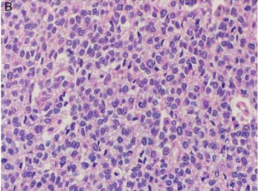
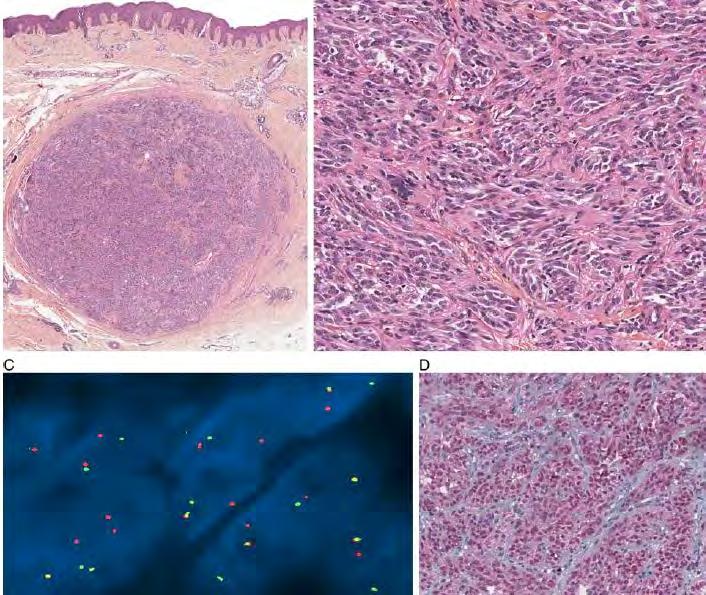
4 Ewing Sarcoma/Primitive Neuroectodermal Tumor (ES/PNET)
5
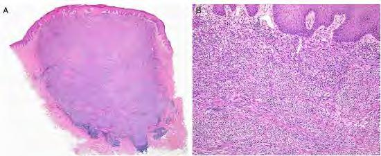
6 Can occur at any body site Extremities most common Followed by trunk and Head&Neck regions Usually small (<2.5cm) and localized to the dermis and subcutis Metastasize less frequently comparing to bone and soft tissue counterparts Clinico-radiological correlation critical to exclude metastasis

7 (Adv Anat Pathol 2013;20:75 85)

")
8 Annals of Diagnostic Pathology 11 (2007)
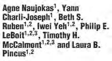
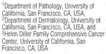
9 Adamantinoma-like ES
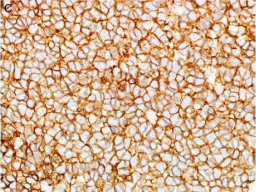
10 CD99


11 Pancytokeratin Desmin (Am J Surg Pathol 2005;29: )
12

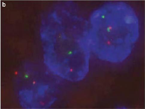
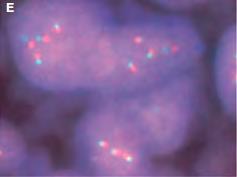
13 Fluorescent In Situ Hybridization (FISH) for EWSR1 J Cutan Pathol 2011: 38:
14 CD99
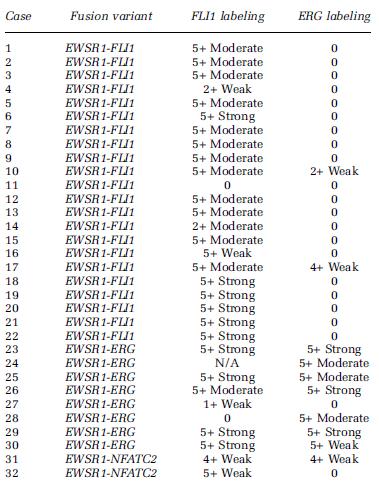
15 ERG IHC ERG is a useful marker for confirming endothelial differentiation in both benign and malignant neoplasms Potentially useful marker to distinguish Leukemia Cutis vs reactive myeloid infiltrates Expression can also be seen in a subset of epithelioid sarcomas and a small percentage of Ewing sarcomas, as well as approximately 45% to 50% of prostatic carcinomas. ERG can be seen in selected bone and soft tissue tumor with cartilaginous differentiation Phosphaturic mesenchymal tumor
16
17


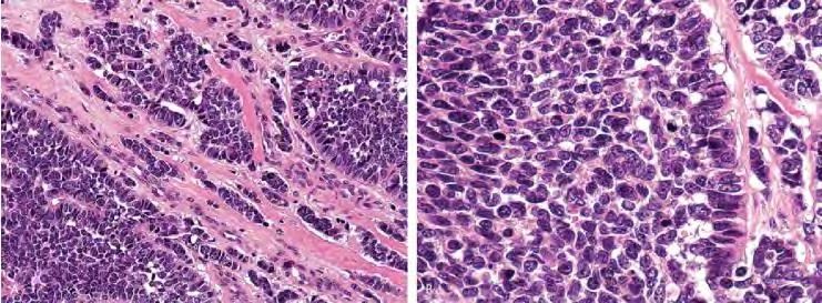
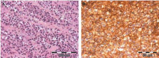
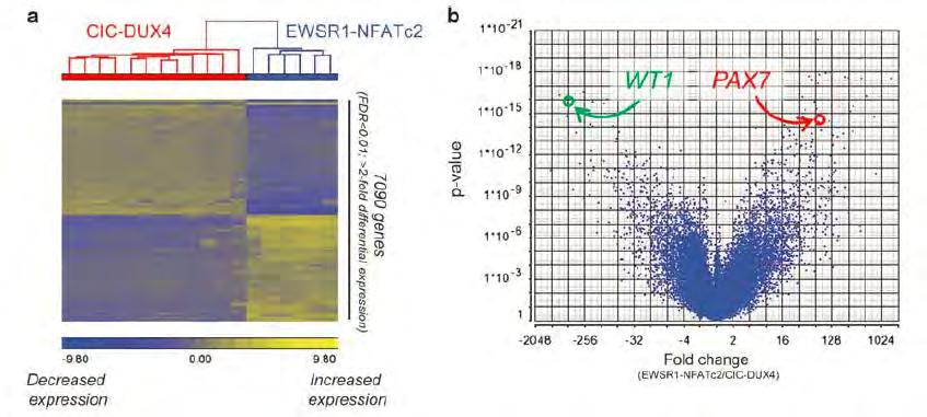
18 Ewing Sarcoma with EWSR1- ERG FLI1 ERG

19 Ewing sarcoma with EWSR1-FLI1
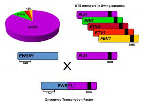
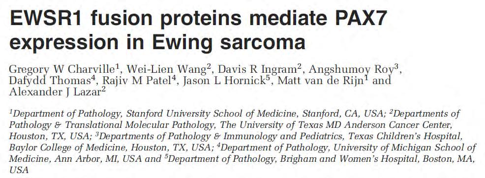
20 EWSR1, FLI1, ERG and their fusion proteins in Ewing Sarcoma
21
22
23
24

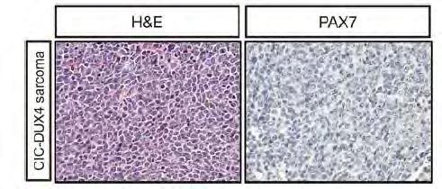
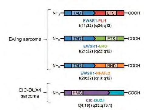
25 Ewing Sarcoma CIC-DUX4
26
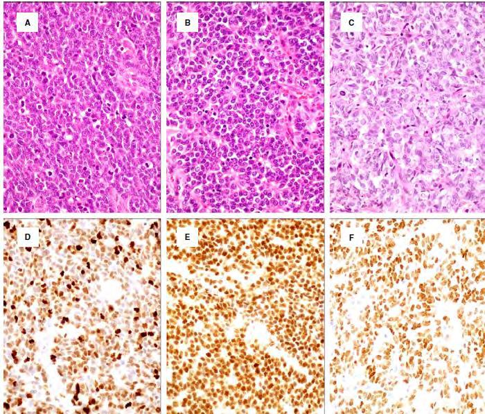
27 PAX7 a sensitive marker for Ewing Sarcoma Positive in NKX2-2 negative cases Also positive in both common and variant forms of Ewing Sarcoma PAX7 expression differentiates Ewing Sarcoma from CIC-DUX4 sarcoma
28
29
30
31 To date IHC focused on the aberrant expression of one of the genes involved in the pathogenic translocation WT1 in DSMRCT (EWSR1-WT1) STAT6 in SFT (NAB2-STAT6) FLI1 in Ewing Sarcoma (ESWR1-FLI1) BCOR in undifferentiated sarcomas PAX7 in ES exploits a characteristic transcriptional read-out of the diseasecausing translocation
32
33 Differential Diagnosis Round blue cell tumors Lymphoblastic lymphoma Merkel cell carcinoma Small cell melanoma Poorly differentiated synovial sarcoma Rhabdomyosarcoma CIC and BCOR re-arranged round cell sarcomas


34 Lymphoblastic lymphoma Diffusely positive for CD99 and Fli-1 Often CD45 negative ERG often negative but may be expressed in myeloid leukemia Always include TdT, CD10 and CD43
35 Metastatic small cell carcinoma Usually lacks CD99 More extensive immunoreactivity for cytokeratins, synaptophysin, chromogranin and CD56 Merkel cell carcinoma Diffusely express neuroendocrine markers More specific cytokeratins such as CK20 IHC for Merkel cell polyoma virus large T antigen Small cell melanoma Strongly positive for Melan-A, HMB45, SOX-10
36
37 Poorly differentiated Synovial Sarcoma Typically more classic areas of monophasic and biphasic synovial sarcoma Positive for high-molecular weight keratins Positive of TLE-1 Negative for FLI-1/ERG Alveolar rhabdomyosarcoma Greater nuclear variability Positive for myogenin and Myo-D1
38
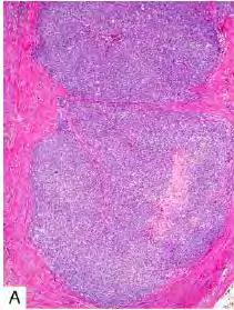
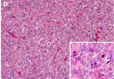
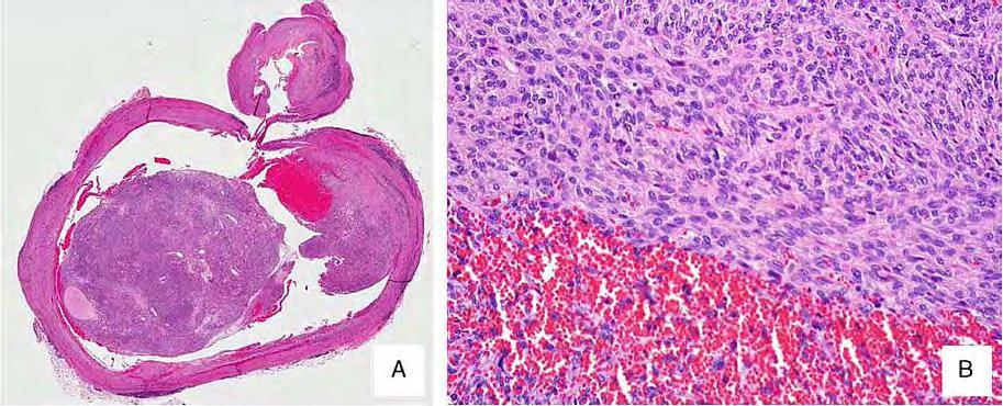
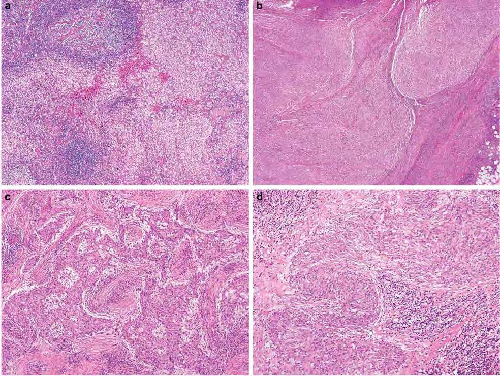
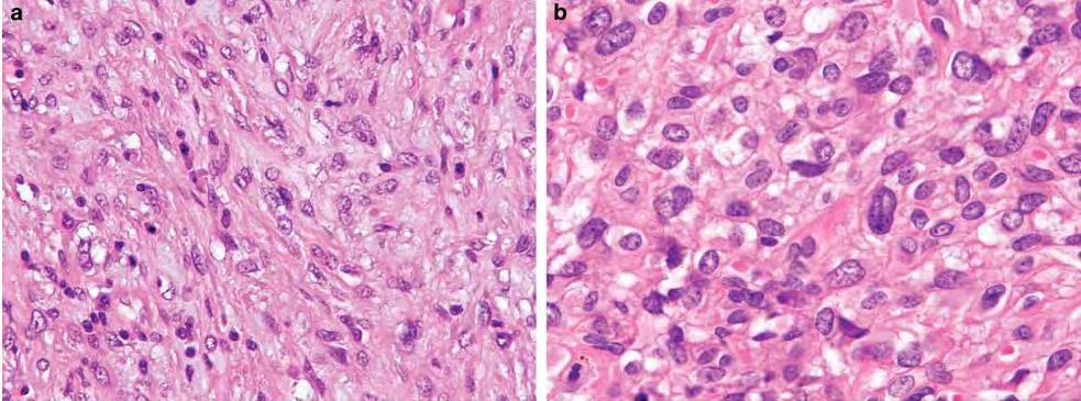
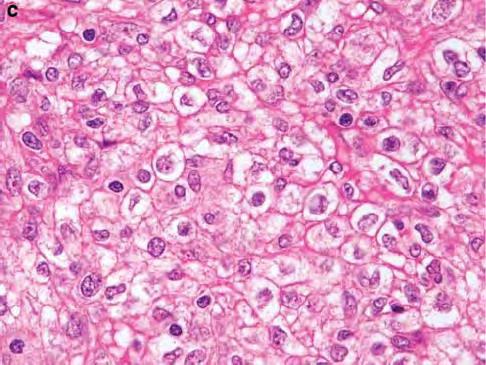
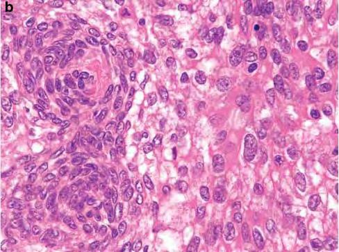
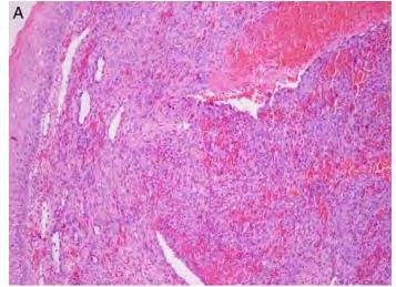
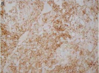
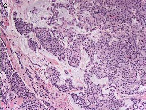
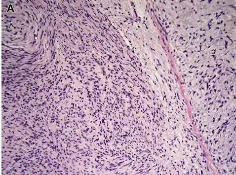
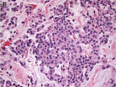
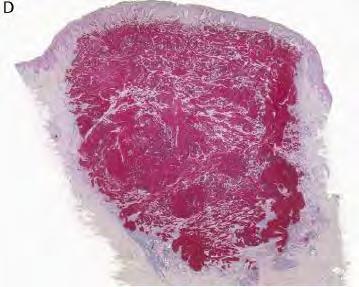
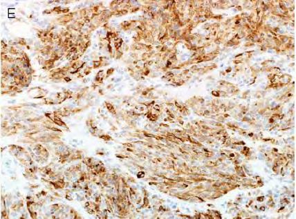
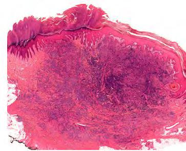
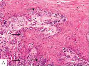
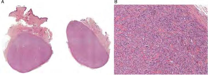
39 Angiomatoid Fibrous Histiocytoma (AFH)


40
41
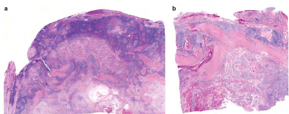
42
43
44
45
46

47 DESMIN EMA CD68
48

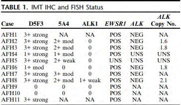
49 t(2;22)(q33;q12) EWSR1/CREB1 t(12;22)(q13;q12)=ewsr1/atf1 t(12;16)(q13;p11)= FUS/ATF1
50 Differential Diagnosis Metastasis involving a lymph node Undifferentiated pleomorphic sarcoma Vascular tumor Benign fibrous histiocytoma with prominent intratumoral hemorrhage Ewing Sarcoma in cases with small cell morphology Follicular dendritic cell tumors
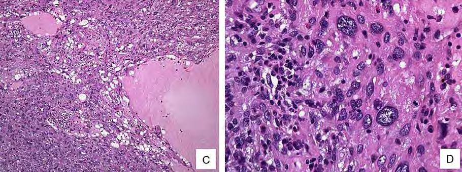
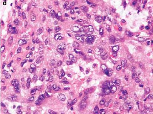
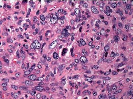
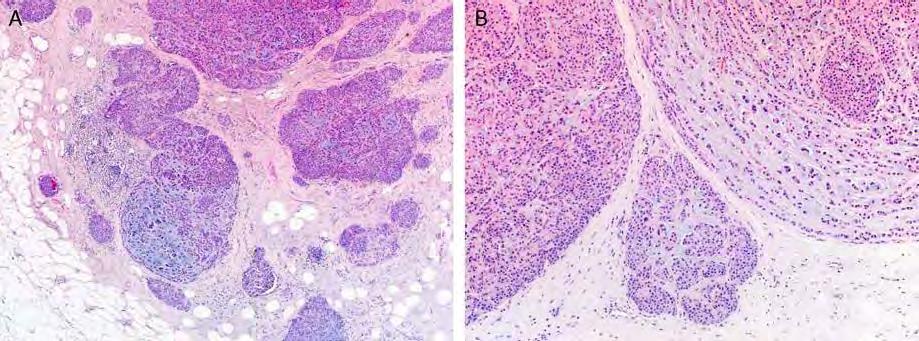
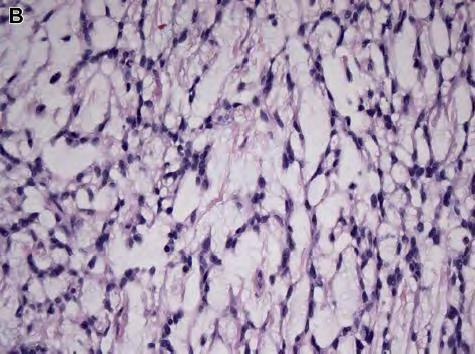
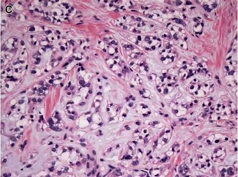
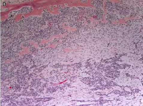
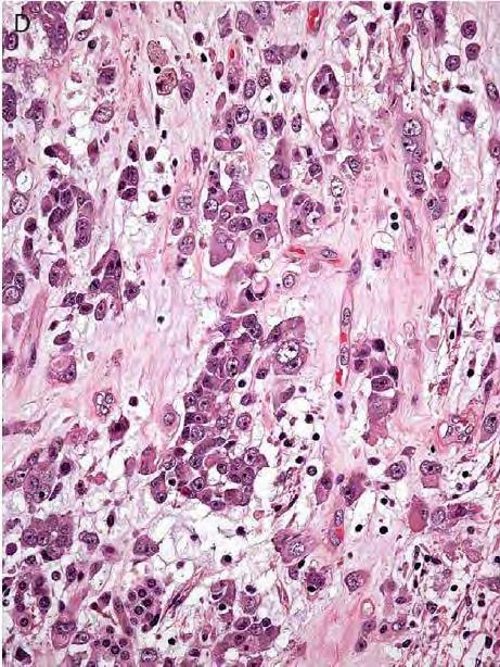
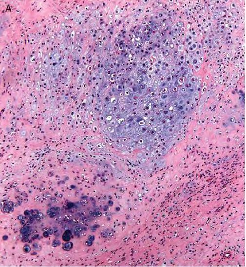
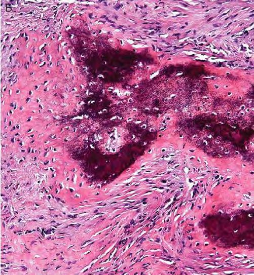
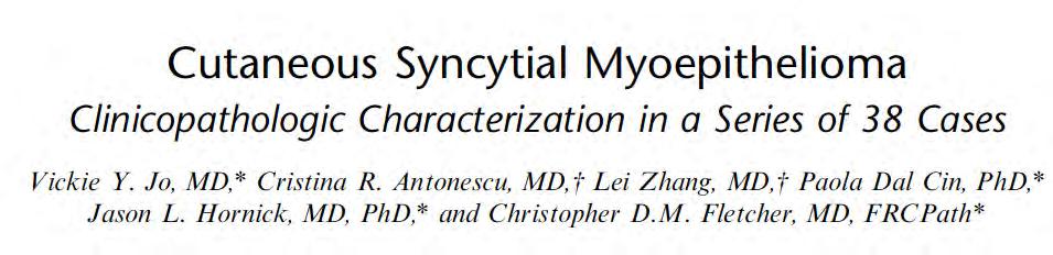
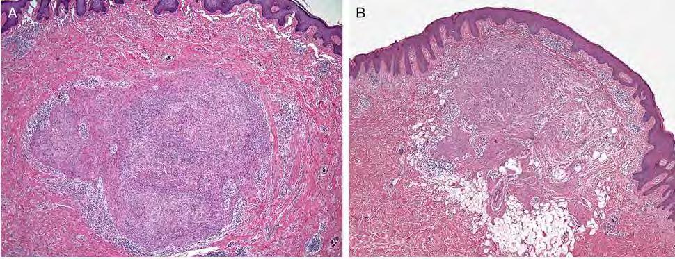
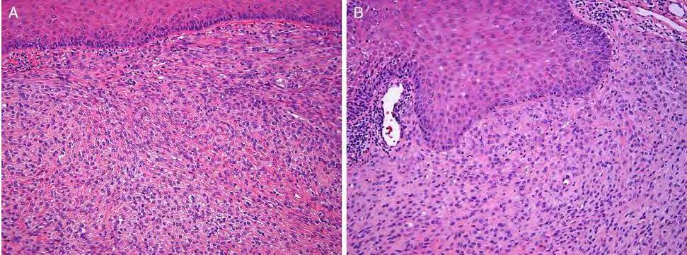
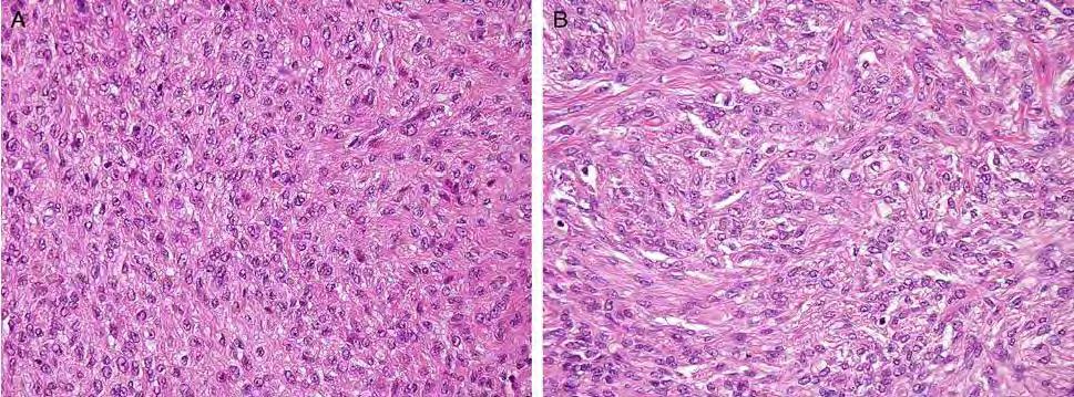
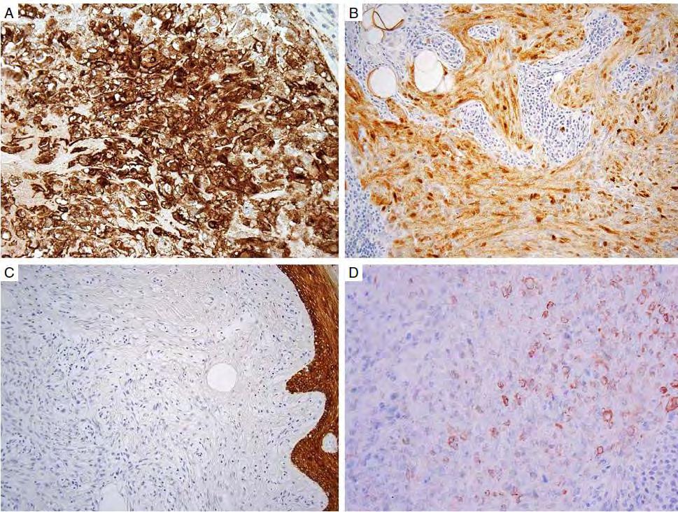
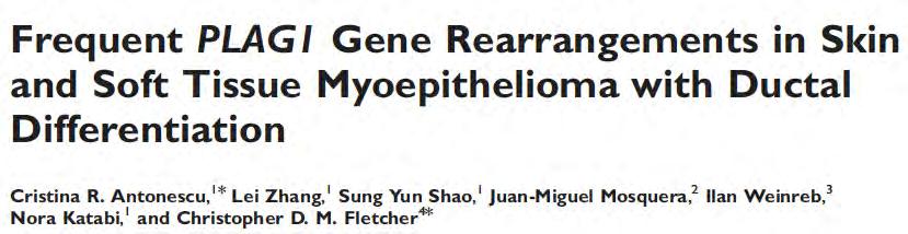
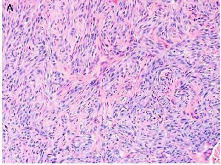
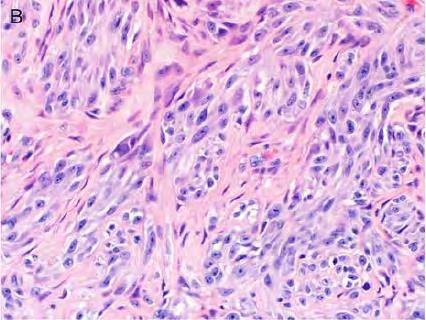
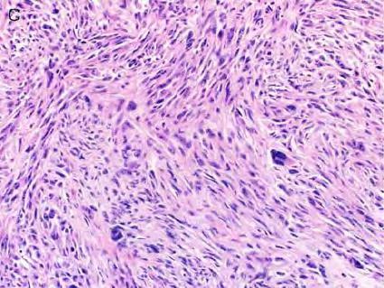
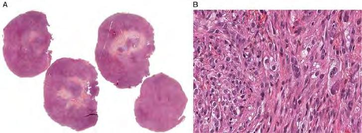
51 Myoepithelial Tumors
52 Cutaneous myoepithelial tumors Chondroid syringoma (mixed tumor) Tubuloductal differentiation Rearrangement of the PLAG1 gene (8q12) Cutaneous myoepithelioma Pure myoepithelial cell population Myoepithelial carcinoma
53
54
55
56


57

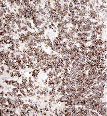
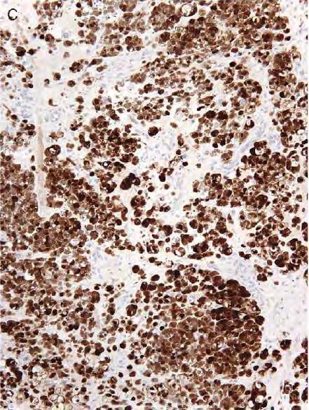
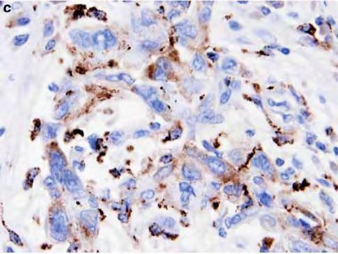
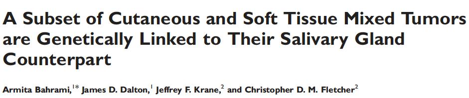
58 S100 EMA AE1/AE3
59
60


61 INI1 Absence of INI1 in myoepithelial carcinomas 10% (adults) 40% (children)
62
63

64 EMA S100 AE1/AE3 GFAP

65 EWSR1-POU5F1 EWSR1-ZNF444 EWSR1-PBX1 EWSR1-ATF1
66

67
68 Differential Diagnosis Epithelioid fibrous histiocytoma Early stage juvenile xanthogranuloma Epithelioid sarcoma Spitz nevus
69 Prognosis and treatment Cutaneous myoepithelioma has a benign clinical course Local recurrence in 20% of cases Syncytial subtype very low local recurrence rate Myoepithelia carcinomas (also contain EWSR1 rearrangements) pursue a more aggressive clinical course
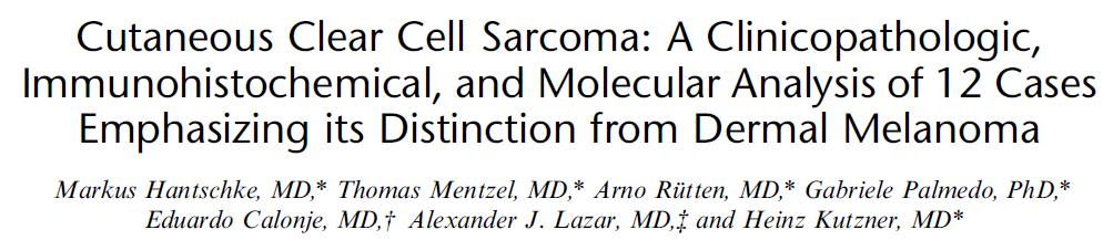
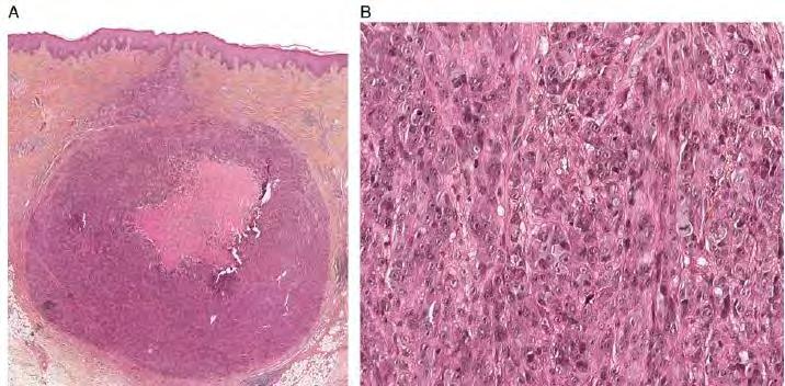
70 Clear Cell Sarcoma
71
72 Usually lesions relatively small (<5cm) Primary cutaneous <1cm Otherwise similar clinical and pathologic features Natural history clinically protracted Multiple local recurrences Late metastases in lymph nodes, lung, bone Wide local excision with adjuvant radiation
73
74 S100-Protein Melan-A
75
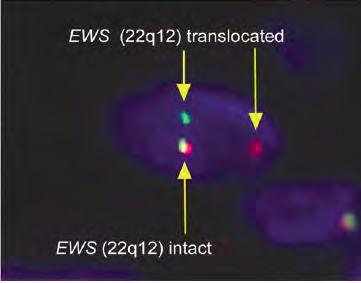
76 t(12;22)(q13;q12) EWSR1-ATF1 t(2;22)(q34;q12) EWSR1-CREB1
77
78
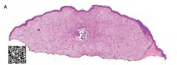
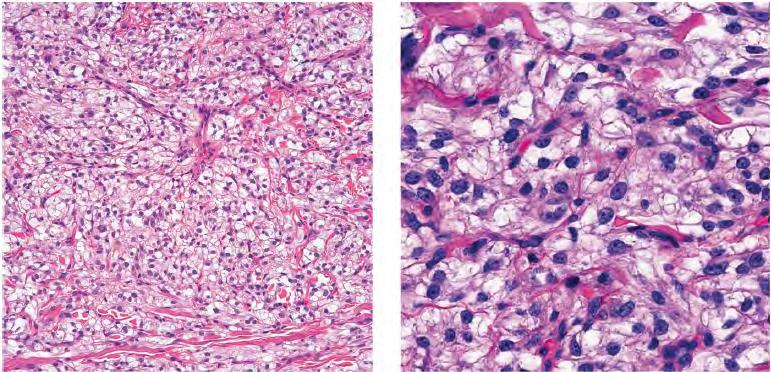
79 Differential Diagnosis Melanoma Perivascular Epithelioid Cell Neoplasm (PEComa) Cellular Blue Nevus Cutaneous Melanocytoma with CRTC1- TRIM11 Fusion

80 Cellular Blue Nevus

81 Melanoma ex blue nevus
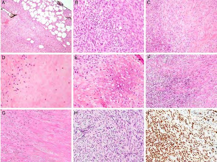
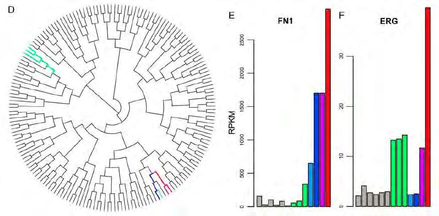
82
83 HMB45
84
85
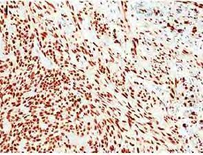
86
87

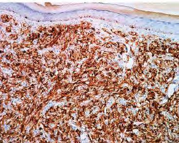
88 S100-Protein NTRK1 HMB45 MelanA TRIM11
89 No activating mutations of the MAP-kinase pathway such as BRAF and NRAS No htert mutations In some cases only a whole gain of chromosome 7 No local recurrences or metastatic evolution (short follow-up)
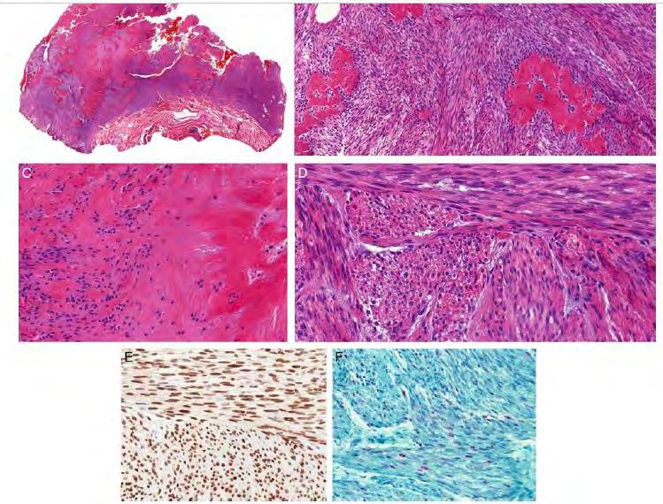
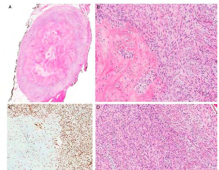
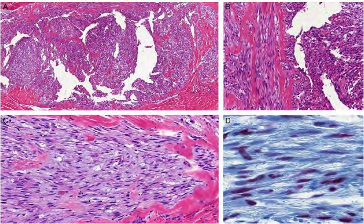
90 Benign/Low Grade Fibroblastic Tumors Calcifying aponeurotic fibroma Recurrent FN1-EGF fusion Fibrous hamartoma of infancy EGFR internal tandem duplications Myofibroma/myopericytoma PDGFR mutations Lipofibromatosis-like neural tumors Recurrent NTRK1-related gene fusions
91
92
93
94 ERG
95 ERG
96
97
98
99
100 EWSR1
101
102
103
104
105 Specific gene breakpoints Cell type in which the fusion occurs Tissue site/microenviroment Epigenetic changes Other genetic alterations
106 Morphology, Morphology, Morphology 100% Job Security!!!


107 KonstantinosLin
Financial disclosures
 Cutaneous Mesenchymal Neoplasms with EWSR1 Rearrangement By Konstantinos Linos MD, FCAP, FASDP Bone, Soft Tissue and Dermatopathology Assistant Professor of Pathology Dartmouth-Hitchc Geisel School of
Financial disclosures
 Mesenchymal Neoplasms with Melanocytic Differentiation By Konstantinos Linos MD, FCAP, FASDP Bone, Soft Tissue and Dermatopathology Assistant Professor of Pathology Dartmouth-Hitchcock Medical Center Geisel
Financial disclosures
 An update on immunohistochemical markers in mesenchymal neoplasms By Konstantinos Linos MD, FCAP, FASDP Assistant Professor of Pathology Geisel School of Medicine at Dartmouth Dartmouth-Hitchcock Medical
Tumores de células pequeñas, redondas y azules: diagnóstico diferencial cuando el tiempo apremia
 Tumores de células pequeñas, redondas y azules: diagnóstico diferencial cuando el tiempo apremia Sílvia Bagué Servei de Patologia Hospital de Sant Pau Barcelona Soft tissue sarcomas Heterogeneous group
Disclosures. An update on ancillary techniques in the diagnosis of soft tissue tumors. Ancillary techniques. Introduction
 Disclosures An update on ancillary techniques in the diagnosis of soft tissue tumors. I have nothing to disclose. Andrew Horvai, MD, PhD Clinical Professor, Pathology Introduction Ancillary techniques
Enterprise Interest Nothing to declare
 Enterprise Interest Nothing to declare Diagnoses one would not like to miss in soft tissue pathology early in your career Marta Sbaraglia, MD Department of Pathology Hospital of Treviso University of Padua
Surgical Pathology Evening Specialty Conference USCAP 2015
 Surgical Pathology Evening Specialty Conference USCAP 2015 John R. Goldblum, M.D. Chairman, Department of Pathology, Cleveland Clinic Professor of Pathology, Cleveland Clinic Lerner College of Medicine
Disclosures. An update on ancillary techniques in the diagnosis of soft tissue tumors. Ancillary techniques. Introduction
 Disclosures An update on ancillary techniques in the diagnosis of soft tissue tumors. I have nothing to disclose. Andrew Horvai, MD, PhD Clinical Professor, Pathology Introduction Ancillary techniques
Klinisch belang van chromosomale translocatie detectie in sarcomen
 Translocations in sarcomas Klinisch belang van chromosomale translocatie detectie in sarcomen Judith V.M.G. Bovée, M.D., Ph.D. Department of Pathology Leiden University Medical Center RNA binding DNA binding
Disclosure of Relevant Financial Relationships
 Ewing and Ewing like sarcomas Using Genetic Signatures in Refining Small Blue Round Cell Tumor Classification Cristina Antonescu, MD Department of Pathology Disclosure of Relevant Financial Relationships
57th Annual HSCP Spring Symposium 4/16/2016
 An Unusual Malignant Spindle Cell Lesion to Involve the Breast Erinn Downs-Kelly, D.O. Associate Professor of Pathology University of Utah & ARUP Laboratories No disclosures Case 39 y/o female with no
1/10/2018. Soft Tissue Tumors Showing Melanocytic Differentiation. Overview. Desmoplastic/ Spindle Cell Melanoma
 2016 MFMER slide-1 2016 MFMER slide-2 2016 MFMER slide-3 Soft Tissue Tumors Showing Melanocytic Differentiation Andrew L. Folpe, M.D. Professor of Laboratory Medicine and Pathology Mayo Clinic, Rochester,
An Overview of Genital Stromal Tumors
 An Overview of Genital Stromal Tumors By Konstantinos Linos MD, FCAP, FASDP Bone, Soft Tissue and Dermatopathology Assistant Professor of Pathology Dartmouth-Hitchcock Medical Center Geisel School of Medicine
Immunohistochemistry in Bone and Soft Tissue Tumors. Sahar Rassi Zankoul, MD
 Immunohistochemistry in Bone and Soft Tissue Tumors Sahar Rassi Zankoul, MD Introduction Bone tumors represent a wide variety of tumors of various origins and malignant potentials. These different tumor
An unusual superficial small round cell sarcoma
 An unusual superficial small round cell sarcoma - Diagnostic problems - Differential diagnosis Antonio Llombart Bosch Isidro Machado Dep. Pathology Univ. Medical School Valencia, Institute of Oncology
3/27/2017. Disclosure of Relevant Financial Relationships
 Ophthalmic Pathology Evening Specialty Conference USCAP 2017 5 th March, 2017 Mukul K. Divatia, MD Assistant Professor Department of Pathology & Genomic Medicine Weill Cornell Medical College Houston Methodist
Lung Tumor Cases: Common Problems and Helpful Hints
 Lung Tumor Cases: Common Problems and Helpful Hints Brandon T. Larsen, MD, PhD Senior Associate Consultant Department of Laboratory Medicine and Pathology Mayo Clinic Arizona Arizona Society of Pathologists
Clinical History. Pediatric Tumors with Involvement of the Head & Neck
 Pediatric Tumors with Involvement of the Head & Neck John Hicks Texas Children s Hospital Baylor College of Medicine Houston, TX NO DISCLOSURES Clinical History 10 Yr-Old Hispanic Male From Mexico with
Case 2. Dr. Sathima Natarajan M.D. Kaiser Permanente Medical Center Sunset
 Case 2 Dr. Sathima Natarajan M.D. Kaiser Permanente Medical Center Sunset History 24 year old male presented with a 3 day history of right flank pain, sharp in nature Denies fever, chills, hematuria or
An Overview of Cutaneous Vascular Neoplasms
 An Overview of Cutaneous Vascular Neoplasms By Konstantinos Linos MD, FCAP, FASDP Bone, Soft Tissue and Dermatopathology Assistant Professor of Pathology Dartmouth-Hitchcock Medical Center Geisel School
SOFT TISSUE TUMOR PATHOLOGY: AN UPDATE
 SOFT TISSUE TUMOR PATHOLOGY: AN UPDATE Jason L. Hornick, MD, PhD July 18, 2013 Department of Pathology Brigham and Women s Hospital Harvard Medical School Boston, MA, USA I have no disclosures. New Soft
Mojca Velikonja Jože Pižem
 Mojca Velikonja Jože Pižem An 81-year old woman presented with an exophytic, wart-like skin lesion on her neck that she had observed for one year. Cryotherapy had been applied twice, but proved unsuccessful.
From Morphology to Molecular Pathology: A Practical Approach for Cytopathologists Part 1-Cytomorphology. Songlin Zhang, MD, PhD LSUHSC-Shreveport
 From Morphology to Molecular Pathology: A Practical Approach for Cytopathologists Part 1-Cytomorphology Songlin Zhang, MD, PhD LSUHSC-Shreveport I have no Conflict of Interest. FNA on Lymphoproliferative
Slide Seminar Spanish Society of Pathology
 Slide Seminar Spanish Society of Pathology John R. Goldblum, M.D. Chairman, Department of Anatomic Pathology Cleveland Clinic Professor of Pathology Cleveland Clinic Lerner College of Medicine 1921 Original
Evening Specialty Conference Bone and Soft Tissue Pathology. Diagnostic pitfalls in bone and soft tissue pathology
 Evening Specialty Conference Bone and Soft Tissue Pathology. Case 1 Elizabeth G Demicco, MD, PhD Mount Sinai Hospital, New York Disclosure of Relevant Financial Relationships USCAP requires that all planners
USCAP 2011: ASDP companion meeting. Steven D. Billings 1
 USCAP 2011: ASDP companion meeting. Steven D. Billings (billins@ccf.org) 1 Spindle cell tumors that make you say, Oh $*&%! This lecture will focus on examples of cutaneous tumors that present particular
Recent Advances In Select Round Cell Sarcomas
 Recent Advances In Select Round Cell Sarcomas Rajiv M. Patel, M.D. Associate Professor of Pathology & Dermatology University of Michigan, Ann Arbor, MI rajivpat@med.umich.edu 1 Translocation Associated
Special slide seminar
 Special slide seminar Tomáš Rozkoš The Fingerland Department of Pathology Charles University Medical Faculty and Faculty Hospital in Hradec Králové Czech Republic Case history, 33 years old resistance
Molecular pathology in soft tissue tumors. Sylvia Höller Pathologie
 Molecular pathology in soft tissue tumors Sylvia Höller Pathologie When do we perform molecular testing? Morphology and IHC are not clearly fitting with an entity some translocations are entity specific
Molecular Diagnosis of Soft Tissue Tumors: Avoid Pitfalls
 Molecular Diagnosis of Soft Tissue Tumors: Avoid Pitfalls Cristina Antonescu, MD Department of Pathology Memorial Sloan-Kettering Cancer Center, New York Overview I. When should we rely on the help of
Charles Halsey, DVM, PhD, DACVP Pfizer, Inc. IHC Resources
 Charles Halsey, DVM, PhD, DACVP Pfizer, Inc. IHC Resources 1 IHC Identification Targets Specimens Controls 2 Tissue controls Trouble Spots 3 The Key to Description IHC Description 4 Intermediate Filaments
Pathology Mystery and Surprise
 Pathology Mystery and Surprise Tim Smith, MD Director Anatomic Pathology Medical University of South Carolina Disclosures No conflicts to declare Some problem cases Kidney tumor Scalp tumor Bladder tumor
Selected Pseudomalignant Soft Tissue Tumors of the Skin and Subcutis
 Selected Pseudomalignant Soft Tissue Tumors of the Skin and Subcutis Andrew L. Folpe, M.D. Professor of Laboratory Medicine and Pathology Mayo Clinic, Rochester, MN folpe.andrew@mayo.edu 2016 MFMER slide-1
Update on Cutaneous Mesenchymal Tumors. Thomas Brenn
 Update on Cutaneous Mesenchymal Tumors Thomas Brenn Cutaneous Mesenchymal Tumours Wide morphological and biological spectrum Myofibroblastic, smooth muscle, neural, vascular, apidocytic, undifferentiated;
Pathology of Sarcoma ELEANOR CHEN, MD, PHD, ASSISTANT PROFESSOR DEPARTMENT OF PATHOLOGY UNIVERSITY OF WASHINGTON
 Pathology of Sarcoma ELEANOR CHEN, MD, PHD, ASSISTANT PROFESSOR DEPARTMENT OF PATHOLOGY UNIVERSITY OF WASHINGTON Presentation outline Background and epidemiology of sarcomas Sarcoma classification Sarcoma
Self assessment case. Dr Saleem Taibjee Dorset County Hospital, Dorchester
 Self assessment case Dr Saleem Taibjee saleemtaibjee@gmail.com Dorset County Hospital, Dorchester Clinical details 34-year-old man: Shave excision Skin tag / papilloma left thigh The best diagnosis is:
C.L. Davis Foundation Descriptive Veterinary Pathology Course
 C.L. Davis Foundation 2015 Descriptive Veterinary Pathology Course IHC Resources IHC Identification Targets Antibodies Antibodies 1 Antibodies Specimens Antigen Retrieval Unmasks antigen epitopes Methods
3/24/2017 DENDRITIC CELL NEOPLASMS: HISTOLOGY, IMMUNOHISTOCHEMISTRY, AND MOLECULAR GENETICS. Disclosure of Relevant Financial Relationships
 DENDRITIC CELL NEOPLASMS: HISTOLOGY, IMMUNOHISTOCHEMISTRY, AND MOLECULAR GENETICS Jason L. Hornick, M.D., Ph.D. Director of Surgical Pathology and Immunohistochemistry Brigham and Women s Hospital Professor
Keywords solitary fibrous tumor, dedifferentiation, dedifferentiated solitary fibrous tumor, STAT6, GRIA2, cytokeratin, rhabdomyosarcomatous
 758452IJSXXX10.1177/1066896918758452International Journal of Surgical PathologyCreytens et al research-article2018 Pitfalls in Pathology Multifocal Cytokeratin Expression in a Dedifferentiated Solitary
The Genetics of Myoepithelial Tumors: salivary glands, soft tissue and bone
 The Genetics of Myoepithelial Tumors: salivary glands, soft tissue and bone Cristina Antonescu, MD Memorial Sloan-Kettering Cancer Center, New York Nothing to declare Disclosure Spectrum of Myoepithelial
Protocol for the Examination of Biopsy Specimens From Pediatric Patients With Ewing Sarcoma
 Protocol for the Examination of Specimens From Pediatric Patients With Ewing Sarcoma Version: EwingSarcoma 4.0.0.0 Protocol Posting Date: February 2019 Accreditation Requirements The use of this protocol
Update on Spitzoid and Blue nevus-like melanocytic lesions Emphasis on molecular studies informing diagnosis, prognosis and therapy
 Update on Spitzoid and Blue nevus-like melanocytic lesions Emphasis on molecular studies informing diagnosis, prognosis and therapy Michael T. Tetzlaff MD, PhD Associate Professor Department of Pathology,
Minimally invasive adenocarcinoma. 5mm or less = microinvasion No necrosis No lymphatic or pleural invasion No spread through air-spaces (STAS)
") Minimally invasive adenocarcinoma 5mm or less = microinvasion No necrosis No lymphatic or pleural invasion No spread through air-spaces (STAS) 2b lepidic acinar 2c papillary micropapillary solid 2d cribriform
Minimally invasive adenocarcinoma 5mm or less = microinvasion No necrosis No lymphatic or pleural invasion No spread through air-spaces (STAS) 2b lepidic acinar 2c papillary micropapillary solid 2d cribriform
What is New in the 2015 WHO Lung Cancer Classification? Zhaolin Xu, MD, FRCPC, FCAP
 What is New in the 2015 WHO Lung Cancer Classification? Zhaolin Xu, MD, FRCPC, FCAP Professor, Dept of Pathology, Dalhousie University, Canada Pulmonary Pathologist and Cytopathologist, QEII HSC Senior
5/10. Pathology Soft tissue tumors. Farah Bhani. Mohammed Alorjani
 5/10 Pathology Soft tissue tumors Mohammed Alorjani Farah Bhani Slides are included in this sheet. Objectives: Soft tissue tumors 1. Describe soft tissue tumors. 2. Understand the classification of soft
Melanoma and the genes: Molecular alterations informing the diagnosis of melanocytic tumors
 Melanoma and the genes: Molecular alterations informing the diagnosis of melanocytic tumors Michael T. Tetzlaff MD, PhD Associate Professor Department of Pathology, Section of Dermatopathology Department
Diplomate of the American Board of Pathology in Anatomic and Clinical Pathology
 A 33-year-old male with a left lower leg mass. Contributed by Shaoxiong Chen, MD, PhD Assistant Professor Indiana University School of Medicine/ IU Health Partners Department of Pathology and Laboratory
ACCME/Disclosures ALK FUSION-POSITIVE MESENCHYMAL TUMORS. Tumor types with ALK rearrangements. Anaplastic Lymphoma Kinase. Jason L.
 Companion Meeting of the International Society of Bone and Soft Tissue Pathology The Evolving Concept of Mesenchymal Tumors ALK FUSION-POSITIVE MESENCHYMAL TUMORS Jason L. Hornick, MD, PhD March 13, 2016
Applications of IHC. Determination of the primary site in metastatic tumors of unknown origin
 Applications of IHC Determination of the primary site in metastatic tumors of unknown origin Classification of tumors that appear 'undifferentiated' by standard light microscopy Precise classification
Michael T. Tetzlaff MD, PhD
 Molecular alterations informing the diagnosis of melanocytic tumors Michael T. Tetzlaff MD, PhD Associate Professor Department of Pathology, Section of Dermatopathology Department of Translational and
Dermatopathology. Dr. Rafael Botella Estrada. Hospital La Fe de Valencia
 Dermatopathology Dr. Rafael Botella Estrada. Hospital La Fe de Valencia Melanoma and mimics Dr. Martin Mihm Malignant lesions result from the accumulation of mutations Class I lesions (benign) Class II
MAPK Pathway. CGH Next Generation Sequencing. Molecular Tools in Care of Patients with Pigmented Lesions 7/20/2017
 Molecular Tools in Care of Patients with Pigmented Lesions Tammie Ferringer, MD Geisinger Medical Center, Danville, PA tferringer@geisinger.edu DISCLOSURE OF RELATIONSHIPS WITH INDUSTRY Tammie Ferringer,
Vernon K. Sondak. Department of Cutaneous Oncology Moffitt Cancer Center Tampa, Florida
 Vernon K. Sondak Department of Cutaneous Oncology Moffitt Cancer Center Tampa, Florida Australasian Melanoma Conference 2016 Sydney, NSW, Australia October 29, 2016 Disclosures Dr. Sondak is a compensated
Diploma Examination. Dermatopathology: First paper. Tuesday 20 March Candidates must answer FOUR questions. Time allowed: 3 hours
 Dermatopathology: First paper Tuesday 20 March 2018 Candidates must answer FOUR questions Time allowed: 3 hours 1. Give an account of the genetic aberrations encountered in Spitzoid neoplasms and how these
Contents Part I Introduction 1 General Description 2 Natural History: Importance of Size, Site, Histopathology
 Contents Part I Introduction 1 General Description... 3 1.1 Introduction... 3 1.2 Incidence and Prevalence... 5 1.3 Predisposing and Genetic Factors... 8 References... 16 2 Natural History: Importance
Technology from Abcam
 CD2 (EP222) CD2 is one of the earliest T-cell lineage restricted antigens to appear during T-cell differentiation and only rare CD2+ cells can be found in the bone marrow. Anti-CD2 is a pan-t-cell antigen
Cheryl M. Coffin, M.D. Goodpasture Professor of Pathology, Microbiology, and Immunology Vanderbilt University Nashville, TN, USA
 Cutaneous Mesenchymal Tumors in Childhood Cheryl M. Coffin, M.D. Goodpasture Professor of Pathology, Microbiology, and Immunology Vanderbilt University Nashville, TN, USA I. Introduction Cutaneous tumors
Fluorescence In Situ Hybridization in the Diagnosis of Soft Tissue Neoplasms: A Review. Munir R. Tanas, MD and John R.
 REVIEW ARTICLE Fluorescence In Situ Hybridization in the Diagnosis of Soft Tissue Neoplasms: A Review Munir R. Tanas, MD and John R. Goldblum, MD Abstract: This paper presents an overview of the role of
Salivary Gland FNA ATYPICAL : Criteria and Controversies
 Salivary Gland FNA ATYPICAL : Criteria and Controversies W.C. Faquin, M.D., Ph.D. Director, Head and Neck Pathology Massachusetts General Hospital Massachusetts Eye and Ear Infirmary Harvard Medical School
HOW MAY THE CLASSIFICATION OF SOFT TISSUE TUMORS EVOLVE?
 Spanish Society of Pathology Zaragoza, May 2011 ARTHUR PURDY STOUT SYMPOSIUM HOW MAY THE CLASSIFICATION OF SOFT TISSUE TUMORS EVOLVE? Christopher D.M. Fletcher, M.D., FRCPath Brigham and Women s Hospital
BSD 2015 Case 19. Female 21. Nodule on forehead. The best diagnosis is:
 BSD 2015 Case 19 Female 21. Nodule on forehead. The best diagnosis is: A. mixed tumour of skin B. porocarcinoma C. nodular hidradenoma D. metastatic adenocarcinoma BSD 2015 Case 19 Female 21 Nodule on
Schedule of Accreditation issued by United Kingdom Accreditation Service 2 Pine Trees, Chertsey Lane, Staines-upon-Thames, TW18 3HR, UK
 2 Pine Trees, Chertsey Lane, Staines-upon-Thames, TW18 3HR, UK Royal National Orthopaedic Hospital NHS Trust Brockley Hill Stanmore Middlesex HA7 4LP Contact: Professor Adrienne Flanagan Tel: +44 (0)20
Case 1. Disclosure. Imaging. Clinical history 5/10/2016. USCAP 2016 Annual Meeting Evening Specialty Conference Bone and Soft tissue Pathology
 Disclosure Dr. Agaram has nothing to disclose Case 1 Narsi Agaram, MBBS USCAP 2016 Annual Meeting Evening Specialty Conference Bone and Soft tissue Pathology Clinical history Imaging 1998 A three month
أملس عضلي غرن = Leiomyosarcoma. Leiomyosarcoma 1 / 5
 Leiomyosarcoma 1 / 5 EPIDEMIOLOGY Exact incidence is unknown, but older studies suggest that leiomyosarcomas comprise approximately 3 percent of soft-tissue sarcomas. Superficial leiomyosarcoma occurs
59 yo male with past medical history of prostate carcinoma, presented with upper abdominal pain
 December 2016 59 yo male with past medical history of prostate carcinoma, presented with upper abdominal pain Contributed by: Divya Sharma, MD. Fellow, Gastrointestinal Pathology, Department of Pathology
Diagnostic Value of Immunohistochemistry in Soft Tissue Tumors
 Original Article DOI: 10.21276/APALM.1637 Diagnostic Value of Immunohistochemistry in Soft Tissue Tumors Sridevi. V*., Susruthan Muralitharan., and Thanka. J Dept of Pathology, SriMuthukumaran Medical
Case Presentation. Maha Akkawi, MD, Fatima Obeidat, MD, Tariq Aladily, MD. Department of Pathology Jordan University Hospital Amman, Jordan
 Case Presentation Maha Akkawi, MD, Fatima Obeidat, MD, Tariq Aladily, MD Department of Pathology Jordan University Hospital Amman, Jordan The 25th Annual Congress of the ADIAP The 8/11/2013 1 5th International
Shintaro Sugita *, Hiroko Asanuma and Tadashi Hasegawa
 Sugita et al. Diagnostic Pathology (2016) 11:37 DOI 10.1186/s13000-016-0486-2 RESEARCH Open Access Diagnostic use of fluorescence in situ hybridization in expert review in a phase 2 study of trabectedin
Advances in the clinicopathological and molecular classification of cutaneous mesenchymal neoplasms
 Histopathology 2016 DOI: 10.1111/his.12930 REVIEW Advances in the clinicopathological and molecular classification of cutaneous mesenchymal neoplasms Danielle C Costigan & Leona A Doyle 1 Department of
Slide seminar: Soft tissue and bone pathology
 Slide seminar: Soft tissue and bone pathology Unusual tumors of bone and soft tissue or unusual presentations of common ones Gunhild Mechtersheimer Institute of Pathology, Heidelberg/DE (Sylvia Höller,
04/09/2018. Salivary Gland Pathology in the Molecular Era Old Friends, Old Foes, & New Acquaintances
 Salivary Gland Pathology in the Molecular Era Old Friends, Old Foes, & New Acquaintances Jennifer L. Hunt, MD, MEd Aubrey J. Hough Jr, MD, Endowed Professor of Pathology Chair of Pathology and Laboratory
NEW IHC A n t i b o d i e s
 NEW IHC Antibodies TABLE OF CONTENTS NEW IHC ANTIBODIES from Cell Marque CITED1 (5H6).... 1 Claudin 7 (5D10F3).... 1 GATA1 (4F5).... 1 Transgelin (2A10C2).... 1 NEW IHC ANTIBODIES using RabMAb Technology
The Enigmatic Spitz Lesion
 The Enigmatic Spitz Lesion The Dawn of Spitz S Spitz Sophie Spitz Melanomas of Childhood ; Am J Pathol 1948 1910-1956 13 children (18 mo - 12 yrs) 12/13 had a benign clinical course Sophie Spitz Born 1910
The role of immunohistochemistry in surgical pathology of the uterine corpus and cervix
 The role of immunohistochemistry in surgical pathology of the uterine corpus and cervix Prof. Ben Davidson, MD PhD Department of Pathology, Norwegian Radium Hospital, Oslo University Hospital, Oslo, Norway
Mesothelioma Pathobasic. Lukas Bubendorf Pathology
 Mesothelioma Pathobasic Lukas Bubendorf Pathology Mechanisms of Asbestos Carcinogenesis in Mesothelioma Asprin High-mobility group protein B1 master switch HMGB1 Initiation/ perpetuation of inflamm. response
3/27/2017. Pulmonary Pathology Specialty Conference. Disclosure of Relevant Financial Relationships. Clinical History:
 Pulmonary Pathology Specialty Conference Saul Suster, M.D. Medical College of Wisconsin Disclosure of Relevant Financial Relationships USCAP requires that all planners (Education Committee) in a position
What I Learned from 3 Cases and 3 Antibodies
 What I Learned from 3 Cases and 3 Antibodies Melinda Sanders, M.D Vanderbilt University Medical Center Professor of Pathology Consultant in Breast Pathology Disclosure of Relevant Financial Relationships
Desmoplastic Melanoma R/O BCC. Clinical Information. 74 y.o. man with lesion on left side of neck r/o BCC
 R/O BCC Sabine Kohler, M.D. Professor of Pathology and Dermatology Dermatopathology Service Stanford University School of Medicine Clinical Information 74 y.o. man with lesion on left side of neck r/o
I have nothing to disclose
 A 47 year old female with multiple lung nodules Disclosure of Relevant Financial Relationships Tamar Giorgadze, MD, PhD Professor of Pathology Medical College of Wisconsin Milwaukee, Wisconsin USCAP requires
Small Round Cell Tumors in Soft Tissue and Bone. ACCME/Disclosure. Adult Small Round Cell Tumors. Ewing Sarcoma 4/13/2016
 Ultrastructural Pathology Society, Seattle, March, 2016 Small Round Cell Tumors in Soft Tissue and Bone ACCME/Disclosure Dr. Fisher has nothing to disclose Cyril Fisher MA MD DSc FRCPath Professor of Tumor
Cutaneous metastases. Thaddeus Mully. University of California, San Francisco Professor, Departments of Pathology and Dermatology
 Cutaneous metastases Thaddeus Mully University of California, San Francisco Professor, Departments of Pathology and Dermatology DISCLOSURE OF RELATIONSHIPS WITH INDUSTRY Thaddeus Mully Course C005 Essential
Biopsy Interpretation of Spindle cell proliferations of the Serosa
 Biopsy Interpretation of Spindle cell proliferations of the Serosa Richard Attanoos, Cardiff. U.K. Disclosure of Relevant Financial Relationships USCAP requires that all planners (Education Committee)
Newer soft tissue entities
 Newer soft tissue entities Examples among fibroblastic tumors Turku, May 6, 2010 Markku Miettinen, M.D. AFIP, Washington, DC Fibroblastic neoplasms Solitary fibrous tumor /Hemangiopericytoma Low-grade
Dermatopathology. Dr. Rafael Botella Estrada. Hospital La Fe de Valencia
 Dermatopathology Dr. Rafael Botella Estrada. Hospital La Fe de Valencia DERMATOPATHOLOGY CASE CHALLENGE: RECOGNIZING MIMIS AND MASQUERADERS Rosalie Elenitsas. University of Pennsylvania Spectrum Lupus
Spindle Cell Lesions Of The Breast. Emad Rakha Professor of Breast Pathology and Consultant Pathologist
 Spindle Cell Lesions Of The Breast Emad Rakha Professor of Breast Pathology and Consultant Pathologist * SCLs comprise a wide spectrum of diseases, ranging from reactive processes to aggressive malignant
Adipocytic Tumours in children
 Università degli Studi di Padova Dipartimento di Medicina Sezione di Anatomia Patologica Generale e Citopatologia Adipocytic Tumours in children Rita Alaggio Basel Seminars in Pathology Paediatric Pathology
LOOK-ALIKES IN SPINDLE AND EPITHELIOID TUMORS: Immunohistochemistry. Cytogenetics Flow cytometry Molecular diagnostics
 LOOK-ALIKES IN SPINDLE AND EPITHELIOID TUMORS: Ultrastructural value and pitfalls in diagnosis Guillermo A Herrera Department of Pathology and Translational Pathobiology Louisiana State University Health
Disclosure. Relevant Financial Relationship(s) None. Off Label Usage None MFMER slide-1
 None. Off Label Usage None MFMER slide-1") Disclosure Relevant Financial Relationship(s) None Off Label Usage None 2013 MFMER slide-1 Case Presentation A 43 year old male, with partial nephrectomy for a right kidney mass 2013 MFMER slide-2 2013
Disclosure Relevant Financial Relationship(s) None Off Label Usage None 2013 MFMER slide-1 Case Presentation A 43 year old male, with partial nephrectomy for a right kidney mass 2013 MFMER slide-2 2013
Small (and large) Blue Cell Tumors of the Skull Base
 Blue Cell Tumors of the Skull Base") Small (and large) Blue Cell Tumors of the Skull Base Jennifer L. Hunt, MD, MEd Aubrey J. Hough Jr, MD, Endowed Professor of Pathology Chair of Pathology and Laboratory Medicine University of Arkansas for
Small (and large) Blue Cell Tumors of the Skull Base Jennifer L. Hunt, MD, MEd Aubrey J. Hough Jr, MD, Endowed Professor of Pathology Chair of Pathology and Laboratory Medicine University of Arkansas for
Difficult Diagnoses and Controversial Entities in Neoplastic Lung
 Difficult Diagnoses and Controversial Entities in Neoplastic Lung Lynette M. Sholl, M.D. Associate Pathologist, Brigham and Women s Hospital Chief, Pulmonary Pathology Service Associate Professor, Harvard
ASCP Resident Review Mini-Course Series Session 2: Renal and Soft Tissue Pathology. Carole Vogler MD, FASCP Ema Dragoescu MD
 178 2011 ASCP Resident Review Mini-Course Series Session 2: Renal and Soft Tissue Pathology Carole Vogler MD, FASCP Ema Dragoescu MD 2011 Annual Meeting Las Vegas, NV AMERICAN SOCIETY FOR CLINICAL PATHOLOGY
DIFFERENTIAL DIAGNOSIS OF SMALL ROUND CELL TUMOURS (SRCT), FLUORESCENCE IN SITU HYBRIDIZATION (FISH)
, FLUORESCENCE IN SITU HYBRIDIZATION (FISH)") POL J PATHOL 2009; 4: 151-162 DIFFERENTIAL DIAGNOSIS OF SMALL ROUND CELL TUMOURS (SRCT), FLUORESCENCE IN SITU HYBRIDIZATION (FISH) AND IMMUNOHISTOCHEMICAL (IHC) STUDY KONRAD PTASZYŃSKI 1, ANNA SZUMERA-CIEĆKIEWICZ
POL J PATHOL 2009; 4: 151-162 DIFFERENTIAL DIAGNOSIS OF SMALL ROUND CELL TUMOURS (SRCT), FLUORESCENCE IN SITU HYBRIDIZATION (FISH) AND IMMUNOHISTOCHEMICAL (IHC) STUDY KONRAD PTASZYŃSKI 1, ANNA SZUMERA-CIEĆKIEWICZ
Original Articles. Utilization of Fluorescence In Situ Hybridization in the Diagnosis of 230 Mesenchymal Neoplasms. An Institutional Experience
 Original Articles Utilization of Fluorescence In Situ Hybridization in the Diagnosis of 230 Mesenchymal Neoplasms An Institutional Experience Munir R. Tanas, MD; Brian P. Rubin, MD, PhD; Raymond R. Tubbs,
Mayo Medical Laboratories
 Mayo Medical Laboratories Virtual Lectures 2014 MFMER 2016 MFMER slide-1 Virtual Lectures Planning Committee Disclosure Summary As a provider accredited by ACCME, College of Medicine, Mayo Clinic (Mayo
21/07/2017. Hobnail endothelial cells are not the same as epithelioid endothelial cells
 UPDATE IN CUTANEOUS VASCULAR S DERMATOPATHOLOGY SESSION BELFAST PATHOLOGY JUNE 21/2017 Dr E Calonje St John s Institute of Dermatology, London, United Kingdom THE FAMILY OF VASCULAR S WITH EPITHELIOID
Objectives. Salivary Gland FNA: The Milan System. Role of Salivary Gland FNA 04/26/2018
 Salivary Gland FNA: The Milan System Dr. Jennifer Brainard Section Head Cytopathology Cleveland Clinic Objectives Introduce the Milan System for reporting salivary gland cytopathology Define cytologic
Differential diagnosis of HCC
 Hepatocellular Carcinoma Quest for an Ideal Immunohistochemical Panel Sanjay Kakar, MD UCSF Differential diagnosis of HCC Hepatocellular lesions Adenoma, FNH, HG dysplasia Adenocarcinoma CholangioCA, metastasis
No financial or other disclosures
 Case 2014-5 Esther N. Bit-Ivan, DO Northwestern University Jason Wang, MD Jason Park, MD Korgun Koral, MD Children s Medical Center Charles Timmons, MD Veena Rajaram, MD No financial or other disclosures
USCAP COMPANION MEETING INTERNATIONAL SOCIETY OF BONE AND SOFT TISSUE PATHOLOGY DENVER, March 2 nd 2008
 1 USCAP COMPANION MEETING INTERNATIONAL SOCIETY OF BONE AND SOFT TISSUE PATHOLOGY DENVER, March 2 nd 2008 THE EVOLUTION OF SOFT TISSUE TUMOUR TAXONOMY: WHAT STILL NEEDS TO BE DONE? Christopher D.M. Fletcher,
Presentation material is for education purposes only. All rights reserved URMC Radiology Page 1 of 98
 Presentation material is for education purposes only. All rights reserved. 2011 URMC Radiology Page 1 of 98 Radiology / Pathology Conference February 2011 Brooke Koltz, Cytopathology Resident Presentation