HISTOPATHOLOGY. Shannon Martinson
|
|
|
- Darcy French
- 5 years ago
- Views:
Transcription

1 HISTOPATHOLOGY Shannon Martinson March 2013


2 Case #1 History: 8 year old beagle Neck pain for the past couple of weeks Paresis, followed by paralysis developed over the past few days
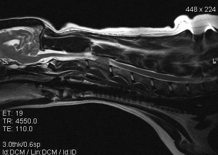

3 Gross Description courtesy of Dr L Pack, AVC
4 Gross Description A nodular mass is present within the second cervical vertebrae. It protrudes into the spinal canal (compressing the spinal cord at this site) courtesy of Dr L Pack, AVC
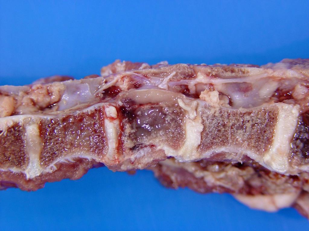
5 Gross Description
6 Gross Description On section, the soft pink gelatinous mass is present within the marrow cavity of the vertebral body with slight dorsal protrusion into the spinal canal.
7 Differential Diagnoses:
8 Differential Diagnoses: Multiple myeloma, osteosarcoma, osteomyelitis
9 Histology
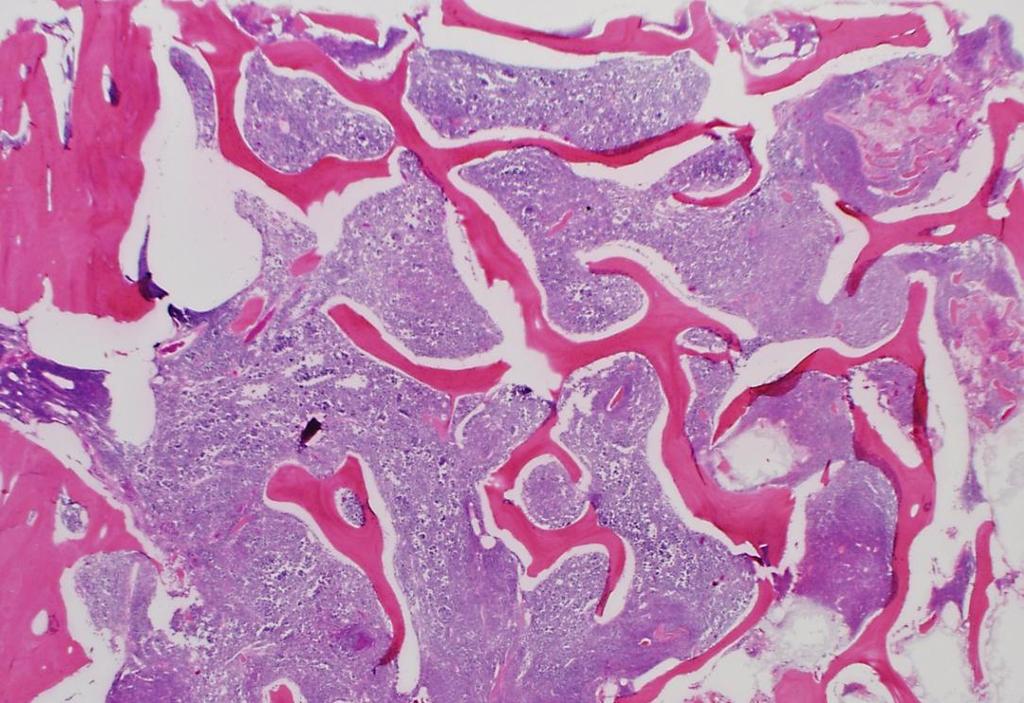
10 Histology
11 Histologic Description:
12 Histologic Description: There is a marked increase in the cellularity of the bone marrow (100% cells / 0% adipose tissue)
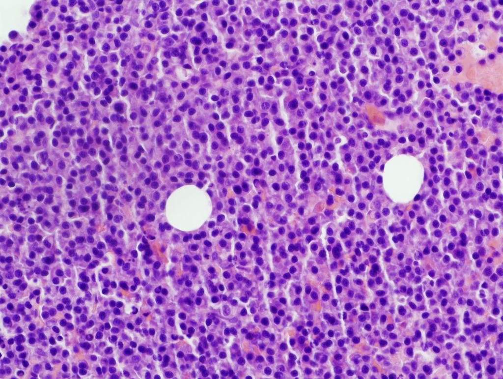
13 Histologic Description:
14 Histologic Description: Sheets of tightly packed neoplastic round cells infiltrate the marrow cavity of the vertebral body
15 Histologic Description:

16 Histologic Description: The cells are round with distinct cell margins and a moderate amount of pale basophilic cytoplasm occasionally with perinuclear clearing. The nuclei are eccentric and round with coarse clumped hyperchromatic chromatin
17 Case #1 Histologic Description (summary): Sheets of tightly packed neoplastic round cells infiltrate the marrow cavity of the vertebral body. The cells are round with distinct cell margins and a moderate amount of pale basophilic cytoplasm with occasional perinuclear clearing. The nuclei are round with clumped chromatin Morphologic Diagnosis:
18 Case #1 Histologic Description (summary): Sheets of tightly packed neoplastic round cells infiltrate the marrow cavity of the vertebral body. The cells are round with distinct cell margins and a moderate amount of pale basophilic cytoplasm with occasional perinuclear clearing. The nuclei are round with clumped chromatin Morphologic Diagnosis: Multiple Myeloma, vertebrae Possible clinical pathology findings:
19 Case #1 Histologic Description (summary): Sheets of tightly packed neoplastic round cells infiltrate the marrow cavity of the vertebral body. The cells are round with distinct cell margins and a moderate amount of pale basophilic cytoplasm with occasional perinuclear clearing. The nuclei are round with clumped chromatin Morphologic Diagnosis: Multiple Myeloma, vertebrae Possible clinical pathology findings: Hypergammaglobulinemia, hypercalcemia, Bence-Jones proteinuria
 of one class Bence-Jones")
20 courtesy of Dr A Matthews AVC Ancillary / helpful diagnostic findings courtesy of Dr. A Matthews, AVC Hypercalcemia increased osteoclastic activity within bone lesions Hypergammaglobulinemia clonal neoplastic plasma cells secrete immunoglobulins (fragments) of one class Bence-Jones proteinuria Immunoglobulin light chains pass through glomerulus into urine Radiographs showed punched out lesions in the vertebrae

21 Case #2 Signalment: 1.5 year old, laying hen History: The bird has been down for a day and has not been eating All images for this case were provided by Dr Heather Fenton, AVC

22 Spleen Liver Bone
23 Spleen Gross Description
24 Spleen Gross Description The spleen is markedly enlarged and misshapen with coalescent yellow-tan nodules scattered throughout
25 Spleen Gross Description

26 Spleen Gross Description These extend into the parenchyma on cut surface replacing most of the normal architecture
27 Spleen Histologic Description

28 Spleen Histologic Description Discrete eosinophilic nodules are present within the spleen.
29 Spleen Histologic Description
30 Spleen Histologic Description The nodules are composed of central regions of necrosis surrounded by a thick cellular rim. What are the cells?
31 Spleen Histologic Description The nodules are composed of central regions of necrosis surrounded by a thick cellular rim. What are the cells?
32 Spleen Histologic Description The nodules are composed of central regions of necrosis surrounded by a thick cellular rim. What are the cells?


33 Spleen Histologic Description The cells consist primarily of epithelioid macrophages and rare heterophils. Admixed with these cells are occasional lymphocytes, plasma cells and fibroblasts and what other cell type?
34 Spleen Histologic Description Scattered multinucleated giant cells
35 Spleen Morphologic Diagnosis
36 Spleen Morphologic Diagnosis Splenitis, granulomatous, multifocal to coalescing, chronic, marked
37 Spleen Acid-fast stain Etiology and Disease Name

38 Spleen Acid-fast stain Etiology and Disease Name Etiology: Mycobacterium avium Disease Name: Avian tuberculosis
39 Case #2 Histologic Description (summary): Scattered throughout the spleen are discrete foci of necrosis surrounded by infiltrates of epithelioid macrophages and multinucleated giant cells admixed with fewer lymphocytes and plasma cells Acid-fast bacilli are present within the lesion Morphologic Diagnosis: Splenitis, granulomatous, multifocal to coalescing, chronic, marked, with intralesional acid-fast bacilli Etiology and Disease Name: Mycobacterium avium Avian tuberculosis

40 Case #2 Avian Tuberculosis Mycobacterium avium complex (serotypes 1, 2 and 3) Can infect an extensive range of species In birds usually chickens, captive wild birds and turkeys Free range farming systems and keeping breeders for several years are conducive to the spread of this disease. Main sources of infection are infected individuals and the contaminated environment (water and soil) Affects multiple organs Granulomatous osteomyelitis and hepatitis

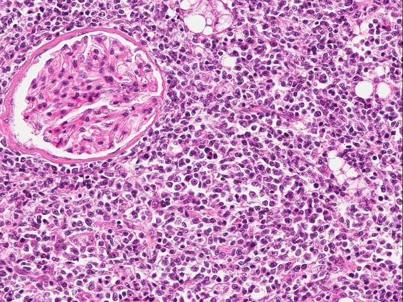
41 Case #3 Signalment: 9 yr-old MC cat History: Cat had been anorexic and was beginning to lose weight. On physical exam the cat was dehydrated. Abdominal palpation revealed enlarged irregular kidneys. Blood chemistry revealed elevated renal enzymes (urea, creatinine).
42

43 Gross Description Marked enlargement of the kidneys Nodular/irregular capsular surface
44 Gross Description
45 Gross Description On cut surface, there are multifocal to coalescing, white, round to irregular masses scattered throughout both of the kidneys
46 Morphologic Diagnosis

47 Morphologic Diagnosis Multiple renal masses, probable malignant neoplasia


48 Need Histology! Feline Infectious Peritonitis (Pyogranulomatous nephritis) Compare previous slides with this case of multifocal granulomatous nephritis due to FIP. These lesions can easily be mistaken for renal lymphoma! Two main differentials for this lesion in cats: Lymphoma and FIP Renal lymphoma
49 Histologic Description


50 Histologic Description At low power, we can appreciate a nodule within the tissue (kidney) that looks more blue than normal suggests an increase in the cellularity of the tissue
51 Histologic Description

52 Histologic Description Description: Densely cellular sheet-like infiltrates efface the normal renal architecture leaving few remaining glomeruli
53 At 40x
54 Histologic Description
55 Histologic Description These cells have distinct cell margins, scant eosinophilic cytoplasm, and round nuclei with stippled chromatin and prominent nucleoli. Mitoses are present.
56 Case #3 Histologic Description (summary): In this section of kidney, most of the normal architecture has been effaced by dense sheet-like cellular infiltrates consisting of large numbers of neoplastic round cells. These neoplastic round cells have small amounts of well-delineated pale acidophilic cytoplasm. The nuclei are round to slightly ovoid with stippled chromatin and distinct nucleoli. Morphologic Diagnosis:
57 Case #3 Histologic Description (summary): In this section of kidney, most of the normal architecture has been effaced by dense sheet-like cellular infiltrates consisting of large numbers of neoplastic round cells. These neoplastic round cells have small amounts of well-delineated pale acidophilic cytoplasm. The nuclei are round to slightly ovoid with stippled chromatin and distinct nucleoli. Morphologic Diagnosis: Renal lymphoma Comments: Lymphoma is the most common renal tumor in the cat. It may cause kidney deficiency/failure by destroying parenchyma or by obstructing urine outflow
58 Questions?
Pathology of the Hematopoietic System. Case studies
 Pathology of the Hematopoietic System Case studies Shannon Martinson, September 2015 Signalment: 9 yr-old MC cat Case Study 1 History: Cat had been anorexic and developed bleeding in the eyes Physical
Pathology of the Hematopoietic System GROSS/HISTO LAB
 Pathology of the Hematopoietic System GROSS/HISTO LAB Paul Hanna (thanks to Dr s Aburto, Martinson & Fenton) Fall 2014 Slide 1 Spleen from a Beaver Give a morphologic diagnosis and possible etiology &
Pathology of the Hematopoietic System - Lab.
 Pathology of the Hematopoietic System - Lab http://people.upei.ca/smartinson/ Shannon Martinson, September 2015 Case #1 Signalment: 96 kg gilt History: Pig from minimal disease herd. Sudden death Case
Inflammation Laboratory 3 Emphasis: Chronic inflammation and healing. Shannon Martinson: VPM 152: April 2013
 Inflammation Laboratory 3 Emphasis: Chronic inflammation and healing Shannon Martinson: http://people.upei.ca/smartinson VPM 152: April 2013 Example A Reproductive tract and colon/rectum from a sheep Previous
Avian Pathology. Bacterial diseases: histo slides. ECVP-ESVP Summer School 2012 Frédérique NGUYEN
 Avian Pathology Bacterial diseases: histo slides ECVP-ESVP Summer School 2012 Frédérique NGUYEN Bacterial diseases: histo slides B1. Turkey. Organs? Morphologic diagnosis? Special procedure? B2. Hen. Organ?
Cellular Pathology. Histopathology Lab #2 (web) Paul Hanna Jan 2018
 Paul Hanna Jan 2018") Cellular Pathology Histopathology Lab #2 (web) Paul Hanna Jan 2018 Slide #91 Clinical History: a necropsy was performed on an aged cat the gross pathological changes included: widespread subcutaneous edema
Cellular Pathology Histopathology Lab #2 (web) Paul Hanna Jan 2018 Slide #91 Clinical History: a necropsy was performed on an aged cat the gross pathological changes included: widespread subcutaneous edema
Burkitt lymphoma. Sporadic Endemic in Africa associated with EBV Translocations involving MYC gene on chromosome 8
 Heme 8 Burkitt lymphoma Sporadic Endemic in Africa associated with EBV Translocations involving MYC gene on chromosome 8 Most common is t(8;14) Believed to be the fastest growing tumor in humans!!!! Morphology
WSC , Conference 9, Case 1. Tissue from a nyala.
 WSC 2009-2010, Conference 9, Case 1. Tissue from a nyala. MICROSCOPIC DESCRIPTION: Heart, atrium (1 pt.): Approximately 40% of the atrial myocardium is replaced by areas of fibrous connective tissue (1
Plasma cell myeloma (multiple myeloma)
") Plasma cell myeloma (multiple myeloma) Common lymphoid neoplasm, present at old age (70 years average) Remember: plasma cells are terminally differentiated B-lymphocytes that produces antibodies. B-cells
Plasma cell myeloma (multiple myeloma) Common lymphoid neoplasm, present at old age (70 years average) Remember: plasma cells are terminally differentiated B-lymphocytes that produces antibodies. B-cells
This is the second learning component (Learning Component 2) in our first learning module (Learning Module 1). In this component we review a very
 in our first learning module (Learning Module 1). In this component we review a very") This is the second learning component (Learning Component 2) in our first learning module (Learning Module 1). In this component we review a very basic response to injury inflammation. We ll look at examples
This is the second learning component (Learning Component 2) in our first learning module (Learning Module 1). In this component we review a very basic response to injury inflammation. We ll look at examples
VETERINARY HEMATOLOGY ATLAS OF COMMON DOMESTIC AND NON-DOMESTIC SPECIES COPYRIGHTED MATERIAL SECOND EDITION
 VETERINARY HEMATOLOGY ATLAS OF COMMON DOMESTIC AND NON-DOMESTIC SPECIES SECOND EDITION COPYRIGHTED MATERIAL CHAPTER ONE HEMATOPOIESIS GENERAL FEATURES All blood cells have a finite life span, but in normal
Hematopathology Lab. Third year medical students
 Hematopathology Lab Third year medical students Objectives Identify the lesion Know the specific name of the lesion Know associated disease Know relevant pathologic background Spherocytes: appear small,
Disorders of Cell Growth & Neoplasia. Histopathology Lab
 Disorders of Cell Growth & Neoplasia Histopathology Lab Paul Hanna April 2010 Case #84 Clinical History: 5 yr-old, West Highland White terrier. skin mass from axillary region. has been present for the
Describing and interpreting gross lesions. Prepared for VPM 4600, May 2018; Shannon Martinson
 Describing and interpreting gross lesions Prepared for VPM 4600, May 2018; Shannon Martinson How to Describe (and Interpret) Lesions Step 1 Step 2 Step 3 Step 4 Look at the specimen: Is it normal or abnormal
MECHANISMS OF HUMAN DISEASE: LABORATORY SESSIONS LYMPHOMA. April 16, 2008
 MECHANISMS OF HUMAN DISEASE: LABORATORY SESSIONS LYMPHOMA April 16, 2008 FACULTY COPY GOAL: Learn the appearance of normal peripheral blood elements and lymph nodes. Recognize abnormal peripheral blood
Pathology of the Alimentary Tract
 Pathology of the Alimentary Tract Lab 2: Lower alimentary tract SI, LI, cecum, and peritoneum GIST in the cecum of a dog Shannon Martinson: http://people.upei.ca/smartinson VPM 221: November, 2011 3 year
Pathology of the Hematopoietic System
 Pathology of the Hematopoietic System Lecture 1: Introduction, Bone Marrow, and Blood Cells http://people.upei.ca/smartinson/ Shannon Martinson, April 2018 Hematopoietic system - Introduction Myeloid Tissue
Pathology of the Hematopoietic System. Lecture 1: Introduction, Bone Marrow, and Blood Cells
 Pathology of the Hematopoietic System Lecture 1: Introduction, Bone Marrow, and Blood Cells Shannon Martinson, March 2010 Hematopoietic system Tizzard, Veterinary Immunology,, 9 th Ed, Saunders Myeloid
2015 Descriptive Vet Path Course. Histo Exam #3 KEY
 2015 Descriptive Vet Path Course Histo Exam #3 KEY Test 3, Slide 1 Tissue from a guinea pig. MORPHOLOGIC DIAGNOSIS: Heart: Multifocally and randomly (1 pt), within the left and right ventricular myocardium
Avian Pathology. QUIZ HISTOPATHOLOGY: Answers
 ECVP/ESVP Summer School 202 Avian Pathology QUIZ HISTOPATHOLOGY: Answers Jérôme ABADIE Frédérique NGUYEN Q Tissue from a Hen Q. Hen Lung. Infiltrating and effacing nearly 60% of the pulmonary parenchyma,
Diagnostic Cytology of Cancer Cases
 Diagnostic Cytology of Cancer Cases Somporn Techangamsuwan Companion Animal Cancer Research Unit (CAC-RU) Department of Pathology, Faculty of Veterinary Science, Chulalongkorn University 1 Tumor or Non-tumor
Case Scenario 1: Thyroid
 Case Scenario 1: Thyroid History and Physical Patient is an otherwise healthy 80 year old female with the complaint of a neck mass first noticed two weeks ago. The mass has increased in size and is palpable.
Respiratory Pathology Lab 2: Lung. Shannon Martinson,
 Respiratory Pathology Lab 2: Lung Shannon Martinson, 2017 http://people.upei.ca/smartinson/ Case 1 Signalment: 9 month old DSH cat History: Poor doer with stunted growth One month of lethargy one day the
PATHOLOGY Intracellular Degeneration LAB 1
 PATHOLOGY Intracellular Degeneration LAB 1 Cellular swelling Liver Organ :- Liver Lesion :- 1. Narrowing of hepatic sinusoids due to the swelling of hepatocyte. 2. The cytoplasm of affected hepatocyte
Liver Lab #2. Bacterial Hepatitis
 Liver Lab #2 Bacterial Hepatitis Case: O12561-04. Adult ewe. Describe the lesion: Multifocal large nodules ranging in size from 1-3.5cm in greatest diameter are present within the liver and are filled
, , 2011 HODGKIN LYMPHOMA
 European Federation of Cytology Societies 4tu Annual Tutorial in Cytopathology Trieste, June 6-10, 2011 HODGKIN LYMPHOMA Classification The World Health Organization Classification of Lymphomas (2001)
FNA of Thyroid. Toward a Uniform Terminology With Management Guidelines. NCI NCI Thyroid FNA State of the Science Conference
 FNA of Thyroid NCI NCI Thyroid FNA State of the Science Conference Toward a Uniform Terminology With Management Guidelines Thyroid Thyroid FNA Cytomorphology NCI Thyroid FNA State of the Science Conference
What s Your Diagnosis??? Renée Fahrenholz, Class of 2012
 Renée Fahrenholz, Class of 2012 What s Your Diagnosis??? Signalment Emma, a 9 year old, Female, Spayed, Domestic Short Haired Feline Presenting Complaint Weight loss, vomited the morning of her visit,
Non-Invasive Follicular Thyroid Neoplasm with Papillary-like Nuclei (NIFTP)
") Papillary Thyroid Carcinoma: Follicular Variant Encapsulated Type Replaced by: Non-Invasive Follicular Thyroid Neoplasm with Papillary-like Nuclei (NIFTP) Lester D. R. Thompson www.lester-thompson.com
Papillary Thyroid Carcinoma: Follicular Variant Encapsulated Type Replaced by: Non-Invasive Follicular Thyroid Neoplasm with Papillary-like Nuclei (NIFTP) Lester D. R. Thompson www.lester-thompson.com
PROBABLE HODGKIN'S DISEASE IN A DOG: REPORT OF A CASE 1
 PROBABLE HODGKIN'S DISEASE IN A DOG: REPORT OF A CASE 1 LEONARD K. STALKER, M.D. Fellow in Surgery, The Mayo Foundation CARL F. SCHLOTTHAUER, D.V.M. AND WILLIAM H. FELDMAN, D.V.M., M.S. Division of Experimental
Histopathology: granulomatous inflammation, including tuberculosis
 Histopathology: granulomatous inflammation, including tuberculosis These presentations are to help you identify basic histopathological features. They do not contain the additional factual information
Kidney Case 1 SURGICAL PATHOLOGY REPORT
 Kidney Case 1 Surgical Pathology Report February 9, 2007 Clinical History: This 45 year old woman was found to have a left renal mass. CT urography with reconstruction revealed a 2 cm medial mass which
Anaplastic Large Cell Lymphoma (of T cell lineage)
") Anaplastic Large Cell Lymphoma (of T cell lineage) Definition T-cell lymphoma comprised of large cells with abundant cytoplasm and pleomorphic, often horseshoe-shaped nuclei CD30+ Most express cytotoxic
Anaplastic Large Cell Lymphoma (of T cell lineage) Definition T-cell lymphoma comprised of large cells with abundant cytoplasm and pleomorphic, often horseshoe-shaped nuclei CD30+ Most express cytotoxic
What s your diagnosis? Malori Marotz. Squirt, an 8month old mix breed puppy. History:
 What s your diagnosis? Malori Marotz Squirt, an 8month old mix breed puppy History: The owner obtained squirt at 12 weeks of age. The owner reported that Squirt was passing soft stools lately and he is
Disturbances of Circulation. Histopathology Lab #2 (Web)
") Disturbances of Circulation Histopathology Lab #2 (Web) Paul Hanna Winter 2015 Slide #96 History: pig was fine in the morning & found dead in the afternoon there was ~100 mls of clear fluid in the pericardial
Disturbances of Circulation Histopathology Lab #2 (Web) Paul Hanna Winter 2015 Slide #96 History: pig was fine in the morning & found dead in the afternoon there was ~100 mls of clear fluid in the pericardial
MORPHOLOGIC DIAGNOSIS: Liver: Hepatitis, necrotizing, multifocal to coalescing, severe, with numerous trichomonads. (3 pt)
") Case 1. Tissue from a pelican. MICROSCOPIC DESCRIPTION: Liver: Approximately 80% (1 pt) of the liver is replaced by multifocal to coalescing areas of coagulative and lytic necrosis. Centrally, within these
Case 1. Tissue from a pelican. MICROSCOPIC DESCRIPTION: Liver: Approximately 80% (1 pt) of the liver is replaced by multifocal to coalescing areas of coagulative and lytic necrosis. Centrally, within these
A case of giant cell tumour of soft parts in a horse Francesco Cian 1, Sarah Whiteoak 2, Jennifer Stewart 1
 A case of giant cell tumour of soft parts in a horse Francesco Cian 1, Sarah Whiteoak 2, Jennifer Stewart 1 1 Animal Health Trust, Newmarket, UK 2 608 Equine and Farm Vets, Rowington, UK Signalment: Horse,
Almost any suspected tumor can be aspirated easily and safely. Some masses are more risky to aspirate including:
 DOES THIS PATIENT HAVE CANCER? USING IN-HOUSE CYTOLOGY TO HELP YOU MAKE THIS DIAGNOSIS. Joyce Obradovich, DVM, Diplomate, ACVIM (Oncology) Animal Cancer & Imaging Center, Canton, Michigan Almost every
number Done by Corrected by Doctor Maha Shomaf
 number 16 Done by Waseem Abo-Obeida Corrected by Zeina Assaf Doctor Maha Shomaf MALIGNANT NEOPLASMS The four fundamental features by which benign and malignant tumors can be distinguished are: 1- differentiation
CINtec p16 INK4a Staining Atlas
 CINtec p16 INK4a Staining Atlas Rating Rating Positive The rating positive will be assigned if the p16 INK4a -stained slide shows a continuous staining of cells of the basal and parabasal cell layers of
Blood Cell Identification Graded
 BCP-21 Blood Cell Identification Graded Case History The patient is a 37-year-old female with a history of multiple sickle cell crises. She now presents with avascular necrosis of the left hip. Laboratory
Pathology lab 4 DONE BY : MORAD ABU QAMAR
 Pathology lab 4 DONE BY : MORAD ABU QAMAR Chronic interstitial inflammation, lung Certain etiologic agents such as viruses are more likely to lead to chronic inflammation, as seen here in the lung of a
Thyroid follicular neoplasms in cytology. Ulrika Klopčič Institute of Oncology, Department of Cytopathology, Ljubljana, Slovenia
 Thyroid follicular neoplasms in cytology Ulrika Klopčič Institute of Oncology, Department of Cytopathology, Ljubljana, Slovenia Lecture overview importance of FNAB in assessing thyroid lesions follicular
Histopathology: chronic inflammation
 Histopathology: chronic inflammation These presentations are to help you identify, and to test yourself on identifying, basic histopathological features. They do not contain the additional factual information
Inflammation Laboratory 1
 Inflammation Laboratory 1 Lab1 Emphasis: The exudates of acute inflammation Descriptions Morphologic Diagnoses Shannon Martinson: http://people.upei.ca/smartinson VPM 152: March 2013 Describing Lesions
Neoplasia 2018 Lecture 2. Dr Heyam Awad MD, FRCPath
 Neoplasia 2018 Lecture 2 Dr Heyam Awad MD, FRCPath ILOS 1. List the differences between benign and malignant tumors. 2. Recognize the histological features of malignancy. 3. Define dysplasia and understand
Avian encephalomyelitis (AE) Epidemic tremor. Dr./ Wafaa Abd El-ghany Assistant Professor of poultry dis., Fac. Vet. Med., Cairo Univ.
 Epidemic tremor. Dr./ Wafaa Abd El-ghany Assistant Professor of poultry dis., Fac. Vet. Med., Cairo Univ.") Avian encephalomyelitis (AE) Epidemic tremor Dr./ Wafaa Abd El-ghany Assistant Professor of poultry dis., Fac. Vet. Med., Cairo Univ. Definition Avian encephalomyelitis (AE) is a viral infection affecting
Avian encephalomyelitis (AE) Epidemic tremor Dr./ Wafaa Abd El-ghany Assistant Professor of poultry dis., Fac. Vet. Med., Cairo Univ. Definition Avian encephalomyelitis (AE) is a viral infection affecting
CONNECTIVE TISSUE (C.T.)
") CONNECTIVE TISSUE (C.T.) Objectives: By the end of this lecture, the student should be able to: 1. Enumerate the general characteristics of C.T. 2. Classify C.T into C.T. proper and special types of C.T.
CONNECTIVE TISSUE (C.T.) Objectives: By the end of this lecture, the student should be able to: 1. Enumerate the general characteristics of C.T. 2. Classify C.T into C.T. proper and special types of C.T.
What s Your Diagnosis?
 What s Your Diagnosis? Signalment: 5 year old MC Belgian Malinois Presenting Complaint: Perineal hernia as well as not eating or defecating History: The patient presented to the KSU VHC on 7/28/2018 for
A Look at the Lymphoid System Rose Raskin, DVM, PhD, DACVP Purdue University West Lafayette, IN
 A Look at the Lymphoid System Rose Raskin, DVM, PhD, DACVP Purdue University West Lafayette, IN The lymphoid organs commonly biopsied include the peripheral and internal lymph nodes, spleen, and occasionally
Participants Identification No. % Evaluation. Mitotic figure Educational Erythrocyte precursor, abnormal 1 0.
 Cell Identification Mitotic figure 212 99.5 Educational Erythrocyte precursor, abnormal BMD-02 The arrowed cell is a mitotic figure. It was correctly identified by 99.5% of the participants. A cell containing
SESSION 1: GENERAL (BASIC) PATHOLOGY CONCEPTS Thursday, October 16, :30am - 11:30am FACULTY COPY
 PATHOLOGY CONCEPTS Thursday, October 16, :30am - 11:30am FACULTY COPY") SESSION 1: GENERAL (BASIC) PATHOLOGY CONCEPTS Thursday, October 16, 2008 9:30am - 11:30am FACULTY COPY GOAL: Describe the basic morphologic (structural) changes which occur in various pathologic conditions.
SESSION 1: GENERAL (BASIC) PATHOLOGY CONCEPTS Thursday, October 16, 2008 9:30am - 11:30am FACULTY COPY GOAL: Describe the basic morphologic (structural) changes which occur in various pathologic conditions.
Cellular Pathology Gross Pathology Laboratory 2 Cell Injury. VPM 152: General Pathology Instructor: Chelsea Martin Winter 2016
 Cellular Pathology Gross Pathology Laboratory 2 Cell Injury VPM 152: General Pathology Instructor: Chelsea Martin Winter 2016 Gross Specimens The following slides consist of images from the specimens presented
WSC , Conference 9. Case 1. Tissue from a rhesus macaque.
 Case 1. Tissue from a rhesus macaque. MICROSCOPIC DESCRIPTION: Esophagus: There is multifocal loss of the mucosal lining (1 pt). In these areas, the denuded subepithelial fibrous connective tissue is infiltrated
Cytology of NeoPlasia
 PEEr reviewed Cytology of NeoPlasia An Essential Component of Diagnosis Anne Barger, DVM, MS, Diplomate ACVP Cytology is a quick, easy, and inexpensive diagnostic tool. It is commonly used for the diagnosis
Lách
 Lách Lách Lách Lách Splenogonadal fusion. Splenic tissue is attached to testicular tissue. Pseudocyst (false or secondary cyst). A, Outer aspect. Pseudocyst (false or secondary cyst). B, Inner surface.
EDUCATIONAL COMMENTARY DIFFERENTIATING IMMATURE PERIPHERAL BLOOD CELLS
 Educational commentary is provided through our affiliation with the American Society for Clinical Pathology (ASCP). To obtain FREE CME/CMLE credits click on Continuing Education on the left side of the
number Done by Corrected by Doctor Mousa Al-Abbadi
 number 11 Done by Husam Abu-Awad Corrected by Muhammad Tarabieh Doctor Mousa Al-Abbadi The possible outcomes of an acute inflammation are the following: 1- A complete resolution in which the tissue returns
Benign and malignant epithelial lesions: Seborrheic keratosis: A common benign pigmented epidermal tumor occur in middle-aged or older persons more
 Benign and malignant epithelial lesions: Seborrheic keratosis: A common benign pigmented epidermal tumor occur in middle-aged or older persons more common on the trunk; but extremities, head and neck are
(Iteceived for publication December 3, 1915)
") TRANSPLANTABLE SARCOMATA OF THE RAT LIVER ARISING IN THE WALLS OF PARASITIC CYSTS G. L. ROHDENBURG, M.D., AND F. D. BULLOCK, M.D. From Colurnbia University, George Crocker Special Re-search Fund, F. C.
TRANSPLANTABLE SARCOMATA OF THE RAT LIVER ARISING IN THE WALLS OF PARASITIC CYSTS G. L. ROHDENBURG, M.D., AND F. D. BULLOCK, M.D. From Colurnbia University, George Crocker Special Re-search Fund, F. C.
Note: The cause of testicular neoplasms remains unknown
 - In the 15- to 34-year-old age group, they are the most common tumors of men. - Tumors of the testis are a heterogeneous group of neoplasms that include: I. Germ cell tumors : 95%; all are malignant.
Case: The patient is a 73 year old woman with vague complaints of dyspepsia and abdominal pain. Upper endoscopy showed features of gastritis and a
 Case: The patient is a 73 year old woman with vague complaints of dyspepsia and abdominal pain. Upper endoscopy showed features of gastritis and a nodular lesion in the body of the stomach. The patient
Cytology of Neoplasms that Occur on the Limbs Rick Alleman, DVM, PhD, DABVP, DACVP
 Cytology of Neoplasms that Occur on the Limbs Rick Alleman, DVM, PhD, DABVP, DACVP I. Introduction The purpose of this material is to provide information that may be useful in the identification of tumors
Inflammation Laboratory 2. Shannon Martinson: VPM 152: March 2012
 Inflammation Laboratory 2 Shannon Martinson: http://people.upei.ca/smartinson VPM 152: March 2012 Reminder - Creating a Morphologic Diagnosis for Inflammatory Lesions Organ and Process Exudate Distribution
By Dr. Mohamed Saad Daoud
 By Dr. Mohamed Saad Daoud Part I Introduction Types of White Blood Cells Genesis of the White Blood Cells Life Span of the White Blood Cells Dr. Mohamed Saad Daoud 2 Leucocytes Introduction: Infectious
Normal thyroid tissue
 Thyroid Pathology Overview Normal thyroid tissue Normal thyroid tissue with follicles filled with colloid. Thyroid cells form follicles, spheres of epithelial cells (always single layered in health, usually
Inflammation Laboratory 1
 Inflammation Laboratory 1 Lab1 Emphasis: The exudates of acute inflammation Descriptions Morphologic Diagnoses Shannon Martinson: http://people.upei.ca/smartinson VPM 152: February 2012 Describing Lesions
TUMOR,NEOPLASM. Pathology Department, Zhejiang University School of Medicine,
 TUMOR,NEOPLASM Pathology Department, Zhejiang University School of Medicine, 马丽琴,maliqin198@zju.edu.cn The points in this chapter What is a neoplasm (conception) Morphology of neoplasm Macroscopy of Neoplasm
HDF Case Whipple s disease
 HDF Case 952556 Whipple s disease 63 yo female complaining of a diarrhea for 2 months, weigth loss (12 Kg in 3 months), and joint pains. Duodenal biopsy performed. Scanning view, enlarged intestinal villi,
Histopathology Description:
 2013-2-1 CANINE HEART Ahmed M. Abubakar BOVINE PATHOLOGY CONTRIBUTING INSTITUTION : The Royal Veterinary college, Dept. of Pathology and Biology Signalment: 11-month-old male Border Collie dog (Canis familiaris)
Endocrine Lab. Heather Fenton VPM 222 November
 Endocrine Lab Heather Fenton VPM 222 November 27 2012 Case 1: Nursery pig Case 1: Nursery pig Description: There are multifocal round (approximately 1cm diameter) firm lesions within the adrenal gland
PATHOLOGY OF LIVER TUMORS
 PATHOLOGY OF LIVER TUMORS Pathobasic, 31.05.2016 WHO Classification Approach to a Liver Mass Lesion in a patient with chronic liver disease? Lesion in a patient without chronic liver disease? Malignant
This is Learning Component 6 in Learning Module 1. We will show examples of features ( things ) including mineral deposits, urates, pigments, dust,
 including mineral deposits, urates, pigments, dust,") This is Learning Component 6 in Learning Module 1. We will show examples of features ( things ) including mineral deposits, urates, pigments, dust, plant material, and amyloid. 1 Calcium salts are the
This is Learning Component 6 in Learning Module 1. We will show examples of features ( things ) including mineral deposits, urates, pigments, dust, plant material, and amyloid. 1 Calcium salts are the
ataxia, head tremors and mild inappentence, was given palliative care
 2013-5-2 Cerebellum, Spleen-Raccoon Ahmed M. Abubakar BOVINE PATHOLOGY CONTRIBUTING INSTITUTION :College of Veterinary Medicine UC Davies Signalment: Wild-caught juvenile male raccoon, ( Procyon lotor)
Individual cells Extracellular matrix
 Connective Tissue Connective Tissue Elements Individual cells Extracellular matrix»fibers» Collagen» Elastic» Reticular»Ground Substance» PG (proteoglycans)» GAG (glycosaminoglycan)» GP (glycoprotein)
Figure 2: Lymph node Cortical follicular (F) and paracortical (PC) atrophy, with narrowing of the cortex relative to the medulla (M).
 and paracortical (PC) atrophy, with narrowing of the cortex relative to the medulla (M).") Figure 1: Lymph node Follicular hyperplasia, with expansion of the follicular germinal centres (F) by large blast cells. Paracortical hyperplasia, with expansion of the paracortex (PC) by small lymphocytes.
Figure 1: Lymph node Follicular hyperplasia, with expansion of the follicular germinal centres (F) by large blast cells. Paracortical hyperplasia, with expansion of the paracortex (PC) by small lymphocytes.
Case year female. Routine Pap smear
 Case 1 57 year female Routine Pap smear Diagnosis? 1. Atypical glandular cells of unknown significance (AGUS) 2. Endocervical AIS 3. Endocervical adenocarcinoma 4. Endometrial adenocarcinoma 5. Adenocarcinoma
VPM Pigment and other tissue deposits. Shannon Martinson
 VPM 152 - Pigment and other tissue deposits Shannon Martinson http://people.upei.ca/smartinson/ Case 1 Signalment: 2 month old heifer beef calf Clinical History: Lateral recumbency for 4 days Tachycardia,
EDUCATIONAL COMMENTARY DISTINGUISHING MORPHOLOGIC LOOK-ALIKES
 EDUCATIONAL COMMENTARY DISTINGUISHING MORPHOLOGIC LOOK-ALIKES Educational commentary is provided through our affiliation with the American Society for Clinical Pathology (ASCP). To obtain FREE CME/CMLE
Lymphoma Tumor Board Quiz! Laboratory Hematology: Basic Cell Morphology
 Lymphoma Tumor Board Quiz! Laboratory Hematology: Basic Cell Morphology CABOT RINGS Cabot rings in a patient with hemolytic anemia. Cabot ring (red arrow) and Howell-Jolly body (blue arrow). Observed in
Disclosures. Parathyroid Pathology. Objectives. The normal parathyroid 11/10/2012
 Disclosures Parathyroid Pathology I have nothing to disclose Annemieke van Zante MD/PhD Assistant Professor of Clinical Pathology Associate Chief of Cytopathology Objectives 1. Review the pathologic features
EDUCATIONAL COMMENTARY MORPHOLOGIC CHANGES IN PERIPHERAL BLOOD CELLS
 EDUCATIONAL COMMENTARY MORPHOLOGIC CHANGES IN PERIPHERAL BLOOD CELLS Educational commentary is provided through our affiliation with the American Society for Clinical Pathology (ASCP). To obtain FREE CME/CMLE
Case 3. Ann T. Moriarty,MD
 Case 3 Ann T. Moriarty,MD Case 3 59 year old male with asymptomatic cervical lymphadenopathy. These images are from a fine needle biopsy of a left cervical lymph node. Image 1 Papanicolaou Stained smear,100x.
Thyroiditis in the differential diagnosis of lymphoma
 Thyroiditis in the differential diagnosis of lymphoma 2nd Pannonia Congress of Pathology Siofok, Hungary, May 17-19, 2012 Božo Krušlin, M.D., Ph.D., Dpt of Pathology, School of Medicine, University of
Compact bone; Many parallel Haversian canals contain: small blood vessels. very small nerve. Interconnected by Volkmann s canals.
 Special characteristics of COMPACT BONE (dense bone) Thick; well vascularized Osteocytes and lamellae Concentric rings around blood vessels Most bones: outer compact bone inner spongy bone Marrow cavity
From Morphology to Molecular Pathology: A Practical Approach for Cytopathologists Part 1-Cytomorphology. Songlin Zhang, MD, PhD LSUHSC-Shreveport
 From Morphology to Molecular Pathology: A Practical Approach for Cytopathologists Part 1-Cytomorphology Songlin Zhang, MD, PhD LSUHSC-Shreveport I have no Conflict of Interest. FNA on Lymphoproliferative
Prelab #4 BLOOD; BONE MARROW; RESPIRATORY; INTEGUEMENT Page 1
 Prelab #4 BLOOD; BONE MARROW; RESPIRATORY; INTEGUEMENT Page 1 Blood Slide 101 This a classic slide of blood cells using a Wright stain. Inspect red blood cells and their appearance. Note the approximate
LYMPH GLAND. By : Group 1
 LYMPH GLAND By : Group 1 ANATOMY LYMPH NODE Lymphatic Organs Red bone marrow Thymus gland Lymph nodes Lymph nodules Spleen Primary organs Secondary organs Lymph Nodes Firm, smooth-surfaced, bean-shaped
Acute Lymphoblastic Leukaemia
 Acute Lymphoblastic Leukaemia Terri Boyer 17 th October 2006 Overview Disease information: Aetiology of ALL proposed theory, contributing factors Symptoms Complications Diagnostic approaches - morphology
EDUCATIONAL COMMENTARY BLOOD CELL IDENTIFICATION
 EDUCATIONAL COMMENTARY BLOOD CELL IDENTIFICATION Educational commentary is provided through our affiliation with the American Society for Clinical Pathology (ASCP). To obtain FREE CME/CMLE credits click
COMPANY OR UNIVERSITY
 CONTRIBUTOR NAME Daniel Heinrich, DVM CONTRIBUTOR EMAIL dheinric@umn.edu COAUTHORS Jed Overmann, DVM, DACVP; Davis Seelig DVM, PhD, DACVP & Matthew Sturos, DVM COMPANY OR UNIVERSITY University of Minnesota
Charalampos Attipa, Rachel Hampel, Simon L. Priestnall, Kate English Pathobiology and Population Sciences, Royal Veterinary College, Hatfield, UK
 SUBCUTANEOUS MASS OVER THE SCAPULA IN A CAT Charalampos Attipa, Rachel Hampel, Simon L. Priestnall, Kate English Pathobiology and Population Sciences, Royal Veterinary College, Hatfield, UK Signalment:
Presented by: Dr. Giuseppe Molinaro Dr. Davide De Biase
 Presented by: Dr. Giuseppe Molinaro Dr. Davide De Biase Dog Spayed Female LABRADOR RETRIEVER 3 Years old VACCINATIONS ANTIPARASITIC COMMERCIAL DIET VOMITING FOR A MONTH DULLNESS WEIGHT LOSS INAPPETANCE
Cell injury, adaptation and death. Unite one Second Lab.
 Cell injury, adaptation and death Unite one Second Lab. The two lung abscesses seen here are examples of liquefactive necrosis in which there is a liquid center in an area of tissue injury. One abscess
MORPHOLOGY OF BONE MARROW ASPIRATES. Dr.Prasanna N Kumar Head Department of Pathology, Oman Medical College, Oman
 MORPHOLOGY OF BONE MARROW ASPIRATES Dr.Prasanna N Kumar Head Department of Pathology, Oman Medical College, Oman BONE MARROW ASPIRATION Sites Sternum Anterior or posterior iliac spines Aspiration from
Pathology of Hematopoietic and Lymphoid tissue
 CONTENTS Pathology of Hematopoietic and Lymphoid tissue White blood cells and lymph nodes Quantitative disorder of white blood cells Reactive lymphadenopathies Infectious lymphadenitis Tumor metastasis
Pathological Classification of Hepatocellular Carcinoma
 3 rd APASL Single Topic Conference: HCC in 3D Pathological Classification of Hepatocellular Carcinoma Glenda Lyn Y. Pua, M.D. HCC Primary liver cancer is the 2 nd most common cancer in Asia HCC is the
Introduction. 23 rd Annual Seminar in Pathology. FLUIDS, Part 1. Pittsburgh, PA Gladwyn Leiman UVMMC, VT
 23 rd Annual Seminar in Pathology Pittsburgh, PA Gladwyn Leiman UVMMC, VT FLUIDS, Part 1 "Blue walls", Claudia Hansen, 2009 Introduction o Challenging to everyone o Almost any benign or malignant process
Endometrial Stromal Tumors
 Endometrial Stromal Tumors WHO Categories: Endometrial Stromal Nodule (ESN) Endometrial Stromal Sarcoma, low grade (LGESS) Endometrial Stromal Sarcoma, high grade (HGESS) Undifferentiated Uterine Sarcoma
Submission of samples. Cytology of Lumps and Bumps. Evaluation of samples. Use caution interpreting. Criteria of malignancy.
 Submission of samples Cytology of Lumps and Bumps Paul Avery VMD, PhD, DACVP paul.avery@colostate.edu Air dry only No wet fixation using formalin or ethanol Stain 1-2 on-site to evaluate quality Send all