The Enigmatic Spitz Lesion
|
|
|
- Phyllis Janel Blair
- 5 years ago
- Views:
Transcription
1 The Enigmatic Spitz Lesion

2 The Dawn of Spitz
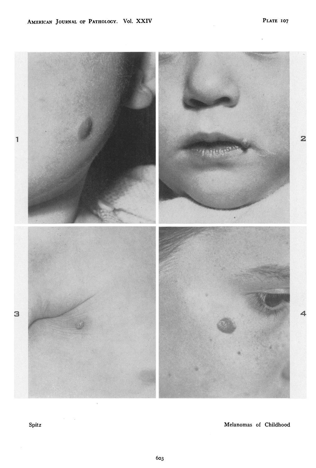
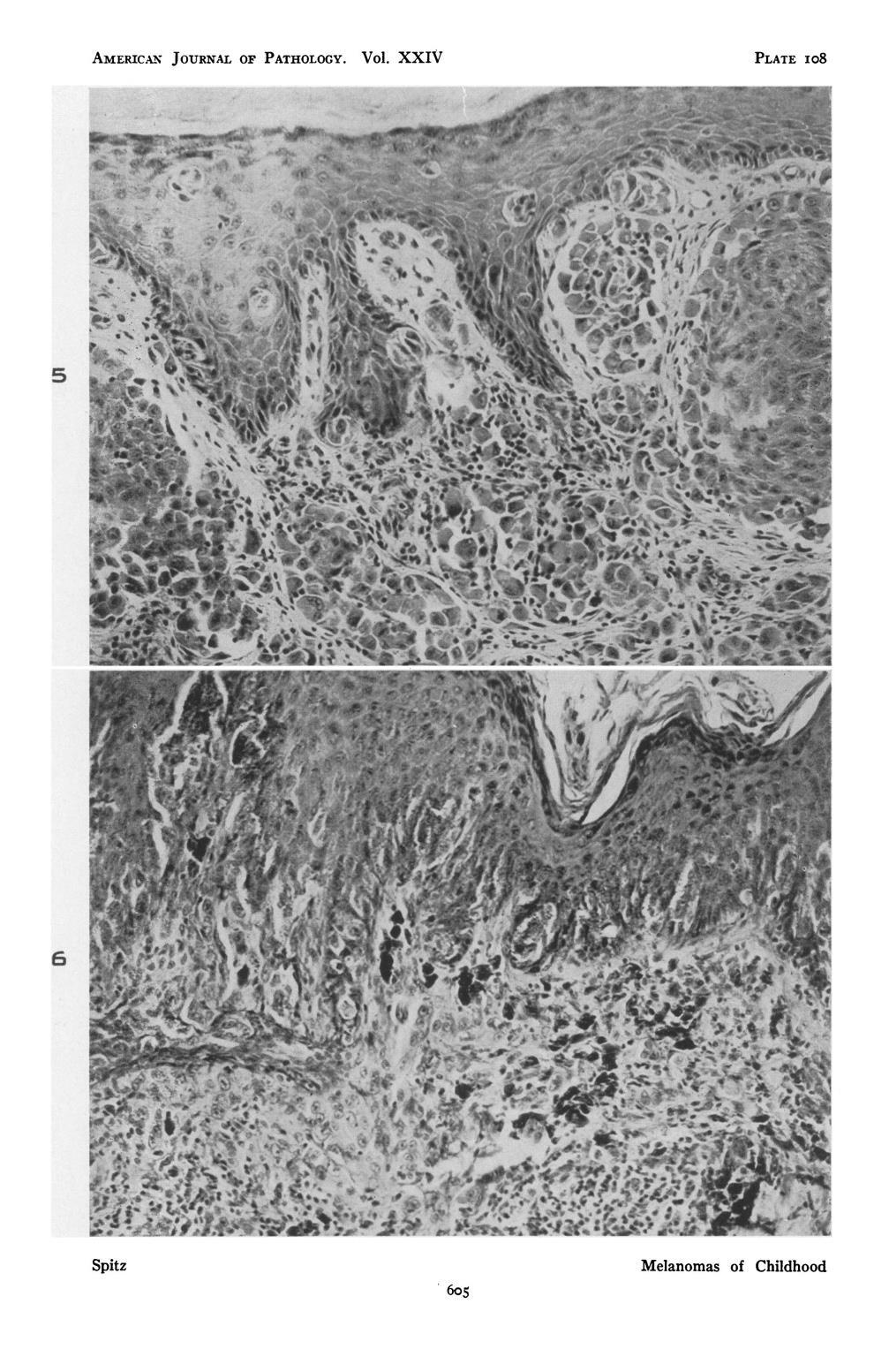
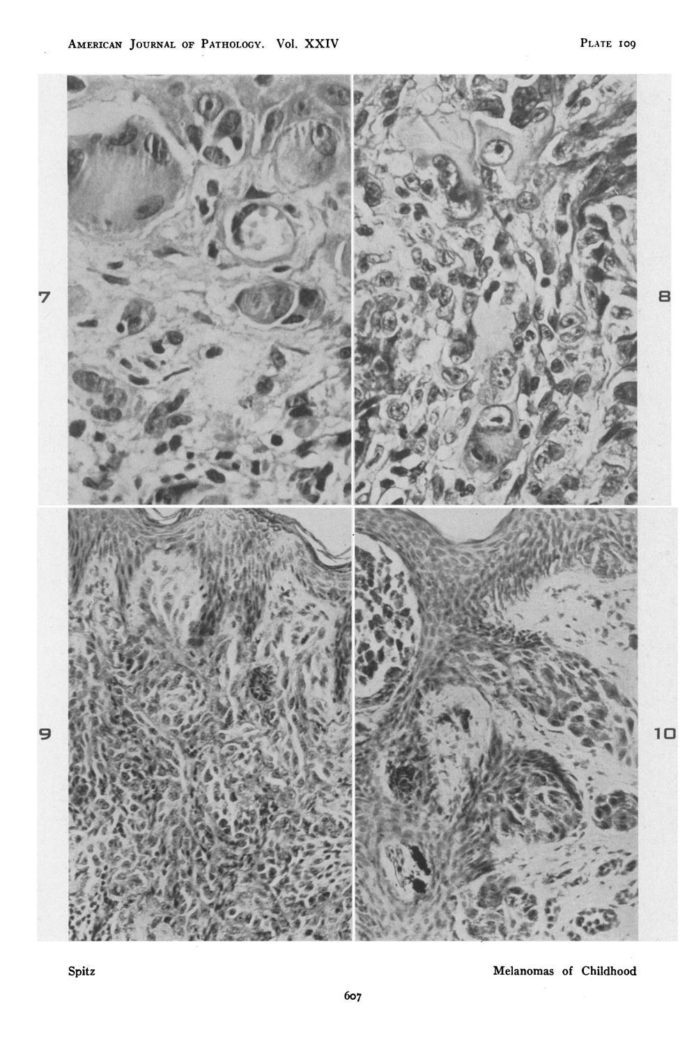
3 S Spitz Sophie Spitz Melanomas of Childhood ; Am J Pathol children (18 mo - 12 yrs) 12/13 had a benign clinical course

4 Sophie Spitz Born 1910 in Nashville AFIP 1944/45: Atlas of Pathology of Tropical Diseases MSK

5 Sophie Spitz

6 Spitz & Allen

7 Am J Pathol 1948
8
9 S Spitz Sophie Spitz Melanomas of Childhood ; Am J Pathol children (18 mo - 12 yrs) 12/13 had a benign clinical course JUVENILE MELANOMA - different from adult tumors

10 Sophie Spitz s Melanomas - Heterogeneous Group of Tumors Am J Pathol 1948 S Spitz 13 children (18 mo -12 yrs) All benign except for one: 12 yo girl Tumor on foot Deeply locate: plantar fascia Probable correct diagnosis: Clear cell sarcoma

11 Juvenile Melanoma A sheep in wolf s clothes
12 Spitz Nevus Kernan JA, Ackerman LV. Spindle cell nevi and epithelioid cell nevi (socalled juvenile melanomas) in children and adults: a clinicopathological study of 27 cases. Cancer. 1960;13: Weedon D, Little J. Spindle and epithelioid cell nevi in children and adults. A review of 211 cases of Spitz nevi. Cancer 1977; 40: Paniago-Pereira C, Maize JC, Ackerman AB. Nevus of large spindle and/or epithelioid cells (Spitz's nevus). Arch Dermatol. 1978; 114:

13 Spitz Nevus Melanoma J Derm Surg 1975
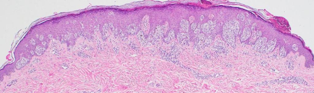
14 Distinction by Microscopy Spitz Nevus Architecture Symmetric Sharply circumscribed Cytology Maturation Spindle and/or epitheloid Other Features Kamino bodies Clefts Melanoma Architecture Asymmetric Ill-defined Cytology No maturation Pagetoid, Pleomorphism Other Features No Kamino bodies Consumption of epidermis


15 Picasso vs DaVinci

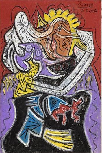
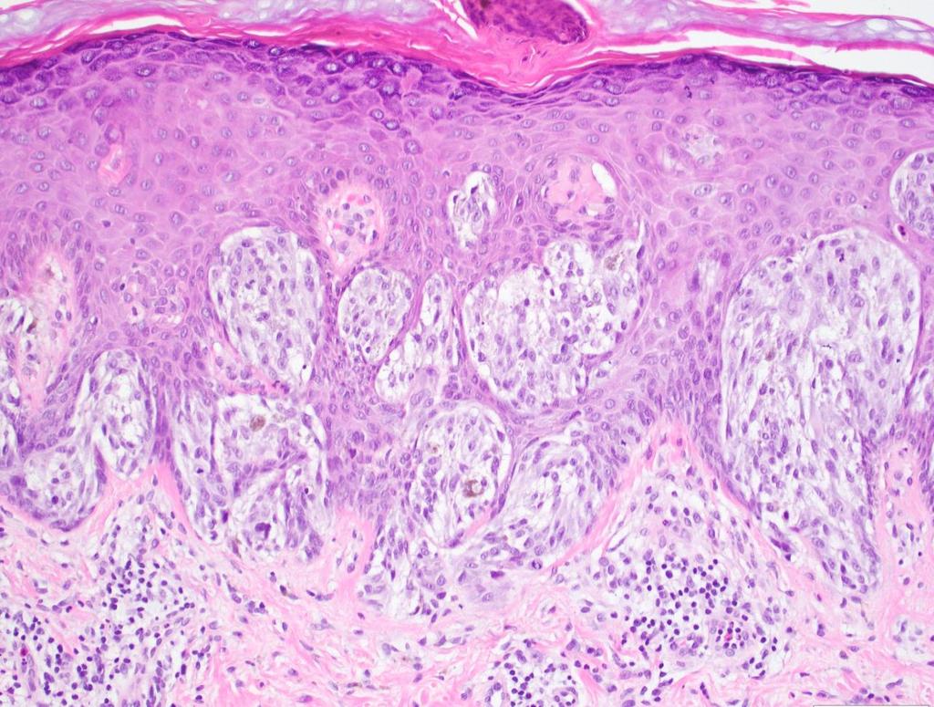
16 Real Picasso vs Fake Picasso
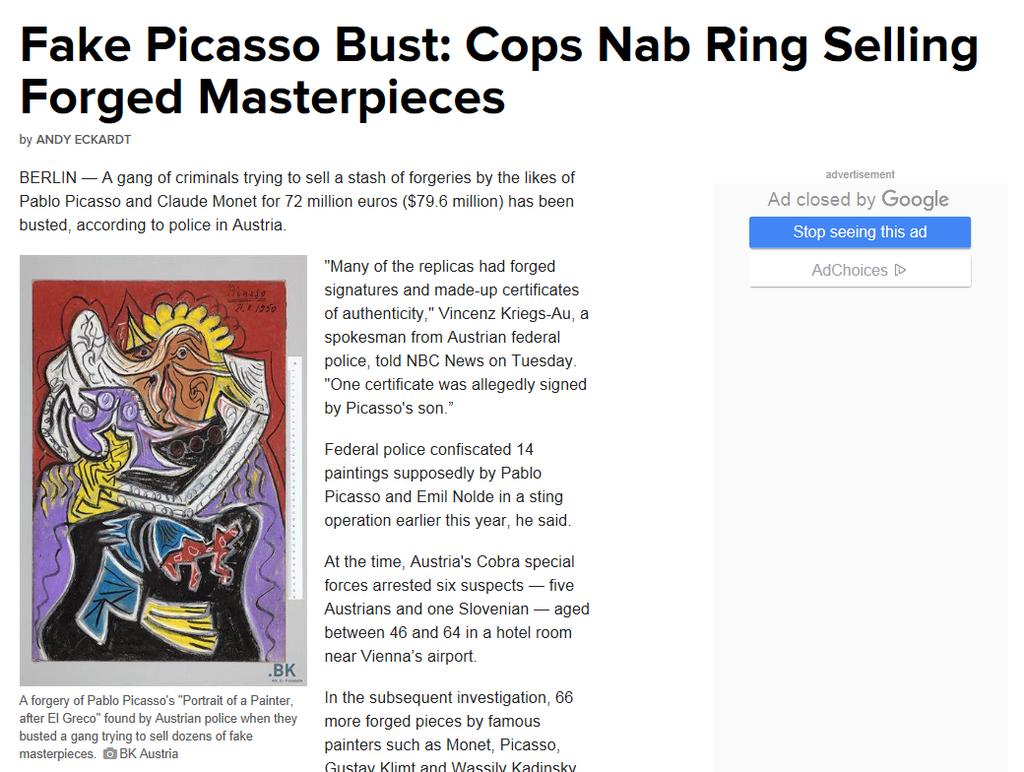
17


18 28 F, left arm Spitz Nevus

19 Spitz Nevus Kamino Bodies

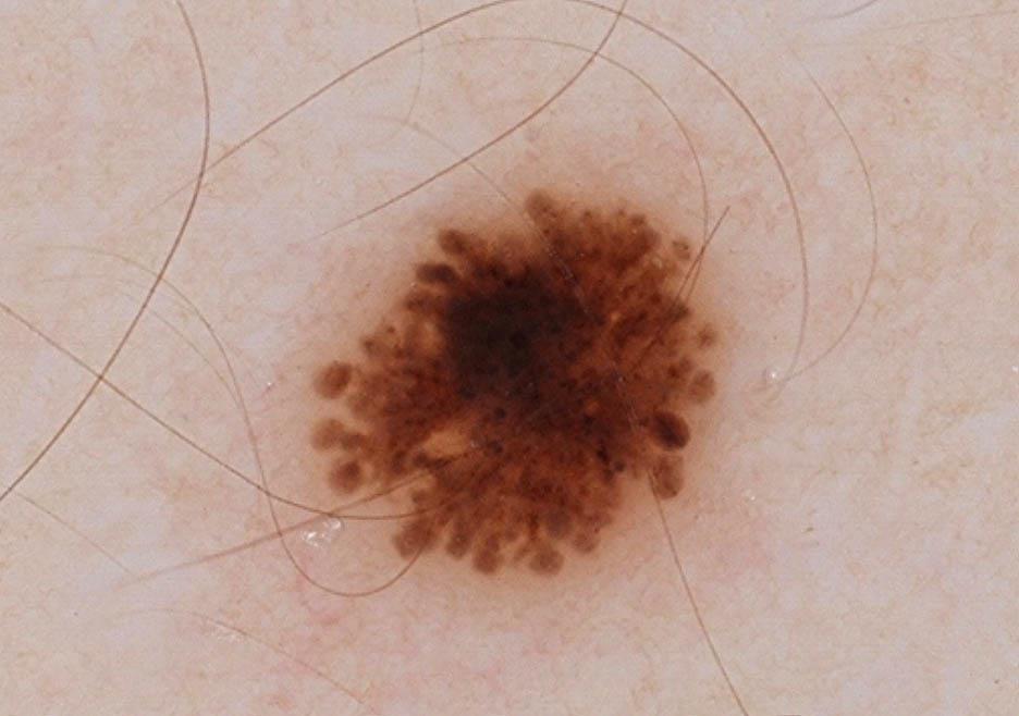

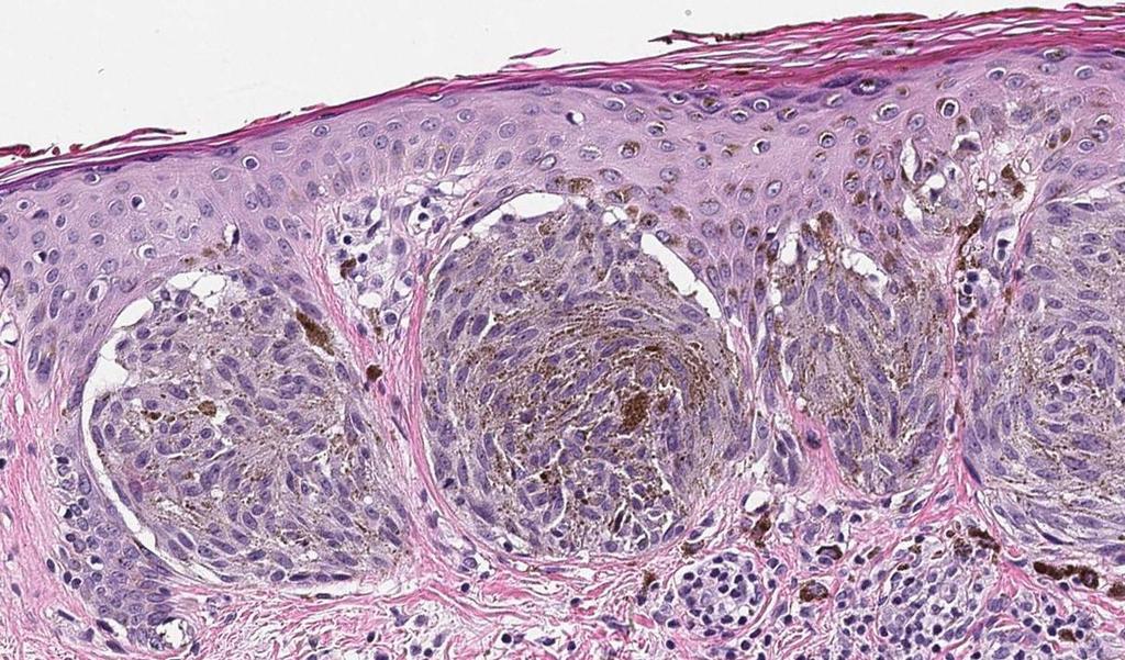
20 Pigmented Spitz Nevus
21 The Spitz Family Grows Spitz nevus, nos Pigmented spindle cell nevus of Reed Desmoplastic Spitz nevus Angiomatoid Spitz nevus Halo Spitz nevus Pagetoid Spitz nevus Others

22 Darth Spitz

23 Reported as Spitz Nevus 28 yo man with dark mole on chest Spitz s nevus


24 Junctional nests of pigmented spindle cells with cleft Maturation Mitotic figures
25 Spitzoid Melanoma 6 yrs later 6 yrs later: LN Metastasis

26 The Fog of Spitz
27
28
29
30


31 Atypical Spitz Tumor or Spitz Nevus?


32 Spitz Nevus, AST or Melanoma? Nevoid Borderline Tumor
33 BAP1 Diagnosis: BAP1-Deficient Nevus (aka Wiesner s Nevus)

34 Surgery to the Rescue
35 J Am Acad Dermatol 2000;42:527-30
36 SLN Findings for Diagnosis If any neoplasm made up of melanocytes metastasizes, it is a melanoma A Spitz-like lesion that metastasizes is a melanoma, pure and simple Mones & Ackerman Am J Dermatopathol 2004;26: AB Ackerman
37
38

39 Dermal Solar Elastotic Material in SLN Am J Surg Pathol 2010; 34:1492-7

40 Mesothelial Cells in LN


41 Subcapsular Melanocytes What do they mean? Mart1 Mart1 Axillary SLN from 44F with mammary adenocarcinoma
42 The Brave New Spitz The Molecularization of Spitz

43 Molecular Classification of Spitz Tumors
44 Desmoplastic Spitz Nevus 33 F SNP-array: 11p


45 Spitz Nevus with ALK Fusions 25 M ALK
46 Alk-Fusion Partners in Spitz lesions TPM3 DCTN1 GTF3C2 NPM1 CLIP1 TPR MLPH (1q21.3) (2p13.1) (2p23.3) (5q35.1) (12q24.31) (1q31.1) (2q37.3)


47 28F NTRK1 Spitz Nevus with NTRK1 expression
48 NTRK-Fusions LMNA-NTRK1 TP53 NTRK1 ETV6 - NTRK3 MYO5A NTRK3 MYH9 NTRK3


49 Atypical Spitz Tumor with ALK-Fusion ALK

50 Atypical Spitz with NTRK1-Fusion
51 Value of Molecular Findings Clarity of Terminology Adjunct Evidence for Diagnosis Possibly Useful for Prognosis and/or Treatment
52 Clarity of Terminology Spitzoid Resembles a Spitz Nevus Spitz/Spitzian Pathway of Spitz Nevus

53 Spitzoid Melanomas


54 Melanomas with BRAF and NRAS Mutations Conventional melanomas with minor spitzoid features
55 Spitzoid Melanoma vs Malignant Spitz Tumor BRAF- or NRAS- mutated conventional melanomas with spitzoid features Melanomas with genomic aberrations typical of Spitz = malignant Spitz tumors
56 Spitz Nevus vs Melanoma Role of Molecular Studies for Diagnostic Accuracy
57 19 F, rt wrist

58 Diagnosis in 2001 Spitz Nevus, Compound Type


59 LN, rt axilla, 2013 S100P


60 FISH for EWSR1 break apart TRANSLOCATION NORMAL


61 Diagnosis Compound Clear Cell Sarcoma J Cutan Pathol 2013; 40: 950-4
62 14 F; rt cheek Diagnosis?
63

64 Path Report: Spitzoid Neoplasm with Severe Atypia

65 Cytogenetics: Gain of 11p 11p

66 14 F, rt cheek Diagnosis: Spitz Nevus
67 Spitzoid Melanoma 6 yrs later FISH Results: Gains in CCDN1 (11q13) and RREB1 (6p25) in > 70% of cells Metastatic melanoma
68 Potential Prognostic or Therapeutic Significance Kinase fusions
69 Spitzoid Neoplasms Spitz Nevi Atypical Spitz Tumors (AST) Spitzoid/Spitzian Melanoma
70 Spitzoid Neoplasms Tools for Dx Conventional H&E IHC Molecular Studies Clinical Correlation
71 Spitz Nevus or Melanoma?
72 Spitzoid Neoplasm
73
74
75 Clinical Findings 68 yo woman New lesion within past year Has doubled in diameter in 3 months
76 Diagnosis: Melanoma

77 Mutation Status: NRASQ61R

78 Cytogenetic Analysis: Melanoma

79 Spitzoid Melanoma of Adults A Diagnostic Problem Wolf in Sheep s Clothing - Modern ancillary methods can reduce diagnostic errors - Better clinical correlation can reduce errors
80 Use of Atypical Spitz Tumor Diagnostic Problem Short Term Work-up incomplete Diagnostic Uncertainty Long Term Work-up did not yield definitive evidence Low Grade Malignant Neoplasm
81
82 Pediatric Melanomas Melanoma death rates (2007, USA): 13.5% ( MSKCC: Death rate of patients with melanoma diagnosed before age 18: 12%
83 Is Spitzoid Melanoma of Childhood a Malignant Melanoma? Cancer 2013; 119: 3911

84 Spitz Rebirth From Juvenile Melanoma to Spitzoid Melanoma of Childhood
85 Summary Spitz nevus vs tumor vs melanoma Spitzoid vs Spitz/Spitzian Molecular Pathology (Diagnosis/Classification)


86 The Spectrum of Spitz Benign Spitz Atypical Spitz Malignant Spitz

87 MSKCC Dept of Pathology 1948
88

89 New Textbook

90 Thank You!
Melanocytic Lesions: Use of Immunohistochemistry and Special Studies Napa Valley 2018
 Melanocytic Lesions: Use of Immunohistochemistry and Special Studies Napa Valley 2018 Victor G. Prieto, MD, PhD Professor Depts. of Pathology and Dermatology University of Texas - MD Anderson Cancer Center
Michael T. Tetzlaff MD, PhD
 Molecular alterations informing the diagnosis of melanocytic tumors Michael T. Tetzlaff MD, PhD Associate Professor Department of Pathology, Section of Dermatopathology Department of Translational and
Update on Spitzoid and Blue nevus-like melanocytic lesions Emphasis on molecular studies informing diagnosis, prognosis and therapy
 Update on Spitzoid and Blue nevus-like melanocytic lesions Emphasis on molecular studies informing diagnosis, prognosis and therapy Michael T. Tetzlaff MD, PhD Associate Professor Department of Pathology,
Case 26 Male 37. Right jawline 5mm nodule?keloid. The best diagnosis is:
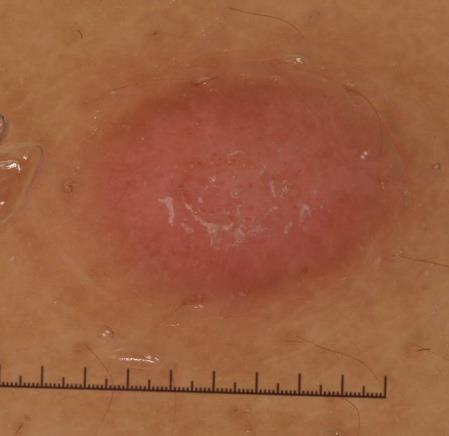
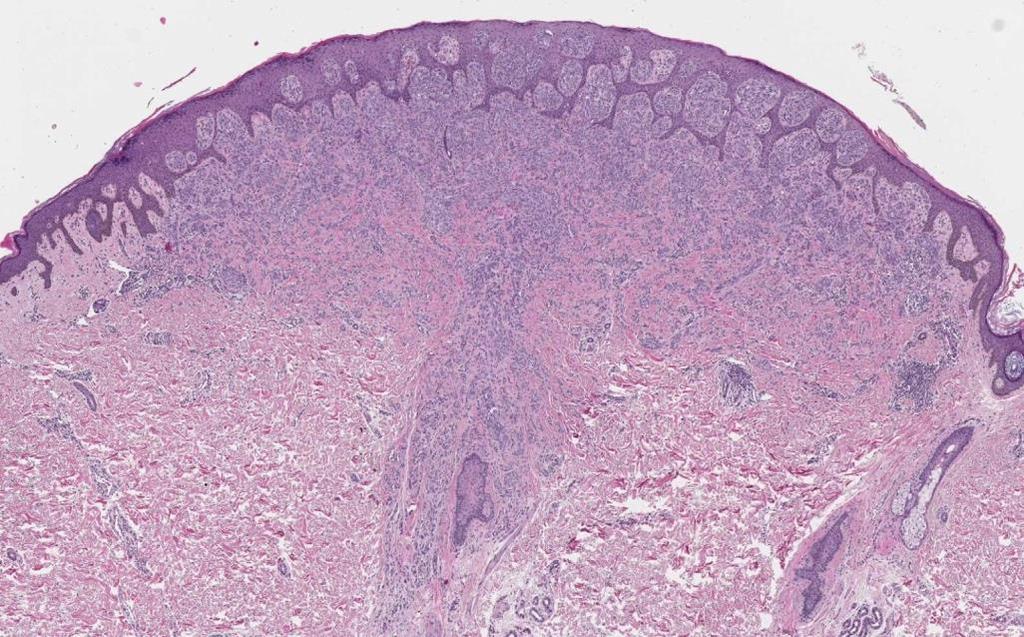
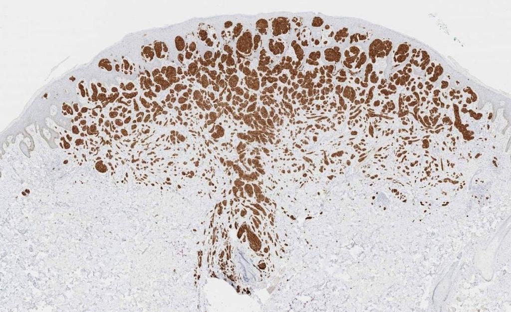
 Case 26 Male 37. Right jawline 5mm nodule?keloid. The best diagnosis is: A. Desmoplastic Spitz naevus B. Atypical Spitz Tumour C. Spitzoid melanoma D. Deep penetrating naevus E. Spitz naevus Case 26: M
10/2/17. MELTUMP, SAMPUS, AST.An Algorithmic Approach to Challenging (Often Borderline) Melanocytic Tumors. An Introduction to SNP Arrays
 Melanocytic Tumors. An Introduction to SNP Arrays") MELTUMP, SAMPUS, AST.An Algorithmic Approach to Challenging (Often ) Melanocytic Tumors An Introduction to SNP Arrays Rajiv M. Patel, M.D. RCPA NZ ASM 2017 (11:45-12:30pm, Saturday, 23-09-17) Why do we
MELTUMP, SAMPUS, AST.An Algorithmic Approach to Challenging (Often ) Melanocytic Tumors An Introduction to SNP Arrays Rajiv M. Patel, M.D. RCPA NZ ASM 2017 (11:45-12:30pm, Saturday, 23-09-17) Why do we
21/07/2017. The «gray zone» of diagnosis is visible. Nevus Atypical nevus Melanoma. Melanoma ex-blue nevus
 Update on the Clinico- Pathological and Molecular Diagnosis of Melanocytic Lesions None to declare Conflicts of interest Belfast pathology Arnaud de la Fouchardière MD, PhD Lyon, France What is new? Today
Melanoma and the genes: Molecular alterations informing the diagnosis of melanocytic tumors
 Melanoma and the genes: Molecular alterations informing the diagnosis of melanocytic tumors Michael T. Tetzlaff MD, PhD Associate Professor Department of Pathology, Section of Dermatopathology Department
Vernon K. Sondak. Department of Cutaneous Oncology Moffitt Cancer Center Tampa, Florida
 Vernon K. Sondak Department of Cutaneous Oncology Moffitt Cancer Center Tampa, Florida Australasian Melanoma Conference 2016 Sydney, NSW, Australia October 29, 2016 Disclosures Dr. Sondak is a compensated
MAPK Pathway. CGH Next Generation Sequencing. Molecular Tools in Care of Patients with Pigmented Lesions 7/20/2017
 Molecular Tools in Care of Patients with Pigmented Lesions Tammie Ferringer, MD Geisinger Medical Center, Danville, PA tferringer@geisinger.edu DISCLOSURE OF RELATIONSHIPS WITH INDUSTRY Tammie Ferringer,
Molecular Aspects of Melanocytic Neoplasia. Iwei Yeh MD, PhD University of California, San Francisco
 Molecular Aspects of Melanocytic Neoplasia Iwei Yeh MD, PhD University of California, San Francisco Thanks to: Boris Bastian Timothy McCalmont Philip LeBoit Beth Ruben Jeff North Laura Pincus Thaddeus
Dermatopathology. Dr. Rafael Botella Estrada. Hospital La Fe de Valencia
 Dermatopathology Dr. Rafael Botella Estrada. Hospital La Fe de Valencia Melanoma and mimics Dr. Martin Mihm Malignant lesions result from the accumulation of mutations Class I lesions (benign) Class II
A PRACTICAL APPROACH TO ATYPICAL MELANOCYTIC LESIONS BIJAN HAGHIGHI M.D, DIRECTOR OF DERMATOPATHOLOGY, ST. JOSEPH HOSPITAL
 A PRACTICAL APPROACH TO ATYPICAL MELANOCYTIC LESIONS BIJAN HAGHIGHI M.D, DIRECTOR OF DERMATOPATHOLOGY, ST. JOSEPH HOSPITAL OBJECTIVES Discuss current trends and changing concepts in our understanding of
Melanocytic proliferations in sundamaged
 Atypical Spitzoid Tumor: What Does It Mean And How Should It Be Managed? Melanocytic proliferations in sundamaged skin Jane L. Messina, Jane L. Messina MD International Melanoma Pathology Working Group
There is NO single Melanoma Stain. > 6000 Mutations in Melanoma. What else can be done to discriminate atypical nevi from melanoma?
 Las Vegas Fall Clinical 2016: The Assessment and Diagnosis of Melanoma Whitney A. High, MD, JD, MEng Associate Professor, Dermatology & Pathology Director of Dermatopathology (Dermatology) University of
Female 18. Deeply pigmented lesion on trunk.?warty naevus?seborrhoeic keratosis?malignant melanoma. The best diagnosis is:
 Female 18. Deeply pigmented lesion on trunk.?warty naevus?seborrhoeic keratosis?malignant melanoma. The best diagnosis is: A. deep penetrating naevus B. naevoid malignant melanoma C. pigment synthesising
6/22/2015. Original Paradigm. Correlating Histology and Molecular Findings in Melanocytic Neoplasms
 6 Correlating Histology and Molecular Findings in Melanocytic Neoplasms Pedram Gerami MD, Associate Professor of Dermatology and Pediatrics at Northwestern University Disclosures: I have been a consultant
Conflict of Interest 9/2/2014. Pathogenesis and Comparison of Atypical Spitz Nevi vs Benign Spitz, and Childhood Melanoma
 Pathogenesis and Comparison of Atypical Spitz Nevi vs Benign Spitz, and Childhood Melanoma Martin C. Mihm Jr., M.D., F.A.C.P. Harvard Medical School Brigham and Women s Hospital Dana Farber Cancer Center
Case 231: F7. Exophytic naevus over left trapezious. Grown over a few weeks. Iniitally flat.?spitz naevus,?malignant
 Case 231: F7. Exophytic naevus over left trapezious. Grown over a few weeks. Iniitally flat.?spitz naevus,?malignant Dermoscopy: coarse vascular structures. c/o A, B, C RAC7750 Case 231: F7. Exophytic
ACCME/Disclosures ALK FUSION-POSITIVE MESENCHYMAL TUMORS. Tumor types with ALK rearrangements. Anaplastic Lymphoma Kinase. Jason L.
 Companion Meeting of the International Society of Bone and Soft Tissue Pathology The Evolving Concept of Mesenchymal Tumors ALK FUSION-POSITIVE MESENCHYMAL TUMORS Jason L. Hornick, MD, PhD March 13, 2016
Ways to get into trouble, ideas on avoiding trouble, and diagnostic approaches to keep trouble at bay
 Pitfalls in the diagnosis of melanocytic tumors Timothy McCalmont, MD University of California, San Francisco Ways to get into trouble, ideas on avoiding trouble, and diagnostic approaches to keep trouble
BAP-oma & BEYOND MICHAEL A NOWAK, MD
 BAP-oma & BEYOND MICHAEL A NOWAK, MD CONFLICTS No conflicts with the content of this lecture BAP-oma Wiesner 2011: Families with multiple tan dome-shaped papules of head, neck, trunk, and extremities.
The Pathology of Neoplasia Part II
 The Pathology of Neoplasia Part II February 2018 PAUL BOGNER, MD A S S O C I A T E P R O F E S S O R O F O N C O L O G Y P A T H O L O G Y A N D D E R M A T O L O G Y Clinical goals of cancer pathology
David B. Troxel, MD. Common Medicolegal Situations: Misdiagnosis of Melanoma
 Common Medicolegal Situations: Misdiagnosis of Melanoma David B. Troxel, MD Medical Director, The Doctors Company, Napa, California Clinical Professor Emeritus, University of California at Berkeley Past
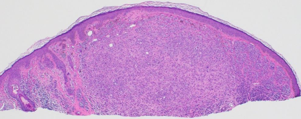
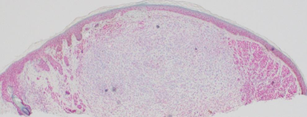
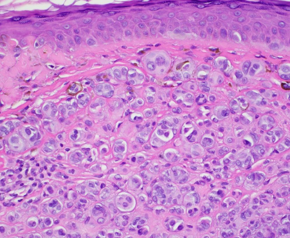
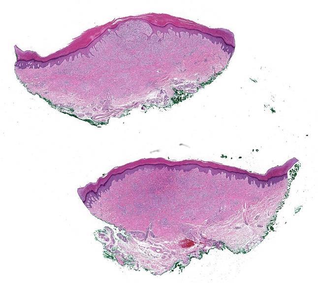
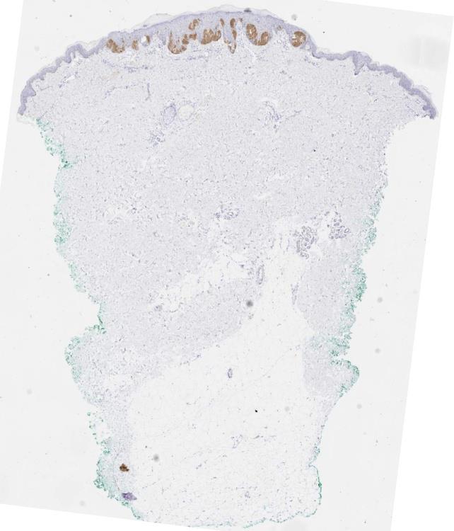
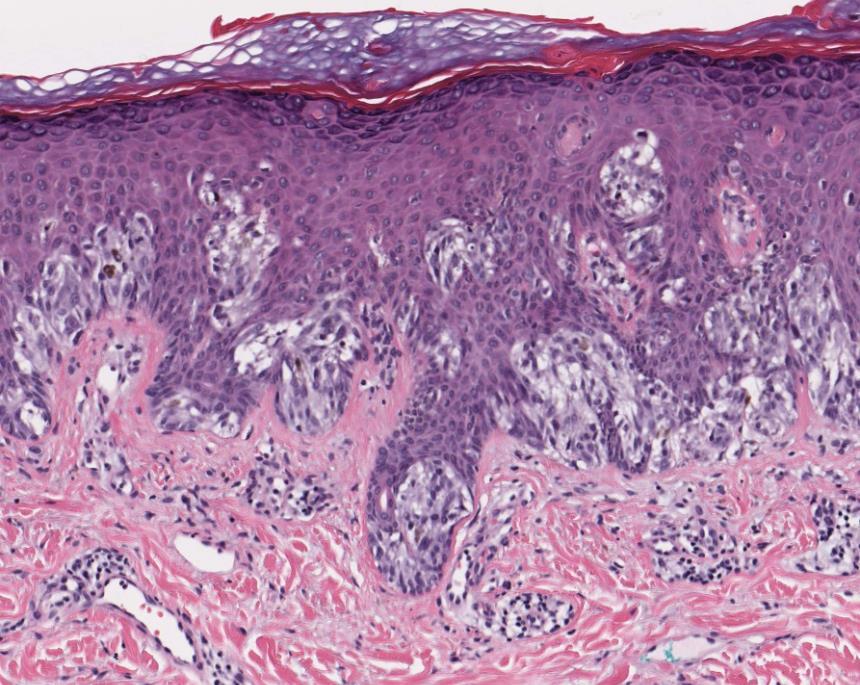
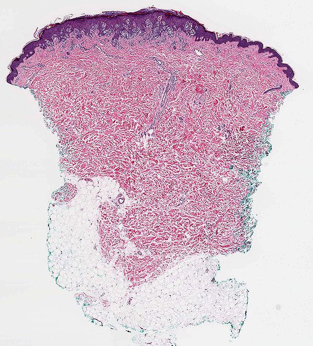
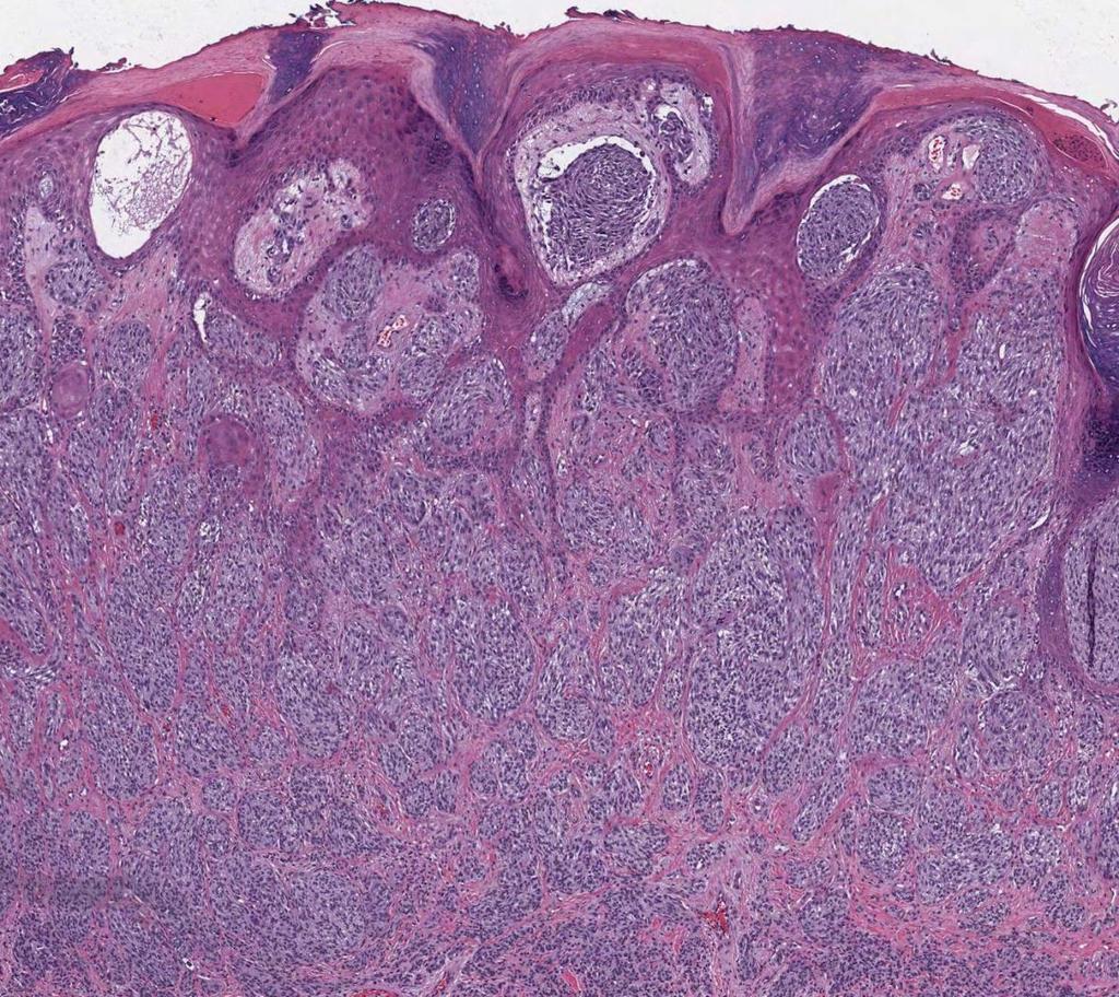
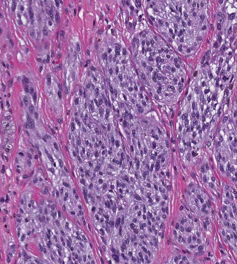
Supplementary Figure 1. Spitzoid Melanoma with PPFIBP1-MET fusion. (a) Histopathology (4x) shows a domed papule with melanocytes extending into the
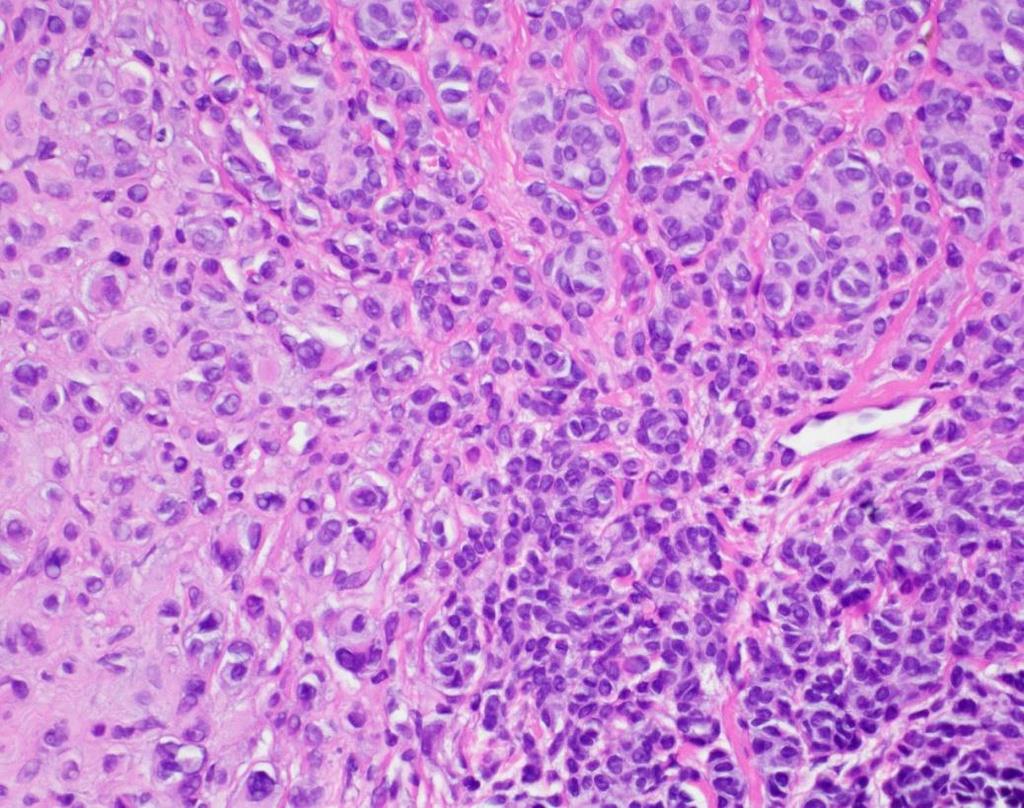
 Histopathology (4x) shows a domed papule with melanocytes extending into the") Supplementary Figure 1. Spitzoid Melanoma with PPFIBP1-MET fusion. (a) Histopathology (4x) shows a domed papule with melanocytes extending into the deep dermis. (b) The melanocytes demonstrate abundant
Supplementary Figure 1. Spitzoid Melanoma with PPFIBP1-MET fusion. (a) Histopathology (4x) shows a domed papule with melanocytes extending into the deep dermis. (b) The melanocytes demonstrate abundant
Diagnoses of Cases 1. Lentigo, other melanosis and the acquired nevus 2. Variations on the acquired nevus 3. Dermal melanocytosis
 Diagnoses of Cases 1. Lentigo, other melanosis and the acquired nevus 1 1A. Lentigo simplex 4 1B. Psoralens and ultraviolet A (PUVA) lentigo 6 1C. Solar lentigo 8 1D. Café au lait macule 10 1E. Ink-spot
Spitz nevi in the classic histopathological pattern - lamb in wolf`s clothing *
 DERMATOPATHOLOGY 91 Spitz nevi in the classic histopathological pattern - lamb in wolf`s clothing * Gustavo Costa Verardino 1 Mayra Carrijo Rochael 1 DOI: http://dx.doi.org/10.1590/abd1806-4841.20153310
Desmoplastic Melanoma R/O BCC. Clinical Information. 74 y.o. man with lesion on left side of neck r/o BCC
 R/O BCC Sabine Kohler, M.D. Professor of Pathology and Dermatology Dermatopathology Service Stanford University School of Medicine Clinical Information 74 y.o. man with lesion on left side of neck r/o
Patricia Chevez-Barrrios AAOOP-USCAP /12/2016
 Biomarkers in Ocular Melanoma Patricia Chévez-Barrios, MD Pathology and Genomic Medicine, Houston Methodist Hospital Professor of Pathology and Laboratory Medicine and Ophthalmology, Weill Cornell Medical
Management of pediatric melanocytic lesions
 Open Journal of Clinical & Medical Case Reports Management of pediatric melanocytic lesions Volume 3 (2017) Issue 8 ISSN 2379-1039 Jin Kim, BS; Emmanuel Gabriel MD, PhD; Weiguo Liu MD, PhD; Lin Lin MD,
2/6/2018. Original Paradigm. Clonal Chromosomal A berrations. Only 20% of Spitz Nevi 95% 6p, 7q, 17q, 20q, 4q,8q, 1q, 11q. Isolated Gain in 11p
 Molecular Diagnostics for Melanocytic Neoplasms: Moving towards a Revolution in the Management of Melanocytic Neoplasms Pedr am Gerami MD Associate Professor of Dermatology, Pathology and Pediatrics at
Genetic Testing: When should it be ordered? Julie Schloemer, MD Dermatology
 Genetic Testing: When should it be ordered? Julie Schloemer, MD Dermatology Outline Germline testing CDKN2A BRCA2 BAP1 Somatic testing Gene expression profiling (GEP) BRAF Germline vs Somatic testing
Malignant tumors of melanocytes: Part 1. Deba P Sarma, MD., Omaha
 Malignant tumors of melanocytes: Part 1 Deba P Sarma, MD., Omaha The melanocytic tumor is one of the most difficult and confusing areas in Dematopathology. It is true that most (95%) of such lesions are
K Blessing, J J H Grant, D S A Sanders, M M Kennedy, A Husain, P Coburn
 J Clin Pathol 2000;53:591 595 591 Papers Pathology, Aberdeen University, Foresterhill, Aberdeen AB25 2ZD, K Blessing Pathology, Birmingham University, Birmingham B15 2TT, D S A Sanders Pathology, Heartlands
F006 Imaging in Dermatology Melanocytic Neoplasia Clinical-Confocal-Pathological-Correlations
 F006 Imaging in Dermatology Melanocytic Neoplasia Clinical-Confocal-Pathological-Correlations Melissa Gill, MD SkinMedical Research and Diagnostics Dobbs Ferry, NY, USA Department of Pathology SUNY Downstate
Case RAC7783. M46. Ear. Mole. r/o MM.?Blue naevus RAC7783
 Case RAC7783. M46. Ear. Mole. r/o MM.?Blue naevus RAC7783 Pie Chart Participants N=74 Benign: 48 N=74 Blue naevus: 38 Intradermal: 12 DPN: 10 Compound 3 Clonal: 3; Spitz 2; Special Site: 1; Congenital:
Cutaneous Mesenchymal Neoplasms with EWSR1 Rearrangement
 Cutaneous Mesenchymal Neoplasms with EWSR1 Rearrangement By Konstantinos Linos MD, FCAP, FASDP Bone, Soft Tissue and Dermatopathology Assistant Professor of Pathology Dartmouth-Hitchcock Medical Center
Financial disclosures
 Mesenchymal Neoplasms with Melanocytic Differentiation By Konstantinos Linos MD, FCAP, FASDP Bone, Soft Tissue and Dermatopathology Assistant Professor of Pathology Dartmouth-Hitchcock Medical Center Geisel
Time to reconsider Spitzoid neoplasms?
 DERMATOLOGY PRACTICAL & CONCEPTUAL www.derm101.com Time to reconsider Spitzoid neoplasms? Carmelo Urso 1 1 Department of Anatomic Pathology, Dermatopathology Section, SM Annunziata Hospital, AUSL Toscana
Enterprise Interest Nothing to declare
 Enterprise Interest Nothing to declare Diagnoses one would not like to miss in soft tissue pathology early in your career Marta Sbaraglia, MD Department of Pathology Hospital of Treviso University of Padua
1/10/2018. Soft Tissue Tumors Showing Melanocytic Differentiation. Overview. Desmoplastic/ Spindle Cell Melanoma
 2016 MFMER slide-1 2016 MFMER slide-2 2016 MFMER slide-3 Soft Tissue Tumors Showing Melanocytic Differentiation Andrew L. Folpe, M.D. Professor of Laboratory Medicine and Pathology Mayo Clinic, Rochester,
Disclosures. An update on ancillary techniques in the diagnosis of soft tissue tumors. Ancillary techniques. Introduction
 Disclosures An update on ancillary techniques in the diagnosis of soft tissue tumors. I have nothing to disclose. Andrew Horvai, MD, PhD Clinical Professor, Pathology Introduction Ancillary techniques
Melanocytic Tumours. Molecular Biology 02/06/2015. Cutaneous Melanocytic Tumours Introduction. Thomas Brenn. Intermediate Malignancy
 Cutaneous Melanocytic Tumours Introduction Melanocytic Tumours: Update on Epidemiology and Molecular Biology Thomas Brenn Wide clinical and morphological spectrum Ranging from benign naevi to melanoma
Society for Pediatric Pathology Spring Meeting Joint Symposium with American Society of Dermatopathology
 Society for Pediatric Pathology 2013 Spring Meeting Joint Symposium with American Society of Dermatopathology Update on Cutaneous Melanocytic, Mesenchymal and Lymphoproliferative Lesions in Children Melanocytic
Guy Perrot (Ги Перро)
") НАУЧНО-ПРАКТИЧЕСКАЯ КОНФЕРЕНЦИЯ (МАСТЕР-КЛАСС) «ПРАКТИЧЕСКИЕ АСПЕКТЫ ДИАГНОСТИКИ И ЛЕЧЕНИЯ МЕЛАНОМЫ КОЖИ» DIAGNOSTIC AND PITFALLS IN MELANOMA Guy Perrot (Ги Перро) MD PHD pathologist, University Hospital
НАУЧНО-ПРАКТИЧЕСКАЯ КОНФЕРЕНЦИЯ (МАСТЕР-КЛАСС) «ПРАКТИЧЕСКИЕ АСПЕКТЫ ДИАГНОСТИКИ И ЛЕЧЕНИЯ МЕЛАНОМЫ КОЖИ» DIAGNOSTIC AND PITFALLS IN MELANOMA Guy Perrot (Ги Перро) MD PHD pathologist, University Hospital
Page 1 of 3. We suggest the following changes:
 Page 1 of 3 Loren E. Clarke, M.D. Myriad Genetic Laboratories, Inc. 320 Wakara Way, Salt Lake City, UT 84108 Phone: 801.883.3470 Email: lclarke@myriad.com Date of Request: June 2017 NCCN Guidelines Panel:
المركب النموذج--- سبيتز وحمة = Type Spitz's Nevus, Compound SPITZ NEVUS 1 / 7
 SPITZ NEVUS 1 / 7 Epidemiology An annual incidence rate of 1.4 cases of Spitz nevus per 100,000 individuals has been estimated in Australia, compared with 25.4 per 100,000 individuals for cutaneous melanoma
Benign and malignant epithelial lesions: Seborrheic keratosis: A common benign pigmented epidermal tumor occur in middle-aged or older persons more
 Benign and malignant epithelial lesions: Seborrheic keratosis: A common benign pigmented epidermal tumor occur in middle-aged or older persons more common on the trunk; but extremities, head and neck are
Salivary Gland FNA ATYPICAL : Criteria and Controversies
 Salivary Gland FNA ATYPICAL : Criteria and Controversies W.C. Faquin, M.D., Ph.D. Director, Head and Neck Pathology Massachusetts General Hospital Massachusetts Eye and Ear Infirmary Harvard Medical School
Which melanoma patients benefit from genetic testing?
 Which melanoma patients benefit from genetic testing? Michael A. Marchetti, MD Assistant Attending, Dermatology Service Memorial Sloan Kettering Cancer Center American Academy of Dermatology Annual Meeting
Associate Clinical Professor of Dermatology MUSC
 Re-excision of Moderately Dysplastic Nevi: Should we or shouldn t we? John C. Maize, Jr, M.D. Dermatologist and Dermatopathologist Trident Dermatology, Charleston SC Associate Clinical Professor of Dermatology
Simulators of melanoma
 Simulators of melanoma Philip E. LeBoit, M.D. Depts. of Pathology and Dermatology University of California, San Francisco Simulators of melanoma Simulators of melanoma in situ Melanocytic Non-melanocytic
Conflicts of Interest
 Challenging Melanocytic Lesions Carlos N. Prieto-Granada M.D. Assistant Professor University of Alabama at Birmingham (UAB) Department of Pathology 2017 AAD Annual Meeting 3/2/17 - Orlando, FL None Conflicts
EARLY ONLINE RELEASE
 EARLY ONLINE RELEASE Note: This article was posted on the Archives Web site as an Early Online Release. Early Online Release articles have been peer reviewed, copyedited, and reviewed by the authors. Additional
Index. Springer-Verlag Berlin Heidelberg 2017 J.A. Plaza, V.G. Prieto, Pathology of Pigmented Skin Lesions, DOI /
 A Acral lentiginous (mucosal lentiginous) melanoma, 483 Acral lentiginous melanoma (ALM) asymmetric and irregular lentiginous junctional growth, 431 clinical features, 427 428 differential diagnosis, 428
Difficult Diagnoses and Controversial Entities in Neoplastic Lung
 Difficult Diagnoses and Controversial Entities in Neoplastic Lung Lynette M. Sholl, M.D. Associate Pathologist, Brigham and Women s Hospital Chief, Pulmonary Pathology Service Associate Professor, Harvard
Melanoma-Back to Basics I Thought I Knew Ya! Paul K. Shitabata, M.D. Dermatopathologist APMG
 Melanoma-Back to Basics I Thought I Knew Ya! Paul K. Shitabata, M.D. Dermatopathologist APMG At tumor board, a surgeon insists that all level II melanomas are invasive since they have broken through the
Molecular Methods in the Diagnosis and Prognostication of Melanoma: Pros & Cons
 Molecular Methods in the Diagnosis and Prognostication of Melanoma: Pros & Cons Ben J. Friedman, MD Senior Staff Physician Department of Dermatology Department of Pathology and Laboratory Medicine Henry
Integrating Fluorescence in situ Hybridization and Genomic Array Results into the Diagnostic Workup of Melanoma
 Integrating Fluorescence in situ Hybridization and Genomic Array Results into the Diagnostic Workup of Melanoma Association for Molecular Pathology United States and Canadian Academy of Pathology Companion
Difficulties in the diagnosis of spitzoid melanocytic lesions
 For reprint orders, please contact reprints@expert-reviews.com Difficulties in the diagnosis of spitzoid melanocytic lesions Expert Rev. Dermatol. 5(5), 549 560 (2010) Stephen H Olsen 1, Rajiv M Patel
Brief Report. Shivanand Gundalli 1, Smita Kadadavar 1, Somil Singhania 1, Rutuja Kolekar 2 INTRODUCTION. Melanocytic Nevus
 Our Dermatology Online Histopathological spectrum of benign melanocytic nevi our experience in a tertiary care centre Shivanand Gundalli 1, Smita Kadadavar 1, Somil Singhania 1, Rutuja Kolekar 2 1 Department
HISTOPATHOLOGIC REPORTING OF MELANOCYTIC SKIN LESIONS. Problems, thoughts, proposals
 HISTOPATHOLOGIC REPORTING OF MELANOCYTIC SKIN LESIONS Problems, thoughts, proposals Gerardo Ferrara Anatomic Pathology Unit Macerata General Hospital AV3 ASUR Marche Macerata, I Aims and scope STANDARDIZATION:
Disclosures. An update on ancillary techniques in the diagnosis of soft tissue tumors. Ancillary techniques. Introduction
 Disclosures An update on ancillary techniques in the diagnosis of soft tissue tumors. I have nothing to disclose. Andrew Horvai, MD, PhD Clinical Professor, Pathology Introduction Ancillary techniques
Diploma Examination. Dermatopathology: First paper. Tuesday 20 March Candidates must answer FOUR questions. Time allowed: 3 hours
 Dermatopathology: First paper Tuesday 20 March 2018 Candidates must answer FOUR questions Time allowed: 3 hours 1. Give an account of the genetic aberrations encountered in Spitzoid neoplasms and how these
Mesothelioma: diagnostic challenges from a pathological perspective. Naseema Vorajee August 2016
 Mesothelioma: diagnostic challenges from a pathological perspective Naseema Vorajee August 2016 Naseema.vorajee@nhls.ac.za Pleural diseases (whether neoplastic, reactive or infective) may have similar
Less Common Variants of Cutaneous Melanoma
 Less Common Variants of Cutaneous Melanoma Raymond L. Barnhill* 1, G. Peter Sarantopoulos 1, and Kapil Gupta 2 1 Department of Pathology and Laboratory Medicine, University of California, Los Angeles,
ARTICLE INFO ABSTRACT
 Melanocytic Pigmentation: A Single Manifestation of Myriad of Pathologies [PP: 05-09] Dr. Swapna Honwad Department of Oral Pathology dr.swapnahonwad@gmail.com Dr. Elsy P. Simon Department of Endodontics
Dermatologica Sinica
 DERMATOLOGICA SINICA 30 (2012) 57e61 Contents lists available at SciVerse ScienceDirect Dermatologica Sinica journal homepage: http://www.derm-sinica.com CASE REPORT Pigmented epithelioid melanocytoma:
F109 Imaging in Dermatology Melanocytic Neoplasia Clinical-Confocal-Pathological-Correlations
 F109 Imaging in Dermatology Melanocytic Neoplasia Clinical-Confocal-Pathological-Correlations Melissa Gill, MD SkinMedical Research and Diagnostics Dobbs Ferry, NY, USA Department of Pathology SUNY Downstate
Pathology of the skin. 2nd Department of Pathology, Semmelweis University
 Pathology of the skin 2nd Department of Pathology, Semmelweis University Histology of the skin Epidermis: Stratum corneum Stratum granulosum Stratum spinosum Stratum basale Dermis: papillary and reticular
Disclosure. Relevant Financial Relationship(s) None. Off Label Usage None MFMER slide-1
 None. Off Label Usage None MFMER slide-1") Disclosure Relevant Financial Relationship(s) None Off Label Usage None 2013 MFMER slide-1 Case Presentation A 43 year old male, with partial nephrectomy for a right kidney mass 2013 MFMER slide-2 2013
Disclosure Relevant Financial Relationship(s) None Off Label Usage None 2013 MFMER slide-1 Case Presentation A 43 year old male, with partial nephrectomy for a right kidney mass 2013 MFMER slide-2 2013
The Relevance of Cytologic Atypia in Cutaneous Neural Tumors
 The Relevance of Cytologic Atypia in Cutaneous Neural Tumors Recent Findings - New Developments New Problems Zsolt B. Argenyi, M.D. Professor of Pathology & Dermatology Director of Dermatopathology Department
5/21/2018. Disclosures. Consulting: Myriad Genetics SciBase. Superficial Atypical Melanocytic Proliferations. SSM, LMM and (some of) their Simulants
 their Simulants") Disclosures Consulting: Myriad Genetics SciBase Superficial Atypical Melanocytic Proliferations SSM, LMM and (some of) their Simulants 1 Melanomas and Nevi. Nevi are important mainly in relation to melanoma
Disclosures Consulting: Myriad Genetics SciBase Superficial Atypical Melanocytic Proliferations SSM, LMM and (some of) their Simulants 1 Melanomas and Nevi. Nevi are important mainly in relation to melanoma
Special slide seminar
 Special slide seminar Tomáš Rozkoš The Fingerland Department of Pathology Charles University Medical Faculty and Faculty Hospital in Hradec Králové Czech Republic Case history, 33 years old resistance
Molecular Diagnosis of Soft Tissue Tumors: Avoid Pitfalls
 Molecular Diagnosis of Soft Tissue Tumors: Avoid Pitfalls Cristina Antonescu, MD Department of Pathology Memorial Sloan-Kettering Cancer Center, New York Overview I. When should we rely on the help of
Melanoma Update: 8th Edition of AJCC Staging System
 Melanoma Update: 8th Edition of AJCC Staging System Rosalie Elenitsas, M.D. Professor of Dermatology Director, Dermatopathology University of Pennsylvania DISCLOSURE OF RELATIONSHIPS WITH INDUSTRY None
57th Annual HSCP Spring Symposium 4/16/2016
 An Unusual Malignant Spindle Cell Lesion to Involve the Breast Erinn Downs-Kelly, D.O. Associate Professor of Pathology University of Utah & ARUP Laboratories No disclosures Case 39 y/o female with no
ACCME/Disclosures. Diagnosing Mesothelioma in Limited Tissue Samples. Papanicolaou Society of Cytopathology Companion Meeting March 12 th, 2016
 Diagnosing Mesothelioma in Limited Tissue Samples Papanicolaou Society of Cytopathology Companion Meeting March 12 th, 2016 Sanja Dacic, MD, PhD University of Pittsburgh ACCME/Disclosures GENERAL RULES
21/07/2017. Hobnail endothelial cells are not the same as epithelioid endothelial cells
 UPDATE IN CUTANEOUS VASCULAR S DERMATOPATHOLOGY SESSION BELFAST PATHOLOGY JUNE 21/2017 Dr E Calonje St John s Institute of Dermatology, London, United Kingdom THE FAMILY OF VASCULAR S WITH EPITHELIOID
- Selected Tumors of the Skin Appendages - Primary vs. Metastasis
 - Selected Tumors of the Skin Appendages - Primary vs. Metastasis Napa Valley 2018 Victor G. Prieto, MD, PhD Chair of Pathology UT MD Anderson Cancer Center vprieto@mdanderson.org Napa Valley in May Introduction
Skin Tumors in Children
 AAD San Diego S021 2018 Skin Tumors in Children Jane M. Grant-Kels, MD,FAAD grant@uchc.edu Founding Chair Emeritus, Derm Dept, UCONN Vice Chair Dept of Dermatology Professor of Dermatology, Pathology and
Rosette-like structures in the spectrum of spitzoid tumors
 J Cutan Pathol 2013: 40: 788 795 doi: 10.1111/cup.12192 John Wiley & Sons. Printed in Singapore Rosette-like structures in the spectrum of spitzoid tumors 2013 John Wiley & Sons A/S. Published by John
Klinisch belang van chromosomale translocatie detectie in sarcomen
 Translocations in sarcomas Klinisch belang van chromosomale translocatie detectie in sarcomen Judith V.M.G. Bovée, M.D., Ph.D. Department of Pathology Leiden University Medical Center RNA binding DNA binding
Copy number and somatic mutations drive tumors
 Detection of copy number alterations, ploidy and loss of heterozygosity across the genome in FFPE specimens Utility for diagnosis and treatment with comparison to FISH-based and as a complement to sequencing
Lung Tumor Cases: Common Problems and Helpful Hints
 Lung Tumor Cases: Common Problems and Helpful Hints Brandon T. Larsen, MD, PhD Senior Associate Consultant Department of Laboratory Medicine and Pathology Mayo Clinic Arizona Arizona Society of Pathologists
Financial disclosures
 Cutaneous Mesenchymal Neoplasms with EWSR1 Rearrangement By Konstantinos Linos MD, FCAP, FASDP Bone, Soft Tissue and Dermatopathology Assistant Professor of Pathology Dartmouth-Hitchc Geisel School of
ARIZONA SOCIETY OF PATHOLOGISTS 13 TH APRIL 2013 HEAD AND NECK CYTOPATHOLOGY. F ZAHRA ALY, MD, PhD
 ARIZONA SOCIETY OF PATHOLOGISTS 13 TH APRIL 2013 HEAD AND NECK CYTOPATHOLOGY F ZAHRA ALY, MD, PhD The main areas sites amenable for cytopathology include lymph nodes, thyroid, major salivary glands especially
Michael T. Tetzlaff MD, PhD
 American Joint Cancer Committee (AJCC) staging system for primary cutaneous melanoma (8 th Edition) and principles of sentinel lymph node evaluation Emphasis on concise and accurate reporting of primary
4/17/2015. Case 1. A 37 year old man with a 2.2 cm solitary left thyroid mass.
 Case 1 A 37 year old man with a 2.2 cm solitary left thyroid mass. Case 1 Case 1 1 Case 1: Diagnosis? A. Benign B. Atypia of undetermined significance/follicular lesion of undetermined significance C.
S everal morphological features are frequently used in the
 1194 ORIGINAL ARTICLE Interobserver reproducibility of histological features in cutaneous malignant melanoma C Urso, F Rongioletti, D Innocenzi, C Saieva, D Batolo, S Chimenti, R Filotico, R Gianotti,
Dermatologica Sinica
 DERMATOLOGICA SINICA 31 (2013) 140e144 Contents lists available at SciVerse ScienceDirect Dermatologica Sinica journal homepage: http://www.derm-sinica.com CASE REPORT Atypical fibroxanthoma-like amelanotic
Update: Morphologic Considerations in Mesothelioma within the Pleural and Peritoneal Cavities. Douglas J. Hartman, MD June 7, 2018
 Update: Morphologic Considerations in Mesothelioma within the Pleural and Peritoneal Cavities Douglas J. Hartman, MD June 7, 2018 Objectives Review Historical Features associated with prognosis Present
Self assessment case. Dr Saleem Taibjee Dorset County Hospital, Dorchester
 Self assessment case Dr Saleem Taibjee saleemtaibjee@gmail.com Dorset County Hospital, Dorchester Clinical details 34-year-old man: Shave excision Skin tag / papilloma left thigh The best diagnosis is:
Gross Appearance & Histology of Skin Cancer. Kyle Mannion M.D. January 21, 2005
 Gross Appearance & Histology of Skin Cancer Kyle Mannion M.D. January 21, 2005 Actinic Keratosis 5-20% will develop squamous/basal cell ca Almost solely from solar damage Usually develop during 4 th decade
Diploma examination. Dermatopathology: First paper. Tuesday 21 March Candidates must answer FOUR questions ONLY. Time allowed: Three hours
 Dermatopathology: First paper Tuesday 21 March 2017 1. Discuss the role of fluorescent in-situ hybridization (FISH) and emerging molecular techniques in the diagnosis of cutaneous melanocytic lesions,
Approximately 2% of the United States population
 Differentiation of Malignant Melanoma From Benign Nevus Using a Novel Genomic Microarray With Low Specimen Requirements Wells M. Chandler, MD; Leslie R. Rowe, MS; Scott R. Florell, MD; Mona S. Jahromi,
Molecular pathology in soft tissue tumors. Sylvia Höller Pathologie
 Molecular pathology in soft tissue tumors Sylvia Höller Pathologie When do we perform molecular testing? Morphology and IHC are not clearly fitting with an entity some translocations are entity specific
MECHANISMS OF HUMAN DISEASE: LABORATORY SESSION PATHOLOGY OF THE SKIN LAB. Friday, February 13, :30 am 11:00 am
 MECHANISMS OF HUMAN DISEASE: LABORATORY SESSION PATHOLOGY OF THE SKIN LAB Friday, February 13, 2009 9:30 am 11:00 am FACULTY COPY GOALS: Describe the basic clinical and morphologic features of various
THE SPITZ NEVUS OFTEN POSES
 OBSERVATION ONLINE FIRST Melanoma Mimic A Case of Multiple Pagetoid Spitz Nevi KaLynne Harris, MD; Scott R. Florell, MD; Jason Papenfuss, MD; Wendy Kohlmann, MS, CGC; Mona Jahromi, BS; Joshua D. Schiffman,
Low-grade serous neoplasia. Robert A. Soslow, MD
 Low-grade serous neoplasia Robert A. Soslow, MD soslowr@mskcc.org Outline Orientation Ovarian tumor overview Non serous borderline tumors Serous borderline tumors Clinical summary Morphologic description
Dilemmas in Cytopathology and Histopathology
 Dilemmas in Cytopathology and Histopathology Yuri E. Nikiforov, MD, PhD Division of Molecular & Genomic Pathology University of Pittsburgh Medical Center, USA Objectives Discuss new WHO classification