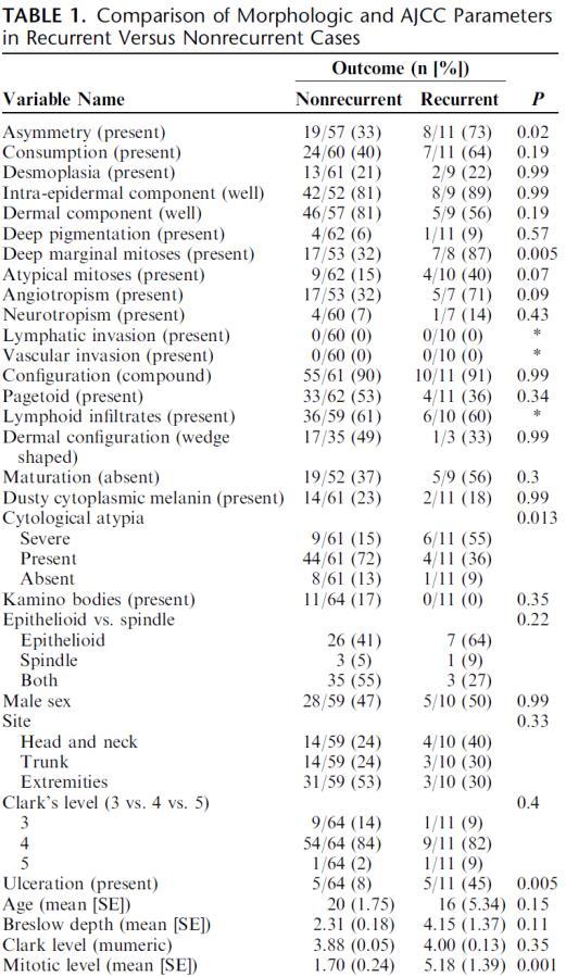
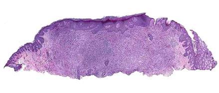
Update on Spitzoid and Blue nevus-like melanocytic lesions Emphasis on molecular studies informing diagnosis, prognosis and therapy
|
|
|
- Philip Stevens
- 5 years ago
- Views:
Transcription


1 Update on Spitzoid and Blue nevus-like melanocytic lesions Emphasis on molecular studies informing diagnosis, prognosis and therapy Michael T. Tetzlaff MD, PhD Associate Professor Department of Pathology, Section of Dermatopathology Department of Translational and Molecular Pathology The University of Texas MD Anderson Cancer Center Executive Officer Translational Research Program The Alliance for Clinical Trials
2 Update on Spitz nevus and Spitzoid Melanoma Spitz nevus family of lesions Spitz nevus Atypical Spitz Tumor (AST) Spitzoid Melanoma????

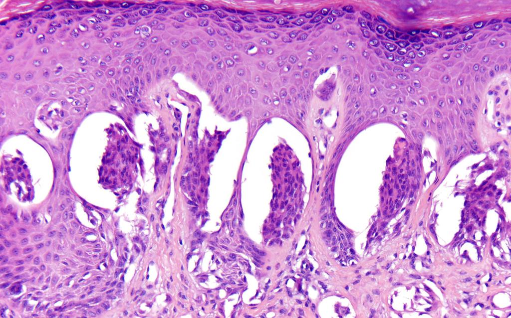
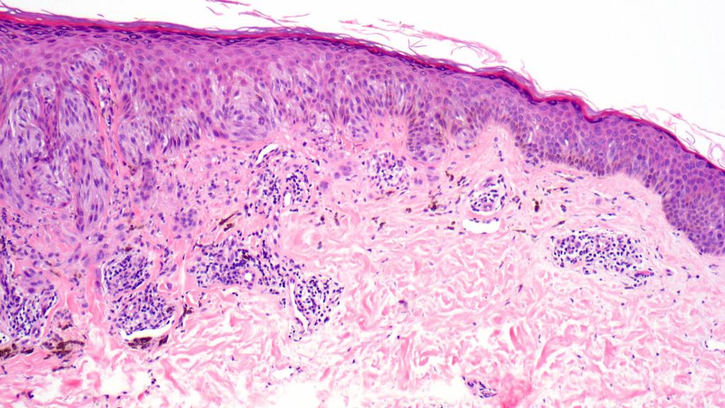
3 Spitz nevus, Spitzoid Melanoma and Atypical Spitz Tumors Pink papule on the cheek of a 9 year old boy Spitz Nevus

4 Spitz Nevus
5
6

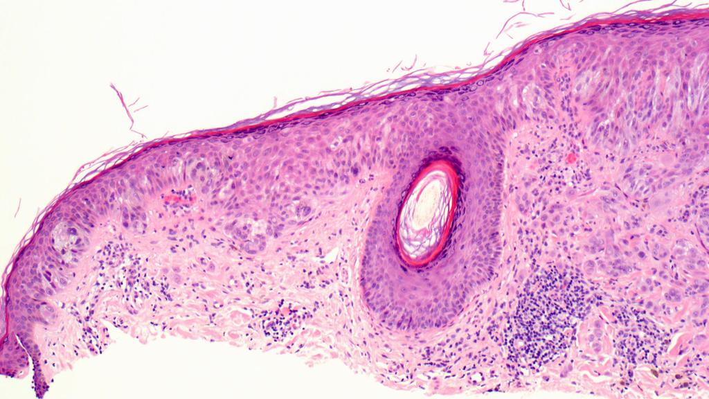
7 Spitz Nevus

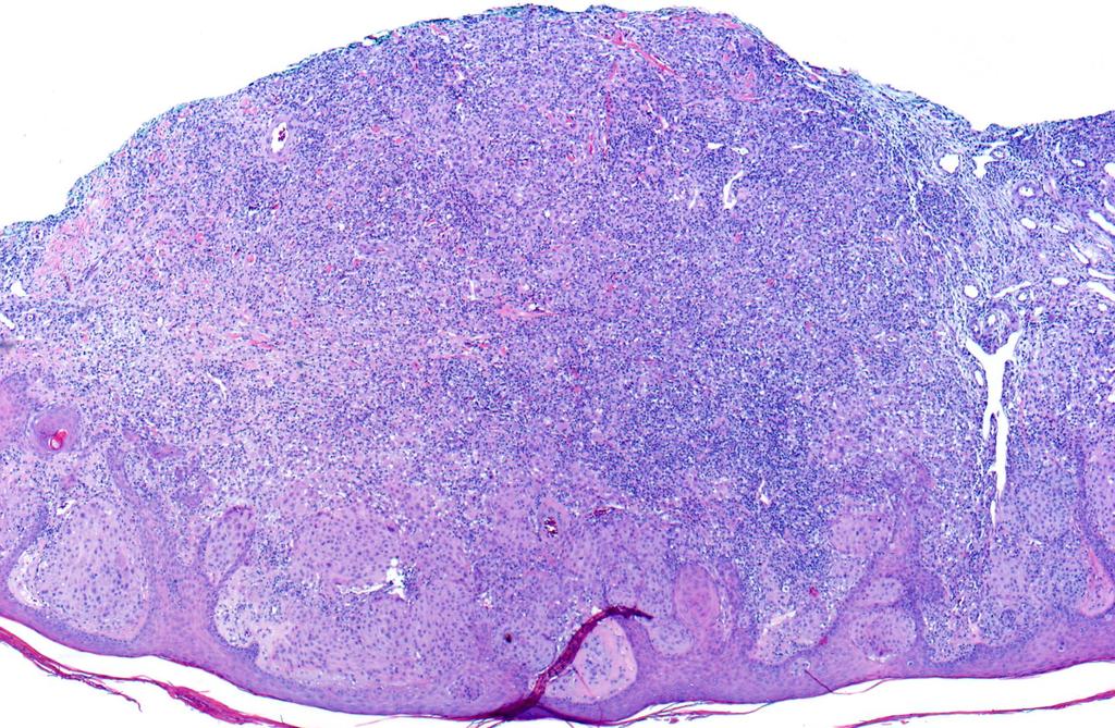
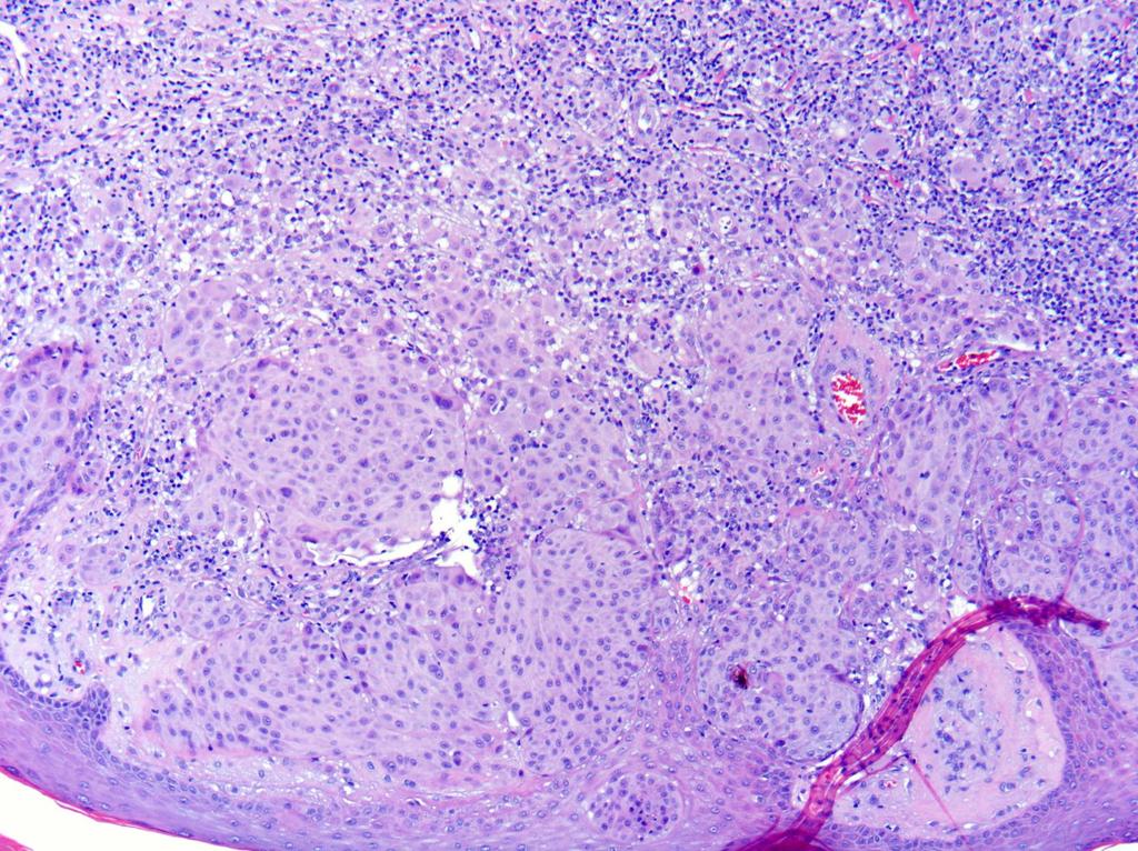
8 Spitz nevus, Spitzoid Melanoma and Atypical Spitz Tumors Changing pink-red papule on the thigh of a 49 year old man
9
10
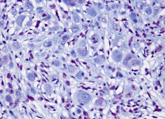
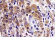
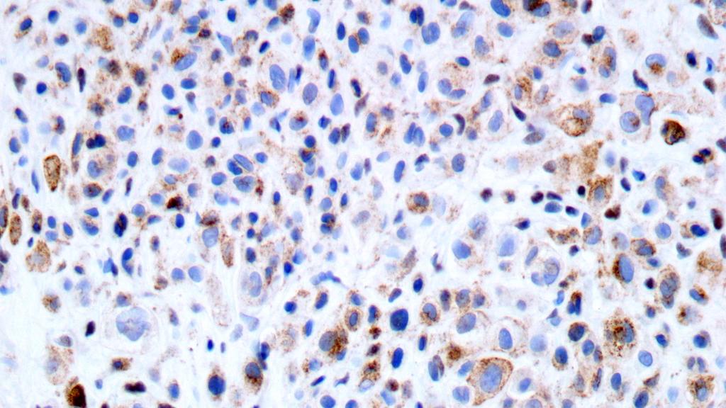
11 Mart-1/Ki-67

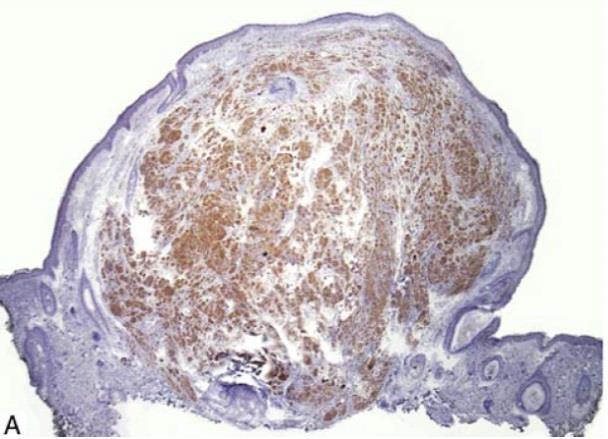
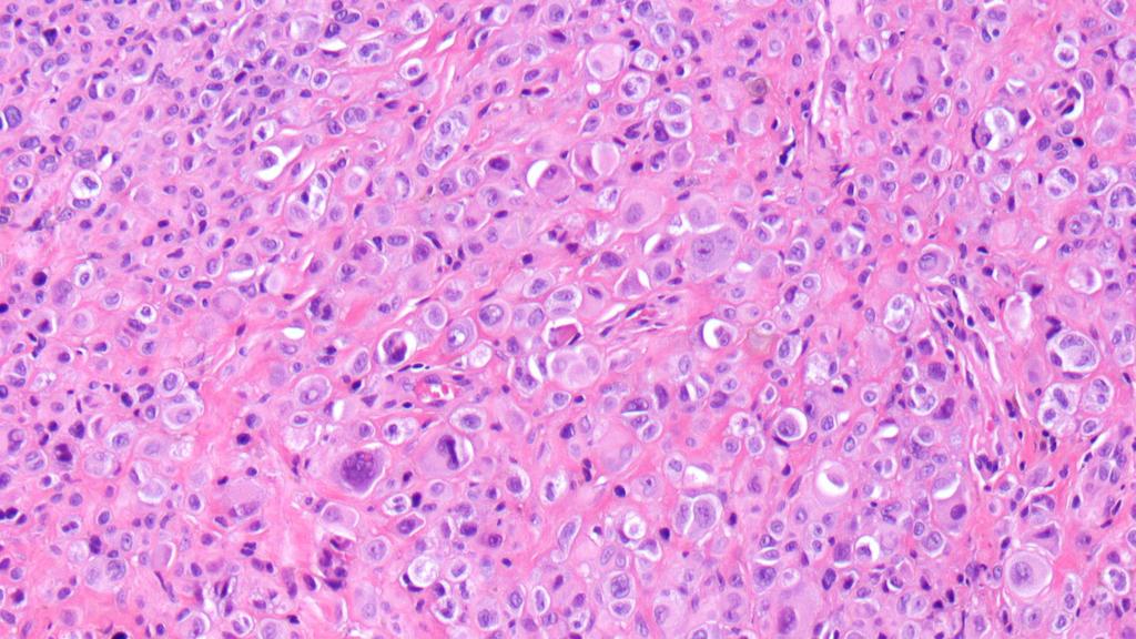
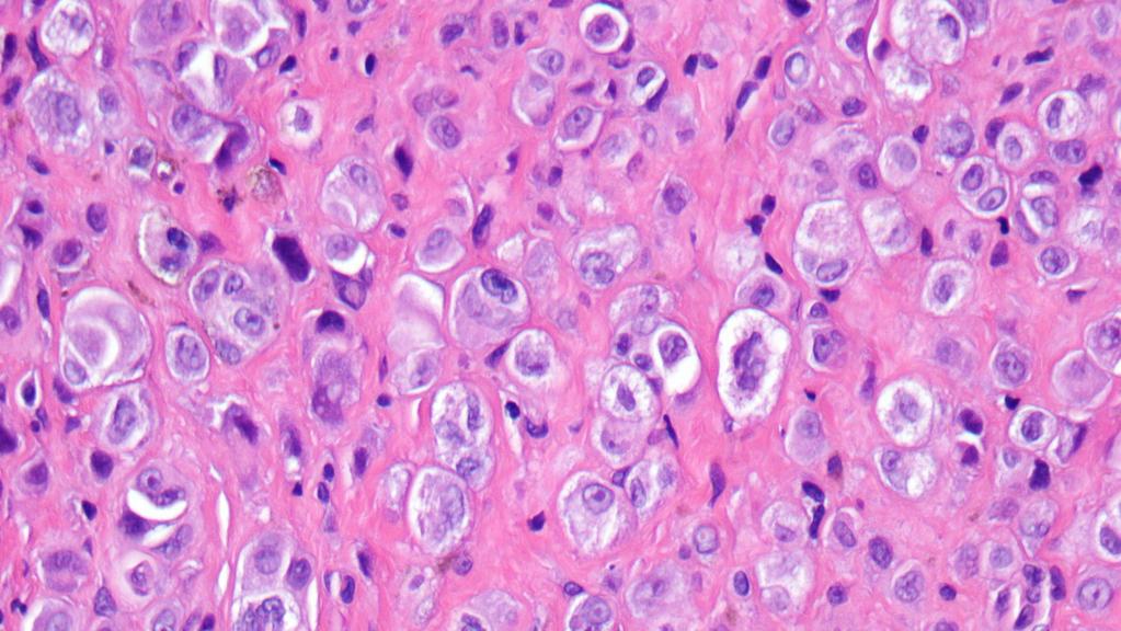
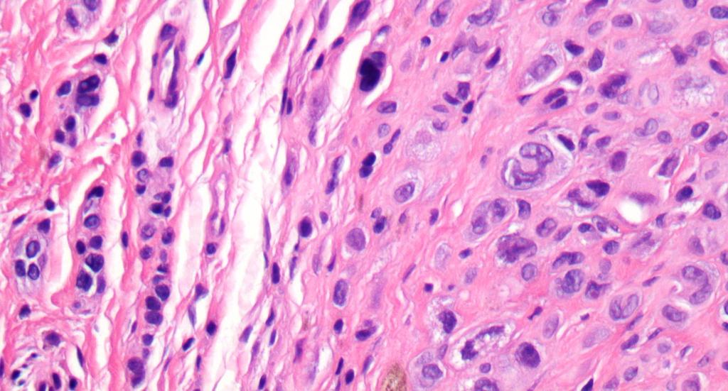
12 Invasive melanoma with Spitz features BAP1
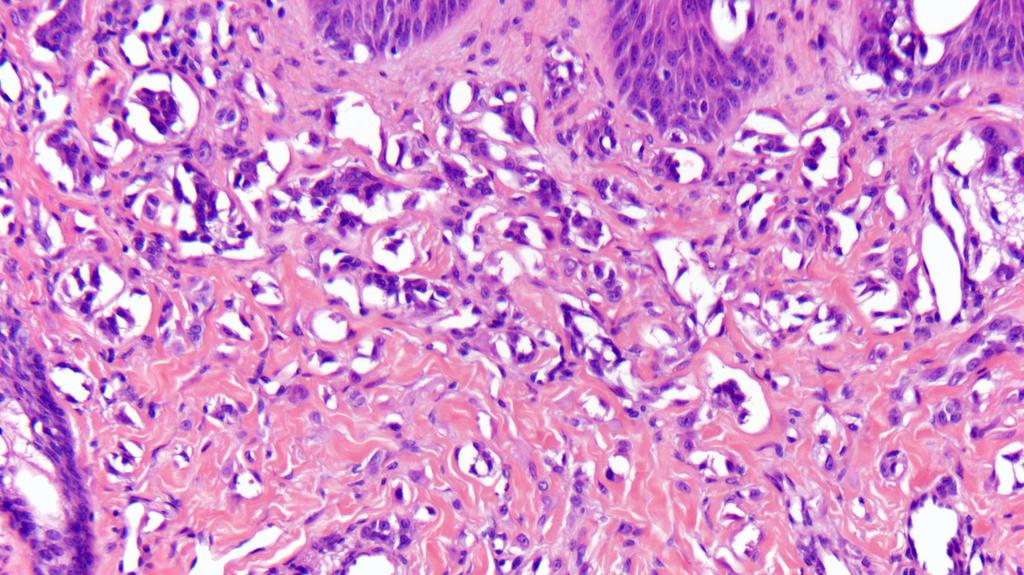
13 Spitz nevus, Spitzoid Melanoma and Atypical Spitz Tumors Spitzoid lesions Spitz nevus Size <10 mm Symmetric Well-circumscribed Epidermal hyperplasia Kamino bodies common Rarely ulcerated Discontinuous intraepidermal proliferation Minimal pagetoid spread confined to center Well spaced cells in dermis Mitotic figures only superficial Monotonous atypia with maturation Spitzoid melanoma Size >10 mm Asymmetric Poorly circumscribed Minimal epidermal changes Kamino bodies uncommon Ulceration not uncommon Continuous intraepidermal proliferation Prominent pagetoid spread including at the edges Dense, sheet-like, expansile Mitotic figures common throughout Prominent cytologic atypia without maturation

14 Reproducible criteria for the distinction among Atypical Spitz Tumors have been historically difficult to establish

15 Predicting lymph node metastases and disease progression among Atypical Spitz Tumors has been historically difficult 57 patients alive without disease Median follow-up: 28.6 and 43.8 months
16 A molecular classification scheme for Spitzoid neoplasia? 11p amplified HRAS mutant Desmoplastic No metastases to date Spitzoid Lesion Molecular-genetic alteration? Predictable clinical course? Reproducible histopathologic criteria? TERT promoter mutant BAP1 deficient BRAFV600E Spitzoid lesions defined by FISH Homozygous deletion of 9p21 Relative loss of 6q23 Translocation associated ROS1 ALK NTRK1 NTRK3 MET BRAF RET
 1 case with gains at 7q21 No evidence of recurrence or metastasis in any of the")
17 Identification of 11p amplified Spitz nevi Performed Comparative Genomic Hybridization (CGH) on 17 Spitz nevi 13 cases with no chromosomal copy number alterations 3 cases showed isolated gains of the entire short arm of chromosome 11 (11p) 1 case with gains at 7q21 No evidence of recurrence or metastasis in any of the lesions

 with >3x gains of 11p HRAS")
 of")
18 Morphology of 11p amplified/hras mutant Spitz nevi FISH for 11p on 102 Spitz nevi: Identified 12 Spitz nevi (11.8%) with >3x gains of 11p HRAS mutations in 8/12 (67%) of 11p Spitz nevi HRAS mutations in 1/21 (5%) of Spitz nevi without 11p


19 11p amplified/hras mutant Spitz nevi HRAS sequencing on 170 Spitz tumors: 24/170 Spitzoid lesions (14%) with HRAS mutation HRAS mutated Spitzoid lesions histopathologically similar to 11p Spitz HRAS mutated Spitzoid lesions did not develop recurrence or metastasis (median follow-up: 10.5 y)
20 11p/HRAS correlates with benign clinical course HRAS sequencing on 170 Spitz tumors: 24/170 Spitzoid lesions (14%) with HRAS mutation HRAS mutated Spitzoid lesions histopathologically similar to 11p Spitz HRAS mutated Spitzoid lesions did not develop recurrence or metastasis (median follow-up: 10.5 y)


21 11p amplified Spitz nevi


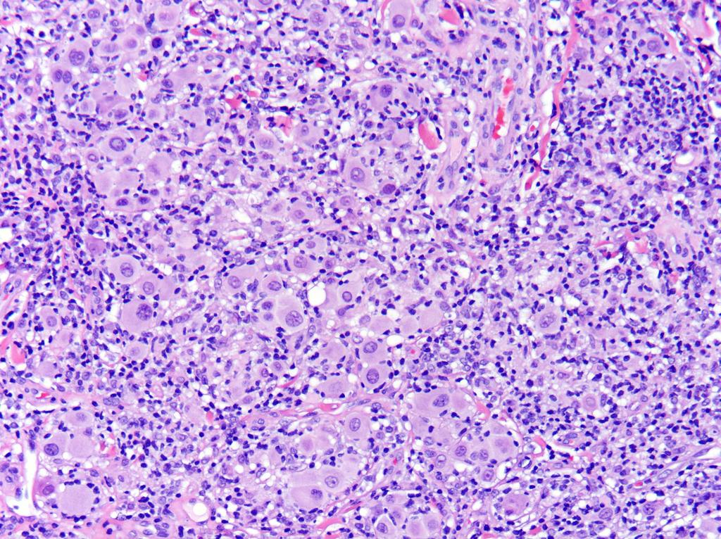
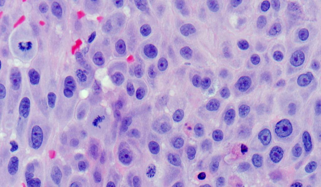
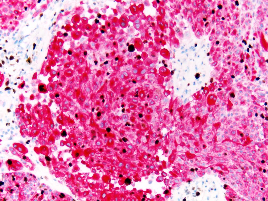
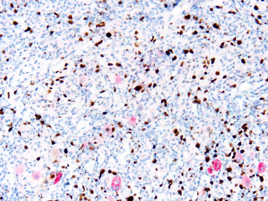
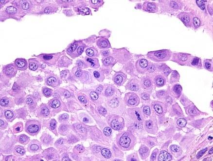
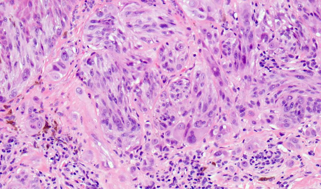
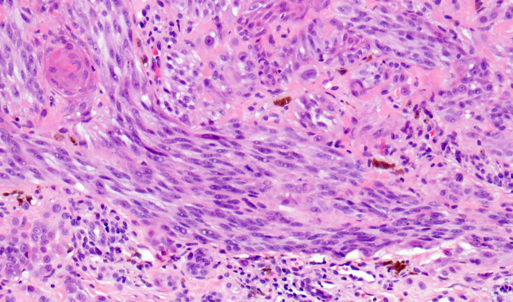
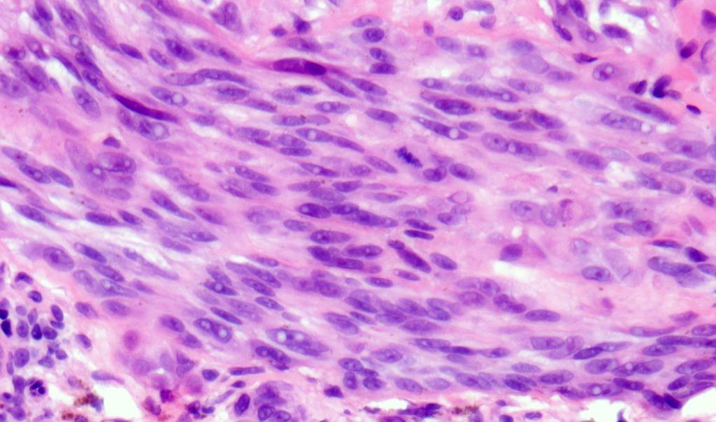
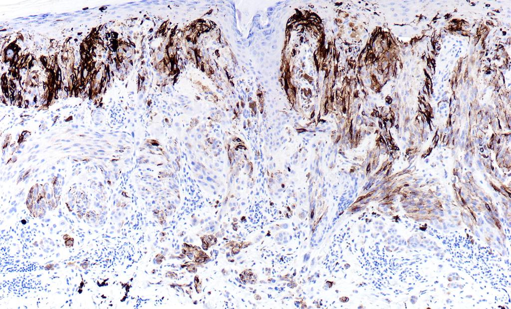
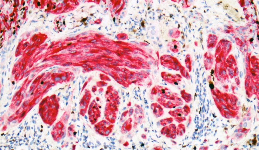
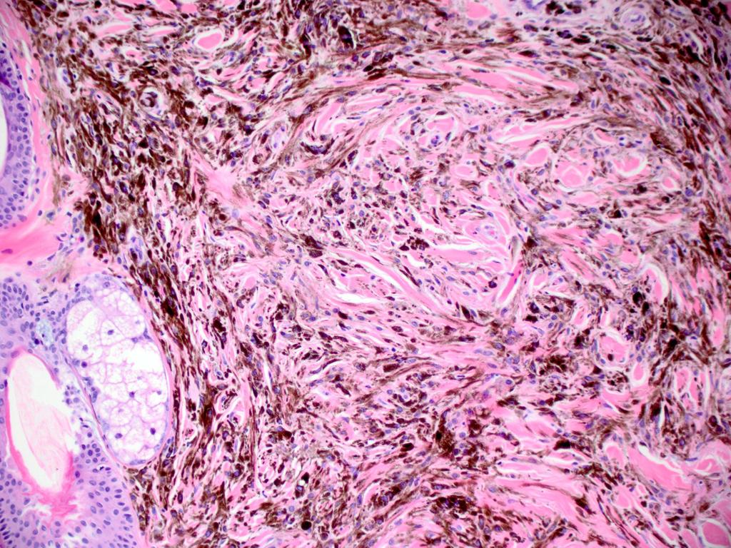
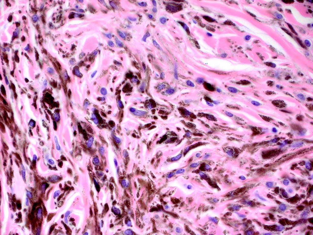
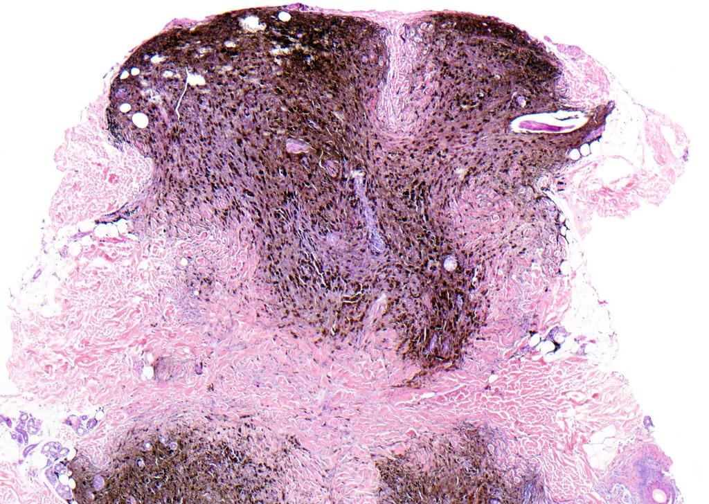
22 BAP1 deficient BRAFV600E Spitz tumors Offspring with numerous papular melanocytic neoplasms with Spitzoid morphology Lesions analyzed by acgh Identified frequent deletions of 3p Sequencing revealed mutations in BAP1.


23 BAP1 deficient BRAFV600E Spitz tumors 32 sporadic Atypical Spitz Tumors assessed by BAP1 IHC: BAP1 negative by IHC in 9 BAP1 mutation in 5 Characteristic features: Trunk Dermal Epithelioid cells Prominent TILS BRAF V600E


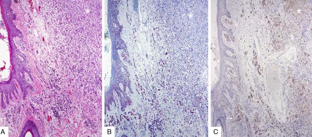
24 BAP1 deficient BRAFV600E Spitz tumors


25 BAP1 deficient BRAFV600E Spitz tumors
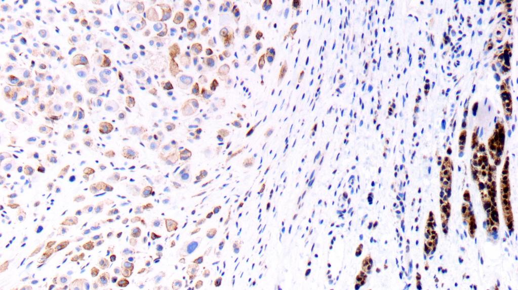
26 BRAF BAP1


27 Translocation resulting in activating kinase fusions are frequent among Spitzoid lesions Different fusion partner gene Spanned the spectrum of Spitzoid neoplasia Each fusion occurred in mutually exclusive fashion of others Constitutively active kinase activity IHC+ Kinase Do specific translocations correlate with a predictable morphology or clinical course?


28 ALK kinase fusions Spitzoid lesions have distinctive histopathologic features Author Busam et al Yeh et al Amin et al Totals Gender Men Women Median age (range) 16 y (2-35y) 12y (5m-64y) 13y (1-38y) Location Extremity Trunk/buttock Head/Neck Diagnosis Spitz nevus 5 6 NS 11 Atypical Spitz tumor NS 34 Spitzoid melanoma No discrete ALK-fusion genotype correlates with a particular clinical phenotype.


29 ALK-rearranged Spitz nevus with atypical features Compound or intradermal Exophytic and/or wedge-shaped silhouette Plexiform growth pattern Fusiform amelanotic melanocytes arranged in intersecting fascicles among dermal collagen bundles Rare cases with metastases restricted to sentinel lymph nodes (SLNs) identified, but long-term follow-up has not demonstrated further aggressive behavior. ALK
 corresponds with a predictable clinical phenotype Constitutively active kinase activity has critical")
30 Translocation resulting in activating kinase fusions are frequent among Spitzoid lesions Early events in Spitzoid neoplasia seen across the spectrum Important oncogenic drivers Not sufficient for malignant transformation No evidence yet that a discrete fusion (5 partner with 3 kinase) corresponds with a predictable clinical phenotype Constitutively active kinase activity has critical therapeutic implications Important unanswered question: Do specific kinase fusions correlate with a discrete histopathologic and/or clinical phenotype?

31 Comparative genomic hybridization (CGH) systematically interrogates the whole genome The relative hybridization of tumor derived probe compared to normal defines the relative composition of tumor DNA throughout the genome Gains show net green signal Losses show net red signal
32 CGH confirms widespread genomic instability in melanoma compared to nevi Applied CGH to 186 melanocytic tumors 132 Melanomas 54 Melanocytic nevi 96% of melanomas carry some We chromosomal can exploit this copy aberration: difference between melanoma and nevi in a diagnostic assay? Gains or losses in discrete fragments of chromosomes
: as a surrogate of CGH")
 6p25 (RREB1)")


33 Fluorescence in situ hybridization (FISH): as a surrogate of CGH Hybridize fluorescently labeled probes: 11q13 (CCND1) 6p25 (RREB1) 6q23 (MYB) Cen6 11q13 6p25 6q23 Cen 6 Assess tumor cell nuclei for abnormalities in the number of fluorescent signals. Gerami P et al. Am J Surg Pathology (8):


34 FISH assay as a surrogate of the genomic instability captured by CGH Cen 6 6p25 6q23 11q13 Gerami P et al. Am J Surg Pathology (8):


:808-817.")
35 Second generation FISH: New probes expand and improve the diagnostic utility of FISH Gerami P et al. Am. J. Surg Pathol (6): cen9 cen9 9p21 9p21 6p25 6p25 8q24 8q24 11q13
36 FISH as a prognostic marker in Spitzoid lesions Assessed 75 Atypical Spitz Tumors by FISH (First and Second generation probes)
37 FISH as a prognostic marker in Spitzoid lesions Assessed 75 Atypical Spitz Tumors by FISH (First and Second generation probes) SLN- SLN+ SLNx SLN+/LN+ Death
38 FISH as a prognostic marker in Spitzoid lesions Assessed 75 Atypical Spitz Tumors by FISH (First and Second generation probes) SLN- SLN+ SLNx SLN+/LN+ Death


39 Homozygous deletion of 9p21 correlates with aggressive behavior in Spitzoid melanoma 9p21 9/11 patients with spread beyond sentinel node and or death, showed homozygous deletion of 9p21 compared to 3/64 in group 1.

40 Case example 38 year old man with an enlarging tan-brown lesion on the shoulder
41
42
43
44
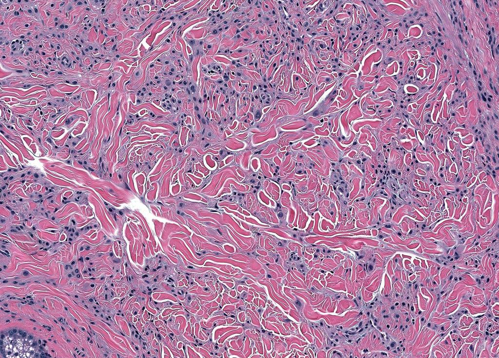
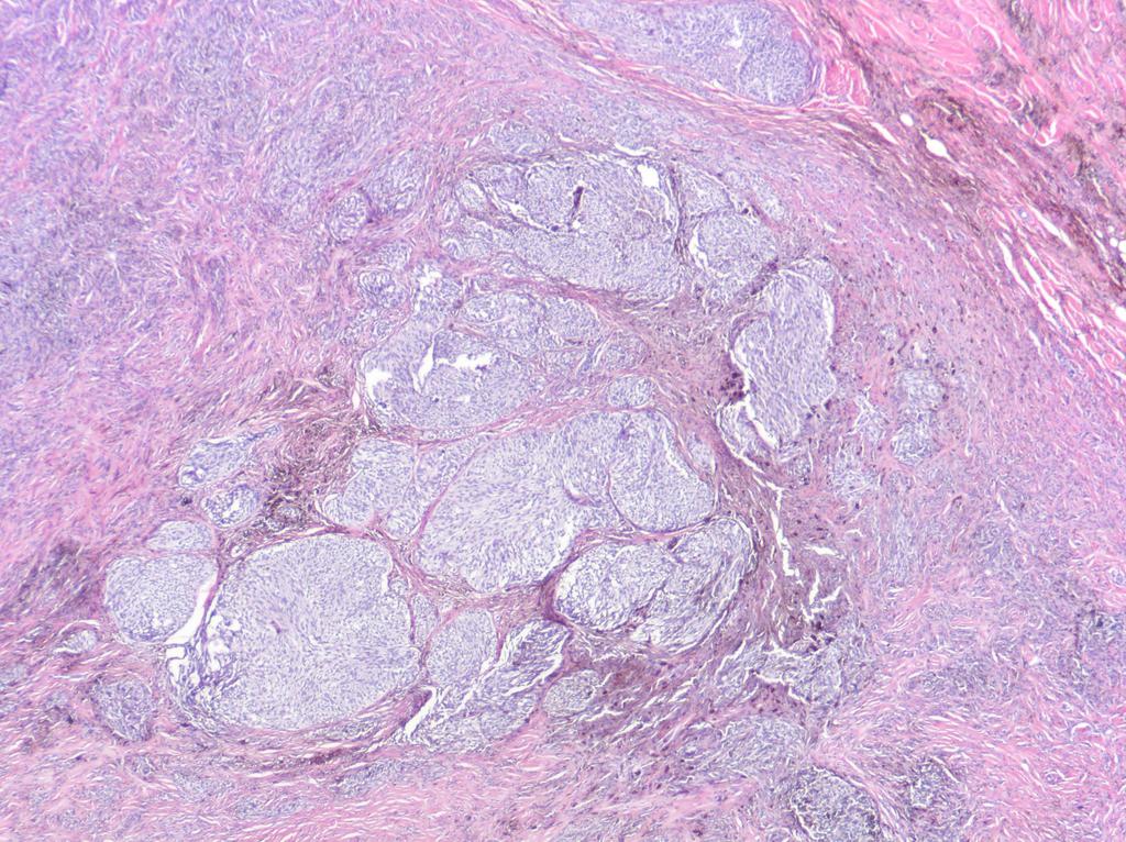
45
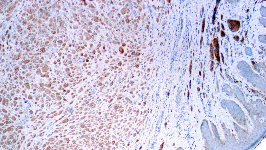
46 HMB-45
47 Mart-1/Ki67
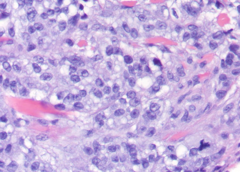
 6.0% >16% 29% >29% Chrom 8: cmyc (8q24) 4.")
 11.")
48 Case example 9p21 cen9 6p25 8q24 11q13 Results Low stringency cut off High stringency cut off Chrom 6: RREB1 (6p25) 6.0% >16% 29% >29% Chrom 8: cmyc (8q24) 4.3% >10% 29% >29% Chrom 9: CDKN2A (9p21)/CEN9 55.0% >10% 29% >29% Chrom 11: CCND1 (11q13) 11.1% >19% 29% >29% Patient with positive SLN (2/3). Currently free of disease (10 mos)


49 TERT promoter mutations occur in melanoma and associate with aggressive clinical course Oncology Reports :
50 Spitzoid lesions with TERT promoter mutations 56 patients with AST and follow-up 21/40 (50%) with at least one positive SLN 9 with extensive nodal metastases 4 with hematogenous metastases who died of disease Mean follow up for remaining 52 patients=32.5 months Clinical Features Age (years) (P =0.03)* <10 10 Gender (P =0.64) Female Male Location (P=0.27) Lower extremity Upper extremity Face Ear Scalp Trunk Race (P=0.70) White Black Other Not specified Lesional diameter (P=0.054) 5 mm 6-10 mm >10 mm Favorable (n=52) 31 (60%) 21 (40%) 30 (60%) 22 (40%) (81%) 2 (5%) 5 (14%) (43%) 23 (45%) 6 (12%) 1 Unfavorable (n=4) 0 (0%) 4 (100%) 3 (75%) 1 (25%) (100%) 0 (0%) 0 (0%) 0 1 (33%) 0 (0%) 2 (67%) 1


51 Spitzoid lesions with TERT promoter mutations 4 patients who died of disease had TERT promoter mutations 0/52 patients alive had TERT promoter mutations 12/49 with favorable outcome had homozygous deletion of 9p21 2/4 patients who died of disease had homozygous deletion of 9p21 All 4 had loss of p16 protein by IHC
52 Spitzoid lesions with TERT promoter mutations TERT promoter mutations are not always associated with poor prognosis in atypical spitzoid tumors. Requena C, Heidenreich B, Kumar R, Nagore E. Pigment Cell Melanoma Res Spitz/Reed Nevi (n=15) AST/SMM (n=9) htert promoter mutations 0/15 2/9 1 of 2 AST/SMMs with htert promoter mutations had positive SLN Both patients alive and without disease (60 and 66 months) htert promoter mutations as marker of bad prognosis could not be demonstrated in our series
53 A molecular classification scheme for Spitzoid neoplasia 11p amplified HRAS mutant Desmoplastic No metastases to date Spitzoid Lesion Molecular-genetic alteration? Predictable clinical course? Reproducible histopathologic? criteria TERT promoter mutant*** BAP1 deficient BRAFV600E Spitzoid lesions defined by FISH Homozygous deletion of 9p21 Relative loss of 6q23 Translocation associated ROS1 ALK NTRK1 NTRK3 MET BRAF RET


54 Spitzoid melanomas share molecular-genetic similarity to conventional melanoma Spitzoid melanomas share significant mutational signature with conventional melanoma and little overlap with Spitz nevi
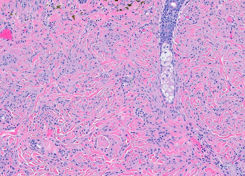
55 Update on blue nevi and blue nevus-like Melanoma Blue nevus family of lesions Common blue nevus Cellular blue nevus Melanoma, blue nevus type Atypical Cellular blue nevus

56 Common blue nevus Dark blue-black (4-10 mm) papule with ill-defined border Anywhere on the body (extremities most common) Variable density of spindled/dendritic melanocytes in the dermis Separated by dermal collagen bundles Abundant coarse intracytoplasmic melanin pigment Considerable range in cellularity and pigmentation
57
58

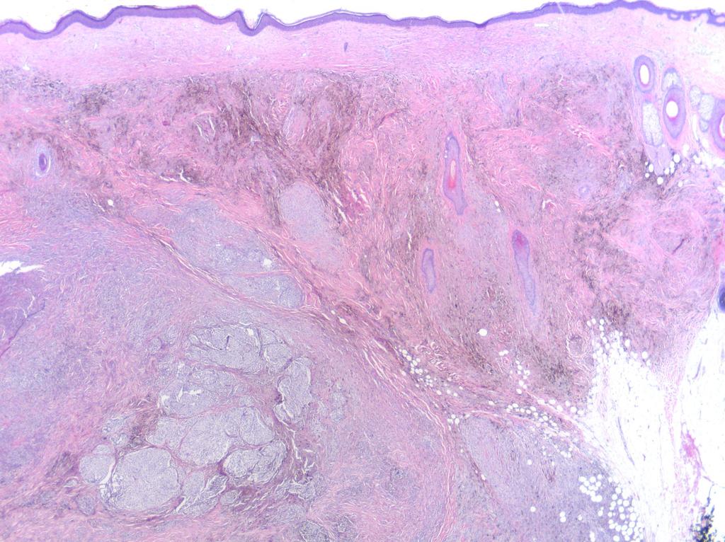
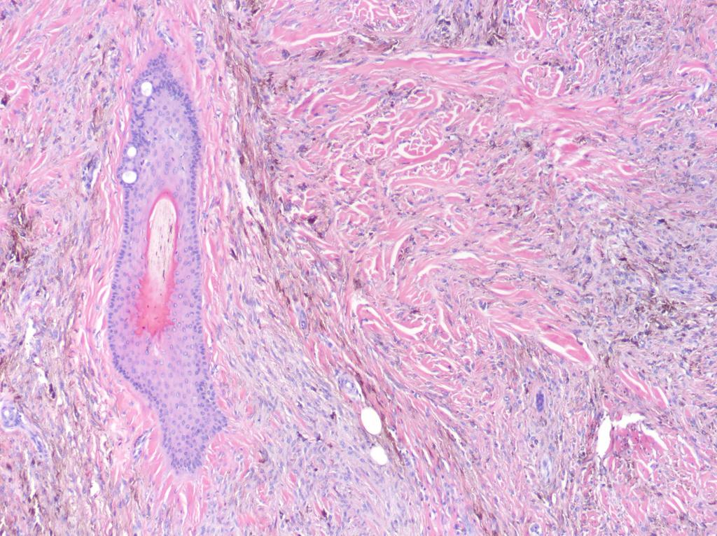
59 Cellular blue nevus Large tumor (>1 cm) raised papule with ill-defined border Lower back and buttocks in young adult Pandermal melanocytic proliferation with a characteristic dumb-bell configuration Plaque/nodule in the upper dermis connected to a nodule in the lower dermis Typically bi-phasic spindled/ pigmented melanocytes admixed with epithelioid melanocytes containing less pigment
60
61


62 Melanoma, blue nevus type
63
64
65
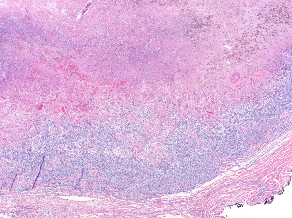
66
67
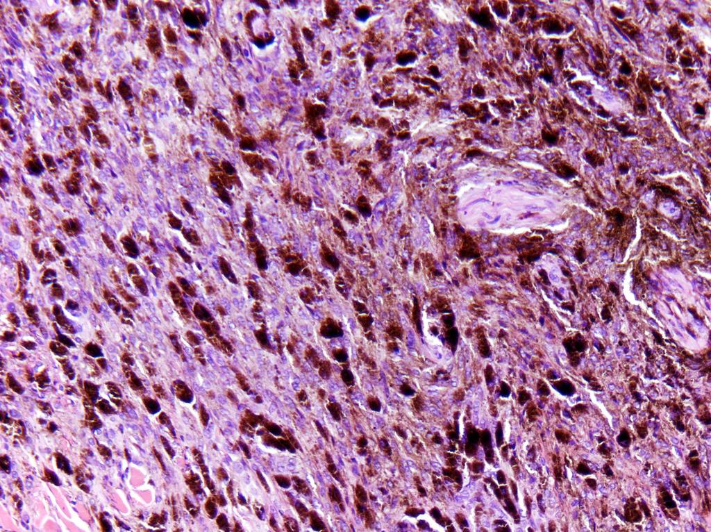
68 Distinguishing among blue nevus-like lesions Blue nevus Cellular Blue nevus Atypical Cellular Blue nevus Melanoma, Blue nevus type Location Mid- to upper dermis Pan-dermal Pan-dermal Pan-dermal and may involve subcutis Cellularity Dendritic melanocytes with coarse pigment Biphasic with dendritic and epithelioid melanocytes Biphasic with dendritic and epithelioid melanocytes May contain or resemble blue nevus/cellular blue nevus Size Small (<10 mm) Can be >10 mm Can be >10 mm >10 mm Symmetry Present Present Present/Absent Usually absent Cytologic atypia Absent Absent Increased Prominent Mitotic figures Usually absent Infrequent Increased High, atypical Necrosis Absent Absent Absent Present HMB-45 Strong, diffuse Strong, diffuse Variable Heterogeneous Ki-67 Low absent Low Slightly increased Elevated (>5-10%) BAP-1 Preserved Preserved Preserved Lost FISH Normal Normal Normal Abnormal (6p25)


69 Chromosomal instability is common in Blue nevus-like melanoma: CGH identifies numerous alterations Copy number changes Cellular Blue nevus Melanoma, Blue nevus type 3/17 (17%) 6/9 (67%) Blue nevi show few (isolated when present) copy number alterations Melanoma, blue nevus type show complex copy number alterations


")

70 Chromosomal instability is common in Blue nevus-like melanoma: FISH studies confirm 100% (12/12) Blue nevi FISH negative 100% (5/5) Melanoma, blue nevus type FISH positive Gains of 6p25 most common


71 GNAQ and GNA11 mutations in blue nevus-like melanomas
 7/11 (64%) 16/18 (89%) 1/11 (9%) 1/18 (6%) 8/11 (73%) GNAQ/GNA11 mutations in benign and malignant blue nevus-like proliferations BAP1 loss typical in melanoma, blue nevus")
72 GNAQ and GNA11 mutations, loss of BAP1 and chromosomal instability in blue nevus-like melanomas Loss of nuclear BAP1 IHC GNAQ mutation (Exon 5) GNA11 mutation (Exon 5) Cellular Blue nevus Melanoma, Blue nevus type 0/24 (0%) 7/11 (64%) 16/18 (89%) 1/11 (9%) 1/18 (6%) 8/11 (73%) GNAQ/GNA11 mutations in benign and malignant blue nevus-like proliferations BAP1 loss typical in melanoma, blue nevus type Chromosomal abnormalities frequent in melanoma, blue nevus type
 only in blue nevus-like melanomas GNAQ/GNA11")

73 Targeted sequencing facilitates distinction among cellular blue nevi and blue nevus like melanomas Mutations in BAP1 (thus far) only in blue nevus-like melanomas GNAQ/GNA11 mutations across the spectrum of blue nevus-like lesions BAP1 and SF3B1 mutated in melanoma, blue nevus type Sequencing analyses likely to be informative in diagnosis of blue nevus like lesions.
74 Questions

75 Thank you Victor G. Prieto MD PhD Jonathan L. Curry MD Carlos A. Torres-Cabala MD PhD Priya Nagarajan MD PhD Phyu Aung MD PhD Doina Ivan MD Department of Pathology, Cleveland Clinic Steven D Billings MD
Michael T. Tetzlaff MD, PhD
 Molecular alterations informing the diagnosis of melanocytic tumors Michael T. Tetzlaff MD, PhD Associate Professor Department of Pathology, Section of Dermatopathology Department of Translational and
Melanoma and the genes: Molecular alterations informing the diagnosis of melanocytic tumors
 Melanoma and the genes: Molecular alterations informing the diagnosis of melanocytic tumors Michael T. Tetzlaff MD, PhD Associate Professor Department of Pathology, Section of Dermatopathology Department
Melanocytic Lesions: Use of Immunohistochemistry and Special Studies Napa Valley 2018
 Melanocytic Lesions: Use of Immunohistochemistry and Special Studies Napa Valley 2018 Victor G. Prieto, MD, PhD Professor Depts. of Pathology and Dermatology University of Texas - MD Anderson Cancer Center
Dermatopathology. Dr. Rafael Botella Estrada. Hospital La Fe de Valencia
 Dermatopathology Dr. Rafael Botella Estrada. Hospital La Fe de Valencia Melanoma and mimics Dr. Martin Mihm Malignant lesions result from the accumulation of mutations Class I lesions (benign) Class II
Case 26 Male 37. Right jawline 5mm nodule?keloid. The best diagnosis is:
 Case 26 Male 37. Right jawline 5mm nodule?keloid. The best diagnosis is: A. Desmoplastic Spitz naevus B. Atypical Spitz Tumour C. Spitzoid melanoma D. Deep penetrating naevus E. Spitz naevus Case 26: M
The Enigmatic Spitz Lesion
 The Enigmatic Spitz Lesion The Dawn of Spitz S Spitz Sophie Spitz Melanomas of Childhood ; Am J Pathol 1948 1910-1956 13 children (18 mo - 12 yrs) 12/13 had a benign clinical course Sophie Spitz Born 1910
21/07/2017. The «gray zone» of diagnosis is visible. Nevus Atypical nevus Melanoma. Melanoma ex-blue nevus
 Update on the Clinico- Pathological and Molecular Diagnosis of Melanocytic Lesions None to declare Conflicts of interest Belfast pathology Arnaud de la Fouchardière MD, PhD Lyon, France What is new? Today
Vernon K. Sondak. Department of Cutaneous Oncology Moffitt Cancer Center Tampa, Florida
 Vernon K. Sondak Department of Cutaneous Oncology Moffitt Cancer Center Tampa, Florida Australasian Melanoma Conference 2016 Sydney, NSW, Australia October 29, 2016 Disclosures Dr. Sondak is a compensated
6/22/2015. Original Paradigm. Correlating Histology and Molecular Findings in Melanocytic Neoplasms
 6 Correlating Histology and Molecular Findings in Melanocytic Neoplasms Pedram Gerami MD, Associate Professor of Dermatology and Pediatrics at Northwestern University Disclosures: I have been a consultant
10/2/17. MELTUMP, SAMPUS, AST.An Algorithmic Approach to Challenging (Often Borderline) Melanocytic Tumors. An Introduction to SNP Arrays
 Melanocytic Tumors. An Introduction to SNP Arrays") MELTUMP, SAMPUS, AST.An Algorithmic Approach to Challenging (Often ) Melanocytic Tumors An Introduction to SNP Arrays Rajiv M. Patel, M.D. RCPA NZ ASM 2017 (11:45-12:30pm, Saturday, 23-09-17) Why do we
MELTUMP, SAMPUS, AST.An Algorithmic Approach to Challenging (Often ) Melanocytic Tumors An Introduction to SNP Arrays Rajiv M. Patel, M.D. RCPA NZ ASM 2017 (11:45-12:30pm, Saturday, 23-09-17) Why do we
Molecular Aspects of Melanocytic Neoplasia. Iwei Yeh MD, PhD University of California, San Francisco
 Molecular Aspects of Melanocytic Neoplasia Iwei Yeh MD, PhD University of California, San Francisco Thanks to: Boris Bastian Timothy McCalmont Philip LeBoit Beth Ruben Jeff North Laura Pincus Thaddeus
There is NO single Melanoma Stain. > 6000 Mutations in Melanoma. What else can be done to discriminate atypical nevi from melanoma?
 Las Vegas Fall Clinical 2016: The Assessment and Diagnosis of Melanoma Whitney A. High, MD, JD, MEng Associate Professor, Dermatology & Pathology Director of Dermatopathology (Dermatology) University of
Update on 8 th Edition Cutaneous AJCC Staging of Primary Cutaneous Melanoma. Michael T. Tetzlaff MD, PhD
 Update on 8 th Edition Cutaneous AJCC Staging of Primary Cutaneous Melanoma Michael T. Tetzlaff MD, PhD Associate Professor Departments of Pathology (Dermatopathology) and Translational and Molecular Pathology
Desmoplastic Melanoma R/O BCC. Clinical Information. 74 y.o. man with lesion on left side of neck r/o BCC
 R/O BCC Sabine Kohler, M.D. Professor of Pathology and Dermatology Dermatopathology Service Stanford University School of Medicine Clinical Information 74 y.o. man with lesion on left side of neck r/o
Michael T. Tetzlaff MD, PhD
 American Joint Cancer Committee (AJCC) staging system for primary cutaneous melanoma (8 th Edition) and principles of sentinel lymph node evaluation Emphasis on concise and accurate reporting of primary
The Pathology of Neoplasia Part II
 The Pathology of Neoplasia Part II February 2018 PAUL BOGNER, MD A S S O C I A T E P R O F E S S O R O F O N C O L O G Y P A T H O L O G Y A N D D E R M A T O L O G Y Clinical goals of cancer pathology
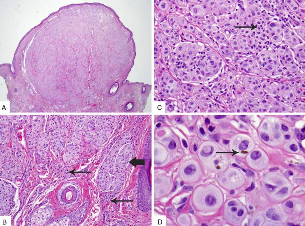
Supplementary Figure 1. Spitzoid Melanoma with PPFIBP1-MET fusion. (a) Histopathology (4x) shows a domed papule with melanocytes extending into the
 Histopathology (4x) shows a domed papule with melanocytes extending into the") Supplementary Figure 1. Spitzoid Melanoma with PPFIBP1-MET fusion. (a) Histopathology (4x) shows a domed papule with melanocytes extending into the deep dermis. (b) The melanocytes demonstrate abundant
Supplementary Figure 1. Spitzoid Melanoma with PPFIBP1-MET fusion. (a) Histopathology (4x) shows a domed papule with melanocytes extending into the deep dermis. (b) The melanocytes demonstrate abundant
Benign and malignant epithelial lesions: Seborrheic keratosis: A common benign pigmented epidermal tumor occur in middle-aged or older persons more
 Benign and malignant epithelial lesions: Seborrheic keratosis: A common benign pigmented epidermal tumor occur in middle-aged or older persons more common on the trunk; but extremities, head and neck are
A PRACTICAL APPROACH TO ATYPICAL MELANOCYTIC LESIONS BIJAN HAGHIGHI M.D, DIRECTOR OF DERMATOPATHOLOGY, ST. JOSEPH HOSPITAL
 A PRACTICAL APPROACH TO ATYPICAL MELANOCYTIC LESIONS BIJAN HAGHIGHI M.D, DIRECTOR OF DERMATOPATHOLOGY, ST. JOSEPH HOSPITAL OBJECTIVES Discuss current trends and changing concepts in our understanding of
Conflict of Interest 9/2/2014. Pathogenesis and Comparison of Atypical Spitz Nevi vs Benign Spitz, and Childhood Melanoma
 Pathogenesis and Comparison of Atypical Spitz Nevi vs Benign Spitz, and Childhood Melanoma Martin C. Mihm Jr., M.D., F.A.C.P. Harvard Medical School Brigham and Women s Hospital Dana Farber Cancer Center
Female 18. Deeply pigmented lesion on trunk.?warty naevus?seborrhoeic keratosis?malignant melanoma. The best diagnosis is:
 Female 18. Deeply pigmented lesion on trunk.?warty naevus?seborrhoeic keratosis?malignant melanoma. The best diagnosis is: A. deep penetrating naevus B. naevoid malignant melanoma C. pigment synthesising
Update on Merkel cell carcinoma
 Merkel cell carcinoma: Diagnosis, staging, sentinel lymph node biopsy and prognostic markers Michael T. Tetzlaff MD, PhD Associate Professor Departments of Pathology (Dermatopathology) and Translational
Merkel cell carcinoma: Diagnosis, staging, sentinel lymph node biopsy and prognostic markers
 Merkel cell carcinoma: Diagnosis, staging, sentinel lymph node biopsy and prognostic markers Michael T. Tetzlaff MD, PhD Associate Professor Departments of Pathology (Dermatopathology) and Translational
Ways to get into trouble, ideas on avoiding trouble, and diagnostic approaches to keep trouble at bay
 Pitfalls in the diagnosis of melanocytic tumors Timothy McCalmont, MD University of California, San Francisco Ways to get into trouble, ideas on avoiding trouble, and diagnostic approaches to keep trouble
Pathology of the skin. 2nd Department of Pathology, Semmelweis University
 Pathology of the skin 2nd Department of Pathology, Semmelweis University Histology of the skin Epidermis: Stratum corneum Stratum granulosum Stratum spinosum Stratum basale Dermis: papillary and reticular
Patricia Chevez-Barrrios AAOOP-USCAP /12/2016
 Biomarkers in Ocular Melanoma Patricia Chévez-Barrios, MD Pathology and Genomic Medicine, Houston Methodist Hospital Professor of Pathology and Laboratory Medicine and Ophthalmology, Weill Cornell Medical
Blue Melanocytic Proliferations
 Blue Melanocytic Proliferations Labib R. Zakka M.D., M.A. Research Fellow Melanoma Program Department of Dermatology Brigham and Women s Hospital Harvard Medical School Conflicts of Interest No conflicts
MAPK Pathway. CGH Next Generation Sequencing. Molecular Tools in Care of Patients with Pigmented Lesions 7/20/2017
 Molecular Tools in Care of Patients with Pigmented Lesions Tammie Ferringer, MD Geisinger Medical Center, Danville, PA tferringer@geisinger.edu DISCLOSURE OF RELATIONSHIPS WITH INDUSTRY Tammie Ferringer,
Michael T. Tetzlaff MD, PhD
 Update on American Joint Cancer Committee (AJCC) staging system for primary cutaneous melanoma Emphasis on concise and accurate reporting of primary and metastatic melanoma for effective risk stratification
Case 231: F7. Exophytic naevus over left trapezious. Grown over a few weeks. Iniitally flat.?spitz naevus,?malignant
 Case 231: F7. Exophytic naevus over left trapezious. Grown over a few weeks. Iniitally flat.?spitz naevus,?malignant Dermoscopy: coarse vascular structures. c/o A, B, C RAC7750 Case 231: F7. Exophytic
Cutaneous Mesenchymal Neoplasms with EWSR1 Rearrangement
 Cutaneous Mesenchymal Neoplasms with EWSR1 Rearrangement By Konstantinos Linos MD, FCAP, FASDP Bone, Soft Tissue and Dermatopathology Assistant Professor of Pathology Dartmouth-Hitchcock Medical Center
المركب النموذج--- سبيتز وحمة = Type Spitz's Nevus, Compound SPITZ NEVUS 1 / 7
 SPITZ NEVUS 1 / 7 Epidemiology An annual incidence rate of 1.4 cases of Spitz nevus per 100,000 individuals has been estimated in Australia, compared with 25.4 per 100,000 individuals for cutaneous melanoma
Genetic Testing: When should it be ordered? Julie Schloemer, MD Dermatology
 Genetic Testing: When should it be ordered? Julie Schloemer, MD Dermatology Outline Germline testing CDKN2A BRCA2 BAP1 Somatic testing Gene expression profiling (GEP) BRAF Germline vs Somatic testing
ACCME/Disclosures ALK FUSION-POSITIVE MESENCHYMAL TUMORS. Tumor types with ALK rearrangements. Anaplastic Lymphoma Kinase. Jason L.
 Companion Meeting of the International Society of Bone and Soft Tissue Pathology The Evolving Concept of Mesenchymal Tumors ALK FUSION-POSITIVE MESENCHYMAL TUMORS Jason L. Hornick, MD, PhD March 13, 2016
I have no relevant conflicts of interest to disclose. John T. Seykora MD PhD Departments of Dermatology & Pathology and Laboratory Medicine
 Molecular Characterization of Stage 1-3 Melanoma: Are we close to accurate prognostication and prediction? I have no relevant conflicts of interest to disclose. John T. Seykora MD PhD Departments of Dermatology
Melanocytic proliferations in sundamaged
 Atypical Spitzoid Tumor: What Does It Mean And How Should It Be Managed? Melanocytic proliferations in sundamaged skin Jane L. Messina, Jane L. Messina MD International Melanoma Pathology Working Group
BAP-oma & BEYOND MICHAEL A NOWAK, MD
 BAP-oma & BEYOND MICHAEL A NOWAK, MD CONFLICTS No conflicts with the content of this lecture BAP-oma Wiesner 2011: Families with multiple tan dome-shaped papules of head, neck, trunk, and extremities.
Case RAC7783. M46. Ear. Mole. r/o MM.?Blue naevus RAC7783
 Case RAC7783. M46. Ear. Mole. r/o MM.?Blue naevus RAC7783 Pie Chart Participants N=74 Benign: 48 N=74 Blue naevus: 38 Intradermal: 12 DPN: 10 Compound 3 Clonal: 3; Spitz 2; Special Site: 1; Congenital:
Financial disclosures
 Mesenchymal Neoplasms with Melanocytic Differentiation By Konstantinos Linos MD, FCAP, FASDP Bone, Soft Tissue and Dermatopathology Assistant Professor of Pathology Dartmouth-Hitchcock Medical Center Geisel
Primary Cutaneous CD30-Positive T-cell Lymphoproliferative Disorders
 Primary Cutaneous CD30-Positive T-cell Lymphoproliferative Disorders Definition A spectrum of related conditions originating from transformed or activated CD30-positive T-lymphocytes May coexist in individual
Melanoma Update: 8th Edition of AJCC Staging System
 Melanoma Update: 8th Edition of AJCC Staging System Rosalie Elenitsas, M.D. Professor of Dermatology Director, Dermatopathology University of Pennsylvania DISCLOSURE OF RELATIONSHIPS WITH INDUSTRY None
A diagnostic algorithm for atypical spitzoid tumors: guidelines for immunohistochemical and molecular assessment
 Modern Pathology (2016), 1 15 2016 USCAP, Inc All rights reserved 0893-3952/16 $32.00 1 A diagnostic algorithm for atypical spitzoid tumors: guidelines for immunohistochemical and molecular assessment
Melanocytic Tumours. Molecular Biology 02/06/2015. Cutaneous Melanocytic Tumours Introduction. Thomas Brenn. Intermediate Malignancy
 Cutaneous Melanocytic Tumours Introduction Melanocytic Tumours: Update on Epidemiology and Molecular Biology Thomas Brenn Wide clinical and morphological spectrum Ranging from benign naevi to melanoma
Reviewers' comments: Reviewer #1 (Remarks to the Author):
:") Reviewers' comments: Reviewer #1 (Remarks to the Author): In this study the authors analysed 18 deep penetrating nevi for oncogenic genomic changes (single nucleotide variations, insertions/deletions,
Reviewers' comments: Reviewer #1 (Remarks to the Author): In this study the authors analysed 18 deep penetrating nevi for oncogenic genomic changes (single nucleotide variations, insertions/deletions,
1/10/2018. Soft Tissue Tumors Showing Melanocytic Differentiation. Overview. Desmoplastic/ Spindle Cell Melanoma
 2016 MFMER slide-1 2016 MFMER slide-2 2016 MFMER slide-3 Soft Tissue Tumors Showing Melanocytic Differentiation Andrew L. Folpe, M.D. Professor of Laboratory Medicine and Pathology Mayo Clinic, Rochester,
21/07/2017. Hobnail endothelial cells are not the same as epithelioid endothelial cells
 UPDATE IN CUTANEOUS VASCULAR S DERMATOPATHOLOGY SESSION BELFAST PATHOLOGY JUNE 21/2017 Dr E Calonje St John s Institute of Dermatology, London, United Kingdom THE FAMILY OF VASCULAR S WITH EPITHELIOID
Diagnoses of Cases 1. Lentigo, other melanosis and the acquired nevus 2. Variations on the acquired nevus 3. Dermal melanocytosis
 Diagnoses of Cases 1. Lentigo, other melanosis and the acquired nevus 1 1A. Lentigo simplex 4 1B. Psoralens and ultraviolet A (PUVA) lentigo 6 1C. Solar lentigo 8 1D. Café au lait macule 10 1E. Ink-spot
Financial disclosures
 Cutaneous Mesenchymal Neoplasms with EWSR1 Rearrangement By Konstantinos Linos MD, FCAP, FASDP Bone, Soft Tissue and Dermatopathology Assistant Professor of Pathology Dartmouth-Hitchc Geisel School of
Springer Healthcare. Staging and Diagnosing Cutaneous Melanoma. Concise Reference. Dirk Schadendorf, Corinna Kochs, Elisabeth Livingstone
 Concise Reference Staging and Diagnosing Cutaneous Melanoma Dirk Schadendorf, Corinna Kochs, Elisabeth Livingstone Extracted from Handbook of Cutaneous Melanoma: A Guide to Diagnosis and Treatment Published
Histopathology of Melanoma
 THE YALE JOURNAL OF BIOLOGY AND MEDICINE 48, 409-416 (1975) Histopathology of Melanoma G. J. WALKER SMITH Department ofpathology, Yale University School ofmedicine, 333 Cedar Street, New Haven, Connecticut
Integrating Fluorescence in situ Hybridization and Genomic Array Results into the Diagnostic Workup of Melanoma
 Integrating Fluorescence in situ Hybridization and Genomic Array Results into the Diagnostic Workup of Melanoma Association for Molecular Pathology United States and Canadian Academy of Pathology Companion
أملس عضلي غرن = Leiomyosarcoma. Leiomyosarcoma 1 / 5
 Leiomyosarcoma 1 / 5 EPIDEMIOLOGY Exact incidence is unknown, but older studies suggest that leiomyosarcomas comprise approximately 3 percent of soft-tissue sarcomas. Superficial leiomyosarcoma occurs
Which melanoma patients benefit from genetic testing?
 Which melanoma patients benefit from genetic testing? Michael A. Marchetti, MD Assistant Attending, Dermatology Service Memorial Sloan Kettering Cancer Center American Academy of Dermatology Annual Meeting
Self assessment case. Dr Saleem Taibjee Dorset County Hospital, Dorchester
 Self assessment case Dr Saleem Taibjee saleemtaibjee@gmail.com Dorset County Hospital, Dorchester Clinical details 34-year-old man: Shave excision Skin tag / papilloma left thigh The best diagnosis is:
Malignant tumors of melanocytes: Part 1. Deba P Sarma, MD., Omaha
 Malignant tumors of melanocytes: Part 1 Deba P Sarma, MD., Omaha The melanocytic tumor is one of the most difficult and confusing areas in Dematopathology. It is true that most (95%) of such lesions are
Disclosure Information. Lecture Outline. Lecture Outline. Introduction. Molecular Pathology of Cutaneous Melanoma. Nothing to disclose
 Molecular Pathology of Cutaneous Melanoma Disclosure Information Nothing to disclose Jonathan L. Curry, MD Assistant Professor of Pathology and Dermatology University of Texas-MD Anderson Cancer Center
David B. Troxel, MD. Common Medicolegal Situations: Misdiagnosis of Melanoma
 Common Medicolegal Situations: Misdiagnosis of Melanoma David B. Troxel, MD Medical Director, The Doctors Company, Napa, California Clinical Professor Emeritus, University of California at Berkeley Past
Guy Perrot (Ги Перро)
") НАУЧНО-ПРАКТИЧЕСКАЯ КОНФЕРЕНЦИЯ (МАСТЕР-КЛАСС) «ПРАКТИЧЕСКИЕ АСПЕКТЫ ДИАГНОСТИКИ И ЛЕЧЕНИЯ МЕЛАНОМЫ КОЖИ» DIAGNOSTIC AND PITFALLS IN MELANOMA Guy Perrot (Ги Перро) MD PHD pathologist, University Hospital
НАУЧНО-ПРАКТИЧЕСКАЯ КОНФЕРЕНЦИЯ (МАСТЕР-КЛАСС) «ПРАКТИЧЕСКИЕ АСПЕКТЫ ДИАГНОСТИКИ И ЛЕЧЕНИЯ МЕЛАНОМЫ КОЖИ» DIAGNOSTIC AND PITFALLS IN MELANOMA Guy Perrot (Ги Перро) MD PHD pathologist, University Hospital
2/6/2018. Original Paradigm. Clonal Chromosomal A berrations. Only 20% of Spitz Nevi 95% 6p, 7q, 17q, 20q, 4q,8q, 1q, 11q. Isolated Gain in 11p
 Molecular Diagnostics for Melanocytic Neoplasms: Moving towards a Revolution in the Management of Melanocytic Neoplasms Pedr am Gerami MD Associate Professor of Dermatology, Pathology and Pediatrics at
Index. Springer-Verlag Berlin Heidelberg 2017 J.A. Plaza, V.G. Prieto, Pathology of Pigmented Skin Lesions, DOI /
 A Acral lentiginous (mucosal lentiginous) melanoma, 483 Acral lentiginous melanoma (ALM) asymmetric and irregular lentiginous junctional growth, 431 clinical features, 427 428 differential diagnosis, 428
Enterprise Interest Nothing to declare
 Enterprise Interest Nothing to declare Diagnoses one would not like to miss in soft tissue pathology early in your career Marta Sbaraglia, MD Department of Pathology Hospital of Treviso University of Padua
Mody. AIS vs. Invasive Adenocarcinoma of the Cervix
 Common Problems in Gynecologic Pathology Michael T. Deavers, M.D. Houston Methodist Hospital, Houston, Texas Common Problems in Gynecologic Pathology Adenocarcinoma in-situ (AIS) of the Cervix vs. Invasive
An update on molecular alterations in melanocytic tumors with emphasis on Spitzoid lesions
 Review Article on Molecular Oncology age 1 of 17 An update on molecular alterations in melanocytic tumors with emphasis on Spitzoid lesions Emmanouil Dimonitsas 1#, Aliki Liakea 2#, Stratigoula Sakellariou
EARLY ONLINE RELEASE
 EARLY ONLINE RELEASE Note: This article was posted on the Archives Web site as an Early Online Release. Early Online Release articles have been peer reviewed, copyedited, and reviewed by the authors. Additional
The Relevance of Cytologic Atypia in Cutaneous Neural Tumors
 The Relevance of Cytologic Atypia in Cutaneous Neural Tumors Recent Findings - New Developments New Problems Zsolt B. Argenyi, M.D. Professor of Pathology & Dermatology Director of Dermatopathology Department
Management of pediatric melanocytic lesions
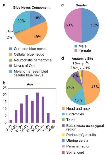
 Open Journal of Clinical & Medical Case Reports Management of pediatric melanocytic lesions Volume 3 (2017) Issue 8 ISSN 2379-1039 Jin Kim, BS; Emmanuel Gabriel MD, PhD; Weiguo Liu MD, PhD; Lin Lin MD,
Histopathology: skin pathology
 Histopathology: skin pathology These presentations are to help you identify, and to test yourself on identifying, basic histopathological features. They do not contain the additional factual information
Diploma examination. Dermatopathology: First paper. Tuesday 21 March Candidates must answer FOUR questions ONLY. Time allowed: Three hours
 Dermatopathology: First paper Tuesday 21 March 2017 1. Discuss the role of fluorescent in-situ hybridization (FISH) and emerging molecular techniques in the diagnosis of cutaneous melanocytic lesions,
Page 1 of 3. We suggest the following changes:
 Page 1 of 3 Loren E. Clarke, M.D. Myriad Genetic Laboratories, Inc. 320 Wakara Way, Salt Lake City, UT 84108 Phone: 801.883.3470 Email: lclarke@myriad.com Date of Request: June 2017 NCCN Guidelines Panel:
The Dermal Melanocytoses. Conflicts of Interest 5/22/2018. The Nevi of Ota and Ito. Martin C. Mihm M.D.
 The Dermal Melanocytoses Martin C. Mihm M.D. Director Mihm Cutaneous Pathology Consultative Service (MCPCS) Brigham and Women s Hospital Director Melanoma Program Brigham and Women s Hospital and Harvard
Society for Pediatric Pathology Spring Meeting Joint Symposium with American Society of Dermatopathology
 Society for Pediatric Pathology 2013 Spring Meeting Joint Symposium with American Society of Dermatopathology Update on Cutaneous Melanocytic, Mesenchymal and Lymphoproliferative Lesions in Children Melanocytic
Benign versus Cancerous Lesions How to tell the difference FMF 2014 Christie Freeman MD, CCFP, DipPDerm, MSc
 1 Benign versus Cancerous Lesions How to tell the difference FMF 2014 Christie Freeman MD, CCFP, DipPDerm, MSc Benign lesions Seborrheic Keratoses: Warty, stuck-on Genetics and birthdays Can start in late
57th Annual HSCP Spring Symposium 4/16/2016
 An Unusual Malignant Spindle Cell Lesion to Involve the Breast Erinn Downs-Kelly, D.O. Associate Professor of Pathology University of Utah & ARUP Laboratories No disclosures Case 39 y/o female with no
Original Articles Primary orbital melanoma in association with cellular blue nevus
 Original Articles Primary orbital melanoma in association with cellular blue nevus Tarek El-Sawy, MD, PhD, a Mathieu F. Bakhoum, PhD, a Michael Tetzlaff, MD, PhD, b Qasiem J. Nasser, MD, a Victor G. Prieto,
Diagnosis of melanocytic proliferations remains one of
 Update on Fluorescence In Situ Hybridization in Melanoma State of the Art Pedram Gerami, MD; Artur Zembowicz, MD, PhD N Context. Recent advances in understanding the molecular basis of melanoma have resulted
Case year old female presented with asymmetric enlargement of the left lobe of the thyroid
 Case 4 22 year old female presented with asymmetric enlargement of the left lobe of the thyroid gland. No information available relative to a prior fine needle aspiration biopsy. A left lobectomy was performed.
Case 4 Diagnosis 2/21/2011 TGB
 Case 4 22 year old female presented with asymmetric enlargement of the left lobe of the thyroid gland. No information available relative to a prior fine needle aspiration biopsy. A left lobectomy was performed.
Dermatopathology: The tumor is composed of keratinocytes which show atypia, increase mitoses and abnormal mitoses.
 Squamous cell carcinoma (SCC): A common malignant tumor of keratinocytes arising in the epidermis, usually from a precancerous condition: 1- UV induced actinic keratosis, usually of low grade malignancy.
Conflicts of Interest
 Challenging Melanocytic Lesions Carlos N. Prieto-Granada M.D. Assistant Professor University of Alabama at Birmingham (UAB) Department of Pathology 2017 AAD Annual Meeting 3/2/17 - Orlando, FL None Conflicts
Diploma Examination. Dermatopathology: First paper. Tuesday 20 March Candidates must answer FOUR questions. Time allowed: 3 hours
 Dermatopathology: First paper Tuesday 20 March 2018 Candidates must answer FOUR questions Time allowed: 3 hours 1. Give an account of the genetic aberrations encountered in Spitzoid neoplasms and how these
Simulators of melanoma
 Simulators of melanoma Philip E. LeBoit, M.D. Depts. of Pathology and Dermatology University of California, San Francisco Simulators of melanoma Simulators of melanoma in situ Melanocytic Non-melanocytic
- Selected Tumors of the Skin Appendages - Primary vs. Metastasis
 - Selected Tumors of the Skin Appendages - Primary vs. Metastasis Napa Valley 2018 Victor G. Prieto, MD, PhD Chair of Pathology UT MD Anderson Cancer Center vprieto@mdanderson.org Napa Valley in May Introduction
Malignant Peripheral Nerve Sheath Tumor
 C H A P T E R 120 Malignant Peripheral Nerve Sheath Tumor Currently, malignant peripheral nerve sheath tumor (MPNST) is the most commonly used generic name for the neoplasms known in the past as neurosarcoma,
04/10/2018. Intraductal Papillary Neoplasms Of Breast INTRADUCTAL PAPILLOMA
 Intraductal Papillary Neoplasms Of Breast Savitri Krishnamurthy MD Professor of Pathology Deputy Division Head The University of Texas MD Anderson Cancer Center 25 th Annual Seminar in Pathology Pittsburgh,
> 6000 Mutations in Melanoma. Tests That Cay Be Employed. FISH for Additions/Deletions. Comparative Genomic Hybridization
 Winter Clinical 2017: The Assessment and Diagnosis of Melanoma Whitney A. High, MD, JD, MEng Associate Professor, Dermatology & Pathology Director of Dermatopathology (Dermatology) University of Colorado
Molecular Methods in the Diagnosis and Prognostication of Melanoma: Pros & Cons
 Molecular Methods in the Diagnosis and Prognostication of Melanoma: Pros & Cons Ben J. Friedman, MD Senior Staff Physician Department of Dermatology Department of Pathology and Laboratory Medicine Henry
Update: Morphologic Considerations in Mesothelioma within the Pleural and Peritoneal Cavities. Douglas J. Hartman, MD June 7, 2018
 Update: Morphologic Considerations in Mesothelioma within the Pleural and Peritoneal Cavities Douglas J. Hartman, MD June 7, 2018 Objectives Review Historical Features associated with prognosis Present
Diplomate of the American Board of Pathology in Anatomic and Clinical Pathology
 A 33-year-old male with a left lower leg mass. Contributed by Shaoxiong Chen, MD, PhD Assistant Professor Indiana University School of Medicine/ IU Health Partners Department of Pathology and Laboratory
Disclosure. Relevant Financial Relationship(s) None. Off Label Usage None MFMER slide-1
 None. Off Label Usage None MFMER slide-1") Disclosure Relevant Financial Relationship(s) None Off Label Usage None 2013 MFMER slide-1 Case Presentation A 43 year old male, with partial nephrectomy for a right kidney mass 2013 MFMER slide-2 2013
Disclosure Relevant Financial Relationship(s) None Off Label Usage None 2013 MFMER slide-1 Case Presentation A 43 year old male, with partial nephrectomy for a right kidney mass 2013 MFMER slide-2 2013
Less Common Variants of Cutaneous Melanoma
 Less Common Variants of Cutaneous Melanoma Raymond L. Barnhill* 1, G. Peter Sarantopoulos 1, and Kapil Gupta 2 1 Department of Pathology and Laboratory Medicine, University of California, Los Angeles,
Whitney A. High, MD, JD, MEng
 ADS Dermatopathology Meeting 2014 Selected Adnexal Tumors Whitney A. High, MD, JD, MEng Associate Professor, Dermatology & Pathology Director of Dermatopathology (Dermatology) University of Colorado School
Genomic analysis of childhood High grade glial (HGG) brain tumors
 brain tumors") Genomic analysis of childhood High grade glial (HGG) brain tumors Linda D Cooley Children s Mercy, Kansas City The Children s Mercy Hospital, 2017 Genomic analysis of childhood High grade glial (HGG) brain
Genomic analysis of childhood High grade glial (HGG) brain tumors Linda D Cooley Children s Mercy, Kansas City The Children s Mercy Hospital, 2017 Genomic analysis of childhood High grade glial (HGG) brain
We are IntechOpen, the world s leading publisher of Open Access books Built by scientists, for scientists. International authors and editors
 We are IntechOpen, the world s leading publisher of Open Access books Built by scientists, for scientists 3,500 108,000 1.7 M Open access books available International authors and editors Downloads Our
Dermatologica Sinica
 DERMATOLOGICA SINICA 30 (2012) 57e61 Contents lists available at SciVerse ScienceDirect Dermatologica Sinica journal homepage: http://www.derm-sinica.com CASE REPORT Pigmented epithelioid melanocytoma:
S everal morphological features are frequently used in the
 1194 ORIGINAL ARTICLE Interobserver reproducibility of histological features in cutaneous malignant melanoma C Urso, F Rongioletti, D Innocenzi, C Saieva, D Batolo, S Chimenti, R Filotico, R Gianotti,
Impact of Prognostic Factors
 Melanoma Prognostic Factors: where we started, where are we going? Impact of Prognostic Factors Staging Management Surgical intervention Adjuvant treatment Suraj Venna, MD Assistant Clinical Professor,
AGGRESSIVE VARIANTS OF PAPILLARY THYROID CARCINOMA DIAGNOSIS AND PROGNOSIS
 AGGRESSIVE VARIANTS OF PAPILLARY THYROID CARCINOMA DIAGNOSIS AND PROGNOSIS PAPILLARY THYROID CARCINOMA Clinical Any age Microscopic to large Female: Male= 2-4:1 Radiation history Lymph nodes Prognosis
Primary enteric adenocarcinoma with predominantly signet ring features of the lung: A case report with clinicopathological and molecular findings
 CASE REPORT Primary enteric adenocarcinoma with predominantly signet ring features of the lung: A case report with clinicopathological and molecular findings Makoto Nagashima 1, Ayako Moriyama 1, Yasuo
LUNG CANCER. pathology & molecular biology. Izidor Kern University Clinic Golnik, Slovenia
 LUNG CANCER pathology & molecular biology Izidor Kern University Clinic Golnik, Slovenia 1 Pathology and epidemiology Small biopsy & cytology SCLC 14% NSCC NOS 4% 70% 60% 50% 63% 62% 61% 62% 59% 54% 51%
Special slide seminar
 Special slide seminar Tomáš Rozkoš The Fingerland Department of Pathology Charles University Medical Faculty and Faculty Hospital in Hradec Králové Czech Republic Case history, 33 years old resistance