Središnja medicinska knjižnica
|
|
|
- Jewel Martin
- 5 years ago
- Views:
Transcription
. pp. 365-70. ISSN 1219-4956 http://www.springer.com/journal/12253 http://www.springerlink.com/content/1219-4956 http://dx.doi.org/10.")
1 Središnja medicinska knjižnica Ulamec M., Džombeta T., Čupić H., Leniček T., Tomas D., Krušlin B. (2012) Periacinar retraction clefting and d2-40 expression in prostatic adenocarcinoma. Pathology Oncology Research, 18 (2). pp ISSN University of Zagreb Medical School Repository
2 Research Paper PERIACINAR RETRACTION CLEFTING AND D2-40 EXPRESSION IN PROSTATIC ADENOCARCINOMA Monika Ulamec 1, Tihana Džombeta 2, Hrvoje Čupić 1,2, Tanja Leniček 1, Davor Tomas 1,2, Božo Krušlin 1,2 1 Ljudevit Jurak Department of Pathology, Sestre milosrdnice University Hospital Centre, Zagreb, Croatia; 2 School of Medicine, University of Zagreb, Department of Pathology, Zagreb, Croatia Correspondence to: Božo Krušlin, MD, PhD Ljudevit Jurak Department of Pathology, Sestre Milosrdnice University Hospital Centre Vinogradska cesta 29, Zagreb, Croatia Phone: ; Fax: bozo.kruslin@kbcsm.hr 1
3 Abstract Retraction clefting is known to appear in various types of tumors, but it has only recently been recognized as a specific histological phenomenon. Previously, it was considered merely a laboratory procedure artifact, but lately, there have been some assumptions that peritumoral retractions actually represent lymphatic spaces. In our study, we analyzed neoplastic glands in 52 specimens of prostatic adenocarcinoma. Immunohistochemical analysis was performed using D2-40 antibody, to highlight lymphatic endothelium and thereby differentiate actual lymph vessels or lymphovascular invasion from periacinar retractions. Our results showed that the number of lymph vessels was significantly lower in tumorous tissue compared to adjacent normal prostatic tissue. On the other hand, the number of lymph vessels in tumorous tissue was significantly higher than the number of lymph vessels mimicking periacinar retractions. Overall, the number of lymph vessels mimicking periacinar clefts was particularly low. These results are in accordance with our previous studies, which had shown that periacinar clefting appears due to lack of basal cells and stromal changes around tumorous acini. Also, these results support our hypothesis that retractions do not represent lymph vessels but should be considered a distinct entity, which is proven to be helpful both as diagnostic and predictive factor. Key words: D2 40; lymphovascular invasion; prostatic adenocarcinoma; retraction clefting 2
4 Introduction Clefting is a well known histological phenomenon described in various carcinoma types beside prostatic carcinoma, mainly in basal cell carcinoma and breast carcinoma. The neoplastic cells of prostatic cancer often appear pulled away from the surrounding stroma, leaving empty spaces that completely or partially encircle the acini; these are called retraction clefts or retraction artifacts and were described for the first time in 1960s in autopsy studies by Halpert and co-workers [1, 2]. Periacinar retraction clefts in prostatic carcinoma serve as a helpful additional criterion in setting of pathohistological diagnosis, particularly in differentiating it from some benign conditions (atrophy, postatrophic hyperplasia, atypical adenomatous hyperplasia), which may mimic prostatic carcinoma [3-8]. They are mostly seen in Gleason grade 3, occasionally appear in grades 2 and 4 but are uncommon in comedo type Gleason grade 5 carcinoma [6]. The origin of clefting is still not clear. At first considered a consequence of inadequate laboratory procedure, it has also been suggested that clefts actually represent pre-lymphatic spaces or lymph vessel compartments [9]. However, in our opinion they probably result from lack of basal cells and/or stromal changes [10, 11]. Some authors suggest that the presence and extent of clefts around tumorous tissue, not only in prostatic adenocarcinoma but also in some other tumors, especially breast carcinoma, can predict nodal metastasis and patients outcome [12-14]. Moreover, the presence of extensive retraction clefting in prostatic carcinoma is associated with more aggressive tumor phenotype and a shorter biochemical recurrence-free interval [15]. A few years ago D2-40, a novel monoclonal antibody directed against mucin-type transmembrane glycoprotein podoplanin, specifically expressed by lymphatic endothelial cells, was introduced [16]. D2-40 was originally raised against M2A antigen, a surface sialoglycoprotein first detected in association with germ cell neoplasia and fetal testicular gonocytes [16]. The main significance of D2-40 lies in the fact that it is a selective marker of 3
5 lymphatic endothelium, it is unreactive with vascular endothelium and can be used to identify lymphatic invasion of primary tumors [17, 18]. In our study, we used D2-40 to detect lymph vessels in prostatic cancer and compare their presence and distribution with periacinar retraction clefts. Aim was to confirm our hypothesis that clefting is a distinct entity unrelated to lymphatic system. Materials and methods Neoplastic glands were analyzed in 52 consecutive specimens obtained from patients who underwent radical prostatectomy due to prostatic adenocarcinoma in the period from January 1st to June 30th Specimens were taken from the archive at the Ljudevit Jurak Department of Pathology, Sestre milosrdnice University Hospital Centre, Zagreb. Overall, 13 of 52 (25%) patients had extraprostatic extension and/or seminal vesicle invasion. Six (11.5%) patients had lymph node metastases. All available sections were examined on both hematoxylin and eosin (HE) and immunostained slides. Lymphatic endothelial lining was highlighted using D2-40 antibody, enabling the distinction between lymphovascular invasion, defined as prostate adenocarcinoma cells within D2-40 positive compartments, and retracted periacinar stroma. Therefore, to evaluate the number of lymph vessels among periacinar clefts, each section was first examined under low magnification field (x100) on HE stained slide, than the same section was examined on D2-40 immunostained slide. The area with highest number of D2-40 positive compartments was selected under lower magnification (x100) and than lymph vessels were counted under high magnification (x400) on 10 high power fields (HPF), in both tumor and surrounding normal prostatic tissue. 4
6 Similarly, thirty glands with most extensive clefts were selected on HE slides under low power magnification, marked, and than lymph vessels were counted in the same area on immunostained slides. The number of lymph vessels mimicking periacinar retraction clefts was counted on the whole mount section under high magnification (x400), on immunostained slides. That referred to lymph vessels appearing to be periglandular retractions on HE slides, but as demonstrated by D2-40, represented lymphovascular invasion. Immunostained sections were examined by three independent pathologists (U.M, T.D. and K.B.) and the final score was determined as a mean value counted by individuals. Tissue samples were fixed in 10% buffered formaldehyde for 24 hours after surgery. Following fixation, prostate samples were cross sectioned through regions and placed into tissue containers. Samples were fixed for another 24 hours, embedded in paraffin, cut at 5 µm and routinely stained with hematoxylin and eosin. Each sample contained at least 5% of tumorous tissue and normal prostatic tissue that was used as an internal control. Lymph vessels were demonstrated with D2-40 antibody (Novocastra, mouse antibody, clone 49, 1:100). Immunohistochemical staining was performed using standard procedures on DAKO TechMate Horizon automated immunostainer using antibodies to D2-40. Briefly, 5 µm tissue sections were deparaffinized. Antigen retrieval for D2-40 was performed in a steamer using citrate buffer (ph 6.0) for 30 minutes. After blocking with hydrogen peroxide and normal goat serum, the sections were incubated with primary monoclonal mouse anti-human antibodies against D2-40 (Novocastra, mouse antibody, clone 49, 1:100) for 30 minutes at 37 C. The sections were incubated sequentially with biotinylated goat anti-mouse immunoglobulin and peroxidase-conjugated streptavidin. Primary antibody was replaced by phosphate-buffered saline in negative control sections. Color was developed by incubation 5
7 with 3, 3 -diaminobenzidine tetrahydrochloride and slides were counterstained by hematoxylin. Statistical analysis was performed using Mann-Whitney U test. Pearson X 2 test was used to compare Gleason score with the number of lymph vessels mimicking periacinar retractions and number of lymph vessels in the area with most extensive periacinar retractions. The level of significance was set at p< Results The number of lymph vessels ranged between 2 and 8 (median value 4) in tumorous tissue and between 3 and 14 (median 6) in surrounding, normal prostatic tissue, counted on 10 HPF. The number of lymph vessels mimicking periacinar clefts ranged between 0 and 6 on whole cross section (median 0) (Figure 1A). In 30 tumorous glands with most extensive retractions, the number of lymph vessels ranged between 0 and 5 (median 1) (Figure 1B). The results are summarized in Table 1. The number of lymph vessels in tumorous tissue was significantly lower than in surrounding, normal prostatic tissue (p<0.001) (Figure 2A). On the other hand, the number of lymph vessels in tumorous tissue was significantly higher than the number of lymph vessels mimicking periacinar retractions in the same area (p<0.001) (Figure 2B). There was also a statistically significant difference between the number of lymph vessels in the area with 30 glands with most extensive retractions and lymph vessels mimicking retractions, the latter being less frequent (p<0.001). (Figure 2C). There was no statistically significant correlation between lymph vessels mimicking periacinar retractions and Gleason score (P= 0,728), or between lymph vessels in area with 30 glands with the most extensive retractions and Gleason score (P=0,690). 6
8 Discussion While periacinar retraction clefts are a common finding in formalin-fixed, paraffinembedded tissue samples, they are usually absent on frozen section material, therefore considered a laboratory procedure artifact. Frequently, periacinar retractions are difficult to distinguish from lymphovascular invasion. Moreover, Irie J et al [9], suggest that clefts actually represent lymph vessel compartments, based on the analysis of breast carcinoma [9]. According to our results, lymph vessels can mimic periacinar clefts inside tumor, but most periacinar retraction clefts do not represent lymphatic spaces. In our study, in areas containing 30 glands with most extensive retractions, the number of lymph vessels was low and ranged from 0 to 5 (median 1) and although some clefts represented lymphovascular invasion, none of these lymphatic spaces were in the area with most prominent clefts. In our previous study, we analyzed periacinar retractions in p63 immunostained material of prostatic intraepithelial neoplasia (PIN) and prostatic carcinoma [19]. We found that retractions are significantly more prominent in prostatic carcinoma, compared to PIN and non-neoplastic glands, in which they only occur sporadically [6,19]. These findings are consistent with previous assumption that clefting is associated with lack of basal cells and stromal changes. Neoplastic glands lack basal cell layer, hence are always p63 negative, PINs stain positive to p63 in the whole circumference of the gland or discontinuously, while normal glands are positive in the whole circumference [20,21]. Stromal change preceding invasive stage of cancer is the appearance of myofibroblasts/fibroblasts which replace smooth muscles, the normal constituent of prostatic stroma. Our previous studies confirmed the presence of myofibroblasts in prostate cancer; especially in Gleason pattern 3 tumors, which is also the pattern with most prominent periacinar retractions [6], but myofibroblasts were not found in surrounding normal tissue [11]. Similar studies have been made in breast carcinoma. The 7
9 study by Acs et al [8] showed the presence of retractions in 60% of invasive carcinomas, and their rare occurence around in situ carcinomas or benign ducts and acini. Recent studies have also focused on potential prognostic significance of retraction clefting in various types of tumors, including prostatic adenocarcinoma. Acs and co-workers found that breast carcinomas associated with lymphovascular invasion and lymph node metastasis had significantly higher percentage of retraction artifacts compared to tumors without these features. Furthermore, extensive retractions were significantly associated with both poor overall prognosis and disease-free interval [12]. Recently, same authors have confirmed these findings in core needle biopsy material of invasive ductal carcinoma [13]. Similar studies showed statistically significant correlation between extensive peritumoral retractions in esophageal squamous cell carcinoma and lymph node metastasis [14]. In our previous study, we examined whether extensive retractions could predict biochemical reccurence-free survival in prostatic carcinoma. The extent of retraction showed a statistically significant positive correlation with preoperative PSA and negative correlation with biochemical disease free survival [15]. Also, tumors associated with seminal vesicle invasion and/or extracapsular extension showed significantly higher percentage of retraction artifact than tumors without these features [15]. Lymphangiogenesis has been associated with poor prognosis in a number of human cancers. Its prognostic significance in prostate cancer is uncertain. Some studies suggest that lymph vessel density inside intratumoral compartment of prostatic carcinoma is reduced [22,23] as it is in our study. Roma et al [22] associated higher peritumoral lymphatic vessel density with higher Gleason score and peritumoral invasion with frequent lymph node metastasis. Cheng et al [23] did not correlate these findings and concluded that quantification of lymphangiogenesis in prostate adenocarcinoma does not offer useful prognostic information. According to our study, the number of lymph vessels is significantly lower in tumorous tissue 8
10 compared to peritumoral compartment and it does not correlate with Gleason score. We have not found any statistically significant correlation between lymph vessel density and nodal metastasis, but any possible conclusions in this regard are hampered by the fact that only six patients in our study group had nodal metastasis, which is insufficient number for reliable statistical analysis. Number of lymph vessels mimicking clefts (lymphovascular invasion) is particularly low. These results support our hypothesis that clefts appear due to lack of basal cells and stromal changes around tumorous acini and that clefts do not represent prelymphatic or lymphatic spaces but should be considered a distinct entity. Acknowledgements This work was supported in part by the Ministry of Science, Education and Sports, Croatia, project numbers and We would like to thank Dr. Milan Milošević from School of Public Health Andrija Štampar, Zagreb, Croatia, for the statistical analysis. References 1. Halpert B, Schmalhorst WR (1996) Carcinoma of the prostate in patients years old. Cancer 1919: Halpert B, Sheehan EA, Schmalhorst WR et al (1963) Carcinoma of the prostate: a survey of 5000 autopsies. Cancer 16: Kruslin B, Tomas D, Mikuz G (2011) Periacinar retraction artifact of prostate. Front Biosci 3: Srigley JR (2004) Benign mimickers of prostatic adenocarcinoma. Mod Pathol 17:
11 5. Herawi M, Parwani AV, Irie J et al (2005) Small glandular proliferations on needle biopsies: most common benign mimickers of prostatic adenocarcinoma sent in for expert second opinion. Am J Surg Pathol 29: Ulamec M, Tomas D, Ensinger C et al (2007) Periacinar retraction clefting in proliferative prostatic atrophy and prostatic carcinoma. J Clin Pathol 60: Kruslin B, Tomas D, Rogatsch H et al (2003) Periacinar retraction clefting in the prostatic needle core biopsies: an important diagnostic criterion or a simple artifact? Virchows Arch 443: Kruslin B, Tomas D, Rogatsch H et al (2005) Correlation of periacinar retraction clefting in needle core biopsies and corresponding prostatectomy specimens of patients with prostatic adenocarcinoma. Int J Surg Pathol 13: Irie J, Manucha V, Ioffe OB et al (2007) Artefact as the pathologist's friend: peritumoral retraction in in situ and infiltrating duct carcinoma of the breast. Int J Surg Pathol 15: Tomas D, Kruslin B (2004) The potential value of (myo)fibroblastic stromal reaction in the diagnosis of prostatic adenocarcinoma. Prostate 61; Tomas D, Ulamec M, Hudolin T et al (2006) Myofibroblastic stromal reaction and expression of tenascin-c and laminin in prostate cancer. Prostate Cancer Prostatic Dis 9: Acs G, Dumoff KL, Solin LJ et al (2007) Extensive retraction artifact correlates with lymphatic invasion and nodal metastasis and predicts poor outcome in early stage breast carcinoma. Am J Surg Pathol 31: Acs G, Paragh G, Chuang ST et al (2009) The presence of micropapillary features and retraction artifact in core needle biopsy material predicts lymph node metastasis in breast carcinoma. Am J Surg Pathol 33:
12 14. Bujas T, Pavić I, Lenicek T et al (2008) Peritumoral retraction clefting correlates with advanced stage squamous cell carcinoma of the esophagus. Pathol Oncol Res 14: Tomas D, Spajic B, Milosevic M et al (2011) Extensive retraction artifacts predict biochemical recurrence-free survival in prostatic carcinoma. Histopathology 58: Marks A, Sutherland DR, Bailey D et al (1999) Characterization and distribution of an oncofetal antigen (M2A antigen) expressed on testicular germ cell tumors. Br J Cancer 80: Kahn HJ, Bailey D, Marks A (2002) Monoclonal antibody D2-40, a new marker of lymphatic endothelium, reacts with Kaposi's and a subset of angiosarcomas. Mod Pathol 15: Kahn HJ, Marks A (2002) A new monoclonal antibody, D2-40, for detection of lymphatic invasion in primary tumors. Lab Invest 82: Kruslin B, Tomas D, Cviko A et al (2006) Periacinar clefting and p63 immunostaining in prostatic intraepithelial neoplasia and prostatic carcinoma. Pathol Oncol Res 12: Weinstein MH, Signoretti S, Loda M (2002) Diagnostic utility of immunohistochemical staining for p63, a sensitive marker of prostatic basal cells. Mod Pathol 15: Davis LD, Zhang W, Merseburger A et al (2002) p63 expression profile in normal and malignant prostate epithelial cells. Anticancer Res 22: Roma AA, Magi-Galluzzi C, Kral MA et al (2006) Peritumoral lymphatic invasion is associated with regional lymph node metastases in prostate adenocarcinoma. Mod Pathol 19: Cheng L, Bishop E, Zhou H et al (2008) Lymphatic vessel density in radical prostatectomy specimens. Hum Pathol 39:
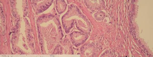
, appearing to be periglandular clefting on HE slide (200x).")
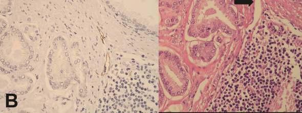
13 Figure 1. A, Lymphovascular invasion, detected with D2-40 immunostaining (inserted slide, 400x), appearing to be periglandular clefting on HE slide (200x). B, Expressed periglandular clefting on HE slide (200x) near lymph vessels evident on D2-40 immunostained slide (inserted slide, 400x). 12
14 13
15 Figure 2. Correlation between the number of lymph vessels in tumor and adjacent prostatic tissue (A), between lymph vessels mimicking periacinar clefting and number of lymph vessels in tumor (B) and between the number of lymph vessels within the area with 30 neoplastic glands with most extensive retractions and lymph vessels mimicking periacinar clefting (C). 14
16 15
17 Table 1. The number of lymph vessels in tumor tissue and surrounding prostatic parenchyma. Specimen Max Median Min number No. of lymph vessels in surrounding prostatic tissue/ 10 HPF No. of lymph vessels in tumorous tissue/ 10HPF No. of lymph vessels mimicking clefting (lymphovascular invasion)/ whole section surface No. of lymph vessels in the area with 30 tumorous glands revealing most extensive retractions 16
University of Zagreb Medical School Repository
 Središnja medicinska knjižnica Tomas, D., Krušlin, B., Rogatsch, H., Schäfer, G., Belicza, M., Mikuz, G. (00) Different Types of Atrophy in the Prostate With and Without Adenocarcinoma. European Urology,
Prostate cancer ~ diagnosis and impact of pathology on prognosis ESMO 2017
 Prostate cancer ~ diagnosis and impact of pathology on prognosis ESMO 2017 Dr Puay Hoon Tan Division of Pathology Singapore General Hospital Prostate cancer (acinar adenocarcinoma) Invasive carcinoma composed
Intraductal carcinoma of the prostate on needle biopsy: histologic features and clinical significance
 & 2006 USCAP, Inc All rights reserved 0893-3952/06 $30.00 www.modernpathology.org Intraductal carcinoma of the prostate on needle biopsy: histologic features and clinical significance Charles C Guo 1 and
INTRADUCTAL LESIONS OF THE PROSTATE. Jonathan I. Epstein
 INTRADUCTAL LESIONS OF THE PROSTATE Jonathan I. Epstein Topics Prostatic intraepithelial neoplasia (PIN) Intraductal adenocarcinoma (IDC-P) Intraductal urothelial carcinoma Ductal adenocarcinoma High Prostatic
A Study of D2-40 Immunohistochemical Expression in Colorectal Carcinomas
 Original Article DOI: 10.21276/APALM.1122 A Study of D2-40 Immunohistochemical Expression in Colorectal Carcinomas Sarvek Bajaj*, Gururajaprasad. C and Suchitha S Department of Pathology, JSS Medical college,
ACCME/Disclosures. Cribriform Lesions of the Prostate. Case
 Cribriform Lesions of the Prostate Ming Zhou, MD, PhD Departments of Pathology and Urology New York University Langone Medical Center New York, NY Ming.Zhou@NYUMC.ORG ACCME/Disclosures The USCAP requires
Although current American Cancer Society guidelines
 ORIGINAL ARTICLE Diffuse Adenosis of the Peripheral Zone in Prostate Needle Biopsy and Prostatectomy Specimens Tamara L. Lotan, MD* and Jonathan I. Epstein, MD*w z Abstract: We have observed a group of
3/28/2017. Disclosure of Relevant Financial Relationships. GU Evening Subspecialty Case Conference. Differential Diagnosis:
 GU Evening Subspecialty Case Conference Rajal B. Shah, M.D. VP, Medical Director, Urologic Pathology Miraca Life Sciences, Irving, Texas Clinical Associate Professor of Pathology Baylor College of Medicine,
Gross appearance of nodular hyperplasia in material obtained from suprapubic prostatectomy. Note the multinodular appearance and the admixture of
 Tiền liệt tuyến Tiền liệt tuyến Gross appearance of nodular hyperplasia in material obtained from suprapubic prostatectomy. Note the multinodular appearance and the admixture of solid and microcystic areas.
ROLE OF PROSTATIC BASAL CELL MARKER IN DIAGNOSIS OF PROSTATIC LESIONS
 Original Research Article Pathology International Journal of Pharma and Bio Sciences ISSN 0975-6299 ROLE OF PROSTATIC BASAL CELL MARKER IN DIAGNOSIS OF PROSTATIC LESIONS SUBATHRA K* Department of pathology,
Coordinate Expression of Cytokeratins 7 and 20 in Prostate Adenocarcinoma and Bladder Urothelial Carcinoma
 Anatomic Pathology / CYTOKERATINS 7 AND 20 IN PROSTATE AND BLADDER CARCINOMAS Coordinate Expression of Cytokeratins 7 and 20 in Prostate Adenocarcinoma and Bladder Urothelial Carcinoma Nader H. Bassily,
Correlation between expression and significance of δ-catenin, CD31, and VEGF of non-small cell lung cancer
 Correlation between expression and significance of δ-catenin, CD31, and VEGF of non-small cell lung cancer X.L. Liu 1, L.D. Liu 2, S.G. Zhang 1, S.D. Dai 3, W.Y. Li 1 and L. Zhang 1 1 Thoracic Surgery,
Uropathology January Jon Oxley
 Uropathology January 2012 Jon Oxley Background to seminar These slides were available to view via the web from scanned slides The junior pathologists answered questions on them via the web The answers
Characterization and significance of MUC1 and c-myc expression in elderly patients with papillary thyroid carcinoma
 Characterization and significance of MUC1 and c-myc expression in elderly patients with papillary thyroid carcinoma Y.-J. Hu 1, X.-Y. Luo 2, Y. Yang 3, C.-Y. Chen 1, Z.-Y. Zhang 4 and X. Guo 1 1 Department
ARTHUR PURDY STOUT SOCIETY COMPANION MEETING: DIFFICULT NEW DIFFERENTIAL DIAGNOSES IN PROSTATE PATHOLOGY. Jonathan I. Epstein.
 1 ARTHUR PURDY STOUT SOCIETY COMPANION MEETING: DIFFICULT NEW DIFFERENTIAL DIAGNOSES IN PROSTATE PATHOLOGY Jonathan I. Epstein Professor Pathology, Urology, Oncology The Reinhard Professor of Urological
Case #1: 75 y/o Male (treated and followed by prostate cancer oncology specialist ).
.") SOLID TUMORS WORKSHOP Cases for review Prostate Cancer Case #1: 75 y/o Male (treated and followed by prostate cancer oncology specialist ). January 2009 PSA 4.4, 20% free; August 2009 PSA 5.2; Sept 2009
SOLID TUMORS WORKSHOP Cases for review Prostate Cancer Case #1: 75 y/o Male (treated and followed by prostate cancer oncology specialist ). January 2009 PSA 4.4, 20% free; August 2009 PSA 5.2; Sept 2009
Središnja medicinska knjižnica
 Središnja medicinska knjižnica Popović A, Demirović A., Spajić B., Štimac G., Krušlin B., Tomas D. (2010) Expression and prognostic role of syndecan-2 in prostate cancer. Prostate Cancer and Prostatic
Prostate Immunohistochemistry. Literature Interpretation: Caveats. Must be aware of staining pattern of antibody in the relevant tissue
 IHC Interpretation: General Principles (1) Prostate Immunohistochemistry Murali Varma Cardiff, UK wptmv@cf.ac.uk Sarajevo Nov 2013 Must be aware of staining pattern of antibody in the relevant tissue Nuclear/cytoplasmic/membranous
Prostate Pathology: Prostate Carcinoma, variants and Gleason Grading (Part 1)
") Prostate Pathology: Prostate Carcinoma, variants and Gleason Grading (Part 1) Jae Y. Ro, MD, PhD June 7, 2012 Ten Leading Cancer Types for the Estimated New Cancer Cases and Deaths By Sex, United States,
Prostate Pathology: Prostate Carcinoma, variants and Gleason Grading (Part 1) Jae Y. Ro, MD, PhD June 7, 2012 Ten Leading Cancer Types for the Estimated New Cancer Cases and Deaths By Sex, United States,
Supplemental Information
 Supplemental Information Prediction of Prostate Cancer Recurrence using Quantitative Phase Imaging Shamira Sridharan 1, Virgilia Macias 2, Krishnarao Tangella 3, André Kajdacsy-Balla 2 and Gabriel Popescu
5/21/2018. Difficulty in Underdiagnosing Prostate Cancer. Diagnosis of Prostate Cancer. Evaluation of Prostate Cancer and Atypical on Needle Biopsy
 Evaluation of Prostate Cancer and Atypical on Needle Biopsy Jonathan I. Epstein Difficulty in Underdiagnosing Prostate Cancer Limited tissue on needle biopsy (1 cm. x
Diagnosis, pathology and prognosis including variant pathology
 PROSTATE CANCER Diagnosis, pathology and prognosis including variant pathology No Conflict of Interest Universitat Autónoma de Barcelona F.Algaba Section of Pathology PROSTATE CANCER Diagnosis, pathology
Update on 2015 WHO Classification of Lung Adenocarcinoma 1/3/ Mayo Foundation for Medical Education and Research. All rights reserved.
 1 Our speaker for this program is Dr. Anja Roden, an associate professor of Laboratory Medicine and Pathology at Mayo Clinic as well as consultant in the Anatomic Pathology Laboratory and co-director of
Senior of Histopathology Department at Khartoum, Radiation and Isotopes Center
 EUROPEAN ACADEMIC RESEARCH Vol. IV, Issue 2/ May 2016 ISSN 2286-4822 www.euacademic.org Impact Factor: 3.4546 (UIF) DRJI Value: 5.9 (B+) Immune Histochemical Evaluation of AMACR (P504S) in Prostatic Adenocarcinoma
Disclosures. Outline. What IS tumor budding?? Tumor Budding in Colorectal Carcinoma: What, Why, and How. I have nothing to disclose
 Tumor Budding in Colorectal Carcinoma: What, Why, and How Disclosures I have nothing to disclose Soo-Jin Cho, MD, PhD Assistant Professor UCSF Dept of Pathology Current Issues in Anatomic Pathology 2017
PSA. HMCK, p63, Racemase. HMCK, p63, Racemase
 Case 1 67 year old male presented with gross hematuria H/o acute prostatitis & BPH Urethroscopy: small, polypoid growth with a broad base emanating from the left side of the verumontanum Serum PSA :7 ng/ml
Department of Diagnostic Pathology, Kochi Red Cross Hospital, Kochi, Japan
 Malaysian J Pathol 2014; 36(3) : 169 173 ORIGINAL ARTICLE Application of combined immunohistochemical panel of AMACR(P504S)/p63 cocktail, cytokeratin 5 and D2-40 to atypical glands in prostatic needle
A712(19)- Test slide, Breast cancer tissues with corresponding normal tissues
- Test slide, Breast cancer tissues with corresponding normal tissues") A712(19)- Test slide, Breast cancer tissues with corresponding normal tissues (formalin fixed) For research use only Specifications: No. of cases: 12 Tissue type: Breast cancer tissues with corresponding
A712(19)- Test slide, Breast cancer tissues with corresponding normal tissues (formalin fixed) For research use only Specifications: No. of cases: 12 Tissue type: Breast cancer tissues with corresponding
Cover Page. The handle holds various files of this Leiden University dissertation
 Cover Page The handle http://hdl.handle.net/1887/55957 holds various files of this Leiden University dissertation Author: Dekker T.J.A. Title: Optimizing breast cancer survival models based on conventional
Gleason Scoring System 2017 JASREMAN DHILLON, MD ASSOCIATE PROFESSOR, DEPARTMENT OF ANATOMIC PATHOLOGY, MOFFITT CANCER CENTER, TAMPA, FLORIDA
 Gleason Scoring System 2017 JASREMAN DHILLON, MD ASSOCIATE PROFESSOR, DEPARTMENT OF ANATOMIC PATHOLOGY, MOFFITT CANCER CENTER, TAMPA, FLORIDA Learners Objectives u Latest changes per ISUP 2014 that impact
Mucin-producing urothelial-type adenocarcinoma of prostate: report of two cases of a rare and diagnostically challenging entity
 & 2005 USCAP, Inc All rights reserved 0893-3952/05 $30.00 www.modernpathology.org Mucin-producing urothelial-type adenocarcinoma of prostate: report of two cases of a rare and diagnostically challenging
Introduction. Key Words: high-grade prostatic intraepithelial neoplasia, HGPIN, radical prostatectomy, prostate biopsy, insignificant prostate cancer
 Prostate cancer after initial high-grade prostatic intraepithelial neoplasia and benign prostate biopsy Premal Patel, MD, 1 Jasmir G. Nayak, MD, 1,2 Zlatica Biljetina, MD, 4 Bryan Donnelly, MD 3, Kiril
They Do Look Alike : Mimics of Prostate Cancer in Biopsy Samples
 They Do Look Alike : in Biopsy Samples Gladell P. Paner, MD Departments of Pathology and Surgery (Urology) University of Chicago, IL USA Gladell.paner@uchospitals.edu Benign in Needle Biopsy 1. Benign
ISPUB.COM. Interpretation Of Prostatic Biopsies: A Review. A Chitale, S Khubchandani INTRODUCTION NON-NEOPLASTIC LESIONS GRADING: GLEASON'S SCORE
 ISPUB.COM The Internet Journal of Urology Volume 3 Number 1 A Chitale, S Khubchandani Citation A Chitale, S Khubchandani.. The Internet Journal of Urology. 2004 Volume 3 Number 1. Abstract The incidence
PROSTATIC ADENOCARCINOMA: DIAGNOSTIC CRITERIA AND IMPORTANT MIMICKERS PROSTATIC ADENOCARCINOMA: DIAGNOSTIC CRITERIA
 PROSTATIC ADENOCARCINOMA: DIAGNOSTIC CRITERIA AND IMPORTANT MIMICKERS PROSTATIC ADENOCARCINOMA: DIAGNOSTIC CRITERIA 1 A good H & E helps! ADENOCARCINOMA DIAGNOSTIC CRITERIA Relatively uniform proliferation
University Journal of Pre and Para Clinical Sciences
 ISSN 2455 2879 Volume 2 Issue 1 2016 Metaplastic carcinoma breast a rare case report Abstract : Metaplastic carcinoma of the breast is a rare malignancy with two distinct cell lines described as a breast
Procedures Needle Biopsy Transurethral Prostatic Resection Suprapubic or Retropubic Enucleation (Subtotal Prostatectomy) Radical Prostatectomy
 Radical Prostatectomy") Prostate Gland Protocol applies to invasive carcinomas of the prostate gland. Protocol web posting date: July 2006 Protocol effective date: April 2007 Based on AJCC/UICC TNM, 6 th edition Procedures Needle
Prostate Gland Protocol applies to invasive carcinomas of the prostate gland. Protocol web posting date: July 2006 Protocol effective date: April 2007 Based on AJCC/UICC TNM, 6 th edition Procedures Needle
MEDICAL POLICY Genetic and Protein Biomarkers for Diagnosis and Risk Assessment of
 POLICY: PG0367 ORIGINAL EFFECTIVE: 08/26/16 LAST REVIEW: 09/27/18 MEDICAL POLICY Genetic and Protein Biomarkers for Diagnosis and Risk Assessment of Prostate Cancer GUIDELINES This policy does not certify
Benign Mimics of Malignancy in Breast Pathology
 Arthur Purdy Stout Society of Surgical Pathologists Companion Meeting Benign Mimics of Malignancy in Breast Pathology Stuart J. Schnitt, M.D. Beth Israel Deaconess Medical Center and Harvard Medical School,
Metachronous anterior urethral metastasis of prostatic ductal adenocarcinoma
 http://dx.doi.org/10.7180/kmj.2016.31.1.66 KMJ Case Report Metachronous anterior urethral metastasis of prostatic ductal adenocarcinoma Jeong Hyun Oh 1, Taek Sang Kim 1, Hyun Yul Rhew 1, Bong Kwon Chun
ASSESSMENT OF GLEASON SYSTEM USE ON DIFFERENT TYPES OF PROSTATIC TISSUE SAMPLES
 ASSESSMENT OF GLEASON SYSTEM USE ON DIFFERENT TYPES OF PROSTATIC TISSUE SAMPLES I. E. PLEŞEA*, B. ZAHARIA*, S. D. ENACHE**, G. MITROI***, P. TOMESCU***, O. T. POP*, P. BADEA****, A. KOŻOKIĆ***** *Department
Immunohistochemical Expression Of Cytokeratin 8 And 18 In Breast Carcinoma.
 ISPUB.COM The Internet Journal of Pathology Volume 13 Number 3 Immunohistochemical Expression Of Cytokeratin 8 And 18 In Breast Carcinoma. B Rattan, A Baghla, M Manjari, P Kakkar, S Kahlon, S Paul Citation
The 2015 World Health Organization Classification for Lung Adenocarcinomas: A Practical Approach
 The 2015 World Health Organization Classification for Lung Adenocarcinomas: A Practical Approach Dr. Carol Farver Director, Pulmonary Pathology Pathology and Laboratory Medicine Institute Objectives Discuss
OMPRN Pathology Matters Meeting 2017
 OMPRN Pathology Matters Meeting 2017 Pathology of Aggressive Prostate Cancer Intraductal Carcinoma and Cribriform Carcinoma Dr. Michelle Downes, Staff Urologic Pathologist Sunnybrook Health Sciences Centre,
Comparison of CD10 expression in stroma of epithelial and mesenchymal tumors of the breast
 Global Advanced Research Journal of Medicine and Medical Science (ISSN: 2315-5159) Vol. 4(1) pp. 051-056, January, 2015 Available online http://garj.org/garjmms/index.htm Copyright 2015 Global Advanced
Original Article CREPT expression correlates with esophageal squamous cell carcinoma histological grade and clinical outcome
 Int J Clin Exp Pathol 2017;10(2):2030-2035 www.ijcep.com /ISSN:1936-2625/IJCEP0009456 Original Article CREPT expression correlates with esophageal squamous cell carcinoma histological grade and clinical
Surgical Pathology Issues of Practical Importance
 Surgical Pathology Issues of Practical Importance Anne Moore, MD Medical Oncology Syed Hoda, MD Surgical Pathology The pathologist is central to the team approach needed to manage the patient with breast
Tumor Angiogenesis in Stage II Colorectal Carcinoma Association With Survival
 ANATOMIC PATHOLOGY Original Article Tumor Angiogenesis in Stage II Carcinoma Association With Survival BARBARA F. BANNER, MD, ROB WHITEHOUSE, STEPHEN P. BAKER, MSPH, AND RICHARD S. SWANSON, MD We studied
LYMPHATIC DRAINAGE AXILLARY (MOSTLY) INTERNAL MAMMARY SUPRACLAVICULAR
 INTERNAL MAMMARY SUPRACLAVICULAR") BREAST LYMPHATIC DRAINAGE AXILLARY (MOSTLY) INTERNAL MAMMARY SUPRACLAVICULAR HISTOLOGY LOBE: (10 in whole breast) LOBULE: (many per lobe) ACINUS/I, aka ALVEOLUS/I: (many per lobule) DUCT(S): INTRA- or
BREAST LYMPHATIC DRAINAGE AXILLARY (MOSTLY) INTERNAL MAMMARY SUPRACLAVICULAR HISTOLOGY LOBE: (10 in whole breast) LOBULE: (many per lobe) ACINUS/I, aka ALVEOLUS/I: (many per lobule) DUCT(S): INTRA- or
1 NORMAL HISTOLOGY AND METAPLASIAS
 1 NORMAL HISTOLOGY AND METAPLASIAS, MD Anatomy and Histology 1 Metaplasias 2 ANATOMY AND HISTOLOGY The female breast is composed of a branching duct system, which begins at the nipple with the major lactiferous
The Role of Cytokeratin 5/6 in Differential Diagnosis of Prostate Tumors
 EUROPEAN ACADEMIC RESEARCH Vol. III, Issue 10/ January 2016 ISSN 2286-4822 www.euacademic.org Impact Factor: 3.4546 (UIF) DRJI Value: 5.9 (B+) The Role of Cytokeratin 5/6 in Differential Diagnosis of Prostate
Immunohistochemical studies (ER & Ki-67) in Proliferative breast lesions adjacent to malignancy
 in Proliferative breast lesions adjacent to malignancy") IOSR Journal of Dental and Medical Sciences (IOSR-JDMS) e-issn: 2279-0853, p-issn: 2279-0861.Volume 13, Issue 3 Ver. IV. (Mar. 2014), PP 84-89 Immunohistochemical studies (ER & Ki-67) in Proliferative
IOSR Journal of Dental and Medical Sciences (IOSR-JDMS) e-issn: 2279-0853, p-issn: 2279-0861.Volume 13, Issue 3 Ver. IV. (Mar. 2014), PP 84-89 Immunohistochemical studies (ER & Ki-67) in Proliferative
Vascular endothelial growth factor (VEGF)-C, VEGF-D, VEGFR-3 and D2-40 expressions in primary breast cancer: Association with lymph node metastasis
-C, VEGF-D, VEGFR-3 and D2-40 expressions in primary breast cancer: Association with lymph node metastasis") Original papers Vascular endothelial growth factor (VEGF)-C, VEGF-D, VEGFR-3 and D2-40 expressions in primary breast cancer: Association with lymph node metastasis Aydan Eroğlu 1, A F, Cevriye Ersöz 2,
Original papers Vascular endothelial growth factor (VEGF)-C, VEGF-D, VEGFR-3 and D2-40 expressions in primary breast cancer: Association with lymph node metastasis Aydan Eroğlu 1, A F, Cevriye Ersöz 2,
Patterns of E.cadherin and Estrogen receptor Expression in Histological Sections of Sudanese Patients with Breast Carcinoma
 Patterns of E.cadherin and Estrogen receptor Expression in Histological Sections of Sudanese Patients with Breast Carcinoma Hadia. Mohammed. Abdalla. Abdalrhman *, Elsadig.A.Adam, Ayda.D.A.Allatif 3,'Namareg.E.Afadul
Pathology of the Prostate. PathoBasic Tatjana Vlajnic
 Pathology of the Prostate PathoBasic 24.01.17 Tatjana Vlajnic Overview Adenocarcinoma of the prostate Grading Special variants Mimickers of prostate adenocarcinoma Atrophy Inflammatory conditions Granulomatous
Research Article Stromal Expression of CD10 in Invasive Breast Carcinoma and Its Correlation with ER, PR, HER2-neu, and Ki67
 SAGE-Hindawi Access to Research International Breast Cancer Volume 20, Article ID 47957, 4 pages doi:0.406/20/47957 Research Article Stromal Expression of CD0 in Invasive Breast Carcinoma and Its Correlation
Supplementary Materials. for Garmy-Susini, et al, Integrin 4 1 signaling is required for lymphangiogenesis and tumor metastasis
 Supplementary Materials for Garmy-Susini, et al, Integrin 4 1 signaling is required for lymphangiogenesis and tumor metastasis 1 Supplementary Figure Legends Supplementary Figure 1: Integrin expression
Original Article CyclinD1 promotes lymph node metastasis by inducing lymphangiogenesis in human ovarian carcinoma
 Int J Clin Exp Pathol 2018;11(7):3726-3731 www.ijcep.com /ISSN:1936-2625/IJCEP0077355 Original Article CyclinD1 promotes lymph node metastasis by inducing lymphangiogenesis in human ovarian carcinoma Minhua
Prostate cancer staging and datasets: The Nitty-Gritty. What determines our pathological reports? 06/07/2018. Dan Berney Maastricht 2018
 Prostate cancer staging and datasets: The Nitty-Gritty What determines our pathological reports? Dan Berney Maastricht 2018 Biopsy reporting. How not to do it. The TNM 8 th edition. Changes good and bad
WHAT SHOULD WE DO WITH TUMOUR BUDDING IN EARLY COLORECTAL CANCER?
 CANCER STAGING TNM and prognosis in CRC WHAT SHOULD WE DO WITH TUMOUR BUDDING IN EARLY COLORECTAL CANCER? Alessandro Lugli, MD Institute of Pathology University of Bern Switzerland Maastricht, June 19
Outcomes Following Negative Prostate Biopsy for Patients with Persistent Disease after Radiotherapy for Prostate Cancer
 Clinical Urology Post-radiotherapy Prostate Biopsy for Recurrent Disease International Braz J Urol Vol. 36 (1): 44-48, January - February, 2010 doi: 10.1590/S1677-55382010000100007 Outcomes Following Negative
Prostatic ductal adenocarcinoma is a subtype of
 ORIGINAL ARTICLE High-grade Prostatic Intraepithelial Neoplasialike Ductal Adenocarcinoma of the Prostate: A Clinicopathologic Study of 28 Cases Fabio Tavora, MD* and Jonathan I. Epstein, MD*w z Abstract:
ACRIN 6666 Therapeutic Surgery Form
 S1 ACRIN 6666 Therapeutic Surgery Form 6666 Instructions: Complete a separate S1 form for each separate area of each breast excised with the intent to treat a cancer (e.g. each lumpectomy or mastectomy).
Hyperplastic, Premalignant and Malignant Lesions of the Prostate Gland
 Jehoram Tei Anim, md, FRCPath; Sitara Abdul Sathar, MB; Mohammed Ejaz Bhatti, MSc From the Department of Pathology, Faculty of Medicine, Kuwait University, Kuwait. Address reprint requests and correspondence
Cluster designation 5 staining of normal and non-lymphoid neoplastic skin*
 J Cutan Pathol 2005: 32: 50 54 Copyright # Blackwell Munksgaard 2005 Blackwell Munksgaard. Printed in Denmark Journal of Cutaneous Pathology Cluster designation 5 staining of normal and non-lymphoid neoplastic
S1.04 Principal clinician. G1.01 Comments. G2.01 *Specimen dimensions (prostate) S2.02 *Seminal vesicles
 S2.02 *Seminal vesicles") Prostate Cancer Histopathology Reporting Proforma (Radical Prostatectomy) Includes the International Collaboration on Cancer reporting dataset denoted by * Family name Given name(s) Date of birth Sex Male
Prostate Cancer Histopathology Reporting Proforma (Radical Prostatectomy) Includes the International Collaboration on Cancer reporting dataset denoted by * Family name Given name(s) Date of birth Sex Male
BREAST PATHOLOGY. Fibrocystic Changes
 BREAST PATHOLOGY Lesions of the breast are very common, and they present as palpable, sometimes painful, nodules or masses. Most of these lesions are benign. Breast cancer is the 2 nd most common cause
3/23/2017. Significant Changes in Prostate Cancer Classification, Grading, Staging and Reporting. Disclosure of Relevant Financial Relationships
 Disclosure of Relevant Financial Relationships Staging and Reporting of Prostate Cancer: Major Changes in 8 th Edition AJCC Staging and CAP Cancer Checklists USCAP requires that all planners (Education
FINALIZED SEER SINQ QUESTIONS
 0076 Source 1: WHO Class CNS Tumors pgs: 33 MP/H Rules/Histology--Brain and CNS: What is the histology code for a tumor originating in the cerebellum and extending into the fourth ventricle described as
WT1, Estrogen Receptor, and Progesterone Receptor as Markers for Breast or Ovarian Primary Sites in Metastatic Adenocarcinoma to Body Fluids
 Anatomic Pathology / WT1, ESTROGEN RECEPTOR, AND PROGESTERONE RECEPTOR IN CYTOLOGY OF BODY FLUIDS WT1, Estrogen Receptor, and Progesterone Receptor as Markers for Breast or Ovarian Primary Sites in Metastatic
5/21/2018. Prostate Adenocarcinoma vs. Urothelial Carcinoma. Common Differential Diagnoses in Urological Pathology. Jonathan I.
 Common Differential Diagnoses in Urological Pathology Jonathan I. Epstein Prostate Adenocarcinoma vs. Urothelial Carcinoma 1 2 NKX3.1 NKX3.1 3 4 5 6 Proposed ISUP Recommendations Option to use PSA as a
Carcinoma mammario: le istologie non frequenti. Valentina Guarneri Università di Padova IOV-IRCCS
 Carcinoma mammario: le istologie non frequenti Valentina Guarneri Università di Padova IOV-IRCCS Histological diversity of breast adenocarcinomas Different histological types are defined according to specific
Gastric Carcinoma with Lymphoid Stroma: Association with Epstein Virus Genome demonstrated by PCR
 Gastric Carcinoma with Lymphoid Stroma: Association with Epstein Virus Genome demonstrated by PCR Pages with reference to book, From 305 To 307 Irshad N. Soomro,Samina Noorali,Syed Abdul Aziz,Suhail Muzaffar,Shahid
Definition of Synoptic Reporting
 Definition of Synoptic Reporting The CAP has developed this list of specific features that define synoptic reporting formatting: 1. All required cancer data from an applicable cancer protocol that are
Prostatic stromal hyperplasia with atypia (PSHA) is a
 is a") Prostatic Stromal Hyperplasia With Atypia Follow-up Study of 18 Cases Deloar Hossain, MD, FRCPC; Isabelle Meiers, MD; Junqi Qian, MD; Gregory T. MacLennan, MD; David G. Bostwick, MD, MBA Context. Prostatic
Prostatic Stromal Hyperplasia With Atypia Follow-up Study of 18 Cases Deloar Hossain, MD, FRCPC; Isabelle Meiers, MD; Junqi Qian, MD; Gregory T. MacLennan, MD; David G. Bostwick, MD, MBA Context. Prostatic
Inverted (hobnail) high grade prostatic intraepithelial neoplasia and invasive inverted pattern
 high grade prostatic intraepithelial neoplasia and invasive inverted pattern") ONCOLOGY LETTERS 10: 2395-2399, 2015 Inverted (hobnail) high grade prostatic intraepithelial neoplasia and invasive inverted pattern MELTEM ÖZNUR 1, SEVIM BAYKAL KOCA 2, PELIN YILDIZ 3, BURAK BAHADIR 4
ONCOLOGY LETTERS 10: 2395-2399, 2015 Inverted (hobnail) high grade prostatic intraepithelial neoplasia and invasive inverted pattern MELTEM ÖZNUR 1, SEVIM BAYKAL KOCA 2, PELIN YILDIZ 3, BURAK BAHADIR 4
MORPHOLOGIC TRANSITIONS BETWEEN PROLIFERATIVE INFLAMMATORY ATROPHY AND HIGH-GRADE PROSTATIC INTRAEPITHELIAL NEOPLASIA
 ADULT UROLOGY MORPHOLOGIC TRANSITIONS BETWEEN PROLIFERATIVE INFLAMMATORY ATROPHY AND HIGH-GRADE PROSTATIC INTRAEPITHELIAL NEOPLASIA MATHEW J. PUTZI AND ANGELO M. DE MARZO ABSTRACT Objectives. To validate
Assessment Run CK19
 Assessment Run 29 200 CK9 The slide to be stained for CK9 comprised:. Appendix, 2. Thyroid gland, 3. Pancreas, 4. Ductal breast carcinoma, 5. Esophagus, 6. Papillary thyroid carcinoma. All tissues were
Basement membrane in lobule.
 Bahram Memar, MD Basement membrane in lobule. Normal lobule-luteal phase Normal lobule-follicular phase Lactating breast Greater than 95% are adenocarcinomas in situ carcinomas and invasive carcinomas.
CD15 and CEA expression in thymic epithelial neoplasms
 Turkish Journal of Cancer Volume 8, No., 8 CD and CEA expression in thymic epithelial neoplasms AYTEKİN AKYOL, AYŞEGÜL ÜNER Hacettepe University, Department of Pathology, Ankara-Turkey ABSTRACT The aim
Chronic inflammation of long-standing duration has
 Inflammatory Atrophy of the Prostate Prevalence and Significance Athanase Billis, MD; Luis A. Magna, MD Context. Recently, prostatic atrophy associated with chronic inflammation has been linked to carcinoma
Histopathology of Endoscopic Resection Specimens from Barrett's Esophagus
 Histopathology of Endoscopic Resection Specimens from Barrett's Esophagus Br J Surg 38 oct. 1950 Definition of Barrett's esophagus A change in the esophageal epithelium of any length that can be recognized
Prognostic Significance of Extranodal Cancer Invasion of Mediastinal Lymph Nodes in Lung Cancer
 Jpn. J. Clin. Oncol. 198, 1 (), 7-1 Prognostic Significance of Extranodal Cancer Invasion of Mediastinal Lymph Nodes in Lung Cancer KEIICHI SUEMASU, M.D. AND TSUGUO NARUKE, M.D. Department of Surgery,
Chapter 2. Understanding My Diagnosis
 Chapter 2. Understanding My Diagnosis With contributions from Nancy L. Brown, Ph.D.,Palo Alto Medical Foundation Research Institute; and Patrick Swift, M.D., Alta Bates Comprehensive Cancer Program o Facts
Kidney, Bladder and Prostate Neoplasia. David Bingham MD
 Kidney, Bladder and Prostate Neoplasia David Bingham MD typical malignant cytology of bladder washings 1 benign 2 malignant typical malignant cytology of bladder washings b Bladder tumor Non invasive papillary
Prostate Case Scenario 1
 Prostate Case Scenario 1 H&P 5/12/16: A 57-year-old Hispanic male presents with frequency of micturition, urinary urgency, and hesitancy associated with a weak stream. Over the past several weeks, he has
STUDY OF PROSTATIC LESION FOR A PERIOD OF FIVE YEARS
 Page222 IJPBS Volume 4 Issue 2 APR-JUN 2014 222-226 Research Article Biological Sciences STUDY OF PROSTATIC LESION FOR A PERIOD OF FIVE YEARS B. Rajashekar Reddy 1, Rameswarapu Suman Babu 1 and Sujatha.P*
Treatment options for the precancerous Atypical Breast lesions. Prof. YOUNG-JIN SUH The Catholic University of Korea
 Treatment options for the precancerous Atypical Breast lesions Prof. YOUNG-JIN SUH The Catholic University of Korea Not so benign lesions? Imaging abnormalities(10% recall) lead to diagnostic evaluation,
Objectives. Atypical Glandular Cells. Atypical Endocervical Cells. Reactive Endocervical Cells
 2013 California Society of Pathologists 66 th Annual Meeting San Francisco, CA Atypical Glandular Cells to Early Invasive Adenocarcinoma: Cervical Cytology and Histology Christina S. Kong, MD Associate
2 to 3% of All New Visceral Cancers Peak Incidence is 6th Decade M:F = 2:1 Grossly is a Bright Yellow, Necrotic Mass with a Pseudocapsule
 GENITOURINARY PATHOLOGY Kathleen M. O Toole, M.D. Renal Cell Carcinoma 2 to 3% of All New Visceral Cancers Peak Incidence is 6th Decade M:F = 2:1 Grossly is a Bright Yellow Necrotic Mass Grossly is a Bright
Prostate Cancer Grading, Staging and Reporting: An Update Cristina Magi-Galluzzi, MD, PhD
 Prostate Cancer Grading, Staging and Reporting: An Update Cristina Magi-Galluzzi, MD, PhD Director, Genitourinary Pathology R.J. Tomsich Pathology & Laboratory Medicine Institute Professor of Pathology,
JMSCR Vol 04 Issue 12 Page December 2016
 JMSCR Vol 4 Issue 12 Page 14471-14478 December 216 www.jmscr.igmpublication.org Impact Factor 5.244 Index Copernicus Value: 83.27 ISSN (e)-2347-176x ISSN (p) 2455-45 DOI: https://dx.doi.org/1.18535/jmscr/v4i12.29
IMMUNOHISTOCHEMICAL EXPRESSION OF TISSUE INHIBITOR OF METALLOPROTEINASE-1 (TIMP-1) IN INVASIVE BREAST CARCINOMA
 IN INVASIVE BREAST CARCINOMA") & IMMUNOHISTOCHEMICAL EXPRESSION OF TISSUE INHIBITOR OF METALLOPROTEINASE-1 (TIMP-1) IN INVASIVE BREAST CARCINOMA Suada Kuskunović*, Svjetlana Radović, Mirsad Dorić, Ajna Hukić, Mirsad Babić, Ivana Tomić,
& IMMUNOHISTOCHEMICAL EXPRESSION OF TISSUE INHIBITOR OF METALLOPROTEINASE-1 (TIMP-1) IN INVASIVE BREAST CARCINOMA Suada Kuskunović*, Svjetlana Radović, Mirsad Dorić, Ajna Hukić, Mirsad Babić, Ivana Tomić,
Maram Abdaljaleel, MD Dermatopathologist and Neuropathologist University of Jordan, School of Medicine
 Maram Abdaljaleel, MD Dermatopathologist and Neuropathologist University of Jordan, School of Medicine The most common non-skin malignancy of women 2 nd most common cause of cancer deaths in women, following
Signet-Ring Cell Change in Benign Prostatic Hyperplasia - A Rare Case Report
 Article ID: WMC00688 ISSN 2046-1690 Signet-Ring Cell Change in Benign Prostatic Hyperplasia - A Rare Case Report Author(s):Dr. Pallavi Bhuyan, Dr. Smita Mahapatra, Dr. Sujata Pujari, Dr. Jayasree Rath
Pancreatobiliary Frozen Section Nightmares
 Pancreatobiliary Frozen Section Nightmares Aatur D. Singhi, MD PhD Assistant Professor University of Pittsburgh Medical Center Department of Pathology singhiad@upmc.edu Objectives Briefly give an overview
Estrogen receptor (ER)
") Assessment Run B7 204 Estrogen receptor (ER) Material The slide to be stained for ER comprised: No. Tissue ER-positivity* ER-intensity*. Uterine cervix 80-90% Moderate to strong 2. Breast carcinoma 0%
Assessment Run B7 204 Estrogen receptor (ER) Material The slide to be stained for ER comprised: No. Tissue ER-positivity* ER-intensity*. Uterine cervix 80-90% Moderate to strong 2. Breast carcinoma 0%
Analysis of immunohistochemical expression of CD10 in the malignant lesions of prostate
 IOSR Journal of Dental and Medical Sciences (IOSR-JDMS) e-issn: 2279-0853, p-issn: 2279-0861.Volume 16, Issue 7 Ver. I (July. 2017), PP 78-82 www.iosrjournals.org Analysis of immunohistochemical expression
NPQR Quality Payment Program (QPP) Measures 21_18247_LS.
 Measures 21_18247_LS.") NPQR Quality Payment Program (QPP) Measures 21_18247_LS MEASURE ID: QPP 99 MEASURE TITLE: Breast Cancer Resection Pathology Reporting pt Category (Primary Tumor) and pn Category (Regional Lymph Nodes)
NPQR Quality Payment Program (QPP) Measures 21_18247_LS MEASURE ID: QPP 99 MEASURE TITLE: Breast Cancer Resection Pathology Reporting pt Category (Primary Tumor) and pn Category (Regional Lymph Nodes)
Asian J Androl 2005; 7 (2): DOI: /j x
: DOI: /j x") Asian J Androl 5; 7 (2): 159 163 DOI: 1.1111/j.1745-7262.5.29.x. Original Article. Mass screening of prostate cancer in a Chinese population: the relationship between pathological features of prostate
Asian J Androl 5; 7 (2): 159 163 DOI: 1.1111/j.1745-7262.5.29.x. Original Article. Mass screening of prostate cancer in a Chinese population: the relationship between pathological features of prostate