The Role of Mast Cells in the Activation of Dermal Fibroblasts. Leah Campeau
|
|
|
- Silvester Booth
- 5 years ago
- Views:
Transcription
1 The Role of Mast Cells in the Activation of Dermal Fibroblasts by Leah Campeau A thesis submitted in partial fulfillment of the requirements for the degree of Master of Science in Experimental Surgery Department of Surgery University of Alberta Leah Campeau,
2 Abstract Introduction: Hypertrophic scar (HTS) formation is a fibroproliferative disorder that commonly follows deep dermal burns with prolonged inflammation. It is characterized by excessive extracellular matrix such as collagen deposition mainly by dermal fibroblasts. Mast cells have been implicated in HTS as they degranulate in response to injury and release pro-inflammatory and profibrotic mediators that may contribute to scar formation via increased fibroblast activity. We hypothesize that mast cell mediators regulate deep dermal fibroblasts to become profibrotic, thus mediating HTS development. Methods: Mast cells were quantified in human HTS and scar tissue from dermal fibrotic mouse models including CXCR4-treated nude mice. In vitro, layered dermal fibroblasts were cultured with conditioned media from activated mast cells and examined for changes in fibroblast activity. The measures determined included MTT cell proliferation assays to assess cell viability and RT-PCR to assess fibrotic gene expression. Liquid chromatography/mass spectrometry analysis of 4-hydroxyproline was measured as an indicator of type I collagen production and flow cytometric analysis of -SMA expression as a measure of myofibroblast differentiation and contractile capacity in layered fibroblasts after exposure to conditioned media from mast cells. Results: In vivo, mast cell densities increased in scar tissues from all dermal fibrotic mouse models and decreased in scar tissues from CXCR4-treated nude mice. In the presence of conditioned media from activated mast cells, fibroblasts showed no significant change in proliferation or gene and protein expression of -SMA and type I collagen but showed general trends suggesting increase proliferation and decreased -SMA expression. - ii -
3 Conclusion: In vivo, mast cells were found to be involved in hypertrophic scar formation. In our in vitro experiments, mast cells may have roles in HTS development but their effects on fibroblasts require further study and the mechanism of how mast cells could selectively influence the activity of superficial and deep fibroblasts warrants further investigation. - iii -
4 Preface This thesis is an original work by Leah Campeau. Portions of chapter 1 of this thesis have been published as Leah Campeau, Jie Ding, Edward E. Tredget, A potential role of CXCL12/CXCR4 chemotactic pathway in wound healing and hypertrophic scar formation, Receptors and Clinical Investigation 2015; 2: e791. doi: /rci.791. Chapter 2 is a manuscript-based portion. I was responsible for performing all experiments, data collection, analysis and manuscript composition. Jie Ding and Edward E. Tredget assisted with manuscript edits, concept formation and manuscript modifications. - iv -
5 Dedication I would like to dedicate this thesis to my parents Guy and Iris Campeau, for without their loving help and support of my work, none of this would be possible. Also, with great thanks to Alexander for encouraging me throughout the completion of this thesis. - v -
6 Acknowledgments I would like to formally thank Edward Tredget, Jie Ding, Dean Befus, Thomas Churchill and Gina Rayat for their patient guidance and expertise in the formulation of my thesis. I also gratefully acknowledge Zengshuan Ma for his technical assistance and support in completing this thesis. This work was financially supported by: Edmonton Civic Employees Charitable Assistance Fund, Department of Surgery and the Fire Fighters Burn Trust Fund - vi -
7 Table of Contents Abstract... ii Preface... iv Dedication... v Acknowledgments... vi Chapter 1. Introduction Structure and Function of the Skin Wound Healing Process Hemostasis Inflammation Proliferation Maturation and Matrix Remodeling Cellular Elements Involved in Wound Healing Fibroblasts and Myofibroblasts Mast Cells Platelets Neutrophils Monocytes Macrophages Keratinocytes Fibrocytes T cells Natural Killer Cells Mediators Involved in Wound Healing vii -
8 1.4.1 Histamine Tryptase and Chymase Transforming Growth Factor (TGF-β) Platelet-Derived Growth Factor (PDGF) Epidermal Growth Factor (EGF) Vascular Endothelial Growth Factor (VEGF) Insulin-Like Growth Factor (IGF-1) Fibroblast Growth Factor-2 (FGF2) Interferon-γ (IFN-γ) Tumor Necrosis Factor (TNF) Interleukins (IL-1α, IL-1β, IL-4, IL-5, IL-6, IL-10, IL-12 and IL-13) CXCL12/CXCR4 Pathway and its Involvement in Wound Healing Hypertrophic Scarring in Humans The Importance of the Inflammatory Response and Cellular Migration in Scar Formation and Fibrosis Mouse Models of Human HTS Summary and Formulation of Thesis Rationale Objectives of the Thesis Tables Figures Bibliography Chapter 2. Quantification of Mast Cells in Scar Tissues from Human and Dermal Fibrotic Mouse Models and the Regulatory Role of Mast Cells on Heterogeneous Dermal Fibroblasts Introduction viii -
9 2.2 Methods Dermal Fibrotic Mouse Models and Scar Tissue Harvest Mast Cell and Fibroblast Co-Culture (4,5-dimethylthiazol-2-y l)-2,5-diphenyltetrazolium Bromide Cell Proliferation Assay Real-Time Reverse Transcription-Polymerase Chain Reaction (RT-PCR) for Collagen-1 and -SMA Gene Expression Flow Cytometry Analysis of Myofibroblast Differentiation by -SMA Expression Liquid Chromatography/Mass Spectrometry (LC/MS) Analysis of 4-Hydroxyproline Statistical Analysis Results Discussion Conclusions Tables Figures Bibliography Chapter 3. Conclusions and Future Directions Bibliography Bibliography Appendices ix -
10 List of Tables Table 1-1: Features of normal, HTS, and deep dermal fibroblasts [187] Table 1-2: Potential activators and mechanisms of mast cell activation [129] Table 1-3: Products release by activated mast cells [129] Table 2-1. Primer sequences for real-time RT-PCR [24] Table 2-2. Average number of mast cells in dermal layers of human HTS tissue x -
11 List of Figures Figure 1-1: Structure of human skin [2] Figure 1-2: Brief overview of the differences between the phases of normal wound healing and excessive scar formation [10] Figure 1-3: Morphology of human superficial (a) and deep dermal fibroblasts (b) [27] Figure 1-4: Human and murine mast cell classification [48] Figure 1-5: Morphology of a resting and activated mast cell following activation induced degranulation [188] Figure 1-6: Role of mast cells in the various stages of wound healing [64] Figure 1-7. The CXCL12/CXCR4 Pathway [187] Figure 1-8: Hypertrophic scar to the trunk 16 months following burn injury [29] Figure 1-9: Excessive contracture in a burn of the hand [26] Figure 1-10: Characteristic imbalances of hypertrophic scar formation [89] Figure 2-1: Quantification of human mast cells in vivo Figure 2-2: Experimental design for co-culture of layered dermal fibroblasts and mast cell conditioned media Figure 2-3: Immunofluorescent staining and mast cell quantification of normal human skin and hypertrophic scar Figure 2-4: Toluidine blue staining of scar tissues (xenografts) for mast cells and mast cell quantification in dermal fibrotic mouse models Figure 2-5: Toluidine blue staining for mast cell quantification in scar tissues (xenografts) of dermal fibrotic nude mice treated with a CXCR4 antagonist xi -
12 Figure 2-6: Effect of mast cell conditioned media on fibroblast cell proliferation Figure 2-7: Effect of mast cell conditioned media on -SMA and collagen gene expression Figure 2-8: Effect of mast cell conditioned media on protein expression of -SMA and type I collagen Figure A1: Levels of cytokines, chemokines and growth factors in conditioned mast cell media Figure A2: Effect of activated and inactivated mast cells on fibroblast proliferation in a transwell insert system Figure A3: Effect of activated and inactivated mast cells on fibroblast fibrotic gene expression in a transwell insert system Figure A4: Effect of activated and inactivated mast cells on fibroblast protein expression of - SMA and collagen in a transwell insert system xii -
13 List of Abbreviations -SMA BALB BSA Alpha smooth muscle actin Bragg albino laboratory bred Bovine serum albumin CCL2 chemokine ligand 2 COL-1 CTGF Type I collagen Connective tissue growth factor CXCL12 C-X-C motif chemokine 12 CXCR4 C-X-C chemokine receptor type 4 ECM EGF FBS FCS Extracellular matrix Epidermal growth factor Fetal bovine serum Fetal calf serum FGF2 Fibroblast growth factor 2 GM-SCF Granulocyte macrophage colony-stimulating factor HPRT1 Hypoxanthine phosphoribosyltransferase 1 HTS IFN- Hypertrophic scar(s) Interferon-gamma IGF-1 Insulin-like growth factor 1 IL LT MC TC /MC T MMP Interleukin Leukotriene Tryptase-chymase/tryptase only positive mast cells Matrix metalloproteinases - xiii -
14 mrna NK cells PBS PE PDGF Messenger ribonucleic acid Natural killer cells Phosphate buffered saline Phycoerythrin Platelet-derived growth factor PGD 2 Prostaglandin D 2 RAG SCF SFM TCR TGF- Th1/Th2 TNF V(D)J VEGF Recombination activating gene Stem cell factor Serum free media T-cell receptor Transforming growth factor beta Type I/II helper T cells Tumor necrosis factor Variable, diversity, joining Vascular endothelial growth factor - xiv -
15 Chapter 1. Introduction Hypertrophic scar (HTS) formation is a fibroproliferative disorder, whose incomplete understanding of its pathophysiology continues to perplex researchers and clinicians alike. This chapter examines the structure of the skin and the burden of HTS. General phases of wound healing will be introduced and the physiological aberrations of the underlying mechanisms of HTS discussed. A brief overview of animal models and the elements of wound healing with respect to HTS research will also be covered. Although there are numerous contributors in HTS development, in this thesis emphasis will be placed on the role of mast cells, their implications in HTS formation and influences on fibroblasts during wound healing. 1.1 Structure and Function of the Skin The skin is the body s largest organ and first line of defense [1]. Its complex three-layered structure consists of the epidermis, dermis and hypodermis, a network of cellular, structural and molecular elements that collectively work together to serve the many functions of the skin [Figure 1-1]. These include protection from the external environment in the form of harmful pathogens or abrasions, heat regulation through sweat glands and blood vessels, containment of internal organs, tissues and vital substances, tactile, temperature and pain sensations and the synthesis and storage of vitamin D [2]. As a physical barrier, the skin on average has an acidic ph of less than 5, which contributes to sustaining the diverse collection of micro flora that aid in defense against foreign invaders [3, 4]. The avascular epidermis is the most superficial layer composed of stratified keratinized epithelium with some melanocytes, adnexal structures and a few other cells types
16 These tightly compacted keratinocytes and their epidermal lipids provide a sturdy outer layer that acts as a permeability barrier, preventing the loss of water and electrolytes in addition to protection against microorganism invasion and aqueous fluids [5, 6]. The middle dermal layer, which can be further divided into the superficial papillary dermis or deeper reticular dermis, is primarily formed by extracellular matrix (ECM), a dense network of collagen and elastic fibers responsible for the skin s elasticity and strength. Interspersed within the ECM is a variety of cell types and structures including but not limited to fibroblasts, mast cells, macrophages, nerve cells, arrector pili muscles, meissner s corpuscles, pacinian corpuscles, sudoriferous glands, sebaceous glands, hair follicles, endothelium and smooth muscle [1, 2]. Various differences between the composition and components of the dermal layers have been documented including cellular density and phenotypes, ECM composition and the presence of other matrix constituents such as veriscan, collagen and decorin [7]. The innermost and thickest layer is the hypodermis or subcutaneous tissue. It is comprised mainly of loose connective tissue and adipose tissue and often contains deeper portions of sweat glands, blood and lymphatic vessels and cutaneous nerves. The hypodermis provides most of the body s fat storage and acts as a shock absorber and storage reservoir for energy. Although it is mentioned when discussing the layers of the skin, it is generally not considered a true part of the skin [2]. During cutaneous injury, this vast and complex system is disrupted and damaged, compromising many of its functions. Wounds penetrating the deeper dermal layers are more prone to excessive scar formation in the form of HTS consequently resulting in a disruption of normal functioning [8]. Studies suggest that fibroblast cell heterogeneity and differing phenotypic characteristics within the dermis are largely a part of why this occurs [9]. Currently, - 2 -
17 emphasis on the significance of fibroblast phenotypes, their interactions with various other immune cells and the overall contributions to scar formation during wound healing are major aspects of HTS research. 1.2 Wound Healing Process The mechanisms of scar formation are both vast and complex, involving numerous cellular components, their migration and subsequent production of numerous mediators, which stimulate effector responses. To understand the pathophysiology and underlying mechanisms of fibroproliferative disorders such as HTS, it is first important to understand the basic process of normal tissue healing in response to injury. The formation of a scar or scar tissue generally consists of three distinct phases, inflammation, cell proliferation and maturation and matrix remodeling, with the antecedent of these being hemostasis [Figure 1-2] [10]. Although these phases can be characterized by specific cellular responses, the finite distinction of when one ends and another begins is ambiguous as they may overlap considerably [11] Hemostasis Hemostasis or the cessation of bleeding, is initiated immediately after injury and prior to inflammation [12]. As blood comes into contact with the open wound and tissue elements including exposed collagen and ECM, platelets are stimulated to release clotting factors and growth factors [13]. Blood vessels constrict and complement and clotting cascades are activated to begin the formation of a fibrin clot [10]. The clot consists primarily of platelets embedded in a mesh of cross-linked fibrin fibers and serves many functions. It acts as a temporary protective shield for the wound, a transportation medium through which cellular migration of inflammatory - 3 -
18 cells may occur and as a potent cytokine and growth factor reservoir during platelet degranulation [11, 14]. Granules, such as α-granules are found in platelets and release a number of cytokines including epidermal growth factor (EGF), platelet-derived growth factor (PDGF), transforming growth factor β (TGF-β), vascular endothelial growth factor (VEGF), fibroblast growth factor 2 (FGF2) and insulin-like growth factor (IGF-1), many of which are involved in chemotactic homing of inflammatory cells, cellular proliferation and fibroblast migration [15, 16] Inflammation Inflammation plays a significant role in stimulating fibrosis for wound closure. In HTS development, this period is prolonged which may be rooted in the formation of an excessively fibrotic scar [10]. During inflammation vasodilation occurs, increasing vascular permeability for invading inflammatory cells [11]. Proliferation and differentiation of these cells is a necessity for the phagocytosis of damaged tissue, bacteria and foreign material [15]. Neutrophils are one of the first cells to arrive on site following injury and work to debride the wound of denatured tissue through protease production [17]. Shortly after, peripheral blood monocytes infiltrate the tissue, differentiate into macrophages and continue to clear the wound of debris. Macrophages secrete a diverse array of fibrogenic and pro-inflammatory cytokines that stimulate collagen expression, further attraction of fibroblasts and smooth muscle cells, and promote reepithelialization, wound closure of epithelial tissues and angiogenesis, the formation of new blood vessels from pre-existing vessels. Consequently, macrophages play a pivotal part in the transition between the inflammatory phase and the proliferative phase, given that the latter is heavily dependent on their cytokine secretory profile [12, 13, 18]. Resident tissue mast cells - 4 -
19 present in the dermis of the skin also contribute to the copious amount of mediators that dictate the proliferative phase. These cells are activated post-injury to degranulate and release a variety of cytokines, lipid mediators, proteinases and growth factors that may contribute to inflammation and fibrotic development to follow [19] Proliferation The proliferative phase involves a number of repair processes for the epidermal and dermal layers of the skin. These include extensive cellular proliferation and associated secretion of cytokines, chemokines and growth factors, ECM deposition, reepithelialization, continued cellular migration and angiogenesis [12, 20]. Many of the mediators released by platelets and macrophages are held within the fibrin clot and act to stimulate cells as they enter the wound area [21]. Fibroblasts are generally regarded as the most significant proliferative cells within this phase [11]. Migratory and resident fibroblasts in conjunction with macrophages, fibrocytes and endothelial cells collectively work to form granulation tissue, which allows for bridging of the wound gap and leads to vascular ingrowth. Activated fibroblasts synthesize type III collagen, ECM and other constituents to form this tissue, which eventually replaces the fibrin clot [10, 22-24]. During injury, blood vessels are often damaged within tissue and need to be repaired. The repair or replacement of these vessels is called angiogenesis and is stimulated by local changes in the tissue environment and a host of cytokines and growth factors [25]. Matrix metalloproteinases (MMPs) degrade and dissect the basement membrane and ECM, allowing endothelial cells to migrate, form tubules and eventually new capillaries [12]
20 Reepithelialization also occurs at the site of injury. Epithelial cells are stimulated to proliferate and migrate to prevent further fluid loss and bacterial invasion [11]. Their migration is mediated via cytokine and growth factor secretion by platelets, macrophages and fibroblasts. Proliferating keratinocytes eventually progress across the granulation tissue produced by fibroblasts until wound closure is achieved, marking the end of the proliferative phase [10, 22] Maturation and Matrix Remodeling The maturation and remodeling phase of scar formation is the longest phase in wound healing. Its main processes constitute ECM modification and collagen deposition. Type III collagen is degraded and replaced by a greater deposition of thicker type I collagen (COL-1) fibers produced by fibroblasts [11, 13, 24]. The new collagen fibers are then broken down and re-arranged in an organized, cross-linked manner that differs from that observed in uninjured tissue [11, 13]. In addition to collagen deposition, wound contraction also occurs in the final stage of scar maturation. Myofibroblasts, expressing the contractile myofilaments of α-smooth muscle actin (α-sma), are responsible for this contraction [12, 26]. The final stage in scar formation may persist for extended periods of time, during which, contraction and ECM remodeling continue to occur until cellular activity ceases and apoptosis occurs [10]. 1.3 Cellular Elements Involved in Wound Healing Fibroblasts and Myofibroblasts One of the most prominent cells in wound healing is the dermal residing fibroblast. During HTS development, the functions of these cells are enhanced, which results in the - 6 -
21 formation of a fibroproliferative lesion in the form of HTS [27]. After injury, they are stimulated by a number of cytokines, growth factors and chemokines such as PDGF, TGF-β and connective tissue growth factor (CTGF) to migrate to the wound area, proliferate and produce various elements that contribute to scar formation [11, 18]. During the proliferative phase of wound healing, fibroblasts proliferate and secrete copious amounts of ECM and complementary substances including fibronectin and collagen type III to create granulation tissue. Upon further scar development and remodeling, their secretory profile shifts to primarily produce type I collagen, which replaces type III collagen [24]. In addition to the fundamental components comprising scar tissue, fibroblasts also produce TGF-β, CTGF, PDGF, IGF-1, VEGF, decorin, a key component in collagen organization, and collagenase, an enzyme that cleaves collagen, and factors that promote keratinocyte activation for reepithelialization [18, 27-29]. Fibroblasts also express matrix MMPs, proteinases involved in the degradation of ECM and proteolytic cleavage of collagen within granulation tissue [30]. MMPs have also been implicated as regulators of inflammation and associated with reepithelialization in wound healing [31]. As wound healing progresses, fibroblasts may differentiate into a phenotype called the myofibroblast, which is responsible for wound contraction. Differentiation is stimulated primarily by profibrotic growth factors such as TGF-β and PDGF and a number of other pathways that regulate differentiation [12, 32]. Myofibroblasts are temporarily found at sites of injury and express α-sma, organized bundles of microfilaments, which function to aid wound contracture and closure during healing [33, 34]. These contractile properties are stabilized through a mechano-transduction system allowing force transmission to surrounding ECM and collagen deposition, resulting in permanent contraction of the wound [26, 35, 36]. In normal - 7 -
22 wound tissue, the myofibroblast population seemingly disappears after reepithelialization, most likely through apoptotic action [37]. However, in HTS, higher levels of myofibroblasts are found in comparison to normal tissue and mature scar, which is likely correlated to their higher resistance to apoptosis, highlighting them in fibrosis [38-40]. Fibroblasts are an immensely heterogeneous population of cells consisting of many distinct phenotypes that dictate their diverse array of functions. In normal adult human skin, at least three subpopulations have been found, each residing in their own niche with distinctive characteristics [41]. These include superficial fibroblasts (SF), which reside in the superficial papillary dermis, deep fibroblasts (DF), which reside in the deeper reticular dermis and fibroblasts associated with hair follicles [42, 43]. All of these subtypes have distinct differences with respect to proliferation and their secretion rates and levels. Wang et al [27] concluded that DF differ from SF in regards to size [1-3], proliferation and their production of a variety of cytokines and other components [Table 1-1]. With respect to HTS fibroblasts, stable phenotypic differences pertaining to cytokine responses have been identified in comparison to uninjured tissue [44]. A recent study conducted by Chun et al [45] showed that fibroblasts undergo dynamic biological changes during HTS formation, characteristic of an increased production of TGF-β, collagen type I and III and VEGF. Analysis of the functional properties of these fibroblasts indicated that deep dermal fibroblasts resemble HTS fibroblasts, substantiating their significance in wound healing [27]. Therefore, through their differentiated state as myofibroblasts, their functional roles in the formation of granulation tissue, remodeling of injured tissue and stimulation of other wound healing processes, it is irrefutable that fibroblasts play imperative roles in wound healing and - 8 -
23 HTS development. However, the full spectrum of their properties and functions in the skin and HTS formation is still unclear Mast Cells Mast cells are granular inflammatory cells that reside within tissues in a mature form. Within the skin they are interspersed between dermal collagen bundles and that contribute to inflammation and vascular changes during wound healing [46, 47]. Generally, mature mast cells do not circulate in the blood stream but rather in an immature form as hematopoietic progenitors, which differentiate upon infiltration into tissue [48]. Chemotaxis of mast cell progenitors may be facilitated by a number of pathways including the C-X-C motif chemokine 12 (CXCL12)/ C-X-C receptor type 4 (CXCR4) pathway [49]. Mast cell phenotypes can be categorized based on their anatomical location, their secretory profiles and their protease expression [Figure 1-4]. The most common differentiation between human mast cell types is their intracellular expression of two serine proteases, tryptase and chymase. The first type (MC TC ) is positive for both tryptase and chymase and generally predominates within the skin and subepithelial regions of bronchial, nasal and GI submucosa. The second type (MC T ) is only tryptase positive and found primarily in alveolar walls and GI mucosa [47, 48, 50]. Both of these phenotypes have differing distributions within tissue and secretory profiles, suggesting they may play distinct roles in many biological processes. Mast cells have a fairly variable distribution within tissues. Initially, it was believed that MC TC and MC T mast cells were the human equivalents of connective tissue and mucosal subtypes previously described in rodents. However, it is now known that both types are present in variable numbers in different tissues. For example, within the dermal layer of skin, both MC TC - 9 -
24 and MC T mast cell phenotypes exist, with MC TC comprising approximately 88% of the total mast cell population and MC T cells, the remaining 12%. This possibly implicates the significance of the MC TC phenotype in wound healing [47]. Within the dermis itself, mast cells often associate with blood vessels, nerve endings, smooth muscle cells, mucus glands and hair follicles, which correlates with many of their functions in allergy and even wound healing [50, 51]. Traditionally, mast cells have been viewed and studied from the perspective of pulmonary research and allergies, IgE activation, their significance in asthma, bronchoconstriction, obstruction and excessive mucus secretion, anaphylaxis and associated symptoms of allergic reactions [52]. However, the necessity and role of mast cells in fibrosis and wound healing remains controversial and less clear. Studies show that mast cells have been found in higher abundance within HTS tissue in comparison to mature scar tissue, suggesting their importance in fibrosis and increased infiltration [53, 54]. Following injury, they become activated and degranulate resulting in morphological changes and release of mediators that stimulate a variety of wound healing processes [Figure 1-5 and Figure 1-6] [19]. The full spectrum of mechanisms behind mast cell activation have not been fully elucidated given the multitude of factors capable of inducing activation through different mechanisms. These activation factors may include pathogens, pathogen products, chemicals, neuropeptides, various cytokines and even physical stimuli such as heat or mechanical injury [Table 1-2] [55-57]. In wound healing, the severity of activation and degranulation of mast cells was found to be contingent upon the distance of the mast cells from the wound edge, as discovered by Weller and his associates [58]. Mast cells are highly granular cells that contain granules with a diverse array of preformed, stored mediators. After activation post-injury, mast cells degranulate, rapidly
25 releasing the contents of their granules into the surrounding tissue [19]. These factors can then stimulate various processes and interact with multiple cell types to aid in the regulation of wound healing. Mast cells may release their granular contents via different methods depending on the stimulant. These may include partial release where individual granules or a particular subset is released, secretory vesicle release where some factors may be released without the loss of granules or complete degranulation where the cells empties the majority of it s granular storage [59]. Mast cells can produce a huge diversity of mediators including cytokines, growth factors, chemokines, proteinases and lipid mediators [Table 1-3] that can promote the inflammatory and proliferative phase, stimulate fibroblasts and play roles in a variety of other cellular repair processes [18, 46, 60]. Histamine, a compound primarily produced by mast cells causes vasodilation and enhances fibroblast collagen production, while TGF-β and IL-4 promote fibroblast proliferation [18, 53, 61]. The expression of prostanoids and leukotrienes contributes to vasodilation and venule permeability, permitting infiltration of circulatory immune cells [62]. For example, leukotriene B4 (LTB4), LTC4 and the prostanoid prostaglandin D2 (PGD2) are all involved in the chemotaxis of neutrophils [63]. Mast cells also release proteases during inflammation, namely the serine proteases tryptase and chymase used for mast cell classification. These proteases have been shown to have many roles in wound healing including promoting neutrophil accumulation, activating resident macrophages, promoting angiogenesis and breaking down ECM to prepare for the proliferation of fibroblasts and endothelial cells by activating numerous MMPs [62, 64]. Tryptase has also been shown to stimulate fibroblast proliferation and type I collagen production [65]. These are only a few of the mediators that mast cells are capable
26 of releasing, thus their influence on HTS development may be even greater than what was previously outlined. As previously mentioned, fibroblasts are the primary cells responsible for excessive ECM production leading to fibrosis. In addition to mediator release, mast cells can also have direct communications with fibroblasts. Recent studies have shown that gap junctions may form between them, enabling direct cell-to-cell communication [66]. These connexons or hemichannels are found on each cell and join to form a porous channel allowing ions and molecules of approximately 1kDa or less to travel from cell to cell through a porous channel. The channels are composed of six transmembrane proteins called connexins. It is believed that connexin 43 and connexin 32 are responsible for the gap junctions that form between mast cells and fibroblasts as they are mutually expressed by both cell types [67, 68]. Thus, mast cells may play roles in a number of wound healing events particularly in inflammation and stimulation of fibroblasts through degranulation and gap junctions with fibroblasts Platelets In the event of an injury, damage to blood vessels occurs. Platelets are the primary component involved in hemostasis and the formation of a fibrin clot [14]. Their degranulation is pertinent to wound healing as it releases a plethora growth factors that act as chemokines to stimulate the migration of inflammatory cells into the wound area. These include but are not limited to TGF-β, FGF2, PDGF, IGF-1, interleukin-1 (IL-1) and tumor necrosis factor (TNF) [18]. Platelets also secrete VEGF, a cytokine that aids in promoting angiogenesis [69]
27 1.3.4 Neutrophils During inflammation, neutrophils are one of the first cells to arrive onsite after injury. Several cytokines and growth factors such as PDGF are responsible for this attraction, as well as the CXCL12/CXCR4 chemotactic pathway [12, 70]. Their primary function is to decontaminate and cleanse the wound area of any foreign bacteria, microorganisms or cellular debris that may be present [71]. However, this is not the only role neutrophils play in wound healing. They also produce a number of pro-inflammatory cytokines, which perpetuate the inflammatory response and may be the first activating signals to fibroblasts [18]. Eventually, neutrophils will undergo apoptosis and become ingested by subsequent macrophage populations [72]. Previously, reports have suggested that although neutrophils are part of the typical scar formation processes, they are not essential for successful wound healing [73]. However, more recent emphasis has been placed on the importance of inflammation in wound closure, thus causing reevaluation of the functional role of neutrophils in wound healing, specifically in preventing infection [71] Monocytes Monocytes circulate within the blood and are capable of differentiating into a number of different cell lineages including, dendritic cells, Langerhans in the skin, macrophages and fibrocytes [18, 74, 75]. During inflammation these cells are chemotactically attracted to the wound site by various pathways including the CXCL12/CXCR4 pathway, where they are stimulated to differentiate into macrophages, a cell type that plays a significant role in HTS development [11, 46, 76]. In addition to macrophages, monocytes can also differentiate into fibrocytes; circulatory cells that can further differentiate into fibroblasts or myofibroblasts [17]. It is clear the differentiation properties of monocytes contribute to the formation of HTS via the
28 cells they give rise to, in addition to greater populations having been documented during prolonged inflammation [10]. Distinct monocyte subpopulations exist and have been characterized in human and murine species by their functions and distinct migratory properties [77, 78]. Naturally, discrepancies are present between species, but parallels can and have been drawn between subset populations in both human and murine systems [79]. The primary two subsets of monocytes can be classified and termed as inflammatory or classical and non-inflammatory or nonclassical by their functions. Inflammatory human/murine monocytes (CD14+/CX3CR1 low Gr1 + ) are so named as they are recruited at sites of inflammation whereas non-inflammatory monocytes (CD16+/CX3CR1 high Gr1 - ) typically invade non-inflamed tissue or reside in the lumen of blood vessels and clear cellular debris [77, 79]. In a recent review, Willenborg and Eming [74] made note of the important role macrophages play in wound repair and brought about the idea that one monocyte subset may be preferentially recruited during inflammation. Given this statement, in conjunction with monocytes being precursors of fibrocytes as well, it could be postulated that monocytes play a more pivotal role in wound healing than currently understood Macrophages Macrophages are mononuclear-derived cells that play a critical role in wound healing. Abnormally increased populations of these cells are found in HTS tissue, alluding to their importance in HTS formation [53]. Their functions include phagocytic activities to clear cellular debris and production of a vast array of cytokines and growth factors that aid in angiogenesis, collagen production, reepithelialization and perpetuation and resolution of the inflammatory response [11, 18]
29 Macrophages can be classified based on two properties, their origin and functional capabilities as determined by their activation pathway and subsequent phenotypic expression [53, 80]. With respect to origin, two types of macrophages exist, resident macrophages, which are present at all times within tissue and recruited macrophages that are derived from circulating monocytes in the blood stream. The former have been shown to play minor roles in the process of wound healing in contrast with their migratory counterparts [25]. Once newly recruited macrophages enter the wound area they can carry out a number of functions that group them roughly into one of two functional groups, classically activated macrophages also known as the M1 subset and alternatively activated macrophages or M2 the subset [81]. Because macrophages have a number of varying functional phenotypes, the M1 and M2 classifications are representative of functional diversifications at extreme ends of a macrophage functional spectrum [25]. M1 macrophages are generally present in the inflammatory phase and play roles in carrying out pro-inflammatory activities, eradication of invading microorganisms and promotion of type I immune responses by producing proinflammatory cytokines such as TNFα, interleukin-1 (IL-1β), and IL-6. [18, 74, 82, 83]. Generally, the M1 type is regarded as anti-fibrotic as it can inhibit fibroblast proliferation, reducing ECM production and inhibit fibrogenesis by inducing fibroblasts to produce more MMP-1, which degrades excessive ECM [25]. Conversely, M2 macrophages are regarded as profibrotic and regulate wound healing through a key cytokine and growth factor secretion profile, some of which include IL-10, TGF-β, VEGF, FGF2, PDGF and IGF-1 [74, 82-84]. Many of the mediators produced by M2 macrophages promote fibrogenesis through stimulation of fibroblast differentiation into myofibroblasts and ECM synthesis [85]
30 The growth factors secreted by macrophages are considered to be some of the most pertinent in wound healing as they are directly involved in the stimulation of fibroblasts, collagen production and angiogenesis [25]. A recent study substantiates the role of macrophages in HTS development as systemic depletion of macrophages in the subacute phase of wound healing in a human HTS-like nude mouse model showed reduced scar formation over time, thus providing evidence of the profibrotic roles of macrophages within fibrosis and HTS development [86] Keratinocytes Keratinocytes are the primary cells involved in reepithelialization and exhibit increased proliferation and differentiation in HTS [87]. They are stimulated to proliferate and migrate over wound granulation tissue to facilitate wound closure. This is modulated by secretion of keratinocyte growth factors from activated fibroblasts [88]. Through their secretory products, keratinocytes aid in regulating fibroblast activities (IL-1α, PDGF), promoting angiogenesis (VEGF) and stimulating other keratinocytes (IL-6) [11, 18, 89]. However, emphasis has been placed on their effect on fibroblast activity. Bellemare et al [90] demonstrated that normal fibroblasts cocultured with HTS keratinocytes exhibited greater ECM deposition than fibroblasts cocultured with keratinocytes derived from normal skin, suggesting a potential significance of abnormal keratinocytes-fibroblast modulation in HTS development [26] Fibrocytes Fibrocytes are spindle-shaped, circulatory cells that exhibit fibroblast-like characteristics and are derived from mononuclear cells, predominantly CD14+ monocytes [91, 92]. Bucala and
31 associates defined the unique cell type as being CD45+/CD34+/CD14- [93]. In addition, a population of CD14- cells possessing mesenchymal and hematopoietic features was described in peripheral [94, 95]. These cells are capable of migration into tissue during injury and are found in increased numbers within HTS when compared to mature scars [17, 91, 96]. They contribute to wound healing primarily by producing ECM and collagen, although to a lesser degree than dermal fibroblasts and by releasing a number of inflammatory cytokines, growth factors and chemokines, including but not limited to IL-6, IL-10, TGF-β, PDGF and TNF [18, 20]. As the wound healing process progresses towards the final maturation and remodeling stage, contractures occur within the tissue by myofibroblasts. A study conducted by Mori et al [36] has confirmed that fibrocytes are capable of differentiating into myofibroblasts, express α-sma and contribute to wound closure by contraction of the granulation tissue. Fibrocytes also possess surface proteins that allow them to act as antigen presenting cells, thus, promoting angiogenesis and upregulation of fibroblast activity [20, 97] T cells During scar formation, CD4 + T cells can differentiate into type 2 helper T cells (Th2) or type 1 helper T cells (Th1), as characterized by their cytokine production patterns. Both subsets have been indicated as immunoregulators in wound healing, with Th2 cells being strongly linked to fibrogenesis and Th1 cells linked to attenuating the formation of tissue fibrosis [10, 18]. The one exception to this pattern is that Th2 cells also produce IL-10, which is an anti-fibrogenic cytokine [98]. Analysis of HTS tissue has shown an overabundance of Th2 cells and their associated cytokines in comparison to normal scar tissue. Conversely low levels of the Th1-17 -
32 subset and their associated cytokines were observed, indicating a Th2 polarized response in HTS formation [99]. As T cells develop, they may become polarized and restricted to producing Th2 (IL-4, IL- 5, IL-10, IL-13 and TGF-β) or Th1 (IFN- -12) cytokine patterns [10]. Analysis has shown that IL-12 and IFN-γ are capable of directing CD4 + T cells to a Th1 pattern and that IL-4 can direct them to a Th2 pattern [100]. In addition, cytokines from each pattern ultimately inhibit one another. This fact poses the idea that once a T cell population starts to become polarized, the cells can then produce cytokines to reinforce that polarization, thus explaining an observed overabundance of Th2 cells in HTS tissue [9, 101]. Although T cells have been characterized as having roles in wound healing, recent studies have suggested that their contributions are not a requisite for HTS formation [102]. A dermal fibrotic nude mouse model developed in our lab has been shown to produce scars that exhibit morphological and histological characteristics of human HTS. This model uses Bragg albino laboratory (BALB) nude mice, which are T cell deficient, implying that T cells are not necessary for HTS development [53, 102, 103]. This finding may bolster the importance of other inflammatory cells such as mast cells and macrophages in wound healing as they produce a number of cytokines and growth factors involved in the fibrotic process [83] Natural Killer Cells Natural killer (NK) cells are large granular lymphocytes derived from CD34 + bone marrow progenitors, whose wide distribution encompasses peripheral blood, spleen and bone marrow under normal conditions [ ]. As a heterogeneous population, two distinct populations have been defined with respect to the density of their surface expression for CD56, a
33 neural cell adhesion molecule. The first subtype is termed CD56 dim, which comprises approximately 90% of circulating NK cells. The second subtype are CD56 bright, which are more commonly found in lymph node and sites of inflammation where they may produce a number of cytokines [ ]. Traditionally these cells are known for their involvement in autoimmunity, infection and cancer immunology, however, they have been recently been shown to participate in wound healing as their presence slows wound closure [110]. Upon activation, they rapidly release various cytokines including antifibrotic IFN- and proinflammatory TNF [105]. Although the extent of involvement of NK cells within wound healing remains limited, recent studies using RAG-1 -/- and RAG-2 -/- γc -/- knockout model capable of developing HTS suggests that the presence of NK cells and associated IFN- levels may aid in attenuating scar thickness during healing [111]. 1.4 Mediators Involved in Wound Healing Wound healing and fibrosis are vast and complex processes, consisting of multiple overlapping phases involving numerous cell types and a diverse array of cytokines, chemokines and growth factors. Here we review a few key mediators involved in these phenomena that activated mast cells produce Histamine Histamine is one of the most well known mediators of mast cells. It is primarily associated with inflammation, pruritus and associated symptoms in allergic reactions [112]. However, histamine also plays a significant role in wound healing as elevated levels have been
34 found in scar tissue. Similar to allergic reactions, histamine is likely the cause of characteristic pruritus in HTS. It is also involved in mediating the inflammatory response through vasodilation and cellular migration into the wound area [10, 113]. In terms of the later phases of wound healing, histamine may contribute to fibroblast proliferation, migration, differentiation, expression of -SMA and collagen synthesis, thus contributing to fibrotic development [10, 19, 114]. Histamine is also involved in angiogenesis as it facilitates endothelial proliferation [113] Tryptase and Chymase Tryptase and chymase are serine proteases expressed by most mast cells resident in the skin, which have a variety of influences in the inflammatory, proliferative and matrix remodeling phases in wound healing [47], Tryptase has been shown to influence fibroblast activity, including stimulation of proliferation, migration and differentiation into myofibroblasts [60]. Alternatively, it can also stimulate cleavage of collagen and other matrix elements during matrix degradation through MMP activation, thus contributing to tissue remodeling. Other antifibrotic effects include inhibition of keratinocyte proliferation via inhibition of EGF [113, 115]. Chymase is a serine protease found in skin tissues, which suggests it may have a significant role in wound healing in addition to its increased expression correlating with the development of fibrosis [116]. Similar to tryptase, it induces degradation of ECM through MMP activation and is capable of inhibiting keratinocyte proliferation, potentially delaying reepithelialization. Chymase also potently enhances fibroblast proliferation, although to a greater extent than tryptase [113]. In the inflammatory phase, chymase promotes activation of a number of inflammatory mediators such as pro-il-1. Conversely, it can inactivate cytokines such as IL- 6 and IL-13 through its cleaving properties. Chymase also increases vascular permeability
35 enabling enhanced cellular infiltration, whose migration is facilitated by its degradation of ECM [117]. It has even been shown to contribute to the fibrotic process through upregulation of TGF- [118]. Thus, tryptase and chymase both play roles in the various phases of wound healing, although the full spectrum of their contributions is not understood Transforming Growth Factor (TGF-β) TGF-β is one of the most highly regarded growth factors involved in the wound healing process. It is secreted by a variety of cells including degranulating platelets, macrophages, T cells, keratinocytes, mast cells and fibroblasts [9]. These growth factors are secreted in their latent forms, thus allowing sustained release throughout the healing process [89]. Three homologous mammalian forms exist (TGF-β 1, 2 and 3), with TGF-β1 being the most prevalent and most investigated [119]. TGF-β1 and TGF-β2 both exhibit profibrotic characteristics and are capable of stimulating their own synthesis in an autocrine fashion. Alternatively, TGF-β3 antagonistically attenuates scar formation and is typically induced in the later stages of scar formation [9, 120]. TGF-β1 and TGF-β2 influence the majority of processes involved in wound healing with an emphasis on promoting ECM and collagen synthesis for granulation tissue. They act as chemoattractants for inflammatory cells such as mast cells and are primary cytokines involved in modulating keratinocyte and fibroblast interactions, aiding contraction by stimulating fibroblast differentiation into myofibroblasts and promoting angiogenesis and reepithelialization [119, 121]. Unusually high levels of TGF-β1 and TGF-β2 have been found in HTS tissue in a number of studies, supporting their profibrotic properties in wound healing [18, 53, 122]. Like its isoforms, TGF-β3 also stimulates the migration of inflammatory cells and fibroblasts and
36 promotes angiogenesis and reepithelialization. However, unlike its counterparts it inhibits scar formation by inhibition of ECM deposition [9, 120]. Collectively, TGF-β isoforms are strongly implicated in fibrosis and wound healing. TGF-β1 and TGF-β2 are highly involved in processes that contribute to tissue development while TGF-β3 primarily antagonizes them [123] Platelet-Derived Growth Factor (PDGF) PDGF is produced through platelet degranulation and secretion by macrophages, keratinocytes, mast cells and fibroblasts. It is said to play a role in each stage of wound healing, with its specific effector functions involving the stimulation of cellular migration, promotion of reepithelialization via the upregulation of IGF-1, promotion of angiogenesis and the upregulation of fibroblast proliferation, differentiation and ECM production [18, 19, 120]. Clearly PDGF has distinct contributory roles to wound healing, which is substantiated by elevated levels observed in HTS tissue [124, 125] Epidermal Growth Factor (EGF) EGF s role is predominantly tied to epithelial cells. It acts as a chemoattractant and potential proliferative stimulator for epithelial cells, thus promoting reepithelialization. Additionally, it has also been implicated in the formation of granulation tissue and fibroblast migration [126]. Platelets, macrophages, mast cells and fibroblasts are all cells that secrete this growth factor [19, 119]
37 1.4.6 Vascular Endothelial Growth Factor (VEGF) As its name indicates, VEGF s primary effect is on endothelial cells. Secreted primarily by platelets, macrophages, and keratinocytes in addition to fibroblasts and mast cells, VEGF works to increase vascular permeability and facilitate endothelial cell migration during angiogenesis [69]. The importance of its role within wound healing has been supported by studies where its reduced expression resulted in aberrant wound healing [127, 128]. In addition, a study conducted by Chun et al [45] indicated that VEGF levels are elevated in scar tissue when compared to normal tissue Insulin-Like Growth Factor (IGF-1) IGF-1 is a profibrotic growth factor whose effects are deemed similar to that of TGF-β. Secreted by cells including platelets, macrophages, mast cells and fibroblasts, it functions as a mitotic factor for fibroblasts, monocytes and endothelial cells. It also stimulates collagen production while decreasing collagenase production by fibroblasts [9, 129, 130]. A number of human and animal studies have confirmed elevated levels of IGF-1 in HTS but not in normal skin [ ]. Under normal conditions in uninjured skin, IGF-1 is not in contact with fibroblasts as it resides in the epidermis. However, in the event of an injury it disperses, becomes incorporated into the ECM and is then able to exert its profibrotic effects on fibroblast activities, thus contributing to HTS development [9] Fibroblast Growth Factor-2 (FGF2) FGF2 also known as basic FGF or bfgf is one of many growth factors that comprise the FGF family. This growth factor is upregulated after injury, in comparison to expressed levels in
38 normal skin [120]. Its effector functions contribute to ECM deposition, angiogenesis and reepithelialization by the stimulation, migration and proliferation of fibroblasts, endothelial cells and keratinocytes to improve scar quality [11, 18]. Cells that are capable of secreting FGF2 include macrophages, mast cells, fibroblasts, endothelial cells as well as platelets. A study conducted by Ortega and associates substantiated the importance of FGF2 in wound healing through the use of FGF2 knockout mice that displayed reduced collagen deposition, delayed wound healing and a delayed rate of reepithelialization after skin injury [18, 119, 134]. Therefore, wound healing is impaired in the absence of FGF Interferon-γ (IFN-γ) IFN-γ is an antifibrotic cytokine that is produced by mast cells and the Th1 subset during wound healing [18, 129]. Functionally, IFN-γ antagonizes a number of fibrotic processes, including inhibition of collagen and TGF-β production, increased stimulation of myofibroblast apoptosis and decreased collagenase production [ ]. Analysis by Tredget et al [99] revealed that IFN-γ levels are reduced in HTS tissue in comparison to normal tissue and mature scar. As IFN- is antifibrotic it has been considered as a therapeutic treatment for fibrosis and HTS. Treatment with IFN- in vitro and in vivo has been shown effective in decreasing collagen synthesis, the formation of new granulation tissue and improved scar fibrosis overall in rats [139]. However, its use clinically in fibrosis has had variable results [ ] Tumor Necrosis Factor (TNF) During wound healing, TNF is produced by platelets, keratinocytes, mast cells, macrophages, neutrophils and fibrocytes [18, 119]. Being a pro-inflammatory cytokine, one of its
39 roles involves stimulating migration of inflammatory cells to the wound site. It also plays a distinct role in decreasing collagen synthesis and inhibiting wound reepithelialization [119, 120, 143]. Recently, TNF has been shown to attenuate fibrosis via diminishing numbers and activation states of profibrotic macrophages [144]. However, the effects of TNF are contingent upon its levels within tissue. At low levels it is capable of promoting wound healing through indirect stimulation of the inflammatory response and increasing growth factor production by macrophages. Conversely, at higher levels it acts as a fibrotic inhibitor by suppressing ECM synthesis while increasing MMP production leading to increased ECM degradation and impaired cell migration and collagen deposition [119] Interleukins (IL-1α, IL-1β, IL-4, IL-5, IL-6, IL-10, IL-12 and IL-13) Interleukins are a family of cytokines originally believed to be produced solely by leukocyte populations. However, it is now known that they are released by a variety of cells and possess a diverse array of effects. Cells responsible for interleukin secretion in wound healing include platelets, neutrophils, macrophages, fibrocytes, mast cells, keratinocytes and T cells, with T cells being the primary source [11, 18]. Prominent interleukins involved in wound healing include IL-1α, IL-1β, IL-4, IL-5, IL-6, IL-8, IL-10, IL-12 and IL-13 [18, 99]. IL-1 (IL-1α and IL- 1β) and IL-8 act as a chemoattractants for various cells and are also a pro-inflammatory cytokine along with IL-6 [ ]. In terms of their influence on fibrogenesis, IL-4, IL-5 and IL-13 are all considered to be profibrotic, thus enhancing the formation of a fibroproliferative scar. Conversely, IL-10 and IL-12 are considered antifibrotic [18]
40 1.5 CXCL12/CXCR4 Pathway and its Involvement in Wound Healing During wound healing there are a multitude of complex processes and pathways that contribute to HTS development. However, some emphasis should be placed on the importance of cellular chemotaxis in wound healing as subsequent processes are contingent upon products that many migratory cells produce. One pathway that has been strongly correlated with cellular migration in wound healing is the CXCL12/CXCR4 chemotactic pathway [Figure 1-7] [29, ]. Cellular migration is regulated by chemokine stimulation [151]. Chemokines are a subset of pro-inflammatory cytokines that act as chemoattractants, stimulating the migration of various cell types [11]. C-X-C motif chemokine 12 (CXCL12) also known as CXCL12 is a chemokine that belongs to the CXC family, where N-terminal cysteines (C) are separated by one or more amino acids (X) [148]. Initially, CXCL12 was believed to be unique in its binding specificity in that it only bound to one receptor, C-X-C chemokine receptor type 4 (CXCR4) and vice versa [152]. However, recent studies have shown that it may also bind with CXCR7 [153]. Expression of CXCR4 is present on bone marrow-derived stem cells, and other circulating cell types including CD14+ monocytes, mast cell progenitors and fibrocytes [49, 148]. CXCL12, which is similarly expressed in human, swine and rat skin is produced by fibroblasts, endothelial cells, myofibroblasts and keratinocytes [29, 154]. Therefore, this pathway facilitates the migration of bone marrow-derived stem cells, or more specifically CD14 + CXCR4 expressing cells into injured tissue [148, 150]. Keratinocyte proliferation is also believed to be stimulated through this pathway, thus promoting reepithelialization and fibroblast activity as keratinocyte and fibroblast interactions upregulate one another [26, 155]. Other mechanisms in wound healing that appear to involve the
41 CXCL12/CXCR4 pathway include angiogenesis via promoting proliferation and migration of endothelial cells and the homing of proangiogenic haematopoietic cells to tissue [154]. The upregulation of the CXCL12/CXCR4 pathway has been observed in studies pertaining to fibroproliferative developments [148, 150]. Ding et al [148] showed signaling of the CXCL12/CXCR4 was upregulated by an increased expression of CXCL12 in tissue and serum as well as increased expression of its receptor CXCR4. This is partly due to the influence of pro-inflammatory cytokines such as IL-1 and TNF. Consequently, greater migratory populations of cells were observed. In accordance with injury depth and HTS formation, deep dermal fibroblasts were found to exhibit greater expression of CXCL12 than superficial fibroblasts, indicating that a greater migratory cellular response may occur in response to deeper injuries [148]. Inhibition of the CXCL12/CXCR4 pathway has significant implications in wound healing with respect to HTS development. A recent study emphasized the role of the CXCL12/CXCR4 pathway in skin inflammation and identified its inhibition as a potential therapeutic strategy [156]. In a more recent study by Ding et al [157] the use of a CXCR4 antagonist, CTCE-9908, a small peptide analog that competitively binds to CXCR4, was tested therapeutically on the CXCL12/CXCR4 pathway in a human dermal fibrotic nude mouse model. A number of HTS scar features were improved including reduced scar thickness, cellularity, vascularity, contraction and thinner and softer engrafted tissue. Macrophage and myofibroblast populations were also observed to decrease, indicating a reduction in chemotaxis of peripheral blood cells and substantiating the significance of CXCL12/CXCR4 signaling and its potential as a therapeutic target for HTS development
42 The CXCL12/CXCR4 pathway is directly involved in the migration of CD14+ CXCR4 expressing cells into injured tissue. Analysis of peripheral blood CD14+ CXCR4 expressing cells in HTS patients revealed that in addition to being found at higher levels, a significant proportion appeared to be monocytes as imaged in scatter plots [148, 150]. As these cells have the capability to differentiate into macrophages and fibrocytes, cells that contribute to fibrosis and HTS formation, upregulation or down regulation of the CXCL12/CXCR4 pathway could significantly bolster or attenuate the fibrotic nature of a developing scar [17, 18]. 1.6 Hypertrophic Scarring in Humans HTS are a type of fibroproliferative disorder of unknown pathophysiology that may follow trauma, various surgical procedures, such as cleft-lip and palate reconstructive surgery or most commonly, thermal injury [158]. Recent reviews addressing the epidemiology of HTS formation designated a prevalence rate that varied between 32% and 72% overall and an incidence rate of 32% to 94% for burn injuries [40, 159]. Physiologically, these types of scars generally manifest themselves, as hard, red, raised and tender [Figure 1-8 & Figure 1-9] [26, 29]. In addition to their unruly appearances, they often cause pruritus, pain, discomfort and contractures leading to restriction of mobility, all unpleasant side effects for the affected individual [160]. The effects of HTS do not stop at disfigurement and discomfort. They can also affect an individual s quality of life, consequently resulting in lowered self-esteem. The latter could further propagate into other issues within society such as social isolation and discrimination [160]. Collectively the cosmetic, physiological and psychological impairments of HTS make the need for effective therapeutic techniques highly desirable
43 Despite the elusive nature of HTS formation, a number of developmental risk factors with significant effects on its progression have been identified. These include: young age, dark skin, being female, the burn site being on the neck or upper limb, meshed skin graphs, time to healing, multiple surgical procedures and injury severity or depth [159]. Although these studies have provided some insight to the prevalence and occurrence rate of HTS formation, more rigorous studies with standardized methodologies need to be developed for further investigation. As many other fibroproliferative disorders have similarities to the wound healing process of HTS, investigation of the pathophysiology of HTS may be therapeutically beneficial for a variety of other fibroproliferative conditions. A number of aberrations from the normal wound healing process have been described for fibroproliferative disorders [9, 18, 26, 40]. Such irregularities include prolonged inflammation [Figure 1-2], abnormalities in cellular migration and proliferation, upregulated synthesis and secretion of ECM, cytokines and proteins, as well as changes in the remodeling of granulation tissue [Figure 1-4] [10, 15]. Prominent outcomes of these aberrations include excess accumulation of ECM due to an imbalance in collagen deposition [Figure 1-10] and lysis and increased cellular infiltration and activity [40, 161, 162]. In addition, aberrant cell populations are found in HTS tissue when compared to normal tissue or even mature scar tissue [163]. Consequently, differences in cytokine and growth factor levels have also been observed. Profibrotic factors are expressed at higher levels whereas antifibrotic factor levels are diminished [15, 164]. It is evident that there are a number of distinctions between mature scar and HTS scar formation. By collectively looking at the migration and functions of various immune cells, cytokines, growth factors and their contributions to prolonged collagen and ECM deposition by
44 fibroblasts we will be able to better understand potential underlying causes of various abnormalities in abnormal scar formation. 1.7 The Importance of the Inflammatory Response and Cellular Migration in Scar Formation and Fibrosis Fibroblasts are regarded as the primary cells in fibrosis as they are responsible for excessive secretion of collagen and ECM, constituents that form the basis of most scar tissue in fibroproliferative disorders [27]. Although these cells play a critical role in fibrosis, their stimulation and effector responses are controlled by the release and activation of growth factors and cytokines from cells during the inflammatory and or proliferative phase. Subsequent processes such as angiogenesis, remodeling and collagen deposition are also heavily contingent upon the same factors [11]. Therefore, although fibroblasts are major effector cells in fibrosis, other migratory immune cells optimize their function in a secondary manner, thus highlighting the importance of these migratory cells and the inflammatory response in scar development. In addition to cytokine and growth factor secretion by activated migratory cells being an important contribution to excessive scar development, it is not the only contribution that migratory cells make to fibrotic development. As previously mentioned, analysis of peripheral blood CD14+ CXCR4 expressing cells in HTS patients with respect to the CXCL12/CXCR4 pathway revealed that in addition to being found at higher levels, a significant proportion appeared to be monocytes [148, 150]. Peripheral blood monocytes have the ability to differentiate into macrophages and fibrocytes, both of which have significant profibrotic functions in wound healing (23,42). Fibrocytes can perform a number of functions similar to that of fibroblasts, contributing to granulation tissue development [91]. In addition, as mast cell
45 progenitors have been found to express CXCR4, an upregulation of the CXCL12/CXCR4 pathway may result in increased mast cell infiltration and activity [49]. Therefore, migratory cells make direct contributions to the wound healing process. Conclusively, fibroblasts in association with the activities they perform are easily one of the most pertinent cells in HTS formation and wound healing in general. However, recognition of the imperative role that inflammation and its associated cells play in the wound healing cannot be diminished. Upregulation in fibroblast activity is facilitated by a number of migratory cells both directly and indirectly as previously described, thus highlighting the importance of inflammatory immune cells, their migration and the CXCL12/CXCR4 pathway. 1.8 Mouse Models of Human HTS As mechanisms of HTS have not yet been fully elucidated, many models including but not limited to the rabbit ear model, porcine model, mechanical load model, ex vivo scar biopsies and the human scratch model have been developed to study this phenomenon [ ]. However, these models are less than ideal as animals generally do not form these types of scars, morphological and physiological differences between species are present, some component of the wound healing process is absent or the model is ethically dubious [169, 170]. The rabbit ear model has been shown to develop scars which resemble HTS in gross appearance and histologically. However, the model results in chondrocyte proliferation in addition to increased ECM deposition and is very different from thermal injury generated HTS in humans, rendering it a more effective model to study the development of potential therapeutics rather than mechanisms of HTS [171]
46 The porcine model uses the female red Duroc pig, whose skin is most similar to humans with regards to epidermal thickness, hair follicle pattern, vascularization and dermal collagen and elastin content. Wounds in this model are similar to patterns of veriscan, decorin and myofibroblasts in HTS in humans but not identical [166, 172]. However, the major drawback to using porcine models is the size of the animal and the associated cost and consequences of handling, storage and feeding making it a less desirable model. The mechanical load model involves applying mechanical stress on wounds of C57/BL6 mice during healing, resulting in scars resembling human HTS. Increased scar thickness, altered arrangement of collagen bundles, hypervascularity, hypercellularity and loss of rete ridges, adnexal structures and hair follicles are all characteristics that this model have in common with human HTS [167]. However, as mechanical stress is used, this model does not accurately reflect burn injuries and other forms of injury. In addition to animal models, studies are also done using human tissue. Human scratch models are the best representatives to eliminate the issues of data transferability from animals to humans. However, protocols involving humans introduce a number of ethical issues into experimentation. Ex vivo models using excised human HTS skin have also been used. As these models allow for analysis of HTS tissue alone, they do not allow us to assess the development of HTS and the immune systems involvement, limiting their use. Our lab has developed a novel mouse model by transplanting human split-thickness skin grafts on the backs of nude mice that demonstrates morphological and histological characteristics of human HTS [103]. Split thickness grafts were used as they yield scars more similar characteristics to human HTS in our dermal fibrotic models compared to full-thickness grafts [53]. From this, further experimentation led to the establishment of a TCRαβ -/- γδ -/-, RAG-1 -/-, and
47 RAG-2 -/- γc -/- grafted mouse model that may be better representations of human HTS as they have the capacity to remodel over time [111]. All mouse models are immunocompromised allowing successful grafting of human skin on the dorsal surface. Nude mice cannot produce functional, mature T cells, as they possess a rudimentary thymus in which thymic epithelial cells fail to differentiate [173]. This is due to the mice being homozygous for the null nu allele in their forkhead box (FOX)N1 gene. This mutation is also responsible for the macroscopic hairless or nude appearance of the mice, giving them their colloquial nickname. Nude mice are capable of producing B cells, however, because they lack functional T cells, they cannot mount a number of immune responses requiring CD4 + and CD8 + T cells, making these mice useful in many fields of immunological and transplantation research [111]. Although, nude mice are generally considered athymic, it is possible for them to generate small yet detectable numbers of CD4 + and CD8 + T cells as they age via extrathymic cellular development [174, 175]. Because of this trait, the use of nude mice is much less favorable and preference is often given to the use of gene knockout mice. RAG-1 -/-, and RAG-2 -/- γc -/- are examples of knockout mice, where the recombination activating gene (RAG) or genomic locus has been knocked out. This prevents the activation of V(D)J (variable, diversity, joining) regions and somatic recombination, resulting in nonfunctional genes for immunoglobulins and T-cell receptors (TCR) [176]. Consequently, lymphocyte differentiation is halted at an early stage and only non-functional B and T cells are produced [177, 178]. Both RAG-1 -/- and RAG-2 -/- γc -/- knockout species are devoid of B cells and T cells. However, RAG-2 -/- γc -/- mice additionally lack natural killer (NK) cells [111]
48 TCR -/- -/- -/- knockout mice lack the genes necessary for successful T cell receptor rearrangement, a process crucial for thymocyte development and T cell differentiation [179]. They are similar to nude mice in that they also lack T cells. However, as they are incapable of developing T cells through extrathymic development over time, they are a more accurate representation of a completely T cell deficient model. 1.9 Summary and Formulation of Thesis Review of literature has clearly illustrated an incomplete pathophysiological explanation of fibrosis including HTS development. Given the mobility, aesthetic and psychological consequences associated with this type of fibrosis, the desire for therapeutic strategies is high. Therefore, further research on the underlying mechanisms of HTS will aid in the development of more efficient therapeutic strategies. Previously, emphasis has been place upon the proliferative and maturation and matrix remodeling phases, in which fibroblasts have an undeniably significant role. However, recently the significance of a prolonged inflammatory phase and the effects of various immune cells and immune responses are now receiving more consideration. Fibrocytes, monocytes and macrophages have been studied and are generally regarded to have significant contributions to wound healing and fibrosis according to previously published literature. T cells, initially thought to be crucial in HTS development are no longer viewed as a requisite in fibrosis as our dermal fibrotic mouse models are capable of forming human HTS-like scar in the absence of T cells. Neutrophils play roles in the initial stages of inflammation but their contributions typically reside within this phase. Thus, the contributions of mast cells in wound healing and fibrosis remain, which although documented to some degree, still remain elusive
49 1.9.1 Rationale As previously discussed, many factors implicate the importance of mast cells in fibrosis and wound healing. Elevated numbers have been found in HTS tissue and they activate in response to injury causing degranulation and release of mediators into surrounding tissue, many of which play roles in mediating the wound healing processes. Their progenitors migrate via the CXCL12/CXCR4 chemotactic pathway, which is upregulated in HTS development and they have direct gap junction communications with fibroblasts [19, 49, 66]. In previous literature, deep dermal fibroblasts have been shown to have similar characteristics to HTS fibroblasts and suggested to play an important role in HTS development. They are believed to be of greater significance than superficial fibroblasts in HTS formation as they are more fibrotic [27]. In previous studies, the effects of mast cells on heterogeneous fibroblasts were assessed using co-culture systems and demonstrated an increase in fibroblast proliferation, contraction and myofibroblast expression and collagen synthesis. However, these studies were conducted using monolayer culture systems or collagen gel contraction models where cells were in direct contact with one another, making it uncertain whether the enhanced fibrotic responses of fibroblasts were do to direct cell-to-cell contact or mast cell mediator release [65, 66, 114, ]. Analysis of mast cell media on fibroblasts will contribute to the understanding of these cell interactions and may elucidate specific effects on the differing characteristics of superficial and deep dermal fibroblast phenotypes
50 1.9.3 Objectives of the Thesis Here we investigate mast cell populations in human scar tissues and tissues from dermal fibrotic mouse models in vivo and the effects of conditioned media of mast cells on layered fibroblasts in vitro. Human and dermal fibrotic mouse model tissues were stained and analyzed for mast cell content and primary human layered fibroblasts were cultured with conditioned media from activated mast cells at a ratio of 1:10. A greater ratio of cells was used to compensate for decreased levels of tryptase and chymase observed in LAD2 cells [184]. Substance P was used as a mast cell activator as it has been shown to activate mast cells, inducing degranulation, it naturally occurs within the human body and it has been found in elevated levels within HTS tissue [185, 186]. Culture media and fibroblasts from in vitro experiments were harvested for analysis of proliferation, gene expression, -SMA expression and collagen production following incubation. Experimental objectives were as follows: 1. To characterize and quantify mast cells in human HTS tissue in vivo. 2. Contrast mast cells in scar tissues from murine dermal fibrotic models with human HTS. 3. Determine if systemic antagonism of the CXCL12/CXCR4 signal pathway inhibits mast cell recruitment from bone marrow in scar tissues of a murine dermal fibrotic model. 4. Explore the role and mechanism of mast cell activation of deep and superficial fibroblast phenotypes in vitro. We hypothesize an increase mast cell infiltration will be observed in scar tissues and antagonism of the CXCL12/CXCR4 pathway will result in decreased mast cell recruitment in vivo. In in vitro experiments, we hypothesize conditioned media from mast cells will mediate deep fibroblasts to fibrosis by upregulating proliferation, -SMA expression and type I collagen production
51 1.10 Tables Table 1-1: Features of normal, HTS, and deep dermal fibroblasts [187] Normal Fibroblasts HTS Fibroblasts Deep Dermal Fibroblasts Cell size Proliferation rate Collagen synthesis Collagenase activity α-sma expression Collagen contraction TGF-β TGF-β T II receptor CTGF Osteopontin Decorin Fibromodulin Biglycan Versican TLRs α-sma, Alpha smooth muscle actin TGF-β, Transforming growth factor beta CTGF, Connective tissue growth factor TLRs, Toll-like receptors
52 Table 1-2: Potential activators and mechanisms of mast cell activation [129]

53 Table 1-3: Products release by activated mast cells [129]

54 1.11 Figures Figure 1-1: Structure of human skin [2]
![Figure 1-2: Brief overview of the differences between the phases of normal wound healing and excessive scar formation [10]](/docs-images/93/114004824/images/55-0.jpg "Illustrative depiction of prolonged inflammatory and proliferative phases in HTS formation as well as a decrease in the")
 and deep dermal fibroblasts (b) [27] Deep fibroblasts are larger in size than")
55 Figure 1-2: Brief overview of the differences between the phases of normal wound healing and excessive scar formation [10] Illustrative depiction of prolonged inflammatory and proliferative phases in HTS formation as well as a decrease in the magnitude of responses within the matrix-remodeling phase. Key mediators and cytokines are mentioned. Figure 1-3: Morphology of human superficial (a) and deep dermal fibroblasts (b) [27] Deep fibroblasts are larger in size than superficial fibroblasts


56 Figure 1-4: Human and murine mast cell classification [48] Figure 1-5: Morphology of a resting and activated mast cell following activation induced degranulation [188]

57 Figure 1-6: Role of mast cells in the various stages of wound healing [64]

 and inhibition of this pathway using a CXCR4 antagonist (b).")
58 Figure 1-7. The CXCL12/CXCR4 Pathway [187] Tissue resident gradients of C-X-C motif chemokine 12 (CXCL12) stimulate cells expressing C- X-C chemokine receptor type 4 (CXCR4) to migrate into the tissue (a) and inhibition of this pathway using a CXCR4 antagonist (b). Figure 1-8: Hypertrophic scar to the trunk 16 months following burn injury [29]
![Figure 1-9: Excessive contracture in a burn of the hand [26] Figure](/docs-images/93/114004824/images/59-0.jpg "1-10: Characteristic imbalances of hypertrophic scar formation [89] -")
59 Figure 1-9: Excessive contracture in a burn of the hand [26] Figure 1-10: Characteristic imbalances of hypertrophic scar formation [89]
60 1.12 Bibliography 1. Bangert C, Brunner P, Stingl G. Immune functions of the skin. Clin Dermatol 2011; 29(4): p Moore KL, Agur AMR, Dalley AF. Essential clinical anatomy. 5th ed. 2015: Wolters Kluwer. 3. Lambers H, Piessens S, Bloem A, Pronk H, Finkel P. Natural skin surface ph is on average below 5, which is beneficial for its resident flora. Int J Cosmet Sci 2006; 28(5): p Grice EA, JA Segre. The skin microbiome. Nat Rev Microbiol 2011; 9(4): p Pappas A. Epidermal surface lipids. Dermatoendocrinol 2009; 1(2): p Elias PM. Skin barrier function. Curr Allergy Astham Rep 2008; 8(4): p Janson DG, Saintigny G, van Adrichem A, Mahé C, El Ghalbzouri A. Different gene expression patterns in human papillary and reticular fibroblasts. J Invest Dermatol 2012; 132(11): p Dunkin CS, Pleat JM, Gillespie PH, Tyler MP, Roberts AH, McGrouther DA. Scarring occurs at a critical depth of skin injury: Precise measurement in a graduated dermal scratch in human volunteers. Plast Reconstr Surg 2007; 119(6): p Ladak A, Tredget EE. Pathophysiology and management of the burn scar. Clin Plast Surg 2009; 36(4): p Gauglitz GG, Korting HC, Pavicic T, Ruzicka T, Jeschke MG. Hypertrophic scarring and keloids: Pathomechanisms and current and emerging treatment strategies. Mol Med 2011; 17(1-2): p
61 11. Broughton G, Janis JE, Attinger CE. The basic science of wound healing. Plast Reconstr Surg 2006; 117(7 Suppl): p. 12S-34S. 12. Baum CL, Arpey CJ. Normal cutaneous wound healing: Clinical correlation with cellular and molecular events. Dermatol Surg 2005; 31(6): p Diegelmann RF, Evans MC. Wound healing: An overview of acute, fibrotic and delayed healing. Front Biosci 2004; 9: p Martin P. Wound healing--aiming for perfect skin regeneration. Science 1997; 276(5309): p Tredget EE, Nedelec B, Scott PG, Ghahary A. Hypertrophic scars, keloids, and contractures. The cellular and molecular basis for therapy. Surg Clin North Am 1997; 77(3): p Nurden AT, Nurden P, Sanchez M, Andia I, Anitua E. Platelets and wound healing. Front Biosci 2008; 13: p Quan TE, Cowper SE, Bucala R. The role of circulating fibrocytes in fibrosis. Curr Rheumatol Rep 2006; 8(2): p van der Veer WM, Bloemen MC, Ulrich MM, Molema G, van Zuijlen PP, E. Middelkoop, Niessen FB. Potential cellular and molecular causes of hypertrophic scar formation. Burns 2009; 35(1): p Wilgus TA, Wulff BC. The importance of mast cells in dermal scarring. Adv Wound Care 2014; 3(4): p Blakaj A, Bucala R. Fibrocytes in health and disease. Fibrogenesis Tissue Repair 2012; 5(Suppl 1): p. S
62 21. Clark RA, Ghosh K, Tonnesen MG. Tissue engineering for cutaneous wounds. J Invest Dermatol 2007; 127(5): p Rhett JM, Ghatnekar GS, Palatinus JA, O'Quinn M, Yost MJ, Gourdie RG. Novel therapies for scar reduction and regenerative healing of skin wounds. Trends Biotechnol 2008; 26(4): p Singer AJ, Clark RA. Cutaneous wound healing. N Engl J Med 1999; 341(10): p Profyris C, Tziotzios C, Do Vale I. Cutaneous scarring: Pathophysiology, molecular mechanisms, and scar reduction therapeutics part i. The molecular basis of scar formation. J Am Acad Dermatol 2012; 66(1): p Mahdavian DB, van der Veer WM, van Egmond M, Niessen FB, Beelen RH. Macrophages in skin injury and repair. Immunobiology 2011; 216(7): p Kwan P, Hori K, Ding J, Tredget EE. Scar and contracture: Biological principles. Hand Clin 2009; 25(4): p Wang J, Dodd C, Shankowsky HA, Scott PG, Tredget EE, Wound Healing Research Group. Deep dermal fibroblasts contribute to hypertrophic scarring. Lab Invest 2008; 88(12): p Agren MS, Taplin CJ, Woessner JF Jr, Eaglstein WH, Mertz PM. Collagenase in wound healing: Effect of wound age and type. J Invest Dermatol 1992; 99(6): p Zhu Z, Ding J, Shankowsky HA, Tredget EE. The molecular mechanism of hypertrophic scar. J Cell Commun Signal 2013; 7(4): p Chakraborti S, Mandal M, Das S, Mandal A, Chakraborti T. Regulation of matrix metalloproteinases: An overview. Mol Cell Biochem 2003; 253(1-2): p
63 31. Gill SE, Parks WC. Metalloproteinases and their inhibitors: Regulators of wound healing. Int J Biochem Cell Biol 2008; 40(6-7): p Grotendorst GR, Rahmanie H, Duncan MR. Combinatorial signaling pathways determine fibroblast proliferation and myofibroblast differentiation. FASEB J 2004; 18(3): p Tomasek JJ, Gabbiani G, Hinz B, Chaponnier C, Brown RA. Myofibroblasts and mechano-regulation of connective tissue remodelling. Nat Rev Mol Cell Biol 2002; 3(5): p Desmoulière A, Chaponnier C, Gabbiani G. Tissue repair, contraction, and the myofibroblast. Wound Repair Regen 2005; 13(1): p Medina A, Ghahary A. Fibrocytes can be reprogrammed to promote tissue remodeling capacity of dermal fibroblasts. Mol Cell Biochem 2010; 344(1-2): p Mori L, Bellini A, Stacey MA, Schmidt M, Mattoli S. Fibrocytes contribute to the myofibroblast population in wounded skin and originate from the bone marrow. Exp Cell Res 2005; 304(1): p Gabbiani G. The myofibroblast in wound healing and fibrocontractive diseases. J Pathol 2003; 200(4): p Hinz B, Phan SH, Thannickal VJ, Galli A, Bochaton-Piallat ML, Gabbiani G. The myofibroblast: One function, multiple origins. Am J Pathol 2007; 170(6): p Moulin V, Larochelle S, Langlois C, Thibault I, Lopez-Valle CA, Roy M. Normal skin wound and hypertrophic scar myofibroblasts have differential responses to apoptotic inductors. J Cell Physiol 2004; 198(3): p
64 40. Widgerow AD. Hypertrophic burn scar evolution and management. WHSA 2013; 6(2): p Sorrell JM, Baber MA, Caplan AL. Site-matched papillary and reticular human dermal fibroblasts differ in their release of specific growth factors/cytokines and in their interaction with keratinocytes. J Cell Physiol 2004; 200(1): p Bressler RS, Bressler CH. Functional anatomy of the skin. Clin Podiatr Med Surg 1989; 6(2): p Reynolds AJ, Jahoda CA. Inductive properties of hair follicle cells. Ann N Y Acad Sci 1991; 642: p Garner WL, Karmiol S, Rodriguez JL, Smith DJ Jr, Phan SH. Phenotypic differences in cytokine responsiveness of hypertrophic scar versus normal dermal fibroblasts. J Invest Dermatol 1993; 101(6): p Chun Q, Zhiyong W, Fei S, Xiqiao W. Dynamic biological changes in fibroblasts during hypertrophic scar formation and regression. Int Wound J 2014; 13(2): p Eming SA, Krieg T, Davidson JM. Inflammation in wound repair: Molecular and cellular mechanisms. J Invest Dermatol 2007; 127(3): p Irani AA, Schechter NM, Craig SS, DeBlois G, Schwartz LB. Two types of human mast cells that have distinct neutral protease compositions. Proc Natl Acad Sci U S A 1986; 83(12): p Galli SJ, Borregaard N, Wynn TA. Phenotypic and functional plasticity of cells of innate immunity: Macrophages, mast cells and neutrophils. Nat Immunol 2011; 12(11): p
65 49. Juremalm M, Hjertson M, Olsson N, Harvima I, Nilsson K, Nilsson G. The chemokine receptor cxcr4 is expressed within the mast cell lineage and its ligand stromal cellderived factor-1alpha acts as a mast cell chemotaxin. Eur J Immunol 2000; 30(12): p Collington SJ, Williams TJ, Weller CL. Mechanisms underlying the localisation of mast cells in tissues. Trends Immunol 2011; 32(10): p da Silva EZ, Jamur MC, Oliver C. Mast cell function: A new vision of an old cell. J Histochem Cytochem 2014; 62(10): p Weller CL, Collington SJ, Williams T, Lamb JR. Mast cells in health and disease. Clin Sci 2011; 120(11): p Wang J, Ding J, Jiao H, Honardoust D, Momtazi M, Shankowsky HA, Tredget EE. Human hypertrophic scar-like nude mouse model: Characterization of the molecular and cellular biology of the scar process. Wound Rep Regen 2011; 19(2): p Kischer CW, Bunce H 3rd, Shetlah MR. Mast cell analyses in hypertrophic scars, hypertrophic scars treated with pressure and mature scars. J Invest Dermatol 1978; 70(6): p Rao KN, Brown MA. Mast cells: Multifaceted immune cells with diverse roles in health and disease. Ann N Y Acad Sci 2088; 1143: p Noli C, Miolo A. The mast cell in wound healing. Vet Dermatol 2001; 12(6): p Theoharides TC, Alysandratos KD, Angelidou A, Delivanis DA, Sismanopoulos N, Zhang B, Asadi S, Vasiadi M, Weng Z, Miniati A, Kalogeromitros D. Mast cells and inflammation. Biochim Biophys Acta 2012; 1822(1): p
66 58. Weller K, Foitzik K, Paus R, Syska W, Maurer M. Mast cells are required for normal healing of skin wounds in mice. FASEB J 2006; 20(13): p Theoharides TC, Kempuraj D, Tagen M, Conti P, Kalogeromitros D. Differential release of mast cell mediators and the pathogenesis of inflammation. Immunol Rev 2007; 217: p Wulff BC, Wilgus TA. Mast cell activity in the healing wound: More than meets the eye? Exp Dermatol 2013; 22(8): p Kupietzky A, Levi-Schaffer F. The role of mast cell-derived histamine in the closure of an in vitro wound. Inflamm Res 1996; 45(4): p Ng MF. The role of mast cells in wound healing. Int Wound J 2010; 7(1): p Metcalfe DD, Baram D, Mekori YA. Mast cel. Physiol Rev 1997; 77(4): p Douaiher J, Succar J, Lancerotto L, Gurish MF, Orgill DP, Hamilton MJ, Krilis SA, Stevens RL. Development of mast cells and importance of their tryptase and chymase serine proteases in inflammation and wound healing, in Adv in immunol, W.A. Frederick, Editor. 2014, Academic Press. p Abe M, Kurosawa M, Ishikawa O, Miyachi Y, Kido H. Mast cell tryptase stimulates both human dermal fibroblast proliferation and type i collagen production. Clin Exp Allergy 1998; 28(12): p Foley TT, Ehrlich HP. Through gap junction communications, co-cultured mast cells and fibroblasts generate fibroblast activities allied with hypertrophic scarring. Plast Reconstr Surg 2013; 131(5): p Oviedo-Orta E, Howard Evans W. Gap junctions and connexin-mediated communication in the immune system. Biochim Biophys Acta 2004; 1662(1 2): p
67 68. Pistorio AL, Ehrlich HP. Modulatory effects of connexin-43 expression on gap junction intercellular communications with mast cells and fibroblasts. J Cell Biochem 2011; 112(5): p Bao P, Kodra A, Tomic-Canic M, Golinko MS, Ehrlich HP, Brem H. The role of vascular endothelial growth factor in wound healing. J Surg Res 2008; 153(2): p Suratt BT, Petty JM, Young SK, Malcolm KC, Lieber JG, Nick JA, Gonzalo JA, Henson PM, Worthen GS. Role of the cxcr4/sdf-1 chemokine axis in circulating neutrophil homeostasis. Blood 2004; 104(2): p Dovi JV, Szpaderska AM, DiPietro LA. Neutrophil function in the healing wound: Adding insult to injury? Thromb Haemost 2004; 92(2): p Witte MB, Barbul A. General principles of wound healing. Surg Clin North Am 1997; 77(3): p Simpson DM, Ross R. The neutrophilic leukocyte in wound repair a study with antineutrophil serum. J Clin Invest 1972; 51(8): p Willenborg S, Eming SA. Macrophages - sensors and effectors coordinating skin damage and repair. J Dtsch Dermatol Ges 2014; 12(3): p Pilling D, Fan T, Huang D, Kaul B, Gomer RH. Identification of markers that distinguish monocyte-derived fibrocytes from monocytes, macrophages, and fibroblasts. PLoS ONE 2009; 4(10): p. e Bleul CC, Fuhlbrigge RC, Casasnovas JM, Aiuti A, Springer TA. A highly efficacious lymphocyte chemoattractant, stromal cell-derived factor 1 (sdf-1). J Exp Med 1996; 184(1): p
68 77. Geissmann F, Jung S, Littman DR. Blood monocytes consist of two principal subsets with distinct migratory properties. Immunity 2003; 19(1): p Strauss-Ayali D, Conrad SM, Mosser DM. Monocyte subpopulations and their differentiation patterns during infection. J Leukoc Biol 2007; 82(2): p Ginhoux F, Jung S. Monocytes and macrophages: Developmental pathways and tissue homeostasis. Nat Rev Immunol 2014; 14(6): p Mantovani A, Sozzani S, Locati M, Allavena P, Sica A. Macrophage polarization: Tumor-associated macrophages as a paradigm for polarized m2 mononuclear phagocytes. Trends Immunol 2002; 23(11): p Murray PJ, Wynn TA. Protective and pathogenic functions of macrophage subsets. Nat Rev Immunol 2011; 11(11): p Sindrilaru A, Scharffetter-Kochanek K. Disclosure of the culprits: Macrophagesversatile regulators of wound healing. Adv Wound Care 2013; 2(7): p Yang J, Zhang L, Yu C, Yang XF, Wang H. Monocyte and macrophage differentiation: Circulation inflammatory monocyte as biomarker for inflammatory diseases. Biomark Res 2013; 2: p Ishida Y, Gao JL, Murphy PM. Chemokine receptor cx3cr1 mediates skin wound healing by promoting macrophage and fibroblast accumulation and function. J Immunol 2008; 180(1): p Song E, Ouyang N, Horbelt M, Antus B, Wang M, Exton MS. Influence of alternatively and classically activated macrophages on fibrogenic activities of human fibroblasts. Cell Immunol 2000; 204(1): p
69 86. Zhu Z, Ding J, Ma Z, Iwashina T, Tredget EE. Systemic depletion of macrophages in the subacute phase of wound healing reduces hypertrophic scar formation. Wound Repair Regen 2016; doi: /wrr TE Hakvoort, Altun V, Ramrattan RS, van der Kwast TH, Benner R, van Zuijlen PP, Vloemans AF, Prens EP. Epidermal participation in post-burn hypertrophic scar development. Virchows Arch 1999; 434(3): p Smola H, Thiekotter G, Fusenig NE. Mutual induction of growth factor gene expression by epidermal-dermal cell interaction. J Cell Biol 1993; 122(2): p Armour A, Scott PG, Tredget EE. Cellular and molecular pathology of hts: Basis for treatment. Wound Rep Regen 2007; Suppl 1: p. S Bellemare J, Roberge CJ, Bergeron D, Lopez-Valle CA, Roy M, Moulin VJ. Epidermis promotes dermal fibrosis: Role in the pathogenesis of hypertrophic scars. J Pathol 2005; 206(1): p Yang L, Scott PG, Dodd C, Medina A, Jiao H, Shankowsky HA, Ghahary A, Tredget EE. Identification of fibrocytes in postburn hypertrophic scar. Wound Rep Regen 2005; 13(4): p Abe R, Donnelly SC, Peng T, Bucala R, Metz CN. Peripheral blood fibrocytes: Differentiation pathway and migration to wound sites. J Immunol 2001; 166(12): p Bucala R., Spiegel LA., Chesney J., Hogan M., Cerami A. Circulating fibrocytes define a new leukocyte subpopulation that mediates tissue repair. Mol Med 1994; 1(71-81.)
70 94. Hu G., Liu P., Feng J., Jin Y. A novel population of mesenchymal progenitors with hematopoietic potential originated from cd14 peripheral blood mononuclear cells. Int J Med Sci 2010; 8(1): p Hu G., Xu JJ., Deng ZH., Feng J., Jin Y. Supernatant of bone marrow mesenchymal stromal cells induces peripheral blood mononuclear cells possessing mesenchymal features. Int J Biol Sci 2011; 7(3): p Yang L, Scott PG, Giuffre J, Shankowsky HA, Ghahary A, Tredget EE. Peripheral blood fibrocytes from burn patients: Identification and quantification of fibrocytes in adherent cells cultured from peripheral blood mononuclear cells. Lab Invest 2002; 82(9): p Wang JF, Jiao H, Stewart TL, Shankowsky HA, Scott PG, Tredget EE. Fibrocytes from burn patients regulate the activities of fibroblasts. Wound Rep Regen 2007; 15(1): p Moore KW, de Waal Malefyt R, Coffman RL, O'Garra A. Interleukin-10 and the interleukin-10 receptor. Annu Rev Immunol 2001; 19: p Tredget EE, Yang L, Delehanty M, Shankowsky H, Scott PG. Polarized th2 cytokine production in patients with hypertrophic scar following thermal injury. J Interferon Cytokine Res 2006; 26(3): p Swain SL. T-cell subsets. Who does the polarizing? Curr Biol 1995; 5(8): p Schulz EG, Mariani L, Radbruch A, Hofer T. Sequential polarization and imprinting of type 1 t helper lymphocytes by interferon-gamma and interleukin-12. Immunity 2009; 30(5): p
71 102. Yang DY, Li SR, Wu JL, Chen YQ, Li G, Bi S, Dai X. Establishment of a hypertrophic scar model by transplanting full-thickness human skin grafts onto the backs of nude mice. Plast Reconstr Surg 2007; 119(1): p Momtazi M, Kwan P, Ding J, Anderson CC, Honardoust D, Goekjian S, Tredget EE. A nude mouse model of hypertrophic scar shows morphologic and histologic characteristics of human hypertrophic scar. Wound Rep Regen 2013; 21(1): p Galy A, Travis M, Cen D, Chen B. Human t, b, natural killer, and dendritic cells arise from a common bone marrow progenitor cell subset. Immunity 1995; 3(4): p Schneider DF, Glenn CH, Faunce DE. Innate lymphocyte subsets and their immunoregulatory roles in burn injury and sepsis. J Burn Care Res 2007; 28(3): p Vivier E, Tomasello E, Baratin M, Walzer T, Ugolini S. Functions of natural killer cells. Nat Immunol 2008; 9(5): p Farag SS, Caligiuri MA. Human natural killer cell development and biology. Blood Rev 2006; 20(3): p Ritz J, Schmidt RE, Michon J, Hercend T, Schlossman SF. Characterization of functional surface structures on human natural killer cells. Adv Immunol 1988; 42: p Cooper MA, Fehniger TA, Caligiuri MA. The biology of human natural killer-cell subsets. Trends Immunol 2001; 22(11): p Brubaker AL, Schneider DF, Kovacs EJ. Neutrophils and natural killer t cells as negative regulators of wound healing. Expert Rev Dermatol 2011; 6(1): p Momtazi M, Ding J, Kwan P, Anderson CC, Honardoust D, Goekjian S, Tredget EE. Morphologic and histologic comparison of hypertrophic scar in nude mice, t-cell
72 receptor, and recombination activating gene knockout mice. Plast Reconstr Surg 2015; 136(6): p White MV. The role of histamine in allergic diseases. J Allergy Clin Immunol 1990; 86(4): p Artuc M, Hermes B, Steckelings UM, Grutzkau A, Henz BM. Mast cells and their mediators in cutaneous wound healing--active participants or innocent bystanders? Exp Dermatol 1999; 8(1): p Gailit J, Marchese MJ, Kew RR, Gruber BL. The differentiation and function of myofibroblasts is regulated by mast cell mediators. J Invest Dermatol 2001; 117(5): p Caughey GH. Mast cell tryptases and chymases in inflammation and host defense. Immunol Rev 2007; 217: p Oskeritzian CA. Mast cells and wound healing. Adv Wound Care 2012; 1(1): p Dai H, Korthuis RJ. Mast cell proteases and inflammation. Drug Discov Today Dis Models 2011; 8(1): p XY Zhao, Zhao LY, Zheng QS, Su Jl, Guan H, Shang FJ, Niu XL, He YP, Lu XL. Chymase induces profibrotic response via transforming growth factor-beta 1/smad activation in rat cardiac fibroblasts. Mol Cell Biochem 2008; 3210(1-2): p Barrientos S, Stojadinovic O, Golinko MS, Brem H, Tomic-Canic M. Growth factors and cytokines in wound healing. Wound Rep Regen 2008; 16(5): p Werner S, Grose R. Regulation of wound healing by growth factors and cytokines. Physiol Rev 2003; 83(3): p
73 121. Olsson N, Piek E, ten Dijke P, Nilsson G. Human mast cell migration in response to members of the transforming growth factor-beta family. J Leukoc Biol 2000; 67(3): p Wang R, Ghahary A, Shen Q, Scott PG, Roy K, Tredget EE. Hypertrophic scar tissues and fibroblasts produce more transforming growth factor-beta1 mrna and protein than normal skin and cells. Wound Rep Regen 2000; 8(2): p Bock O, Yu H, Zitron S, Bayat A, Ferguson MW, Mrowietz U. Studies of transforming growth factors beta 1-3 and their receptors i and ii in fibroblast of keloids and hypertrophic scars. Acta Derm Venereol 2005; 85(3): p Krotzsch-Gomez FE, Furuzawa-Carballeda J, Reyes-Marquez R, Quiroz-Hernandez E, Diaz de Leon L. Cytokine expression is downregulated by collagen-polyvinylpyrrolidone in hypertrophic scars. J Invest Dermatol 1998; 111(5): p Niessen FB, Andriessen MP, Schalkwijk J, Visser L, Timens W. Keratinocyte-derived growth factors play a role in the formation of hypertrophic scars. J Pathol 2001; 194(2): p Hardwicke J, Schmaljohann D, Boyce D, Thomas D. Epidermal growth factor therapy and wound healing--past, present and future perspectives. Surgeon 2008; 6(3): p Frank S, Hubner G, Breier G, Longaker MT, Greenhalgh DG, Werner S. Regulation of vascular endothelial growth factor expression in cultured keratinocytes. Implications for normal and impaired wound healing. J Biol Chem 1995; 270(21): p Swift ME, Kleinman HK, DiPietro LA. Impaired wound repair and delayed angiogenesis in aged mice. Lab Invest 1999; 79(12): p
74 129. Galli SJ, Nakae S, Tsai M. Mast cells in the development of adaptive immune responses. Nat Immunol 2005; 6(2): p Gartner MH, Benson JD, Caldwell MD. Insulin-like growth factors i and ii expression in the healing wound. J Surg Res 1992; 52(4): p Ghahary A, Shen YJ, Wang R, Scott PG, Tredget EE. Expression and localization of insulin-like growth factor-1 in normal and post-burn hypertrophic scar tissue in human. Mol Cell Biochem 1998; 183(1-2): p Aarabi S, Longaker MT, Gurtner GC. Hypertrophic scar formation following burns and trauma: New approaches to treatment. PLoS Med 2007; 4(9): p. e Ghahary A, Shen YJ, Nedelec B, Scott PG, Tredget EE. Enhanced expression of mrna for insulin-like growth factor-1 in post-burn hypertrophic scar tissue and its fibrogenic role by dermal fibroblasts. Mol Cell Biochem 1995; 148(1): p Ortega S., Ittmann M, Tsang SH, Ehrlich M, Basilico C. Neuronal defects and delayed wound healing in mice lacking fibroblast growth factor 2. Proc Natl Acad Sci U S A 1998; 95(10): p Ghahary A, Shen YJ, Nedelec B, Scott PG, Tredget EE. Interferons gamma and alpha-2b differentially regulate the expression of collagenase and tissue inhibitor of metalloproteinase-1 messenger rna in human hypertrophic and normal dermal fibroblasts. Wound Rep Regen 1995; 3(2): p Higashi K, Inagaki Y, Fujimori K, Nakao A, Kaneko H, Nakatsuka I. Interferon-gamma interferes with transforming growth factor-beta signaling through direct interaction of yb-1 with smad3. J Biol Chem 2003; 278(44): p
75 137. Tredget EE, R Wang, Shen Q, Scott RG, Ghahary A. Transforming growth factor-beta mrna and protein in hypertrophic scar tissues and fibroblasts: Antagonism by ifn-alpha and ifn-gamma in vitro and in vivo. J Interferon Cytokine Res 2000; 20(2): p Yokozeki M, Baba Y, Shimokawa H, Moriyama K, Kuroda T. Interferon-gamma inhibits the myofibroblastic phenotype of rat palatal fibroblasts induced by transforming growth factor-beta1 in vitro. FEBS Lett 1999; 442(1): p Laato M, Heino J, Gerdin B, Kahari VM, Niinikoski J. Interferon-gamma-induced inhibition of wound healing in vivo and in vitro. Ann Chir Gynaecol 2001; 90: p Fusiak T, GC Smaldone, Condos R. Pulmonary fibrosis treated with inhaled interferongamma (ifn-gamma). J Aerosol Med Pulm Drug Deliv 2015; 28(5): p Kalra S, Utz JP, Ryu JH. Interferon gamma-1b therapy for advanced idiopathic pulmonary fibrosis. Mayo Clin Proc 2003; 78(9): p Larrabee WF, East CA, Jaffe HS, Stephenson C, Peterson KE. Intralesional interferon gamma treatment for keloids and hypertrophic scars. Arch Otolaryngol Head Neck Surg 1990; 116(10): p Kitzis V, Engrav LH, Quinn LS. Transient exposure to tumor necrosis factor-alpha inhibits collagen accumulation by cultured hypertrophic scar fibroblasts. J Surg Res 1999; 87(1): p Redente E, Keith R, Janssen W, Henson P, Ortiz L, Downey GP, Bratton DL, Riches DW. Tumor necrosis factor-α accelerates the resolution of established pulmonary fibrosis in mice by targeting profibrotic lung macrophage. Am J Respir Cell Mol Biol 2014; 50(4): p
76 145. Gallucci RM, Simeonova PP, Matheson JM, Kommineni C, Guriel JL, Sugawara T, Luster MI. Impaired cutaneous wound healing in interleukin-6-deficient and immunosuppressed mice. FASEB J 2000; 14(15): p Iocono JA, Colleran KR, Remick DG, Gillespie BW, Ehrlich HP, Garner WL. Interleukin-8 levels and activity in delayed-healing human thermal wounds. Wound Rep Regen 2000; 8(3): p Thomay AA, Daley JM, Sabo E, Worth PJ, Shelton LJ, Harty MW, Reichner JS, Albina JE. Disruption of interleukin-1 signaling improves the quality of wound healing. Am J Pathol 2009; 174(6): p Ding J, Hori K, Zhang R, Marcoux Y, Honardoust D, Shankowsky HA, Tredget EE. Stromal cell-derived factor 1 (sdf-1) and its receptor cxcr4 in the formation of postburn hypertrophic scar (hts). Wound Rep Regen 2011; 19(5): p Hu C, Yong X, Li C, Liu D, Chen L, Hu J, Teng M, Zhang D, Fan Y, Liang G. Cxcl12/cxcr4 axis promotes mesenchymal stem cell mobilization to burn wounds and contributes to wound repair. J Surg Res 2013; 183(1): p Xu J, Mora A, Shim H, Stecenko A, Brigham K, Rojas M. Role of the sdf-1/cxcr4 axis in the pathogenesis of lung injury and fibrosis. Am J Respir Cell Mol Biol 2007; 37(3): p Gillitzer R, Goebeler M. Chemokines in cutaneous wound healing. J Leukoc Biol 2001; 69(4): p Choi WT, An J. Biology and clinical relevance of chemokines and chemokine receptors cxcr4 and ccr5 in human diseases. Exp Biol Med 2011; 236(6): p
77 153. Tarnowski M, Liu R, Wysoczynski M, Ratajczak J, Kucia M, Ratajczak MZ. Cxcr7: A new sdf-1-binding receptor in contrast to normal cd34(+) progenitors is functional and is expressed at higher level in human malignant hematopoietic cells. Eur J Haematol 2010; 85(6): p Toksoy A, Muller V, Gillitzer R, Goebeler M. Biphasic expression of stromal cellderived factor-1 during human wound healing. BJD 2007; 157(6): p Florin L, Maas-Szabowski N, Werner S, Szabowski A, Angel P. Increased keratinocyte proliferation by jun-dependent expression of ptn and sdf-1 in fibroblasts. J Cell Sci 2005; 118(Pt 9): p Zgraggen S, Huggenberger R, Kerl K, Detmar M. An important role of the sdf-1/cxcr4 axis in chronic skin inflammtion. PLoS ONE 2014; 9(4): p. e Ding J, Ma Z, Liu H, Kwan P, Iwashina T, Shankowsky HA, Wong D, Tredget EE. The therapeutic potential of a c-x-c chemokine receptor type 4 (cxcr-4) antagonist on hypertrophic scarring in vivo. Wound Rep Regen 2014; 22(5): p Am Soltani, Francis CS, Motamed A, Karatsonyi AL, Hammoudeh JA, Sanchez-Lara PA, Reinisch JF, Urata MM. Hypertrophic scarring in cleft lip repair: A comparison of incidence among ethnic groups. Clin Epidemiol 2012; 4: p Lawrence JW, Mason ST, Schomer K, Klein M. Epidemiology and impact of scarring after burn injury: A systematic review of the literature. J Burn Care Res 2012; 33(1): p Engrav LH, Garner WL, Tredget EE. Hypertrophic scar, wound contraction and hyperhypopigmentation. J Burn Care Res 2007; 28(4): p
78 161. Stella M, Castagnoli C, Gangemi EN. Postburn scars: An update. Int J Low Extrem Wounds 2008; 7(3): p ST O'Sullivan, O'Shaughnessy M, O'Connor TP. Aetiology and management of hypertrophic scars and keloids. Ann R Coll Surg Engl 1996; 78(3 ( Pt 1)): p Beer T, Baldwin H, West L, Gallagher P, Wright D. Mast cells in pathological and surgical scars. Br J Ophthalmol 1998; 82(6): p Kovacs EJ, DiPietro LA. Fibrogenic cytokines and connective tissue production. FASEB J 1994; 8(11): p O Kloeters, Tandara A, Mustoe TA. Hypertrophic scar model in the rabbit ear: A reproducible model for studying scar tissue behavior with new observations on silicone gel sheeting for scar reduction. Wound Rep Regen 2007; Suppl 1: p. S Zhu KQ, Engrav LH, Cole JK, Matsumura H, Piepkorn M, Isik FF, Carrougher GJ, Muangman PM, Yunusov MY, Yang TM. The female, red duroc pig as an animal model of hypertrophic scarring and the potential role of the cones of skin. Burns 2003; 29(7): p Aarabi S, Bhatt KA, Shi Y, Paterno J, Chang EI, Loh SA, Holmes JW, Longaker MT, Yee H, Gurtner GC. Mechanical load initiates hypertrophic scar formation through decreased cellular apoptosis. FASEB J 2007; 21(12): p Seo BF, Lee JY, Jung SN Models of abnormal scarring. Periodical DOI: /2013/ Ramos ML, Gragnani A, Ferreira LM. Is there an ideal animal model to study hypertrophic scarring? J Burn Care Res 2008; 29(2): p
79 170. van den Broek LJ, Limandjaja GC, Niessen FB, Gibbs S. Human hypertrophic and keloid scar models: Principles, limitations and future challenges from a tissue engineering perspective. Exp Dermatol 2014; 23(6): p Reid RR, Mogford JE, Butt R, degiorgio-miller A, Mustoe TA. Inhibition of procollagen c-proteinase reduces scar hypertrophy in a rabbit model of cutaneous scarring. Wound Rep Regen 2006; 14(2): p Sullivan TP, Eaglstein WH, Davis SC, Mertz P. The pig as a model for human wound healing. Wound Rep Regen 2001; 9(2): p Su DM, Navarre S, Oh WJ, Condie BG, Manley NR. A domain of foxn1 required for crosstalk-dependent thymic epithelial cell differentiation. Nat Immunol 2003; 4(11): p Hilbert DM, Holmes KL, Anderson AO, Rudikoff S. Long-term thymic reconstitution by peripheral cd4 and cd8 single-positive lymphocytes. Eur J Immunol 1993; 23(10): p Kennedy JD, Pierce CW, Lake JP. Extrathymic t cell maturation. Phenotypic analysis of t cell subsets in nude mice as a function of age. J Immunol 1992; 148(6): p Schatz DG, Oettinger MA, Baltimore D. The v(d)j recombination activating gene, rag-1. Plast Reconstr Surg 2005; 115(4): p Swanson PC. The bounty of rags: Recombination signal complexes and reaction outcomes. Immunol Rev 2004; 200: p Thompson CB. Rag knockouts deliver a one/two punch. Curr Biol 1992; 2(4): p Mombaerts P, Clarke AR, Rudnicki MA, Iacomini J, Itohara S, Lafaille JJ, Wang L, Ichikawa Y, Jaenisch R, Hooper ML. Mutations in t-cell antigen receptor genes alpha
80 and beta block thymocyte development at different stages. Nature 1992; 360(6401): p Garbuzenko E, Nagler A, Pickholtz D, Gillery P, Reich R, Maquart FX, Levi-Schaffer F. Human mast cells stimulate fibroblast proliferation, collagen synthesis and lattice contraction: A direct role for mast cells in skin fibrosis. Clin Exp Allergy 2002; 32(2): p Hildebrand KA, Zhang M, Befus AD, Salo PT, Hart DA. A myofibroblast-mast cellneuropeptide axis of fibrosis in post-traumatic joint contractures: An in vitro analysis of mechanistic components. J Orthop Res 2014; 32(10): p Moyer KE, Saggers GC, Ehrlich HP. Mast cells promote fibroblast populated collagen lattice contraction through gap junction intercellular communication. Wound Rep Regen 2004; 12(3): p Trautmann A, Krohne G, Brocker EB, Klein CE. Human mast cells augment fibroblast proliferation by heterotypic cell-cell adhesion and action of il-4. J Immunol 1998; 160(10): p Guhl S, Babina M, Neou A, Zuberbier T, Artuc M. Mast cell lines hmc-1 and lad2 in comparison with mature human skin mast cells--drastically reduced levels of tryptase and chymase in mast cell lines. Exp Dermatol 2010; 19(9): p Kulka M, Sheen CH, Tancowny BP, Grammer LC, Schleimer RP. Neuropeptides activate human mast cell degranulation and chemokine production. Immunol 2008; 123(3): p
81 186. Scott J. R., Fau Muangman P. R., Fau Tamura R. N., Fau Zhu K. Q., Liang Z. Fau, Anthony J Fau - Engrav Loren H., Fau Engrav L. H., Gibran N. S. Substance p levels and neutral endopeptidase activity in acute burn wounds and hypertrophic scar Ding J., Tredget EE. The cellular and molecular basis of fibroproliferative scarring following thermal injury. (Chapter) (2016) The Scar Book: Formation, Rehabilitation, Management, and Prevention (accepted) Lorentz A, Baumann A, Vitte J, Blank U. The snare machinery in mast cell secretion. Front Immunol 2012; 3: p
82 Chapter 2. Quantification of Mast Cells in Scar Tissues from Human and Dermal Fibrotic Mouse Models and the Regulatory Role of Mast Cells on Heterogeneous Dermal Fibroblasts 2.1 Introduction Hypertrophic scar (HTS) formation is a fibroproliferative disorder of unknown pathophysiology with detrimental physiological, aesthetic and psychological consequences [1-3]. Development of HTS is primarily characterized by excessive wound contraction, increased cellular infiltration and activity, and excessive extracellular matrix (ECM) and collagen deposition. ECM is produced primarily by dermal residing fibroblasts sustained in a hyperactive state by a variety of inflammatory cytokines, growth factors and other mediators [4, 5]. Heterogeneous fibroblast populations with distinct characteristics have been found in superficial and deep dermal layers of the skin, with deep dermal fibroblasts more closely resembling HTS fibroblasts [6]. This in conjunction with injury depth as a highly predictive risk of HTS formation, suggests deep dermal fibroblasts may be critical in formation of HTS [7]. HTS formation is characterized by a prolonged inflammatory phase, highlighting a significant role for inflammatory immune cells. Evidence indicates mast cells may play a pivotal role in wound healing and fibrosis as elevated numbers have been documented in HTS tissue. Upregulation of the CXCL12/CXCR4 chemotactic pathway, a migratory pathway of mast cell progenitors is also observed during HTS development [8, 9]. During injury, mast cells become activated leading to degranulation and release of mediators, which may stimulate a number of wound healing mechanisms. In vitro, mast cells enhanced fibroblast proliferation, contraction,
83 myofibroblast expression and collagen production in monolayer co-cultures and collagen gel contraction models implicating direct cell-to-cell contact [10-14]. The first objective of this study was to characterize and quantify mast cells in human HTS tissues in vivo. The second and third objectives were to contrast mast cells in scar tissues from murine dermal fibrotic models with human HTS and determine if systemic antagonism of the SDF-1/CXCR4 signal pathway inhibits mast recruitment from bone marrow in scar tissues of a murine dermal fibrotic model. The fourth and final objective was to explore the role and mechanism of mast cell activation on deep and superficial fibroblast phenotypes in vitro. We hypothesized that increased infiltration of mast cells will be observed in scar tissues, systemic antagonism of the CXCL12/CXCR4 pathway will inhibit mast cell recruitment, and that mast cells mediate the fibrotic activities of deep dermal fibroblasts to fibrosis. This may help elucidate underlying mechanisms and the development of novel therapeutic strategies for HTS and other fibroproliferative disorders. 2.2 Methods Dermal Fibrotic Mouse Models and Scar Tissue Harvest Animals and skin samples Animals used in this study included 4-6 week old Bagg albino laboratory bred (BALB)/cnu/nu nude mice, TCR -/- -/- -/- (B6.129P2-Tcrb tm1mom Tcrd tm1mom /J), RAG-1 -/- (B6.129S7- Rag1 tm1mom /J) (Jackson Laboratories, Bar Harbor, ME) and RAG-2 -/- c -/- (C57BL/6J x C57BL/10SgSnA1)-[KO] c-[ko]rag2 (Taconic Farms Inc, Hudson, NY) mice weighing ~25 g. Normal human skin for grafting on immunodeficient mice was obtained from abdominoplasty patients following informed consent. Paired normal and HTS tissue was also obtained from burn
84 patients with clinical characteristics of HTS, fixed in 10% formalin for at least 24 hours, processed and embedded in paraffin. Preparation of human skin grafts Skin grafts were prepared and transplanted as previously described [15, 16]. Briefly normal human skin was obtained after informed consent from patients undergoing elective abdominoplasty and excess subcutaneous fat was removed. Split thickness skin grafts were harvested using a dermatome set at 0.3 mm avoiding skin abnormalities such as striae or scars. Tissue grafts were cut using a scalpel and a 2.0 x 1.5 cm plastic template. Grafts were stored in sterile normal saline, placed on ice and subsequently grafted. Establishment of dermal fibrotic mouse models All animal protocols were carried out using previously established protocols approved by the University of Alberta Animal Care and Use Committee and in accordance with the standards of the Canadian Council on Animal Care [15, 16]. Briefly, immunodeficient mice were purchased and conditioned in a virus antibody free biocontainment facility for two weeks prior to grafting and for the duration of the experiment with 12-hour light and dark cycles. Hair was removed under isofurane anesthetic (Halocarbon Laboratories, River Edge, NJ) with a commercial hair remover (Nair hair remover Church & Dwight Co., Inc. Princeton, NJ) and disinfected with iodine. A 2.0 x 1.5 cm section using a plastic template was marked on the dorsal skin of the animal and dissected, elevated and excised using straight scissors, leaving the panniculus carnosus intact. Control animals were grafted with full thickness mouse skin of the same strain and experimental animals were grafted with split thickness human xenografts
85 Wounds were sutured using 4-0, silk suture (Ethicon, Somerville, NJ). The four corners of the graft were sutured first, followed by two addition sutures between each corner stitch, for a total of 12 stitches. Grafts were dressed with non-adherent petrolatum (Xeroform, Covidien, Mansfield, MA) and dry gauze was tied over the bolus dressing to ensure adherence of the grafts to the underlying wound bed. Mice were given water, containing the antibiotic ketoprofen and food ad libitum during healing. Treatment, harvesting and processing of skin biopsies BALB/c-nu/nu nude mice, TCR -/- -/- -/-, RAG-1 -/- and RAG-2 -/- c -/- were injected subcutaneously with 0.02 ml of the narcotic analgesia, hydromorphone (hydromorphone HP 10 diluted to 0.05 mg/ml, Sandoz, Boucherville, QC) once, immediately after grafting for pain management. After 7 days, sutures and dressings were removed. Animals were euthanized using isoflurane anesthetic at 30, 60, 120 and 180 days postoperatively and xenograft scar biopsies were collected off the panniculus carnosus using sharp scissors and forceps. Sections were fixed in 10% formalin (Zinc Formal Fixx, Thermo Fisher Scientific Inc, Waltham, MA) for 24 hours, processed and embedded in paraffin. Paraffin sections were cut to 5 m and mounted on glass slides for staining. For CXCR4 antagonist experiments, animals were given novo-trimel analgesic (Novopharm Ltd, Toronto, Canada) for the first week after grafting for pain management. Treatment included CTCE-9908, a CXCR4 antagonist for experimental animals or PBS for sham animals, injected subcutaneously each day for 2 weeks and then once a week thereafter. The CXCR4 antagonist was dissolved in water at 100 mg/kg dissolved and diluted in 100-uL of PBS. Animals were euthanized at 14 (1 week) and 56 days (8 weeks) postoperatively, xenograft scar biopsies were collected, fixed in 10% formalin (Zinc Formal Fixx, Thermo Fisher
86 Scientific Inc, Waltham, MA), processed, embedded in paraffin, cut to 5 m and mounted on glass slides for subsequent staining and analysis [17] Quantification of Mast Cells in Scar Tissues Toluidine blue staining and quantification of mast cell numbers in scar tissues Paraffin embedded sections cut at 5 m were incubated at 60 C for 20 minutes before deparaffinization and rehydration in two changes of xylene (Thermo Fisher Scientific Inc, Waltham, MA), five changes of ethanol in descending concentrations (100%, 95%, 90%, 80%, 70%) and distilled water, each for 5 minutes [15]. Sections were then stained for 3-4 minutes using a toluidine blue, ph 2.2 (IHC World, Ellicott City, MD) and rinsed repeatedly in distilled water. Slides were dehydrated quickly by dipping 10 times each in ethanol (90%, 100%), cleared in 2 changes of xylene (Thermo Fisher Scientific Inc, Waltham, MA) for 3 minutes each and allowed to dry. Mounting was performed using Permount (Thermo Fisher Scientific Inc, Waltham, MA). The number of mast cells in eight arbitrarily chosen, random high power fields (HPF, 400x) within human xenografts, excluding adjacent murine skin, were quantified for each section. Mast cells were identified as reddish-purple cells with a granular appearance and density was calculated using the following formula, density= average number of mast cells per HPF area of a single HPF in mm 2. Fluorescent staining and quantification of mast cells in human skin Human skin biopsies were fixed in 10% formalin for at least 24 hours, embedded in paraffin and mounted on glass slides were incubated at 60 C for 20 minutes before
87 deparaffinization and rehydration in two changes of xylene (Thermo Fisher Scientific Inc, Waltham, MA), five changes of ethanol in descending concentrations (100%, 95%, 90%, 80%, 70%) and distilled water, each for 5 minutes [15]. Sections then underwent heat-mediated antigen retrieval in sodium citrate buffer, ph 6.0 in a conventional pressure cooker for approximately 10 minutes, or just prior to boiling and cooled for 20 minutes. Image-iT RX Signal Enhancer (Thermo Fisher Scientific Inc, Waltham, MA) was used for 30 minutes to enhance signal and mask autofluorescence from subsequent staining. It was followed by incubation with 10% goat serum for 1 hour. After washing three times each for 5 minutes in phosphate buffered saline (PBS), sections were incubated with a 1:500 dilution of an anti-huma mast cell tryptase primary rabbit antibody (ab Abcam, Cambridge, UK) for 16 hours at 4 C and then washed three times in PBS. Subsequently slides were incubated with a 1:350 dilution of Alexa Fluor 546 goat anti-rabbit secondary antibody (Thermo Fisher Scientific Inc, Waltham, MA) for 1.5 hours. Washing and mounting of sections with ProLong Gold Antifade with DAPI (Thermo Fisher Scientific Inc, Waltham, MA) was then performed prior to image analysis. Sections were photographed using NIS Elements Imaging Software on a Nikon Eclipse Ti-E inverted microscope and mast cell density was determined [Figure 2-1]. Briefly, ImageJ software was used to determine dermal thickness of the sample and divide it into five arbitrary representative layers of equal thickness. Mast cells were then counted in 100 m width sections within the each layer. Thickness of each layer varied from section to section. Counts were conducted in triplicate and subsequently converted to percentages of total mast cell content within all five dermal layers using the following formula, mast cell density layerx = number of mast cells in layer X sum of mast cells in layers
88 2.2.3 Mast Cell and Fibroblast Co-Culture LAD2 human mast cell line The LAD2 [18] human mast cell line was used in all experiments. Cells were cultured in serum free media (StemPro-34 SFM, Thermo Fisher Scientific Inc, Waltham, MA) supplemented with 2 mm L-glutamine (Thermo Fisher Scientific Inc, Waltham, MA), 100 U/ml penicillin, 100 ug/ml streptomycin (Thermo Fisher Scientific Inc, Waltham, MA) and 100 ng/ml recombinant human SCF (PeproTech, Rocky Hill, NJ) at 37 C, 5% CO 2 and 9%% humidity. During expansion, cells were maintained between concentrations of 1x10 5 and 5x10 5 cells/ml and half of the medium was replaced weekly. Cells were not permitted to grow beyond a concentration of 5x10 5 cells/ml. Paired superficial and deep fibroblasts Discarded normal human skin was obtained after informed consent from patients undergoing elective abdominoplasty. Fibroblasts were cultured from superficial and deep dermal layers of skin harvested as previously described [6]. Briefly, a dermatome set at approximately 0.5 mm was used to horizontally cut the dermis into five layers, layer 1 being the most superficial and layer 5 the deepest after removal of the epidermis. Culture of fibroblasts from layers 1 and 5 was conducted as previously described [19]. Briefly, dermal specimens were minced into small pieces less than 0.5 mm in any dimension in Dulbecco s modified Eagle s medium (DMEM) supplemented with 10% fetal calf serum (FCS). Tissue was then washed six times with medium and spread into 60 x 15 mm Petri dishes. Glass coverslips and a drop of silicone lubricant was used to immobilize tissue, prior to the addition of 3 ml of 10% FCS- DMEM. Tissue fragments were then incubated at 37 C, 5% CO 2 and 95% humidity. Media was
89 replaced every 5 days for 4 weeks, after which, cells, mainly fibroblasts were trypsinized (0.25%, wt/vol) and transferred to 75-cm 2 culture flasks. Upon confluent cell growth, cells were passaged to new 75-cm 2 flasks. Cell culture and expansion of fibroblasts used DMEM supplemented with 10% fetal bovine serum (FBS) and 1% antibiotics. Passages 4-6 were used in this study. Preparation of conditioned mast cell media Mast cell at the desired concentrations were suspended in supplemented StemPro-34 SFM and activated once with substance P (Sigma-Aldrich Canada Co., Oakville, ON) at a concentration of 0.5 g/ml. Media was collected at 1 hour post-activation, centrifuged at 1250 RPM at 4 C for 5 minutes and filtered (0.8 m) prior to being added to seeded fibroblasts [Figure 2-2]. Fibroblast culture with conditioned media from mast cells Cells were cultured at a ratio of 1:10 for fibroblasts to mast cells in conditioned media in a 60:40 ratio of DMEM with 2% FBS to StemPro-34 media. Superficial and deep dermal fibroblasts suspended in DMEM with 2% FBS were seeded in 12-well plates at 4.5 x 10 4 cells per well and incubated at 37 C, 5% CO 2 for 3-4 hours to permit cell adherence. Conditioned media from mast cells was then loaded into wells and incubated for 48 hours. Media alone was used for control wells. Culture media and fibroblasts were collected for collagen production assessment, fibroblast proliferation, gene expression and myofibroblast differentiation assays
90 (4,5-dimethylthiazol-2-y l)-2,5-diphenyltetrazolium Bromide Cell Proliferation Assay The colorimetric 3-(4,5-Dimethylthiazol-2-yl)-2,5-diphenyltetrazolium bromide (MTT) cell proliferation assay was used to detect fibroblast proliferation as an indirect measurement of viability, where yellow tetrazole (MTT) is reduced by the mitochondria of living cells into purple formazan. MTT was dissolved in PBS and incubated with fibroblasts following co-culture with mast cells or mast cell media at a concentration of 0.45 mg/ml for 4 hours at 37 C. After incubation, media was removed and formazan crystals were dissolved using DMSO. Absorbance was subsequently measured by spectrophotometry of 100 ul of solution in a 96-well plate at 550 nm Real-Time Reverse Transcription-Polymerase Chain Reaction (RT-PCR) for Collagen-1 and -SMA Gene Expression To quantify gene expression of -SMA and COL-1, fibroblasts were lysed with TRIzol reagent (Thermo Fisher Scientific Inc, Waltham, MA) and extracted using an RNeasy Mini Kit (QIAGEN Sciences, Germantown, MD). Complementary DNA (cdna) synthesis was done using 0.5 g of RNA and a cdna Synthesis Kit (Thermo Fisher Scientific Inc, Waltham, MA). Real-time RT-PCR was conducted using a total volume of 25 l consisting of Power SYBR Green PCR Master Mix (Thermo Fisher Scientific Inc, Waltham, MA) with 1 M primers (Table 1) and 5 l of cdna diluted at 1:10. Human hypoxanthine phosphoribosyltransferase 1 (HPRT1) was used as an internal standard. Analysis and amplification was carried out using StepOnePlus RT-PCR System (AB Applied Biosystems) and relative gene expression was measured as cycle thresholds (Ct) and normalized with HPRT1 control Ct values for samples. Relative gene expression was calculated using the following formula,
91 relative expression = 2 -[(CtE tg -CtE cg )-(CtC tg -CtC cg )], where tg=target group, cg=control group, E=experimental group, C=control group Flow Cytometry Analysis of Myofibroblast Differentiation by -SMA Expression After incubation, media was removed and fibroblasts were harvested and blocked with a human FcR blocking reagent (MACS Miltenyi Biotec Inc, Auburn, CA). Cells were fixed with 4% paraformaldehyde for 10 minutes and permeabilized with 0.5% saponin in PBS for 10 minutes. After washing with 0.1% saponin in 1% BSA-PBS, cells were stained with anti -SMA PE conjugated mouse monoclonal antibody (R&D Systems Inc, Minneapolis, MN) for 15 minutes. A mouse IgG2A PE antibody was used as an isotype control. Cells were washed again, fixed in 1% paraformaldehyde and the level of PE fluorescence was measured by flow cytometry using a 488 nm filter (BD FACSCanto II, Becton Dickinson). Data was acquired on 10,000 PE positive cells per sample with fluorescent signals at logarithmic gain using BD FACSDiva software. Only viable cells were gated Liquid Chromatography/Mass Spectrometry (LC/MS) Analysis of 4-Hydroxyproline Co-culture media was collected and analyzed for 4-hydroxyproline, a major component of collagen, as an indicator of collagen production by fibroblasts. Prior to co-culture, ascorbic acid (500 g/ml), proline (11.5 g/ml) and -aminopropionitrile (50 g/ml) were added to fibroblast culture media. After incubation, 500 l of culture media was collected and subject to analysis by LC/MS. Protein within the media was precipitated with acetonitrile and centrifuged for 15 minutes at 4 C. The precipitate was then hydrolyzed using 6 N HCl at 110 C overnight,
92 dried and mixed with a known amount of HYP-D 3 n-butyl ester. Final LC/MS analysis was conducted on an HP1100 LC linked to an HP 1100 Mass Selective detector monitoring the ions 188 (N-butyl ester of 4-hydroxyproline) and 191 (HYP-d 3 n-butyl ester of 4-hydroxyproline). Results were displayed as ng/ml of 4-hydroxyproline obtained by reference to a standard curve of 4-hydroxyproline run under identical conditions. Wells with fibroblasts alone were set up as negative controls Statistical Analysis Three independent experiments were conducted in replicates of five. Analysis was performed using STATA for Macintosh version 13.0 (College Station, TX). Data are graphically displayed as the mean ± the standard error of the mean. Statistical comparisons between groups were performed using ANOVA with multiple comparisons using Bonferroni and Dunnett s tests. A p-value of 0.05 was considered significant. 2.3 Results Increased mast cell density in deep dermal layers of human HTS tissues Using immunofluorescent staining we observed increased mast cell numbers and distribution in human HTS tissue [Table 2-2] and quantified the percentage of mast cells in five dermal layers in paired normal human skin and HTS tissue [Figure 2-3a]. Statistically significant differences were only found between layers 1 in comparison to layers 4 and 5 in normal skin [Figure 2-3b]
93 Increased mast cell density in scar tissues from mouse models Using toluidine blue staining, we quantified mast cells in human skin grafts in mouse models as reddish-purple staining cells with a granular appearance [Figure 2-4a]. Mast cells resembled those in human hypertrophic scar but were observably lighter and less prominent than normal murine mast cells [Figure 2-4b]. Mast cell densities increased in human skin grafts and remained elevated in TCR -/- -/-, RAG-1 -/- and RAG-2 -/- c -/- mice in comparison to normal split thickness human skin, whereas nude mice exhibited a significant increase at 30 and 120 days but not at 60 and 180 days. However, statistically significant differences were not found between time points for all strains [Figure 2-4c]. A CXCR4 antagonist decreases mast cell density Mast cell quantification was conducted using toluidine blue staining and counting the number of granular, reddish-purple cells in dermal fibrotic nude mouse models treated with the CXCR4 receptor antagonist CTCE-9908 [Figure 2-5a]. Mast cell densities significantly decreased in treated mice 8 weeks after grafting, but not after 2 weeks, in comparison to untreated controls. Mast cell conditioned media significantly increased at 8 weeks post-grafting compared to 2 weeks post-grafting [Figure 2-5b]. Conditioned media of mast cells had no significant effect on fibroblast proliferation Fibroblast proliferation was evaluated using a MTT proliferation assay [Figure 2-6]. Results indicate conditioned media from mast cells had no significant effect on the proliferation of superficial and deep fibroblasts in vitro. In addition, no differences were observed in optical
94 density readings between DF and SF in culture in the presence or absence of conditioned media from mast cells. Conditioned media of mast cells had no significant effect on fibrotic gene expression in fibroblasts Using RT-PCR we measured gene expression for the contractile protein -smooth muscle actin ( -SMA) and collagen I (COL-1) as indicators of myofibroblast differentiation and collagen production. Conditioned media of mast cells had no significant effect on the mrna expression of -SMA and COL-1 in SF and DF [Figure 2-7]. Conditioned media of mast cells had no significant effect on -SMA protein expression by fibroblasts Protein expression of -SMA in SF and DF was assessed using flow cytometry analysis. Conditioned mast cell media had no significant effect on the percentage of cells positive for - SMA protein expression in SF and DF [Figure 2-8]. Conditioned media of mast cells had no effect on collagen production A major constituent of collagen is 4-hydroxyproline, which aids in collagen stability [20]. Liquid chromatography/mass spectrometry (LC/MS) was used to assess 4-hydroxyproline levels as an indication of collagen production. Results showed no significant effect of conditioned media from mast cells on collagen production by SF or DF in culture [Figure 2-8]
95 2.4 Discussion Previous literature suggests a significant role of mast cells in wound healing. Studies have shown increased mast cell infiltration in human HTS tissue and HTS animal models, however, the nature of the involvement of these cells remains uncertain. Our characterization of mast cell number in normal human skin and hypertrophic scar demonstrated significant differences between layer 1 and layers 4 and 5 in normal skin. The lack of statistical significances is believed to be due to the small sample size used. Unfortunately, this sample size could not be controlled, as access to paired normal and HTS tissues samples from patients are very rare. However, observing general trends within the data, it appears that mast cell density gradually decline in normal skin with depth and may increase in the deeper layers of the dermis of human HTS tissue. Raw data from mast cell quantification suggests that overall mast content increases in HTS scar in comparison to normal skin [Table 2-2]. Although these observations are merely trends, they provide preliminary insight into the differences in mast cell density within dermal layers of normal human and HTS tissue until, more samples become available for analysis. Similarly, elevated levels of stained mast cells were observed in scar tissue (xenografts) from RAG-1 -/- and RAG-2 -/- c -/- and TCR -/- -/- -/- dermal fibrotic mouse models. Mast cell infiltration increased significantly at the 30 day time point and remained elevated until the 180 day time point in comparison to split thickness normal human skin. No statistically significant differences were found between the time points for each strain. In contrast to RAG-1 -/- and RAG-2 -/- c -/- and TCR -/- -/- -/- mice, BALB/c nude mice exhibited an increase at the 30 and 120 day time points but a decrease at 60 and 180 days. Statistically, no significant differences were found between densities at any of the time points, suggesting that elevated mast cell numbers were present at all time points. Observed decreases
96 and variability in mast cell density can potentially be explained on the basis of three factors, mast cell formalin sensitivity, possible morphological changes such as granular content associated with migration and immunological differences between the strains of mice. Previous literature has determined that the majority of dermal mast cells are sensitive to formalin fixation resulting in non-positive staining of mast cells using toluidine blue [21]. As our samples were fixed with formalin, it is possible that a significant number of mast cells were present in the dermis but stained poorly or not at all, leading to decreased densities and variability. Faint staining in all mouse models and decreased mast cell density in nude mice may also be the result of a morphological change of infiltrating mast cell progenitors and immunologic deficiencies between mice, which may include aberrations in normal granule content, thus affecting staining. It is postulated that circulating murine progenitors infiltrate the xenografted human tissue due to chemokine stimulation by the wound healing process. The difference in environment within human tissue may cause mast cell progenitors to differentiate into a unique mast cell phenotype that deviates from normal skin mast cells, resulting in an altered staining capacity. This is supported by observed differences in the appearance of proliferative xenografts mast cells and murine mast cells in normal mouse skin. However, as other immunodeficient mice did not yield the same results as nude mice, this explanation may be incomplete. In association with morphological changes in mast cell phenotype, the immunologic background of mice may also have an influence. BALB/c nude are T cell deficient although they can acquire mature T cells through extrathymic development over time. Alternatively, TCR -/- - /- -/- knockout mice are completely deficient of T cells and RAG-1 -/-, RAG-2 -/- c -/- knockout mice are deficient in T cells and B cells, with RAG-2 -/- c -/- additionally lacking NK cells [22]
97 Data are suggestive that T cells developed through extrathymic T cell maturation may influence mast cell recruitment and infiltration. These immune cell deficiencies are not believed to impact HTS formation; however, we cannot rule out that they may have some bearing on mast cell infiltration and differentiation. As mature mast cells are generally considered not to proliferate in tissue, increased numbers are likely the result of infiltration of hematopoietic progenitors from peripheral blood [23]. The CXCL12/CXCR4 chemotactic pathway is upregulated in HTS development and has been shown to facilitate cellular migration of immune cells during fibrosis [24]. As mast cell progenitors express the CXCR4 receptor, migration into the dermis is likely facilitated by this pathway [9]. Greater expression of CXCL12 mrna by DF fibroblasts in comparison to SF may also explain an increased infiltration of mast cells in deeper dermal layers of human HTS tissue [24]. Involvement of the CXCL12/CXCR4 pathway in mast cell migration was confirmed in a dermal fibrotic nude mouse model treated with the CXCR4 antagonist, CTCE Significant decreases in mast cell density were observed after 8 weeks in treated mice in contrast to untreated control mice. The lack of differences at 2 weeks can potentially be explained by the elapsed time not being sufficient for mast cell recruitment into the tissue to reach a level of statistical significance. Our characterization of mast cell densities in human HTS tissue and dermal fibrotic mouse models suggested an overall increase in mast cell content and a decrease in mast cell content when the CXCL12/CXCR4 chemotactic pathway is inhibited, suggesting a role for mast cells in HTS formation. In our final objective of this study we analyzed the mechanism of involvement of mast cells on fibroblasts in vitro. Conditioned mast cell media from 1 hour of activation was tested in
98 culture with layered fibroblasts as previous reports indicate LAD2 mast cells are capable of achieving maximum degranulation in approximately 10 minutes [25]. Studies have shown mast cell communication with fibroblasts enhances their fibrotic activity possibly contributing to fibrosis but also some inhibitory effects on fibroblasts as well [Table 2-3] [11, 15, 26-29]. We had initially hypothesized that conditioned mast cell media might regulate deep fibroblasts to become profibrotic, thus mediating HTS development. However, we did not obtain our expected results. Our data demonstrated no change in fibroblast proliferation and fibrotic gene and protein expression of -SMA and COL-1 after culture with conditioned media from mast cells. Although statistical significance was not observed, likely due to a small experimental group (n=3), clear trends were observed within our experimental data as discussed below. Fibroblast proliferation appeared to slightly increase for both SF and DF fibroblasts after culture with conditioned media from mast cells, possibly indicating a profibrotic role for mast cells as they may contribute to increased populations of fibroblasts in the dermis of HTS tissue, potentially leading to a greater amount of ECM and collagen deposition. It should be noted that the increased trend observed in fibroblast proliferation might be the result of substance P s effect on fibroblasts. However, as substance P is relatively unstable at low concentrations used in these experiments and rendered inactive after a short period of time [30]. Therefore, it is unlikely that it was still active in the mast cell conditioned media after the 1-hour incubation time. In addition to fibroblast proliferation, gene and protein expression of -SMA and type I collagen were analyzed and showed evident trends. The contractile protein -SMA was used as an indication of myofibroblast differentiation in culture and mast cell conditioned media appeared to decrease -SMA mrna and protein expression for SF and DF, which may possibly indicate a decrease in myofibroblast differentiation and a potential role for mast cells in delaying
99 wound contraction and potentially HTS formation. Collagen, a primary constituent of HTS produced primarily by fibroblasts, showed an apparent decrease in its mrna expression in only SF and no apparent changes in trends for protein expression of SF or DF, suggesting that mast cells do not affect collagen production by fibroblasts [31]. Although statistical significance was not found in our experimental results, evident trends were observed. Further experiments would need to be conducted to confirm the validity of these trends and whether they would be rendered statistically significant with a greater sample size. The general consensus of previous literature seems to encompass profibrotic effects of mast cells on fibroblasts. Previous studies demonstrated an increase in both -SMA and collagen when mast cells were cultured in direct cell-to-cell contact with fibroblasts [11, 12] and no change when cultured with conditioned media [27], which is not consistent with our results. However, there is literature that demonstrates inhibitory effects as well, which is similar to some of the trends observed in our data [Table 2-3]. Differences between our data and previous studies may be explained on the basis of methodological differences between experiments, primarily the culture system used, mast cell origin and the presence of an activator. Our experiments used a separated culture system where conditioned media from mast cells was cultured with fibroblasts and no cell-to-cell contact was permitted. However, most previous models demonstrating profibrotic effects of mast cells on fibroblasts used direct cell-to-cell contact culture models. The observed profibrotic effects of mast cells on fibroblasts may have been due to direct cell contact, which could have elicited changes in fibroblast behavior, resulting in increased -SMA and type I collagen production. In addition, fibroblasts produce SCF, a mast cell activator that may have stimulated and activated the mast cells in culture in the absence of an external activator, which in conjunction with direct
100 cell-to-cell contact may have led to a change in fibroblast activity as opposed to mast cell mediators alone [32]. Results from previous conditioned media studies used similar methodologies to our experiments and were consistent with our statistically significant results suggesting mast cells have no effect on fibroblasts but not with our observed trends, which may suggest an antifibrotic effect. Explanations for why previous conditioned media studies demonstrated no effect of conditioned media from mast cells on fibroblasts may be due to the type of mast cells used. In one study by Moyer et al [27] an immature human mast cell (HMC-1) line was used with is known for its paucity in granules [33]. Therefore, in the event of activation and degranulation, very little contents would be released, which may explain why no notable changes were observed in fibroblasts. In another study conducted by Foley et al [11], a rat mast cell line was used in the absence of an activator. Therefore, the mast cells may not have been activated leading to degranulation and mediator release, thus causing a change in fibroblast activity. Our experiments eliminated these issues by implementing the naturally occurring mast cell activator, substance P and using a more mature human mast cell line (LAD2), with increased granule content in comparison to the HMC-1 cell line. The effects of mast cell conditioned media on fibroblasts set aside, our data also showed trends suggesting differences between SF and DF responses. Notably these results were not statistically significant as n was so small and more experiments would be needed to confirm. Previous literature has found SF and DF have differing characteristics, with DF more closely resembling HTS fibroblasts [6]. This weakly mirrored in our experiments as cellular proliferation and gene and protein expression of -SMA and protein expression of collagen appeared to be
101 greater in DF than SF, however, further experimentation is necessary to confirm these trends in our data. Our results suggest mast cells do not have any effect on the proliferation, fibrotic gene expression and protein expression of -SMA and type I collagen of fibroblasts. However, trends seem to be evident in the data. More experiments are required to determine if the observed trends become significant with a sample size larger than n=3 or if the absence of an effect of conditioned media from mast cells on fibroblasts is true. In addition to determining if a phenomenon is present in our experimental data upon analysis of a larger sample size, further experimentation on specific mast cell mediators, the necessity of mast cell activation for fibroblast regulation and experimental replication using primary human mast cells may be pursued. We performed preliminary analysis of conditioned media from mast cells and showed increased levels of chemokine ligand 2 (CCL2), granulocyte macrophage colony-stimulating factor (GM-CSF), TNF, IL-1 and IL-12 after activation [Figure A1]. CCL2 is a chemokine involved in the migration of monocytes during wound healing, which may contribute to the inflammatory and proliferative phase as monocytes are capable of differentiating into macrophages and fibrocytes [34]. GM-SCF is a glycoprotein that stimulates bone marrow progenitor cells to differentiate into macrophages and granulocytes and upregulates proinflammatory cytokines, thus possibly contributing to hypercellularity in wound healing [35, 36]. TNF is a proinflammatory cytokine, when present in low amounts contributes to wound healing through stimulation of the inflammatory response. Conversely, when present in high amounts it may act as a fibrotic inhibitor via suppression of ECM synthesis and impaired cellular migration, which may explain our observed decrease in collagen gene expression by SF [37]. IL-1 is a proinflammatory cytokine shown to upregulate of fibroblast proliferation, which may
102 explain increased proliferation in SF and DF in our data [38]. IL-12 is an antifibrotic cytokine and a potent inducer of IFN- production, another antifibrotic cytokine known to attenuate a variety of fibrotic processes during fibrosis including inhibition of collagen production and stimulation of myofibroblast apoptosis [39-41]. It also plays a role in mediating early inflammatory responses and angiogenesis [42]. Preliminary analysis of conditioned media from mast cells showed increased levels in proinflammatory and antifibrotic cytokines whose roles in wound healing are consistent with the observed increase in fibroblast proliferation and may relate to the decreases in gene and protein expression of -SMA and gene expression for collagen in our cultures. An additional preliminary study was conducted assessing mast cell activation. Inactivated and substance P activated mast cells were suspended in a permeable transwell insert in wells with seeded fibroblasts. Data showed no significant differences between inactivated and activated mast cell results except in the case of proliferation for DF, which may suggest mast cell activation may not be necessary for fibroblast regulation after 48 hours in culture (Figure A2-4). This model differed from our initial conditioned media approach as the mast cells were simultaneously in media with fibroblasts but not in direct cell-to-cell contact. The lack of differences between inactivated mast cells and activated mast cells with fibroblasts may be due to release of SCF by fibroblasts, causing activation of the mast cells rendering differences between inactivated and activated mast cells effects on fibroblasts indiscernible. Therefore, the study should be repeated implementing the conditioned media from mast cells as described in this chapter. With an n of 3, our results show no significant effects of conditioned media from mast cells on fibroblast proliferation, and -SMA and type I collagen gene and protein expression in
103 culture. Trends suggest possible profibrotic roles for mast cells with respect to fibroblast proliferation and antifibrotic roles with regards to -SMA expression. Similarly, trends suggesting differences between SF and DF phenotypes were observed. More experiments are needed to determine if these trends are significant with an increased sample size or if mast cells do not affect fibroblasts in this culture system. 2.5 Conclusions In conclusion, we observed elevated mast cell numbers in human HTS tissue and scar tissues from dermal fibrotic mouse models, with an increase in mast cell density in deep dermal layers of human HTS skin. Mast cell infiltration was inhibited by a CXCR4 antagonist, indicating a role for the CXCL12/CXCR4 chemotactic pathway in mast cell migration during fibrosis. In vitro, conditioned media from LAD2 mast cells showed no significant change in fibroblast proliferation or gene and protein expression of -SMA and type I collagen in layered fibroblasts but showed general trends suggesting increase proliferation and decreased -SMA expression. Taken together these results indicate that mast cells may have roles in HTS development but their effects on fibroblasts require further study
104 2.6 Tables Table 2-1. Primer sequences for real-time RT-PCR [24] Gene Forward sequence Reverse sequence HPRT cgg ctt gct cga gat gtg at gca cac aga ggg cta cga tgt COL-1 gcc tcg gag gaa act ttg c tcc ggt tga ttt ctc atc ata gc -SMA ctg ttc cag cca tcc ttc at ccg tga tct cct tct gca tt -SMA, alpha smooth muscle actin; COL-1, type 1 collagen; HPRT1, hypoxanthine phosphoribosyltransferase 1 Table 2-2. Average number of mast cells in dermal layers of human HTS tissue Dermal Layer Patient 1 Patient 2 Patient 3 Normal HTS Normal HTS Normal HTS Layer Layer Layer Layer Layer Layers Layer 1, most superficial layer; layer 5, deepest layer
105 Table 2-3. Literature overview of the effects of mast cells on fibroblast proliferation, -SMA expression and collagen expression in culture Culture system Cell-cell contact [43] Transwell [43] Fibroblast Source F FRF FGS FZSN FPJ F Mast cell Source HMC-1/H.skin HMC-1/H.skin HMC-1/H.skin HMC-1/H.skin HMC-1/H.skin HMC-1/H.skin Activator Proliferation -SMA/Gel contraction* N/A + N/A + N/A + N/A n N/A n N/A n Collagen Cell-cell contact [27] Cell-cell contact [27] Conditioned media [27] FS FS FS N/A HMC-1 HMC-1 supernatant 48/80 N/A 48/80 +* ++* ++* Cell-cell contact [44] JC JC JC HMC-1 HMC-1 N/A SP +* ++* +++* Cell-cell contact [12] FS HMC-1 N/A +, +* Cell-cell contact [11] Conditioned media [11] FS FS RMC-1 RMC-1 supernatant N/A N/A +, +* n + Conditioned media [13] FS HMC-1 sonicates N/A + +* + Cell-cell contact [26] Conditioned media [26] NRH NRH RPMC RPMC N/A N/A ---* --* sonicates NRH RPMC N/A -* + supernatant NRH GPHMC culture N/A -* medium +, increase; -, decrease; n, no observed change; N/A, not applicable or not used; HMC-1, human mast cell line; RMC-1, rat mast cell line; RPMC, rat peritoneal mast cells; GPHMC, guinea pig heart mast cells; F , fetal skin; FRF, adult foreskin-passage 6; GFS, child foreskin-passage 5; FZSN, child foreskin-passage 3-4; FPJ, adult foreskin-passage 5; FS, foreskin; JC, human elbow joint capsule-derived; NRH, neonatal rat heart; 48/80, compound 48/80; SP, substance P;
.")
106 2.7 Figures Figure 2-1: Quantification of human mast cells in vivo Full dermal thickness of section from the rete ridges was measured using ImageJ software and divided evenly into five arbitray layers of equal dermal thickness (1-5). Mast cells were then counted in sections with a width of 100 m for each layer individually (red lines) and converted to percentages of the total number of mast cells using the following formula, Mast cell density layerx = number of mast cells in layer X sum of mast cells in layers


107 Figure 2-2: Experimental design for co-culture of layered dermal fibroblasts and mast cell conditioned media
108 Figure 2-3: Immunofluorescent staining and mast cell quantification of normal human skin and hypertrophic scar. (a) Representative immunofluorescent images of normal human skin and hypertrophic scar stained for mast cell detection demonstrating increased mast cell numbers in hypertrophic scar (200x) (b). Quantification of the percentage of mast cells in normal human and hypertrophic scar demonstrates a significant decrease in mast cells percentages between layer 1 and layers 4-5 within normal human tissue. Normal human skin also appears to gradually decline in mast cell percentages with increased dermal depth, whereas HTS tissue appears to increase with dermal depth. *p
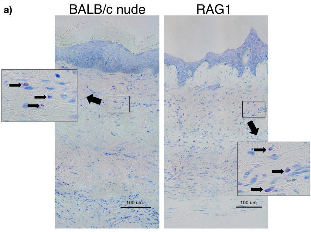
109 - 95 -
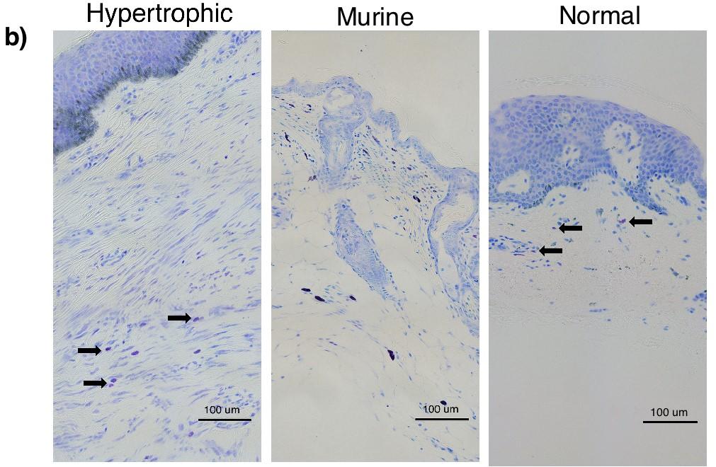
110 - 96 -
 taken at 30 days postoperatively demonstrate an increase in mast cell density for BALB/c-nu/nu nude, RAG-1 -/-, RAG-2 -/- c -/- and TCR -/- -/- -/- mice (a) in comparison")
111 Figure 2-4: Toluidine blue staining of scar tissues (xenografts) for mast cells and mast cell quantification in dermal fibrotic mouse models. Representative images (400x) taken at 30 days postoperatively demonstrate an increase in mast cell density for BALB/c-nu/nu nude, RAG-1 -/-, RAG-2 -/- c -/- and TCR -/- -/- -/- mice (a) in comparison to normal human split thickness skin (b). Cells appear lighter in comparison to mast cells in normal murine tissue and resemble mast cells in human HTS tissue (b). Quantification of mast cell density in 8 random, high-power fields under 400x magnification demonstrated an increase in mast cell number in the proliferative xenografts of all strains when compared to normal split thickness human skin (c). Significant differences in mast cell density were not observed within any strain of mice at the different time points *p
 of dermal fibrotic nude mice treated with a CXCR4 antagonist.")
112 Figure 2-5: Toluidine blue staining for mast cell quantification in scar tissues (xenografts) of dermal fibrotic nude mice treated with a CXCR4 antagonist
 demonstrate a significant decrease in mast cell density at 8 weeks in")
113 Representative images (400x) demonstrate a decrease in mast cell density in mice treated with a CXCR4 antagonist at 8 weeks in comparison to controls (a). Quantification of mast cell density in 8 random, high-power fields under 400x magnification in scar tissues (xenografts) demonstrate a significant decrease in mast cell density at 8 weeks in comparison to control mice and a significant increase in density at 8 weeks compared to 2 weeks in control mice (b). *p 0.05 Figure 2-6: Effect of mast cell conditioned media on fibroblast cell proliferation. Fibroblasts were cultured with 1 hour conditioned media from activated mast cells and an MTT assay was used to assess cell proliferation after 48 hours of incubation. Proliferation significantly increased for superficial and deep fibroblasts (SF and DF) following culture with mast cell conditioned media in comparison to being cultured alone. n=3 *p
114 Figure 2-7: Effect of mast cell conditioned media on -SMA and collagen gene expression. Fibroblasts were cultured with 1 hour conditioned media from substance P activated mast cells and RT-PCR was used to assess fibrotic gene expression. Messenger RNA expression of -SMA decreased in superficial and deep fibroblasts (SF and DF) and COL-1 only in SF following culture with mast cell conditioned media in comparison to being cultured alone. n=3 *p
115 Figure 2-8: Effect of mast cell conditioned media on protein expression of -SMA and type I collagen. Fibroblasts were cultured with conditioned media from substance P activated mast cells and flow cytometry and LC/MS were used to assess -SMA and 4-hydroxyproline protein expression. Mast cell conditioned media treatment decreased protein expression of -SMA (a) in superficial and deep fibroblasts (SF and DF) but no change was observed for collagen (b) in comparison to fibroblasts alone. DF had greater protein expression of -SMA and type 1 collagen in comparison to SF in the presence and absence of mast cell conditioned media. n=3 *p
116 2.8 Bibliography 1. Engrav LH, Garner WL, Tredget EE. Hypertrophic scar, wound contraction and hyperhypopigmentation. J Burn Care Res 2007; 28(4): p Kwan P, Hori K, Ding J, Tredget EE. Scar and contracture: Biological principles. Hand Clin 2009; 25(4): p Zhu Z, Ding J, Shankowsky HA, Tredget EE. The molecular mechanism of hypertrophic scar. J Cell Commun Signal 2013; 7(4): p Stella M, Castagnoli C, Gangemi EN. Postburn scars: An update. Int J Low Extrem Wounds 2008; 7(3): p Widgerow AD. Hypertrophic burn scar evolution and management. WHSA 2013; 6(2): p Wang J, Dodd C, Shankowsky HA, Scott PG, Tredget EE, Wound Healing Research Group. Deep dermal fibroblasts contribute to hypertrophic scarring. Lab Invest 2008; 88(12): p Dunkin CS, Pleat JM, Gillespie PH, Tyler MP, Roberts AH, McGrouther DA. Scarring occurs at a critical depth of skin injury: Precise measurement in a graduated dermal scratch in human volunteers. Plast Reconstr Surg 2007; 119(6): p Beer T, Baldwin H, West L, Gallagher P, Wright D. Mast cells in pathological and surgical scars. Br J Ophthalmol 1998; 82(6): p Juremalm M, Hjertson M, Olsson N, Harvima I, Nilsson K, Nilsson G. The chemokine receptor cxcr4 is expressed within the mast cell lineage and its ligand stromal cell
117 derived factor-1alpha acts as a mast cell chemotaxin. Eur J Immunol 2000; 30(12): p Abe R, Donnelly SC, Peng T, Bucala R, Metz CN. Peripheral blood fibrocytes: Differentiation pathway and migration to wound sites. J Immunol 2001; 166(12): p Foley TT, Ehrlich HP. Through gap junction communications, co-cultured mast cells and fibroblasts generate fibroblast activities allied with hypertrophic scarring. Plast Reconstr Surg 2013; 131(5): p Gailit J, Marchese MJ, Kew RR, Gruber BL. The differentiation and function of myofibroblasts is regulated by mast cell mediators. J Invest Dermatol 2001; 117(5): p Garbuzenko E, Nagler A, Pickholtz D, Gillery P, Reich R, Maquart FX, Levi-Schaffer F. Human mast cells stimulate fibroblast proliferation, collagen synthesis and lattice contraction: A direct role for mast cells in skin fibrosis. Clin Exp Allergy 2002; 32(2): p Hildebrand A, Romaris M, Rasmussen LM, Heinegard D, Twardzik DR, Border WA, Ruoslahti E. Interaction of the small interstitial proteoglycans biglycan, decorin and fibromodulin with transforming growth factor beta. Biochem J 1994; 302(Pt 2): p Wang J, Ding J, Jiao H, Honardoust D, Momtazi M, Shankowsky HA, Tredget EE. Human hypertrophic scar-like nude mouse model: Characterization of the molecular and cellular biology of the scar process. Wound Rep Regen 2011; 19(2): p
118 16. Honardoust D., Kwan P., Momtazi M., Ding J., Tredget EE. Novel methods for the investigation of human hypertrophic scarring and other dermal fibrosis. Methods Mol Biol 2013; 1037: p Ding J, Ma Z, Liu H, Kwan P, Iwashina T, Shankowsky HA, Wong D, Tredget EE. The therapeutic potential of a c-x-c chemokine receptor type 4 (cxcr-4) antagonist on hypertrophic scarring in vivo. Wound Rep Regen 2014; 22(5): p Kirshenbaum AS., Akin C., Wu Y., Rottem M., Goff JP., Beaven MA., Rao VK., Metcalfe DD. Characterization of novel stem cell factor responsive human mast cell lines lad 1 and 2 established from a patient with mast cell sarcoma/leukemia; activation following aggregation of fcîµri or fcî³ri. Leuk Res 2003; 27(8): p Tredget EE, Shen YJ, Liu G, Forsyth N, Smith C, Robertson HA, Scott PG, Ghahary A. Regulation of collagen synthesis and messenger rna levels in normal and hypertrophic scar fibroblasts in vitro by interferon alfa-2b. Wound Rep Regen 1993; 1(3): p Shoulders MD, Raines RT. Collagen structure and stability. Ann Rev Biochem 2009; 78: p Marshall JS, Ford GP, Bell EB. Formalin sensitivity and differential staining of mast cells in human dermis. Br J Dermatol 1987; 117(1): p Momtazi M, Ding J, Kwan P, Anderson CC, Honardoust D, Goekjian S, Tredget EE. Morphologic and histologic comparison of hypertrophic scar in nude mice, t-cell receptor, and recombination activating gene knockout mice. Plast Reconstr Surg 2015; 136(6): p Dahlin JS, Hallgren J. Mast cell progenitors: Origin, development and migration to tissues. Mol Immunol 2015; 63(1): p
119 24. Ding J, Ma Z, Shankowsky HA, Medina A, Tredget EE. Deep dermal fibroblast profibrotic characteristics are enhanced by bone marrow-derived mesenchymal stem cells. Wound Rep Regen 2013; 21(3): p Kulka M, Sheen CH, Tancowny BP, Grammer LC, Schleimer RP. Neuropeptides activate human mast cell degranulation and chemokine production. Immunol 2008; 123(3): p de Almeida A., Mustin D., Forman MF., Brower GL., Janicki JS., Carver W. Effects of mast cells on the behavior of isolated heart fibroblasts: Modulation of collagen remodeling and gene expression. J Cell Physiol 2002; 191(1): p Moyer KE, Saggers GC, Ehrlich HP. Mast cells promote fibroblast populated collagen lattice contraction through gap junction intercellular communication. Wound Rep Regen 2004; 12(3): p Kischer CW, Bunce H 3rd, Shetlah MR. Mast cell analyses in hypertrophic scars, hypertrophic scars treated with pressure and mature scars. J Invest Dermatol 1978; 70(6): p Abe M, Kurosawa M, Ishikawa O, Miyachi Y, Kido H. Mast cell tryptase stimulates both human dermal fibroblast proliferation and type i collagen production. Clin Exp Allergy 1998; 28(12): p Description and general use: Merck Index, 11th ed., p. 1398, # Profyris C, Tziotzios C, Do Vale I. Cutaneous scarring: Pathophysiology, molecular mechanisms, and scar reduction therapeutics part i. The molecular basis of scar formation. J Am Acad Dermatol 2012; 66(1): p
120 32. Linenberger ML., Jacobson FW., Bennett LG., Broudy VC., Martin FH., Abkowitz JL. Stem cell factor production by human marrow stromal fibroblasts. Exp Hematol 1995; 23(10): p Butterfield JH, Weiler D, Dewald G, Gleich GJ. Establishment of an immature mast cel lline from a patient with mast cell leukemia. Leuk Res 1988; 12(4): p Deshmane SL, Kremlev S, Amini S, Sawaya BE. Monocyte chemoattractant protein-1 (mcp-1): An overview. J Interferon Cytokine Res 2009; 29(6): p Fang Y, Gong SJ, Xu YH, Hambly BD, Bao S. Impaired cutaneous wound healing in granulocyte/macrophage colony-stimulating factor knockout mice. Br J Dermatol 2007; 157(3): p Fang Y, Shen J, Yao M, Beagley KW, Hambly BD, Bao S. Granulocyte-macrophage colony-stimulating factor enhances wound healing in diabetes via upregulation of proinflammatory cytokines. Br J Dermatol 2010; 162(3): p Abreu MT. Toll-like receptor signalling in the intestinal epithelium: How bacterial recognition shapes intestinal function. Nat Rev Immunol 2010; 10(2): p Postlethwaite AE, Raghow R, Stricklin GP, Poppleton H, Seyer JM, Kang AH. Modulation of fibroblast functions by interleukin 1: Increased steady-state accumulation of type i procollagen messenger rnas and stimulation of other functions but not chemotaxis by human recombinant interleukin 1 alpha and beta. J Cell Biol 1988; 106(2): p Ghahary A, Shen YJ, Nedelec B, Scott PG, Tredget EE. Interferons gamma and alpha-2b differentially regulate the expression of collagenase and tissue inhibitor of
121 metalloproteinase-1 messenger rna in human hypertrophic and normal dermal fibroblasts. Wound Rep Regen 1995; 3(2): p Keane MP, Belperio JA, Burdick MD, Strieter RM. Il-12 attenuates bleomycin-induced pulmonary fibrosis. Am J Physiol Lung Cell Mol Physiol 2001; 281(1): p. L Tredget EE, R Wang, Shen Q, Scott RG, Ghahary A. Transforming growth factor-beta mrna and protein in hypertrophic scar tissues and fibroblasts: Antagonism by ifn-alpha and ifn-gamma in vitro and in vivo. J Interferon Cytokine Res 2000; 20(2): p Matias MAT, Saunus JM, Ivanovski S, Walsh LJ, Farah CS. Accelerated wound healing phenotype in interleukin 12/23 deficient mice. J Inflamm (Lond) 2011; 8: p Trautmann A, Krohne G, Brocker EB, Klein CE. Human mast cells augment fibroblast proliferation by heterotypic cell-cell adhesion and action of il-4. J Immunol 1998; 160(10): p Hildebrand KA, Zhang M, Befus AD, Salo PT, Hart DA. A myofibroblast-mast cellneuropeptide axis of fibrosis in post-traumatic joint contractures: An in vitro analysis of mechanistic components. J Orthop Res 2014; 32(10): p
122 Chapter 3. Conclusions and Future Directions Our results have indicated that mast cells are involved in hypertrophic scarring in our in vivo study but their effects on fibroblasts require further study in vitro. As our sample size was very small, further exploration should be conducted to clarify the effects of mast cells on fibroblasts and determine if the trends observed within our data are an actual phenomenon. If replication does not yield significant differences, it may suggest cell-to-cell contact is the primary method of communication between mast cells and fibroblasts yielding fibrotic changes in fibroblast activity. Conversely, should these trends be found significant within a larger sample size, it would be important to discern whether these differences are due to methodology discrepancies or are another regulatory effect of mast cells on fibroblasts. Our experiments did not involve direct cell-to-cell contact whereas previous literature did [1-5]. It is possible that direct cellular contact is necessary for profibrotic results to be observed, which may explain the absence of change in our experiments. Additional replications of conditioned media experiments should be conducted to determine if the trends observed are valid in a larger sample size, alongside cultures implementing direct cell-to-cell contact to determine if the profibrotic effects reported in previous literature can also be replicated. Experiments should also be performed using primary human mast cells. As LAD2s are a moderately differentiated mast cell line, they possess a number of differences in comparison to primary human mast cells in addition to not being fully mature. Previous literature has documented that LAD2 cells produce smaller amounts of mediators, in particular tryptase and chymase, which may explain differences in our results [6, 7]. Other aspects of mast cell activation on fibroblasts that could be investigated include assessment of mast cell media following our preliminary analysis of conditioned mast cell media,
123 and assessment of the necessity for mast cell activation in culture. Comparisons could be made between conditioned media from LAD2 cells and primary human mast cells allowing us to distinguish differences in cytokine, chemokine and growth factor levels and interpret which mediators may be primarily responsible for any observed trends in vitro. Analysis of mast cell activation in conditioned media and cell-to-cell contact culture systems will also elucidate the importance of mast cell activation on fibroblast regulation. Additionally, external activators such as substance P and production of SCF by fibroblasts could be compared in culture to determine if an external activator is even necessary in direct cell-to-cell cultures for mast cell activation. Collectively, our research in conjunction with these future experiments will further define the effects that mast cells have on fibroblasts and potentially identify factors which may lead to the development of therapeutic strategies for the treatment and prevention of HTS development and other fibroproliferative disorders. 3.1 Bibliography 1. Wang J, Ding J, Jiao H, Honardoust D, Momtazi M, Shankowsky HA, Tredget EE. Human hypertrophic scar-like nude mouse model: Characterization of the molecular and cellular biology of the scar process. Wound Rep Regen 2011; 19(2): p Foley TT, Ehrlich HP. Through gap junction communications, co-cultured mast cells and fibroblasts generate fibroblast activities allied with hypertrophic scarring. Plast Reconstr Surg 2013; 131(5): p
124 3. Moyer KE, Saggers GC, Ehrlich HP. Mast cells promote fibroblast populated collagen lattice contraction through gap junction intercellular communication. Wound Rep Regen 2004; 12(3): p Kischer CW, Bunce H 3rd, Shetlah MR. Mast cell analyses in hypertrophic scars, hypertrophic scars treated with pressure and mature scars. J Invest Dermatol 1978; 70(6): p Abe M, Kurosawa M, Ishikawa O, Miyachi Y, Kido H. Mast cell tryptase stimulates both human dermal fibroblast proliferation and type i collagen production. Clin Exp Allergy 1998; 28(12): p Guhl S, Babina M, Neou A, Zuberbier T, Artuc M. Mast cell lines hmc-1 and lad2 in comparison with mature human skin mast cells--drastically reduced levels of tryptase and chymase in mast cell lines. Exp Dermatol 2010; 19(9): p Kulka M, Sheen CH, Tancowny BP, Grammer LC, Schleimer RP. Neuropeptides activate human mast cell degranulation and chemokine production. Immunol 2008; 123(3): p
125 Bibliography Description and general use: Merck Index, 11th ed., p. 1398, #8834. Aarabi S, Bhatt KA, Shi Y, Paterno J, Chang EI, Loh SA, Holmes JW, Longaker MT, Yee H and Gurtner GC (2007). Mechanical load initiates hypertrophic scar formation through decreased cellular apoptosis. FASEB J 21(12): Aarabi S, Longaker MT and Gurtner GC (2007). Hypertrophic scar formation following burns and trauma: new approaches to treatment. PLoS Med 4(9): e234. Abe M, Kurosawa M, Ishikawa O, Miyachi Y and Kido H (1998). Mast cell tryptase stimulates both human dermal fibroblast proliferation and type I collagen production. Clin Exp Allergy 28(12): Abe R, Donnelly SC, Peng T, R. Bucala and Metz CN (2001). Peripheral blood fibrocytes: differentiation pathway and migration to wound sites. J Immunol 166(12): Abreu MT (2010). Toll-like receptor signalling in the intestinal epithelium: how bacterial recognition shapes intestinal function. Nat Rev Immunol 10(2): Agren MS, Taplin CJ, Woessner JF Jr, Eaglstein WH and Mertz PM (1992). Collagenase in wound healing: effect of wound age and type. J Invest Dermatol 99(6): Am, S., Francis CS, Motamed A, Karatsonyi AL, Hammoudeh JA, Sanchez-Lara PA, Reinisch JF and Urata MM (2012). Hypertrophic scarring in cleft lip repair: a comparison of incidence among ethnic groups. Clin Epidemiol 4: Armour A, Scott PG and Tredget EE (2007). Cellular and molecular pathology of HTS: basis for treatment. Wound Rep Regen Suppl 1: S
126 Artuc M, Hermes B, Steckelings UM, Grutzkau A and Henz BM (1999). Mast cells and their mediators in cutaneous wound healing--active participants or innocent bystanders? Exp Dermatol 8(1): Bangert C, Brunner P and Stingl G (2011). Immune Functions of the Skin. Clin Dermatol 29(4): Bao P, Kodra A, Tomic-Canic M, Golinko MS, Ehrlich HP and Brem H (2008). The Role of Vascular Endothelial Growth Factor in Wound Healing. J Surg Res 153(2): Barrientos S, Stojadinovic O, Golinko MS, Brem H and Tomic-Canic M (2008). Growth factors and cytokines in wound healing. Wound Rep Regen 16(5): Baum CL and Arpey CJ (2005). Normal cutaneous wound healing: clinical correlation with cellular and molecular events. Dermatol Surg 31(6): Beer T, Baldwin H, West L, Gallagher P and Wright D (1998). Mast cells in pathological and surgical scars. Br J Ophthalmol 82(6): Bellemare J, Roberge CJ, Bergeron D, Lopez-Valle CA, Roy M and Moulin VJ (2005). Epidermis promotes dermal fibrosis: role in the pathogenesis of hypertrophic scars. J Pathol 206(1): 1-8. Blakaj A and Bucala R (2012). Fibrocytes in health and disease. Fibrogenesis Tissue Repair 5(Suppl 1): S6. Bleul CC, Fuhlbrigge RC, Casasnovas JM, Aiuti A and Springer TA (1996). A highly efficacious lymphocyte chemoattractant, stromal cell-derived factor 1 (SDF-1). J Exp Med 184(1):
127 Bock O, Yu H, Zitron S, Bayat A, Ferguson MW and Mrowietz U (2005). Studies of transforming growth factors beta 1-3 and their receptors I and II in fibroblast of keloids and hypertrophic scars. Acta Derm Venereol 85(3): Bressler RS and Bressler CH (1989). Functional anatomy of the skin. Clin Podiatr Med Surg 6(2): Broughton G, Janis JE and Attinger CE (2006). The basic science of wound healing. Plast Reconstr Surg 117(7 Suppl): 12S-34S. Brubaker AL, Schneider DF and Kovacs EJ (2011). Neutrophils and natural killer T cells as negative regulators of wound healing. Expert Rev Dermatol 6(1): 5-8. Bucala, R., L. Spiegel, J. Chesney, M. Hogan and A. Cerami (1994). Circulating fibrocytes define a new leukocyte subpopulation that mediates tissue repair. Mol Med 1(71-81.). Butterfield JH, Weiler D, Dewald G and Gleich GJ (1988). Establishment of an immature mast cel lline from a patient with mast cell leukemia. Leuk Res 12(4): Caughey GH (2007). Mast cell tryptases and chymases in inflammation and host defense. Immunol Rev 217: Chakraborti S, Mandal M, Das S, Mandal A and Chakraborti T (2003). Regulation of matrix metalloproteinases: an overview. Mol Cell Biochem 253(1-2): Choi, W. and An J (2011). Biology and clinical relevance of chemokines and chemokine receptors CXCR4 and CCR5 in human diseases. Exp Biol Med 236(6): Chun Q, Zhiyong W, Fei S and Xiqiao W (2014). Dynamic biological changes in fibroblasts during hypertrophic scar formation and regression. Int Wound J 13(2): Clark RA, Ghosh K and Tonnesen MG (2007). Tissue engineering for cutaneous wounds. J Invest Dermatol 127(5):
128 Collington SJ, Williams TJ and Weller CL (2011). Mechanisms underlying the localisation of mast cells in tissues. Trends Immunol 32(10): Cooper MA, Fehniger TA and Caligiuri MA (2001). The biology of human natural killer-cell subsets. Trends Immunol 22(11): da Silva EZ, Jamur MC and Oliver C (2014). Mast Cell Function: A New Vision of an Old Cell. J Histochem Cytochem 62(10): Dahlin JS and Hallgren J (2015). Mast cell progenitors: Origin, development and migration to tissues. Mol Immunol 63(1): Dai H and Korthuis RJ (2011). Mast Cell Proteases and Inflammation. Drug Discov Today Dis Models 8(1): de Almeida, A., D. Mustin, M. Forman, G. Brower, J. Janicki and W. Carver (2002). Effects of mast cells on the behavior of isolated heart fibroblasts: modulation of collagen remodeling and gene expression. J Cell Physiol 191(1): Deshmane SL, Kremlev S, Amini S and Sawaya BE (2009). Monocyte Chemoattractant Protein-1 (MCP-1): An Overview. J Interferon Cytokine Res 29(6): Desmoulière A, Chaponnier C and Gabbiani G (2005). Tissue repair, contraction, and the myofibroblast. Wound Repair Regen 13(1): Diegelmann RF and Evans MC (2004). Wound healing: an overview of acute, fibrotic and delayed healing. Front Biosci 9: Ding J, Hori K, Zhang R, Marcoux Y, Honardoust D, Shankowsky HA and Tredget EE (2011). Stromal cell-derived factor 1 (SDF-1) and its receptor CXCR4 in the formation of postburn hypertrophic scar (HTS). Wound Rep Regen 19(5):
129 Ding J, Ma Z, Liu H, Kwan P, Iwashina T, Shankowsky HA, Wong D and Tredget EE (2014). The therapeutic potential of a C-X-C chemokine receptor type 4 (CXCR-4) antagonist on hypertrophic scarring in vivo. Wound Rep Regen 22(5): Ding J, Ma Z, Shankowsky HA, Medina A and Tredget EE (2013). Deep dermal fibroblast profibrotic characteristics are enhanced by bone marrow-derived mesenchymal stem cells. Wound Rep Regen 21(3): Ding, J. and E. Tredget The cellular and molecular basis of fibroproliferative scarring following thermal injury. (Chapter) (2016) The Scar Book: Formation, Rehabilitation, Management, and Prevention (accepted). Douaiher J, Succar J, Lancerotto L, Gurish MF, Orgill DP, Hamilton MJ, Krilis SA and Stevens RL (2014). Development of Mast Cells and Importance of Their Tryptase and Chymase Serine Proteases in Inflammation and Wound Healing. Adv in Immunol. W. A. Frederick, Academic Press. Volume 122: Dovi JV, Szpaderska AM and DiPietro LA (2004). Neutrophil function in the healing wound: adding insult to injury? Thromb Haemost 92(2): Dunkin CS, Pleat JM, Gillespie PH, Tyler MP, Roberts AH and McGrouther DA (2007). Scarring occurs at a critical depth of skin injury: precise measurement in a graduated dermal scratch in human volunteers. Plast Reconstr Surg 119(6): Elias, P. (2008). Skin Barrier Function. Curr Allergy Astham Rep 8(4): Eming SA, Krieg T and Davidson JM (2007). Inflammation in wound repair: molecular and cellular mechanisms. J Invest Dermatol 127(3): Engrav LH, Garner WL and Tredget EE (2007). Hypertrophic scar, wound contraction and hyper-hypopigmentation. J Burn Care Res 28(4):
130 Fang Y, Gong SJ, Xu YH, Hambly BD and Bao S (2007). Impaired cutaneous wound healing in granulocyte/macrophage colony-stimulating factor knockout mice. Br J Dermatol 157(3): Fang Y, Shen J, Yao M, Beagley KW, Hambly BD and Bao S (2010). Granulocytemacrophage colony-stimulating factor enhances wound healing in diabetes via upregulation of proinflammatory cytokines. Br J Dermatol 162(3): Farag SS and Caligiuri MA (2006). Human natural killer cell development and biology. Blood Rev 20(3): Florin L, Maas-Szabowski N, Werner S, Szabowski A and Angel P (2005). Increased keratinocyte proliferation by JUN-dependent expression of PTN and SDF-1 in fibroblasts. J Cell Sci 118(Pt 9): Foley TT and Ehrlich HP (2013). Through gap junction communications, co-cultured mast cells and fibroblasts generate fibroblast activities allied with hypertrophic scarring. Plast Reconstr Surg 131(5): Frank S, Hubner G, Breier G, Longaker MT, Greenhalgh DG and Werner S (1995). Regulation of vascular endothelial growth factor expression in cultured keratinocytes. Implications for normal and impaired wound healing. J Biol Chem 270(21): Fusiak T, S. GC and Condos R (2015). Pulmonary Fibrosis Treated with Inhaled Interferongamma (IFN-gamma). J Aerosol Med Pulm Drug Deliv 28(5): Gabbiani G (2003). The myofibroblast in wound healing and fibrocontractive diseases. J Pathol 200(4): Gailit J, Marchese MJ, Kew RR and Gruber BL (2001). The differentiation and function of myofibroblasts is regulated by mast cell mediators. J Invest Dermatol 117(5):
131 Galli SJ, Borregaard N and Wynn TA (2011). Phenotypic and functional plasticity of cells of innate immunity: macrophages, mast cells and neutrophils. Nat Immunol 12(11): Galli SJ, Nakae S and Tsai M (2005). Mast cells in the development of adaptive immune responses. Nat Immunol 6(2): Gallucci RM, Simeonova PP, Matheson JM, Kommineni C, Guriel JL, Sugawara T and Luster MI (2000). Impaired cutaneous wound healing in interleukin-6-deficient and immunosuppressed mice. FASEB J 14(15): Galy A, Travis M, Cen D and Chen B (1995). Human T, B, natural killer, and dendritic cells arise from a common bone marrow progenitor cell subset. Immunity 3(4): Garbuzenko E, Nagler A, Pickholtz D, Gillery P, Reich R, Maquart FX and Levi-Schaffer F (2002). Human mast cells stimulate fibroblast proliferation, collagen synthesis and lattice contraction: a direct role for mast cells in skin fibrosis. Clin Exp Allergy 32(2): Garner WL, Karmiol S, Rodriguez JL, Smith DJ Jr and Phan SH (1993). Phenotypic differences in cytokine responsiveness of hypertrophic scar versus normal dermal fibroblasts. J Invest Dermatol 101(6): Gartner MH, Benson JD and Caldwell MD (1992). Insulin-like growth factors I and II expression in the healing wound. J Surg Res 52(4): Gauglitz GG, Korting HC, Pavicic T, Ruzicka T and Jeschke MG (2011). Hypertrophic scarring and keloids: pathomechanisms and current and emerging treatment strategies. Mol Med 17(1-2):
132 Geissmann F, Jung S and Littman DR (2003). Blood monocytes consist of two principal subsets with distinct migratory properties. Immunity 19(1): Ghahary A, Shen YJ, Nedelec B, Scott PG and Tredget EE (1995). Interferons gamma and alpha-2b differentially regulate the expression of collagenase and tissue inhibitor of metalloproteinase-1 messenger RNA in human hypertrophic and normal dermal fibroblasts. Wound Rep Regen 3(2): Ghahary A, Shen YJ, B. Nedelec, Scott PG and Tredget EE (1995). Enhanced expression of mrna for insulin-like growth factor-1 in post-burn hypertrophic scar tissue and its fibrogenic role by dermal fibroblasts. Mol Cell Biochem 148(1): Ghahary A, Shen YJ, Wang R, Scott PG and Tredget EE (1998). Expression and localization of insulin-like growth factor-1 in normal and post-burn hypertrophic scar tissue in human. Mol Cell Biochem 183(1-2): 1-9. Gill SE and Parks WC (2008). Metalloproteinases and their inhibitors: regulators of wound healing. Int J Biochem Cell Biol 40(6-7): Gillitzer R and Goebeler M (2001). Chemokines in cutaneous wound healing. J Leukoc Biol 69(4): Ginhoux F and Jung S (2014). Monocytes and macrophages: developmental pathways and tissue homeostasis. Nat Rev Immunol 14(6): Grice EA and S. JA (2011). The skin microbiome. Nat Rev Microbiol 9(4): Grotendorst GR, Rahmanie H and Duncan MR (2004). Combinatorial signaling pathways determine fibroblast proliferation and myofibroblast differentiation. FASEB J 18(3):
133 Guhl S, Babina M, Neou A, Zuberbier T and Artuc M (2010). Mast cell lines HMC-1 and LAD2 in comparison with mature human skin mast cells--drastically reduced levels of tryptase and chymase in mast cell lines. Exp Dermatol 19(9): Hardwicke J, Schmaljohann D, Boyce D and Thomas D (2008). Epidermal growth factor therapy and wound healing--past, present and future perspectives. Surgeon 6(3): Higashi K, Y. Inagaki, Fujimori K, Nakao A, Kaneko H and Nakatsuka I (2003). Interferongamma interferes with transforming growth factor-beta signaling through direct interaction of YB-1 with Smad3. J Biol Chem 278(44): Hilbert DM, Holmes KL, Anderson AO and Rudikoff S (1993). Long-term thymic reconstitution by peripheral CD4 and CD8 single-positive lymphocytes. Eur J Immunol 23(10): Hildebrand A, Romaris M, Rasmussen LM, Heinegard D, Twardzik DR, Border WA and Ruoslahti E (1994). Interaction of the small interstitial proteoglycans biglycan, decorin and fibromodulin with transforming growth factor beta. Biochem J 302(Pt 2): Hildebrand KA, Zhang M, Befus AD, Salo PT and Hart DA (2014). A myofibroblast-mast cellneuropeptide axis of fibrosis in post-traumatic joint contractures: an in vitro analysis of mechanistic components. J Orthop Res 32(10): Hinz B, Phan SH, Thannickal VJ, Galli A, Bochaton-Piallat ML and Gabbiani G (2007). The myofibroblast: one function, multiple origins. Am J Pathol 170(6): Honardoust, D., P. Kwan, M. Momtazi, J. Ding and E. Tredget (2013). Novel methods for the investigation of human hypertrophic scarring and other dermal fibrosis. Methods Mol Biol 1037:
134 Hu C, Yong X, C. Li, Liu D, Chen L, Hu J, Teng M, Zhang D, Fan Y and Liang G (2013). CXCL12/CXCR4 axis promotes mesenchymal stem cell mobilization to burn wounds and contributes to wound repair. J Surg Res 183(1): Hu, G., P. Liu, J. Feng and Y. Jin (2010). A novel population of mesenchymal progenitors with hematopoietic potential originated from CD14 peripheral blood mononuclear cells. Int J Med Sci 8(1): Hu, G., J. Xu, Z. Deng, J. Feng and Y. Jin (2011). Supernatant of bone marrow mesenchymal stromal cells induces peripheral blood mononuclear cells possessing mesenchymal features. Int J Biol Sci 7(3): Iocono JA, Colleran KR, Remick DG, Gillespie BW, Ehrlich HP and Garner WL (2000). Interleukin-8 levels and activity in delayed-healing human thermal wounds. Wound Rep Regen 8(3): Irani AA, Schechter NM, Craig SS, DeBlois G and Schwartz LB (1986). Two types of human mast cells that have distinct neutral protease compositions. Proc Natl Acad Sci U S A 83(12): Ishida Y, Gao JL and Murphy PM (2008). Chemokine receptor CX3CR1 mediates skin wound healing by promoting macrophage and fibroblast accumulation and function. J Immunol 180(1): Janson DG, Saintigny G, van Adrichem A, Mahé C and El Ghalbzouri A (2012). Different gene expression patterns in human papillary and reticular fibroblasts. J Invest Dermatol 132(11): Juremalm M, Hjertson M, Olsson N, Harvima I, Nilsson K and Nilsson G (2000). The chemokine receptor CXCR4 is expressed within the mast cell lineage and its ligand
135 stromal cell-derived factor-1alpha acts as a mast cell chemotaxin. Eur J Immunol 30(12): Kalra S, Utz JP and Ryu JH (2003). Interferon gamma-1b therapy for advanced idiopathic pulmonary fibrosis. Mayo Clin Proc 78(9): Keane MP, Belperio JA, Burdick MD and Strieter RM (2001). IL-12 attenuates bleomycininduced pulmonary fibrosis. Am J Physiol Lung Cell Mol Physiol 281(1): L92-7. Kennedy JD, Pierce CW and Lake JP (1992). Extrathymic T cell maturation. Phenotypic analysis of T cell subsets in nude mice as a function of age. J Immunol 148(6): Kirshenbaum, A., C. Akin, Y. Wu, M. Rottem, J. Goff, M. Beaven, V. Rao and D. Metcalfe (2003). Characterization of novel stem cell factor responsive human mast cell lines LAD 1 and 2 established from a patient with mast cell sarcoma/leukemia; activation following aggregation of FcεRI or FcγRI. Leuk Res 27(8): Kischer CW, Bunce H 3rd and Shetlah MR (1978). Mast cell analyses in hypertrophic scars, hypertrophic scars treated with pressure and mature scars. J Invest Dermatol 70(6): Kitzis V, Engrav LH and Quinn LS (1999). Transient exposure to tumor necrosis factor-alpha inhibits collagen accumulation by cultured hypertrophic scar fibroblasts. J Surg Res 87(1): Kovacs EJ and DiPietro LA (1994). Fibrogenic cytokines and connective tissue production. FASEB J 8(11): Krotzsch-Gomez FE, Furuzawa-Carballeda J, Reyes-Marquez R, Quiroz-Hernandez E and Diaz de Leon L (1998). Cytokine expression is downregulated by collagenpolyvinylpyrrolidone in hypertrophic scars. J Invest Dermatol 111(5):
136 Kulka M, Sheen CH, Tancowny BP, Grammer LC and Schleimer RP (2008). Neuropeptides activate human mast cell degranulation and chemokine production. Immunol 123(3): Kupietzky A and Levi-Schaffer F (1996). The role of mast cell-derived histamine in the closure of an in vitro wound. Inflamm Res 45(4): Kwan P, Hori K, Ding J and Tredget EE (2009). Scar and contracture: biological principles. Hand Clin 25(4): Laato M, Heino J, Gerdin B, Kahari VM and Niinikoski J (2001). Interferon-gamma-induced inhibition of wound healing in vivo and in vitro. Ann Chir Gynaecol 90: Ladak A and Tredget EE (2009). Pathophysiology and management of the burn scar. Clin Plast Surg 36(4): Lambers H, Piessens S, Bloem A, Pronk H and Finkel P (2006). Natural skin surface ph is on average below 5, which is beneficial for its resident flora. Int J Cosmet Sci 28(5): Larrabee WF, East CA, Jaffe HS, Stephenson C and Peterson KE (1990). Intralesional interferon gamma treatment for keloids and hypertrophic scars. Arch Otolaryngol Head Neck Surg 116(10): Lawrence JW, Mason ST, Schomer K and Klein M (2012). Epidemiology and impact of scarring after burn injury: a systematic review of the literature. J Burn Care Res 33(1): Linenberger, M., F. Jacobson, L. Bennett, V. Broudy, F. Martin and J. Abkowitz (1995). Stem cell factor production by human marrow stromal fibroblasts. Exp Hematol 23(10):
137 Lorentz A, Baumann A, Vitte J and Blank U (2012). The SNARE Machinery in Mast Cell Secretion. Front Immunol 3: 143. Mahdavian DB, van der Veer WM, van Egmond M, Niessen FB and Beelen RH (2011). Macrophages in skin injury and repair. Immunobiology 216(7): Mantovani A, Sozzani S, Locati M, Allavena P and Sica A (2002). Macrophage polarization: tumor-associated macrophages as a paradigm for polarized M2 mononuclear phagocytes. Trends Immunol 23(11): Marshall JS, Ford GP and Bell EB (1987). Formalin sensitivity and differential staining of mast cells in human dermis. Br J Dermatol 117(1): Martin, P. (1997). Wound healing--aiming for perfect skin regeneration. Science 276(5309): Matias MAT, Saunus JM, Ivanovski S, Walsh LJ and Farah CS (2011). Accelerated wound healing phenotype in Interleukin 12/23 deficient mice. J Inflamm (Lond) 8: 39. Medina A and Ghahary A (2010). Fibrocytes can be reprogrammed to promote tissue remodeling capacity of dermal fibroblasts. Mol Cell Biochem 344(1-2): Metcalfe DD, Baram D and Mekori YA (1997). Mast cel. Physiol Rev 77(4): Mombaerts P, Clarke AR, Rudnicki MA, Iacomini J, Itohara S, Lafaille JJ, Wang L, Ichikawa Y, Jaenisch R and Hooper ML (1992). Mutations in T-cell antigen receptor genes alpha and beta block thymocyte development at different stages. Nature 360(6401): Momtazi M, Ding J, Kwan P, Anderson CC, Honardoust D, Goekjian S and Tredget EE (2015). Morphologic and Histologic Comparison of Hypertrophic Scar in Nude Mice, T-Cell Receptor, and Recombination Activating Gene Knockout Mice. Plast Reconstr Surg 136(6):
138 Momtazi M, Kwan P, Ding J, Anderson CC, Honardoust D, Goekjian S and Tredget EE (2013). A nude mouse model of hypertrophic scar shows morphologic and histologic characteristics of human hypertrophic scar. Wound Rep Regen 21(1): Moore KL, Agur AMR and Dalley AF (2015). Essential Clinical Anatomy, Wolters Kluwer. Moore KW, de Waal Malefyt R, Coffman RL and O'Garra A (2001). Interleukin-10 and the interleukin-10 receptor. Annu Rev Immunol 19: Mori L, Bellini A, Stacey MA, Schmidt M and Mattoli S (2005). Fibrocytes contribute to the myofibroblast population in wounded skin and originate from the bone marrow. Exp Cell Res 304(1): Moulin V, Larochelle S, Langlois C, Thibault I, Lopez-Valle CA and Roy M (2004). Normal skin wound and hypertrophic scar myofibroblasts have differential responses to apoptotic inductors. J Cell Physiol 198(3): Moyer KE, Saggers GC and Ehrlich HP (2004). Mast cells promote fibroblast populated collagen lattice contraction through gap junction intercellular communication. Wound Rep Regen 12(3): Murray PJ and Wynn TA (2011). Protective and pathogenic functions of macrophage subsets. Nat Rev Immunol 11(11): Ng MF (2010). The role of mast cells in wound healing. Int Wound J 7(1): Niessen FB, Andriessen MP, Schalkwijk J, Visser L and Timens W (2001). Keratinocytederived growth factors play a role in the formation of hypertrophic scars. J Pathol 194(2): Noli C and Miolo A (2001). The mast cell in wound healing. Vet Dermatol 12(6):
139 Nurden AT, Nurden P, Sanchez M, Andia I and Anitua E (2008). Platelets and wound healing. Front Biosci 13: O, K., Tandara A and Mustoe TA (2007). Hypertrophic scar model in the rabbit ear: a reproducible model for studying scar tissue behavior with new observations on silicone gel sheeting for scar reduction. Wound Rep Regen Suppl 1: S40-5. Olsson N, Piek E, ten Dijke P and Nilsson G (2000). Human mast cell migration in response to members of the transforming growth factor-beta family. J Leukoc Biol 67(3): Ortega, S., Ittmann M, Tsang SH, Ehrlich M and Basilico C (1998). Neuronal defects and delayed wound healing in mice lacking fibroblast growth factor 2. Proc Natl Acad Sci U S A 95(10): Oskeritzian CA (2012). Mast Cells and Wound Healing. Adv Wound Care 1(1): Oviedo-Orta E and Howard Evans W (2004). Gap junctions and connexin-mediated communication in the immune system. Biochim Biophys Acta 1662(1 2): Pappas A (2009). Epidermal surface lipids. Dermatoendocrinol 1(2): Pilling D, Fan T, Huang D, Kaul B and Gomer RH (2009). Identification of markers that distinguish monocyte-derived fibrocytes from monocytes, macrophages, and fibroblasts. PLoS ONE 4(10): e7475. Pistorio AL and Ehrlich HP (2011). Modulatory Effects of Connexin-43 Expression on Gap Junction Intercellular Communications with Mast Cells and Fibroblasts. J Cell Biochem 112(5): Postlethwaite AE, Raghow R, Stricklin GP, Poppleton H, Seyer JM and Kang AH (1988). Modulation of fibroblast functions by interleukin 1: increased steady-state accumulation of type I procollagen messenger RNAs and stimulation of other functions
140 but not chemotaxis by human recombinant interleukin 1 alpha and beta. J Cell Biol 106(2): Profyris C, Tziotzios C and Do Vale I (2012). Cutaneous scarring: Pathophysiology, molecular mechanisms, and scar reduction therapeutics Part I. The molecular basis of scar formation. J Am Acad Dermatol 66(1): Quan TE, Cowper SE and Bucala R (2006). The role of circulating fibrocytes in fibrosis. Curr Rheumatol Rep 8(2): Ramos ML, Gragnani A and Ferreira LM (2008). Is there an ideal animal model to study hypertrophic scarring? J Burn Care Res 29(2): Rao KN and Brown MA (2088). Mast cells: multifaceted immune cells with diverse roles in health and disease. Ann N Y Acad Sci 1143: Redente E, Keith R, Janssen W, Henson P, Ortiz L, Downey GP, Bratton DL and Riches DW (2014). Tumor Necrosis Factor-α Accelerates the Resolution of Established Pulmonary Fibrosis in Mice by Targeting Profibrotic Lung Macrophage. Am J Respir Cell Mol Biol 50(4): Reid RR, Mogford JE, Butt R, degiorgio-miller A and Mustoe TA (2006). Inhibition of procollagen C-proteinase reduces scar hypertrophy in a rabbit model of cutaneous scarring. Wound Rep Regen 14(2): Reynolds AJ and Jahoda CA (1991). Inductive properties of hair follicle cells. Ann N Y Acad Sci 642: Rhett JM, Ghatnekar GS, Palatinus JA, O'Quinn M, Yost MJ and Gourdie RG (2008). Novel therapies for scar reduction and regenerative healing of skin wounds. Trends Biotechnol 26(4):
141 Ritz J, Schmidt RE, Michon J, Hercend T and Schlossman SF (1988). Characterization of functional surface structures on human natural killer cells. Adv Immunol 42: Schatz DG, Oettinger MA and Baltimore D (2005). The V(D)J recombination activating gene, RAG-1. Plast Reconstr Surg 115(4): Schneider DF, Glenn CH and Faunce DE (2007). Innate lymphocyte subsets and their immunoregulatory roles in burn injury and sepsis. J Burn Care Res 28(3): Schulz EG, Mariani L, Radbruch A and Hofer T (2009). Sequential polarization and imprinting of type 1 T helper lymphocytes by interferon-gamma and interleukin-12. Immunity 30(5): Scott, J. R., M. P. R. Fau, T. R. N. Fau, Z. K. Q. Fau, Z. F. Liang, L. H. Anthony J Fau - Engrav, E. L. H. Fau and N. S. Gibran (1130). Substance P levels and neutral endopeptidase activity in acute burn wounds and hypertrophic scar. Seo BF, Lee JY and Jung SN (2013) Models of abnormal scarring. Biomed Res Int DOI: /2013/ Shoulders MD and Raines RT (2009). Collagen Structure and Stability. Ann Rev Biochem 78: Simpson DM and Ross R (1972). The neutrophilic leukocyte in wound repair a study with antineutrophil serum. J Clin Invest 51(8): Sindrilaru A and Scharffetter-Kochanek K (2013). Disclosure of the Culprits: Macrophages- Versatile Regulators of Wound Healing. Adv Wound Care 2(7): Singer AJ and Clark RA (1999). Cutaneous wound healing. N Engl J Med 341(10): Smola H, Thiekotter G and Fusenig NE (1993). Mutual induction of growth factor gene expression by epidermal-dermal cell interaction. J Cell Biol 122(2):
142 Song E, Ouyang N, Horbelt M, Antus B, Wang M and Exton MS (2000). Influence of alternatively and classically activated macrophages on fibrogenic activities of human fibroblasts. Cell Immunol 204(1): Sorrell JM, Baber MA and Caplan AL (2004). Site-matched papillary and reticular human dermal fibroblasts differ in their release of specific growth factors/cytokines and in their interaction with keratinocytes. J Cell Physiol 200(1): ST, O. S., O'Shaughnessy M and O'Connor TP (1996). Aetiology and management of hypertrophic scars and keloids. Ann R Coll Surg Engl 78(3 ( Pt 1)): Stella M, Castagnoli C and Gangemi EN (2008). Postburn scars: an update. Int J Low Extrem Wounds 7(3): Strauss-Ayali D, Conrad SM and Mosser DM (2007). Monocyte subpopulations and their differentiation patterns during infection. J Leukoc Biol 82(2): Su DM, Navarre S, Oh WJ, Condie BG and Manley NR (2003). A domain of Foxn1 required for crosstalk-dependent thymic epithelial cell differentiation. Nat Immunol 4(11): Sullivan TP, Eaglstein WH, Davis SC and Mertz P (2001). The pig as a model for human wound healing. Wound Rep Regen 9(2): Suratt BT, Petty JM, Young SK, Malcolm KC, Lieber JG, Nick JA, Gonzalo JA, Henson PM and Worthen GS (2004). Role of the CXCR4/SDF-1 chemokine axis in circulating neutrophil homeostasis. Blood 104(2): Swain SL (1995). T-cell subsets. Who does the polarizing? Curr Biol 5(8): Swanson PC (2004). The bounty of RAGs: recombination signal complexes and reaction outcomes. Immunol Rev 200:
143 Swift ME, Kleinman HK and DiPietro LA (1999). Impaired wound repair and delayed angiogenesis in aged mice. Lab Invest 79(12): Tarnowski M, Liu R, Wysoczynski M, Ratajczak J, Kucia M and Ratajczak MZ (2010). CXCR7: a new SDF-1-binding receptor in contrast to normal CD34(+) progenitors is functional and is expressed at higher level in human malignant hematopoietic cells. Eur J Haematol 85(6): TE, H., Altun V, Ramrattan RS, van der Kwast TH, Benner R, van Zuijlen PP, Vloemans AF and Prens EP (1999). Epidermal participation in post-burn hypertrophic scar development. Virchows Arch 434(3): Theoharides TC, Alysandratos KD, Angelidou A, Delivanis DA, Sismanopoulos N, Zhang B, Asadi S, Vasiadi M, Weng Z, Miniati A and Kalogeromitros D (2012). Mast cells and inflammation. Biochim Biophys Acta 1822(1): Theoharides TC, Kempuraj D, Tagen M, Conti P and Kalogeromitros D (2007). Differential release of mast cell mediators and the pathogenesis of inflammation. Immunol Rev 217: Thomay AA, Daley JM, Sabo E, Worth PJ, Shelton LJ, Harty MW, Reichner JS and Albina JE (2009). Disruption of Interleukin-1 Signaling Improves the Quality of Wound Healing. Am J Pathol 174(6): Thompson, C. (1992). RAG knockouts deliver a one/two punch. Curr Biol 2(4): Toksoy A, Muller V, Gillitzer R and Goebeler M (2007). Biphasic expression of stromal cellderived factor-1 during human wound healing. BJD 157(6):
144 Tomasek JJ, Gabbiani G, Hinz B, Chaponnier C and Brown RA (2002). Myofibroblasts and mechano-regulation of connective tissue remodelling. Nat Rev Mol Cell Biol 3(5): Trautmann A, Krohne G, Brocker EB and Klein CE (1998). Human mast cells augment fibroblast proliferation by heterotypic cell-cell adhesion and action of IL-4. J Immunol 160(10): Tredget EE, Nedelec B, Scott PG and Ghahary A (1997). Hypertrophic scars, keloids, and contractures. The cellular and molecular basis for therapy. Surg Clin North Am 77(3): Tredget EE, W. R, Shen Q, R. Scott and Ghahary A (2000). Transforming growth factor-beta mrna and protein in hypertrophic scar tissues and fibroblasts: antagonism by IFNalpha and IFN-gamma in vitro and in vivo. J Interferon Cytokine Res 20(2): Tredget EE, Shen YJ, Liu G, Forsyth N, Smith C, Robertson HA, Scott PG and Ghahary A (1993). Regulation of collagen synthesis and messenger RNA levels in normal and hypertrophic scar fibroblasts in vitro by interferon alfa-2b. Wound Rep Regen 1(3): Tredget EE, Yang L, Delehanty M, Shankowsky H and Scott PG (2006). Polarized Th2 cytokine production in patients with hypertrophic scar following thermal injury. J Interferon Cytokine Res 26(3): van den Broek LJ, Limandjaja GC, Niessen FB and Gibbs S (2014). Human hypertrophic and keloid scar models: principles, limitations and future challenges from a tissue engineering perspective. Exp Dermatol 23(6):
145 van der Veer WM, Bloemen MC, Ulrich MM, Molema G, van Zuijlen PP, M. E. and Niessen FB (2009). Potential cellular and molecular causes of hypertrophic scar formation. Burns 35(1): Vivier E, Tomasello E, Baratin M, Walzer T and Ugolini S (2008). Functions of natural killer cells. Nat Immunol 9(5): Wang J, Ding J, Jiao H, Honardoust D, Momtazi M, Shankowsky HA and Tredget EE (2011). Human hypertrophic scar-like nude mouse model: characterization of the molecular and cellular biology of the scar process. Wound Rep Regen 19(2): Wang J, Dodd C, Shankowsky HA, Scott PG, Tredget EE and Wound Healing Research Group (2008). Deep dermal fibroblasts contribute to hypertrophic scarring. Lab Invest 88(12): Wang JF, Jiao H, Stewart TL, Shankowsky HA, Scott PG and Tredget EE (2007). Fibrocytes from burn patients regulate the activities of fibroblasts. Wound Rep Regen 15(1): Wang R, Ghahary A, Shen Q, Scott PG, Roy K and Tredget EE (2000). Hypertrophic scar tissues and fibroblasts produce more transforming growth factor-beta1 mrna and protein than normal skin and cells. Wound Rep Regen 8(2): Weller CL, Collington SJ, Williams T and Lamb JR (2011). Mast cells in health and disease. Clin Sci 120(11): Weller K, Foitzik K, Paus R, Syska W and Maurer M (2006). Mast cells are required for normal healing of skin wounds in mice. FASEB J 20(13): Werner S and Grose R (2003). Regulation of wound healing by growth factors and cytokines. Physiol Rev 83(3):
146 White MV (1990). The role of histamine in allergic diseases. J Allergy Clin Immunol 86(4): Widgerow AD (2013). Hypertrophic Burn Scar Evolution and Management. WHSA 6(2): Wilgus TA and Wulff BC (2014). The Importance of Mast Cells in Dermal Scarring. Adv Wound Care 3(4): Willenborg S and Eming SA (2014). Macrophages - sensors and effectors coordinating skin damage and repair. J Dtsch Dermatol Ges 12(3): Witte MB and Barbul A (1997). General principles of wound healing. Surg Clin North Am 77(3): Wulff BC and T. Wilgus (2013). Mast cell activity in the healing wound: more than meets the eye? Exp Dermatol 22(8): Xu J, Mora A, Shim H, Stecenko A, Brigham K and Rojas M (2007). Role of the SDF- 1/CXCR4 Axis in the Pathogenesis of Lung Injury and Fibrosis. Am J Respir Cell Mol Biol 37(3): XY, Z., Zhao LY, Zheng QS, Su Jl, Guan H, Shang FJ, Niu XL, He YP and Lu XL (2008). Chymase induces profibrotic response via transforming growth factor-beta 1/Smad activation in rat cardiac fibroblasts. Mol Cell Biochem 3210(1-2): Yang DY, Li SR, Wu JL, Chen YQ, Li G, Bi S and Dai X (2007). Establishment of a hypertrophic scar model by transplanting full-thickness human skin grafts onto the backs of nude mice. Plast Reconstr Surg 119(1):
147 Yang J, Zhang L, Yu C, Yang XF and Wang H (2013). Monocyte and macrophage differentiation: circulation inflammatory monocyte as biomarker for inflammatory diseases. Biomark Res 2: 1. Yang L, Scott PG, Dodd C, Medina A, Jiao H, Shankowsky HA, Ghahary A and Tredget EE (2005). Identification of fibrocytes in postburn hypertrophic scar. Wound Rep Regen 13(4): Yang L, Scott PG, Giuffre J, Shankowsky HA, Ghahary A and Tredget EE (2002). Peripheral blood fibrocytes from burn patients: identification and quantification of fibrocytes in adherent cells cultured from peripheral blood mononuclear cells. Lab Invest 82(9): Yokozeki M, Baba Y, Shimokawa H, Moriyama K and Kuroda T (1999). Interferon-gamma inhibits the myofibroblastic phenotype of rat palatal fibroblasts induced by transforming growth factor-beta1 in vitro. FEBS Lett 442(1): Zgraggen S, Huggenberger R, Kerl K and Detmar M (2014). An Important Role of the SDF- 1/CXCR4 Axis in Chronic Skin Inflammtion. PLoS ONE 9(4): e Zhu KQ, Engrav LH, Cole JK, Matsumura H, Piepkorn M, Isik FF, Carrougher GJ, Muangman PM, Yunusov MY and Yang TM (2003). The female, red Duroc pig as an animal model of hypertrophic scarring and the potential role of the cones of skin. Burns 29(7): Zhu Z, Ding J, Ma Z, Iwashina T and Tredget EE (2016). Systemic Depletion of Macrophages in the Subacute Phase of Wound Healing Reduces Hypertrophic Scar Formation. Wound Repair Regen doi: /wrr
148 Zhu Z, Ding J, Shankowsky HA and Tredget EE (2013). The molecular mechanism of hypertrophic scar. J Cell Commun Signal 7(4):
149 Appendices To assess the necessity of mast cell activation for fibroblast regulation we performed a preliminary co-culture (n=3) with layered fibroblasts, inactivated mast cells (imc) and activated mast cells (amc) in a transwell insert system. Cells were cultured at a ratio of 1:10 for fibroblasts to mast cells in a 60:40 ratio of DMEM with 2% FBS to StemPro-34 media. Superficial and deep dermal fibroblasts suspended in DMEM with 2% FBS were seeded in 12- well plates and incubated at 37 C, 5% CO 2 for 3-4 hours to permit cell adherence. imc and amc were then loaded into permeable transwell inserts (0.4 m) within the wells and incubated for 48 hours. Culture media and fibroblasts were collected for collagen production assessment, fibroblast proliferation, gene expression and myofibroblast differentiation assays as previously described. In addition, samples (n=1) of activated and inactivated conditioned mast cell media were analyzed at 1 hour for mediator content
150 Figure A1: Levels of cytokines, chemokines and growth factors in conditioned mast cell media. Levels of chemokine ligand 2 (CCL2), granulocyte macrophage colony stimulating factor (GM- SCF), TNF, IL-1 and IL-12 appeared to increase after mast cell activation. n=
 and inactivated (imc) LAD2 mast cells for 48 hours and evaluated for cell proliferation.")
151 Figure A2: Effect of activated and inactivated mast cells on fibroblast proliferation in a transwell insert system Superficial (SF) and deep dermal fibroblasts (DF) were co-cultured with activated (amc) and inactivated (imc) LAD2 mast cells for 48 hours and evaluated for cell proliferation. Mast cell activation significantly increased proliferation in SF when compared to SF alone but not in comparison to SF+SF+iMC. DF proliferation was also increased by mast cell activation relative to DF alone and DF+iMC. n=3 *p
152 Figure A3: Effect of activated and inactivated mast cells on fibroblast fibrotic gene expression in a transwell insert system
153 Superficial (SF) and deep dermal fibroblasts (DF) were co-cultured with activated (amc) and inactivated (imc) LAD2 mast cells for 48 hours and evaluated for fibrotic gene expression of - SMA and collagen. SF showed significant decreases in -SMA mrna when mast cells were activated, relative to SF alone or SF+iMC. DF showed no differences at all in -SMA mrna expression. No differences were detected in COL-1 gene expression for either SF or DF. n=3 *p

154 Figure A4: Effect of activated and inactivated mast cells on fibroblast protein expression of - SMA and collagen in a transwell insert system
Anatomy and Physiology I Student Outline The Integumentary System. Integumentary System. Page 1
 Anatomy and Physiology I Student Outline The Integumentary System Integumentary System Page 1 Have a very clear understanding of the each particular tissue and their unique functions in each layer of the
1. Introduction (Open your text to the image of a cross section of skin) i. Organ of the Integument. Connective Tissues. Epithelial Tissues
 i. Organ of the Integument. Connective Tissues. Epithelial Tissues") Integumentary System 1. Introduction (Open your text to the image of a cross section of skin) A. Integumentary System i. Organ of the Integument a. Tissues Connective Tissues * Tissue / Location Relationships
Integumentary System 1. Introduction (Open your text to the image of a cross section of skin) A. Integumentary System i. Organ of the Integument a. Tissues Connective Tissues * Tissue / Location Relationships
Tissue repair. (3&4 of 4)
") Tissue repair (3&4 of 4) What will we discuss today: Regeneration in tissue repair Scar formation Cutaneous wound healing Pathologic aspects of repair Regeneration in tissue repair Labile tissues rapid
Tissue repair (3&4 of 4) What will we discuss today: Regeneration in tissue repair Scar formation Cutaneous wound healing Pathologic aspects of repair Regeneration in tissue repair Labile tissues rapid
Healing & Repair. Tissue Regeneration
 Healing & Repair Dr. Srikumar Chakravarthi Repair & Healing: Are they same? Repair :Regeneration of injured cells by cells of same type, as with regeneration of skin/oral mucosa (requires basement membrane)
Lymphoid System: cells of the immune system. Answer Sheet
 Lymphoid System: cells of the immune system Answer Sheet Q1 Which areas of the lymph node have most CD3 staining? A1 Most CD3 staining is present in the paracortex (T cell areas). This is towards the outside
INFLAMMATION & REPAIR
 INFLAMMATION & REPAIR Lecture 7 Chemical Mediators of Inflammation Winter 2013 Chelsea Martin Special thanks to Drs. Hanna and Forzan Course Outline i. Inflammation: Introduction and generalities (lecture
ACTIVATION OF T LYMPHOCYTES AND CELL MEDIATED IMMUNITY
 ACTIVATION OF T LYMPHOCYTES AND CELL MEDIATED IMMUNITY The recognition of specific antigen by naïve T cell induces its own activation and effector phases. T helper cells recognize peptide antigens through
Question 1. Kupffer cells, microglial cells and osteoclasts are all examples of what type of immune system cell?
 Abbas Chapter 2: Sarah Spriet February 8, 2015 Question 1. Kupffer cells, microglial cells and osteoclasts are all examples of what type of immune system cell? a. Dendritic cells b. Macrophages c. Monocytes
Four Types of Vertebrate Tissue
 BIO 121 Molecular Cell Biology Lecture Section IV A. Cells in the Context of Tissue, Organ and Organismal Architecture B. Wound Healing Four Types of Vertebrate Tissue 1.Epithelium 2.Connective Tissue
Immunological Lung Diseases
 Emphysema and Fibrosis Universitätsklinik für Pneumologie Prof. Thomas Geiser Head Div. of Pulmonary Medicine and Laboratory of Lung Research, MU50 thomas.geiser@insel.ch The healthy lung: The pathway
LECTURE OUTLINE: CTP (Connective Tissues Proper) (Ordinary Connective Tissues)
 (Ordinary Connective Tissues)") LECTURE OUTLINE: CTP (Connective Tissues Proper) (Ordinary Connective Tissues) General Definition: Tissues composed of cells embedded in an extracellular (intercellular) matrix, consisting of ground substance
LECTURE OUTLINE: CTP (Connective Tissues Proper) (Ordinary Connective Tissues) General Definition: Tissues composed of cells embedded in an extracellular (intercellular) matrix, consisting of ground substance
HYPERSENSITIVITY REACTIONS D R S H O AI B R AZ A
 HYPERSENSITIVITY REACTIONS D R S H O AI B R AZ A HYPERSENSITIVITY REACTIONS Are exaggerated immune response upon antigenic stimulation Individuals who have been previously exposed to an antigen are said
Overview of the Lymphoid System
 Overview of the Lymphoid System The Lymphoid System Protects us against disease Lymphoid system cells respond to Environmental pathogens Toxins Abnormal body cells, such as cancers Overview of the Lymphoid
PBS Class #2 Introduction to the Immune System part II Suggested reading: Abbas, pgs , 27-30
 PBS 803 - Class #2 Introduction to the Immune System part II Suggested reading: Abbas, pgs. 15-25, 27-30 Learning Objectives Compare and contrast the maturation of B and T lymphocytes Compare and contrast
INNATE IMMUNITY Non-Specific Immune Response. Physiology Unit 3
 INNATE IMMUNITY Non-Specific Immune Response Physiology Unit 3 Protection Against Infection The body has several defenses to protect itself from getting an infection Skin Mucus membranes Serous membranes
ANATOMY OF THE IMMUNE SYSTEM
 Immunity Learning objectives Explain what triggers an immune response and where in the body the immune response occurs. Understand how the immune system handles exogenous and endogenous antigen differently.
Cytokines modulate the functional activities of individual cells and tissues both under normal and pathologic conditions Interleukins,
 Cytokines http://highered.mcgraw-hill.com/sites/0072507470/student_view0/chapter22/animation the_immune_response.html Cytokines modulate the functional activities of individual cells and tissues both under
Innate Immunity: Nonspecific Defenses of the Host
 PowerPoint Lecture Presentations prepared by Bradley W. Christian, McLennan Community College C H A P T E R 16 Innate Immunity: Nonspecific Defenses of the Host Host Response to Disease Resistance- ability
Immune System AP SBI4UP
 Immune System AP SBI4UP TYPES OF IMMUNITY INNATE IMMUNITY ACQUIRED IMMUNITY EXTERNAL DEFENCES INTERNAL DEFENCES HUMORAL RESPONSE Skin Phagocytic Cells CELL- MEDIATED RESPONSE Mucus layer Antimicrobial
Cell-Derived Inflammatory Mediators
 Cell-Derived Inflammatory Mediators Introduction about chemical mediators in inflammation Mediators may be Cellular mediators cell-produced or cell-secreted derived from circulating inactive precursors,
2/5/2019. Organ System: Skin or Integumentary System. Hypodermis (or superficial fascia) Integumentary System - Learn and Understand
 Integumentary System - Learn and Understand") Integumentary System - Learn and Understand Skin is an organ comprised of all four tissues Each layer of the skin contributes to one or more of its numerous functions Skin is both strong and flexible Keratinization
Integumentary System - Learn and Understand Skin is an organ comprised of all four tissues Each layer of the skin contributes to one or more of its numerous functions Skin is both strong and flexible Keratinization
INTEGUMENTARY SYSTEM PART I: FUNCTIONS & EPIDERMIS
 INTEGUMENTARY SYSTEM PART I: FUNCTIONS & EPIDERMIS Integumentary System Cutaneous membrane Epidermis (5-layers) made up of epithelial tissue only Dermis (2-layers) contains connective tissue, vessels,
Tissue renewal and Repair. Nisamanee Charoenchon, PhD Department of Pathobiology, Faculty of Science
 Tissue renewal and Repair Nisamanee Charoenchon, PhD Email: nisamanee.cha@mahidol.ac.th Department of Pathobiology, Faculty of Science Topic Objectives 1. Describe processes of tissue repair, regeneration
Anatomy Ch 6: Integumentary System
 Anatomy Ch 6: Integumentary System Introduction: A. Organs are body structures composed of two or more different tissues. B. The skin and its accessory organs make up the integumentary system. Types of
Innate vs Adaptive Response
 General Immunology Innate vs Adaptive Response Innate- non-specific (4 types of barriers) anatomic- ato mechanical ca (skin), ph, mucous, normal flora Physiologic- temperature, ph, chemicals (lysozyme,
After this presentation and discussion, the participants should be able to:
 Tissue Repair Robert F. Diegelmann, Ph.D. OBJECTIVES After this presentation and discussion, the participants should be able to: 1. Define the biochemical responses to tissue injury 2. Describe the mechanisms
Immunology Lecture- 1
 Immunology Lecture- 1 Immunology and Immune System Immunology: Study of the components and function of the immune system Immune System a network collected from cells, tissues organs and soluble factors
Lab 7: Integumentary System Hamilton ANSWERS TO PRE- LAB ASSIGNMENTS
 Lab 7: Integumentary System Hamilton ANSWERS TO PRE- LAB ASSIGNMENTS Pre-Lab Activity 1: 1. a. epidermis b. dermis c. hypodermis d. adipose tissue e. hair f. sebaceous gland g. sweat gland 2. a Pre-Lab
2. The normal of the gut, and vagina keep the growth of pathogens in check. 3. in the respiratory tract sweep out bacteria and particles.
 Chapter 39 Immunity I. Three Lines of Defense A. Surface Barriers to Invasion 1. is an important barrier. 2. The normal of the gut, and vagina keep the growth of pathogens in check. 3. in the respiratory
Lymphatic System. Where s your immunity idol?
 Lymphatic System Where s your immunity idol? Functions of the Lymphatic System Fluid Balance Drains excess fluid from tissues Lymph contains solutes from plasma Fat Absorption Lymphatic system absorbs
Connective Tissue (CT)
") Connective Tissue (CT) YONG-MEI CHEN ( 陈咏梅 ) Dept. of Anatomy, Histology & Embryology Peking Union Medical College Tel:69156461 E-mail address: pumc_he@126.com Content Introduction of CT 1. Origin 2. Compositions
Connective Tissue (CT) YONG-MEI CHEN ( 陈咏梅 ) Dept. of Anatomy, Histology & Embryology Peking Union Medical College Tel:69156461 E-mail address: pumc_he@126.com Content Introduction of CT 1. Origin 2. Compositions
Immunology. Prof. Nagwa Mohamed Aref (Molecular Virologist & Immunology)
") Host Defenses Overview and Nonspecific Defenses I Immunology Prof. Nagwa Mohamed Aref (Molecular Virologist & Immunology) The Nature of Host Defenses 2 3 4 1st line of defense - intact skin mucous membranes
Host Defenses Overview and Nonspecific Defenses I Immunology Prof. Nagwa Mohamed Aref (Molecular Virologist & Immunology) The Nature of Host Defenses 2 3 4 1st line of defense - intact skin mucous membranes
10. Which of the following immune cell is unable to phagocytose (a) neutrophils (b) eosinophils (c) macrophages (d) T-cells (e) monocytes
 neutrophils (b) eosinophils (c) macrophages (d) T-cells (e) monocytes") Chapter 2. Acute and chronic inflammation(6): 1. In acute inflammation, which events occur in the correct chronological order? (Remembered from 2000, 2004 exam.) p50 (a) transient vasoconstriction, stasis
Chapter 2. Acute and chronic inflammation(6): 1. In acute inflammation, which events occur in the correct chronological order? (Remembered from 2000, 2004 exam.) p50 (a) transient vasoconstriction, stasis
1. The scavenger receptor, CD36, functions as a coreceptor for which TLR? a. TLR ½ b. TLR 3 c. TLR 4 d. TLR 2/6
 Allergy and Immunology Review Corner: Cellular and Molecular Immunology, 8th Edition By Abul K. Abbas, MBBS, Andrew H. H. Lichtman, MD, PhD and Shiv Pillai, MBBS, PhD. Chapter 4 (pages 62-74): Innate Immunity
Sheet #7. Dr. Heba Kalbouneh. Dr. Heba Kalbouneh. Dr. Heba Kalbouneh
 Sheet #7 Dr. Heba Kalbouneh Dr. Heba Kalbouneh Dr. Heba Kalbouneh Connective tissue The differences between epithelial and connective tissue - Epithelial cells are tightly packed (no or minimal spaces
Which molecules of the initial phase of wound healing may be used as markers for early detection of skin damage?
 Which molecules of the initial phase of wound healing may be used as markers for early detection of skin damage? L.H. Cornelissen October 2004 BMTE 04.53 Promotor: prof.dr.ir. F.P.T. Baaijens Coach: dr.ir.
1. Overview of Innate Immunity
 Chapter 15: Innate Immunity 1. Overview of Innate Immunity 2. Inflammation & Phagocytosis 3. Antimicrobial Substances 1. Overview of Innate Immunity Chapter Reading pp. 449-456 The Body s Defenses The
Most abundant and widely distributed tissues in the body Binds, support, and strengthen body tissues, protect and insulate internal organ, serve as
 Connective tissue Most abundant and widely distributed tissues in the body Binds, support, and strengthen body tissues, protect and insulate internal organ, serve as major transport system, compartmentalizes
Connective tissue CONNECTIVE TISSUE Part I
 Connective tissue CONNECTIVE TISSUE Part I Part 1 Connective Tissue Found everywhere in the body (app. 50% of body weight) Includes the most abundant and widely distributed tissues General features of
MACROPHAGE "MONOCYTES" SURFACE RECEPTORS
 LECTURE: 13 Title: MACROPHAGE "MONOCYTES" SURFACE RECEPTORS LEARNING OBJECTIVES: The student should be able to: Describe the blood monocytes (size, and shape of nucleus). Enumerate some of the monocytes
ACTIVATION AND EFFECTOR FUNCTIONS OF CELL-MEDIATED IMMUNITY AND NK CELLS. Choompone Sakonwasun, MD (Hons), FRCPT
, FRCPT") ACTIVATION AND EFFECTOR FUNCTIONS OF CELL-MEDIATED IMMUNITY AND NK CELLS Choompone Sakonwasun, MD (Hons), FRCPT Types of Adaptive Immunity Types of T Cell-mediated Immune Reactions CTLs = cytotoxic T lymphocytes
ACTIVATION AND EFFECTOR FUNCTIONS OF CELL-MEDIATED IMMUNITY AND NK CELLS Choompone Sakonwasun, MD (Hons), FRCPT Types of Adaptive Immunity Types of T Cell-mediated Immune Reactions CTLs = cytotoxic T lymphocytes
The Adaptive Immune Response. B-cells
 The Adaptive Immune Response B-cells The innate immune system provides immediate protection. The adaptive response takes time to develop and is antigen specific. Activation of B and T lymphocytes Naive
The Skinny of the Immune System
 The Skinny of the Immune System Robert Hostoffer, DO, FACOP, FAAP Associate Professor of Pediatrics Case Western Reserve University, Cleveland, Ohio Overview 1. Immune system of the skin 2. Immune Players
Nonspecific External Barriers skin, mucous membranes
 Immune system Chapter 36 BI 103 Plant-Animal A&P Levels of Defense Against Disease Nonspecific External Barriers skin, mucous membranes Physical barriers? Brainstorm with a partner If these barriers are
Hole s Essentials of Human Anatomy & Physiology
 Hole s Essentials of Human Anatomy & Physiology David Shier Jackie Butler Ricki Lewis Created by Dr. Melissa Eisenhauer Head Athletic Trainer/Assistant Professor Trevecca Nazarene University Chapter 6
Epithelial Tissue lining, covering, glandular tissue > Function protect, absorption, filtration, secretion, excretion
 Chapter 4: TISSUES IX. Tissues Intro Epithelial Tissue lining, covering, glandular tissue > Function protect, absorption, filtration, secretion, excretion Connective Tissue most widespread tissue type
Nonspecific Defenses of the Host. Chapter 16
 Nonspecific Defenses of the Host Chapter 16 I. Introduction: Overview of host defenses A. Resistance Ability to ward off disease through body defenses 1. Nonspecific All body defenses that protect one
Immunology lecture: 14. Cytokines: Main source: Fibroblast, but actually it can be produced by other types of cells
 Immunology lecture: 14 Cytokines: 1)Interferons"IFN" : 2 types Type 1 : IFN-Alpha : Main source: Macrophages IFN-Beta: Main source: Fibroblast, but actually it can be produced by other types of cells **There
The Immune System. These are classified as the Innate and Adaptive Immune Responses. Innate Immunity
 The Immune System Biological mechanisms that defend an organism must be 1. triggered by a stimulus upon injury or pathogen attack 2. able to counteract the injury or invasion 3. able to recognise foreign
HISTOLOGY Lecture TWO DR. ASHRAF SAID
 HISTOLOGY Lecture TWO DR. ASHRAF SAID Start Of this lecture TISSUES TISSUE: A DEFINITION A group of connected and interdependent cells that cooperate to perform a specific function CONNECTIVE TISSUE The
1) Mononuclear phagocytes : 2) Regarding acute inflammation : 3) The epithelioid cells of follicular granulomas are :
 Mononuclear phagocytes : 2) Regarding acute inflammation : 3) The epithelioid cells of follicular granulomas are :") Pathology Second 1) Mononuclear phagocytes : - Are the predominant cells in three day old wounds - Are common in liver, spleen and pancreasd - Produce fibroblast growth factor - Secrete interferon-g -
Pathology Second 1) Mononuclear phagocytes : - Are the predominant cells in three day old wounds - Are common in liver, spleen and pancreasd - Produce fibroblast growth factor - Secrete interferon-g -
Integumentary System
 Integumentary System Overview Functions 1. Protection 2. Excretion of wastes 3. Maintenance of T b 4. Synthesis of vitamin D 3 5. Storage of lipids 6. Detection of sensory stimuli Epidermis Tissue types
General Biology. A summary of innate and acquired immunity. 11. The Immune System. Repetition. The Lymphatic System. Course No: BNG2003 Credits: 3.
 A summary of innate and acquired immunity General iology INNATE IMMUNITY Rapid responses to a broad range of microbes Course No: NG00 Credits:.00 External defenses Invading microbes (pathogens). The Immune
DEBRIDEMENT: ANATOMY and PHYSIOLOGY. Professor Donald G. MacLellan Executive Director Health Education & Management Innovations
 DEBRIDEMENT: ANATOMY and PHYSIOLOGY Professor Donald G. MacLellan Executive Director Health Education & Management Innovations ANATOMY and PHYSIOLOGY Epidermal Layers ECM Structure Dermis Structure Skin
7/10/18. Introduction. Integumentary System. Physiology. Anatomy. Structure of the Skin. Epidermis
 Introduction Integumentary System Chapter 22 Skin is largest and heaviest organ of body (7% of body weight) Houses receptors for touch, heat, cold, movement, and vibration No other body system is more
Contents 1 Introduction Structure and function of the skin Wound definition and implications 2 Phases and processes during healing Inflammatory phase
 Mathematical Models for Wound Healing Events Part 1: Biological background E. Javierre 1, F. J. Vermolen 2, P. Moreo 1,3, J. M. García-Aznar 1,3, M. Doblaré 1,3 1 Centro de Investigación Biomédica en Red
Chapter 17. The Lymphatic System and Immunity. Copyright 2010, John Wiley & Sons, Inc.
 Chapter 17 The Lymphatic System and Immunity Immunity Innate Immunity Fast, non-specific and no memory Barriers, ph extremes, Phagocytes & NK cells, fever, inflammation, complement, interferon Adaptive
Chapter 7 Wound Healing
 Chapter 7 Wound Healing 長庚皮膚科 2015.12.11 R2 劉人鳳 CHAPTER SUMMARY Wound healing occurs in orderly, overlapping phases: the inflammatory, the proliferative, and the remodeling phases. The depth of the wound
Innate Immunity. Bởi: OpenStaxCollege
 Innate Immunity Bởi: OpenStaxCollege The vertebrate, including human, immune system is a complex multilayered system for defending against external and internal threats to the integrity of the body. The
Effector T Cells and
 1 Effector T Cells and Cytokines Andrew Lichtman, MD PhD Brigham and Women's Hospital Harvard Medical School 2 Lecture outline Cytokines Subsets of CD4+ T cells: definitions, functions, development New
Integumentary System and Body Membranes
 Integumentary System and Body Membranes The Skin and its appendages hair, nails, and skin glands Anatomy/Physiology NHS http://www.lab.anhb.uwa.edu.au/mb140/corepages/integumentary/integum.htm I. System
THE BIOLOGY OF PLATELET-GEL THERAPY
 THE BIOLOGY OF PLATELET-GEL THERAPY The synopsis of normal healing includes a well known sequence of coordinated phases. The unique process leading to healing is ontologically partitioned in three sequential
Healing and Repair. Dr. Nabila Hamdi MD, PhD
 Healing and Repair Dr. Nabila Hamdi MD, PhD 1 ILOs Know the classification of human cells according to their ability for proliferation. Understand the mechanism of cellular regeneration. Identify the types
Impact of Asthma in the U.S. per Year. Asthma Epidemiology and Pathophysiology. Risk Factors for Asthma. Childhood Asthma Costs of Asthma
 American Association for Respiratory Care Asthma Educator Certification Prep Course Asthma Epidemiology and Pathophysiology Robert C. Cohn, MD, FAARC MetroHealth Medical Center Cleveland, OH Impact of
Chapter 13 Lymphatic and Immune Systems
 The Chapter 13 Lymphatic and Immune Systems 1 The Lymphatic Vessels Lymphoid Organs Three functions contribute to homeostasis 1. Return excess tissue fluid to the bloodstream 2. Help defend the body against
Basis of Immunology and
 Basis of Immunology and Immunophysiopathology of Infectious Diseases Jointly organized by Institut Pasteur in Ho Chi Minh City and Institut Pasteur with kind support from ANRS & Université Pierre et Marie
Skin (Integumentary System) Wheater, Chap. 9
 Wheater, Chap. 9") Skin (Integumentary System) Wheater, Chap. 9 Skin (Integument) Consists of skin and associated derivatives Largest organ of body (21 ft 2 ; 9 lbs.; has 11 miles of blood vessels) Functions: Protection
Skin (Integumentary System) Wheater, Chap. 9 Skin (Integument) Consists of skin and associated derivatives Largest organ of body (21 ft 2 ; 9 lbs.; has 11 miles of blood vessels) Functions: Protection
vi Preface Table 2 Association of Fibrosis With Types of Injury: Representative Examples
 Fibrosis or scar, defined pathologically as inappropriate repair by connective tissue, is increasingly recognized as an important feature of many chronic diseases (Table 1), and as such, represents an
Hematopoiesis. Hematopoiesis. Hematopoiesis
 Chapter. Cells and Organs of the Immune System Hematopoiesis Hematopoiesis- formation and development of WBC and RBC bone marrow. Hematopoietic stem cell- give rise to any blood cells (constant number,
Chapter 3, Part A (Pages 37-45): Leukocyte Migration into Tissues
: Leukocyte Migration into Tissues") Allergy and Immunology Review Corner: Chapter 3, Part A (pages 37-45) of Cellular and Molecular Immunology (Seventh Edition), by Abul K. Abbas, Andrew H. Lichtman and Shiv Pillai. Chapter 3, Part A (Pages
Allergy and Immunology Review Corner: Chapter 3, Part A (pages 37-45) of Cellular and Molecular Immunology (Seventh Edition), by Abul K. Abbas, Andrew H. Lichtman and Shiv Pillai. Chapter 3, Part A (Pages
Cutaneous Immunology: Innate Immune Responses. Skin Biology Lecture Series
 Cutaneous Immunology: Innate Immune Responses Skin Biology Lecture Series The Immune Response: Innate and Adaptive Components Source: Wolff, Goldsmith, Katz, Gilchrest, Paller, Leffell. Fitzpatrick s Dermatology
Adaptive immune responses: T cell-mediated immunity
 MICR2209 Adaptive immune responses: T cell-mediated immunity Dr Allison Imrie allison.imrie@uwa.edu.au 1 Synopsis: In this lecture we will discuss the T-cell mediated immune response, how it is activated,
WOUND CARE UPDATE. -Commonly Used Skin Substitute Products For Wound. -Total Contact Casting. Jack W. Hutter DPM, FACFAS, C. ped.
 WOUND CARE UPDATE -Commonly Used Skin Substitute Products For Wound Closure -Total Contact Casting Jack W. Hutter DPM, FACFAS, C. ped. Commonly Used Skin Substitute Products for Wound Closure why are they
CONNECTIVE TISSUE (C.T.)
") CONNECTIVE TISSUE (C.T.) Objectives: By the end of this lecture, the student should be able to: 1. Enumerate the general characteristics of C.T. 2. Classify C.T into C.T. proper and special types of C.T.
CONNECTIVE TISSUE (C.T.) Objectives: By the end of this lecture, the student should be able to: 1. Enumerate the general characteristics of C.T. 2. Classify C.T into C.T. proper and special types of C.T.
MIcro451 Immunology. Prof. Nagwa Mohamed Aref (Molecular Virologist & Immunology)
") Host Defenses Overview and Nonspecific Defenses I-C MIcro451 Immunology Prof. Nagwa Mohamed Aref (Molecular Virologist & Immunology) Inflammation Phagocytosis Interferon Complement 14.2 The Second Line
Host Defenses Overview and Nonspecific Defenses I-C MIcro451 Immunology Prof. Nagwa Mohamed Aref (Molecular Virologist & Immunology) Inflammation Phagocytosis Interferon Complement 14.2 The Second Line
Medical Virology Immunology. Dr. Sameer Naji, MB, BCh, PhD (UK) Head of Basic Medical Sciences Dept. Faculty of Medicine The Hashemite University
 Head of Basic Medical Sciences Dept. Faculty of Medicine The Hashemite University") Medical Virology Immunology Dr. Sameer Naji, MB, BCh, PhD (UK) Head of Basic Medical Sciences Dept. Faculty of Medicine The Hashemite University Human blood cells Phases of immune responses Microbe Naïve
Medical Virology Immunology Dr. Sameer Naji, MB, BCh, PhD (UK) Head of Basic Medical Sciences Dept. Faculty of Medicine The Hashemite University Human blood cells Phases of immune responses Microbe Naïve
Cytokines, adhesion molecules and apoptosis markers. A comprehensive product line for human and veterinary ELISAs
 Cytokines, adhesion molecules and apoptosis markers A comprehensive product line for human and veterinary ELISAs IBL International s cytokine product line... is extremely comprehensive. The assays are
CYTOKINE RECEPTORS AND SIGNAL TRANSDUCTION
 CYTOKINE RECEPTORS AND SIGNAL TRANSDUCTION What is Cytokine? Secreted popypeptide (protein) involved in cell-to-cell signaling. Acts in paracrine or autocrine fashion through specific cellular receptors.
Science that studies adverse skin effects and the substances that produce them
 Science that studies adverse skin effects and the substances that produce them Leena A. Nylander-French, Ph.D., CIH 159 Rosenau Tel: 966.3826 E-mail: leena_french@unc.edu Occupational skin diseases are
Unit I Problem 9 Histology: Basic Tissues of The Body
 Unit I Problem 9 Histology: Basic Tissues of The Body - What is the difference between cytology and histology? Cytology: it is the study of the structure and functions of cells and their contents. Histology:
Disease causing organisms Resistance Immunity
 Part 1 Disease causing organisms Resistance Immunity Bacteria Most common pathogens Anthrax Cholera Staphylococcus epidermidis bacteria Bacterial diseases Tuberculosis Cholera Bubonic Plague Tetanus Effects
Chapter 2 (pages 22 33): Cells and Tissues of the Immune System. Prepared by Kristen Dazy, MD, Scripps Clinic Medical Group
: Cells and Tissues of the Immune System. Prepared by Kristen Dazy, MD, Scripps Clinic Medical Group") Allergy and Immunology Review Corner: Cellular and Molecular Immunology, 8th Edition By Abul K. Abbas, MBBS; Andrew H. H. Lichtman, MD, PhD; and Shiv Pillai, MBBS, PhD. Chapter 2 (pages 22 33): Cells and
Allergy and Immunology Review Corner: Cellular and Molecular Immunology, 8th Edition By Abul K. Abbas, MBBS; Andrew H. H. Lichtman, MD, PhD; and Shiv Pillai, MBBS, PhD. Chapter 2 (pages 22 33): Cells and
Uncovering the mechanisms of wound healing and fibrosis
 Any Questions??? Ask now or contact support support@sabiosciences.com 1-888-503-3187 International customers: SABio@Qiagen.com Uncovering the mechanisms of wound healing and fibrosis Webinar related questions:
Part III Innate and Adaptive Immune Cells: General Introduction
 Innate and Adaptive Immune Cells: General Introduction Iván López-Expósito As an organ specialized in food digestion and nutrient absorption, the intestinal mucosa presents a huge surface area (almost
11/8/2012. Chapter 6 Part 1 Objectives: Skin = Integument = Cutaneous Membrane. The Structure of Skin. Epidermis
 Chapter 6 Part 1 Objectives: Define organ, and associate the skin as an organ of the integumentary system. List the general functions of the skin. Describe the structure of the layers of the skin. Summarize
Skin. Kristine Krafts, M.D.
 Skin Kristine Krafts, M.D. Skin Lecture Objectives Describe the functions of skin. Describe the structure, location and function of the cell types found in epidermis: keratinocytes, melanocytes, Langerhans
Role of Inflammation in Pulmonary Hypertension
 Role of Inflammation in Pulmonary Hypertension K. R. Stenmark University of Colorado Denver, USA Prominent Fibroproliferative Changes are Observed in the Lung Vasculature of Infants With Pulmonary Arterial
Integumentary System
 Chapter 5 Integumentary System 5-1 Skin: composed of dermis and epidermis Dermis. Gives structural strength. C.T. with many fibers, fibroblasts, macrophages. Some adipocytes and blood vessels. Contains
The Integumentary System: An Overview
 The Integumentary System: An Overview Functions: Protective covering Helps regulate body temperature Retards water loss from deeper tissues Houses sensory receptors Synthesizes biochemicals Excretes small
B. Incorrect! The ectoderm does not produce the dermis. C. Incorrect! The dermis is derived from the mesoderm.
 Human Anatomy - Problem Drill 04: The Integumentary System Question No. 1 of 10 Instructions: (1) Read the problem and answer choices carefully, (2) Work the problems on paper as 1. From the inner cell
IL-24 AND ITS ROLE IN WOUND HEALING
 IL-24 AND ITS ROLE IN WOUND HEALING Nancy J. Poindexter, Ryan Williams, Garth Powis, Sunil Chada, and Elizabeth A. Grimm & Introgen Therapeutics, Inc., Houston, TX IL-24/MDA 24/MDA-77 is a Tumor Suppressor
1. Lymphatic vessels recover about of the fluid filtered by capillaries. A. ~1% C. ~25% E. ~85% B. ~10% D. ~50%
 BIOL2030 Huaman A&P II -- Exam 3 -- XXXX -- Form A Name: 1. Lymphatic vessels recover about of the fluid filtered by capillaries. A. ~1% C. ~25% E. ~85% B. ~10% D. ~50% 2. Special lymphatic vessels called
Endeavour College of Natural Health endeavour.edu.au
 Endeavour College of Natural Health endeavour.edu.au BIOH122 Human Biological Science 2 Session 8 Immune System 1 Bioscience Department Endeavour College of Natural Health endeavour.edu.au Session Plan
The Immune System: Innate and Adaptive Body Defenses Outline PART 1: INNATE DEFENSES 21.1 Surface barriers act as the first line of defense to keep
 The Immune System: Innate and Adaptive Body Defenses Outline PART 1: INNATE DEFENSES 21.1 Surface barriers act as the first line of defense to keep invaders out of the body (pp. 772 773; Fig. 21.1; Table
This section covers the basic knowledge of normal skin structure and function required to help understand how skin diseases occur.
 Background Knowledge Functions of normal skin Background Knowledge This section covers the basic knowledge of normal skin structure and function required to help understand how skin diseases occur. Learning
Ch. 4: Skin and Body Membranes
 Ch. 4: Skin and Body Membranes I. Body Membranes A. Function of body membranes 1. Cover body surfaces 2. Line body cavities 3. Form protective sheets around organs II. Classification of Body Membranes
Review Questions: Janeway s Immunobiology 8th Edition by Kenneth Murphy
 Review Questions: Janeway s Immunobiology 8th Edition by Kenneth Murphy Chapter 11 (pages 429-460): Dynamics of Adaptive Immunity prepared by Kelly von Elten, Walter Reed National Military Medical Center,
M.Sc. III Semester Biotechnology End Semester Examination, 2013 Model Answer LBTM: 302 Advanced Immunology
 Code : AS-2246 M.Sc. III Semester Biotechnology End Semester Examination, 2013 Model Answer LBTM: 302 Advanced Immunology A. Select one correct option for each of the following questions:- 2X10=10 1. (b)
Introduction. Skin and Body Membranes. Cutaneous Membranes Skin 9/14/2017. Classification of Body Membranes. Classification of Body Membranes
 Introduction Skin and Body Membranes Body membranes Cover surfaces Line body cavities Form protective and lubricating sheets around organs Classified in 5 categories Epithelial membranes 3 types- cutaneous,