المركب النموذج--- سبيتز وحمة = Type Spitz's Nevus, Compound SPITZ NEVUS 1 / 7
|
|
|
- Piers Garrett
- 5 years ago
- Views:
Transcription

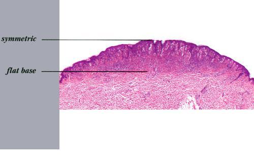

1 SPITZ NEVUS 1 / 7
2 Epidemiology An annual incidence rate of 1.4 cases of Spitz nevus per 100,000 individuals has been estimated in Australia, compared with 25.4 per 100,000 individuals for cutaneous melanoma during the same time interval. Among melanocytic nevi excised in children, 1 percent to 8 percent of cases are interpreted as Spitz nevus. In a case series of 308 patients with Spitz nevi reported by Allen, 15 percent of lesions occurred in adolescents and adults, the oldest patient being 56 years of age. There appears to be no gender predilection for Spitz nevus. Published cases have been described primarily in whites. Etiology and Pathogenesis It may be presumed that Spitz nevi are derived from the same progenitor cells that give rise to epidermal melanocytes and nevomelanocytes. Unlike common acquired nevi and congenital nevi, the B-RAF mutation does not appear to be involved. Amplifications of chromosome 11p and H-RAS and activating mutations of H-RAS have been noted in a subset of Spitz nevi. 84 Heritable genetic factors have not been investigated systematically, but there is a single report of multiple tumors in identical twin boys. 82 The role of trauma in the histogenesis of Spitz nevi is speculative. One case has been reported at the site of bacille Calmette-Guérin vaccination. 85 SPITZ NEVUS AT A GLANCE 2 / 7
3 HISTORY The rate of growth, period of stabilization, and rate of regression of Spitz nevi are not known. The duration of solitary Spitz nevus before presentation is usually less than 9 months. Lesions usually show an increase in radial size over time, some gradual and others rapid. Based on patients with eruptive Spitz nevi, regression has been reported to occur and may even occur in some lesions while new lesions are appearing. CUTANEOUS LESIONS The most common variety of Spitz nevus is solitary, asymptomatic, pink or red, hairless, firm, and dome-shaped. Some Spitz nevi may resemble a keloid. The surface is commonly smooth, and the borders may fade into surrounding skin. Verrucous, scaly, stippled, crusted, or (rarely) eroded lesions have been noted. Spitz nevi are usually asymptomatic, but pruritus, tenderness, and/or bleeding may occur. 82 A halo of depigmentation has been associated with several cases of Spitz nevus. 33,88 Spitz nevi are part of a spectrum in which some also include PSCN and deep penetrating nevi. Spitz nevi can also present as widespread eruptive lesions or in a grouped manner as multiple agminated lesions consisting of red, red-brown, brown, or dark-brown papules or nodules, with a fine stippled surface. Agminated Spitz nevi often occur in the early years of life within a background of congenital (sometimes acquired) macular pigmentation (nevus spilus) or occasionally within a hypopigmented plaque. Spitz nevi may also develop as single or multiple lesions in a large CNN. The diameter of Spitz nevi ranges from several millimeters to several centimeters, the average being 8 mm in one series. 82 In 73 percent of patients in one series, 81 the tumor diameter was 6 mm or less, and in 94 percent of patients, the tumor diameter was less than 10 mm. Most cases are described as superficial papules or nodules, although subcutaneous involvement may occur. 87 In one series of 43 3 / 7
4 patients, 82 the anatomic location included the head or neck in 18, upper extremities in 9, torso in 9, and lower extremities in 7. Spitz nevi tend to spare palms, soles, and mucous membranes. RELATED PHYSICAL FINDINGS There is no known association with other somatic abnormalities. Laboratory Tests HISTOPATHOLOGY Unlike ordinary nevi and melanomas, melanocytic cells in Spitz nevi are large often twice the size of epidermal basal keratinocytes, with prominent mononuclear or multinucleated giant cells in the epidermis and/or dermis. Mitoses, usually few in number, are detected in one-half the cases, whereas atypical mitoses are uncommon in Spitz nevi. In contrast to melanoma, the melanocytic cells in Spitz nevi show progressive maturation with increasing depth, becoming smaller and more similar to ordinary nevomelanocytes, with the overall distribution of cells in the dermis being wedge-shaped, with narrowing of the wedge toward the subcutaneous fat. Coalescent eosinophilic globules (Kamino bodies), periodic acid-schiff-positive and diastase-resistant (resembling colloid bodies), have been reported in 60 percent of Spitz nevi. Similar globules may be detected in 2 percent of melanomas and 0.9 percent of typical acquired nevi, but the globules are smaller in size, more difficult to find, single rather than coalescent, and commonly periodic acid-schiff-negative. 4 / 7
5 Melanocytic elements are usually arranged in well-circumscribed nests, although there may be permeation of the epidermis by single cells or small groups of cells. In those cases with epidermal nests, artifactual clefts are usually seen above the nests in half the cases, a finding rarely observed in melanoma. The epidermis is usually hyperplastic, with elongated and bulbous pegs and knobs extending into the dermis, although thinning and even ulceration may occur rarely. The dermal inflammatory cell infiltrate may be slight or marked, band-like, and mainly at the base or patchy around blood vessels and/or intermixing with tumor cells. Although melanin was observed in all 13 patients originally described by Spitz, more recent studies have determined that melanin was moderate in 10 percent of cases and heavy in 5 percent. 88 Although the bizarre histopathologic features and frequent occurrence of dermal inflammation may cause diagnostic confusion, Spitz nevus usually can be differentiated from melanoma. The histologic features of Spitz nevus in children and adults are similar. SPECIAL TESTS A subgroup of Spitz nevi have a gain of chromosome 11p and amplification or activation of H-RAS. These markers are not routinely used in the clinical setting but may be helpful in the future for discriminating these nevi from other melanocytic neoplasias. Complications Misinterpretation of benign or malignant behavior of the lesion could result in unnecessary or insufficient treatment, respectively. Prognosis and Clinical Course 5 / 7
6 The natural history of the Spitz nevus is largely unknown. Based on patients with eruptive Spitz nevi, it is clear that spontaneous regression can occur. A concern is that some lesions diagnosed as Spitz nevi may progress to melanoma or represent melanoma at the outset. Differential Diagnosis of Spitz Nevi Nonpigmented/pink 1. o Amelanotic melanoma 2. o Juvenile xanthogranuloma 3. o Hemangioma 4. o Pyogenic granuloma 5. o Molluscum contagiosum 6. o Intradermal nevus 7. o Solitary mastocytoma 8. o Granuloma 9. o Clear cell acanthoma 10. o Insect bite reaction 11. o Dermatofibroma 12. o Appendageal tumor 13. o Keloid Pigmented 1. o Blue nevus 2. o Deep penetrating nevus 3. o Pigmented intradermal nevus 4. o Melanoma 5. o Hematoma Warty variants 1. o Verruca vulgaris 2. o Seborrheic keratoses 6 / 7
7 3. o Epidermal nevus 4. o Epidermolytic acanthoma Treatment Complete excision with a clear margin of normal skin is generally sufficient treatment for Spitz nevi. Given the difficulty of confidently excluding the possibility of melanoma in certain cases, a wider margin of normal skin may be prudent for histopathologically worrisome lesions. 90 Incompletely excised lesions may recur in as many as 7 percent to 16 percent of patients. 82,91-93 Although some authors have advocated sentinel node biopsies in these lesions, this may only add to the confusion because Spitz nevus cells may be present in the regional lymph nodes but not necessarily indicate malignancy. 94 Management of patients who have numerous Spitz nevi requires individual judgment and periodic examination for new or unstable lesions. Prevention The role of UVR exposure in the development or progression of Spitz nevi is unknown. 7 / 7
LENTIGO SIMPLEX. Epidemiology
 LENTIGO SIMPLEX Epidemiology The frequency of lentigo simplex in children and adults has not been determined. There does not appear to be a racial or gender predilection. Lentigo simplex is the most common
Benign versus Cancerous Lesions How to tell the difference FMF 2014 Christie Freeman MD, CCFP, DipPDerm, MSc
 1 Benign versus Cancerous Lesions How to tell the difference FMF 2014 Christie Freeman MD, CCFP, DipPDerm, MSc Benign lesions Seborrheic Keratoses: Warty, stuck-on Genetics and birthdays Can start in late
Benign and malignant epithelial lesions: Seborrheic keratosis: A common benign pigmented epidermal tumor occur in middle-aged or older persons more
 Benign and malignant epithelial lesions: Seborrheic keratosis: A common benign pigmented epidermal tumor occur in middle-aged or older persons more common on the trunk; but extremities, head and neck are
Pathology of the skin. 2nd Department of Pathology, Semmelweis University
 Pathology of the skin 2nd Department of Pathology, Semmelweis University Histology of the skin Epidermis: Stratum corneum Stratum granulosum Stratum spinosum Stratum basale Dermis: papillary and reticular
أملس عضلي غرن = Leiomyosarcoma. Leiomyosarcoma 1 / 5
 Leiomyosarcoma 1 / 5 EPIDEMIOLOGY Exact incidence is unknown, but older studies suggest that leiomyosarcomas comprise approximately 3 percent of soft-tissue sarcomas. Superficial leiomyosarcoma occurs
Brief Report. Shivanand Gundalli 1, Smita Kadadavar 1, Somil Singhania 1, Rutuja Kolekar 2 INTRODUCTION. Melanocytic Nevus
 Our Dermatology Online Histopathological spectrum of benign melanocytic nevi our experience in a tertiary care centre Shivanand Gundalli 1, Smita Kadadavar 1, Somil Singhania 1, Rutuja Kolekar 2 1 Department
Dermoscopy: Recognizing Top Five Common In- Office Diagnoses
 Dermoscopy: Recognizing Top Five Common In- Office Diagnoses Vu A. Ngo, DO Department of Family Medicine and Dermatology Choctaw Nation Health Services Authority Learning Objectives Introduction to dermoscopy
Dermatopathology: The tumor is composed of keratinocytes which show atypia, increase mitoses and abnormal mitoses.
 Squamous cell carcinoma (SCC): A common malignant tumor of keratinocytes arising in the epidermis, usually from a precancerous condition: 1- UV induced actinic keratosis, usually of low grade malignancy.
الفتوي الاصفر الحبيبوم = Xanthogranuloma_Juvenile JUVENILE XANTHOGRANULOMA 1 / 9
 JUVENILE XANTHOGRANULOMA 1 / 9 Clinical Findings CUTANEOUS LESIONS JXG is a benign, self-healing disorder that is characterized by asymptomatic yellowish papulonodular lesions of the skin and other organs
Desmoplastic Melanoma R/O BCC. Clinical Information. 74 y.o. man with lesion on left side of neck r/o BCC
 R/O BCC Sabine Kohler, M.D. Professor of Pathology and Dermatology Dermatopathology Service Stanford University School of Medicine Clinical Information 74 y.o. man with lesion on left side of neck r/o
Dermatopathology. Dr. Rafael Botella Estrada. Hospital La Fe de Valencia
 Dermatopathology Dr. Rafael Botella Estrada. Hospital La Fe de Valencia Melanoma and mimics Dr. Martin Mihm Malignant lesions result from the accumulation of mutations Class I lesions (benign) Class II
Histopathology of Melanoma
 THE YALE JOURNAL OF BIOLOGY AND MEDICINE 48, 409-416 (1975) Histopathology of Melanoma G. J. WALKER SMITH Department ofpathology, Yale University School ofmedicine, 333 Cedar Street, New Haven, Connecticut
Appendix : Dermoscopy
 Go Back to the Top To Order, Visit the Purchasing Page for Details APP Appendix : Dermoscopy Dermoscopy, also known as dermatoscopy, epiluminoscopy and epiluminescent microscopy, is an effective non-invasive
Female 18. Deeply pigmented lesion on trunk.?warty naevus?seborrhoeic keratosis?malignant melanoma. The best diagnosis is:
 Female 18. Deeply pigmented lesion on trunk.?warty naevus?seborrhoeic keratosis?malignant melanoma. The best diagnosis is: A. deep penetrating naevus B. naevoid malignant melanoma C. pigment synthesising
Yes. Breaking Bad II: Dermoscopy of Pink-ish Things. Does it Fit? Yes 6/17/2018. Yes. Joslyn Kirby, MD, MS, MEd
 Breaking Bad II: Dermoscopy of Pink-ish Things Joslyn Kirby, MD, MS, MEd Yes Observe Yes Step 2. Fit a Benign Nevus Pattern? Does it Fit? Step 1: Melanocytic? pigment network, globules, homogeneous? No
Citation The Journal of Dermatology, 37(8), available at
, available at") NAOSITE: Nagasaki University's Ac Title Two cases of blaschkitis with promi Author(s) Utani, Atsushi Citation The Journal of Dermatology, 37(8), Issue Date 2010-08 URL Right http://hdl.handle.net/10069/25634
NAOSITE: Nagasaki University's Ac Title Two cases of blaschkitis with promi Author(s) Utani, Atsushi Citation The Journal of Dermatology, 37(8), Issue Date 2010-08 URL Right http://hdl.handle.net/10069/25634
Birthmarks: When to worry, when to reassure
 Birthmarks: When to worry, when to reassure Aimee Smidt, MD, FAAD, FAAP Associate Professor, Depts of Dermatology and Pediatrics University of New Mexico School of Medicine November 2016 Goals and Objectives
Histopathology: skin pathology
 Histopathology: skin pathology These presentations are to help you identify, and to test yourself on identifying, basic histopathological features. They do not contain the additional factual information
LUMPS AND BUMPS: AN ORGANIZED APPROACH TO DIAGNOSIS AND MANAGEMENT
 LUMPS AND BUMPS: AN ORGANIZED APPROACH TO DIAGNOSIS AND MANAGEMENT Tammy P. Than, M.S., O.D., F.A.A.O. The University of Alabama at Birmingham / School of Optometry 1716 University Blvd. Birmingham, AL
Conflict of Interest 9/2/2014. Pathogenesis and Comparison of Atypical Spitz Nevi vs Benign Spitz, and Childhood Melanoma
 Pathogenesis and Comparison of Atypical Spitz Nevi vs Benign Spitz, and Childhood Melanoma Martin C. Mihm Jr., M.D., F.A.C.P. Harvard Medical School Brigham and Women s Hospital Dana Farber Cancer Center
MECHANISMS OF HUMAN DISEASE: LABORATORY SESSION PATHOLOGY OF THE SKIN LAB. Friday, February 13, :30 am 11:00 am
 MECHANISMS OF HUMAN DISEASE: LABORATORY SESSION PATHOLOGY OF THE SKIN LAB Friday, February 13, 2009 9:30 am 11:00 am FACULTY COPY GOALS: Describe the basic clinical and morphologic features of various
Melanoma: The Basics. What is a melanocyte?
 Melanoma: The Basics What is a melanocyte? A melanocyte is a normal cell, found in the skin, which produces melanin. Melanin is a black or dark brown pigment that is seen in the skin, hair, and parts of
MECHANISMS OF HUMAN DISEASE: LABORATORY SESSION PATHOLOGY OF THE SKIN LAB. Friday, February 12, :30 am 11:00 am
 MECHANISMS OF HUMAN DISEASE: LABORATORY SESSION PATHOLOGY OF THE SKIN LAB Friday, February 12, 2012 9:30 am 11:00 am FACULTY COPY GOALS: Describe the basic clinical and morphologic features of various
Disclosure. Objectives. PAFP CME Conference Lou Mancano MD, FAAFP Reading Health System November 18, 2016
 PAFP CME Conference Lou Mancano MD, FAAFP Reading Health System November 18, 2016 1 Disclosure The speaker has no conflict of interest, financial agreement, or working affiliation with any group or organization.
IT S FUNDAMENTAL MY DEAR WATSON! A SHERLOCKIAN APPROACH TO DERMATOLOGY
 IT S FUNDAMENTAL MY DEAR WATSON! A SHERLOCKIAN APPROACH TO DERMATOLOGY Skin, Bones, and other Private Parts Symposium Dermatology Lectures by Debra Shelby, PhD, DNP, FNP-BC, FADNP, FAANP Debra Shelby,
A PRACTICAL APPROACH TO ATYPICAL MELANOCYTIC LESIONS BIJAN HAGHIGHI M.D, DIRECTOR OF DERMATOPATHOLOGY, ST. JOSEPH HOSPITAL
 A PRACTICAL APPROACH TO ATYPICAL MELANOCYTIC LESIONS BIJAN HAGHIGHI M.D, DIRECTOR OF DERMATOPATHOLOGY, ST. JOSEPH HOSPITAL OBJECTIVES Discuss current trends and changing concepts in our understanding of
Common Benign Lesions and Skin Cancers. 22nd May 2015 Dr Mark Foley
 Common Benign Lesions and Skin Cancers 22nd May 2015 Dr Mark Foley Thank you for downloading this file. This intended to supplement the presentation given at the NZ Wound Care Conference, it is not intended
Gross Appearance & Histology of Skin Cancer. Kyle Mannion M.D. January 21, 2005
 Gross Appearance & Histology of Skin Cancer Kyle Mannion M.D. January 21, 2005 Actinic Keratosis 5-20% will develop squamous/basal cell ca Almost solely from solar damage Usually develop during 4 th decade
Questions. Answers. Share your photos and diagnoses with us!
 Illustrated quizzes on problems seen in everyday practice CASE 1 A 66-year-old male presents with ruddy-brown, pruritic papules on his chest and back that have been present for several years. The patient
Basal cell carcinoma 5/28/2011
 Goal of this Presentation A practical approach to the diagnosis of cutaneous carcinomas and their mimics Thaddeus Mully, MD University of California San Francisco To review common non-melanoma skin cancers
Lid Lesions: Relax or Refer
 Lid Lesions: Relax or Refer Blair Lonsberry, MS, OD, MEd., FAAO Professor of Optometry Pacific University College of Optometry blonsberry@pacificu.edu Agenda Benign vs. Malignant lesions Benign Eyelid
Photo Quiz Self-Test Your Diagnostic Acumen
 Do You Know Your Nevi? Case 1: The parents of a 3-year-old girl seek medical evaluation of the nodules on their daughter s back. The lesions have been present since birth and have grown with the child.
Clinical characteristics
 Skin Cancer Fernando Vega, MD Seattle Healing Arts Clinical characteristics Precancerous lesions Common skin cancers ACTINIC KERATOSIS Precancerous skin lesions Actinic keratoses Dysplastic melanocytic
comedo-like openings (clods, brown or orange & circles) milia-like cysts (dots or clods, white) 1/29/18 Dotted vessels are also commonly seen in SCC
 milia-like cysts (dots or clods, white) 1/29/18 Dotted vessels are also commonly seen in SCC") Brown circles Dotted vessels are also commonly seen in SCC Step1 1. Nevus (unequivocal) 2. DF/IDN 3. BCC 4. SCC Network Patchy network Peripheral network & central hypopigmentation DF: network with central
Brown circles Dotted vessels are also commonly seen in SCC Step1 1. Nevus (unequivocal) 2. DF/IDN 3. BCC 4. SCC Network Patchy network Peripheral network & central hypopigmentation DF: network with central
Lagophthalmos. Lagophthalmos: signs. Lagophthalmos: clinical tips. Lagophthalmos: treatment plan. Madarosis
 Lagophthalmos Def: incomplete closure of the eyelid SX: FBS, irritation, red, burn, dry, chronic morning corneal irritation Lagophthalmos: signs 2-5 mm lid separation with slit lamp during blink can force
العصوي الوعاي ي الورام = angiomatosis Bacillary
 1 / 7 BACILLARY ANGIOMATOSIS Epidemiology BA is most commonly seen in patients with acquired immunodeficiency syndrome (AIDS) and a CD4 count less than 50 cells/mm 3, with an incidence of 1.2 cases per
BAP-oma & BEYOND MICHAEL A NOWAK, MD
 BAP-oma & BEYOND MICHAEL A NOWAK, MD CONFLICTS No conflicts with the content of this lecture BAP-oma Wiesner 2011: Families with multiple tan dome-shaped papules of head, neck, trunk, and extremities.
Polypoid Melanoma, A Virulent Variant of the Nodular Growth Pattern
 Polypoid Melanoma, A Virulent Variant of the Nodular Growth Pattern ELIZABETH A. MANCI, M.D., CHARLES M. BALCH, M.D..TARIQ M. MURAD, M.D., PH.D., AND SENG/JAW SOONG, PH.D. Manci, Elizabeth A., Balch, Charles
Non-Melanocytic Pattern Dermoscopy
 Non-Melanocytic Pattern Dermoscopy I have no conflicts of interest to disclose Except that I LOVE dermoscopy Michelle Tarbox, MD Assistant Professor of Dermatology and Dermatopathology Texas Tech University
MELANOMA IN ADOLESCENTS AND YOUNG ADULTS
 Cancer in Adolescents and Young Adults (AYA) Working Group MELANOMA IN ADOLESCENTS AND YOUNG ADULTS Emmanouil Saloustros MD, DSc General Hospital of Heraklion Venizelio Heraklion, Crete, Greece ESMO Preceptorship
Simulators of melanoma
 Simulators of melanoma Philip E. LeBoit, M.D. Depts. of Pathology and Dermatology University of California, San Francisco Simulators of melanoma Simulators of melanoma in situ Melanocytic Non-melanocytic
Objectives. 1. Recognizing benign skin lesions. 2.Know which patients will likely need surgical intervention.
 The Joy of Pediatric Skin Dr. Claire Sanger University of Kentucky Plastic & Reconstructive Surgery Objectives 1. Recognizing benign skin lesions 2.Know which patients will likely need surgical intervention.
Non-melanocytic Patterns
 Non-melanocytic Lesions Non-melanocytic Patterns Michelle Tarbox, MD Assistant Professor of Dermatology and Dermatopathology Texas Tech University Health Sciences Center 2018 Seborrheic keratoses Acanthotic
6/17/2018. Breaking Bad (Part 1) Dermoscopy of Brown(ish) Things. Bad?
 Dermoscopy of Brown(ish) Things. Bad?") Breaking Bad (Part 1) Dermoscopy of Brown(ish) Things Jennie T. Clarke, MD ssociate Professor of Dermatology University of Utah School of Medicine Bad? 1 Brown(ish) Things Bad Melanoma Pigmented basal
Breaking Bad (Part 1) Dermoscopy of Brown(ish) Things Jennie T. Clarke, MD ssociate Professor of Dermatology University of Utah School of Medicine Bad? 1 Brown(ish) Things Bad Melanoma Pigmented basal
Skin lesions The Good and the Bad. Dr Virginia Hubbard Ipswich Hospital NHS Trust Barts and the London School of Medicine and Dentistry
 Skin lesions The Good and the Bad Dr Virginia Hubbard Ipswich Hospital NHS Trust Barts and the London School of Medicine and Dentistry Case 1 32 year old woman Australian Lesion on back New hair growing
=ﻰﻤاﻤﺤﻠا ﺔﻴﻘﻠﺤﻠا ﺔذﺒاﻨﻠا
 1 / 15 Erythema Annulare Centrifugum and Other Figurate Erythemas The figurate erythemas include a variety of eruptions characterized by annular and polycyclic lesions. Classification of this group has
NEOPLASMS OF THE SURFACE EPITHELIUM (KERATINOCYTES)
") NEOPLASMS OF THE SURFACE EPITHELIUM (KERATINOCYTES) Papillary Lesions Precancerous Lesions Keratinocyte Proliferations Carcinomas Melanotic Lesions Melanomas Normal Mucosa Keratin layer Spinous layer Basal
NEOPLASMS OF THE SURFACE EPITHELIUM (KERATINOCYTES) Papillary Lesions Precancerous Lesions Keratinocyte Proliferations Carcinomas Melanotic Lesions Melanomas Normal Mucosa Keratin layer Spinous layer Basal
Guy Perrot (Ги Перро)
") НАУЧНО-ПРАКТИЧЕСКАЯ КОНФЕРЕНЦИЯ (МАСТЕР-КЛАСС) «ПРАКТИЧЕСКИЕ АСПЕКТЫ ДИАГНОСТИКИ И ЛЕЧЕНИЯ МЕЛАНОМЫ КОЖИ» DIAGNOSTIC AND PITFALLS IN MELANOMA Guy Perrot (Ги Перро) MD PHD pathologist, University Hospital
НАУЧНО-ПРАКТИЧЕСКАЯ КОНФЕРЕНЦИЯ (МАСТЕР-КЛАСС) «ПРАКТИЧЕСКИЕ АСПЕКТЫ ДИАГНОСТИКИ И ЛЕЧЕНИЯ МЕЛАНОМЫ КОЖИ» DIAGNOSTIC AND PITFALLS IN MELANOMA Guy Perrot (Ги Перро) MD PHD pathologist, University Hospital
David B. Troxel, MD. Common Medicolegal Situations: Misdiagnosis of Melanoma
 Common Medicolegal Situations: Misdiagnosis of Melanoma David B. Troxel, MD Medical Director, The Doctors Company, Napa, California Clinical Professor Emeritus, University of California at Berkeley Past
Squamous papilloma Squamous acanthoma Keratoacanthoma Verruca vulgaris Condyloma acuminatum Focal epithelial hyperplasia Sino nasal papilloma
 Benign tumors Epithelial origin Squamous papilloma Squamous acanthoma Keratoacanthoma Verruca vulgaris Condyloma acuminatum Focal epithelial hyperplasia Sino nasal papilloma Squamous papilloma Exophytic
Update on Spitzoid and Blue nevus-like melanocytic lesions Emphasis on molecular studies informing diagnosis, prognosis and therapy
 Update on Spitzoid and Blue nevus-like melanocytic lesions Emphasis on molecular studies informing diagnosis, prognosis and therapy Michael T. Tetzlaff MD, PhD Associate Professor Department of Pathology,
Primary Cutaneous CD30-Positive T-cell Lymphoproliferative Disorders
 Primary Cutaneous CD30-Positive T-cell Lymphoproliferative Disorders Definition A spectrum of related conditions originating from transformed or activated CD30-positive T-lymphocytes May coexist in individual
Pigmented lesions of the Oral cavity
 Oral medicine أ.م.د احسان عبد هللا كميل Pigmented lesions of the Oral cavity Pigmented oral lesions are a large group of disorders in which the dark or brown color is the essential clinical characteristic.
Doctors of Optometry Course Notes
 Doctors of Optometry Course Notes OD19 1CE COPE: 43871-AS Eyelid Lumps and Bumps Sunday, February 26, 2017 2:40 pm 3:30 pm Regency C 3 rd Floor Presenter: Blair Lonsberry, OD, FAAO Dr. Lonsberry is a Full
Melanoma. Kaushik Mukherjee MD A. Scott Pearson MD
 Melanoma Kaushik Mukherjee MD A. Scott Pearson MD Disclosures You still have to study Not all inclusive No Western blots Extensive use of Google Image Search and Sabiston Melanoma Basics 8 th most common
Melanoma-Back to Basics I Thought I Knew Ya! Paul K. Shitabata, M.D. Dermatopathologist APMG
 Melanoma-Back to Basics I Thought I Knew Ya! Paul K. Shitabata, M.D. Dermatopathologist APMG At tumor board, a surgeon insists that all level II melanomas are invasive since they have broken through the
Selected Pseudomalignant Soft Tissue Tumors of the Skin and Subcutis
 Selected Pseudomalignant Soft Tissue Tumors of the Skin and Subcutis Andrew L. Folpe, M.D. Professor of Laboratory Medicine and Pathology Mayo Clinic, Rochester, MN folpe.andrew@mayo.edu 2016 MFMER slide-1
Management of pediatric melanocytic lesions
 Open Journal of Clinical & Medical Case Reports Management of pediatric melanocytic lesions Volume 3 (2017) Issue 8 ISSN 2379-1039 Jin Kim, BS; Emmanuel Gabriel MD, PhD; Weiguo Liu MD, PhD; Lin Lin MD,
Abrupt Intralesional Color Change on Dermoscopy as a New Indicator of Early Superficial Spreading Melanoma in a Japanese Woman
 Published online: June 24, 2015 1662 6567/15/0072 0123$39.50/0 This is an Open Access article licensed under the terms of the Creative Commons Attribution-NonCommercial 3.0 Unported license (CC BY-NC)
Pathology. Skin Tumor. Bayan N. Mohammad 15/10/2015. Mohammad al-orjani. Page 0 of 23
 #7 35 Pathology Skin Tumor Bayan N. Mohammad 15/10/2015 Mohammad al-orjani Page 0 of 23 بسم هللا الرحمن الرحيم GREETINGS This lecture is about skin tumors, all the slides are included and every slide will
Actinic keratosis (AK): Dr Sarma s simple guide
: Dr Sarma s simple guide") Actinic keratosis (AK): Dr Sarma s simple guide Actinic keratosis is a very common lesion that you will see in your day-to-day practice. First, let me explain the name Actinic keratosis. It means keratosis
Actinic keratosis (AK): Dr Sarma s simple guide Actinic keratosis is a very common lesion that you will see in your day-to-day practice. First, let me explain the name Actinic keratosis. It means keratosis
Malignant Peripheral Nerve Sheath Tumor
 C H A P T E R 120 Malignant Peripheral Nerve Sheath Tumor Currently, malignant peripheral nerve sheath tumor (MPNST) is the most commonly used generic name for the neoplasms known in the past as neurosarcoma,
Malignant tumors of melanocytes: Part 1. Deba P Sarma, MD., Omaha
 Malignant tumors of melanocytes: Part 1 Deba P Sarma, MD., Omaha The melanocytic tumor is one of the most difficult and confusing areas in Dematopathology. It is true that most (95%) of such lesions are
Learning Objectives. Tanning. The Skin. Classic Features. Sun Reactive Skin Type Classification. Skin Cancers: Preventing, Screening and Treating
 Learning Objectives Skin Cancers: Preventing, Screening and Treating Robert A. Baldor, MD, FAAFP Professor, Family Medicine & Community Health University of Massachusetts Medical School Distinguish the
Spectrum of clinical presentations
 Spectrum of clinical presentations Case History A 7-day-old male patient born full-term via uncomplicated vaginal delivery was seen for multiple erythematous red-brown purpuric lesions that were present
Melanoma Update: 8th Edition of AJCC Staging System
 Melanoma Update: 8th Edition of AJCC Staging System Rosalie Elenitsas, M.D. Professor of Dermatology Director, Dermatopathology University of Pennsylvania DISCLOSURE OF RELATIONSHIPS WITH INDUSTRY None
Eruptive Tumors of the Follicular Infundibulum: An Unexpected Diagnosis of Hypopigmented Macules
 Dermatol Ther (Heidelb) (2015) 5:207 211 DOI 10.1007/s13555-015-0079-0 CASE REPORT Eruptive Tumors of the Follicular Infundibulum: An Unexpected Diagnosis of Hypopigmented Macules Poonkiat Suchonwanit.
SKIN SONOGRAPHY IN CHILDREN. CRISTIAN J. GARCIA MD Santiago, Chile
 SKIN SONOGRAPHY IN CHILDREN CRISTIAN J. GARCIA MD Santiago, Chile I HAVE NO DISCLOSURES OBJECTIVES RELEVANCE OF SKIN LESIONS IN CHILDREN ROLEN OF THE RADIOLOGIST CLINICAL CORRELATION US TECHNIQUE NORMAL
Acral Melanoma in Japan
 Acral Melanoma in Japan MAKOTO SEUI, M.D., HIDEAKI TAKEMATSU, M.D., MICHIKO HOSOKAWA, M.D., MASAAKI OBATA, M.D., YASUSHI TOMITA, M.D., TAIZO KATO, M.D., MASAAKI TAKAHASHI, M.D., AND MARTIN C. MIHM, JR.,
We are IntechOpen, the world s leading publisher of Open Access books Built by scientists, for scientists. International authors and editors
 We are IntechOpen, the world s leading publisher of Open Access books Built by scientists, for scientists 3,500 108,000 1.7 M Open access books available International authors and editors Downloads Our
Malignant Melanoma Early Stage. A guide for patients
 This melanoma patient brochure is designed to help educate melanoma patients and their caregivers. It was developed under the guidance of Dr. Michael Smylie, Professor, Department of Oncology, University
Toby Maurer, MD University of California, San Francisco. Lifetime risk of an American developing melanoma
 Distinguishing Pigmented Skin Lesions and Melanoma Toby Maurer, MD University of California, San Francisco Epidemiology of Melanoma Lifetime risk of an American developing melanoma 1935: 1 in 1500 1980:
Supplementary Figure 1. Spitzoid Melanoma with PPFIBP1-MET fusion. (a) Histopathology (4x) shows a domed papule with melanocytes extending into the
 Histopathology (4x) shows a domed papule with melanocytes extending into the") Supplementary Figure 1. Spitzoid Melanoma with PPFIBP1-MET fusion. (a) Histopathology (4x) shows a domed papule with melanocytes extending into the deep dermis. (b) The melanocytes demonstrate abundant
Supplementary Figure 1. Spitzoid Melanoma with PPFIBP1-MET fusion. (a) Histopathology (4x) shows a domed papule with melanocytes extending into the deep dermis. (b) The melanocytes demonstrate abundant
Principles of Anatomy and Physiology
 Principles of Anatomy and Physiology 14 th Edition CHAPTER 5 The Integumentary System Introduction The organs of the integumentary system include the skin and its accessory structures including hair, nails,
Identifying Skin Cancer. Mary S. Stone MD Professor of Dermatology and Pathology University of Iowa Carver College of Medicine March, 2018
 Identifying Skin Cancer Mary S. Stone MD Professor of Dermatology and Pathology University of Iowa Carver College of Medicine March, 2018 American Cancer Society web site Skin Cancer Melanoma Non-Melanoma
Dermatology for the PCP Deanna G. Brown, MD, FAAD Susong Dermatology Consulting Staff at CHI Memorial
 Dermatology for the PCP Deanna G. Brown, MD, FAAD Susong Dermatology Consulting Staff at CHI Memorial Cutaneous Oncology for the PCP Deanna G. Brown, MD, FAAD Susong Dermatology Consulting Staff at CHI
DERMATOLOGY ROTATION: COMPETENCY-BASED GOALS AND OBJECTIVES
 UNC DIVISION OF PLASTIC AND RECONSTRUCTIVE SURGERY DERMATOLOGY ROTATION: COMPETENCY-BASED GOALS AND OBJECTIVES MEDICAL KNOWLEDGE A. Anatomy/Physiology/Embryology Goal: The resident will have knowledge
Clinical Pathological Conference. Malignant Melanoma of the Vulva
 Clinical Pathological Conference Malignant Melanoma of the Vulva History F/48 Chinese Married Para 1 Presented in September 2004 Vulval mass for 2 months Associated with watery and blood stained discharge
ISPUB.COM. Seborrheic Keratosis: A Pictorial Review of the Histopathologic Variations. D Sarma, S Repertinger
 ISPUB.COM The Internet Journal of Dermatology Volume 7 Number 2 Seborrheic Keratosis: A Pictorial Review of the Histopathologic Variations D Sarma, S Repertinger Citation D Sarma, S Repertinger.. The Internet
Basics in Dermoscopy
 Basics in Dermoscopy Manal Bosseila Professor of Dermatology, Cairo University Member of European Academy Dermatology & Venereology EADV Member of International Dermoscopy Society IDS Member of Aesthetic
ARTICLE INFO ABSTRACT
 Melanocytic Pigmentation: A Single Manifestation of Myriad of Pathologies [PP: 05-09] Dr. Swapna Honwad Department of Oral Pathology dr.swapnahonwad@gmail.com Dr. Elsy P. Simon Department of Endodontics
Diseases of the vulva
 Diseases of the vulva 1. Bartholin Cyst - Infection of the Bartholin gland produces an acute inflammation within the gland (adenitis) and may result in an abscess. Bartholin duct cysts - Are relatively
Maligna Melanoma and Atypical Fibroxanthoma: An Unusual Collision Tumour G Türkcü 1, A Keleş 1, U Alabalık 1, D Uçmak 2, H Büyükbayram 1 ABSTRACT
 Maligna Melanoma and Atypical Fibroxanthoma: An Unusual Collision Tumour G Türkcü 1, A Keleş 1, U Alabalık 1, D Uçmak 2, H Büyükbayram 1 ABSTRACT Two different neoplasia in the same biopsy material called
Periocular Malignancies
 Periocular Malignancies Andrew Gurwood, O.D., F.A.A.O., Dipl. Marc Myers, O.D., F.A.A.O. Drs. Myers and Gurwood have no financial interests to disclose. Course Description Discussion of the most common
Cutaneous Malignancies: A Primer COPYRIGHT. Marissa Heller, M.D.
 Cutaneous Malignancies: A Primer Marissa Heller, M.D. Associate Director of Dermatologic Surgery Department of Dermatology Beth Israel Deaconess Medical Center December 10, 2016 Skin Cancer Non-melanoma
Among the benign intraepithelial melanocytic proliferations, Inflamed Conjunctival Nevi. Histopathological Criteria. Resident Short Reviews
 Resident Short Reviews Inflamed conjunctival nevi (ICN) may suggest malignancy because of their rapid growth and atypical histology. The objective of this study was to characterize the diagnostic features
Lumps and Bumps: The Dermatology of Lid Lesions
 Lumps and Bumps: The Dermatology of Lid Lesions Thomas J. Joly, MD, PhD Assistant Professor of Ophthalmology Eastern Virginia Medical School Ophthalmic Plastic Surgery Service Virginia Eye Consultants
Lichenoid Tissue Reaction in Malignant Melanoma A Potential Diagnostic Pitfall
 natomic Pathology / LICHENOID TISSUE RECTION IN MLIGNNT MELNOM Lichenoid Tissue Reaction in Malignant Melanoma Potential Diagnostic Pitfall CPT Scott R. Dalton, MC, US, 1,3 Capt Matt. aptista, USF, MC,
Glistening, Skin-Colored Nodule
 To Print: Click your browser's PRINT button. NOTE: To view the article with Web enhancements, go to: http://www.medscape.com/viewarticle/436334 Medscape Dermatology Clinic Glistening, Skin-Colored Nodule
MODULE 1. LOCAL AND GENERAL CRITERIA IN PIGMENTED MELANOCYTIC LESIONS.
 DERMOSCOPY TEACHING PROGRAMME Dermoscopy Teaching Programme Module 1 MODULE 1. LOCAL AND GENERAL CRITERIA IN PIGMENTED MELANOCYTIC LESIONS. Dermoscopy is a non-invasive in vivo technique that provides
Toby Maurer, MD University of California, San Francisco. Lifetime risk of an American developing melanoma
 Distinguishing Pigmented Skin Lesions and Melanoma Toby Maurer, MD University of California, San Francisco Epidemiology of Melanoma Lifetime risk of an American developing melanoma 1935: 1 in 1500 1980:
SEBACEOUS NEOPLASMS. Dr. Prachi Saraogi Clinical Fellow in Dermatology
 SEBACEOUS NEOPLASMS Dr. Prachi Saraogi Clinical Fellow in Dermatology Sebaceous neoplasms Sebaceous adenoma (Benign) Sebaceous carcinoma (Malignant) SEBACEOUS ADENOMA Benign tumours composed of incompletely
International Journal of Research in Health Sciences Available online at: Original Article
 International Journal of Research in Health Sciences Available online at: http://www.ijrhs.org/ Original Article Histopathological types of skin tumors in Barak Valley: A hospital based study 1 Dr. Swagata
Melanocytic Lesions: Use of Immunohistochemistry and Special Studies Napa Valley 2018
 Melanocytic Lesions: Use of Immunohistochemistry and Special Studies Napa Valley 2018 Victor G. Prieto, MD, PhD Professor Depts. of Pathology and Dermatology University of Texas - MD Anderson Cancer Center
Clinicopathologic Self- Assessment S003 AAD 2017
 Clinicopathologic Self- Assessment S003 AAD 2017 Clay J. Cockerell, M.D. Director, Cockerell Dermatopathology Director, Division of Dermatopathology UT Southwestern Medical Center July 2017 No relevant
STUDY. Scott W. Menzies, MB,BS, PhD; Karin Westerhoff, MD; Harold Rabinovitz, MD; Alfred W. Kopf, MD; William H. McCarthy, MBBS, MEd; Brian Katz
 STUDY Surface Microscopy of Pigmented Basal Cell Carcinoma Scott W. Menzies, MB,BS, PhD; Karin Westerhoff, MD; Harold Rabinovitz, MD; Alfred W. Kopf, MD; William H. McCarthy, MBBS, MEd; Brian Katz Objectives:
Blue Melanocytic Proliferations
 Blue Melanocytic Proliferations Labib R. Zakka M.D., M.A. Research Fellow Melanoma Program Department of Dermatology Brigham and Women s Hospital Harvard Medical School Conflicts of Interest No conflicts
Skin Cancer. 5 Warning Signs. American Osteopathic College of Occupational and Preventive Medicine OMED 2012, San Diego, Monday, October 8, 2012 C-1
 Skin Cancer AMERICAN OSTEOPATHIC COLLEGE OF OCCUPATIONAL & PREVENTIVE MEDICINE OMED 2012 October 8, 2012 E. Robert Wanat II, D.O., M.P.H. Learning Objectives: Identify the 3 Basic Types of Skin Cancer
VACAVILLE DERMATOLOGY
 Connecting the Dots on those Spots NANDAN V. KAMATH, M.D. VACAVILLE DERMATOLOGY Sources All of the photos were taken with permission from the Dermnet NZ website - Dermnet New Zealand after communicating