2/6/2018. Original Paradigm. Clonal Chromosomal A berrations. Only 20% of Spitz Nevi 95% 6p, 7q, 17q, 20q, 4q,8q, 1q, 11q. Isolated Gain in 11p
|
|
|
- Henry O’Connor’
- 5 years ago
- Views:
Transcription


1 Molecular Diagnostics for Melanocytic Neoplasms: Moving towards a Revolution in the Management of Melanocytic Neoplasms Pedr am Gerami MD Associate Professor of Dermatology, Pathology and Pediatrics at Northwestern University Co-dir ector of Melanoma Program, Northwestern Skin Cancer Institute Disclosures: I have been a consultant to Abbott Molecular, Neogenomics, Castle Biosciences, Myriad Genomics, and Derm Tech Int. Summary 95% of melanomas have clonal chromosomal aberrations including gains on 7q, 8q, 6p, 1q, 20q, 17 and 3 as well as losses on 9p,10, 6q and 8p Conv ersely, nevi inf requently have chromosomal aberrations: 20% of spitz nevi have isolated gain in 11p Original Paradigm FISH Clonal Chromosomal A berrations Common Gains Common Deletions Melanoma 95% 6p, 7q, 17q, 20q, 4q,8q, 1q, 11q 9p, 10, 21q Nevi Only 20% of Spitz Nevi Isolated Gain in 11p 1
M ulti-center retrospective case c ontrolled s tudy 2 )I nclusion criteria: Diagnosis of")
FISH evaluation with probes targeting 6 p25, 6 q23, C ep 6, 11q13, 9 p21, and 8 q24 was performed on all c ases blinded to the c linical outcome Data Collection and")
Expansile nodular growth 10)Epidermal Consumption 11)Epithelioid versus spindle morphology 12)Complete clinical follow up including sentinel node status 13)FISH")
2 Melanoma Four P robe FISH A ssay: 6 p25 8 q q13 9 p21 Sensitivity:86% Specificity:95% Multi-center collaborative study on Spitz tumors involving Northwestern Univ, UCSF, Univ of Michigan, MD Anderson, Memorial Sloan Kettering and Sydney Melanoma Unit Study Design 1 )M ulti-center retrospective case c ontrolled s tudy 2 )I nclusion criteria: Diagnosis of AST 5 years f ollow up no adverse event or tumor spread beyond a s entinel lymph node or les s than 5 years of follow up if there was evidence of tumor s pread beyond the sentinel lymph node 3 )FISH evaluation with probes targeting 6 p25, 6 q23, C ep 6, 11q13, 9 p21, and 8 q24 was performed on all c ases blinded to the c linical outcome Data Collection and Analysis The following data points were collected for each case: 1)Age 2)Sex 3)Anatomic site 4)Breslow depth 5)Mitotic count 6)Clark Level 7)Ulceration status 8)Presence or Absence of kamino bodies 9)Expansile nodular growth 10)Epidermal Consumption 11)Epithelioid versus spindle morphology 12)Complete clinical follow up including sentinel node status 13)FISH data 2
 M itotic Clark Positive FU Time (months) (Yrs) (mm) Rate (/mm 2 ) Level FISH Frequency of FISH results for good (1a,1b,1x) vs.")


3 Summary of clinical, histological, and molecular data by clinical stage Clinical N Average Sex Average Ulceration Average Average % Patients with Average Stage Age Ratio (M :F:NA) Breslow Status (Y: N) M itotic Clark Positive FU Time (months) (Yrs) (mm) Rate (/mm 2 ) Level FISH Frequency of FISH results for good (1a,1b,1x) vs. bad (2,4) outcome Spitzoid patients Patient outcome Fisher s FISH result Good (1a,1b,1x) Bad (2,4) exact test Positive 8 5 6p 0.02 Negative 56 6 Positive 8 1 6q 1.00 Negative p Cep 6 Positive 2 1 Negative M : 31F: 5NA 2.3 7Y: 57N a 13 1b 11q 9p Positive 8 5 Negative 56 6 Positive 3 9 Negative < x M : 4 F: 1NA 3.2 4Y: 4N q Positive 1 1 Negative FISH outcome Positive Negative 49 0 < M : 1F 6.6 1Y: 2N *NA- not available FISH old probe set Positive 15 6 Negative FISH new probe set Positive 9 11 Negative 55 0 < Cytogenetic risk Low risk 55 0 Intermediate 6 2 High risk 3 9 < Multivariate Analysis Comparing Variables for Group 1 versus Group 2 through 4 Variable Estimate Odds Ratio 95% CI for OR p-value Mitotic F9P (+ vs. -) < Footnote: backward elimination method was used to derive the final multivariable models 3

4 4
 4 of 9 patients with ASTs with")

 Distant metastasis and death from disease from ASTs uncommon but when it does happen most cases")

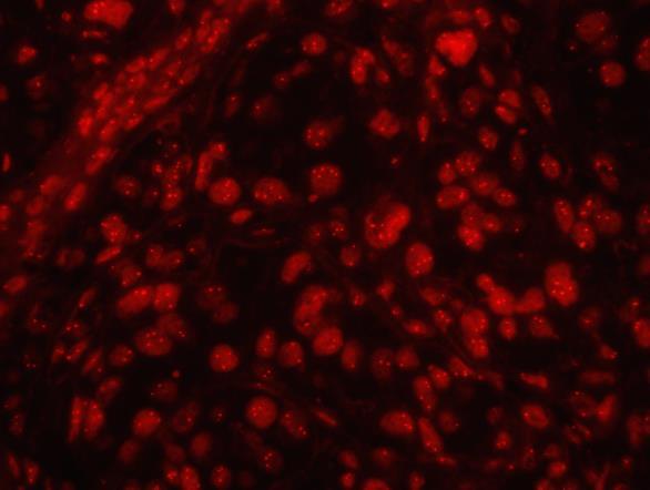
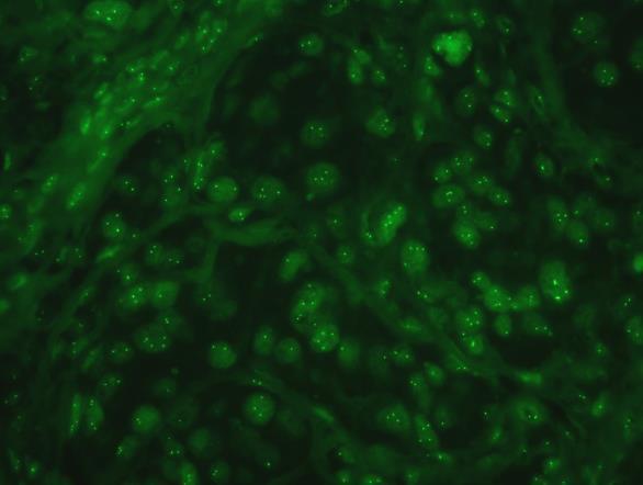
5 Recurring Themes A mong A STs With Ree Aggressive Clinical Course : 1) 9 of 11 cases with disease progression beyond the sentinel had homozygous 9p21 deletions 2) 4 of 9 patients with ASTs with homozygous 9p21 deletion developed recurrent in transit metastasis in the skin in addition to sentinel and non-sentinel lymph node involvement all of whom are still alive. One patient with up to 8 years of follow up. 3) Distant metastasis and death from disease from ASTs uncommon but when it does happen most cases likely to have homozygous 9p21 deletion and likely to occur with a more protracted course compared to conventional melanomas Chimeric proteins resulting f rom translocations involving receptor tyrosine kinases Mutually exclusive fusion proteins identified in 72/140 (51%) Spitzoid neoplasms studied 5


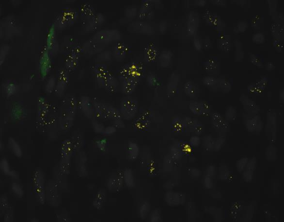
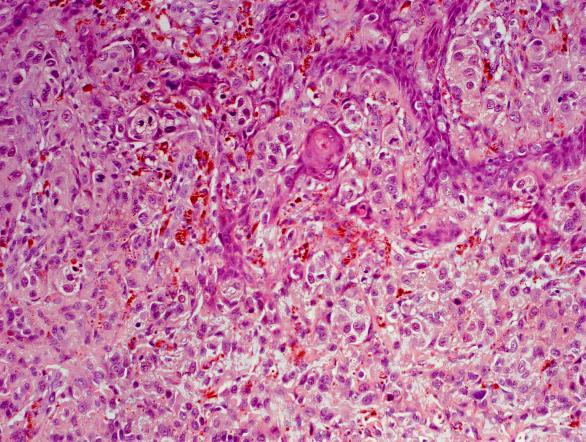
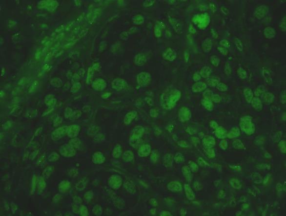
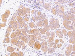
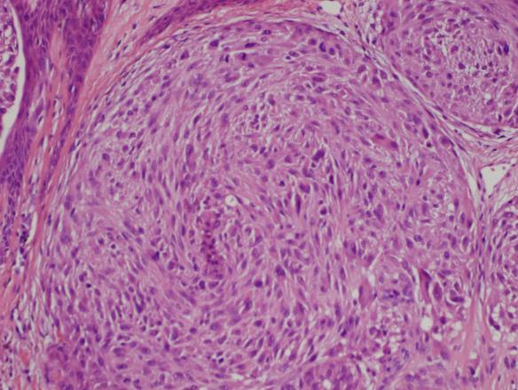
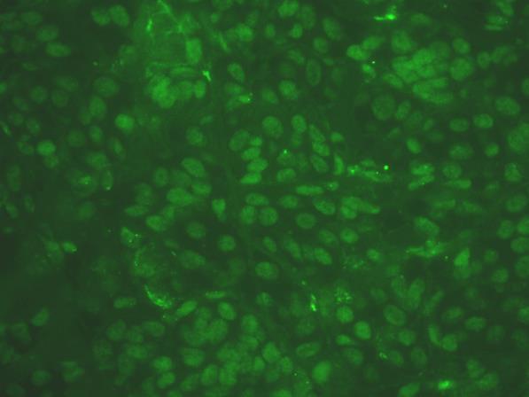
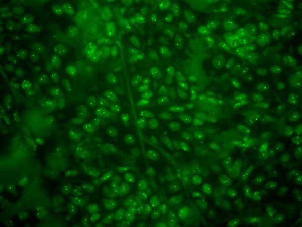
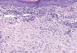
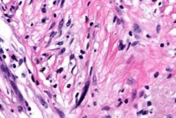
6 ALK positive Spitz Nevus


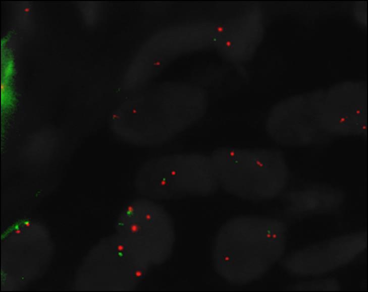
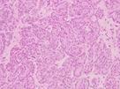
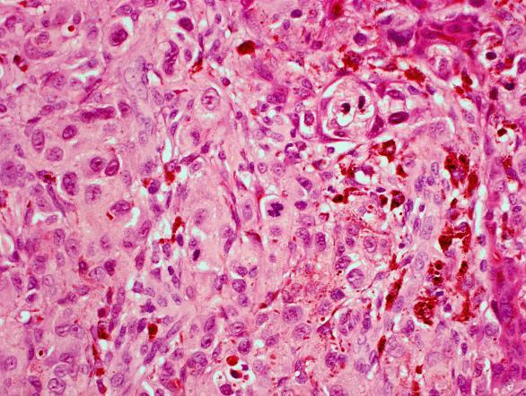
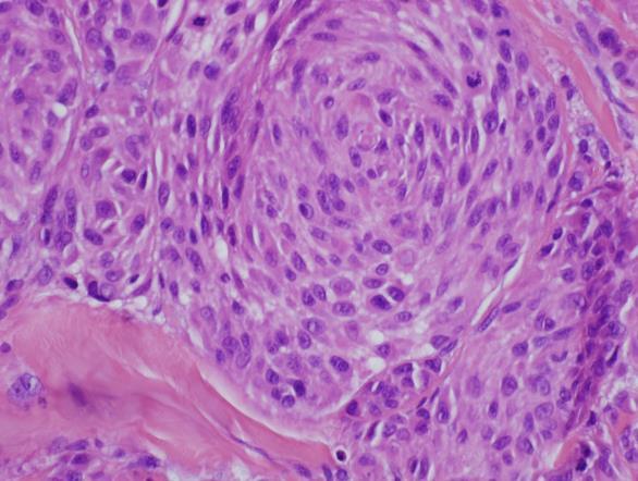
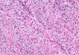
7 NTRK positive Spitz tumor 7
8 8


9 Figure 4 Figure 4. This boxplot demonstrates the range of lesion sizes by fusion group with the black bar representing the median and the overlying boxes representing the 25 th -75 th percentiles. As can be seen in the figure, the 25 th -75 th percentile ALK-fused cases do not overlap with any of the other subgroups, indicating that the majority of ALK-translocations were significantly larger than non-alk-fusions. 9

Abdel-Rahman described 1 family with uveal melanoma, lung adenocarcinoma and meningiomas Family 1 : c.")
 3. Cutaneous Melanoma 4. Renal Cell Carcinoma 5. Atypical Spitz Tumors/BAPomas 6.")
:153-9.")

10 Recently a number of familial melanoma syndromes involving BAP1 have been described: 1)Wiesneret al described 2 families with uveal melanoma, cutaneous melanoma, nevi with atypical epithelioid c ell component and grey zone lesions with atypical epithelioid cell c omponent Family1: c.1305 del6 Family2: c a>g 2)Testa et al described 2 families with uveal melanoma and mesothelioma Family 1 : p.gin684x Family 2 : p.iie72fsx7 3 )Abdel-Rahman described 1 family with uveal melanoma, lung adenocarcinoma and meningiomas Family 1 : c. 799 C->T Associated Malignancies BAP1 = BRCA1-associated protein-1 Patients with germline mutations in BAP1 were found to be at increased risk for: 1. Uveal melanoma 2. Mesothelioma (no asbestos exposure) 3. Cutaneous Melanoma 4. Renal Cell Carcinoma 5. Atypical Spitz Tumors/BAPomas 6. Basal Cell Carcinoma *Other tumors such as meningioma, cholangiocaricinoma, breast and lung carcinoma have been seen in multiple carriers and may be associated with the syndrome. Image: Carbone M, Yang H, Pass HI, Krausz T, Testa JR, Gaudino G. BAP1 and cancer. Nat Rev Cancer Mar;13(3): d oi: /nrc3459. Review. PubMed PMID: ; PubMed Central PMCID: PMC Nuclear deubiquitinating protein that interacts with several other proteins, including BRCA1 Structural architecture in interaction with other proteins not completely understood but has been shown to be involved with: DNA damage response Cell cycle regulation Cell growth Chromatin dynamics 10

11 8 New Families Identified 4 families identified after a dermatologist diagnosed a BAP-1 Deficient Tumor in a young patient (ages 10-32) and asked about family history 1 family identified after a patient in her 40 s was diagnosed with a BAP1 associated nevoid melanoma Family 4 Family 5 Median Age of Onset and Prevalence of Characteristic Tumors in BAP1 Patients Tumor/Malignancy Number of Cases Estimated Penetrance Median Age Literature Median Age Our Study Median Age General Population UvealMelanoma 61/215 cases 28% Mesothelioma 48/215 cases 22% Cutaneous Melanoma 38/215 cases 18% BAP1 Deficient Tumors 33/215 cases 17% Renal Cell Carcinoma 20/215 cases 9% Median Ages of Onset of Associated Tumors Prevalence in Patients with Skin Examinations Of the 53 patients with a documented TBSE by a dermatologist, 40 (75%) were found to have at least one BDT on clinical exam. The number of BDTs in BAP1 patients thought to increase as the patient ages. 11
 Of the 29 patients age >56 without a mesothelioma diagnosis, 66% had truncating")


12 Histologic Presentation Mesothelioma Truncating Mutations Of the 48 patients diagnosed mesothelioma, 96% (n=46) carried truncating mutations in BAP1. The 2 mesothelioma patients without truncating mutations were diagnosed at age 71 and 72 (median age of diagnosis = 56) Of the 29 patients age >56 without a mesothelioma diagnosis, 66% had truncating mutations (n=19) Truncating mutation occurs before the nuclear localization sequence BAP1 protein accumulates in cell cytoplasm Abberrant BAP1 protein has been shown to form amyloid aggregates in cell cytoplasm Inflammation and cytotoxicity involved in mesothelioma pathogenesis? Reported Exonic Mutations in BAP1 Hierarchy of Risk f or Distant Metastasis in Melanocytic Neoplasms with Spitzoid Morphology Conventional Melanoma Most Aggressive Spitzoid Melanoma with Homozygous 9p21 Deletion Intermediate ASTs with no evidence of copy number aberrations Low Risk ASTs with 6q23 Deletion Low Risk ASTs with 3p21 Deletion/BAPomas Low Risk ASTs with 11p gains Low Risk Chromosomal Aberrations New Paradigm Melanoma 95% Spitz tumors Chimeric Fusion Proteins: Ros, AlK, NTRK1, BRAF, RET Cellular functions represented in GEP signature 54 g enes initially assessed and then narrowed to 28 with 3 additional controls Migration/chemotaxi s/metastasis CXCL14 SPP1 CLCA2 S100A9 S100A8 Differentiation/ proliferation CRABP2 SPRRIB BTG1 Common Gains 6p, 7q, 17q, 20q, 4q,8q, 1q, 11q Isolated Gain in 11p, can have gains in 7q Chemokine/secreted molecules Gap junction/cellular adhesion CCL14 MGP SPP1 GJA1 DSC1 PPL Cell surface receptors Structural proteins TACSTD2 CLCA2 ROBO1 MGP SPP1 CST6 Common Deletions 9p, 10, 21q 3p21, 6q23, heterozygous loss of 9p21 Lymphocytic invasion Gerami, Clin Cancer Res 2013 LTA4H Angiogenesis regulator Transcription factor TRIM29 Extracellular functions CXCL14 KRT6B KRT
 Validation Set (n = 104) Age, median yrs (range) 61 (23-89) 58 (18-94) Follow-up, median yrs (range) 4.9 (0.0-13.7) 5.7 (0.0-11.")
13 % free of metastasis % free of metastasis 1 st intended use: Identify the node negative patients who have aggressive disease Validation Study #1: Background demographics Characteristics Training Set (n = 164) Validation Set (n = 104) Age, median yrs (range) 61 (23-89) 58 (18-94) Follow-up, median yrs (range) 4.9 ( ) 5.7 ( ) Patients with Stage I and II melanoma Quantifies expression of 31 genes from primary tumor Applies a validation algorithm Classifies patients as low vs high risk with strong accuracy Class 1 test result: Low risk of metastasis within 5 years Class 2 test result: High risk of metastasis within 5 years Identification of high risk patients allows them the opportunity to access further evaluation, treatment, and monitoring with the goal of improving long-term survival 73 AJCC Stage n (%) n (%) 0 15 (9%) 0 (0%) I 63 (38%) 56 (54%) II 67 (41%) 34 (33%) III 18 (11%) 12 (11%) IV 1 (1%) 2 (2%) Breslow Thickness Median mm (range) 1.86 ( ) 1.4 ( ) 1mm 46 (28%) 45 (43%) > 1mm 101 (62%) 58 (56%) Mitotic Index 1/mm 2 43 (26%) 29 (28%) > 1/mm 2 82 (50%) 53 (51%) Ulceration Absent 104 (63%) 65 (63%) Present 46 (28%) 28 (27%) Growth Pattern Superficial spreading 75 (46%) 56 (54%) Nodular 47 (29%) 25 (24%) Desmoplastic/lentigo maligna/ 25 (15%) 10 (10%) acral lentiginous Gerami, Clin Cancer Res 2015 Censor date: May GEP A ccuracy: Disease-free survival prediction all cases Tr aining Set n=104 p< Validation Set n=164 n=104 5-yr DFS Class 1 = 91% Class 2 = 25% ROC = Accuracy = 83% Sensitivity = 85% Gerami, Clin Cancer Res 2015 Censor Date: May, yr DFS Class 1 = 97% Class 2 = 31% ROC = Accuracy = 86% Sensitivity = 89% 75 SLNB v s. GEP 1 st and 2 nd Validation Studies with SLNB pr ocedure Patients with SLN Procedure (n=217) SLN positive = 58 Met = 37 Non = 21 SLN Results SLN negative = 159 Met = 70 Non = 89 PPV = 64% NPV = 56% Class 2 = 141 Met = 91 GEP Results 100% Non = 50 Class 1 = 76 Met = 16 Non = 60 PPV = 65% NPV = 79% DecisionDx-Melanoma Improves Prediction Over SLNB Negative Status for Distant Metastasis-Free Survival DMFS 100% 75% 50% 25% 0% 0 n=217 p< SLNB 4 6 Time (years) 8 SLNB- SLNB+ 10 SLNB- (n=159) SLNB+ (n=58) Events yr DMFS 64% 42% 75% 50% 25% 0% DecisionDx in SLNB- Patients 0 n=159 p< Time (years) Class 1/SLNB- (n=67) Class 1/ SLNB- Class 2/ SLNB Class 2/SLNB- (n=92) Events yr DMFS 86% 49% 13

14 Previously unreported validation cohort of 523 patients Cox Regression Analysis of 523 Patients Comparison of GEP and SLN in 523 patients GEP plus SLN in combination Independent Validation of 357 Previously Unreported Stage I and II patients RFS in validation cohor t of 264 pr eviously unr eported stage I patients 14

 events E G I R Non-invasive biopsy (tape strip) Highly accurate technology Objective findings Excisional Biopsy n=368 p<0.")
; manuscript in preparation Scientific Rationale Results of Validation Study: Total series sensitivity:91% Total series")
15 % free of metastasis Independent cohort of 93 previously unreported stage II cases 166 previously unreported stage III patients DecisionDx-Melanoma Identifies ~70% of SLNB Negative Patients who had Distant Metastasis Adhesive Patch Testing DMFS by SLN Status DecisionDx in SLNB- Patients Clinically Suspicious Melanocytic Lesion (mole) ABCDE criteria or Dermascope 5-yr SLN- Events SLNB- DMFS n=216 80% 42 5-yr SLNB+ SLN+ Events DMFS Class 1/SLN- (n=106) Class 2/SLN- (n=110) 5-yr DMFS Events 90% 13 71% 29 Identified ~70% (29/42) events E G I R Non-invasive biopsy (tape strip) Highly accurate technology Objective findings Excisional Biopsy n=368 p< n=152 53% 69 Time (years) RNA Expression Continue observation Zager et al. J ClinOncol 2016;34 (suppl; abstr 9581); manuscript in preparation Scientific Rationale Results of Validation Study: Total series sensitivity:91% Total series specificity:69% Sensitivity in consecutive series: 79% Specificity in consecutive series: 80% 89 15
 13 immune")
 S100A7")


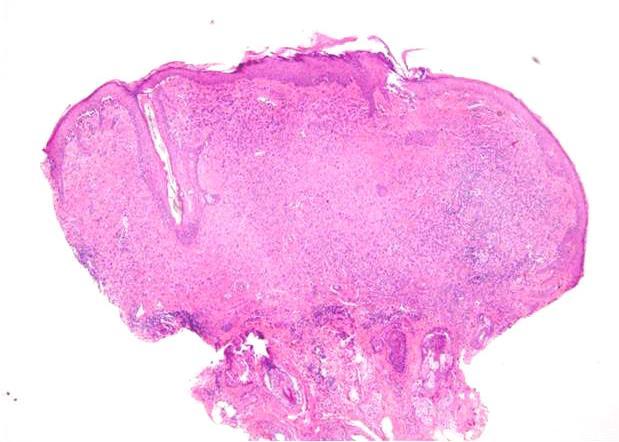
16 mrna Expression for Diagnosis of Melanocytic Neoplasms From Myriad Evaluated 859 total lesions by mrna expression profiling focusing on 23 genes Gene Signature 1) 13 immune related genes 2) 1 cell differentiation gene 3) 9 housekeeping genes A 23 gene expression signature for differentiating melanoma from nevi PRAME (2 Amplicons) S100A7 S100A8 S100A9 S100A12 PI3 S100A Group PRAME Housekeeper Group Algorithm Score Immune Signaling Group IRF1 CCL5 CXCL9 CXCL10 CD38 LCP2 PTPRC SELL Scale and Threshold Values Training set of 464 and Validation set of year old male, upper back Pretest: Dysplastic nevus with moderate to severe atypia +2.2 (Malignant) 3/3: Melanoma in situ Case


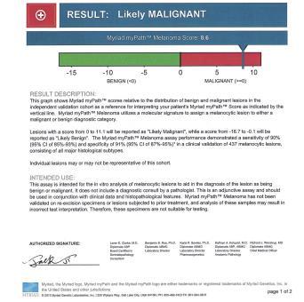

17 year old male; face Pretest: Desmoplastic melanoma 71 year old male, face: Excision Score: +3.8 Case
 H&E")


18 Excision Specimen (Block A4) H&E Melan- A 74 year old female, cheek Pretest: Inflamed nevus S100 Sox-10 Score: +3.1 = False positive 51 year old female, mid back Pretest: Melanoma 34 year old femal, My histologic diagnosis: Spindle Cell nevus of Reed Score: = False negative Immune: -2.4 S100A: -7.5 PRAME: +3.3 Case Score: +6.5 Immune: S100A: +3.4 PRAME: +2.3 Case Myriad Score 4.2 Conclusions from metaanalysis: most useful for predicting prognosis in stage III patients. Most studies show relatively low detection rate of patients with relapse in stage I or II patients Best results in stage III patients but more data needed and more standardization as far as markers assessed and timing of the assessment 18
6/22/2015. Original Paradigm. Correlating Histology and Molecular Findings in Melanocytic Neoplasms
 6 Correlating Histology and Molecular Findings in Melanocytic Neoplasms Pedram Gerami MD, Associate Professor of Dermatology and Pediatrics at Northwestern University Disclosures: I have been a consultant
> 6000 Mutations in Melanoma. Tests That Cay Be Employed. FISH for Additions/Deletions. Comparative Genomic Hybridization
 Winter Clinical 2017: The Assessment and Diagnosis of Melanoma Whitney A. High, MD, JD, MEng Associate Professor, Dermatology & Pathology Director of Dermatopathology (Dermatology) University of Colorado
Update on Lymph Node Management in Melanoma
 Update on Lymph Node Management in Melanoma John T. Vetto MD, FACS Professor of Surgery Division of Surgical Oncology Oregon Health & Science University Portland, Oregon Lymph Nodes in Melanoma Outline
True or False? Nearly twice as many SLNB negative compared to SLNB positive patients will ultimately die of metastatic melanoma.
 qrt-pcr Based Methods for Prognosis of Melanoma Pedr am Gerami MD Pr ofessor of Dermatology, Pathology and Pediatrics Nor thwestern University Dir ector Melanoma Program, Northwestern Skin Cancer Institute
I have no relevant conflicts of interest to disclose. John T. Seykora MD PhD Departments of Dermatology & Pathology and Laboratory Medicine
 Molecular Characterization of Stage 1-3 Melanoma: Are we close to accurate prognostication and prediction? I have no relevant conflicts of interest to disclose. John T. Seykora MD PhD Departments of Dermatology
Which melanoma patients benefit from genetic testing?
 Which melanoma patients benefit from genetic testing? Michael A. Marchetti, MD Assistant Attending, Dermatology Service Memorial Sloan Kettering Cancer Center American Academy of Dermatology Annual Meeting
Genetic Testing: When should it be ordered? Julie Schloemer, MD Dermatology
 Genetic Testing: When should it be ordered? Julie Schloemer, MD Dermatology Outline Germline testing CDKN2A BRCA2 BAP1 Somatic testing Gene expression profiling (GEP) BRAF Germline vs Somatic testing
Impact of Prognostic Factors
 Melanoma Prognostic Factors: where we started, where are we going? Impact of Prognostic Factors Staging Management Surgical intervention Adjuvant treatment Suraj Venna, MD Assistant Clinical Professor,
Vernon K. Sondak. Department of Cutaneous Oncology Moffitt Cancer Center Tampa, Florida
 Vernon K. Sondak Department of Cutaneous Oncology Moffitt Cancer Center Tampa, Florida Australasian Melanoma Conference 2016 Sydney, NSW, Australia October 29, 2016 Disclosures Dr. Sondak is a compensated
Michael T. Tetzlaff MD, PhD
 Molecular alterations informing the diagnosis of melanocytic tumors Michael T. Tetzlaff MD, PhD Associate Professor Department of Pathology, Section of Dermatopathology Department of Translational and
Case 26 Male 37. Right jawline 5mm nodule?keloid. The best diagnosis is:
 Case 26 Male 37. Right jawline 5mm nodule?keloid. The best diagnosis is: A. Desmoplastic Spitz naevus B. Atypical Spitz Tumour C. Spitzoid melanoma D. Deep penetrating naevus E. Spitz naevus Case 26: M
Melanoma and the genes: Molecular alterations informing the diagnosis of melanocytic tumors
 Melanoma and the genes: Molecular alterations informing the diagnosis of melanocytic tumors Michael T. Tetzlaff MD, PhD Associate Professor Department of Pathology, Section of Dermatopathology Department
Update on Spitzoid and Blue nevus-like melanocytic lesions Emphasis on molecular studies informing diagnosis, prognosis and therapy
 Update on Spitzoid and Blue nevus-like melanocytic lesions Emphasis on molecular studies informing diagnosis, prognosis and therapy Michael T. Tetzlaff MD, PhD Associate Professor Department of Pathology,
Molecular Aspects of Melanocytic Neoplasia. Iwei Yeh MD, PhD University of California, San Francisco
 Molecular Aspects of Melanocytic Neoplasia Iwei Yeh MD, PhD University of California, San Francisco Thanks to: Boris Bastian Timothy McCalmont Philip LeBoit Beth Ruben Jeff North Laura Pincus Thaddeus
Gene Expression Profiling in Malignancies: New Insights into Cancer Care
 Gene Expression Profiling in Malignancies: New Insights into Cancer Care Surgical Grand Rounds February 8, 2017 Martin Fleming, M.D. Chief, Division of Surgical Oncology West Cancer Center University of
MAPK Pathway. CGH Next Generation Sequencing. Molecular Tools in Care of Patients with Pigmented Lesions 7/20/2017
 Molecular Tools in Care of Patients with Pigmented Lesions Tammie Ferringer, MD Geisinger Medical Center, Danville, PA tferringer@geisinger.edu DISCLOSURE OF RELATIONSHIPS WITH INDUSTRY Tammie Ferringer,
Gene Expression Profiling for Cutaneous Melanoma
 Applies to all products administered or underwritten by Blue Cross and Blue Shield of Louisiana and its subsidiary, HMO Louisiana, Inc.(collectively referred to as the Company ), unless otherwise provided
There is NO single Melanoma Stain. > 6000 Mutations in Melanoma. What else can be done to discriminate atypical nevi from melanoma?
 Las Vegas Fall Clinical 2016: The Assessment and Diagnosis of Melanoma Whitney A. High, MD, JD, MEng Associate Professor, Dermatology & Pathology Director of Dermatopathology (Dermatology) University of
Gene Expression Profiling for Cutaneous Melanoma
 Gene Expression Profiling for Cutaneous Melanoma Policy Number: 2.04.146 Last Review: 8/1/2018 Origination: 08/2018 Next Review: 8/1/2019 Policy Blue Cross and Blue Shield of Kansas City (Blue KC) will
The Enigmatic Spitz Lesion
 The Enigmatic Spitz Lesion The Dawn of Spitz S Spitz Sophie Spitz Melanomas of Childhood ; Am J Pathol 1948 1910-1956 13 children (18 mo - 12 yrs) 12/13 had a benign clinical course Sophie Spitz Born 1910
David B. Troxel, MD. Common Medicolegal Situations: Misdiagnosis of Melanoma
 Common Medicolegal Situations: Misdiagnosis of Melanoma David B. Troxel, MD Medical Director, The Doctors Company, Napa, California Clinical Professor Emeritus, University of California at Berkeley Past
Dermatopathology. Dr. Rafael Botella Estrada. Hospital La Fe de Valencia
 Dermatopathology Dr. Rafael Botella Estrada. Hospital La Fe de Valencia Melanoma and mimics Dr. Martin Mihm Malignant lesions result from the accumulation of mutations Class I lesions (benign) Class II
21/07/2017. The «gray zone» of diagnosis is visible. Nevus Atypical nevus Melanoma. Melanoma ex-blue nevus
 Update on the Clinico- Pathological and Molecular Diagnosis of Melanocytic Lesions None to declare Conflicts of interest Belfast pathology Arnaud de la Fouchardière MD, PhD Lyon, France What is new? Today
Medical Policy An independent licensee of the Blue Cross Blue Shield Association
 Gene Expression Profiling for Cutaneous Melanoma Page 1 of 28 Medical Policy An independent licensee of the Blue Cross Blue Shield Association Title: Gene Expression Profiling for Cutaneous Melanoma Professional
A PRACTICAL APPROACH TO ATYPICAL MELANOCYTIC LESIONS BIJAN HAGHIGHI M.D, DIRECTOR OF DERMATOPATHOLOGY, ST. JOSEPH HOSPITAL
 A PRACTICAL APPROACH TO ATYPICAL MELANOCYTIC LESIONS BIJAN HAGHIGHI M.D, DIRECTOR OF DERMATOPATHOLOGY, ST. JOSEPH HOSPITAL OBJECTIVES Discuss current trends and changing concepts in our understanding of
Desmoplastic Melanoma R/O BCC. Clinical Information. 74 y.o. man with lesion on left side of neck r/o BCC
 R/O BCC Sabine Kohler, M.D. Professor of Pathology and Dermatology Dermatopathology Service Stanford University School of Medicine Clinical Information 74 y.o. man with lesion on left side of neck r/o
The Pathology of Neoplasia Part II
 The Pathology of Neoplasia Part II February 2018 PAUL BOGNER, MD A S S O C I A T E P R O F E S S O R O F O N C O L O G Y P A T H O L O G Y A N D D E R M A T O L O G Y Clinical goals of cancer pathology
Melanocytic Lesions: Use of Immunohistochemistry and Special Studies Napa Valley 2018
 Melanocytic Lesions: Use of Immunohistochemistry and Special Studies Napa Valley 2018 Victor G. Prieto, MD, PhD Professor Depts. of Pathology and Dermatology University of Texas - MD Anderson Cancer Center
Protocol applies to melanoma of cutaneous surfaces only.
 Melanoma of the Skin Protocol applies to melanoma of cutaneous surfaces only. Procedures Biopsy (No Accompanying Checklist) Excision Re-excision Protocol revision date: January 2005 Based on AJCC/UICC
Corporate Medical Policy
 Corporate Medical Policy Gene Expression Profiling for Cutaneous Melanoma File Name: Origination: Last CAP Review: Next CAP Review: Last Review: gene_expression_profiling_for_cutaneous_melanoma 5/2018
Melanoma Update: 8th Edition of AJCC Staging System
 Melanoma Update: 8th Edition of AJCC Staging System Rosalie Elenitsas, M.D. Professor of Dermatology Director, Dermatopathology University of Pennsylvania DISCLOSURE OF RELATIONSHIPS WITH INDUSTRY None
Malignant tumors of melanocytes : Part 3. Deba P Sarma, MD., Omaha
 Malignant tumors of melanocytes : Part 3 Deba P Sarma, MD., Omaha Let s go over one case of melanoma using the following worksheet. Of the various essential information that needs to be included in the
Ways to get into trouble, ideas on avoiding trouble, and diagnostic approaches to keep trouble at bay
 Pitfalls in the diagnosis of melanocytic tumors Timothy McCalmont, MD University of California, San Francisco Ways to get into trouble, ideas on avoiding trouble, and diagnostic approaches to keep trouble
Gene Expression Profiling for Melanoma
 Medical Policy Manual Genetic Testing, Policy No. 29 Gene Expression Profiling for Melanoma Next Review: April 2019 Last Review: April 2018 Effective: June 1, 2018 IMPORTANT REMINDER Medical Policies are
Talk to Your Doctor. Fact Sheet
 Talk to Your Doctor Hearing the words you have skin cancer is overwhelming and would leave anyone with a lot of questions. If you have been diagnosed with Stage I or II cutaneous melanoma with no apparent
Update on Genetic Testing for Melanoma
 Update on Genetic Testing for Melanoma Emily Y. Chu, M.D., Ph.D. Assistant Professor of Dermatology & Pathology and Laboratory Medicine Hospital of the University of Pennsylvania February 18, 2018 AAD
Challenges in Melanoma Diagnosis and Management
 Challenges in Melanoma Diagnosis and Management Winter Clinical Dermatology Conference - Hawaii Darrell S. Rigel, MD MS Clinical Professor of Dermatology New York University Medical Center DISCLOSURE OF
Patricia Chevez-Barrrios AAOOP-USCAP /12/2016
 Biomarkers in Ocular Melanoma Patricia Chévez-Barrios, MD Pathology and Genomic Medicine, Houston Methodist Hospital Professor of Pathology and Laboratory Medicine and Ophthalmology, Weill Cornell Medical
Cutaneous Melanoma: Epidemiology (USA) The Sentinel Node in Head and Neck Melanoma. Cutaneous Melanoma: Epidemiology (USA)
 The Sentinel Node in Head and Neck Melanoma. Cutaneous Melanoma: Epidemiology (USA)") The Sentinel Node in Head and Neck Melanoma Cutaneous Melanoma: Epidemiology (USA) 6 th leading cause of cancer among men and women 68,720 new cases of invasive melanoma in 2009 8,650 deaths from melanoma
The Sentinel Node in Head and Neck Melanoma Cutaneous Melanoma: Epidemiology (USA) 6 th leading cause of cancer among men and women 68,720 new cases of invasive melanoma in 2009 8,650 deaths from melanoma
Dr. Brent Doolan, BSc MBBS MPH
 Impact of partial biopsies on the need for complete excisional surgery in the management of cutaneous melanomas: A multi-centre review Dr. Brent Doolan, BSc MBBS MPH Peter MacCallum Cancer Centre, Melbourne
An Overview of Melanoma. Harriet Kluger, M.D. Associate Professor Section of Medical Oncology Yale Cancer Center
 An Overview of Melanoma Harriet Kluger, M.D. Associate Professor Section of Medical Oncology Yale Cancer Center Melanoma Statistics Median age at presentation 45-55 55 years Incidence: 2003 54,200 cases
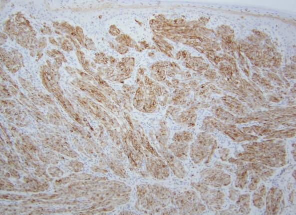
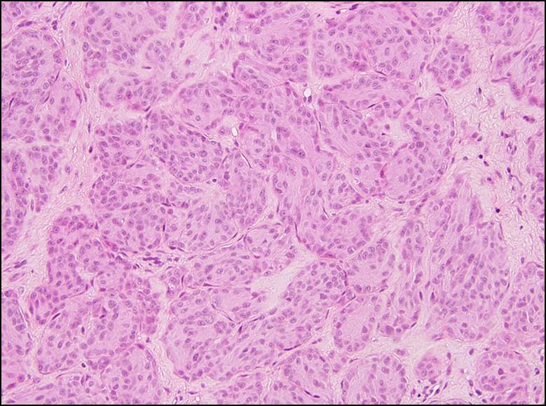
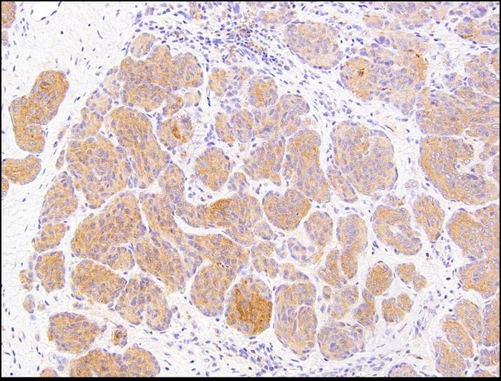
Supplementary Figure 1. Spitzoid Melanoma with PPFIBP1-MET fusion. (a) Histopathology (4x) shows a domed papule with melanocytes extending into the
 Histopathology (4x) shows a domed papule with melanocytes extending into the") Supplementary Figure 1. Spitzoid Melanoma with PPFIBP1-MET fusion. (a) Histopathology (4x) shows a domed papule with melanocytes extending into the deep dermis. (b) The melanocytes demonstrate abundant
Supplementary Figure 1. Spitzoid Melanoma with PPFIBP1-MET fusion. (a) Histopathology (4x) shows a domed papule with melanocytes extending into the deep dermis. (b) The melanocytes demonstrate abundant
Melanocytic proliferations in sundamaged
 Atypical Spitzoid Tumor: What Does It Mean And How Should It Be Managed? Melanocytic proliferations in sundamaged skin Jane L. Messina, Jane L. Messina MD International Melanoma Pathology Working Group
Updates on Melanoma: Are You Following the Latest Guidelines of Care? Jerry Brewer, MD
 Updates on Melanoma: Are You Following the Latest Guidelines of Care? Jerry Brewer, MD Disclosure Statement Update on Melanoma Are You Following the Latest Guidelines of Care? I, Jerry D. Brewer, MD, do
Dermatopathology: The tumor is composed of keratinocytes which show atypia, increase mitoses and abnormal mitoses.
 Squamous cell carcinoma (SCC): A common malignant tumor of keratinocytes arising in the epidermis, usually from a precancerous condition: 1- UV induced actinic keratosis, usually of low grade malignancy.
Epithelial Cancer- NMSC & Melanoma
 Epithelial Cancer- NMSC & Melanoma David Chin MB, BCh, BAO, LRCP, LRCS (Ireland) MCh(MD), PhD (UQ), FRCS, FRACS (Plast) Plastic & Reconstructive Surgeon Visiting Scientist Melanoma Genomic Group & Drug
Management of pediatric melanocytic lesions
 Open Journal of Clinical & Medical Case Reports Management of pediatric melanocytic lesions Volume 3 (2017) Issue 8 ISSN 2379-1039 Jin Kim, BS; Emmanuel Gabriel MD, PhD; Weiguo Liu MD, PhD; Lin Lin MD,
Melanoma Underwriting Presented at 2018 AHOU Conference. Hank George FALU
 Melanoma Underwriting Presented at 2018 AHOU Conference Hank George FALU MELANOMA EPIDEMIOLOGY 70-80,000 American cases annually Majority are in situ or thin > 20% are diagnosed age 45 8-9,000 melanoma
10/2/17. MELTUMP, SAMPUS, AST.An Algorithmic Approach to Challenging (Often Borderline) Melanocytic Tumors. An Introduction to SNP Arrays
 Melanocytic Tumors. An Introduction to SNP Arrays") MELTUMP, SAMPUS, AST.An Algorithmic Approach to Challenging (Often ) Melanocytic Tumors An Introduction to SNP Arrays Rajiv M. Patel, M.D. RCPA NZ ASM 2017 (11:45-12:30pm, Saturday, 23-09-17) Why do we
MELTUMP, SAMPUS, AST.An Algorithmic Approach to Challenging (Often ) Melanocytic Tumors An Introduction to SNP Arrays Rajiv M. Patel, M.D. RCPA NZ ASM 2017 (11:45-12:30pm, Saturday, 23-09-17) Why do we
Identifying Skin Cancer. Mary S. Stone MD Professor of Dermatology and Pathology University of Iowa Carver College of Medicine March, 2018
 Identifying Skin Cancer Mary S. Stone MD Professor of Dermatology and Pathology University of Iowa Carver College of Medicine March, 2018 American Cancer Society web site Skin Cancer Melanoma Non-Melanoma
Molecular Methods in the Diagnosis and Prognostication of Melanoma: Pros & Cons
 Molecular Methods in the Diagnosis and Prognostication of Melanoma: Pros & Cons Ben J. Friedman, MD Senior Staff Physician Department of Dermatology Department of Pathology and Laboratory Medicine Henry
1. Opdivo + Ipilumimab is now the first line therapy for metastatic melanoma.
 Melanoma UpToDate: Introduction: Risk Factors: 1. Opdivo + Ipilumimab is now the first line therapy for metastatic melanoma. Median age = 50 yrs Incidence is rising - Sun exposure: UVB (290-320nm) > UVA
Update on 8 th Edition Cutaneous AJCC Staging of Primary Cutaneous Melanoma. Michael T. Tetzlaff MD, PhD
 Update on 8 th Edition Cutaneous AJCC Staging of Primary Cutaneous Melanoma Michael T. Tetzlaff MD, PhD Associate Professor Departments of Pathology (Dermatopathology) and Translational and Molecular Pathology
Melanoma. Kaushik Mukherjee MD A. Scott Pearson MD
 Melanoma Kaushik Mukherjee MD A. Scott Pearson MD Disclosures You still have to study Not all inclusive No Western blots Extensive use of Google Image Search and Sabiston Melanoma Basics 8 th most common
You Are Going to Cut How Much Skin? Locoregional Surgical Treatment. Justin Rivard MD, MSc, FRCSC September 21, 2018
 You Are Going to Cut How Much Skin? Locoregional Surgical Treatment Justin Rivard MD, MSc, FRCSC September 21, 2018 Presenter Disclosure Faculty/Speaker: Justin Rivard Relationships with financial sponsors:
Springer Healthcare. Staging and Diagnosing Cutaneous Melanoma. Concise Reference. Dirk Schadendorf, Corinna Kochs, Elisabeth Livingstone
 Concise Reference Staging and Diagnosing Cutaneous Melanoma Dirk Schadendorf, Corinna Kochs, Elisabeth Livingstone Extracted from Handbook of Cutaneous Melanoma: A Guide to Diagnosis and Treatment Published
Female 18. Deeply pigmented lesion on trunk.?warty naevus?seborrhoeic keratosis?malignant melanoma. The best diagnosis is:
 Female 18. Deeply pigmented lesion on trunk.?warty naevus?seborrhoeic keratosis?malignant melanoma. The best diagnosis is: A. deep penetrating naevus B. naevoid malignant melanoma C. pigment synthesising
Primary Cutaneous Melanoma Pathology Reporting Proforma DD MM YYYY. *Tumour site. *Specimen laterality. *Specimen type
 Primary Cutaneous Melanoma Pathology Reporting Proforma Includes the International Collaboration on Cancer reporting dataset denoted by * Family name Given name(s) Date of birth DD MM YYYY Sex Male Female
Molecular Enhancement of Sentinel Node Evaluation
 Cochran Illustrations 060104 Molecular Enhancement of Sentinel Node Evaluation Alistair Cochran, MD and Rong Huang MD Departments of Pathology and Laboratory Medicine and Surgery, David Geffen School of
Benign and malignant epithelial lesions: Seborrheic keratosis: A common benign pigmented epidermal tumor occur in middle-aged or older persons more
 Benign and malignant epithelial lesions: Seborrheic keratosis: A common benign pigmented epidermal tumor occur in middle-aged or older persons more common on the trunk; but extremities, head and neck are
Financial disclosures
 Mesenchymal Neoplasms with Melanocytic Differentiation By Konstantinos Linos MD, FCAP, FASDP Bone, Soft Tissue and Dermatopathology Assistant Professor of Pathology Dartmouth-Hitchcock Medical Center Geisel
WHAT DOES THE PATHOLOGY REPORT MEAN?
 Melanoma WHAT IS MELANOMA? Melanoma is a type of cancer that affects cells called melanocytes. These cells are found mainly in skin but also in the lining of other areas such as nose and rectum, and also
Epidemiology. Objectives 8/28/2017
 Case based Discussion of Head and Neck Melanoma: Review of Epidemiology, Risk Factors, Identification, Treatments and Prevention Jacqueline M. Doucette MS FNP-C Objectives Define and identify melanoma
Toby Maurer, MD University of California, San Francisco. Lifetime risk of an American developing melanoma
 Distinguishing Pigmented Skin Lesions and Melanoma Toby Maurer, MD University of California, San Francisco Epidemiology of Melanoma Lifetime risk of an American developing melanoma 1935: 1 in 1500 1980:
Metastatic Melanoma. Cynthia Kwong February 16, 2017 SUNY Downstate Medical Center Department of Surgery Grand Rounds
 Metastatic Melanoma Cynthia Kwong February 16, 2017 SUNY Downstate Medical Center Department of Surgery Grand Rounds Case Presentation 77 year old male with previous history of scalp melanoma and thyroid
ACCME/Disclosures ALK FUSION-POSITIVE MESENCHYMAL TUMORS. Tumor types with ALK rearrangements. Anaplastic Lymphoma Kinase. Jason L.
 Companion Meeting of the International Society of Bone and Soft Tissue Pathology The Evolving Concept of Mesenchymal Tumors ALK FUSION-POSITIVE MESENCHYMAL TUMORS Jason L. Hornick, MD, PhD March 13, 2016
Toby Maurer, MD University of California, San Francisco. Lifetime risk of an American developing melanoma
 Distinguishing Pigmented Skin Lesions and Melanoma Toby Maurer, MD University of California, San Francisco Epidemiology of Melanoma Lifetime risk of an American developing melanoma 1935: 1 in 1500 1980:
Case Scenario 1 Worksheet. Primary Site C44.4 Morphology 8743/3 Laterality 0 Stage/ Prognostic Factors
 CASE SCENARIO 1 9/10/13 HISTORY: Patient is a 67-year-old white male and presents with lesion located 4-5cm above his right ear. The lesion has been present for years. No lymphadenopathy. 9/10/13 anterior
Page 1 of 3. We suggest the following changes:
 Page 1 of 3 Loren E. Clarke, M.D. Myriad Genetic Laboratories, Inc. 320 Wakara Way, Salt Lake City, UT 84108 Phone: 801.883.3470 Email: lclarke@myriad.com Date of Request: June 2017 NCCN Guidelines Panel:
Malignant Melanoma in Turkey: A Single Institution s Experience on 475 Cases
 Malignant Melanoma in Turkey: A Single Institution s Experience on 475 Cases Faruk Tas, Sidika Kurul, Hakan Camlica and Erkan Topuz Institute of Oncology, Istanbul University, Istanbul, Turkey Received
Multispectral Digital Skin Lesion Analysis. Summary
 Subject: Multispectral Digital Skin Lesion Analysis Page: 1 of 8 Last Review Status/Date: March 2016 Multispectral Digital Skin Lesion Analysis Summary There is interest in noninvasive devices that will
Dilemmas in Cytopathology and Histopathology
 Dilemmas in Cytopathology and Histopathology Yuri E. Nikiforov, MD, PhD Division of Molecular & Genomic Pathology University of Pittsburgh Medical Center, USA Objectives Discuss new WHO classification
Malignant tumors of melanocytes: Part 1. Deba P Sarma, MD., Omaha
 Malignant tumors of melanocytes: Part 1 Deba P Sarma, MD., Omaha The melanocytic tumor is one of the most difficult and confusing areas in Dematopathology. It is true that most (95%) of such lesions are
Michael T. Tetzlaff MD, PhD
 Update on American Joint Cancer Committee (AJCC) staging system for primary cutaneous melanoma Emphasis on concise and accurate reporting of primary and metastatic melanoma for effective risk stratification
Patient age and cutaneous malignant melanoma: Elderly patients are likely to have more aggressive histological features and poorer survival
 MOLECULAR AND CLINICAL ONCOLOGY 7: 1083-1088, 2017 Patient age and cutaneous malignant melanoma: Elderly patients are likely to have more aggressive histological features and poorer survival FARUK TAS
BAP-oma & BEYOND MICHAEL A NOWAK, MD
 BAP-oma & BEYOND MICHAEL A NOWAK, MD CONFLICTS No conflicts with the content of this lecture BAP-oma Wiesner 2011: Families with multiple tan dome-shaped papules of head, neck, trunk, and extremities.
MELANOMA IN ADOLESCENTS AND YOUNG ADULTS
 Cancer in Adolescents and Young Adults (AYA) Working Group MELANOMA IN ADOLESCENTS AND YOUNG ADULTS Emmanouil Saloustros MD, DSc General Hospital of Heraklion Venizelio Heraklion, Crete, Greece ESMO Preceptorship
Topics for Discussion. Malignant Melanoma. Surgical Treatment. Current Treatment of Cutaneous Melanoma 5/17/2013. Lymph Regional nodes:
 Topics for Discussion What is a sentinel lymph node (SLN)? Utility of sentinel lymph biopsies: therapeutic or staging? Current Treatment of Cutaneous Melanoma Carlos Corvera, M.D. Associate Professor of
Melanoma Case Scenario 1
 Melanoma Case Scenario 1 History and physical 11/5/16 Patient is a single, 48-year-old male in good health who presented to his primary physician for a yearly physical exam during which a 3.4 x 2.8 x 1.5
Michael T. Tetzlaff MD, PhD
 American Joint Cancer Committee (AJCC) staging system for primary cutaneous melanoma (8 th Edition) and principles of sentinel lymph node evaluation Emphasis on concise and accurate reporting of primary
Translating Evidence into Practice: Primary Cutaneous Melanoma Guidelines. Sentinel Lymph Node Biopsy
 American Academy of Dermatology 2018 Annual Meeting San Diego, CA, February 17, 2018 Translating Evidence into Practice: Primary Cutaneous Melanoma Guidelines. Sentinel Lymph Node Biopsy Christopher Bichakjian,
SKIN CANCER. Most common cancer diagnosis 40% of all cancers
 SKIN CANCER Most common cancer diagnosis 40% of all cancers OBJECTIVES Review common and uncommon cancers of the skin. Special emphasis on melanoma and dysplastic nevus Review pathology/tnm/staging, which
Melanoma and Dermoscopy. Disclosure Statement: ABCDE's of melanoma. Co-President, Usatine Media
 Melanoma and Dermoscopy Richard P. Usatine, MD, FAAFP Professor, Family and Community Medicine Professor, Dermatology and Cutaneous Surgery Medical Director, University Skin Clinic University of Texas
Rebecca Vogel, PGY-4 March 5, 2012
 Rebecca Vogel, PGY-4 March 5, 2012 Historical Perspective Changes In The Staging System Studies That Started The Talk Where We Go From Here Cutaneous melanoma has become an increasingly growing problem,
Skin Cancer. 5 Warning Signs. American Osteopathic College of Occupational and Preventive Medicine OMED 2012, San Diego, Monday, October 8, 2012 C-1
 Skin Cancer AMERICAN OSTEOPATHIC COLLEGE OF OCCUPATIONAL & PREVENTIVE MEDICINE OMED 2012 October 8, 2012 E. Robert Wanat II, D.O., M.P.H. Learning Objectives: Identify the 3 Basic Types of Skin Cancer
1/10/2018. Soft Tissue Tumors Showing Melanocytic Differentiation. Overview. Desmoplastic/ Spindle Cell Melanoma
 2016 MFMER slide-1 2016 MFMER slide-2 2016 MFMER slide-3 Soft Tissue Tumors Showing Melanocytic Differentiation Andrew L. Folpe, M.D. Professor of Laboratory Medicine and Pathology Mayo Clinic, Rochester,
Benign versus Cancerous Lesions How to tell the difference FMF 2014 Christie Freeman MD, CCFP, DipPDerm, MSc
 1 Benign versus Cancerous Lesions How to tell the difference FMF 2014 Christie Freeman MD, CCFP, DipPDerm, MSc Benign lesions Seborrheic Keratoses: Warty, stuck-on Genetics and birthdays Can start in late
Clinical Case Conference Melanoma
 Clinical Case Conference Melanoma Epidemiology ~60,000 cases and 8,000 deaths per year in US Caucasian:African American = 10:1 15% arise from existing nevi 91% are cutaneous 15% are LN+ at presentation
Melanoma Case Scenario 1
 Melanoma Case Scenario 1 History and physical 11/5/16 Patient is a single, 48-year-old male in good health who presented to his primary physician for a yearly physical exam during which a 3.4 x 2.8 x 1.5
Melanoma Quality Reporting
 Melanoma Quality Reporting September 1, 2013 December 31, 2016 Laurence McCahill, MD Surgical Oncologist Metro Health Surgical Oncology Metro Health Professional Building 2122 Health Drive SW Wyoming,
Update on SLN and Melanoma: DECOG and MSLT-II. Gordon H. Hafner, MD, FACS
 Update on SLN and Melanoma: DECOG and MSLT-II Gordon H. Hafner, MD, FACS No disclosures The surgery of malignant disease is not the surgery of organs, it is of the lymphatic system. Lord Moynihan Lymph
Disclosures. SLNB for Melanoma 25/02/2014 SENTINEL LYMPH NODE BIOPSY FOR MELANOMA: CURRENT GUIDELINES AND THEIR CLINICAL APPLICATION
 8 th Canadian Melanoma Conference February 22, 2014 Rimrock Resort Hotel, Banff, Alberta SENTINEL LYMPH NODE BIOPSY FOR MELANOMA: CURRENT GUIDELINES AND THEIR CLINICAL APPLICATION Christopher Bichakjian,
Cutaneous Malignancies: A Primer COPYRIGHT. Marissa Heller, M.D.
 Cutaneous Malignancies: A Primer Marissa Heller, M.D. Associate Director of Dermatologic Surgery Department of Dermatology Beth Israel Deaconess Medical Center December 10, 2016 Skin Cancer Non-melanoma
Melanoma and Mimickers
 Melanoma and Mimickers Kara Walton, MD Assistant Professor of Dermatology and Dermatopathology Medical College of Wisconsin Disclosures No relevant financial disclosures 1 Objectives Recognize common benign
Diagnoses of Cases 1. Lentigo, other melanosis and the acquired nevus 2. Variations on the acquired nevus 3. Dermal melanocytosis
 Diagnoses of Cases 1. Lentigo, other melanosis and the acquired nevus 1 1A. Lentigo simplex 4 1B. Psoralens and ultraviolet A (PUVA) lentigo 6 1C. Solar lentigo 8 1D. Café au lait macule 10 1E. Ink-spot
Melanocytic Tumours. Molecular Biology 02/06/2015. Cutaneous Melanocytic Tumours Introduction. Thomas Brenn. Intermediate Malignancy
 Cutaneous Melanocytic Tumours Introduction Melanocytic Tumours: Update on Epidemiology and Molecular Biology Thomas Brenn Wide clinical and morphological spectrum Ranging from benign naevi to melanoma
Clinical Pathological Conference. Malignant Melanoma of the Vulva
 Clinical Pathological Conference Malignant Melanoma of the Vulva History F/48 Chinese Married Para 1 Presented in September 2004 Vulval mass for 2 months Associated with watery and blood stained discharge
Clinical characteristics
 Skin Cancer Fernando Vega, MD Seattle Healing Arts Clinical characteristics Precancerous lesions Common skin cancers ACTINIC KERATOSIS Precancerous skin lesions Actinic keratoses Dysplastic melanocytic
ACCME/Disclosures. Diagnosing Mesothelioma in Limited Tissue Samples. Papanicolaou Society of Cytopathology Companion Meeting March 12 th, 2016
 Diagnosing Mesothelioma in Limited Tissue Samples Papanicolaou Society of Cytopathology Companion Meeting March 12 th, 2016 Sanja Dacic, MD, PhD University of Pittsburgh ACCME/Disclosures GENERAL RULES