Radiologic and pathologic correlation of non-mass like breast lesions on US and MRI: Benign, high risk, versus malignant
|
|
|
- Cordelia Ball
- 5 years ago
- Views:
Transcription
1 Radiologic and pathologic correlation of non-mass like breast lesions on US and MRI: Benign, high risk, versus malignant Poster No.: C-1161 Congress: ECR 2013 Type: Educational Exhibit Authors: J. Kwak, B. K. Seo, S. E. Song, K. R. Cho, O. H. Woo, S. H Cha ; Ansan/KR, Seoul/KR, Ansan-si, Gyeonggi-do/KR Keywords: Breast, MR, Biopsy, Pathology DOI: /ecr2013/C-1161 Any information contained in this pdf file is automatically generated from digital material submitted to EPOS by third parties in the form of scientific presentations. References to any names, marks, products, or services of third parties or hypertext links to thirdparty sites or information are provided solely as a convenience to you and do not in any way constitute or imply ECR's endorsement, sponsorship or recommendation of the third party, information, product or service. ECR is not responsible for the content of these pages and does not make any representations regarding the content or accuracy of material in this file. As per copyright regulations, any unauthorised use of the material or parts thereof as well as commercial reproduction or multiple distribution by any traditional or electronically based reproduction/publication method ist strictly prohibited. You agree to defend, indemnify, and hold ECR harmless from and against any and all claims, damages, costs, and expenses, including attorneys' fees, arising from or related to your use of these pages. Please note: Links to movies, ppt slideshows and any other multimedia files are not available in the pdf version of presentations. Page 1 of 24
2 Learning objectives Dynamic contrast enhanced magnetic resonance imaging (DCE-MRI) High risk screening, Pre-operative staging, Post-treatment follow-up > Radiologists assesses the morphology as well as kinetics of the lesion following the breast imaging reporting and data system (BI-RADS) lexicon Non-mass like enhancement is the predominant morphology of ductal carcinoma in situ (DCIS) which exhibits a variety of kinetic curve shapes 1.To provide descriptions of non-mass-like lesion on MRI and US 2.To show correlations of radiologic findings of non-mass like lesions on breast US and MRI with pathologic features 3.To classify non-mass like lesions into three types, benign, high-risk, and malignant lesions and demonstrate radiologic characteristics among pathologic diagnosis. 4.To provide guidelines for interpretation of non-mass like breast lesions on US and MRI Page 2 of 24
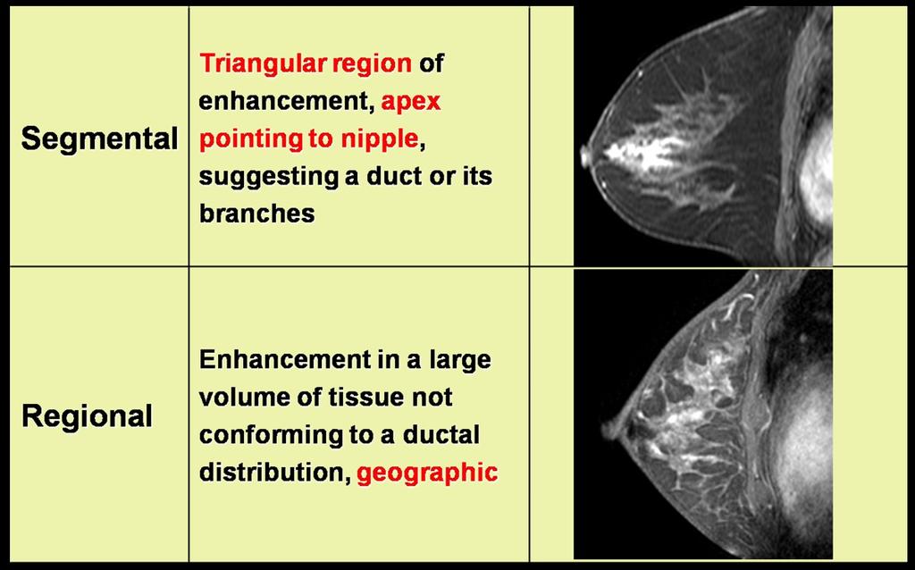
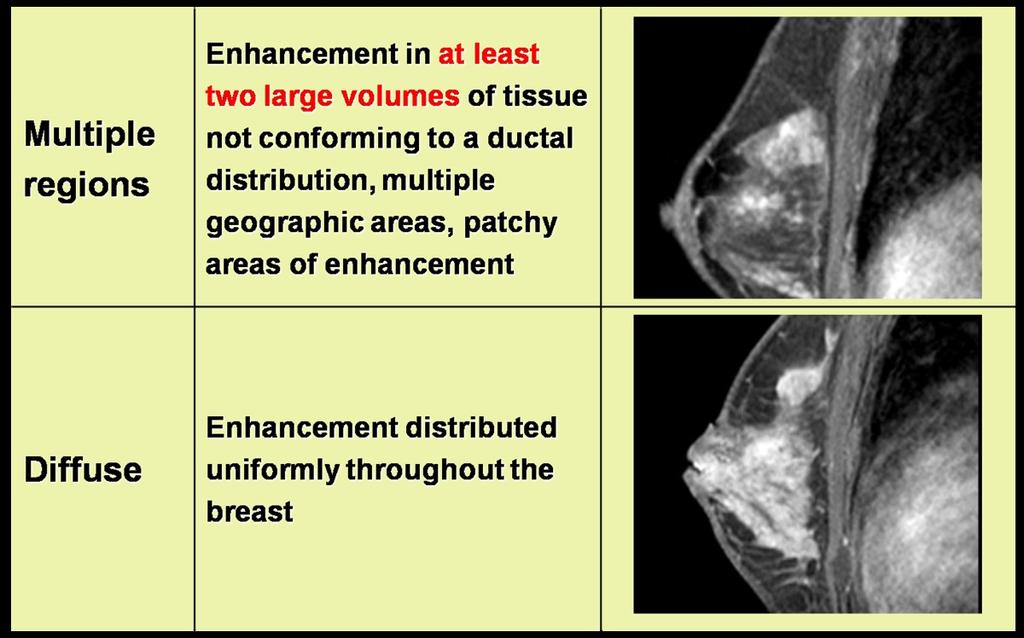
3 Background Non-mass-like lesions does not exist in standardized terminology of BI-RADS-US lexicon. DCIS and invasive lobular carcinoma that usually manifest as a non-mass-like lesion on US. Common benign pathologic disease such as fibrocystic change and fibroadenoma occasionally manifest as non-mass like lesions on US and MRI. However, image interpretations of non-mass like lesions are not familiar to radiologists and needs more experiences! <Distribution of NMLE>(Fig.1~3) Distribution Positive predictive malignancy Focal Not known Linear or branching ductal 24%- 85% Regional 21% Marked regional 59% Segmental 78% value (PPV) for value (PPV) for Linear or branching ductal Benign lesions : linear-ductal pattern Malignant lesion: branching-ductal pattern Differential diagnosis of ductal enhancement ;Atypical ductal hyperplasia, lobular carcinoma in situ and DCIS Fibrocystic change, ductal epithelial hyperplasia, and fibrosis Internal enhancement of NMLE (Fig4~6) Internal Enhancement Positive predictive malignancy Reticular Not known Stippled 25% Page 3 of 24
4 Heterogeneous 53% Clumped 60% Homogeneous 67% *Classification by Uematsu et al. Milk duct system: ''single ductal'' hypoechoic ''multiple ductal'' hypoechoic areas Intraductal lesions growing in the milk duct Glandular tissue ''non-ductal'' hypoechoic areas: Focal/ Segmental Lesion that differs from the surrounding glandular tissue or the same area in the contralateral breast Con#ned asymmetry with an indistinct shape on two different projections *Classification by Sotome et al. Duct dilatation DCIS, IDC with a predominant intraductal component, intraductal papilloma, ductal epithelial hyperplasia Multi-vesicular pattern DCIS, Matopathy Low echo area in the mammary gland DCIS, IDC with a predominant intraductal component, ILC, mastopathy Various pathology presented as NMLE Benign High risk Malignant Fibrocystic change Atypical ductal hyperplasia Ductal Carcinoma in situ Page 4 of 24
5 Ductal hyperplasia epithelial Intraductal papilloma Invasive ductal carcinoma Matopathy (focal adenosis) Lobular carcinoma in situ Invasive tubular carcinoma Hormonal Stimulation Invasive lobular carcinoma Inflammatory change Page 5 of 24

6 Images for this section: Fig. 1: Distribution of NMLE Page 6 of 24
7 Fig. 2: Distribution of NMLE Page 7 of 24

8 Fig. 3: Distribution of NMLE Fig. 4: Internal Enhancement Pattern of NMLE Page 8 of 24


9 Fig. 5: Internal Enhancement Pattern of NMLE Page 9 of 24
10 Fig. 6: Internal Enhancement Pattern of NMLE Page 10 of 24
11 Imaging findings OR Procedure details Fig.7 A-54-year-old-woman with invasive ductal carcinoma in right breast A. On US, non-mass-like-lesion is seen in 12 O'clock direction of left breast and the lesion is confirmed as fibrocystic change by core biopsy. B. On axial contrast enhanced T1WI, non-mass-like enhancement with focal distribution and internal stippled enhancement is seen in upper mid portion of left breast. Fig.8 A-48-year-old-woman with invasive ductal carcinoma in right breast A. On US, non-mass-like-lesion is seen in 10 O'clock direction of left breast and the lesion is confirmed as fibrocystic change by core biopsy. B. On sagittal contrast enhanced T1WI, non-mass-like enhancement with segmental distribution and internal clumped enhancement is seen in upper inner portion of left breast. Fig.9 A-52-year-old-woman with previous history of paraffin injection in her breasts A. On US, a single ductal non-mass-like-lesion is seen in 10 O'clock direction of right breast and the lesion is confirmed as moderate ductal epithelial hyperplasia by core biopsy. B. On axial contrast enhanced T1WI, non-mass-like enhancement with ductal distribution and internal homogeneous enhancement is seen in upper outer portion of right breast. Fig.10 A-60-year-old-woman with invasive ductal carcinoma in left breast A. On US, non-mass-like-lesion is seen in 6 O'clock direction of right breast and the lesion is confirmed as chronic granulomatous inflammation with necrosis by core biopsy. Page 11 of 24
12 B. On axial contrast enhanced T1WI, non-mass-like enhancement with regional distribution and internal heterogeneous enhancement is seen in upper outer portion of right breast. Fig.11 A-55-year-old-woman with hormone replacement therapy On axial contrast enhanced T1WI, bilateral, symmetric non-mass-like enhancements with regional distribution and internal stippled enhancement are seen in upper outer portion of both breasts. Fig.12 A-33-year-old-woman with no clinical symptom On US, dilated duct with intraductal echogenic material is seen in subareolar area of right breast. The lesion is confirmed as atypical ductal hyperplasia with fibrocystic change by surgical excision with US-guided needled localization. Fig.13 A-44-year-old-woman with no clinical symptom A. On left mammography, indistinct and round microcalcifications are regionally distributed in subareolar area of left breast. B. On US, duct ectasia with internal microcalcifications and intraductal echogenic material is seen in left subareolar area. C. On axial contrast enhanced T1WI, non-mass-like enhancement with segmental distribution and internal heterogeneous enhancement is seen in upper outer portion of right breast. The lesion is confirmed as ductal carcinoma in situ, low grade by breast conserving therapy. Fig.14 A-53-year-old-woman with ductalcarcinoma insitu in left breast A.On MMG, a focal asymmetric density with clustered microcalcifications is seen.b.on US, ductal change is seen. C,D. On MRI and a CAD system, a non-mass-like enhancement shows linear distribution and internal clumped enhancement. Page 12 of 24
13 E. MR-guided mammotome is done and the lesion is confirmed as DCIS. Fig.15 A-47-year-old-woman with invasive ductal carcinoma in left breast A.On MMG, a focal asymmetric density with clustered microcalcifications is seen.b.on US, ductal change is seen. C. On CT, non-mass like enhancement is seen. D,E. On MRI and CAD system, non-mass like enhancement shows segmetal distriburion and internal heterogeneous enhancement. F. On gross specimen, the lesion is confirmed as invasive ductal carcinoma. Fig.16 A-56-year-old-woman with invasive ductal carcinoma in left breast A.On MMG, segmentally distributed microcalcifications are regionally distrubuted in LUIQ.B.On US, indistinct irregular shaped hypoechoic mass is seen in left breast. C,D. On CT and MRI, non-mass like enhancement shows regional distribution and heterogeneous enhancement. Fig.17 A-44-year-old-woman with no clinical symptom A. On US, a non-ductal hypoechoic area is seen in subareolar area of left breast. B. On axial contrast enhanced T1WI, non-mass-like enhancement with regional distribution and internal clumped enhancement is seen in her left breast. The lesion is confirmed as invasive lobular carcinoma by breast conserving therapy. Page 13 of 24


14 Images for this section: Fig. 7: Fibrocystic Change Fig. 8: Fibrocystic Change Page 14 of 24


15 Fig. 9: Ductal epithelial hyperplasia Fig. 10: Granulomatous Mastitis Page 15 of 24

16 Fig. 11: Hormonal Replacement Treatment Page 16 of 24


17 Fig. 16: Invasive Ductal Carcinoma Fig. 15: Invasive Ductal Carcinoma Page 17 of 24

18 Fig. 14: DCIS Page 18 of 24

19 Fig. 13: DCIS Page 19 of 24


20 Fig. 17: Invasive lobular carcinoma Page 20 of 24
21 Fig. 12: Atypical Ducal Hyperplasia Page 21 of 24
22 Conclusion Non-mass like lesions on breast US and MRI have various pathologic diagnoses from benign to malignant lesions. On US, non-mass like lesions associated with segmental or linear distributed suspicious microcalcifications or intraductal nodules prefer malignancy. On dynamic MRI, non-mass like lesions with ductal or segmental distribution and heterogeneous or clumped enhancements prefer malignancy. On the other hand, kinetic assessment on MRI is not helpful to distinguish benign from malignant non-mass like lesions. Page 22 of 24

23 Images for this section: Fig. 18: Guidelines for interpretation of non-mass like breast lesions on US and MRI Page 23 of 24
24 References Breast Cancer 2007, 14(4): Breast Cancer 2012, 19(4): Radiology 2006, 238(1):42-53 Radiology 2001, 219(2): AJR 2003, 181(2): Radiologic clinics of North America 2004, 42(5): , vii. AJR 2006, 187(2): Medical physics 2008, 35(7): Page 24 of 24
Radiologic and pathologic correlation of non-mass like breast lesions on US and MRI: Benign, high risk, versus malignant
 Radiologic and pathologic correlation of non-mass like breast lesions on US and MRI: Benign, high risk, versus malignant Poster No.: C-1161 Congress: ECR 2013 Type: Educational Exhibit Authors: J. Kwak,
Pathologic outcomes of coarse heterogeneous calcifications detected on mammography
 Pathologic outcomes of coarse heterogeneous calcifications detected on mammography Poster No.: C-1957 Congress: ECR 2011 Type: Scientific Paper Authors: H. J. Lim, K. R. Cho, K. W. Hwang, B. K. Seo, O.
Radiologic Findings of Mucocele-like Tumors of the breast: Can we differentiate pure benign from associated with high risk lesions?
 Radiologic Findings of Mucocele-like Tumors of the breast: Can we differentiate pure benign from associated with high risk lesions? Poster No.: C-0332 Congress: ECR 2014 Type: Educational Exhibit Authors:
Evaluation of BI-RADS 3 lesions in women with a high risk of hereditary breast cancer.
 Evaluation of BI-RADS 3 lesions in women with a high risk of hereditary breast cancer. Poster No.: C-0346 Congress: ECR 2014 Type: Scientific Exhibit Authors: A. Thomas 1, R. Dominguez Oronoz 1, S. Roche
Atypical ductal hyperplasia diagnosed at ultrasound guided biopsy of breast mass
 Atypical ductal hyperplasia diagnosed at ultrasound guided biopsy of breast mass Poster No.: C-1483 Congress: ECR 2014 Type: Authors: Keywords: DOI: Scientific Exhibit J. Cho, J. Chung, E. S. Cha, J. E.
Breast calcification: Management and Pictorial Review
 Breast calcification: Management and Pictorial Review Poster No.: C-0692 Congress: ECR 2014 Type: Educational Exhibit Authors: V. de Lara Bendahan, M. F. Ramos Solis, A. Amador Gil, C. 1 2 3 2 4 4 Gómez
Vacuum-assisted breast biopsy using computer-aided 3.0 T- MRI guidance: diagnostic performance in 173 lesions
 Vacuum-assisted breast biopsy using computer-aided 3.0 T- MRI guidance: diagnostic performance in 173 lesions Poster No.: C-2870 Congress: ECR 2017 Type: Scientific Exhibit Authors: A. Pozzetto, L. Camera,
Spectrum of findings of sclerosing adenosis at breast MRI.
 Spectrum of findings of sclerosing adenosis at breast MRI. Poster No.: C-0738 Congress: ECR 2012 Type: Scientific Exhibit Authors: F. Vasselli 1, F. Pediconi 2, M. Telesca 2, M. Luciani 2, V. Casali 2,
MRI BI-RADS: How to make it out?
 MRI BI-RADS: How to make it out? Poster No.: C-1850 Congress: ECR 2016 Type: Educational Exhibit Authors: M. Ben Ammar, A. Ben Miled, O. Ghdes, S. Harguem, A. Gaja, N. Mnif; Tunis/TN Keywords: Breast,
Hyperechoic breast lesions can be malignant.
 Hyperechoic breast lesions can be malignant. Poster No.: C-0041 Congress: ECR 2015 Type: Educational Exhibit Authors: G. Babu, R. bradley; Edinburgh/UK Keywords: Breast, Ultrasound, Biopsy, Cancer DOI:
Categorical Classification of Spiculated Mass on Breast MRI
 Categorical Classification of Spiculated Mass on Breast MRI Poster No.: C-1974 Congress: ECR 2013 Type: Authors: Scientific Exhibit Y. Kanda 1, S. Kanao 2, M. Kataoka 2, K. Togashi 2 ; 1 Kyoto City/JP,
Triple-negative breast cancer: which typical features can we identify on conventional and MRI imaging?
 Triple-negative breast cancer: which typical features can we identify on conventional and MRI imaging? Poster No.: C-1862 Congress: ECR 2013 Type: Educational Exhibit Authors: V. Bertani 1, A. Gualano
Excisional biopsy or long term follow-up results in breast high-risk lesions diagnosed at core needle biopsy
 Excisional biopsy or long term follow-up results in breast high-risk lesions diagnosed at core needle biopsy Poster No.: C-2515 Congress: ECR 2015 Type: Authors: Scientific Exhibit Ö. S. Okcu 1, A. Oktay
Aims and objectives. Page 2 of 10
 Diagnostic performance of automated breast volume scanner (ABVS) versus hand-held ultrasound (HHUS) as second look for breast lesions detected only on magnetic resonance imaging. Poster No.: C-1701 Congress:
Cairo/EG, Khartoum/SD, London/UK Biological effects, Diagnostic procedure, Ultrasound, Mammography, Breast /ecr2015/C-0107
 Role of sono-mammography in the evaluation of clinically palapble breast masses during pregnancy & lactation with differentaition between true patholgical & false physiological lobular hyperlpasia.sudanese
Digital breast tomosynthesis (DBT) occult breast cancers: clinical, radiological and histopathological features.
 occult breast cancers: clinical, radiological and histopathological features.") Digital breast tomosynthesis (DBT) occult breast cancers: clinical, radiological and histopathological features. Poster No.: C-1707 Congress: ECR 2015 Type: Scientific Exhibit Authors: V. Vinci 1, A. Iqbal
Digital breast tomosynthesis (DBT) occult breast cancers: clinical, radiological and histopathological features. Poster No.: C-1707 Congress: ECR 2015 Type: Scientific Exhibit Authors: V. Vinci 1, A. Iqbal
BI-RADS 3 category, a pain in the neck for the radiologist which technique detects more cases?
 BI-RADS 3 category, a pain in the neck for the radiologist which technique detects more cases? Poster No.: B-0966 Congress: ECR 2013 Type: Scientific Paper Authors: J. Etxano Cantera, I. Simon-Yarza, G.
BI-RADS 3, 4 and 5 lesions on US: Five categories and their diagnostic efficacy and pitfalls in interpretation
 BI-RADS 3, 4 and 5 lesions on US: Five categories and their diagnostic efficacy and pitfalls in interpretation e-poster: C-118 Congress: ECR 2008 Type: Educational Exhibit Topic: Breast / Ultrasound Authors:
Breast cancer tumor size: Correlation between MRI and histopathology
 Breast cancer tumor size: Correlation between MRI and histopathology Poster No.: C-0409 Congress: ECR 2010 Type: Topic: Scientific Exhibit Breast Authors: H. Khan, M. Hoosein, M. Alattar, S. Tenant, L.
DCIS of the Breast--MRI findings with mammographic correlation.
 DCIS of the Breast--MRI findings with mammographic correlation. Poster No.: C-1560 Congress: ECR 2013 Type: Educational Exhibit Authors: N. B. Ibrahim, P. Morris, S. ANANDAN; Burlington, MA/US Keywords:
Sonographic and Mammographic Features of Phyllodes Tumours of the Breast: Correlation with Histological Grade
 Sonographic and Mammographic Features of Phyllodes Tumours of the Breast: Correlation with Histological Grade Poster No.: C-0046 Congress: ECR 2014 Type: Authors: Keywords: DOI: Scientific Exhibit C. Y.
Intracystic Papillary Carcinoma of the Breast: Clinical and Radiological Findings with Histopathologic Correlation
 Intracystic Papillary Carcinoma of the Breast: Clinical and Radiological Findings with Histopathologic Correlation Poster No.: C-2078 Congress: ECR 2015 Type: Scientific Exhibit Authors: S. S. AlSharif
Correlation Between BIRADS Classification and Ultrasound -guided Tru-Cut Biopsy Results of Breast Lesions: Retrospective Analysis of 285 Patients
 Correlation Between BIRADS Classification and Ultrasound -guided Tru-Cut Biopsy Results of Breast Lesions: Retrospective Analysis of 285 Patients Poster No.: C-1433 Congress: ECR 2014 Type: Scientific
Role of positron emission mammography (PEM) for assessment of axillary lymph node status in patients with breast cancer
 for assessment of axillary lymph node status in patients with breast cancer") Role of positron emission mammography (PEM) for assessment of axillary lymph node status in patients with breast cancer Poster No.: C-1260 Congress: ECR 2011 Type: Scientific Paper Authors: K. M. Kulkarni,
Role of positron emission mammography (PEM) for assessment of axillary lymph node status in patients with breast cancer Poster No.: C-1260 Congress: ECR 2011 Type: Scientific Paper Authors: K. M. Kulkarni,
Breast Lesion Excision System-Intact (BLES): A Stereotactic Method of Biopsy of Suspicius Non-Palpable Mammographic Lesions.
: A Stereotactic Method of Biopsy of Suspicius Non-Palpable Mammographic Lesions.") Breast Lesion Excision System-Intact (BLES): A Stereotactic Method of Biopsy of Suspicius Non-Palpable Mammographic Lesions. Poster No.: C-1595 Congress: ECR 2014 Type: Authors: Scientific Exhibit I. Georgiou
Breast Lesion Excision System-Intact (BLES): A Stereotactic Method of Biopsy of Suspicius Non-Palpable Mammographic Lesions. Poster No.: C-1595 Congress: ECR 2014 Type: Authors: Scientific Exhibit I. Georgiou
Slowly growing malignant nodules and rapidly growing benign nodules: Evaluation of the value of volume doubling time
 Slowly growing malignant nodules and rapidly growing benign nodules: Evaluation of the value of volume doubling time Poster No.: C-208 Congress: ECR 2009 Type: Educational Exhibit Topic: Chest Authors:
Triple Negative Breast Cancer: Clinical Presentation and Multimodality Imaging Characteristics
 Triple Negative Breast Cancer: Clinical Presentation and Multimodality Imaging Characteristics Poster No.: R-0141 Congress: RANZCR-AOCR 2012 Type: Scientific Exhibit Authors: O. H. Woo, S. Jang, K. R.
Microcalcifications detected on mammography classified as BIRADS 4 and 5 and their correlations with histopatologic findigns
 Microcalcifications detected on mammography classified as BIRADS 4 and 5 and their correlations with histopatologic findigns Poster No.: C-0401 Congress: ECR 2010 Type: Educational Exhibit Topic: Breast
Intracystic papillary carcinoma of the breast
 Intracystic papillary carcinoma of the breast Poster No.: C-1932 Congress: ECR 2011 Type: Educational Exhibit Authors: V. Dimarelos, F. TZIKOS, N. Kotziamani, G. Rodokalakis, 1 2 3 1 1 1 2 T. MALKOTSI
Breast ultrasound appearances after Mammotome vacuumassisted
 Breast ultrasound appearances after Mammotome vacuumassisted biopsy. Poster No.: C-1924 Congress: ECR 2011 Type: Educational Exhibit Authors: R. Patel 1, G. R. Kaplan 2 ; 1 London/UK, 2 Herts/UK Keywords:
3-marker technique for the localisation and delineation of residual tumour bed following neoadjuvant chemotherapy in patients within the I-SPY 2 trial
 3-marker technique for the localisation and delineation of residual tumour bed following neoadjuvant chemotherapy in patients within the I-SPY 2 trial Poster No.: C-2125 Congress: ECR 2016 Type: Authors:
Quantitative imaging of hepatic cirrhosis on abdominal CT images
 Quantitative imaging of hepatic cirrhosis on abdominal CT images Poster No.: C-0556 Congress: ECR 2013 Type: Authors: Keywords: DOI: Scientific Exhibit S. Kido, A. Nakamura, Y. Hirano; Ube/JP Cirrhosis,
Malignant transformation of fibroadenomas
 Malignant transformation of fibroadenomas Poster No.: C-2503 Congress: ECR 2013 Type: Educational Exhibit Authors: L. N. Elias, M. A. Rudner, L. M. Yano, P. C. Moraes, Y. 1 1 1 1 1 1 2 1 2 Chang, M. B.
Characterisation of cervical lymph nodes by US and PET-CT
 Characterisation of cervical lymph nodes by US and PET-CT Poster No.: C-1807 Congress: ECR 2010 Type: Educational Exhibit Topic: Head and Neck Authors: J. I. Garcia Gomez; Mexico City/MX Keywords: cervical
Single cold nodule in Graves' disease: benign vs malignant
 Single cold nodule in Graves' disease: benign vs malignant Poster No.: C-0073 Congress: ECR 2011 Type: Authors: Keywords: DOI: Scientific Paper L. I. Sonoda, M. Halim, K. Balan; Cambridge/UK Head and neck,
THI-RADS. US differentiation of thyroid lesions.
 THI-RADS. US differentiation of thyroid lesions. Poster No.: C-0864 Congress: ECR 2015 Type: Scientific Exhibit Authors: A. N. Sencha, Y. Patrunov, M. S. Mogutov, E. Penyaeva, A. 1 1 1 2 1 1 1 2 Gruzdev,
THI-RADS. US differentiation of thyroid lesions.
 THI-RADS. US differentiation of thyroid lesions. Poster No.: C-0864 Congress: ECR 2015 Type: Scientific Exhibit Authors: A. N. Sencha, Y. Patrunov, M. S. Mogutov, E. Penyaeva, A. 1 1 1 2 1 1 1 2 Gruzdev,
Breast asymmetries in mammography: Management
 Breast asymmetries in mammography: Management Poster No.: C-1026 Congress: ECR 2015 Type: Educational Exhibit Authors: V. de Lara Bendahan 1, F. J. Hidalgo Ramos 2, J. L. Ortega Garcia 3, Keywords: DOI:
Cierny-Mader classification of chronic osteomyelitis: Preoperative evaluation with cross-sectional imaging
 Cierny-Mader classification of chronic osteomyelitis: Preoperative evaluation with cross-sectional imaging Poster No.: C-590 Congress: ECR 2009 Type: Topic: Educational Exhibit Musculoskeletal Authors:
Diffuse high-attenuation within mediastinal lymph nodes on non-enhanced CT scan: Usefulness in the prediction of benignancy
 Diffuse high-attenuation within mediastinal lymph nodes on non-enhanced CT scan: Usefulness in the prediction of benignancy Poster No.: C-1785 Congress: ECR 2012 Type: Authors: Keywords: DOI: Scientific
Correlation between lesion type and the additional value of digital breast tomosynthesis
 Correlation between lesion type and the additional value of digital breast tomosynthesis Poster No.: C-1604 Congress: ECR 2011 Type: Scientific Exhibit Authors: C. Van Ongeval, L. Cockmartin, A. Van Steen,
MR-guided prostatic biopsy at 3T: the role of PI-RADS-score: a histopahologic-radiologic correlation
 MR-guided prostatic biopsy at 3T: the role of PI-RADS-score: a histopahologic-radiologic correlation Poster No.: B-0969 Congress: ECR 2015 Type: Scientific Paper Authors: A. Malich; Nordhausen/DE Keywords:
64-MDCT imaging of the pancreas: Scan protocol optimisation by different scan delay regimes
 64-MDCT imaging of the pancreas: Scan protocol optimisation by different scan delay regimes Poster No.: C-051 Congress: ECR 2009 Type: Scientific Exhibit Topic: Abdominal and Gastrointestinal Authors:
Spiculated breast masses on MRI: Which category should we choose, 4 or 5?
 Spiculated breast masses on MRI: Which category should we choose, 4 or 5? Poster No.: C-1394 Congress: ECR 2015 Type: Scientific Exhibit Authors: N. Onishi, S. Kanao, M. Kataoka, M. Kawai, M. Iima, A.
Granulomatous mastitis: Radio-pathologic correlation and management
 Granulomatous mastitis: Radio-pathologic correlation and management Poster No.: C-1418 Congress: ECR 2014 Type: Educational Exhibit Authors: S. E. Song, B. K. Seo, K. R. Cho, O. H. Woo, Y.-S. Kim ; 1 1
Breast Pathology in Men: Radiologic-Pathologic Correlation
 Breast Pathology in Men: Radiologic-Pathologic Correlation Poster No.: C-0243 Congress: ECR 2012 Type: Scientific Exhibit Authors: G. Garrido; Málaga/ES Keywords: Breast, Ultrasound, Mammography, Biopsy,
Diffusion-weighted MRI (DWI) "claw sign" is useful in differentiation of infectious from degenerative Modic I signal changes of the spine
 claw sign is useful in differentiation of infectious from degenerative Modic I signal changes of the spine") Diffusion-weighted MRI (DWI) "claw sign" is useful in differentiation of infectious from degenerative Modic I signal changes of the spine Poster No.: C-0894 Congress: ECR 2012 Type: Scientific Exhibit
Diffusion-weighted MRI (DWI) "claw sign" is useful in differentiation of infectious from degenerative Modic I signal changes of the spine Poster No.: C-0894 Congress: ECR 2012 Type: Scientific Exhibit
Ethanol ablation of benign thyroid cysts and predominantly cystic thyroid nodules: factors that predict outcome.
 Ethanol ablation of benign thyroid cysts and predominantly cystic thyroid nodules: factors that predict outcome. Poster No.: C-0322 Congress: ECR 2014 Type: Authors: Keywords: DOI: Scientific Exhibit J.
Cavitary lung lesion: Two different diagnosis with similar appearence
 Cavitary lung lesion: Two different diagnosis with similar appearence Poster No.: P-0043 Congress: ESTI 2015 Type: Educational Poster Authors: M. Yesildag, H. Kalkan, K. Ödev; Konya/TR Keywords: Infection,
Idiopathic dilatation of the pulmonary artery : radiographic and MDCT features in 6 cases
 Idiopathic dilatation of the pulmonary artery : radiographic and MDCT features in 6 cases Poster No.: P-0075 Congress: ESTI 2014 Type: Authors: Educational Poster J. J. Woo 1, K. Y. Lee 2, Y. Cho 1, J.
Prostate biopsy: MR imaging to the rescue
 Prostate biopsy: MR imaging to the rescue Poster No.: C-1855 Congress: ECR 2014 Type: Educational Exhibit Authors: N. V. V. B. Marques 1, J. Ip 1, A. Loureiro 2, J. Niza 1, M. Palmeiro 2, Keywords: DOI:
Cognitive target MRI-TRUS fusion biopsies of MRI detected PIRADS 4 and 5 lesions
 Cognitive target MRI-TRUS fusion biopsies of MRI detected PIRADS 4 and 5 lesions Poster No.: B-0704 Congress: ECR 2015 Type: Scientific Paper Authors: P. P. van Westerveld, J. Vriesema, J. H. W. van den
Identification and numbering of lumbar vertebrae using various anatomical landmarks on MRI of lumbosacral spine
 Identification and numbering of lumbar vertebrae using various anatomical landmarks on MRI of lumbosacral spine Poster No.: C-2125 Congress: ECR 2015 Type: Authors: Scientific Exhibit S. patil 1, A. M.
Invasive lobular carcinoma of the breast; spectrum of imaging findings.
 Invasive lobular carcinoma of the breast; spectrum of imaging findings. Poster No.: C-0847 Congress: ECR 2014 Type: Educational Exhibit Authors: D. Mandich, T. Diaz de Bustamante, L. Koren, M. Arroyo,
Radiological features of Legionella Pneumophila Pneumonia
 Radiological features of Legionella Pneumophila Pneumonia Poster No.: E-0048 Congress: ESTI 2012 Type: Scientific Exhibit Authors: M. Vinciguerra, L. Stefanetti, E. Teti, G. Argentieri, L. G. 1 1 1 1 1
Monitoring neo-adjuvant chemotherapy: comparison of contrast-enhanced spectral mammography (CESM) and MRI versus breast cancer characteristics
 and MRI versus breast cancer characteristics") Monitoring neo-adjuvant chemotherapy: comparison of contrast-enhanced spectral mammography (CESM) and MRI versus breast cancer characteristics Poster No.: B-1062 Congress: ECR 2016 Type: Scientific Paper
Monitoring neo-adjuvant chemotherapy: comparison of contrast-enhanced spectral mammography (CESM) and MRI versus breast cancer characteristics Poster No.: B-1062 Congress: ECR 2016 Type: Scientific Paper
AFib is the most common cardiac arrhythmia and its prevalence and incidence increases with age (Fuster V. et al. Circulation 2006).
.") Feasibility, image quality and radiation dose of coronary CT angiography (CCTA) in patients with atrial fibrillation using a new generation 256 multi-detector CT (MDCT) Poster No.: C-2378 Congress: ECR
Feasibility, image quality and radiation dose of coronary CT angiography (CCTA) in patients with atrial fibrillation using a new generation 256 multi-detector CT (MDCT) Poster No.: C-2378 Congress: ECR
CDIS: what's beyond microcalcifications? - Pictorial essay
 CDIS: what's beyond microcalcifications? - Pictorial essay Poster No.: C-1096 Congress: ECR 2014 Type: Educational Exhibit Authors: R. N. Lucas, C. A. S. Ruano, I. Oliveira, J. M. G. Lourenco, Z. 1 1 1
The role of apparent diffusion coefficient (ADC) and relative ADC in the evaluation of breast masses
 and relative ADC in the evaluation of breast masses") The role of apparent diffusion coefficient (ADC) and relative ADC in the evaluation of breast masses Poster No.: C-1749 Congress: ECR 2014 Type: Scientific Exhibit Authors: U. Aksoy Ozcan 1, A. Öz 2, S.
The role of apparent diffusion coefficient (ADC) and relative ADC in the evaluation of breast masses Poster No.: C-1749 Congress: ECR 2014 Type: Scientific Exhibit Authors: U. Aksoy Ozcan 1, A. Öz 2, S.
Biliary tree dilation - and now what?
 Biliary tree dilation - and now what? Poster No.: C-1767 Congress: ECR 2012 Type: Educational Exhibit Authors: I. Ferreira, A. B. Ramos, S. Magalhães, M. Certo; Porto/PT Keywords: Pathology, Diagnostic
Adenomyosis by myometrial Invasion of endometriosis: Comparison with typical adenomyosis
 Adenomyosis by myometrial Invasion of endometriosis: Comparison with typical adenomyosis Poster No.: C-1294 Congress: ECR 2010 Type: Scientific Exhibit Topic: Genitourinary Authors: S. Moon, H. K. Lim,
Tissue characterisation, Cancer, Quality assurance /ecr2015/B-0553
 Role of DWI at prostatic lesions at 3T-MRI in the discrimination of grading: correlation of imaging, quantitative analysis and pathology at 189 MR-guided prostate biopsies Poster No.: B-0553 Congress:
Purpose. Methods and Materials. Results
 Prevalence and significance of hypoattenuating hepatic lesions deemed too small to characterise: How are we following up these lesions and what are the outcomes? Poster No.: C-014 Congress: ECR 2009 Type:
Assessment of extent of disease: digital breast tomosynthesis (DBT) versus full-field digital mammography (FFDM)
 versus full-field digital mammography (FFDM)") Assessment of extent of disease: digital breast tomosynthesis (DBT) versus full-field digital mammography (FFDM) Poster No.: C-1237 Congress: ECR 2012 Type: Scientific Paper Authors: N. Seo 1, H. H. Kim
Assessment of extent of disease: digital breast tomosynthesis (DBT) versus full-field digital mammography (FFDM) Poster No.: C-1237 Congress: ECR 2012 Type: Scientific Paper Authors: N. Seo 1, H. H. Kim
Evaluation of thyroid nodules: prediction and selection of malignant nodules for FNA (cytology)
") Evaluation of thyroid nodules: prediction and selection of malignant nodules for FNA (cytology) Poster No.: C-0221 Congress: ECR 2014 Type: Authors: Keywords: DOI: Scientific Exhibit E. Papadaki, I. Tritou,
Evaluation of thyroid nodules: prediction and selection of malignant nodules for FNA (cytology) Poster No.: C-0221 Congress: ECR 2014 Type: Authors: Keywords: DOI: Scientific Exhibit E. Papadaki, I. Tritou,
Popliteal pterygium syndrome
 Popliteal pterygium syndrome Poster No.: C-1816 Congress: ECR 2011 Type: Educational Exhibit Authors: L. B. S. Santos, J. L. D. O. Schiavon, O. O. Guimaraes Neto, 1 1 2 3 1 1 C. A. P. Braga, R. S. LEMOS,
Intrahepatic cholangiocarcinoma: diffusion-weighted MR imaging findings
 Intrahepatic cholangiocarcinoma: diffusion-weighted MR imaging findings Poster No.: C-1979 Congress: ECR 2013 Type: Authors: Keywords: DOI: Scientific Exhibit S. Schmidt, A. Pomoni, F. Becce, A. Denys,
Targeted MRI/TRUS fusion-guided biopsy in men with previous negative prostate biopsies: initial experience.
 Targeted MRI/TRUS fusion-guided biopsy in men with previous negative prostate biopsies: initial experience. Poster No.: C-0382 Congress: ECR 2016 Type: Scientific Exhibit Authors: A. I. Gromov 1, V. V.
High density thrombi of pulmonary embolism on precontrast CT scan: Is it dangerous?
 High density thrombi of pulmonary embolism on precontrast CT scan: Is it dangerous? Poster No.: C-1753 Congress: ECR 2013 Type: Authors: Keywords: DOI: Scientific Exhibit B. Y. Lee, H. R. KIM, J. I. Jung,
The solitary pulmonary nodule: Assessing the success of predicting malignancy
 The solitary pulmonary nodule: Assessing the success of predicting malignancy Poster No.: C-0829 Congress: ECR 2010 Type: Scientific Exhibit Topic: Chest Authors: R. W. K. Lindsay, J. Foster, K. McManus;
Effect of intravenous contrast medium administration on prostate diffusion-weighted imaging
 Effect of intravenous contrast medium administration on prostate diffusion-weighted imaging Poster No.: C-1766 Congress: ECR 2015 Type: Authors: Keywords: DOI: Scientific Exhibit J. Bae, C. K. Kim, S.
Valsalva-manoeuvre or prone belly position for computed tomography (CT) scan when an orbita varix is suspected: a single-case study.
 scan when an orbita varix is suspected: a single-case study.") Valsalva-manoeuvre or prone belly position for computed tomography (CT) scan when an orbita varix is suspected: a single-case study. Poster No.: C-0512 Congress: ECR 2012 Type: Authors: Keywords: DOI:
Valsalva-manoeuvre or prone belly position for computed tomography (CT) scan when an orbita varix is suspected: a single-case study. Poster No.: C-0512 Congress: ECR 2012 Type: Authors: Keywords: DOI:
Basic low - field MR imaging of meniscal injuries in children.
 Basic low - field MR imaging of meniscal injuries in children. Poster No.: C-2365 Congress: ECR 2012 Type: Authors: Keywords: DOI: Scientific Exhibit A. Yakimov, M. Nikonova, E. Prokhorova, D. Vybornov,
The Role of Radionuclide Lymphoscintigraphy in the Diagnosis of Lymphedema of the Extremities
 The Role of Radionuclide Lymphoscintigraphy in the Diagnosis of Lymphedema of the Extremities Poster No.: C-1229 Congress: ECR 2014 Type: Authors: Keywords: DOI: Scientific Exhibit M. Osher 1, A. Pallas
MRI features of Triple-negative breast cancer: our experience.
 MRI features of Triple-negative breast cancer: our experience. Poster No.: C-1852 Congress: ECR 2013 Type: Scientific Exhibit Authors: V. Bertani, A. Gualano, V. Londero, A. Dal Col, M. Marcon, P. 1 2
Diffuse Alveolar Hemorrhage: Initial and Follow-up HRCT Features
 Diffuse Alveolar Hemorrhage: Initial and Follow-up HRCT Features Poster No.: E-0037 Congress: ESTI 2012 Type: Authors: Keywords: Scientific Exhibit M. Y. Kim; Seoul/KR Lung, CT-High Resolution, CT, Computer
Ductal carcinoma in situ: ultrasound, mammography and MRI features with pathologic correlation
 Ductal carcinoma in situ: ultrasound, mammography and MRI features with pathologic correlation Poster No.: C-2252 Congress: ECR 2013 Type: Educational Exhibit Authors: L. Fernandes, H. A. M. R. Tinto,
A pictorial essay depicting CT and MR characteristic of adrenal pathologies: Indian study
 A pictorial essay depicting CT and MR characteristic of adrenal pathologies: Indian study Poster No.: C-0703 Congress: ECR 2011 Type: Educational Exhibit Authors: A. J. B. Baxi, K. L. Tourani, N. R. Thanugonda,
Pharmacokinetic evaluation of DCIS
 Pharmacokinetic evaluation of DCIS Poster No.: C-0412 Congress: ECR 2010 Type: Topic: Authors: Keywords: DOI: Scientific Exhibit Breast H. G. Toonen, R. Mann, H. Huisman, J. Veltman, C. Boetes; Nijmegen/NL
Imaging spectrum of angiosarcoma of breast
 Imaging spectrum of angiosarcoma of breast Poster No.: C-1097 Congress: ECR 2015 Type: Educational Exhibit Authors: T. Sehgal, S. K. Ramani, N. Nair, A. Patil, M. H. Thakur; Mumbai/ IN Keywords: Neoplasia,
Evaluation of surgical margins by specimen in impalpable breast carcinoma: a radiopathological correlation
 Evaluation of surgical margins by specimen in impalpable breast carcinoma: a radiopathological correlation Poster No.: C-1146 Congress: ECR 2014 Type: Scientific Exhibit Authors: D. Mandich, L. Koren,
Pleomorphic adenoma head and neck
 Pleomorphic adenoma head and neck Poster No.: C-1042 Congress: ECR 2015 Type: Educational Exhibit Authors: M. E. Pérez Montilla, I. Bravo Rey, E. Roldán Romero, F. BravoRodríguez; Cordoba/ES Keywords:
Computed tomography and Modified RECIST criteria for assessment of response in malignant pleural mesothelioma
 Computed tomography and Modified RECIST criteria for assessment of response in malignant pleural mesothelioma Poster No.: C-0729 Congress: ECR 2013 Type: Scientific Exhibit Authors: A. Marin, I. Pozek,
Soft tissues lymphoma, the great pretender. MRI diagnostic keys.
 Soft tissues lymphoma, the great pretender. MRI diagnostic keys. Poster No.: C-2133 Congress: ECR 2015 Type: Educational Exhibit Authors: L. Caminero, M. E. Banegas Illescas, M. L. Rozas, M. Y. Torres,
PI-RADS classification: prognostic value for prostate cancer grading
 PI-RADS classification: prognostic value for prostate cancer grading Poster No.: C-1622 Congress: ECR 2014 Type: Scientific Exhibit Authors: I. Platzek, A. Borkowetz, T. Paulus, T. Brauer, M. Wirth, M.
Imaging findings of malignancies arising within benign breast lesions
 Imaging findings of malignancies arising within benign breast lesions Poster No.: C-0194 Congress: ECR 2015 Type: Authors: Educational Exhibit F. Can 1, Ö. S. Okcu 2, A. Oktay 2, I. G. Bilgen 3 ; 1 Kutahya/TR,
Seemingly isolated greater trochanter fractures do not exist
 Seemingly isolated greater trochanter fractures do not exist Poster No.: B-0950 Congress: ECR 2012 Type: Scientific Paper Authors: D. Dunker, J. H. Göthlin, M. Geijer ; Gothenburg/SE, Lund/SE Keywords:
Feasibility of magnetic resonance elastography using myofascial phantom model
 Feasibility of magnetic resonance elastography using myofascial phantom model Poster No.: C-0971 Congress: ECR 2013 Type: Scientific Exhibit Authors: H. J. Kang, J.-S. Yoon, S.-J. Hong, C.-H. Oh, S. H.
Inflammatory Breast Carcinoma: Mammographic, Ultrasonographic, MRI and Pathologic Findings
 Inflammatory Breast Carcinoma: Mammographic, Ultrasonographic, MRI and Pathologic Findings Poster No.: C-2248 Congress: ECR 2013 Type: Educational Exhibit Authors: L. Fernandes, J. Lopes Dias, H. A. M.
Comparison of Image quality in temporal bone MRI at 3T using 2D selective RF excitation versus a routine SPACE sequence
 Comparison of Image quality in temporal bone MRI at 3T using 2D selective RF excitation versus a routine SPACE sequence Poster No.: C-1065 Congress: ECR 2015 Type: Authors: Keywords: DOI: Scientific Exhibit
gg4-related inflammatory pseudotumour of the trigeminal nerve: imaging findings and clinical features
 gg4-related inflammatory pseudotumour of the trigeminal nerve: imaging findings and clinical features Poster No.: C-2603 Congress: ECR 2013 Type: Scientific Exhibit Authors: Y. Kawamura, Y. Kikuchi, I.
Scientific Exhibit Authors: V. Moustakas, E. Karallas, K. Koutsopoulos ; Rodos/GR, 2
 Diagnosis of Acute Appendicitis: the role of Color Doppler Ultrasound as first-line imaging method and evaluation of the higher diagnostic performances of CT against its disadvantages. Poster No.: C-0708
"Ultrasound measurements of the lateral ventricles in neonates: A comparison of multiple measurements methods."
 "Ultrasound measurements of the lateral ventricles in neonates: A comparison of multiple measurements methods." Poster No.: C-1557 Congress: ECR 2014 Type: Authors: Keywords: DOI: Educational Exhibit I.
Role of ultrasound in the evaluation of the ileocecal valve
 Role of ultrasound in the evaluation of the ileocecal valve Poster No.: C-1581 Congress: ECR 2010 Type: Scientific Exhibit Topic: GI Tract Authors: M. Mohammed, M. Hussain, U. Momin, S. Lakhtakia, N. D.
The imaging evaluation of breast implants
 The imaging evaluation of breast implants Poster No.: C-0654 Congress: ECR 2014 Type: Authors: Keywords: DOI: Educational Exhibit N. Rotaru; Chisinau/MD Neoplasia, Education and training, Cancer, Imaging
Strain histogram analysis for elastography in breast cancer diagnosis
 Strain histogram analysis for elastography in breast cancer diagnosis Poster No.: C-1854 Congress: ECR 2015 Type: Scientific Exhibit Authors: J. F. Carlsen, C. Ewertsen, S. Sletting, M. B. Nielsen; Copenhagen/DK
PGMI classification of screening mammograms prior to interval cancer. Comparison with radiologists' consensus classification.
 PGMI classification of screening mammograms prior to interval cancer. Comparison with radiologists' consensus classification. Poster No.: C-0673 Congress: ECR 2016 Type: Authors: Keywords: DOI: Scientific
Computed tomographic dacryocystography as compared with X-ray dacryocystography in patients with dacryostenosis
 Computed tomographic dacryocystography as compared with X-ray dacryocystography in patients with dacryostenosis Poster No.: C-1887 Congress: ECR 2016 Type: Authors: Keywords: DOI: Educational Exhibit M.
MRI in Patients with Forefoot Pain Involving the Metatarsal Region
 MRI in Patients with Forefoot Pain Involving the Metatarsal Region Poster No.: C-0151 Congress: ECR 2015 Type: Authors: Keywords: DOI: Scientific Exhibit R. Vukojevi#, M. Mustapic, D. Marjan; Zagreb/HR
Lifetime risk of radiation-induced cancer from screening mammography
 Lifetime risk of radiation-induced cancer from screening mammography Poster No.: C-0558 Congress: ECR 2015 Type: Scientific Exhibit Authors: R. M. K. M.Ali, A. England, P. Hogg; Manchester/UK Keywords: